Embed Size (px)
Citation preview
449ISSN 1863-7221 (print) | eISSN 1864-8312 (online)
© Senckenberg Gesellschaft für Naturforschung, 2018.
76 (3): 449 – 462
7.12.2018
Integrative taxonomy of genus Electrogena (Ephemeroptera: Heptageniidae): the role of innovative morphological analyses for species delimitation
Marek Polášek*, 1, Roman J. Godunko 2, 3, Sereina Rutschmann 4, Marek Svitok 5, 6, Milan Novikmec 5 & Světlana Zahrádková 1
1 Masaryk University, Department of Botany and Zoology, Kotlářská 2, CZ-61137 Brno, Czech Republic; Marek Polášek * [[email protected]]; Světlana Zahrádková [[email protected]] — 2 Czech Academy of Sciences, Biology Centre, Institute of Entomology, Branišovská 31, CZ-37005 České Budějovice, Czech Republic; Roman J. Godunko [[email protected]] — 3 State Museum of Natural History, National Academy of Sciences of Ukraine, Teatralna 18, UA-79008 Lviv, Ukraine — 4 Department of Biochemistry, Genetics and Immunology, Univer-sity of Vigo, 36310, Vigo, Spain; Sereina Rutschmann [[email protected]] — 5 Faculty of Ecology and Environmental Sciences, Technical University in Zvolen, T. G. Masaryka 24, 96053 Zvolen, Slovakia; Marek Svitok [[email protected]]; Milan Novikmec [[email protected]] — 6 Department of Ecosystem Biology, Faculty of Science, University of South Bohemia, Branišovská 1760, CZ-37005 České Budějovice, Czech Republic — * Corresponding author
Accepted 17.ix.2018. Published online at www.senckenberg.de/arthropod-systematics on 23.xi.2018.Editors in charge: Steffen Pauls & Klaus-Dieter Klass
Abstract. The easy, fast and correct identification of species is a crucial aspect of biology and its applications, such as biomonitoring and nature conservation. One of the groups that are common but not easily to identify are mayflies at the larval stage. In recent years, many attempts to species identification using modern and non-traditional methods have been made. Two different approaches are used in most cases: i) DNA taxonomy and ii) modern image processing and classification. In this study, we combined both to describe the intrageneric genetical and morphological variability of the Central European representatives of the genus Electrogena Zurwerra & Tomka, 1985 – one of several mayfly groups with unclear taxonomy. We compared the classical morphometric method for Electrogena species identifica-tion with non-traditional Fourier shape descriptor analysis. In particular, we used Linear Discriminant Analysis and Fourier analysis to distinguish among operational taxonomic units defined by generalised mixed Yule-coalescent (GMYC) approach based on cytochrome c oxidase subunit I (cox1) barcoding gene. Our results demonstrate that the use of modern morphometric methods can significantly improve the proportion of correctly identified individuals to species level. Moreover, the Fourier shape descriptor based analysis revealed with a remarkably low error rate the geographically separated sub-species within the genus Electrogena. Our findings show the possibility of computer-aided mayfly (and possibly other insect orders) taxa identification based on selected body part shapes. These approaches might significantly improve routine invertebrate identification and comparability of identification results across different countries and/or among research teams. Better identification can in turn lead to higher robustness of metacommunity studies, ecological status assessment and other science and practice targeted studies based on invertebrate sampling and identification.
Key words. Ephemeroptera, barcoding, GMYC, geographic distribution, morphometry, Fourier outline analysis.
1. Introduction
Mayflies are hemimetabolous freshwater insects re-pre sented by more than 3000 described species world-wide (BarBer-James et al. 2008). Due to the general threat of freshwater environments (cf. DuDgeon et al. 2006) and the importance of mayflies as indicators of climatic and anthropogenic changes (cf. Brittain
2008), the knowledge of alpha taxonomy and diversity of mayflies (and aquatic insects in general) is very im-portant. Routine identification of mayfly taxa for pur-poses of monitoring their distribution and water qual-ity assessment is mostly based on the larval material. However, the discrimination of some mayfly species
Polášek et al.: Integrative taxonomy of genus Electrogena
450
based on larval stages remains difficult and in some cases impossible. One group of mayflies with ambiguous taxonomy is the genus Electrogena Zurwerra & Tomka, 1985. At present, the genus Electrogena includes about 40 species from the Palearctic and Oriental region (Kluge 2004). In total, 22 species are known to occur in Europe (ex-cluding Turkey and the Caucasus), and four species in Central Europe – Electrogena affinis (Eaton, 1885), Electrogena lateralis (Curtis, 1834), Electrogena ujhelyii (Sowa, 1981), and Electrogena quadrilineata (Lan-da, 1970). The status of Electrogena samalorum (Landa, 1982) is considered as a junior synonym of E. ujhelyii. The synonymy was established by Zurwerra & tomKa (1986) with reference to the personal communication of Dietrich Braasch, but no other details or comments were given. Later, the synonymy was also supported by Kluge (2004), but also without any details. Most of the European species of the genus Electrogena were originally described as representatives of other Heptageniidae genera belonging to the subfamily Ecdyonurinae, mostly as species of the genus Ecdyonurus Eaton, 1868 or Heptagenia Walsh, 1863. The genus Electrogena was designated on the basis of the former Ecdyonurus lateralis group using the electrophoretic separation of allozymes – biochemical products of sev-eral enzyme-coding genes (Zurwerra & tomKa 1985). In the studies by Zurwerra & tomKa (1985) and Zur-werra et al. (1987), the position of the genus Electrogena among the other valid genera and the relationships within the genus were established. A recent study by Yanai et al. (2017) provides a revised generic concept of the Ecdy-onurinae mayflies, revealing the former species Electrogena zebrata as belonging to the newly described genus Anapos Yanai & Sartori gen.n. (2017). Further Yanai et al. (2017) investigate the phylogenetic relationships among seven Electrogena species based on morphologi-cal and molecular characters. Most recently, the study by wagner et al. (2017) newly described the species Electrogena brulini Wagner, 2017 focusing on Alpine representatives of the genus Electrogena. While these previous studies have contributed to the consolidation of the generic concepts within Ecdyonurinae, much work remains to be undertaken to establish the relationships within the genus Electrogena, focusing on more popula-tion genetic levels in order to identify species and their population structures. The discrimination of Central European Electrogena species remains an open quest. Despite several studies on larval morphology and identification of mayflies in-cluding Electrogena species (schoenemunD 1930; lanDa 1969), the discriminating characters were not unified. Significant contributions to the identification and dis-crimination of Electrogena species based on a unified set of morphological characters of larvae have been car-ried out by Belfiore and co-workers (i.e., Belfiore 1994, 1995, 1996, 1997; Belfiore & Desio 1995; Belfiore et al. 1997, 1999, 2000; Belfiore & sartori 1999; righetti & Belfiore 2000). Belfiore (1994) established the sys-
tem of well-defined larval characters divided into three groups, including meristic, ratio and descriptional/quali-tative characters. Each character is coded according to the type of character and relevant body part. These es-tablished morphological characters have been used for several more recent taxonomic works like description of Electrogena braaschi (Sowa, 1984) (goDunKo 2000; go-DunKo et al. 2002), redescription of E. quadrilineata with notes on related species (KłonowsKa-olejniK 2004), de-scription of Electrogena gibedede Sroka & Godunko, 2012 from Caucasus (sroKa & goDunKo 2012), descrip-tion of larvae from Ecdyonurus venosus group (haYBach 1999) and description of new species E. brulini (wagner et al. 2017). Several morphological characters proposed in haYBach (1999) were adopted for this study. Based on Belfiore’s work we assume that the appli-cation of the set of qualitative and quantitative larval characters mentioned above should be sufficient for dis-tinguishing the Central European Electrogena species. However, most of the considered studies have been deal-ing with Alpine or Italian Electrogena species. Therefore a comparison of all Central European species (including E. quadrilineata) has not been carried out yet. And de-spite the opinion that E. samalorum is a junior synonym of E. ujhelyii, the taxonomic position of E. samalorum remains unclear: the high intraspecific variability of E. ujhelyii discussed in Belfiore (1996) could indicate that E. ujhelyii is a complex of cryptic species. Moreover, the autecological preferences of both species are stated dif-ferently (sowa 1981; lanDa & solDán 1982) which also suggests presence of cryptic species. In the present study, we examined the morphological and genetic differences among Central European Electrogena representatives. To delineate the lineages based on the barcoding gene, we applied the generalised mixed Yule-coalescent (GMYC) approach (fuJisawa & Barra-clough 2013). Further, we compared the larval morpho-logical differences among the operational taxonomic units (OTUs) revealed by molecular analysis (GMYC OTUs). We combined multidimensional analyses on a set of morphological characters (based on Belfiore’s system) and Fourier outline analysis of selected body parts. We discuss the cryptic diversity and geographic distribution of selected GMYC OTUs, and provide a revision of lar-val morphological differences among Electrogena spe-cies. A particular emphasis has been placed on E. ujhelyii and possible E. samalorum.
2. Material and methods
2.1. Field sampling
Larvae of Central European Electrogena species were collected between 2006 and 2014, using the kick-sam-pling method and hand-net sampling. We focused on ma-terial from the Czech Republic and Slovakia because this
451
ARTHROPOD SYSTEMATICS & PHYLOGENY — 76 (3) 2018
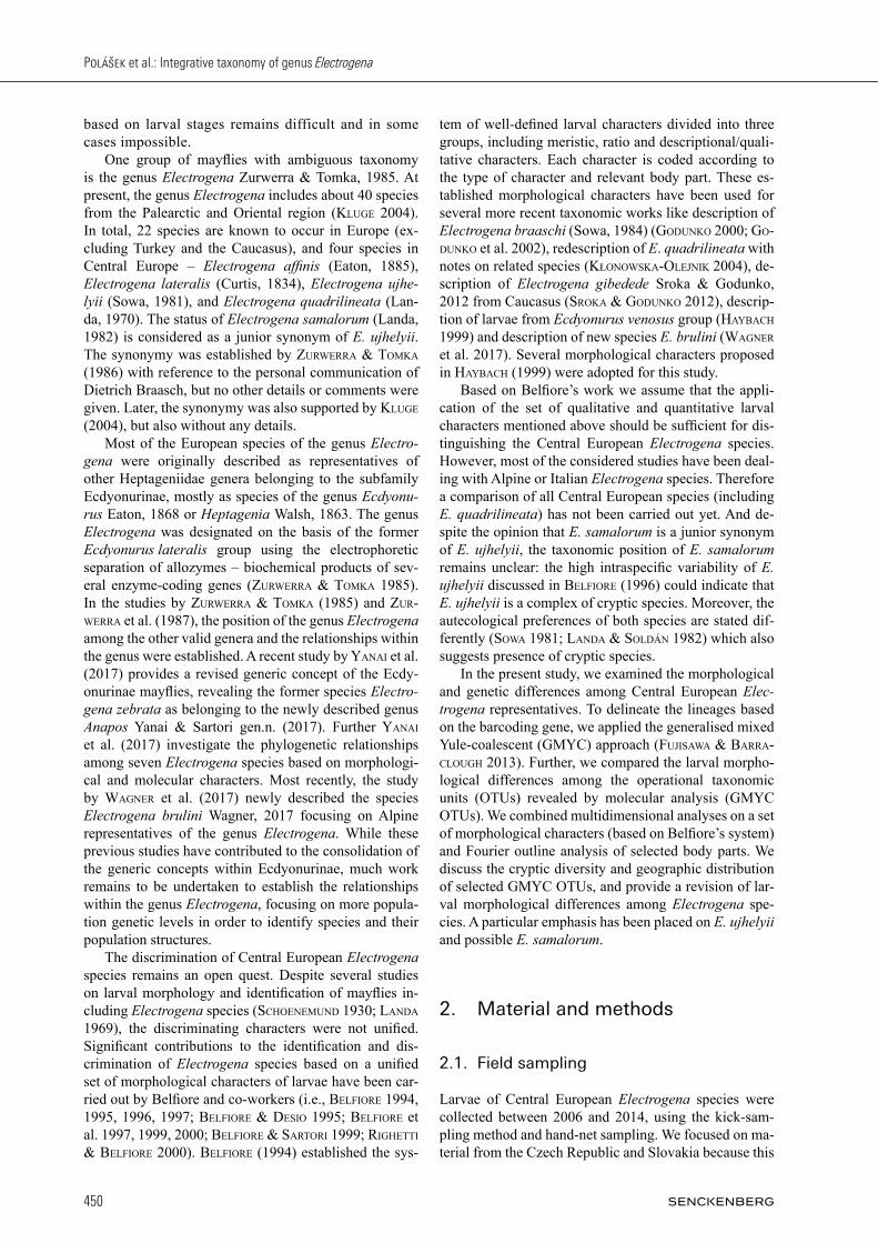
is the area of occurrence of the putative cryptic species. The rest of the collected material came from Hungary (one site was sampled – a brook near Aszófő village, type locality of E. ujhelyii), Austria, Poland, and Switzerland (see Fig. 1). The collected material was preserved in 99% ethanol and stored at 4°C. Altogether, we used 112 in-dividuals for morphological analyses, of which 69 indi-viduals were used for the molecular analyses (for details see Supplement Table S1).
2.2. DNA extraction, amplification, and sequencing
We selected one to two specimens per locality for the molecular analysis. Ideally, two legs (if possible, the right fore and middle leg) were used for DNA extrac-tion. The DNA was extracted and purified using the DNeasy Blood & Tissue Kit (Qiagen, Hilden, Germany). We sequenced the 658-bp fragment of the mitochon-drial cytochrome c oxidase subunit I (cox1) gene, using the primer pair LCO1490 and HCO2198 (folmer et al. 1994). The polymerase chain reaction (PCR) included an initial denaturation (2 min at 94°C) followed by 37 thermocycles of denaturation (30 s at 94°C), annealing (45 s at 45°C), extension (50 s at 72°C), and a final exten-
sion phase (7 min at 72°C). The length of the amplified products was pre-checked via agarose gel electrophore-sis. All amplified PCR products were purified using the QIAquick PCR Purification Kit (Qiagen, Hilden, Germa-ny). The purified PCR products were used as templates for cycle sequencing reactions with BigDye® Terminator v3.1 (Applied Biosystems), following the manufacturer´s instructions. Forward and reverse sequences were read with a capillary sequencer (Applied Biosystems) at the Department of Botany and Zoology, Masaryk University, Brno, Czech Republic. All sequences were assembled and edited with Sequencher® v5.3 (Gene Codes Corpo-ration, Ann Arbor, MI USA).
2.3. DNA taxonomy
Genetic species delineation was carried out by combin-ing newly sequenced cox1 data with 25 published Electrogena sequences available from GenBank (carDoni et al. 2015; gattolliat et al. 2015; waKimura et al. 2016). As outgroup we used Baetis rhodani (GenBank acc. no KF438126) sensu rutschmann et al. (2017b). The cox1-sequences were aligned using MAFFT v7.221 (Katoh & stanDleY 2013). To check for stop codons and the oc-currence of indels, we translated the nucleotide sequence
Fig. 1. Map of sampling sites with GMYC OTUs occurrence. A: Map of Central Europe covering the majority of sampled sites. B: Map of Switzerland with one sampled site. Position of both figured regions is highlighted by red rectangle in the bottom-right overview map. Coloured dots indicate occurrence of different GMYC OTUs, bicoloured dots indicate two different GMYC OTUs occurring at one local-ity. — Colours: violet – affinis, red – eastern ujhelyii, blue – lateralis, orange – quadrilineata, green – western ujhelyii.
Polášek et al.: Integrative taxonomy of genus Electrogena
452
alignment into an amino acid alignment using MEGA 5 (tamura et al. 2011). Identical haplotypes were removed using the Perl script collapsetypes_v4.5.pl (chesters 2013). The sequence alignment was partitioned into co-don positions (first codon position, second codon posi-tion, and third codon position). The best-fitting model of molecular evolution was selected for each codon position separately with jModelTest v2.1 (guinDon & gascuel 2003; DarriBa et al. 2012) using the Bayesian informa-tion criterion (BIC). The selected available models were: GTR + I for codon position one, HKY for codon position two, and GTR + Γ for codon position three. Since this partitioning-scheme resulted in very low effective sam-ple sizes (ESS) for preliminary tree inferences, we used the codon-specific model of sequence evolution HKY112 + CP112 + Γ112 (SRD60, shapiro et al. 2006) for the final analysis. An ultrametric gene tree was calculated using a relaxed molecular clock and a coalescent prior sensu monaghan et al. (2009) in BEAST v2.3.1 (BoucKaert et al. 2014). For the tree reconstruction, we conducted six independent runs with 30 million generations each. We used Tracer v1.7.1 (ramBaut et al. 2018) to assess convergence between runs by inspecting marginal densi-ties and ESS values, and estimated the number of burn-in generations for each run. All runs resulted in trees with very similar topologies (i.e., each putative GMYC spe-cies was recovered as monophyletic clade, except for one run for which E. lateralis from Switzerland and France, was not recovered as monophyletic clade, see Results). The runs were combined in LogCombiner v2.3 (ramBaut & DrummonD 2015), after removing the first 10% (i.e., 3000 trees) of each run as burn-in. Maximum clade credibility trees were inferred with TreeAnnotator v2.3 (ramBaut & DrummonD 2015). A single threshold GMYC analysis was performed on the ultrametric gene tree, using the Splits package (http://r-forge.r-project.org/projects/splits/) for R v3 (R Core Team, 2017).
2.4. Morphology and morphometry
For 13 Palearctic Electrogena species, we obtained a set of 13 numerical and four qualitative characters based on previous work by Belfiore (i.e., Belfiore 1981, 1994, 1995, 1996; Belfiore & Desio 1995; Belfiore et al. 1997, 1999, 2000; Belfiore & sartori 1999; righetti & Bel-fiore 2000; KłonowsKa-olejniK 2004). The minimum, mean and maximum of numerical characters were taken from mentioned papers when available; the status of qual-itative characters was noted. When a certain numerical character or status of qualitative character of a single spe-cies differed between two (or more) papers, we used the information from the more recent publication. A set of several individuals from each locality were chosen regarding the condition of the preserved mate-rial. Only individuals with minimal body damage were chosen for morphological analyses. The individuals in-cluded in the morphological analyses were selected with a particular emphasis on maximal morphological vari-
ability within a single locality – only specimens of dif-ferent size, age and sex classes (without young larvae) from one locality were included. At least one individual from each locality was analysed: the voucher specimen used for the molecular study. The maximum number of examined individuals from one locality was five. Each specimen was labelled by a unique identification code, stored separately in a plastic phial, and processed as de-scribed below. The photographs of gill plates in water medium were taken for preserving the exact shape of the soft parts, which are usually deformed in the non-water mounting medium. The whole habitus and details of the head were also captured. The mouthparts, in some cases also the head capsule, 1st, 4th and 7th tracheal gill plate, and fore, mid and hind legs were mounted into Liquido-Faure on microslides. A binocular stereomicroscope Olympus SZX9 with mounted digital camera Olympus C9090 was used for preparing the microslides and capturing the photographic material. For more detailed imaging of some morpho-logical characters, the microscope Olympus BX41 with mounted digital camera was used. The images were pre-processed using the software QuickPHOTO MICRO v3.1 (www.promicra.cz), and further image adjustment was done with the GNU Image Manipulation Program v2.8 (GIMP; www.gimp.org).
2.4.1. Morphological characters
The set of 16 quantitative numerical, seven ratio and six qualitative characters were defined, mostly based on those described in Belfiore’s articles mentioned above. Binary matrices for each character state were used for coding presence or absence of certain qualitative charac-ters. The presence (in data matrix coded as 1) or absence (coded as 0) of each binary variable (highlighted in bold) was marked. The description of morphological charac-ters and respective indexes are listed in the Appendix.
2.4.2. Morphometric data analyses
We used the matrix with character values (continuous for quantitative, binary for qualitative characters) of each examined individual (see Supplement Table S1) for mor-phometric analyses. Each individual was assigned to the OTU according to the species assignment obtained via GMYC approach (GMYC OTUs). The minimum, mean and maximum values for each numerical character were calculated. To illustrate the differences between given GMYC OTUs, the boxplots (the 25th and 75th percen-tiles) of numerical characters and histograms of qualita-tive characters represented as dummy variables presence/absence frequencies are used for visual presentation of differences between them. The significance of each nu-merical character was expressed using Kruskal-Wallis H test between groups (GMYC OTUs). To distinguish i) among individuals from all available GMYC OTUs and ii) between the closely related individ-uals within E. cf. ujhelyii (e.g., E. ujhelyii sensu stricto
453
ARTHROPOD SYSTEMATICS & PHYLOGENY — 76 (3) 2018
and possible E. samalorum together), we performed a Linear Discriminant Analysis (LDA). For this analysis, the missing values in the character data matrix (in the case when the value of certain characters was not pos-sible to count due to missing body part or body part dam-age) were estimated using factorial analysis for mixed data (FAMD) method (auDigier et al. 2014). Before LDA analysis, we reduced the number of morphological vari-ables to remove the collinearity and improve the separa-tion power of variables. To select the most informative variables, we used the proportion of between-group and within-group variance of each variable as a criterion. We chose two different sets of variables, discriminating all GMYC OTUs (full dataset) and E. cf. ujhelyii GMYC OTUs (reduced dataset) respectively. Additionally, we presented the equation for distin-guishing between closely related E. cf. ujhelyii individu-als using selected morphological characters. The success rate of identifying both Electrogena and E. cf. ujhelyii in-dividuals using LDA analyses of numerical characters is presented as leave-one-out cross-validation success rates.
2.4.3. Outline analysis
Images of the 1st, 4th and 7th tracheal gill, glossa, labrum and metafemora were captured as mentioned above. The images were trimmed on 1000 × 1000 px image and ad-justed, thus the target body part was approximately in the middle of the picture. All images were manually trans-formed to binary black and white images using GIMP v2.8. The outlines of these shapes were extracted, and the elliptic Fourier analysis was performed (for details see clauDe 2008). Shapes were approximated by 32 harmon-ics. Principal component analysis (PCA) on harmonic coefficients was calculated to describe the highest shape variability among individuals for each character. Linear Discrimination Analysis was performed on PCA scores to test the possibility of discriminating the GMYC OTUs using morphological shapes. To compare the classical morphometry methods and modern image-based analy-ses, we used the same data partitioning as for the mor-phometric dataset. We performed all analyses on the com-plete dataset including all GMYC OTUs and the reduced dataset including only E. cf. ujhelyii. The success rate of identification Electrogena OTUs using image-based LDA analyses is presented as leave-one-out cross-validation confusion matrix. All operations were performed using the package Momocs (Bonhomme et al. 2014) in R.
3. Results
3.1. GMYC analysis
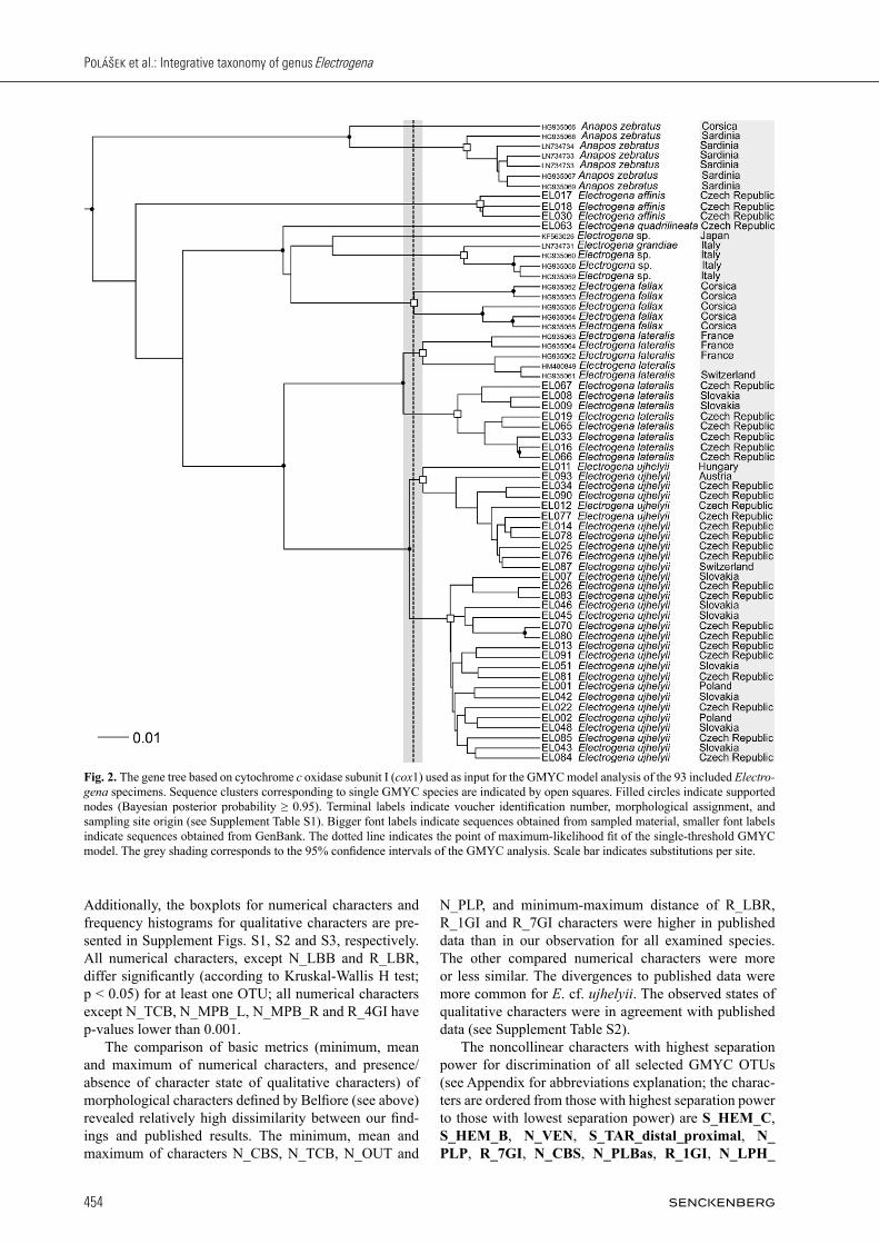
In total we found 65 unique haplotypes within the 94 se-quences. The GMYC model was significant (likelihood ratio: 14.03, P = 0.003). The model delineated twelve
putative species, composed of eight distinct clusters and four singletons (Fig. 2). The 95% confidence interval (CI, defined as 2 log likelihood units) ranged from ten to 13 species and six to nine clusters. All following results are based on the final model as presented in Fig. 2. We detected ten putative GMYC species of the ge-nus Electrogena. In detail, the model detected the spe-cies E. affinis, E. grandiae (including three unassigned specimens indicated as E. sp.), and E. fallax, each as in-dividual GMYC clusters; E. quadrilineata and one unas-signed Electrogena specimen from Japan (E. sp.) as sin-gleton. The morphospecies Anapos zebratus (formerly E. zebrata) formed two putative GMYC species, including a singleton from Corsica, and a cluster of six haplotypes from Sardinia. The two morphospecies E. lateralis and E. cf. ujhelyii were each divided into two putative GMYC species. In case of E. lateralis, one putative GMYC species included individuals from France and Switzerland, the other one comprised individuals from the Czech Republic and Slo-vakia. Electrogena cf. ujhelyii consisted of two clusters with a geographic contact line in the middle of the Czech Republic (Fig. 1). The first putative GMYC species com-prised specimens from Switzerland, Austria, Hungary, and Czech Republic (i.e., Bohemia – western part of the Czech Republic). The second putative GMYC species included specimens from Czech Republic (i.e., Moravia – eastern part of the Czech Republic), Slovakia, and Po-land. Interestingly, at one locality both putative GMYC species from E. cf. ujhelyii group occurred together (lo-cality Klíčava river, Lány, North-West of the Czech Re-public).
3.2. Morphometry
The overview of available information on morphological characters taken from published data is listed in Supple-ment Table S2. Data were available for species E. affinis; Electrogena antalyensis (Braasch & Kazanci, 1986); Electrogena calabra Belfiore, 1995; Electrogena fallax (Hagen, 1864); Electrogena galileae (Demoulin, 1973); Electrogena grandiae (Belfiore, 1981); Electrogena gridelii (Grandi, 1953); Electrogena hyblaea Belfiore, 1994; E. lateralis; Electrogena lunaris Belfiore & Scil-litani, 1997; Electrogena malickyi (Braasch, 1983); E. quadrilineata and E. ujhelyii. Based on the results of the GMYC analysis, we sepa-rated the sampled material available for morphological analyses into five GMYC OTUs: affinis, lateralis, quadrilineata, easternujhelyii and westernujhelyii. These categories are used in all subsequent morphological ana-lyses. The minimum, mean and maximum of 23 numerical (discrete and ratio) characters selected for this study were calculated from a morphological dataset of 112 individu-als classified in five GMYC OTUs (see Supplement Table S3). The frequencies of qualitative traits occur-rences are also listed in Supplementary file (Table S4).
Polášek et al.: Integrative taxonomy of genus Electrogena
454
Additionally, the boxplots for numerical characters and frequency histograms for qualitative characters are pre-sented in Supplement Figs. S1, S2 and S3, respectively. All numerical characters, except N_LBB and R_LBR, differ significantly (according to Kruskal-Wallis H test; p < 0.05) for at least one OTU; all numerical characters except N_TCB, N_MPB_L, N_MPB_R and R_4GI have p-values lower than 0.001. The comparison of basic metrics (minimum, mean and maximum of numerical characters, and presence/absence of character state of qualitative characters) of morphological characters defined by Belfiore (see above) revealed relatively high dissimilarity between our find-ings and published results. The minimum, mean and maximum of characters N_CBS, N_TCB, N_OUT and
N_PLP, and minimum-maximum distance of R_LBR, R_1GI and R_7GI characters were higher in published data than in our observation for all examined species. The other compared numerical characters were more or less similar. The divergences to published data were more common for E. cf. ujhelyii. The observed states of qualitative characters were in agreement with published data (see Supplement Table S2). The noncollinear characters with highest separation power for discrimination of all selected GMYC OTUs (see Appendix for abbreviations explanation; the charac-ters are ordered from those with highest separation power to those with lowest separation power) are S_HEM_C, S_HEM_B, N_VEN, S_TAR_distal_proximal, N_PLP, R_7GI, N_CBS, N_PLBas, R_1GI, N_LPH_
Fig. 2. The gene tree based on cytochrome c oxidase subunit I (cox1) used as input for the GMYC model analysis of the 93 included Electrogena specimens. Sequence clusters corresponding to single GMYC species are indicated by open squares. Filled circles indicate supported nodes (Bayesian posterior probability ≥ 0.95). Terminal labels indicate voucher identification number, morphological assignment, and sampling site origin (see Supplement Table S1). Bigger font labels indicate sequences obtained from sampled material, smaller font labels indicate sequences obtained from GenBank. The dotted line indicates the point of maximum-likelihood fit of the single-threshold GMYC model. The grey shading corresponds to the 95% confidence intervals of the GMYC analysis. Scale bar indicates substitutions per site.
455
ARTHROPOD SYSTEMATICS & PHYLOGENY — 76 (3) 2018
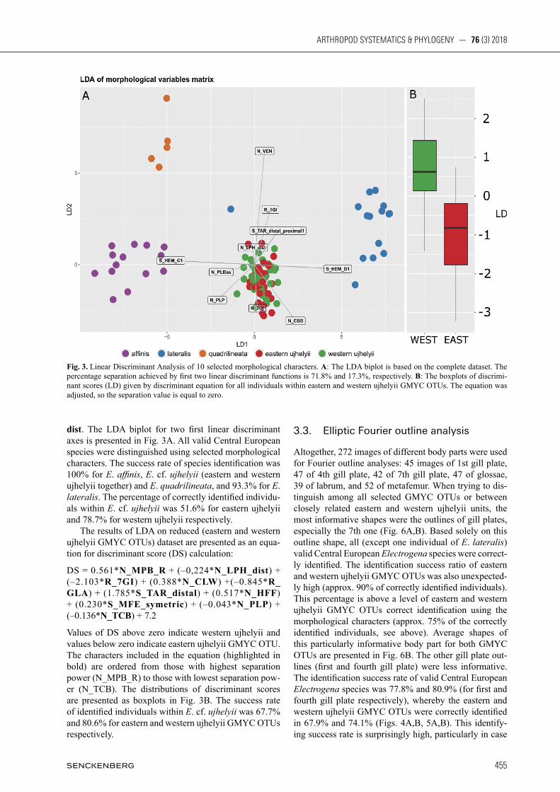
dist. The LDA biplot for two first linear discriminant axes is presented in Fig. 3A. All valid Central European species were distinguished using selected morphological characters. The success rate of species identification was 100% for E. affinis, E. cf. ujhelyii (eastern and western ujhelyii together) and E. quadrilineata, and 93.3% for E. lateralis. The percentage of correctly identified individu-als within E. cf. ujhelyii was 51.6% for eastern ujhelyii and 78.7% for western ujhelyii respectively. The results of LDA on reduced (eastern and western ujhelyii GMYC OTUs) dataset are presented as an equa-tion for discriminant score (DS) calculation:
DS = 0.561*N_MPB_R + (–0,224*N_LPH_dist) + (–2.103*R_7GI) + (0.388*N_CLW) +(–0.845*R_GLA) + (1.785*S_TAR_distal) + (0.517*N_HFF) + (0.230*S_MFE_symetric) + (–0.043*N_PLP) + (–0.136*N_TCB) + 7.2
Values of DS above zero indicate western ujhelyii and values below zero indicate eastern ujhelyii GMYC OTU. The characters included in the equation (highlighted in bold) are ordered from those with highest separation power (N_MPB_R) to those with lowest separation pow-er (N_TCB). The distributions of discriminant scores are presented as boxplots in Fig. 3B. The success rate of identified individuals within E. cf. ujhelyii was 67.7% and 80.6% for eastern and western ujhelyii GMYC OTUs respectively.
3.3. Elliptic Fourier outline analysis
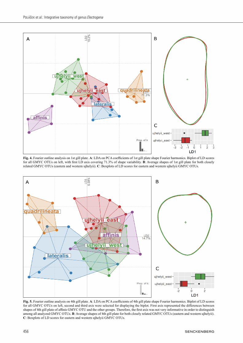
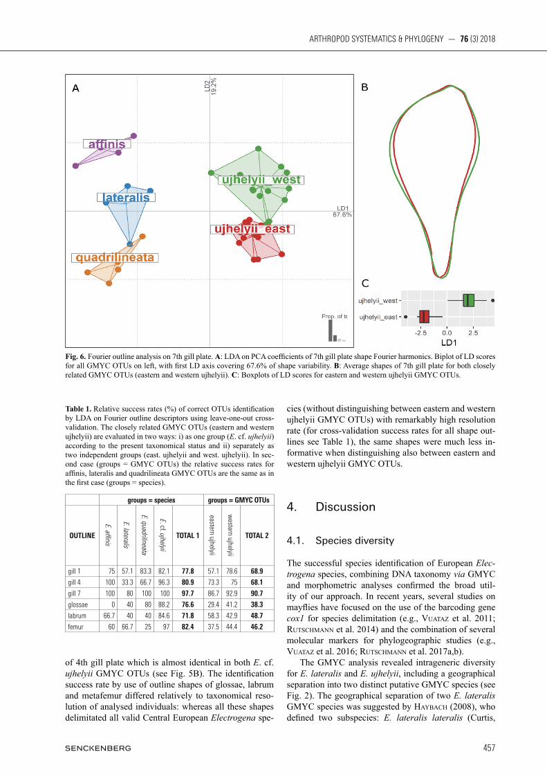
Altogether, 272 images of different body parts were used for Fourier outline analyses: 45 images of 1st gill plate, 47 of 4th gill plate, 42 of 7th gill plate, 47 of glossae, 39 of labrum, and 52 of metafemur. When trying to dis-tinguish among all selected GMYC OTUs or between closely related eastern and western ujhelyii units, the most informative shapes were the outlines of gill plates, especially the 7th one (Fig. 6A,B). Based solely on this outline shape, all (except one individual of E. lateralis) valid Central European Electrogena species were correct-ly identified. The identification success ratio of eastern and western ujhelyii GMYC OTUs was also unexpected-ly high (approx. 90% of correctly identified individuals). This percentage is above a level of eastern and western ujhelyii GMYC OTUs correct identification using the morphological characters (approx. 75% of the correctly identified individuals, see above). Average shapes of this particularly informative body part for both GMYC OTUs are presented in Fig. 6B. The other gill plate out-lines (first and fourth gill plate) were less informative. The identification success rate of valid Central European Electrogena species was 77.8% and 80.9% (for first and fourth gill plate respectively), whereby the eastern and western ujhelyii GMYC OTUs were correctly identified in 67.9% and 74.1% (Figs. 4A,B, 5A,B). This identify-ing success rate is surprisingly high, particularly in case
Fig. 3. Linear Discriminant Analysis of 10 selected morphological characters. A: The LDA biplot is based on the complete dataset. The percentage separation achieved by first two linear discriminant functions is 71.8% and 17.3%, respectively. B: The boxplots of discrimi-nant scores (LD) given by discriminant equation for all individuals within eastern and western ujhelyii GMYC OTUs. The equation was adjusted, so the separation value is equal to zero.
Polášek et al.: Integrative taxonomy of genus Electrogena
456
Fig. 4. Fourier outline analysis on 1st gill plate. A: LDA on PCA coefficients of 1st gill plate shape Fourier harmonics. Biplot of LD scores for all GMYC OTUs on left, with first LD axis covering 71.3% of shape variability. B: Average shapes of 1st gill plate for both closely related GMYC OTUs (eastern and western ujhelyii). C: Boxplots of LD scores for eastern and western ujhelyii GMYC OTUs.
Fig. 5. Fourier outline analysis on 4th gill plate. A: LDA on PCA coefficients of 4th gill plate shape Fourier harmonics. Biplot of LD scores for all GMYC OTUs on left, second and third axis were selected for displaying the biplot. First axis represented the differences between shapes of 4th gill plate of affinis GMYC OTU and the other groups. Therefore, the first axis was not very informative in order to distinguish among all analyzed GMYC OTUs. B: Average shapes of 4th gill plate for both closely related GMYC OTUs (eastern and western ujhelyii). C: Boxplots of LD scores for eastern and western ujhelyii GMYC OTUs.
457
ARTHROPOD SYSTEMATICS & PHYLOGENY — 76 (3) 2018
of 4th gill plate which is almost identical in both E. cf. ujhelyii GMYC OTUs (see Fig. 5B). The identification success rate by use of outline shapes of glossae, labrum and metafemur differed relatively to taxonomical reso-lution of analysed individuals: whereas all these shapes delimitated all valid Central European Electrogena spe-
cies (without distinguishing between eastern and western ujhelyii GMYC OTUs) with remarkably high resolution rate (for cross-validation success rates for all shape out-lines see Table 1), the same shapes were much less in-formative when distinguishing also between eastern and western ujhelyii GMYC OTUs.
4. Discussion
4.1. Species diversity
The successful species identification of European Electrogena species, combining DNA taxonomy via GMYC and morphometric analyses confirmed the broad util-ity of our approach. In recent years, several studies on mayflies have focused on the use of the barcoding gene cox1 for species delimitation (e.g., VuataZ et al. 2011; rutschmann et al. 2014) and the combination of several molecular markers for phylogeographic studies (e.g., VuataZ et al. 2016; rutschmann et al. 2017a,b). The GMYC analysis revealed intrageneric diversity for E. lateralis and E. ujhelyii, including a geographical separation into two distinct putative GMYC species (see Fig. 2). The geographical separation of two E. lateralis GMYC species was suggested by haYBach (2008), who defined two subspecies: E. lateralis lateralis (Curtis,
Fig. 6. Fourier outline analysis on 7th gill plate. A: LDA on PCA coefficients of 7th gill plate shape Fourier harmonics. Biplot of LD scores for all GMYC OTUs on left, with first LD axis covering 67.6% of shape variability. B: Average shapes of 7th gill plate for both closely related GMYC OTUs (eastern and western ujhelyii). C: Boxplots of LD scores for eastern and western ujhelyii GMYC OTUs.
Table 1. Relative success rates (%) of correct OTUs identification by LDA on Fourier outline descriptors using leave-one-out cross-validation. The closely related GMYC OTUs (eastern and western ujhelyii) are evaluated in two ways: i) as one group (E. cf. ujhelyii) according to the present taxonomical status and ii) separately as two independent groups (east. ujhelyii and west. ujhelyii). In sec-ond case (groups = GMYC OTUs) the relative success rates for affinis, lateralis and quadrilineata GMYC OTUs are the same as in the first case (groups = species).
groups = species groups = GMYC OTUs
OUTLINE
E. affinis
E. lateralis
E. quadrilineata
E. cf. ujhelyii
TOTAL 1
eastern ujhelyii
western ujhelyii
TOTAL 2
gill 1 75 57.1 83.3 82.1 77.8 57.1 78.6 68.9
gill 4 100 33.3 66.7 96.3 80.9 73.3 75 68.1
gill 7 100 80 100 100 97.7 86.7 92.9 90.7
glossae 0 40 80 88.2 76.6 29.4 41.2 38.3
labrum 66.7 40 40 84.6 71.8 58.3 42.9 48.7
femur 60 66.7 25 97 82.4 37.5 44.4 46.2
Polášek et al.: Integrative taxonomy of genus Electrogena
458
1834) from Germany and Ireland, and E. lateralis concii (Grandi, 1953) from Italy, Switzerland and Austria; both subspecies have different egg structure. Recent molecular data also evidenced a geographic clustering of E. lateralis, including specimens from the type local-ity in the United Kingdom and from France (Yanai et al. 2017). Although specimens included in our analysis from France and Switzerland were not available for mor-phological analysis, we assume that the separation of two putative GMYC species corresponds with Haybach‘s findings: the Alpine populations of E. lateralis belong to subspecies E. lateralis concii, and the Czech and Slovak population belong to subspecies E. lateralis lateralis. The geographic clustering of the two putative E. ujhelyii GMYC species was rather unexpected since there is no previous evidence from the literature. The western E. ujhelyii GMYC species distribution could be described as Alpine-Pannonian; the eastern E. ujhelyii species dis-tribution could be described as Carpathian. According to the type locality of the species E. ujhelyii (Aszófő, Hungary) and E. samalorum (Podhoroď, Slovakia), the western ujhelyii GMYC OTU represents the originally described species E. ujhelyii, and the eastern ujhelyii GMYC OTU might represent E. samalorum or an un-known species. Our data indicate distinct intraspecific genetic vari-ability for the different Electrogena species. While the within-group genetic distances among E. affinis speci-mens were detected as rather high, the cox1-sequences of E. quadrilineata specimens were recovered as identical haplotype. Notably the analysed E. quadrilineata speci-mens came from three localities (Czech Republic and Austria) and E. affinis from three nearby localities in the Czech Republic. The geographical sampling of the species E. affinis, E. quadrilineata, E. grandiae, and E. fallax was rather restricted (i.e., Czech Republic, Italy, and Corsica). All previous molecular work on these species (carDoni et al. 2015; gattollliat et al. 2015; Yanai et al. 2017) in-cluded specimens from the same geographic areas, with the exception of Yanai et al. (2017), who included for E. quadrilineata specimens from Czech Republic and Aus-tria. Further molecular analyses on a wide geographical scale will be required to confirm the status of these spe-cies. We confirmed the monophyletic clustering of the re-cently proposed genus Anapos Yanai & Sartori, 2017, including A. zebratus (formerly classified as E. zebrata) (Yanai et al. 2017). The results of our GMYC analysis are in agreement with previous studies, including the ex-istence of two distinct A. zebratus lineages (gattolliat et al. 2015; Yanai et al. 2017).
4.2. Morphology
The observed differences between measured and pub-lished values of numerical and qualitative characters illustrate the risks of applying defined numerical mor-
phologic characters. The resulting value of these charac-ters strongly depends on the available equipment (such as preparation tools and microscope) and personal ex-perience. For example, the description of the character N_CBS (a number of comb-shaped bristles on the fore margin of galeo-lacinia) does not define the threshold be-tween comb-shaped bristle and other-type bristles, which are also present (including a number of the transitional types) on the fore margin of galeo-lacinia. This could explain the differences between our own measured and available published values of this character. The other numerical characters which differed significantly were based on small morphological structures (N_TCB) or counts of very fine setae (N_OUT, N_PLP). The differ-ences between our measurements and published values of ratio characters could be caused by the different meth-od of data collecting. While Belfiore and other authors measured the values from hand-made drawings, we used the photographs of microslide mounted body parts. How-ever, these characters are useful when they have extreme values and can be interpreted as presence or absence of a remarkable state of character. For example, the char-acter N_HFF (the number of long hairs on the fore mar-gin of femora) can be used for the identification of E. affinis characterised by a high number of these long hairs, whereas the other Electrogena species have a few of them (E. galileae) or none (rest of Electrogena species). On the other hand, the qualitative characters are quite informative, and the valid Central European Electrogena species can be distinguished solely on the basis of char-acters S_HEM (distinct light spots for E. affinis and E. quadrilineata, two lighter smudges for E. lateralis and unicolour head for E. cf. ujhelyii) and S_BFE(i) (pointed bristles for E. affinis and spatula-like bristles on the fore femora for E. quadrilineata). The LDA results confirmed the high potential of qualitative characters, with the char-acter S_HEM being the most informative character for distinguishing Central European Electrogena species. Additionally, we can consider the qualitative characters S_TAR (colouration of tarsus) and S_BFE(i) (shape of bristles on upper side of fore femora) as informative as S_HEM because of high intercorrelation among these characters. The success rate of valid Central European Electrogena species identification using the LDA on the set of most informative qualitative and quantitative characters was almost 100%. Therefore, we can confirm the usability of the system of qualitative morphological characters for Central European Electrogena valid spe-cies identification. The equation for discriminating the eastern and west-ern ujhelyii GMYC OTU derived from LDA results based on the reduced dataset showed a surprisingly high separation power (67.7% and 80.6% cross-validation success rate, respectively). However, the equation was mostly based on characters, whose values often differed from published ones. Some of the other characters (N_HFF and S_TAR_distal) were included because of few atypical specimens with exceptional values of those char-acters (and therefore easily recognizable in the dataset).
459
ARTHROPOD SYSTEMATICS & PHYLOGENY — 76 (3) 2018
Hence, it is questionable if this equation based partly on problematic morphologic characters is applicable for dis-tinguishing the closely related E. cf. ujhelyii individuals. The Fourier outline shape analysis provided the most exciting results. The percentage of correctly identified individuals in the dataset using the most informative outlines depended on the taxonomical resolution of the analysed specimens. When we tried to distinguish among currently taxonomic valid species of Electrogena (e.g., eastern and western ujhelyii GMYC OTUs classified as one species – E. cf. ujhelyii), the observed success rate varied from almost 100% to approx. 80% depending on which body part was assessed. This was comparable to the success rate of the LDA model on a set of ten most informative qualitative and quantitative characters. The main contribution of the Fourier shape descriptors analy-sis improved the success rate to distinguish the eastern and western ujhelyii GMYC OTUs. In this case, the LDA model correctly identified more than 90% of the speci-mens using only the 7th gill plate shape. Moreover, the model successfully distinguished between two individu-als belonging to different GMYC OTU but occurred at a single locality (Klíčava river, Lány, Czech Republic). Compared to the success rate of LDA based on the ten most informative traditional characters, the Fourier out-line analysis was more accurate using only one character. When we take into account the uncertainty of measuring (or obtaining) the morphological characters entering the model (discussed above), the Fourier descriptor based analysis is the only reliable way to distinguish between eastern and western ujhelyii GMYC OTUs. However, we did not find any character to confident-ly differentiate the eastern and western ujhelyii GMYC OTU. Therefore, we decided not to make any statement on taxonomy of those geographically separated (GMYC based) putative species. Even though we suppose, that eastern ujhelyii represents the former species E. samalorum, further investigation of different life stages (eggs, adults) is needed to reject (or support) the recent syn-onymy of E. ujhelyii and E. samalorum. Moreover, the comparison of type material of both later species has been done, but due to the extent of this work, the results cannot be included in this study and will be published soon. Nevertheless, no significant differences between larval or adult type individuals were recognised (Polášek unpubl.). The comparison of different species (or GMYC spe-cies) identification approaches showed that using mod-ern approaches to shape analysis significantly improves the accuracy of species identification. However, the most recent approaches to automated species identification involve deep neural networks or other computer vision techniques for image (i.e., whole body or body part) classification. Computer vision has been used to clas-sify specimens in different genera (larios et al. 2008), visually easily separable species (wen & guYer 2012; BoniecKi et al. 2015) or even closely related species and genera (faVret & sieracKi 2016), including a high pro-portion of correctly identified specimens. In this study
we demonstrated that using a non-traditional approach (such as outline Fourier descriptors) can significantly im-prove our ability to identify taxonomical units defined by DNA-taxonomy methods, which are not identifiable using comprehensive traditional morphometric meth-ods. Additionally, the high separation power of easily captured qualitative characters, such as colouration of the head, could indicate the high potential of computer vision use for mayfly species identification. Therefore, the DNA-taxonomy-supported set of diagnostic shapes or other image-based data could contribute to the routine species identification, for example as a computer-vision extension of electronic identification keys.
5. Acknowledgements
This work was supported by the Specific Research Project of Masaryk University (MUNI/A/0816/2017) for main author, by institutional support RVO: 60077344 for RJG and by the Slovak Research and Development Agency (APVV SK-CZ-187-11) for MS. SR was supported by the Swiss National Science Foundation (Early PostDoc.Mobility fellowship P2SKP3_158698) and a fel-lowship from the Janggen-Pöhn-Stiftung. MS was also supported from European Regional Development Fund-Project “Mechanisms and dynamics of macromolecular complexes: from single mol-ecules to cells” (No. CZ.02.1.01/0.0/0.0/15_003/0000441). We would like to thank Tomáš Soldán (Czech Academy of Sciences), Małgorzata Kłonowska-Olejnik (Jagiellonian University, Poland) and Sandra Knispel (Musée cantonal de zoologie, Switzerland) for providing material for the molecular and morphological analyses. This research would not be possible without the invaluable help, consultations and material of recently passed away Tomáš Soldán (Czech Academy of Sciences).
6. References
auDigier V., husson f., Josse J. 2016. A principal component method to impute missing values for mixed data. – Advances in Data Analysis and Classification 10(1): 5 – 26. doi:10.1007/s11634-014-0195-1
BarBer-James h.m., gattolliat J.-l., sartori m., huBBarD m.D. 2008. Global diversity of mayflies (Ephemero ptera, Insecta) in freshwater. – Hydrobiologia 595(1): 339 – 350. doi:10.1007/s10750-007-9028-y
Belfiore c. 1981. On the Italian species of the Ecdyonurus lateralis group (Ephemero ptera, Heptageniidae). – Aquatic Insects 3(3): 171 – 178. doi:10.1080/01650428109361059
Belfiore c. 1994. Taxonomic characters for species identifica-tion in the genus Electrogena Zurwerra and Tomka, with a de-scription of Electrogena hyblaea sp. n. from Sicily (Ephemero-ptera, Heptageniidae). – Aquatic Insects 16(4): 193 – 199. doi: 10.1080/01650429409361555
Belfiore c. 1995. Description of Electrogena calabra n. sp., a new species from Southern Italy (Ephemero ptera, Heptage-niidae). – Annales de Limnologie 31(1): 29 – 34. doi:10.1051/limn/1995001
Belfiore c. 1996. Identification and discrimination of Electrogena species by numerical methods (Ephemero ptera: Hepta-geniidae). – Systematic Entomology 21(1): 1 – 13. doi:10.1111/ j.1365-3113.1996.tb00595.x
Belfiore c. 1997. Taxonomic characters and discrimination of spe-cies in the genus Electrogena Zurwerra & Tomka (Ephemero-
Polášek et al.: Integrative taxonomy of genus Electrogena
460
ptera, Heptageniidae). Pp. 427 – 433 in: lanDolt P., sartori M. (eds), Ephemero ptera & Pleco ptera: Biology, Ecology, System-atics. – MTL, Fribourg.
Belfiore c., Desio f. 1995. Taxonomy and distribution of Electrogena ujhelyii (SOWA, 1981). – Annalen des Naturhistorischen Museums in Wien, Serie B für Botanik und Zoologie 97(B): 151 – 154.
Belfiore c., scillitani g., picariello o., catauDo a. 1997. Mor-phological and electrophoretic evidence for a new species of Electrogena from central Italy: Description of E. lunaris sp. n. (Ephemero ptera: Heptageniidae). – Aquatic Insects 19(3): 129 – 140. doi:10.1080/01650429709361647
Belfiore c., haYBach a., KlonowsKa-oleJniK m. 1999. Taxonomy and phenetic relationships of Electrogena affinis (Eaton, 1883) (Ephemero ptera : Heptageniidae). – Annales de Limnologie - In-ternational Journal of Limnology 35(4): 245 – 256. doi:10.1051/limn/1999033
Belfiore c., sartori m. 1999. Redescription of Electrogena galileae (Demoulin, 1973) (Ephemero ptera, Heptageniidae). – Re-vue Suisse de Zoologie 106(4): 1025 – 1034. doi:10.5962/bhl.part.80114
Belfiore c., tanatmis m., KaZanci n. 2000. Taxonomy of Electrogena antalyensis (Kazanci & Braasch, 1986) (Ephemero-ptera, Heptageniidae). – Aquatic Insects 22(4): 261 – 270. doi:10.1076/0165-0424(200010)22:4;1-Y;FT261
Bonhomme V., picq s., gaucherel c., clauDe J. 2014. Momocs : Outline Analysis Using R. – Journal of Statistical Software 56(13): 1 – 24. doi:10.18637/jss.v056.i13
BoucKaert r., heleD J., Kühnert D., Vaughan t., wu c.-h., Xie D., sucharD m.a., ramBaut a., DrummonD a.J. 2014. BEAST 2: a software platform for Bayesian evolutionary analysis. – PLoS Computational Biology 10(4): e1003537. doi:10.1371/jour nal.pcbi.1003537
Brittain J.e. 2008. Mayflies, biodiversity and climate change. Pp. 1 – 14 in: hauer F.R., stanforD J., newell R. (eds), Internation-al Advances in the Ecology, Zoogeography and Systematics of Mayflies and Stoneflies. – University of California Publications in Entomology. doi:10.1525/california/9780520098688.003.0001
carDoni s., tenchini r., ficulle i., pireDDa r., simeone m.c., Belfiore c. 2015. DNA barcode assessment of Mediterranean mayflies (Ephemero ptera), benchmark data for a regional ref-erence library for rapid biomonitoring of freshwaters. – Bio-chemical Systematics and Ecology 62: 36 – 50. doi:10.1016/j.bse.2015.07.035
chesters D. 2013. collapsetypes_v4.5. – URL <https://source-forge.net/projects/collapsetypes> [accessed 01 December 2017].
clauDe J. 2008. Morphometrics with R. 1st edn. – Springer-Verlag, New York. 317 pp. doi:10.1007/978-0-387-77789-4
DarriBa D., taBoaDa g.l., Doallo r., posaDa D. 2012. jMod-elTest 2: more models, new heuristics and parallel computing. – Nature Methods 9(8): 772. doi:10.1038/nmeth.2109
DuDgeon D., arthington a.h., gessner m.o., KawaBata Z.-i., Knowler D.J., léVêque c., naiman r.J., prieur-richarD a.-h., soto D., stiassnY m.l.J., sulliVan c.a. 2006. Freshwater biodiversity: importance, threats, status and conservation chal-lenges. – Biological Reviews 81(2): 163 – 182. doi:10.1017/S1464793105006950
faVret c., sieracKi J.m. 2016. Machine vision automated species identification scaled towards production levels. – Systematic En-tomology 41(1): 133 – 143. doi:10.1111/syen.12146
folmer o., BlacK m., hoeh w., lutZ r., VriJenhoeK r. 1994. DNA primers for amplification of mitochondrial cytochrome c oxidase subunit I from diverse metazoan invertebrates. – Mo-lecular Marine Biology and Biotechnology 3(5): 294 – 299.
fuJisawa t., Barraclough t.g. 2013. Delimiting species using single-locus data and the Generalized Mixed Yule Coalescent approach: a revised method and evaluation on simulated data sets. – Systematic Biology 62(5): 707 – 724. doi:10.1093/sysbio/syt033
gattolliat J.-l., caVallo e., VuataZ l., sartori m. 2015. DNA barcoding of Corsican mayflies (Ephemero ptera) with implica-
tions on biogeography, systematics and biodiversity. – Arthropod Systematics & Phylogeny 73(1): 3 – 18.
goDunKo r.J. 2000. Little known species of the genera Rhithrogena and Electrogena (Ephemero ptera, Heptageniidae) from Ukraine. – Vestnik Zoologii 14: 60 – 66.
goDunKo r.J., proKopoV g.a., KlonowsKa-oleJniK m. 2002. Complementary description of the winged stages of Electrogena braaschi (Ephemero ptera, Heptageniidae). – Vestnik Zoologii 36(5): 73 – 76.
guinDon s., gascuel o. 2003. A simple, fast, and accurate algorithm to estimate large phylogenies by maximum likelihood. – System-atic Biology 52(5): 696 – 704. doi:10.1080/10635150390235520
haYBach a. 1999. Beitrag zur Larvaltaxonomie der Ecdyonurus venosus-Gruppe in Deutschland. – Lauterbornia 37: 113 – 150.
haYBach a. 2008. Egg structure morphology gives evidence for a cryptic subspecies of Electrogena lateralis (Curtis, 1834) in Southern Europe: Electrogena lateralis concii (GRANDI, 1953) nov. stat. [Ephemero ptera, Heptageniidae]. – Ephemera 9(1): 1 – 7.
Katoh K., stanDleY D.m. 2013. MAFFT multiple sequence align-ment software version 7: improvements in performance and usability. – Molecular Biology and Evolution 30(4): 772 – 780. doi:10.1093/molbev/mst010
KłonowsKa-olejniK M. 2004. Redescription of Electrogena quadrilineata (Landa, 1969) from type material (Ephemero ptera, Heptageniidae). – Aquatic Insects 26(2): 85 – 95. doi:10.1080/01650420412331325828
Kluge n. 2004. The phylogenetic system of Ephemero ptera. 1st edn. – Kluwer Academic Publishers, Dordrecht. 442 pp. doi:10.1007/978-94-007-0872-3
lanDa V. 1969. Fauna ČSSR: Jepice - Ephemero ptera. – Academia, Praha. 352 pp.
lanDa V., solDán t. 1982. Ecdyonurus samalorum sp. n. from Czechoslovakia (Ephemero ptera, Heptageniidae). – Acta Ento-mologica Bohemoslovaca 79: 31 – 36.
larios n., Deng h., Zhang w., sarpola m., Yuen J., paasch r., molDenKe a., lYtle D.a., correa s.r., mortensen e.n., sha-piro l.g., Dietterich t.g. 2008. Automated insect identification through concatenated histograms of local appearance features: feature vector generation and region detection for deformable objects. – Machine Vision and Applications 19(2): 105 – 123. doi:10.1007/s00138-007-0086-y
monaghan m.t., wilD r., elliot m., fuJisawa t., BalKe m., in-warD D.J.g., lees D.c., ranaiVosolo r., eggleton p., Barra-clough t.g. 2009. Accelerated species inventory on Madagascar using coalescent-based models of species delineation. – System-atic Biology 58(3): 298 – 311. doi:10.1093/sysbio/syp027
ramBaut a., DrummonD a.J. 2015. LogCombiner v2.3. – URL: <http://beast.community/logcombiner> [accessed 01 December 2017].
ramBaut A., DrummonD a.J., Xie D., Baele g., sucharD m.a. 2018. Posterior summarisation in Bayesian phylogenetics using Tracer 1.7. – Systematic Biology 67(5): 901 – 904. doi: 10.1093/sysbio/syy032
righetti B., Belfiore c. 2000. Additions à la faune des Éphémères de France (5): Electrogena grandiae (Belfiore, 1981) et com-paraison biométrique de différentes populations [Ephemero-ptera, Heptageniidae]. – Ephemera 1(2): 123 – 130.
rutschmann s., gattolliat J.-l., hughes s.J., BáeZ m., sartori m., monaghan m.t. 2014. Evolution and island endemism of morphologically cryptic Baetis and Cloeon species (Ephemero-ptera, Baetidae) on the Canary Islands and Madeira. – Freshwa-ter Biology 59(12): 2516 – 2527. doi:10.1111/fwb.12450
rutschmann s., Detering h., simon s., freDslunD J., monaghan m.t. 2017a. DiscoMark: Nuclear marker discovery from ortho-logous sequences using draft genome data. – Molecular Ecology Resources 17: 257 – 266. doi: 10.1111/1755-0998.12576
rutschmann s., Detering h., simon s., funK D.h., gattolliat J.-l., hughes s.J., raposeiro p.m., Desalle r., sartori m., mona-ghan m.t. 2017b. Colonization and diversification of aquatic in-sects on three Macaronesian archipelagos using 59 nuclear loci
461
ARTHROPOD SYSTEMATICS & PHYLOGENY — 76 (3) 2018
derived from a draft genome. – Molecular Phylogenetics and Evolution 107: 27 – 38. doi:10.1016/j.ympev.2016.10.007
schoenemunD e. 1930. Eintagsfliegen oder Ephemero ptera. 19th edn. – Fischer Verlag, Jena. 106 pp.
shapiro B., ramBaut a., DrummonD a.J. 2006. Choosing appropri-ate substitution models for the phylogenetic analysis of protein-coding sequences. – Molecular Biology and Evolution 23(1): 7 – 9. doi:10.1093/molbev/msj021
sroKa p., goDunKo r.J. 2012. Mayflies of the Caucasus Mountains. I. A new species of the genus Electrogena Zurwerra & Tomka, 1985. – Zootaxa 3222: 28 – 45. doi:10.11646/zootaxa.4231.1.4
sowa r. 1981. Taxonomy and ecology of Ecdyonurus ujhelyii sp. n. (Ephemero ptera, Heptageniidae) from the tributaries of Lake Balaton. – Acta Hydrobiologica 23(4): 375 – 380.
tamura K., peterson D., peterson n., stecher g., nei m., Kumar s. 2011. MEGA5: Molecular evolutionary genetics analysis us-ing maximum likelihood, evolutionary distance, and maximum parsimony methods. – Molecular Biology and Evolution 28(10): 2731 – 2739. doi:10.1093/molbev/msr121
VuataZ l., sartori m., wagner a., monaghan m.t. 2011. To-ward a DNA taxonomy of Alpine Rhithrogena (Ephemero-ptera: Heptageniidae) using a Mixed Yule-Coalescent Analysis of mitochondrial and nuclear DNA. – PLoS ONE 6(5): e19728. doi:10.1371/journal.pone.0019728
VuataZ l., rutschmann s., monaghan m.t., sartori m. 2016. Molecular phylogeny and timing of diversification in Alpine Rhithrogena (Ephemero ptera: Heptageniidae). – BMC Evolu-tionary Biology 16(1): 194. doi:10.1186/s12862-016-0758-1
wagner a., VuataZ l., sartori m. 2017. Electrogena brulini sp. nov. and E. vipavensis Zurwerra & Tomka, 1986 syn. nov. re-vealed by integrative taxonomy of E. gridellii (Grandi, 1953) (Ephemero ptera: Heptageniidae). – Zootaxa 4362(3): 359. doi: 10.11646/zootaxa.4362.3.3
waKimura K., taKemon Y., taKaYanagi a., ishiwata s., watanaBe K., taniDa K., shimiZu n., Kato m. 2016. Characterization of genes for histone H3, 18S rRNA, and cytochrome oxidase sub-unit I of East Asian mayflies (Ephemero ptera). – DNA Barcodes 4(1): 1 – 25. doi:10.1515/dna-2016-0001
wen c., guYer D. 2012. Image-based orchard insect automated identification and classification method. – Computers and Elec - tronics in Agriculture 89: 110 – 115. doi:10.1016/j.compag.2012. 08.008
Yanai Z., sartori m., Dor r., Dorchin n. 2017. Molecular phy-logeny and morphological analysis resolve a long-standing controversy over generic concepts in Ecdyonurinae mayflies (Ephemero ptera: Heptageniidae). – Systematic Entomology 42(1): 182 – 193. doi:10.1111/syen.12203
Zurwerra a., tomKa i. 1985. Electrogena gen. nov., eine neue Gattung der Heptageniidae [Ephemero ptera]. – Entomologische Berichte Luzern 13: 99 – 104.
Zurwerra a., tomKa i. 1986. Drei neue Arten der Gattung Electrogena Zurwerra & Tomka 1985, aus Südeuropa (Ephemero ptera, Heptageniidae). – Bulletin de la Societé Fribourgeoise des Sci-ences Naturelles 75(1/2): 216 – 230.
Zurwerra a., metZler m., tomKa i. 1987. Biochemical system-atics and evolution of the European Heptageniidae (Ephemero-ptera). – Archiv für Hydrobiologie 109(4): 481 – 510.
7. Appendix
7.1. Quantitative (numeric) characters
N_LBB – Number of median bristles on dorsal side of labrum. Only the short and stout bristles, maximally 2.5 × longer than first, medial bristles, were counted. These usually reach to the frontal edge of labrum – the row then continues up to the lateral lobes of the labrum, surrounded by other thin and long bristles. This character was used for the first time to describe the Electrogena larvae.
N_CBS – Number of comb-shaped bristles on fore margin of galea-lacinia. If possible, the mean of the number of comb-shaped bristles on the left and right galea-lacinia was calculated. For details see Belfiore (1996) and haYBach (1999).
N_TCB – Number of pointed teeth on 5th comb-shaped bristle from medially. If possible, the mean of the number of pointed teeth on the 5th comb-shaped bristle on the left and right galea-lacinia was calculated. For details see Belfiore (1996) and haYBach (1999).
N_OUT – Number of hairs near outer edge of galea-lacinia, from palpus to fore margin. Character used in Belfiore (1996), arrange-ment of hairs in the Electrogena species slightly differs from ar-rangement of hairs in genus Ecdyonurus pictured in haYBach (1999). Entire lateral edge of galea-lacinia taken into account, no different types of hairs (in haYBach (1999) defined as N_DOR and N_OUT) observed.
N_PLP – Number of hairs near fore margin of first segment of max-illary palp. Arrangement of hairs is similar to haYBach (1999), the
character also used in Belfiore (1996). If possible, average number of hairs in left and right maxillary palp calculated.
N_PLS – Number of stout bristles on hind margin of first segment of maxillary palp. Arrangement of bristles is similar to haYBach (1999). If possible, average number of bristles in left and right maxillary palp calculated. This character was used for the first time to describe the Electrogena larvae.
N_PLH – Number of long hairs on hind margin of first segment of maxillary palp. Arrangement of hairs is similar to haYBach (1999). Only long and thin hairs near the basis of first segment of maxillary palp taken into account. If possible, average number of hairs in left and right maxillary palp calculated.
N_PLBas – Number of hairs at base of maxillary palp. Arrange-ment of hairs is similar to haYBach (1999). If possible, average number of hairs in left and right maxilla calculated.
N_VEN – Number of hairs on ventral basal part of maxilla. Ar-rangement of hairs is similar to haYBach (1999). If possible, aver-age number of hairs in left and right maxilla calculated. This char-acter was used for the first time to describe the Electrogena larvae.
N_MPB(l) – Number of bristles near left prostheca. Stout, long bristles following incisive parts counted. This character was used for the first time to describe the Electrogena larvae.
N_MPB(r) – Number of bristles near right prostheca. Stout, long bristles following incisive parts counted.
N_LPH_prox – Number of bristles on hind margin of first segment of labial palp – proximal part. Arrangement of bristles slightly dif-fers from arrangement stated in haYBach (1999). Character N_LPH in this article describes number of bristles and hairs in dorsolateral
Polášek et al.: Integrative taxonomy of genus Electrogena
462
part of labial palp, in the article counted for bristles and hairs sepa-rately. In our approach, only one type of bristles was recognised and counted. Area covered by bristles in Central European Electrogena species is divided into two discontinued parts – proximal and distal part. Number of bristles of each part counted separately. This char-acter was used for the first time to describe the Electrogena larvae.
N_LPH_dist – Number of bristles on hind margin of first segment of labial palp - distal part. Details discussed above. This character was used for the first time to describe the Electrogena larvae.
N_CLW – Number of teeth on pretarsal claws. If possible, average of fore, mid and hind leg counted.
N_BVF – Number of bristles on ventral side of femora near hind margin. If possible, number of these on profemora counted.
N_HFF – Number of long hairs (at least twice as long as neighbour-ing bristles) on fore margin of femora. If possible, number of these on profemora counted. This character was used for the first time to describe the Electrogena larvae.
7.2. Quantitative (ratio) characters
R_LBR – Total width of labrum divided by mean width of lateral lobes of labrum. For details see Belfiore (1996).
R_GLA – Distance between glossae (outer distance divided by in-ner distance between glossae). For details see Belfiore (1996).
R_GLB – Width of glossae (outer distance divided by width of glossae). For details see Belfiore (1996).
R_1GI – Relative width of 1st gill plate.
R_4GI – Relative width of 4th gill plate.
R_7GI – Relative width of 7th gill plate.
R_3FEM – Relative width of metafemora (length/width ratio of metafemora). This character was used for the first time to describe the Electrogena larvae.
7.3. Qualitative characters
S_BFE(i) – Shape of setae on upper surface of profemora. Four types of this character were recognised: pointed,roundpointed, bottlelike, and spatulalike. Shape of setae evaluated in central area of upper side of profemora. In case of indecisive type of char-acter, both types (states of the character) were recorded.
S_BFE(iii) – Shape of setae on upper surface of metafemora. For details see above (S_BFE(i)).
S_MFE – Shape of metafemora. Two types of this character recog-nised: symmetrical and asymmetrical. State of this character es-timated by expert judgement; evaluation was based on comparing convexity of fore and hind margin of metafemora. This character was used for the first time to describe the Electrogena larvae.
S_TAR – Marking on the tarsi. Four types of this character were recognised: light,unicolourdark, distallydarkened and distally andproximallydarkened.
S_HEM – Markings on the head. Three types were re cognized: unicolorbrownhead with no light spots on front edge of head capsule (type A); head with twolightersmudges in the middle on frontal edge of head capsule (type B); head with distinct lightspots on frontal edge of head capsule (type C). Overall colour of head (from yellowish to dark brown) and light dots or smudges in central area of head capsule not taken into account.
S_HLB – Extension of pilosity on lateral lobes. Two types were recognised: pilosity of hypopharynxdoesnot overlap apex of superlinguae, or pilosityofhypopharynxoverlapsapex of super-linguae. Only long, fine setae taken into account.
Authors’ contributions
M.P., M.S. and M.N. contributed to the material sampling, M.P. processed and sequenced all material, S.R. performed molecular part of analyses, M.P. and R.J.G. collaborated on morphological part of analyses, all authors collaborated on manuscript preparation with M.P. as a leading author and S.Z. as a supervisor.
Electronic Supplement Fileat http://www.senckenberg.de/arthropod-systematics
File 1: polasek&al-electrogenaheptageniidae-asp2018-electronic supplement-1.pdf — Table S1. A list of all analysed specimens with country and locality specification and GenBank accession numbers for cox1 mitochondrial gene sequences. — Table S2. Nu-merical and qualitative characters of European Electrogena species based on available publications. — Table S3. Minimum, mean and maximum for all numerical characters used in this study. — Table S4. Percentage representation of qualitative character states used in this study. — Fig. S1. Boxplots of discrete numerical characters for defined GMYC OTUs. — Fig. S2. Boxplots of ratio numerical characters for defined GMYC OTUs. — Fig. S3. The histograms of percental representation of qualitative character states.
GenBank accession numbersat https://www.ncbi.nlm.nih.gov
GenBank accession numbers for all used sequences are available in Supplement file. Continuous series of 69 GenBank accession num-bers start with MH093830 and ends with MH093898.