Embed Size (px)
Citation preview
Mickiewicz et al. Critical Care (2015) 19:11 DOI 10.1186/s13054-014-0729-0
RESEARCH Open Access
Integration of metabolic and inflammatorymediator profiles as a potential prognosticapproach for septic shock in the intensivecare unitBeata Mickiewicz1, Patrick Tam2, Craig N Jenne2, Caroline Leger2, Josee Wong3, Brent W Winston3,Christopher Doig4, Paul Kubes5*, Hans J Vogel1* and for the Alberta Sepsis Network
Abstract
Introduction: Septic shock is a major life-threatening condition in critically ill patients and it is well known thatearly recognition of septic shock and expedient initiation of appropriate treatment improves patient outcome.Unfortunately, to date no single compound has shown sufficient sensitivity and specificity to be used as aroutine biomarker for early diagnosis and prognosis of septic shock in the intensive care unit (ICU). Therefore,the identification of new diagnostic tools remains a priority for increasing the survival rate of ICU patients. In thisstudy, we have evaluated whether a combined nuclear magnetic resonance spectroscopy-based metabolomicsand a multiplex cytokine/chemokine profiling approach could be used for diagnosis and prognostic evaluation ofseptic shock patients in the ICU.
Methods: Serum and plasma samples were collected from septic shock patients and ICU controls (ICU patientswith the systemic inflammatory response syndrome but not suspected of having an infection). 1H Nuclear magneticresonance spectra were analyzed and quantified using the targeted profiling methodology. The analysis of theinflammatory mediators was performed using human cytokine and chemokine assay kits.
Results: By using multivariate statistical analysis we were able to distinguish patient groups and detect specificmetabolic and cytokine/chemokine patterns associated with septic shock and its mortality. These metabolites andcytokines/chemokines represent candidate biomarkers of the human response to septic shock and have thepotential to improve early diagnosis and prognosis of septic shock.
Conclusions: Our findings show that integration of quantitative metabolic and inflammatory mediator data can beutilized for the diagnosis and prognosis of septic shock in the ICU.
IntroductionApproximately 18 million cases of sepsis occur everyyear worldwide with a mortality rate reaching almost30% [1]. However, it has been reported that detectingsepsis, especially at an early stage, improves patientoutcome and reduces the mortality rate [2]. Therefore, itis critical to identify new diagnostic tools and develop
* Correspondence: [email protected]; [email protected] Institute for Chronic Diseases, University of Calgary, 3280 HospitalDrive Northwest, Calgary, AB, T2N 4N1, Canada1Bio-NMR-Centre, Department of Biological Sciences, University of Calgary,2500 University Drive Northwest, Calgary, AB, T2N 1N4, CanadaFull list of author information is available at the end of the article
© 2015 Mickiewicz et al.; licensee BioMed CenCommons Attribution License (http://creativecreproduction in any medium, provided the orDedication waiver (http://creativecommons.orunless otherwise stated.
prognostic approaches to improve patient care anddecrease the sepsis death rate.In recent years, several studies have been performed to
describe and identify biomarkers that could be used inthe diagnosis and management of sepsis [3]. This previouswork has suggested that sepsis could be diagnosed bymeasuring increased levels of particular proteins in bloodsuch as plasma C-reactive protein, inflammatory cytokines(for example tumor necrosis factor α (TNF-α), interleukin-1(IL-1) and IL-6), procalcitonin or lipopolysaccharide-bindingprotein [4]. It has also been reported that the concentrationsof lactate or different plasma amino acids can be elevated
tral. This is an Open Access article distributed under the terms of the Creativeommons.org/licenses/by/4.0), which permits unrestricted use, distribution, andiginal work is properly credited. The Creative Commons Public Domaing/publicdomain/zero/1.0/) applies to the data made available in this article,
Mickiewicz et al. Critical Care (2015) 19:11 Page 2 of 12
during the disease [3,5,6]. However, insufficient sensitivityand specificity of the reported compounds currentlyimpede their usage as standard tools for early diagnosis ofsepsis [3,7,8]. Therefore, integrated and multifacetedmedical approaches supported by effective diagnostic tools,such as a combination of various biomarkers, may improvethe identification and the prognosis for sepsis in intensivecare units (ICUs) [3,9]. Such an integrated approach, basedon merging different data sets, could also create broaderand more detailed insight into the nature of the diseasethan can be achieved using one individual approach.In this study, we have combined metabolomics and
multiplex cytokine/chemokine data to investigate itspotential for the diagnosis and prognosis of septic shock.It has previously been demonstrated that nuclear magneticresonance (NMR) spectroscopy-based metabolomics is avery efficient approach for the discovery of molecularmarkers of sepsis in animals models [10-12] and withinhumans [13,14]. In addition, it has been reported thatmultiplex analysis of cytokines can be used for biomarkeridentification and quantification in lipopolysaccharide-stimulated human plasma samples [15]. However, only alimited number of studies have demonstrated success inusing a multiplex cytokine/chemokine profiling approachfor the prediction of sepsis in clinical settings [16-18]. More-over, to date integration of metabolomics and inflammatorymediator data to identify correlations between immunefeatures and metabolic phenotypes during infection has onlybeen described in an animal model [19]. By using 1H NMRspectroscopy and multiplex technology we were able toidentify and quantify specific metabolites and inflammatorymediators potentially involved in the septic shock response.Using multivariate statistical analysis we could generatepowerful models for diagnosis and prognosis of septicshock. This study presents a promising approach forimproving patient care and patient outcome in the ICUand deserves further evaluation in other clinical settings,such as the emergency department.
MethodsSample collectionThe study received approval from the Conjoint HealthResearch Ethics Board of the University of Calgary. Thesamples were collected in accordance with the guidelinesof the Tri-Council policy statement and as part of theCritical Care Epidemiological and Biological TissueResource. All patients, or their next of kin, providedwritten informed consent for participation in this study.The protocol of sample collection has been previouslydescribed in detail [14]. Briefly, blood was drawn frompatients more than 18 years old that were admittedto the ICU of the Foothills Medical Center or thePeter Lougheed Hospital in Calgary (AB, Canada). Thestudy includes samples collected from septic shock patients
who met criteria for septic shock as defined by theAmerican- European consensus statement on sepsis[20,21] and samples obtained from ICU control patients(ICU patients with the systemic inflammatory responsesyndrome (SIRS) but not suspected of having an infection).All samples were collected within 24 hours of patientadmission to the ICU. Serum was obtained by collectingthe blood into a sterile silicone-coated vacutainer (BD,Franklin Lakes, NJ, USA) and allowing the blood to clot for45 min at room temperature. Plasma was obtained bycollecting the blood into a sodium-heparin-containingvacutainer (BD) and processed immediately after collection.The samples were then centrifuged at 1200 x g for 15 min,collected into one each 15 mL tube and frozen at −80°C.The samples were thawed once and aliquoted into 250 μLaliquots that were stored at −80°C until used.
NMR spectroscopy and metabolite concentration profilingThe protocol for the sample preparation and NMRspectral acquisition has been previously described in detail[13,14]. Briefly, filtration (3-kiloDalton (kDa) NanoSepmicrocentrifuge filters; Pall, Inc., East Hills, NY, USA) ofserum samples (V = 250 μL) was followed by adding tothe filtrated samples 80 μL of phosphate-buffered solution(0.5 M NaH2PO4 containing 2.5 mM 2,2-dimethyl-2-silapentane-5-sulfonate, DSS), 10 μL of sodium azide(1 M NaN3) and D2O. The final volume of each samplewas 400 μL and the pH was in the range of 7.0 ± 0.04.NMR spectra were obtained on a 600 MHz BrukerUltrashield Plus NMR spectrometer (Bruker BioSpinLtd., Milton, ON, Canada) using a standard Bruker 1Dspectroscopy presaturation pulse sequence (noesypr1d)with optimal water suppression and a mixing time of100 ms [22,23]. The spectra were manually corrected(phasing, baseline correction, referencing to the DSS peakat 0.0 ppm) in the Chenomx NMR Suite 6.1 software(Chenomx Inc., Edmonton, AB, Canada) [23]. Thetargeted profiling methodology was used for metaboliteidentification and quantification [23]. If the metabolitepeaks could not be distinguished from the noise in NMRspectra, the peaks were assigned with zero value andconsidered as missing data.
Cytokine/chemokine profilingThe analysis of the inflammatory mediators in humanplasma samples was performed using two human cytokineand chemokine assay kits (Bio-Plex Pro Human Cytokine21-plex Assay and Bio-Plex Pro Human Cytokine 27-plexAssay), which were obtained from Bio-Rad Laboratories,Inc. (Hercules, CA, USA). Samples were assayed accordingto the manufacturer’s specifications and the plates wereread on a Luminex 200 apparatus (Applied CytometrySystems, Sheffield, UK). The acquisition and analysis ofthese samples were performed with Bio-Plex Manager 6.0
Mickiewicz et al. Critical Care (2015) 19:11 Page 3 of 12
(Bio-Rad Laboratories, Inc.). If the coefficient of variancebetween two replicates was more than 20% the data wasconsidered as a missing value.
Statistical modelingThe SIMCA-P+ 12.0.1 software (Umetrics, Malmo,Sweden) was applied to create and analyze multivariatemodels. All metabolites or inflammatory mediatorswith more than 50% missing values were excludedfrom the analysis. Data preprocessing (median fold changenormalization, logarithmic transformation, centering andunit variance scaling [24]) was first conducted separatelyfor the metabolomics and cytokine/chemokine datasetand then for the combined dataset.Principal component analysis (PCA) was used to
summarize the variation in each data matrix and toshow outlying samples, that is samples that are situatedoutside of the 95% confidence interval of the Hotelling’sT-squared distribution (elliptic or spherical area in thescore scatter plots) [25]. Following this, supervisedorthogonal partial least squares discriminant analysis(OPLS-DA) was applied [26]. For the integrated meta-bolomics and cytokine/chemokine data and for theage-sex-matched (age within 5 years) septic shock survivorsand nonsurvivors, the OPLS-DA models were based onpotentially relevant metabolites selected in two-sample ttests with P value less than 0.2 as a threshold [27].To validate the statistical significance of each OPLS-DAmodel R2Y and Q2 metrics were calculated based onsevenfold cross-validation (CV) [28] (for the mortalitymodel a fourfold CV was used due to the smaller numberof samples [25]). The R2Y parameter describes thepercentage of variation explained by the model and Q2describes the predictive ability of the model. The differencebetween R2Y and Q2 values provides reliable informationabout the model’s goodness-of-fit and if the differenceexceeds 0.2 to 0.3 it indicates an overfitted model andthe presence of irrelevant predictors [25].To reveal the most important metabolites and cytokines/
chemokines associated with septic shock and mortalitythe OPLS-DA regression coefficients were calculatedfrom the input data. The OPLS coefficients were multipliedby the scaling weights for better interpretation [29] andonly metabolites and cytokines/chemokines with significantchanges in concentration (P <0.05) were consideredas potential biomarkers.Additionally, an area under the receiver operating
characteristic curve (AUROC) [30] was calculated foreach OPLS-DA model (Metz ROC Sofware, The Universityof Chicago, IL, USA). Specificity, sensitivity and accuracywere determined on the basis of sample class predictionduring the cross-validation (Y-predcv, predictive Y variables,in the SIMCA-P+ software). The results of the ROC ana-lysis were then compared to the predictive values of acute
physiology and chronic health evaluation (APACHE) IIscores [31] and sequential organ failure assessment (SOFA)scores [32] collected for the patients upon admittanceto the ICU.
ResultsProfiled samplesIn total 57 samples (37 septic shock patients and 20 ICUcontrols) were retrospectively selected from the ICUtissue bank for this study. The demographic and clinicalcharacteristics of all the patients enrolled in the study areshown in Table 1. Overall 60 metabolites and 45 cytokines/chemokines were assigned and profiled in the samples. Atotal of 1.8% missing values was observed in the NMRdataset and a total of 0.7% missing values was detected inthe cytokine/chemokine dataset. In both datasets themissing values were randomly distributed. We haverecently already presented an analysis of the metabolomicsdata for a slightly larger cohort [14]. However, the numberof samples analyzed here, as well the normalizationapplied to the NMR data, are distinct from the previ-ous study. The different normalization was requiredto allow for the subsequent integration of the NMRand multiplex data.
Predictive models for metabolomics andcytokine/chemokine dataFigure 1A presents the score scatter plot for the com-bined dataset. Similar plots for the individual NMR andcytokine/chemokine datasets are shown in Additionalfile 1. Three principal components (PCs) were calculatedto build the PCA models for metabolomics, cytokine/chemokine (Additional file 1) and the combined dataset(Figure 1A).The percentage of variation explained byeach component is as follows: for the metabolomicsdata: PC1 = 13.6%, PC2 = 12.1% and PC3 = 11.5%; for thecytokine/chemokine data: PC1 = 34%, PC2 = 12.4% andPC3 = 6.8%; and for the combined dataset: PC1 = 18.1%,PC2 = 11.1% and PC3 = 9.5%. Some of the septic shocksamples (one sample for the metabolomics and cytokine/chemokine model and three samples for the combineddataset) appear far outside of the area of 95% confidenceinterval of the Hotelling’s T-squared distribution. It is wellknown that such outliers may disturb the model andincorrectly influence the results [25], thus in the next stepsof statistical analysis the data for these samples wereexcluded. The outlying sample detected in the NMRdataset was exactly the same as in the cytokine/chemokinemodel. This same outlier was also observed in thecombined dataset and from the clinical data this outlyingsample was collected from the oldest patient in thewhole cohort (88 years old) who was assessed withthe admission SOFA score = 0 and who did not surviveduring the ICU stay.
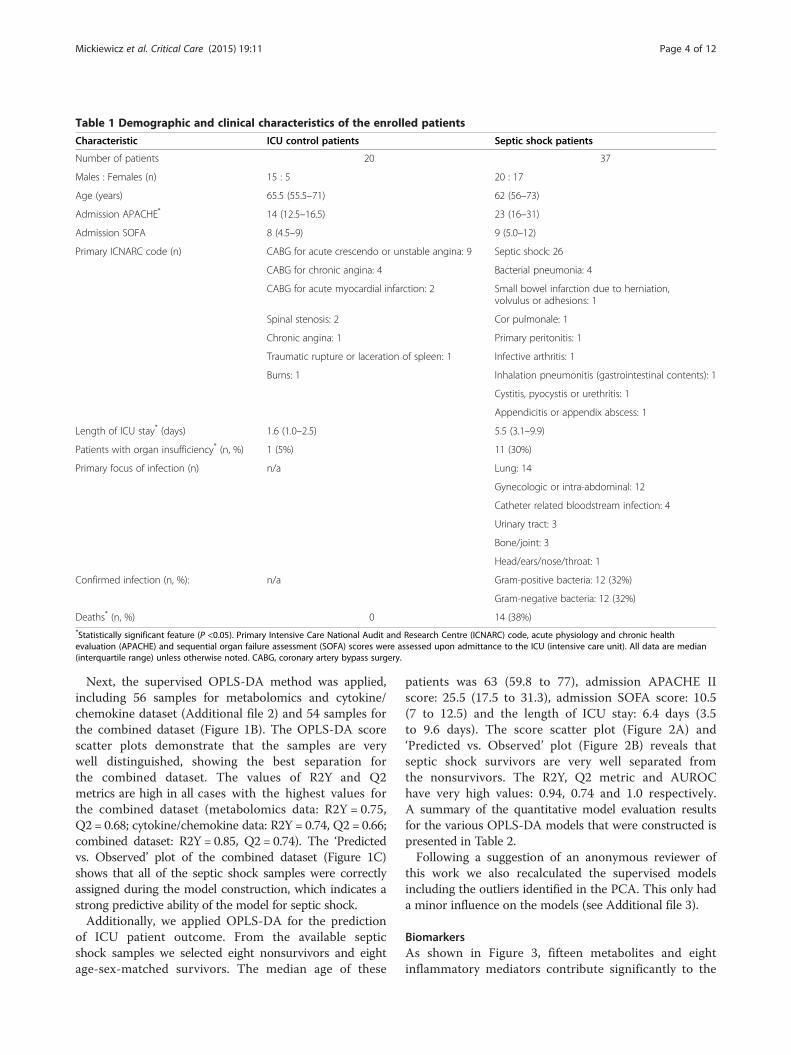
Table 1 Demographic and clinical characteristics of the enrolled patients
Characteristic ICU control patients Septic shock patients
Number of patients 20 37
Males : Females (n) 15 : 5 20 : 17
Age (years) 65.5 (55.5–71) 62 (56–73)
Admission APACHE* 14 (12.5–16.5) 23 (16–31)
Admission SOFA 8 (4.5–9) 9 (5.0–12)
Primary ICNARC code (n) CABG for acute crescendo or unstable angina: 9 Septic shock: 26
CABG for chronic angina: 4 Bacterial pneumonia: 4
CABG for acute myocardial infarction: 2 Small bowel infarction due to herniation,volvulus or adhesions: 1
Spinal stenosis: 2 Cor pulmonale: 1
Chronic angina: 1 Primary peritonitis: 1
Traumatic rupture or laceration of spleen: 1 Infective arthritis: 1
Burns: 1 Inhalation pneumonitis (gastrointestinal contents): 1
Cystitis, pyocystis or urethritis: 1
Appendicitis or appendix abscess: 1
Length of ICU stay* (days) 1.6 (1.0–2.5) 5.5 (3.1–9.9)
Patients with organ insufficiency* (n, %) 1 (5%) 11 (30%)
Primary focus of infection (n) n/a Lung: 14
Gynecologic or intra-abdominal: 12
Catheter related bloodstream infection: 4
Urinary tract: 3
Bone/joint: 3
Head/ears/nose/throat: 1
Confirmed infection (n, %): n/a Gram-positive bacteria: 12 (32%)
Gram-negative bacteria: 12 (32%)
Deaths* (n, %) 0 14 (38%)*Statistically significant feature (P <0.05). Primary Intensive Care National Audit and Research Centre (ICNARC) code, acute physiology and chronic healthevaluation (APACHE) and sequential organ failure assessment (SOFA) scores were assessed upon admittance to the ICU (intensive care unit). All data are median(interquartile range) unless otherwise noted. CABG, coronary artery bypass surgery.
Mickiewicz et al. Critical Care (2015) 19:11 Page 4 of 12
Next, the supervised OPLS-DA method was applied,including 56 samples for metabolomics and cytokine/chemokine dataset (Additional file 2) and 54 samples forthe combined dataset (Figure 1B). The OPLS-DA scorescatter plots demonstrate that the samples are verywell distinguished, showing the best separation forthe combined dataset. The values of R2Y and Q2metrics are high in all cases with the highest values forthe combined dataset (metabolomics data: R2Y = 0.75,Q2 = 0.68; cytokine/chemokine data: R2Y = 0.74, Q2 = 0.66;combined dataset: R2Y = 0.85, Q2 = 0.74). The ‘Predictedvs. Observed’ plot of the combined dataset (Figure 1C)shows that all of the septic shock samples were correctlyassigned during the model construction, which indicates astrong predictive ability of the model for septic shock.Additionally, we applied OPLS-DA for the prediction
of ICU patient outcome. From the available septicshock samples we selected eight nonsurvivors and eightage-sex-matched survivors. The median age of these
patients was 63 (59.8 to 77), admission APACHE IIscore: 25.5 (17.5 to 31.3), admission SOFA score: 10.5(7 to 12.5) and the length of ICU stay: 6.4 days (3.5to 9.6 days). The score scatter plot (Figure 2A) and‘Predicted vs. Observed’ plot (Figure 2B) reveals thatseptic shock survivors are very well separated fromthe nonsurvivors. The R2Y, Q2 metric and AUROChave very high values: 0.94, 0.74 and 1.0 respectively.A summary of the quantitative model evaluation resultsfor the various OPLS-DA models that were constructed ispresented in Table 2.Following a suggestion of an anonymous reviewer of
this work we also recalculated the supervised modelsincluding the outliers identified in the PCA. This only hada minor influence on the models (see Additional file 3).
BiomarkersAs shown in Figure 3, fifteen metabolites and eightinflammatory mediators contribute significantly to the
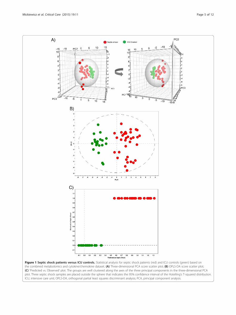
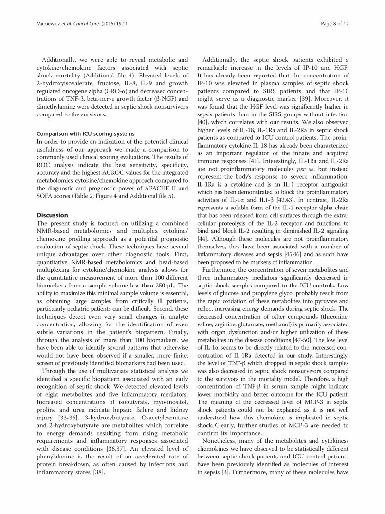
Figure 1 Septic shock patients versus ICU controls. Statistical analysis for septic shock patients (red) and ICU controls (green) based onthe combined metabolomics and cytokine/chemokine dataset. (A) Three-dimensional PCA score scatter plot; (B) OPLS-DA score scatter plot;(C) ‘Predicted vs. Observed’ plot. The groups are well clustered along the axes of the three principal components in the three-dimensional PCAplot. Three septic shock samples are placed outside the sphere that indicates the 95% confidence interval of the Hotelling’s T-squared distribution.ICU, intensive care unit; OPLS-DA, orthogonal partial least squares discriminant analysis; PCA, principal component analysis.
Mickiewicz et al. Critical Care (2015) 19:11 Page 5 of 12
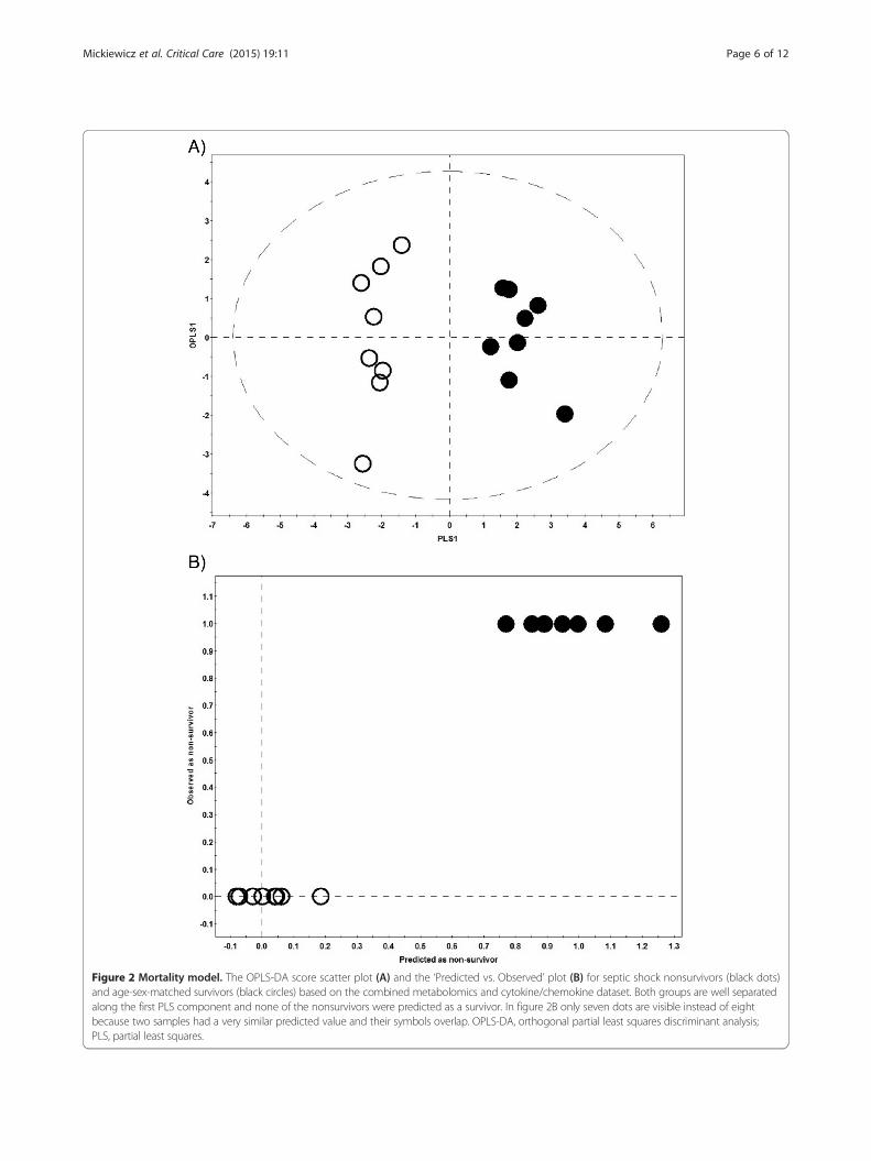
Figure 2 Mortality model. The OPLS-DA score scatter plot (A) and the ‘Predicted vs. Observed’ plot (B) for septic shock nonsurvivors (black dots)and age-sex-matched survivors (black circles) based on the combined metabolomics and cytokine/chemokine dataset. Both groups are well separatedalong the first PLS component and none of the nonsurvivors were predicted as a survivor. In figure 2B only seven dots are visible instead of eightbecause two samples had a very similar predicted value and their symbols overlap. OPLS-DA, orthogonal partial least squares discriminant analysis;PLS, partial least squares.
Mickiewicz et al. Critical Care (2015) 19:11 Page 6 of 12
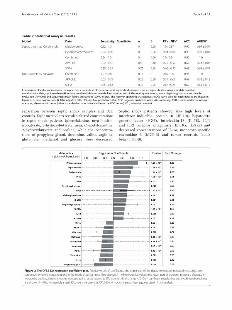
Table 2 Statistical analysis results
Model Data Sensitivity : Specificity α β PPV : NPV ACC AUROC
Septic shock vs. ICU controls Metabolomics 0.92 : 1.0 0 0.08 1.0 : 0.87 0.95 0.99 ± 0.01
Cytokines/chemokines 0.94 : 0.90 0.1 0.06 0.94 : 0.90 0.93 0.99 ± 0.01
Combined 0.94 : 1.0 0 0.06 1.0 : 0.91 0.96 1.0
APACHE 0.82 : 0.42 0.58 0.18 0.71 : 0.57 0.67 0.74 ± 0.07
SOFA 0.85 : 0.25 0.75 0.15 0.66 : 0.50 0.63 0.64 ± 0.07
Nonsurvivors vs. survivors Combined 1.0 : 0.88 0.13 0 0.89 : 1.0 0.94 1.0
APACHE 0.63 : 0.75 0.25 0.38 0.71 : 0.67 0.69 0.78 ± 0.12
SOFA 0.75 : 0.63 0.38 0.25 0.67 : 0.71 0.69 0.81 ± 0.11
Comparison of statistical measures for septic shock patients vs. ICU controls and septic shock nonsurvivors vs. septic shock survivors models based onmetabolomics data, cytokine/chemokine data, combined dataset (metabolites together with inflammatory mediators), acute physiology and chronic healthevaluation (APACHE) and sequential organ failure assessment (SOFA) scores. The receiver operating characteristic (ROC) curve plots for each dataset are shown inFigure 4. α, false positive rate; β, false negative rate; PPV, positive predictive value; NPV, negative predictive value; ACC, accuracy; AUROC, area under the receiveroperating characteristic curve (value ± standard error as calculated from the ROC curves); ICU, intensive care unit.
Mickiewicz et al. Critical Care (2015) 19:11 Page 7 of 12
separation between septic shock samples and ICUcontrols. Eight metabolites revealed altered concentrationsin septic shock patients (phenylalanine, myo-inositol,isobutyrate, 3-hydroxybutyrate, urea, O-acetylcarnitine,2-hydroxybutyrate and proline) while the concentra-tions of propylene glycol, threonine, valine, arginine,glutamate, methanol and glucose were decreased.
Figure 3 The OPLS-DA regression coefficient plot. Positive values of coefficcytokine/chemokine concentrations in the septic shock samples (fold change >metabolite and cytokine/chemokine concentrations, as compared to ICU controare shown (P <0.05, two-sample t test). ICU, intensive care unit; OPLS-DA, orthog
Septic shock patients showed also high levels ofinterferon-inducible protein-10 (IP-10), hepatocytegrowth factor (HGF), interleukin-18 (IL-18), IL-1and IL-2 receptor antagonists (IL-1Ra, IL-2Ra) anddecreased concentrations of IL-1α, monocyte-specificchemokine 3 (MCP-3) and tumor necrosis factorbeta (TNF-β).
ients (the upper part of the diagram) indicate increased metabolite and1) while negative values (the lower part of diagram) present a decrease inls (fold change <1). Only significant metabolites and cytokines/chemokinesonal partial least squares discriminant analysis.
Mickiewicz et al. Critical Care (2015) 19:11 Page 8 of 12
Additionally, we were able to reveal metabolic andcytokine/chemokine factors associated with septicshock mortality (Additional file 4). Elevated levels of2-hydroxyisovalerate, fructose, IL-8, IL-9 and growthregulated oncogene alpha (GRO-α) and decreased concen-trations of TNF-β, beta-nerve growth factor (β-NGF) anddimethylamine were detected in septic shock nonsurvivorscompared to the survivors.
Comparison with ICU scoring systemsIn order to provide an indication of the potential clinicalusefulness of our approach we made a comparison tocommonly used clinical scoring evaluations. The results ofROC analysis indicate the best sensitivity, specificity,accuracy and the highest AUROC values for the integratedmetabolomics-cytokine/chemokine approach compared tothe diagnostic and prognostic power of APACHE II andSOFA scores (Table 2, Figure 4 and Additional file 5).
DiscussionThe present study is focused on utilizing a combinedNMR-based metabolomics and multiplex cytokine/chemokine profiling approach as a potential prognosticevaluation of septic shock. These techniques have severalunique advantages over other diagnostic tools. First,quantitative NMR-based metabolomics and bead-basedmultiplexing for cytokine/chemokine analysis allows forthe quantitative measurement of more than 100 differentbiomarkers from a sample volume less than 250 μL. Theability to maximize this minimal sample volume is essential,as obtaining large samples from critically ill patients,particularly pediatric patients can be difficult. Second, thesetechniques detect even very small changes in analyteconcentration, allowing for the identification of evensubtle variations in the patient’s biopattern. Finally,through the analysis of more than 100 biomarkers, wehave been able to identify several patterns that otherwisewould not have been observed if a smaller, more finite,screen of previously identified biomarkers had been used.Through the use of multivariate statistical analysis we
identified a specific biopattern associated with an earlyrecognition of septic shock. We detected elevated levelsof eight metabolites and five inflammatory mediators.Increased concentrations of isobutyrate, myo-inositol,proline and urea indicate hepatic failure and kidneyinjury [33-36]. 3-hydroxybutyrate, O-acetylcarnitineand 2-hydroxybutyrate are metabolites which correlateto energy demands resulting from rising metabolicrequirements and inflammatory responses associatedwith disease conditions [36,37]. An elevated level ofphenylalanine is the result of an accelerated rate ofprotein breakdown, as often caused by infections andinflammatory states [38].
Additionally, the septic shock patients exhibited aremarkable increase in the levels of IP-10 and HGF.It has already been reported that the concentration ofIP-10 was elevated in plasma samples of septic shockpatients compared to SIRS patients and that IP-10might serve as a diagnostic marker [39]. Moreover, itwas found that the HGF level was significantly higher insepsis patients than in the SIRS groups without infection[40], which correlates with our results. We also observedhigher levels of IL-18, IL-1Ra and IL-2Ra in septic shockpatients as compared to ICU control patients. The proin-flammatory cytokine IL-18 has already been characterizedas an important regulator of the innate and acquiredimmune responses [41]. Interestingly, IL-1Ra and IL-2Raare not proinflammatory molecules per se, but insteadrepresent the body’s response to severe inflammation.IL-1Ra is a cytokine and is an IL-1 receptor antagonist,which has been demonstrated to block the proinflammatoryactivities of IL-1α and IL1-β [42,43]. In contrast, IL-2Rarepresents a soluble form of the IL-2 receptor alpha chainthat has been released from cell surfaces through the extra-cellular proteolysis of the IL-2 receptor and functions tobind and block IL-2 resulting in diminished IL-2 signaling[44]. Although these molecules are not proinflammatorythemselves, they have been associated with a number ofinflammatory diseases and sepsis [45,46] and as such havebeen proposed to be markers of inflammation.Furthermore, the concentration of seven metabolites and
three inflammatory mediators significantly decreased inseptic shock samples compared to the ICU controls. Lowlevels of glucose and propylene glycol probably result fromthe rapid oxidation of these metabolites into pyruvate andreflect increasing energy demands during septic shock. Thedecreased concentration of other compounds (threonine,valine, arginine, glutamate, methanol) is primarily associatedwith organ dysfunction and/or higher utilization of thesemetabolites in the disease conditions [47-50]. The low levelof IL-1α seems to be directly related to the increased con-centration of IL-1Ra detected in our study. Interestingly,the level of TNF-β which dropped in septic shock sampleswas also decreased in septic shock nonsurvivors comparedto the survivors in the mortality model. Therefore, a highconcentration of TNF-β in serum sample might indicatelower morbidity and better outcome for the ICU patient.The meaning of the decreased level of MCP-3 in septicshock patients could not be explained as it is not wellunderstood how this chemokine is implicated in septicshock. Clearly, further studies of MCP-3 are needed toconfirm its importance.Nonetheless, many of the metabolites and cytokines/
chemokines we have observed to be statistically differentbetween septic shock patients and ICU control patientshave been previously identified as molecules of interestin sepsis [3]. Furthermore, many of these molecules have
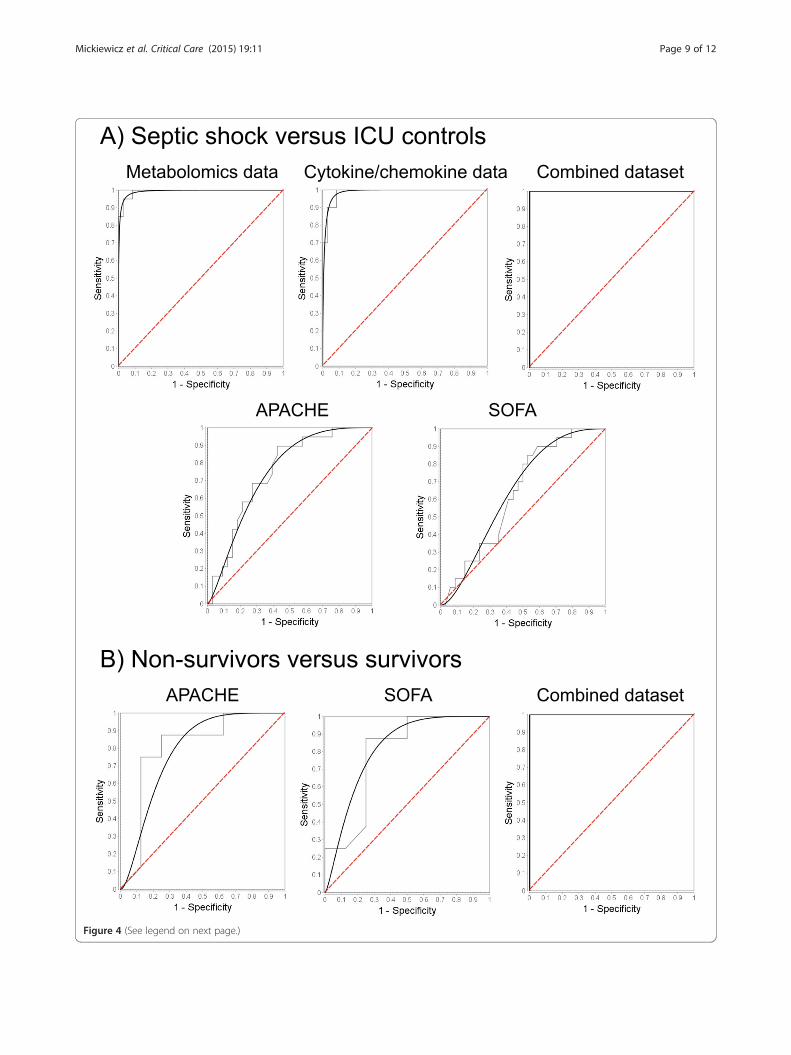
Figure 4 (See legend on next page.)
Mickiewicz et al. Critical Care (2015) 19:11 Page 9 of 12
(See figure on previous page.)Figure 4 The receiver operating characteristic (ROC) curve plots. The ROC plots for (A) septic shock patients vs. intensive care unit (ICU) controlsand (B) septic shock nonsurvivors vs. septic shock survivors models based on the metabolomics data, cytokine/chemokine data and the combineddataset (metabolites together with inflammatory mediators), acute physiology and chronic health evaluation (APACHE) and sequential organ failureassessment (SOFA) scores. Black line - fit line, grey line - empirical data, red dashed line - the chance curve. To further show the details of these curvesin the range of false positive fraction the Additional file 5 shows the ROC curves redrawn with a decimal logarithm scale for the horizontal axes.
Mickiewicz et al. Critical Care (2015) 19:11 Page 10 of 12
been tested as possible point-of-care diagnostic markersin sepsis but none of the identified markers alone havebeen adapted into a successful diagnostic test for sepsis[3]. This failure is likely the result of the multifacetednature of sepsis; a marker that demonstrates significantassociation with one group of septic patients may notcorrelate with all septic patients. As a result, the use ofsingle biomarkers in diagnosis of sepsis has not, andlikely will not, result in the development of successfulpoint-of-care testing.It has previously been demonstrated that applying
metabolomics or multiple cytokine assays separately al-lows for an identification of specific markers associatedwith sepsis severity. However, to date only metabolomicsstudies of sepsis have described potentially predictivevalues. The multiplex inflammatory mediators studiesdid not propose any predictive model that might be usedfor early diagnosis of septic shock [16]. Recently, anotherstudy has assessed a multiple cytokine profiling approachto distinguish SIRS and various forms of sepsis within agroup of emergency department patients [17]. Indeed, inthis study the authors were able to describe individualmediators independently associated with septic shock.However, the global statistical analysis could not identifyany significant differences between the patient groups. Inlight of these previous reports, our integrated metaboliteand cytokine/chemokine study can represent a potentiallypromising methodology for the prediction of septic shock.The combination of biomarkers such as metabolites andinflammatory mediators yields better results and predictivevalues than studies previously published and modelsconstructed based on separate datasets only (Table 2).Additionally, we were able to construct a model for
mortality prediction which represents a much betterprognostic ability than the commonly used APACHE IIand SOFA scores. It should be noted that the applicationof multiple cytokine assays to predict septic shock outcomehas already been described [18]. However, these authorscould only observe a significant mortality odds ratio whenusing the cytokine/chemokine data collected more than24 hours after patient enrollment. In contrast, our resultsare based on blood samples obtained at an earlier stage ofpatient admission to the ICU (not more than 24 hours). Itis well known that the first hours following patient diag-nosis are the most important for patient survival andprognosis of patient outcome at this time is very crucial. Asimilar approach has been described in a recent study in
which the authors attempted to integrate metabolomics,proteomics and clinical variables to predict the survival ofadult sepsis patients [51]. Although they performed abroad proteomics analysis by mass spectrometry, theyconcluded that these results were at best semi-quantitative and they could not incorporate them intheir predictive model. Moreover, they also mentionedthat their proteome analysis was not sensitive enoughto reliably measure cytokines/chemokines in theirsamples. Since cytokines are known to play an importantrole during sepsis, we have used a targeted and quantitativecytokine/chemokine proteomics multiplex approach in thisstudy. Our data clearly illustrate that it is possible tointegrate quantitative metabolic and cytokine/chemokineproteomic data in a bigger biomarker panel. Furthermore,our study describes a mortality model that is only basedon integrated bio-fluid components. This approach maybe advantageous to avoid a possible bias associated with asubjective diagnosis by critical care staff. Be that as itmay, our method can also easily be extended to includequantitative clinical variables and severity scores.
ConclusionsThis study indicates that an integrated metabolic andcytokine/chemokine profiling approach of blood samplesmight serve as a promising tool for the early diagnosisand prognosis of septic shock during the first hours ofpatient admission to the ICU. However, this studyshould be considered as an initial step of applyingintegrated metabolomics and inflammatory mediatorprofiling approach in a clinical setting. Beyond doubt,our results should be validated in other clinical settingsand within larger groups of patients to confirm its applic-ability throughout different ICUs. Additionally, correlationof metabolic and cytokine/chemokine profiles with theseverity of sepsis, as well as validation of the modelin an early sepsis patient population, would providefurther insight into disease mechanisms and could beused to target new therapies in the future.
Key messages
� Integration of metabolic and inflammatory mediatorprofiling data might serve as a reliable diagnosticand prognostic tool for septic shock.
� A total of fifteen metabolites and eight inflammatorymediators had a significant influence on the
Mickiewicz et al. Critical Care (2015) 19:11 Page 11 of 12
separation between septic shock samples andICU controls.
� A receiver operating characteristic analysis indicatedan excellent predictive ability of the integratedmetabolomics/inflammatory mediator models whencompared to the conventionally used ICU scoringsystems.
Additional files
Additional file 1: Three-dimensional PCA score scatter plots.Three-dimensional PCA score scatter plots obtained for 37 septic shockpatients (red) and 20 ICU controls (green) based on the (A) metabolicdata and (B) cytokine/chemokine data. The groups are well clusteredalong the axes of the three principal components. One septic shocksample is placed outside the sphere that describes the 95% confidenceinterval of the Hotelling’s T-squared distribution.
Additional file 2: OPLS-DA score scatter plots. The OPLS-DA scorescatter plots obtained for 36 septic shock patients (red) and 20 ICUcontrols (green) based on the (A) metabolic data and (B) cytokine/chemokine data.
Additional file 3: The summary table. Comparison of statisticalmeasures calculated for the supervised OPLS-DA models without excludingoutliers to the results presented in the manuscript.
Additional file 4: The OPLS-DA regression coefficient plot. Positivevalues of coefficients (the upper part of the diagram) indicates increasedmetabolite or inflammatory mediator concentrations in septic shocknonsurvivor samples while negative values (the lower part of diagram)present a decrease in metabolite or inflammatory mediator concentrations,as compared to the age-sex-matched septic shock survivors. Only significantmetabolites are shown (P <0.05, two-sample t test).
Additional file 5: The receiver operating characteristic (ROC) curvesplotted with a decimal logarithmic scale for the horizontal axes. TheROC plots for (A) septic shock patients vs. ICU controls and (B) septicshock nonsurvivors vs. septic shock survivors models based on themetabolomics data, cytokine/chemokine data and the combined dataset(metabolites together with inflammatory mediators), APACHE (acutephysiology and chronic health evaluation) and SOFA (sequential organfailure assessment) scores. Black line - fit line, red dashed line - the chancecurve (that is the diagonal of the ROC curve when plotted in linear scale).
AbbreviationsACC: accuracy; APACHE: acute physiology and chronic health evaluation;AUROC: area under the receiver operating characteristic curve;CABG: coronary artery bypass surgery; CV: cross-validation; GRO-α:growth-regulated oncogene alpha; HGF: hepatocyte growth factor;ICNARC: Intensive Care National Audit and Research Centre; ICU: intensivecare unit; IL-1: interleukin 1; IL-2: interleukin 2; IL-18: interleukin-18;IL-1Ra: interleukin-1 receptor antagonist; IL-1α: interleukin-1α; IL-2Ra:interleukin-2 receptor antagonist; IL-8: interleukin-8; IL-9: interleukin-9;IP-10: interferon-inducible protein-10; kDa: kiloDaltons; MCP-3: monocyte-specificchemokine 3; NMR: nuclear magnetic resonance; NPV: negative predictive value;OPLS-DA: orthogonal partial least squares discriminant analysis; PC: principalcomponent; PCA: principal component analysis; PLS: partial least squares;PPV: positive predictive value; SIRS: systemic inflammatory response syndrome;SOFA: sequential organ failure assessment; TNF-α: tumor necrosis factor alpha;TNF-β: tumor necrosis factor beta; β-NGF: beta nerve growth factor.
Competing interestsThe authors declare that they have no competing interests.
Authors’ contributionsBM was involved in the conception and design of the study, carrying outNMR experiments, collection and assembly of data, analysis andinterpretation of the data, drafting the manuscript, statistical expertise andcritical revision of the article for important intellectual content. PT carried out
cytokine/chemokine profiling experiments and provided the data forstatistical analysis. CNJ participated in cytokine/chemokine profilingexperiments, data interpretation and drafting the manuscript. CL coordinatedcytokine/chemokine profiling experiments and helped to draft themanuscript. JW supplied blood samples for NMR and cytokine/chemokineprofiling experiments and helped to draft the manuscript. BWW coordinatedblood samples collection and helped to draft the manuscript. CDparticipated in the design of the study, its coordination and samples supply.PK was involved in conception and design of the study, critical revision ofthe manuscript for important intellectual content and final approval of thearticle. HJV was involved in conception and design of the study, criticalrevision of the manuscript for important intellectual content and finalapproval of the manuscript. All authors read and approved the finalmanuscript.
AcknowledgementsWe would like to thank Dr. Deane McIntyre and Dr. Rustem Shaykhutdinovfor technical support and for spectrometer maintenance.The study was supported by the Alberta Sepsis Network, which in turn isfunded by Alberta Innovates Health Solutions (AIHS). HJV and PK holdScientist awards from AIHS, while CNJ is funded by a fellowship from AIHS.CCEPTR receives funding from the Alberta Sepsis Network and from theCanada Foundation for Innovation. PK is also the holder of the Calvin,Phoebe and Joan Snyder Chair in Critical Care Medicine which contributedto this research.The funding agencies had no influence on the design, analysis andmanuscript preparation for this study.
Author details1Bio-NMR-Centre, Department of Biological Sciences, University of Calgary,2500 University Drive Northwest, Calgary, AB, T2N 1N4, Canada. 2SnyderTranslational Laboratory, Department of Critical Care Medicine, University ofCalgary, 3280 Hospital Drive Northwest, Calgary, AB, T2N 4N1, Canada.3Critical Care Epidemiologic and Biologic Tissue Resource, Department ofCritical Care Medicine, University of Calgary, 3280 Hospital Drive Northwest,Calgary, AB, T2N 4N1, Canada. 4Department of Community Health Sciences,University of Calgary, 3280 Hospital Drive Northwest, Calgary, AB, T2N 4N1,Canada. 5Snyder Institute for Chronic Diseases, University of Calgary, 3280Hospital Drive Northwest, Calgary, AB, T2N 4N1, Canada.
Received: 15 April 2014 Accepted: 23 December 2014
References1. Slade E, Tamber PS, Vincent JL. The Surviving Sepsis Campaign: raising
awareness to reduce mortality. Crit Care. 2003;7:1–2.2. Dellinger RP, Carlet JM, Masur H, Gerlach H, Calandra T, Cohen J, et al.
Surviving Sepsis Campaign guidelines for management of severe sepsis andseptic shock. Crit Care Med. 2004;32:858–73.
3. Pierrakos C, Vincent JL. Sepsis biomarkers: a review. Crit Care. 2010;14:R15.4. Marshall JC, Vincent JL, Fink MP, Cook DJ, Rubenfeld G, Foster D, et al.
Measures, markers, and mediators: toward a staging system for clinicalsepsis. A report of the Fifth Toronto Sepsis Roundtable, Toronto, Ontario,Canada, October 25–26, 2000. Crit Care Med. 2003;31:1560–7.
5. Bakker J, Coffernils M, Leon M, Gris P, Vincent JL. Blood lactate levels aresuperior to oxygen-derived variables in predicting outcome in human septicshock. Chest. 1991;99:956–62.
6. Chiarla C, Giovannini I, Siegel JH, Boldrini G, Castagneto M. The relationshipbetween plasma taurine and other amino acid levels in human sepsis.J Nutr. 2000;130:2222–7.
7. Faix JD. Established and novel biomarkers of sepsis. Biomark Med.2011;5:117–30.
8. Holmes CL, Russell JA, Walley KR. Genetic polymorphisms in sepsis andseptic shock: role in prognosis and potential for therapy. Chest.2003;124:1103–15.
9. Levy MM, Dellinger RP, Townsend SR, Linde-Zwirble WT, Marshall JC, Bion J, et al.The Surviving Sepsis Campaign: results of an international guideline-basedperformance improvement program targeting severe sepsis. Crit Care Med.2010;38:367–74.
Mickiewicz et al. Critical Care (2015) 19:11 Page 12 of 12
10. Hoerr V, Zbytnuik L, Leger C, Tam PP, Kubes P, Vogel HJ. Gram-negative andGram-positive bacterial infections give rise to a different metabolic responsein a mouse model. J Proteome Res. 2012;11:3231–45.
11. Izquierdo-Garcia JL, Nin N, Ruiz-Cabello J, Rojas Y, de Paula M,Lopez-Cuenca S, et al. A metabolomic approach for diagnosis of experimentalsepsis. Intensive Care Med. 2011;37:2023–32.
12. Lin ZY, Xu PB, Yan SK, Meng HB, Yang GJ, Dai WX, et al. A metabonomicapproach to early prognostic evaluation of experimental sepsis by (1)H NMRand pattern recognition. NMR Biomed. 2009;22:601–8.
13. Mickiewicz B, Vogel HJ, Wong HR, Winston BW. Metabolomics as a novelapproach for early diagnosis of pediatric septic shock and its mortality.Am J Respir Crit Care Med. 2013;187:967–76.
14. Mickiewicz B, Duggan GE, Winston BW, Doig C, Kubes P, Vogel HJ.Metabolic profiling of serum samples by 1H nuclear magnetic resonancespectroscopy as a potential diagnostic approach for septic shock. Crit CareMed. 2014;42:1140–9.
15. Prabhakar U, Eirikis E, Davis HM. Simultaneous quantification ofproinflammatory cytokines in human plasma using the LabMAP assay.J Immunol Methods. 2002;260:207–18.
16. Bozza FA, Salluh JI, Japiassu AM, Soares M, Assis EF, Gomes RN, et al.Cytokine profiles as markers of disease severity in sepsis: a multiplexanalysis. Crit Care. 2007;11:R49.
17. Lvovschi V, Arnaud L, Parizot C, Freund Y, Juillien G, Ghillani-Dalbin P, et al.Cytokine profiles in sepsis have limited relevance for stratifying patients inthe emergency department: a prospective observational study. PLoS One.2011;6:e28870.
18. Fjell CD, Thair S, Hsu JL, Walley KR, Russell JA, Boyd J. Cytokines andsignaling molecules predict clinical outcomes in sepsis. PLoS One.2013;8:e79207.
19. Saric J, Li JV, Swann JR, Utzinger J, Calvert G, Nicholson JK, et al. Integratedcytokine and metabolic analysis of pathological responses to parasiteexposure in rodents. J Proteome Res. 2010;9:2255–64.
20. Bernard GR, Artigas A, Brigham KL, Carlet J, Falke K, Hudson L, et al. TheAmerican-European Consensus Conference on ARDS. Definitions,mechanisms, relevant outcomes, and clinical trial coordination. Am JRespir Crit Care Med. 1994;149:818–24.
21. Levy MM, Fink MP, Marshall JC, Abraham E, Angus D, Cook D, et al. 2001SCCM/ESICM/ACCP/ATS/SIS International Sepsis Definitions Conference.Crit Care Med. 2003;31:1250–6.
22. Nicholson JK, Foxall PJ, Spraul M, Farrant RD, Lindon JC. 750 MHz 1H and1H-13C NMR spectroscopy of human blood plasma. Anal Chem.1995;67:793–811.
23. Weljie AM, Newton J, Mercier P, Carlson E, Slupsky CM. Targeted profiling:quantitative analysis of 1H NMR metabolomics data. Anal Chem.2006;78:4430–42.
24. van den Berg RA, Hoefsloot HC, Westerhuis JA, Smilde AK, van der Werf MJ.Centering, scaling, and transformations: improving the biologicalinformation content of metabolomics data. BMC Genomics. 2006;7:142.
25. Eriksson L, Johansson E, Kettaneh-Wold N, Trygg J, Wikstrom C, Wold S.Multi- and megavariate data analysis part I: basic principles and applications.Umeå, Sweden: Umetrics AB; 2006.
26. Trygg J, Wold S. Orthogonal projections to latent structures (O-PLS).J Chemometrics. 2002;16:119–28.
27. Hosmer DW, Lemeshow S. Applied logistic regression. New York:Wiley; 2000.
28. Picard RR, Cook DR. Cross-validation of regression models. J Am Stat Assoc.1984;79:575–83.
29. Trygg J, Holmes E, Lundstedt T. Chemometrics in metabonomics. J ProteomeRes. 2007;6:469–79.
30. Metz CE. Basic principles of ROC analysis. Semin Nucl Med. 1978;8:283–98.31. Knaus WA, Draper EA, Wagner DP, Zimmerman JE. APACHE II: a severity of
disease classification system. Crit Care Med. 1985;13:818–29.32. Vincent JL, de Mendonca A, Cantraine F, Moreno R, Takala J, Suter PM, et al.
Use of the SOFA score to assess the incidence of organ dysfunction/failurein intensive care units: results of a multicenter, prospective study. Workinggroup on “sepsis-related problems” of the European Society of IntensiveCare Medicine. Crit Care Med. 1998;26:1793–800.
33. Cerra FB, Caprioli J, Siegel JH, McMenamy RR, Border JR. Proline metabolismin sepsis, cirrhosis and general surgery. The peripheral energy deficit.Ann Surg. 1979;190:577–86.
34. Landaas S, Jakobs C. The occurrence of 2-hydroxyisovaleric acid in patientswith lactic acidosis and ketoacidosis. Clin Chim Acta. 1977;78:489–93.
35. Mao H, Wang H, Wang B, Liu X, Gao H, Xu M, et al. Systemic metabolicchanges of traumatic critically ill patients revealed by an NMR-basedmetabonomic approach. J Proteome Res. 2009;8:5423–30.
36. Wishart DS, Knox C, Guo AC, Eisner R, Young N, Gautam B, et al. HMDB:a knowledgebase for the human metabolome. Nucleic Acids Res.2009;37:D603–10.
37. Wolfe RR, Shaw JH, Durkot MJ. Energy metabolism in trauma and sepsis:the role of fat. Prog Clin Biol Res. 1983;111:89–109.
38. Wannemacher Jr RW, Klainer AS, Dinterman RE, Beisel WR. The significanceand mechanism of an increased serum phenylalanine-tyrosine ratio duringinfection. Am J Clin Nutr. 1976;29:997–1006.
39. Punyadeera C, Schneider EM, Schaffer D, Hsu HY, Joos TO, Kriebel F, et al. Abiomarker panel to discriminate between systemic inflammatory responsesyndrome and sepsis and sepsis severity. J Emerg Trauma Shock. 2010;3:26–35.
40. Sekine K, Fujishima S, Aikawa N. Plasma hepatocyte growth factor isincreased in early-phase sepsis. J Infect Chemother. 2004;10:110–4.
41. Gracie JA, Robertson SE, McInnes IB. Interleukin-18. J Leukoc Biol.2003;73:213–24.
42. Corbett GT, Roy A, Pahan K. Gemfibrozil, a lipid-lowering drug, upregulatesIL-1 receptor antagonist in mouse cortical neurons: implications for neuronalself-defense. J Immunol. 2012;189:1002–13.
43. Dinarello CA. The role of the interleukin-1-receptor antagonist in blockinginflammation mediated by interleukin-1. N Engl J Med. 2000;343:732–4.
44. Rubinstein MP, Kovar M, Purton JF, Cho JH, Boyman O, Surh CD, et al.Converting IL-15 to a superagonist by binding to soluble IL-15R{alpha}.Proc Natl Acad Sci U S A. 2006;103:9166–71.
45. Arend WP, Malyak M, Guthridge CJ, Gabay C. Interleukin-1 receptor antagonist:role in biology. Annu Rev Immunol. 1998;16:27–55.
46. Saito K, Wagatsuma T, Toyama H, Ejima Y, Hoshi K, Shibusawa M, et al.Sepsis is characterized by the increases in percentages of circulatingCD4 + CD25+ regulatory T cells and plasma levels of soluble CD25.Tohoku J Exp Med. 2008;216:61–8.
47. Faure M, Chone F, Mettraux C, Godin JP, Bechereau F, Vuichoud J, et al.Threonine utilization for synthesis of acute phase proteins, intestinalproteins, and mucins is increased during sepsis in rats. J Nutr.2007;137:1802–7.
48. Freund H, Atamian S, Holroyde J, Fischer JE. Plasma amino acids aspredictors of the severity and outcome of sepsis. Ann Surg. 1979;190:571–6.
49. Freund HR, Ryan Jr JA, Fischer JE. Amino acid derangements in patientswith sepsis: treatment with branched chain amino acid rich infusions. AnnSurg. 1978;188:423–30.
50. Poeze M, Luiking YC, Breedveld P, Manders S, Deutz NE. Decreased plasmaglutamate in early phases of septic shock with acute liver dysfunction is anindependent predictor of survival. Clin Nutr. 2008;27:523–30.
51. Langley RJ, Tsalik EL, van Velkinburgh JC, Glickman SW, Rice BJ, Wang C, et al.An integrated clinico-metabolomic model improves prediction of death insepsis. Sci Transl Med. 2013;5:195ra195.
Submit your next manuscript to BioMed Centraland take full advantage of:
• Convenient online submission
• Thorough peer review
• No space constraints or color figure charges
• Immediate publication on acceptance
• Inclusion in PubMed, CAS, Scopus and Google Scholar
• Research which is freely available for redistribution
Submit your manuscript at www.biomedcentral.com/submit