Embed Size (px)
Citation preview
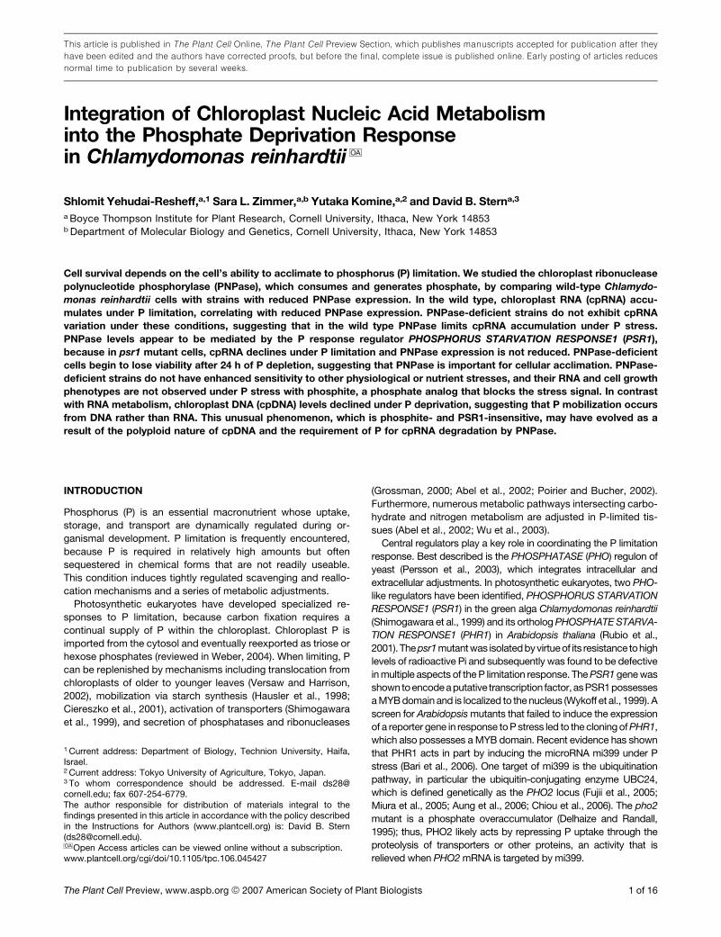
Integration of Chloroplast Nucleic Acid Metabolisminto the Phosphate Deprivation Responsein Chlamydomonas reinhardtii OA
Shlomit Yehudai-Resheff,a,1 Sara L. Zimmer,a,b Yutaka Komine,a,2 and David B. Sterna,3
a Boyce Thompson Institute for Plant Research, Cornell University, Ithaca, New York 14853b Department of Molecular Biology and Genetics, Cornell University, Ithaca, New York 14853
Cell survival depends on the cell’s ability to acclimate to phosphorus (P) limitation. We studied the chloroplast ribonuclease
polynucleotide phosphorylase (PNPase), which consumes and generates phosphate, by comparing wild-type Chlamydo-
monas reinhardtii cells with strains with reduced PNPase expression. In the wild type, chloroplast RNA (cpRNA) accu-
mulates under P limitation, correlating with reduced PNPase expression. PNPase-deficient strains do not exhibit cpRNA
variation under these conditions, suggesting that in the wild type PNPase limits cpRNA accumulation under P stress.
PNPase levels appear to be mediated by the P response regulator PHOSPHORUS STARVATION RESPONSE1 (PSR1),
because in psr1 mutant cells, cpRNA declines under P limitation and PNPase expression is not reduced. PNPase-deficient
cells begin to lose viability after 24 h of P depletion, suggesting that PNPase is important for cellular acclimation. PNPase-
deficient strains do not have enhanced sensitivity to other physiological or nutrient stresses, and their RNA and cell growth
phenotypes are not observed under P stress with phosphite, a phosphate analog that blocks the stress signal. In contrast
with RNA metabolism, chloroplast DNA (cpDNA) levels declined under P deprivation, suggesting that P mobilization occurs
from DNA rather than RNA. This unusual phenomenon, which is phosphite- and PSR1-insensitive, may have evolved as a
result of the polyploid nature of cpDNA and the requirement of P for cpRNA degradation by PNPase.
INTRODUCTION
Phosphorus (P) is an essential macronutrient whose uptake,
storage, and transport are dynamically regulated during or-
ganismal development. P limitation is frequently encountered,
because P is required in relatively high amounts but often
sequestered in chemical forms that are not readily useable.
This condition induces tightly regulated scavenging and reallo-
cation mechanisms and a series of metabolic adjustments.
Photosynthetic eukaryotes have developed specialized re-
sponses to P limitation, because carbon fixation requires a
continual supply of P within the chloroplast. Chloroplast P is
imported from the cytosol and eventually reexported as triose or
hexose phosphates (reviewed in Weber, 2004). When limiting, P
can be replenished by mechanisms including translocation from
chloroplasts of older to younger leaves (Versaw and Harrison,
2002), mobilization via starch synthesis (Hausler et al., 1998;
Ciereszko et al., 2001), activation of transporters (Shimogawara
et al., 1999), and secretion of phosphatases and ribonucleases
(Grossman, 2000; Abel et al., 2002; Poirier and Bucher, 2002).
Furthermore, numerous metabolic pathways intersecting carbo-
hydrate and nitrogen metabolism are adjusted in P-limited tis-
sues (Abel et al., 2002; Wu et al., 2003).
Central regulators play a key role in coordinating the P limitation
response. Best described is the PHOSPHATASE (PHO) regulon of
yeast (Persson et al., 2003), which integrates intracellular and
extracellular adjustments. In photosynthetic eukaryotes, two PHO-
like regulators have been identified, PHOSPHORUS STARVATION
RESPONSE1 (PSR1) in the green alga Chlamydomonas reinhardtii
(Shimogawara et al., 1999) and its ortholog PHOSPHATE STARVA-
TION RESPONSE1 (PHR1) in Arabidopsis thaliana (Rubio et al.,
2001).The psr1mutantwas isolatedbyvirtueof its resistance tohigh
levels of radioactive Pi and subsequently was found to be defective
in multiple aspects of the P limitation response. The PSR1 gene was
showntoencodeaputative transcription factor, asPSR1possesses
a MYB domain and is localized to the nucleus (Wykoff et al., 1999). A
screen for Arabidopsis mutants that failed to induce the expression
of a reporter gene in response to P stress led to the cloning of PHR1,
which also possesses a MYB domain. Recent evidence has shown
that PHR1 acts in part by inducing the microRNA mi399 under P
stress (Bari et al., 2006). One target of mi399 is the ubiquitination
pathway, in particular the ubiquitin-conjugating enzyme UBC24,
which is defined genetically as the PHO2 locus (Fujii et al., 2005;
Miura et al., 2005; Aung et al., 2006; Chiou et al., 2006). The pho2
mutant is a phosphate overaccumulator (Delhaize and Randall,
1995); thus, PHO2 likely acts by repressing P uptake through the
proteolysis of transporters or other proteins, an activity that is
relieved when PHO2 mRNA is targeted by mi399.
1 Current address: Department of Biology, Technion University, Haifa,Israel.2 Current address: Tokyo University of Agriculture, Tokyo, Japan.3 To whom correspondence should be addressed. E-mail [email protected]; fax 607-254-6779.The author responsible for distribution of materials integral to thefindings presented in this article in accordance with the policy describedin the Instructions for Authors (www.plantcell.org) is: David B. Stern([email protected]).OA Open Access articles can be viewed online without a subscription.www.plantcell.org/cgi/doi/10.1105/tpc.106.045427
This article is published in The Plant Cell Online, The Plant Cell Preview Section, which publishes manuscripts accepted for publication after they
have been edited and the authors have corrected proofs, but before the final, complete issue is published online. Early posting of articles reduces
normal time to publication by several weeks.
The Plant Cell Preview, www.aspb.org ª 2007 American Society of Plant Biologists 1 of 16
Although the importance of chloroplast P metabolism is clear,
how it is regulated under nutrient stress is largely unknown. This
study focuses on polynucleotide phosphorylase (PNPase), a
nucleus-encoded and chloroplast-localized ribonuclease that
has a role in polyadenylation-stimulated RNA degradation (Kudla
et al., 1996; Lisitsky et al., 1997; Nishimura et al., 2004). A readily
reversible enzyme (Yehudai-Resheff et al., 2001), PNPase poly-
merizes poly(A)-rich tails on mRNA fragments generated by
endonucleolytic cleavage, a reaction that primarily consumes
nucleotide diphosphates (NDPs) and generates Pi. Because of
its high affinity for poly(A), PNPase binds to these same tails and
rapidly degrades the fragment in the 39 to 59 direction through a
phosphorylytic, or P-requiring, reaction. Instability conferred by
polyadenylation is a common feature of prokaryotes and organ-
elles (Slomovic et al., 2006) and contrasts with the stabilizing
effect of poly(A) tails in the cytosol (Dreyfus and Regnier, 2002).
Furthermore, as a phosphorylytic enzyme, PNPase differs from
most cytosolic ribonucleases, which are hydrolytic and often
activated under P deficiency (Green, 1994).
Because the twin anabolic and catabolic activities of PNPase
either generate or consume Pi, the enzyme has the potential to
affect P pools in addition to, or by way of, regulating RNA levels.
Indeed, the Arabidopsis mutant rif10 overcomes the inhibition of a
key enzyme in the plastid isoprenoid pathway through the inacti-
vation of PNPase, which in turn affects metabolic flux (Sauret-
Gueto et al., 2006). Using Chlamydomonas, we explored whether
strains defective in PNPase expression or regulation would also be
defective in acclimation to P limitation. Here, we present evidence
that chloroplast DNA (cpDNA) and RNA (cpRNA) levels are both
adjusted to optimize cell acclimation and that correct regulation of
PNPase may be required for cell survival under P limitation.
RESULTS
Creation of PNPase-Deficient Strains
A Chlamydomonas PNP cDNA was identified through an EST
project and extended using RT-PCR (see Methods). Based on
available genomic sequence (Joint Genome Institute version 3.0),
Chlamydomonasappears tohavea singlegeneencodingPNPase,
in contrast with the duplicated genes found in higher plants, which
specify plastid (Hayes et al., 1996) and mitochondrial (Perrin et al.,
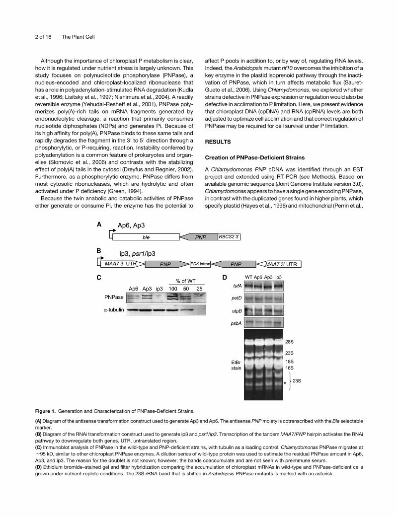
Figure 1. Generation and Characterization of PNPase-Deficient Strains.
(A) Diagram of the antisense transformation construct used to generate Ap3 and Ap6. The antisense PNP moiety is cotranscribed with the Ble selectable
marker.
(B) Diagram of the RNAi transformation construct used to generate ip3 and psr1/ip3. Transcription of the tandem MAA7/PNP hairpin activates the RNAi
pathway to downregulate both genes. UTR, untranslated region.
(C) Immunoblot analysis of PNPase in the wild-type and PNP-deficient strains, with tubulin as a loading control. Chlamydomonas PNPase migrates at
;95 kD, similar to other chloroplast PNPase enzymes. A dilution series of wild-type protein was used to estimate the residual PNPase amount in Ap6,
Ap3, and ip3. The reason for the doublet is not known; however, the bands coaccumulate and are not seen with preimmune serum.
(D) Ethidium bromide–stained gel and filter hybridization comparing the accumulation of chloroplast mRNAs in wild-type and PNPase-deficient cells
grown under nutrient-replete conditions. The 23S rRNA band that is shifted in Arabidopsis PNPase mutants is marked with an asterisk.
2 of 16 The Plant Cell

2004) isozymes. The Chlamydomonas cDNA sequence (GenBank
accession number DQ492261) predicts a protein with the catalytic
core domains I and II and the KH and S1 RNA binding domains,
which are conserved in bacterial, mitochondrial, and chloroplast
PNPases (Yehudai-Resheff et al., 2003).
We designed constructs for antisense RNA or RNA interfer-
ence (RNAi) inhibition of PNP expression (Figures 1A and 1B,
respectively) and obtained multiple colonies for each construct.
The effects of both antisense expression and RNAi were highly
variable, as judged by RT-PCR and immunoblot analysis of PNP
expression. For reasons discussed below, three strains were
selected for detailed analysis: Ap6, Ap3, and ip3. As shown in
Figure 1C, an antibody raised against a portion of the Chlamy-
domonas protein revealed that PNPase accumulation in Ap6 was
;25% of that in the wild type, whereas the other antisense RNA–
derived strain, Ap3, was only slightly reduced relative to the wild
type, retaining ;75% of the wild-type level based on multiple
immunoblots. By contrast, the RNAi strain was strongly affected,
with residual PNPase being estimated at 10 to 20% of the wild-
type level, also based on multiple experiments. PNPase gener-
ally appeared as a doublet, probably reflecting two slightly
different forms of the protein, which accumulate in parallel and,
for this study, were considered in total. The low overall abun-
dance of PNPase (even in the wild type) precludes precise
quantification, but in general, Ap3, Ap6, and ip3 constitute an
allelic series with successively decreasing amounts of PNPase.
In Arabidopsis, a cosuppressed line was studied that was
largely depleted of the chloroplast PNPase (Walter et al., 2002).
This line failed to fully process the 23S rRNA, as revealed by a
mobility shift on an ethidium bromide–stained gel. In addition,
novel psbA and rbcL transcripts accumulated with 39 end exten-
sions. In contrast with those results, none of the Chlamydomonas
strains showed visible differences when total RNA was analyzed
by gel electrophoresis and ethidium bromide staining, nor were
migration differences observed for the four chloroplast mRNAs
tested (Figure 1D). This suggests either that residual PNPase is
sufficient for RNA maturation functions or that it is redundant, for
example with RNase II, as is the case in Escherichia coli (Donovan
and Kushner, 1986), when these Chlamydomonas strains are
grown under standard, nutrient-replete conditions.
Chloroplast RNA Accumulates in Response
to Phosphate Limitation
The catalytic activity of chloroplast PNPase is reversible in vitro,
in which a relatively high NDP:Pi ratio favors polymerization and a
low ratio favors degradation (Yehudai-Resheff et al., 2001). As
discussed in the Introduction, given that PNPase uses and
consumes major P-containing metabolites, its activity might be
relevant to the P limitation response. A simple way of concep-
tualizing this potential integration is to hypothesize that under P
limitation, PNPase-catalyzed RNA degradation and thus the
consumption of P would slow, whereas polymerization would
be favored, assuming that the NDP concentration remained
relatively high. This same polymerization reaction would release
Pi as a form of P mobilization.
As a first step, cpRNA levels were measured after P depletion.
Wild-type or PNPase-deficient strains were first grown in the
presence of P, then the culture was divided into two portions, one
with P and one without. Samples were taken over a 48-h time
course, and at the end, the starved culture was transferred back
to P-containing medium for 2 h. Figure 2A shows a representa-
tive gel blot that reveals that in wild-type cells, accumulation of
the petD and tufA chloroplast transcripts increased substantially
under P limitation. This increase is most evident at the 24- and
48-h time points and was fully reversed 2 h after P was restored
(Figure 2A, lane þ2). This accumulation is consistent with dimin-
ished PNPase catabolic activity along with continued transcrip-
tion, and the 24 h delay in the manifestation of the phenomenon
could be explained as the time taken for the Pi concentration in
the chloroplast to change under P starvation.
Figure 2A also shows that this phenomenon was not observed
in Ap6 cells, which under þP conditions have similar cpRNA
accumulation as the wild type (Figure 1D). In�P conditions, Ap6
mRNA levels remained constant, reacting to neither P depletion
nor the readdition of P to the culture. This finding implicates
decreased PNPase exonucleolytic activity as being responsible
for cpRNA accumulation in wild-type cells acclimating to P
deficiency; in Ap6, the initial PNPase level is low, so minimal
activity changes might be expected under P limitation (Figure
3B). Two additional strains, Ap3 and ip3, were also examined
Figure 2. Chloroplast RNA Accumulation under P Limitation.
(A) Wild-type and Ap6 cells were grown to early log phase in TAP
medium, then collected and resuspended in TAP (þP) or TA (�P)
medium. Total RNA was isolated immediately (0) or after 6, 24, or 48 h.
An additional sample was obtained from cells in TA, after they were
transferred to TAP medium for an additional 2 h (þ2). A total of 3 mg of
RNA was loaded in each lane, and blots were hybridized with the
experimental probes listed at left. To demonstrate the equality of loading,
gels were stained with ethidium bromide.
(B) Ap3 and ip3 cells were analyzed as described for (A), except that the
0 time point was taken before division of the culture and the 6-h time
point was omitted.
PNPase and Phosphate Deprivation 3 of 16
(Figure 2B). Ap3 showed slight abundance increases under P
limitation, with a subsequent reduction when P was restored.
This phenotype was intermediate between those of the wild type
and Ap6, consistent with the relative residual levels of PNPase.
Strain ip3, however, showed no variation in RNA accumulation.
These results confirm that PNPase deficiency is associated with
a lack of cpRNA accumulation in response to P limitation.
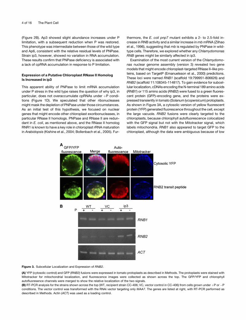
Expression of a Putative Chloroplast RNase II Homolog
Is Increased in ip3
This apparent ability of PNPase to limit mRNA accumulation
under P stress in the wild type raises the question of why ip3, in
particular, does not overaccumulate cpRNAs under þP condi-
tions (Figure 1D). We speculated that other ribonucleases
might mask the depletion of PNPase under those circumstances.
As an initial test of this hypothesis, we focused on nuclear
genes that might encode other chloroplast exoribonucleases, in
particular RNase II homologs. PNPase and RNase II are redun-
dant in E. coli, as mentioned above, and the RNase II homolog
RNR1 is known to have a key role in chloroplast rRNA maturation
in Arabidopsis (Kishine et al., 2004; Bollenbach et al., 2005). Fur-
thermore, the E. coli pnp7 mutant exhibits a 2- to 2.5-fold in-
crease in RNB activity and a similar increase in rnb mRNA (Zilhao
et al., 1996), suggesting that rnb is regulated by PNPase in wild-
type cells. Therefore, we explored whether any Chlamydomonas
RNB genes might be similarly affected in ip3.
Examination of the most current version of the Chlamydomo-
nas nuclear genome assembly (version 3) revealed two gene
models that might encode chloroplast-targeted RNase II–like pro-
teins, based on TargetP (Emanuelsson et al., 2000) predictions.
These loci were named RNB1 (scaffold 19:799951-806626) and
RNB2 (scaffold 11:108345-114817). To gain evidence for subcel-
lular localization, cDNAs encoding the N-terminal 189 amino acids
(RNB1) or 115 amino acids (RNB2) were fused to a green fluores-
cent protein (GFP)–encoding gene, and the proteins were ex-
pressed transiently in tomato (Solanum lycopersicum) protoplasts.
As shown in Figure 3A, a cytosolic version of yellow fluorescent
protein (YFP) generated fluorescence throughout the cell, except
the large vacuole. RNB2 fusions were clearly targeted to the
chloroplasts, because chlorophyll autofluorescence colocalized
with the GFP signal but not with the Mitotracker signal, which
labels mitochondria. RNB1 also appeared to target GFP to the
chloroplast, although the data were ambiguous because of low
Figure 3. Subcellular Localization and Expression of RNB2.
(A) YFP (cytosolic control) and GFP (RNB2) fusions were expressed in tomato protoplasts as described in Methods. The protoplasts were stained with
Mitotracker for mitochondrial localization, and fluorescence images were collected as shown across the top. The GFP/YFP and chlorophyll
autofluoresence channels were merged to show the relative localization of the two signals.
(B) RT-PCR analysis for the strains shown across the top (WT, recipient strain CC-406; VC, vector control in CC-406) from cells grown under þP or �P
conditions. The vector control was transformed with the RNAi vector targeting only MAA7. The genes are listed at right, with RT-PCR performed as
described in Methods. Actin (ACT) was used as a loading control.
4 of 16 The Plant Cell
expression of the fusion protein (data not shown). We conclude
that RNB2 very likely encodes a chloroplast RNase II homolog
and that RNB1 may encode a second plastid homolog.
We next examined the expression of RNB1 and RNB2 in wild-
type, ip3, or vector control cells grown under þP or �P condi-
tions, as shown in Figure 3B. In multiple experiments, little or no
variation was seen for the RNB1 transcript level between strains
or growth conditions. On the other hand, RNB2 mRNA was
reproducibly found at an increased level in ip3 compared with the
wild type or the vector control in bothþP and�P conditions. This
increase was not quantified, but examination of the gels in Figure
3B and in other experiments was consistent with a twofold to
threefold difference compared with the actin control. If this
reflects an increase of RNase II–like activity in ip3 chloroplasts,
this might account for the lack of increased cpRNA when cells
are grown under nutrient-replete conditions (Figure 1D) and may
help to explain the failure of cpRNA levels to change under P
deficiency even in partially PNPase-deficient strains (Figure 2).
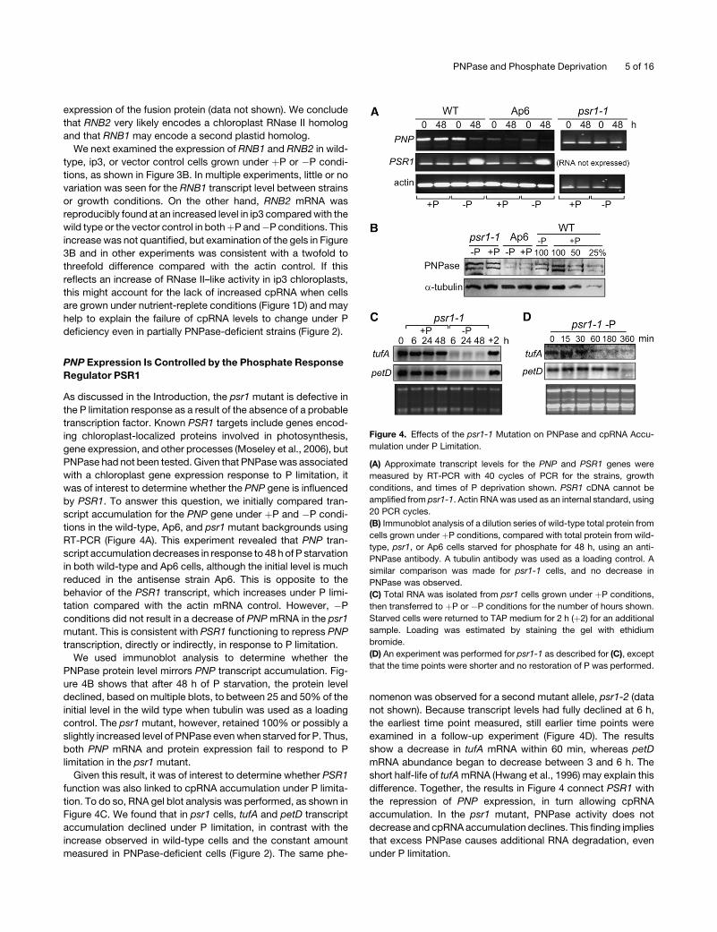
PNP Expression Is Controlled by the Phosphate Response
Regulator PSR1
As discussed in the Introduction, the psr1 mutant is defective in
the P limitation response as a result of the absence of a probable
transcription factor. Known PSR1 targets include genes encod-
ing chloroplast-localized proteins involved in photosynthesis,
gene expression, and other processes (Moseley et al., 2006), but
PNPase had not been tested. Given that PNPase was associated
with a chloroplast gene expression response to P limitation, it
was of interest to determine whether the PNP gene is influenced
by PSR1. To answer this question, we initially compared tran-
script accumulation for the PNP gene under þP and �P condi-
tions in the wild-type, Ap6, and psr1 mutant backgrounds using
RT-PCR (Figure 4A). This experiment revealed that PNP tran-
script accumulation decreases in response to 48 h of P starvation
in both wild-type and Ap6 cells, although the initial level is much
reduced in the antisense strain Ap6. This is opposite to the
behavior of the PSR1 transcript, which increases under P limi-
tation compared with the actin mRNA control. However, �P
conditions did not result in a decrease of PNP mRNA in the psr1
mutant. This is consistent with PSR1 functioning to repress PNP
transcription, directly or indirectly, in response to P limitation.
We used immunoblot analysis to determine whether the
PNPase protein level mirrors PNP transcript accumulation. Fig-
ure 4B shows that after 48 h of P starvation, the protein level
declined, based on multiple blots, to between 25 and 50% of the
initial level in the wild type when tubulin was used as a loading
control. The psr1 mutant, however, retained 100% or possibly a
slightly increased level of PNPase even when starved for P. Thus,
both PNP mRNA and protein expression fail to respond to P
limitation in the psr1 mutant.
Given this result, it was of interest to determine whether PSR1
function was also linked to cpRNA accumulation under P limita-
tion. To do so, RNA gel blot analysis was performed, as shown in
Figure 4C. We found that in psr1 cells, tufA and petD transcript
accumulation declined under P limitation, in contrast with the
increase observed in wild-type cells and the constant amount
measured in PNPase-deficient cells (Figure 2). The same phe-
nomenon was observed for a second mutant allele, psr1-2 (data
not shown). Because transcript levels had fully declined at 6 h,
the earliest time point measured, still earlier time points were
examined in a follow-up experiment (Figure 4D). The results
show a decrease in tufA mRNA within 60 min, whereas petD
mRNA abundance began to decrease between 3 and 6 h. The
short half-life of tufA mRNA (Hwang et al., 1996) may explain this
difference. Together, the results in Figure 4 connect PSR1 with
the repression of PNP expression, in turn allowing cpRNA
accumulation. In the psr1 mutant, PNPase activity does not
decrease and cpRNA accumulation declines. This finding implies
that excess PNPase causes additional RNA degradation, even
under P limitation.
Figure 4. Effects of the psr1-1 Mutation on PNPase and cpRNA Accu-
mulation under P Limitation.
(A) Approximate transcript levels for the PNP and PSR1 genes were
measured by RT-PCR with 40 cycles of PCR for the strains, growth
conditions, and times of P deprivation shown. PSR1 cDNA cannot be
amplified from psr1-1. Actin RNA was used as an internal standard, using
20 PCR cycles.
(B) Immunoblot analysis of a dilution series of wild-type total protein from
cells grown under þP conditions, compared with total protein from wild-
type, psr1, or Ap6 cells starved for phosphate for 48 h, using an anti-
PNPase antibody. A tubulin antibody was used as a loading control. A
similar comparison was made for psr1-1 cells, and no decrease in
PNPase was observed.
(C) Total RNA was isolated from psr1 cells grown under þP conditions,
then transferred to þP or �P conditions for the number of hours shown.
Starved cells were returned to TAP medium for 2 h (þ2) for an additional
sample. Loading was estimated by staining the gel with ethidium
bromide.
(D) An experiment was performed for psr1-1 as described for (C), except
that the time points were shorter and no restoration of P was performed.
PNPase and Phosphate Deprivation 5 of 16
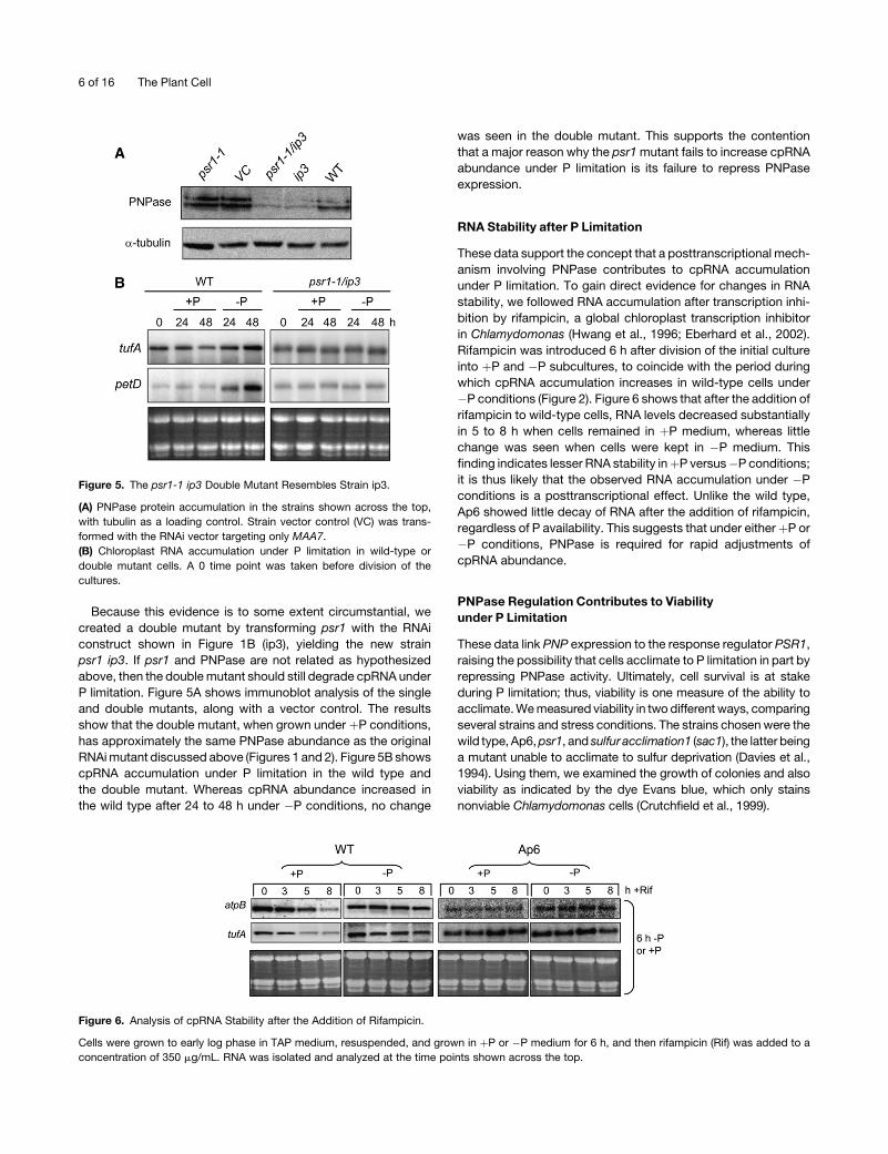
Because this evidence is to some extent circumstantial, we
created a double mutant by transforming psr1 with the RNAi
construct shown in Figure 1B (ip3), yielding the new strain
psr1 ip3. If psr1 and PNPase are not related as hypothesized
above, then the double mutant should still degrade cpRNA under
P limitation. Figure 5A shows immunoblot analysis of the single
and double mutants, along with a vector control. The results
show that the double mutant, when grown under þP conditions,
has approximately the same PNPase abundance as the original
RNAi mutant discussed above (Figures 1 and 2). Figure 5B shows
cpRNA accumulation under P limitation in the wild type and
the double mutant. Whereas cpRNA abundance increased in
the wild type after 24 to 48 h under �P conditions, no change
was seen in the double mutant. This supports the contention
that a major reason why the psr1 mutant fails to increase cpRNA
abundance under P limitation is its failure to repress PNPase
expression.
RNA Stability after P Limitation
These data support the concept that a posttranscriptional mech-
anism involving PNPase contributes to cpRNA accumulation
under P limitation. To gain direct evidence for changes in RNA
stability, we followed RNA accumulation after transcription inhi-
bition by rifampicin, a global chloroplast transcription inhibitor
in Chlamydomonas (Hwang et al., 1996; Eberhard et al., 2002).
Rifampicin was introduced 6 h after division of the initial culture
into þP and �P subcultures, to coincide with the period during
which cpRNA accumulation increases in wild-type cells under
�P conditions (Figure 2). Figure 6 shows that after the addition of
rifampicin to wild-type cells, RNA levels decreased substantially
in 5 to 8 h when cells remained in þP medium, whereas little
change was seen when cells were kept in �P medium. This
finding indicates lesser RNA stability inþP versus�P conditions;
it is thus likely that the observed RNA accumulation under �P
conditions is a posttranscriptional effect. Unlike the wild type,
Ap6 showed little decay of RNA after the addition of rifampicin,
regardless of P availability. This suggests that under eitherþP or
�P conditions, PNPase is required for rapid adjustments of
cpRNA abundance.
PNPase Regulation Contributes to Viability
under P Limitation
These data link PNP expression to the response regulator PSR1,
raising the possibility that cells acclimate to P limitation in part by
repressing PNPase activity. Ultimately, cell survival is at stake
during P limitation; thus, viability is one measure of the ability to
acclimate. We measured viability in two different ways, comparing
several strains and stress conditions. The strains chosen were the
wild type, Ap6, psr1, and sulfur acclimation1 (sac1), the latter being
a mutant unable to acclimate to sulfur deprivation (Davies et al.,
1994). Using them, we examined the growth of colonies and also
viability as indicated by the dye Evans blue, which only stains
nonviable Chlamydomonas cells (Crutchfield et al., 1999).
Figure 6. Analysis of cpRNA Stability after the Addition of Rifampicin.
Cells were grown to early log phase in TAP medium, resuspended, and grown in þP or �P medium for 6 h, and then rifampicin (Rif) was added to a
concentration of 350 mg/mL. RNA was isolated and analyzed at the time points shown across the top.
Figure 5. The psr1-1 ip3 Double Mutant Resembles Strain ip3.
(A) PNPase protein accumulation in the strains shown across the top,
with tubulin as a loading control. Strain vector control (VC) was trans-
formed with the RNAi vector targeting only MAA7.
(B) Chloroplast RNA accumulation under P limitation in wild-type or
double mutant cells. A 0 time point was taken before division of the
cultures.
6 of 16 The Plant Cell
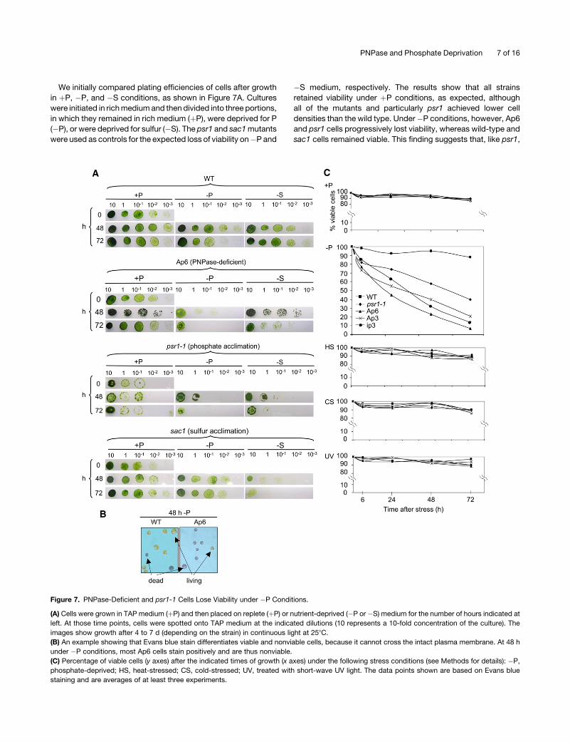
We initially compared plating efficiencies of cells after growth
in þP, �P, and �S conditions, as shown in Figure 7A. Cultures
were initiated in rich medium and then divided into three portions,
in which they remained in rich medium (þP), were deprived for P
(�P), or were deprived for sulfur (�S). The psr1 and sac1 mutants
were used as controls for the expected loss of viability on�P and
�S medium, respectively. The results show that all strains
retained viability under þP conditions, as expected, although
all of the mutants and particularly psr1 achieved lower cell
densities than the wild type. Under�P conditions, however, Ap6
and psr1 cells progressively lost viability, whereas wild-type and
sac1 cells remained viable. This finding suggests that, like psr1,
Figure 7. PNPase-Deficient and psr1-1 Cells Lose Viability under �P Conditions.
(A) Cells were grown in TAP medium (þP) and then placed on replete (þP) or nutrient-deprived (�P or �S) medium for the number of hours indicated at
left. At those time points, cells were spotted onto TAP medium at the indicated dilutions (10 represents a 10-fold concentration of the culture). The
images show growth after 4 to 7 d (depending on the strain) in continuous light at 258C.
(B) An example showing that Evans blue stain differentiates viable and nonviable cells, because it cannot cross the intact plasma membrane. At 48 h
under �P conditions, most Ap6 cells stain positively and are thus nonviable.
(C) Percentage of viable cells (y axes) after the indicated times of growth (x axes) under the following stress conditions (see Methods for details): �P,
phosphate-deprived; HS, heat-stressed; CS, cold-stressed; UV, treated with short-wave UV light. The data points shown are based on Evans blue
staining and are averages of at least three experiments.
PNPase and Phosphate Deprivation 7 of 16
Ap6 cannot acclimate to �P conditions. A complementary
picture was observed under �S conditions. In this case, the
wild type and Ap6 remained viable, whereas sac1 lost viability.
This result suggested that Ap6 was defective in its P limitation,
but not its S limitation, response.
Because P and S limitation are both nutrient stresses, and
because of possible crosstalk between the response pathways
(Moseley et al., 2006), we tested PNPase-deficient strains under
other types of stress, in case the strains lost viability under P
limitation as a result of a general stress intolerance, the S
limitation results notwithstanding. For a more direct measure of
cell viability, we used Evans blue, whose staining of wild-type
versus Ap6 cells subjected to 48 h of P limitation is shown as an
example in Figure 7B (the dye only stains dead cells). The
experiment for which results are shown in Figure 7C included the
three PNPase-deficient strains studied here as well as the wild
type and psr1. The top two graphs show that all strains retained
high viability for 72 h under þP conditions but that psr1 and the
PNPase-deficient strains lost viability under �P conditions, as
expected. The same strains were then subjected to heat, cold,
and UV light stress, using conditions established previously for
Chlamydomonas. Under all of these conditions, no appreciable
difference was observed when comparing the wild type with
other strains. Thus, neither Ap6 nor other PNPase-deficient
strains have a general sensitivity to abiotic stress.
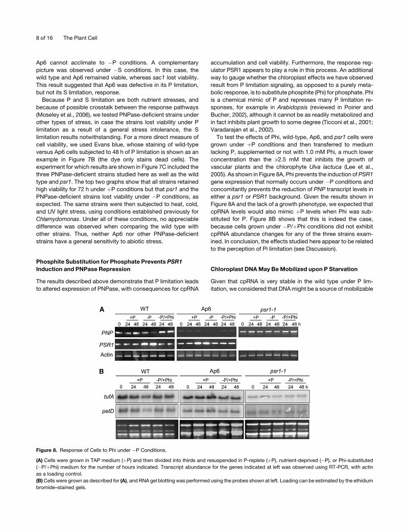
Phosphite Substitution for Phosphate Prevents PSR1
Induction and PNPase Repression
The results described above demonstrate that P limitation leads
to altered expression of PNPase, with consequences for cpRNA
accumulation and cell viability. Furthermore, the response reg-
ulator PSR1 appears to play a role in this process. An additional
way to gauge whether the chloroplast effects we have observed
result from P limitation signaling, as opposed to a purely meta-
bolic response, is to substitute phosphite (Phi) for phosphate. Phi
is a chemical mimic of P and represses many P limitation re-
sponses, for example in Arabidopsis (reviewed in Poirier and
Bucher, 2002), although it cannot be as readily metabolized and
in fact inhibits plant growth to some degree (Ticconi et al., 2001;
Varadarajan et al., 2002).
To test the effects of Phi, wild-type, Ap6, and psr1 cells were
grown under þP conditions and then transferred to medium
lacking P, supplemented or not with 1.0 mM Phi, a much lower
concentration than the >2.5 mM that inhibits the growth of
vascular plants and the chlorophyte Ulva lactuca (Lee et al.,
2005). As shown in Figure 8A, Phi prevents the induction of PSR1
gene expression that normally occurs under �P conditions and
concomitantly prevents the reduction of PNP transcript levels in
either a psr1 or PSR1 background. Given the results shown in
Figure 8A and the lack of a growth phenotype, we expected that
cpRNA levels would also mimic þP levels when Phi was sub-
stituted for P. Figure 8B shows that this is indeed the case,
because cells grown under �P/þPhi conditions did not exhibit
cpRNA abundance changes for any of the three strains exam-
ined. In conclusion, the effects studied here appear to be related
to the perception of Pi limitation (see Discussion).
Chloroplast DNA May Be Mobilized upon P Starvation
Given that cpRNA is very stable in the wild type under P lim-
itation, we considered that DNA might be a source of mobilizable
Figure 8. Response of Cells to Phi under �P Conditions.
(A) Cells were grown in TAP medium (þP) and then divided into thirds and resuspended in P-replete (þP), nutrient-deprived (�P), or Phi-substituted
(�P/þPhi) medium for the number of hours indicated. Transcript abundance for the genes indicated at left was observed using RT-PCR, with actin
as a loading control.
(B) Cells were grown as described for (A), and RNA gel blotting was performed using the probes shown at left. Loading can be estimated by the ethidium
bromide–stained gels.
8 of 16 The Plant Cell
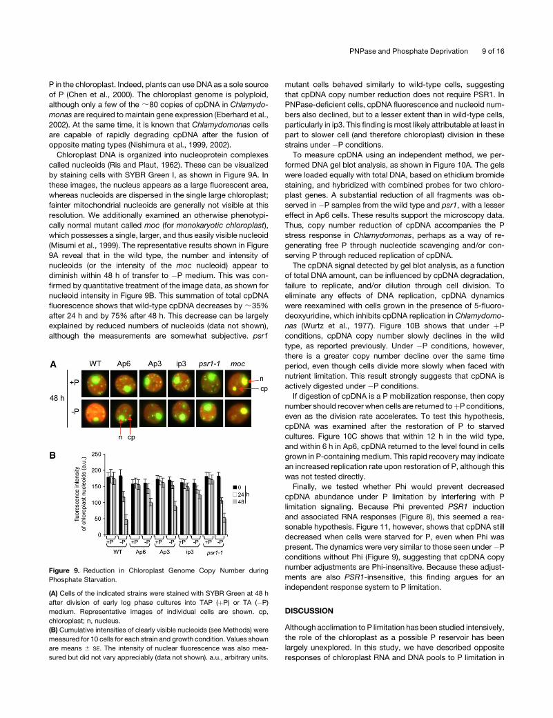
P in the chloroplast. Indeed, plants can use DNA as a sole source
of P (Chen et al., 2000). The chloroplast genome is polyploid,
although only a few of the ;80 copies of cpDNA in Chlamydo-
monas are required to maintain gene expression (Eberhard et al.,
2002). At the same time, it is known that Chlamydomonas cells
are capable of rapidly degrading cpDNA after the fusion of
opposite mating types (Nishimura et al., 1999, 2002).
Chloroplast DNA is organized into nucleoprotein complexes
called nucleoids (Ris and Plaut, 1962). These can be visualized
by staining cells with SYBR Green I, as shown in Figure 9A. In
these images, the nucleus appears as a large fluorescent area,
whereas nucleoids are dispersed in the single large chloroplast;
fainter mitochondrial nucleoids are generally not visible at this
resolution. We additionally examined an otherwise phenotypi-
cally normal mutant called moc (for monokaryotic chloroplast),
which possesses a single, larger, and thus easily visible nucleoid
(Misumi et al., 1999). The representative results shown in Figure
9A reveal that in the wild type, the number and intensity of
nucleoids (or the intensity of the moc nucleoid) appear to
diminish within 48 h of transfer to �P medium. This was con-
firmed by quantitative treatment of the image data, as shown for
nucleoid intensity in Figure 9B. This summation of total cpDNA
fluorescence shows that wild-type cpDNA decreases by ;35%
after 24 h and by 75% after 48 h. This decrease can be largely
explained by reduced numbers of nucleoids (data not shown),
although the measurements are somewhat subjective. psr1
mutant cells behaved similarly to wild-type cells, suggesting
that cpDNA copy number reduction does not require PSR1. In
PNPase-deficient cells, cpDNA fluorescence and nucleoid num-
bers also declined, but to a lesser extent than in wild-type cells,
particularly in ip3. This finding is most likely attributable at least in
part to slower cell (and therefore chloroplast) division in these
strains under �P conditions.
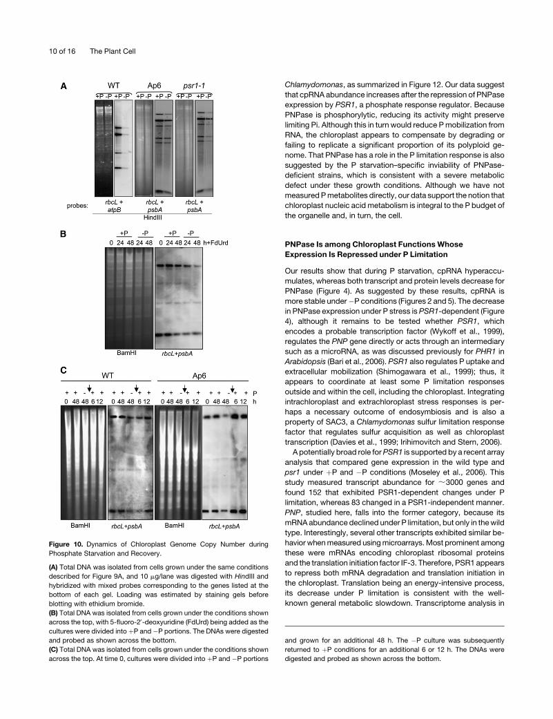
To measure cpDNA using an independent method, we per-
formed DNA gel blot analysis, as shown in Figure 10A. The gels
were loaded equally with total DNA, based on ethidium bromide
staining, and hybridized with combined probes for two chloro-
plast genes. A substantial reduction of all fragments was ob-
served in �P samples from the wild type and psr1, with a lesser
effect in Ap6 cells. These results support the microscopy data.
Thus, copy number reduction of cpDNA accompanies the P
stress response in Chlamydomonas, perhaps as a way of re-
generating free P through nucleotide scavenging and/or con-
serving P through reduced replication of cpDNA.
The cpDNA signal detected by gel blot analysis, as a function
of total DNA amount, can be influenced by cpDNA degradation,
failure to replicate, and/or dilution through cell division. To
eliminate any effects of DNA replication, cpDNA dynamics
were reexamined with cells grown in the presence of 5-fluoro-
deoxyuridine, which inhibits cpDNA replication in Chlamydomo-
nas (Wurtz et al., 1977). Figure 10B shows that under þP
conditions, cpDNA copy number slowly declines in the wild
type, as reported previously. Under �P conditions, however,
there is a greater copy number decline over the same time
period, even though cells divide more slowly when faced with
nutrient limitation. This result strongly suggests that cpDNA is
actively digested under �P conditions.
If digestion of cpDNA is a P mobilization response, then copy
number should recover when cells are returned toþP conditions,
even as the division rate accelerates. To test this hypothesis,
cpDNA was examined after the restoration of P to starved
cultures. Figure 10C shows that within 12 h in the wild type,
and within 6 h in Ap6, cpDNA returned to the level found in cells
grown in P-containing medium. This rapid recovery may indicate
an increased replication rate upon restoration of P, although this
was not tested directly.
Finally, we tested whether Phi would prevent decreased
cpDNA abundance under P limitation by interfering with P
limitation signaling. Because Phi prevented PSR1 induction
and associated RNA responses (Figure 8), this seemed a rea-
sonable hypothesis. Figure 11, however, shows that cpDNA still
decreased when cells were starved for P, even when Phi was
present. The dynamics were very similar to those seen under�P
conditions without Phi (Figure 9), suggesting that cpDNA copy
number adjustments are Phi-insensitive. Because these adjust-
ments are also PSR1-insensitive, this finding argues for an
independent response system to P limitation.
DISCUSSION
Although acclimation to P limitation has been studied intensively,
the role of the chloroplast as a possible P reservoir has been
largely unexplored. In this study, we have described opposite
responses of chloroplast RNA and DNA pools to P limitation in
Figure 9. Reduction in Chloroplast Genome Copy Number during
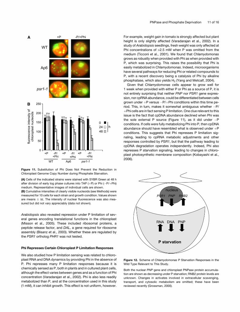
Phosphate Starvation.
(A) Cells of the indicated strains were stained with SYBR Green at 48 h
after division of early log phase cultures into TAP (þP) or TA (�P)
medium. Representative images of individual cells are shown. cp,
chloroplast; n, nucleus.
(B) Cumulative intensities of clearly visible nucleoids (see Methods) were
measured for 10 cells for each strain and growth condition. Values shown
are means 6 SE. The intensity of nuclear fluorescence was also mea-
sured but did not vary appreciably (data not shown). a.u., arbitrary units.
PNPase and Phosphate Deprivation 9 of 16
Chlamydomonas, as summarized in Figure 12. Our data suggest
that cpRNA abundance increases after the repression of PNPase
expression by PSR1, a phosphate response regulator. Because
PNPase is phosphorylytic, reducing its activity might preserve
limiting Pi. Although this in turn would reduce P mobilization from
RNA, the chloroplast appears to compensate by degrading or
failing to replicate a significant proportion of its polyploid ge-
nome. That PNPase has a role in the P limitation response is also
suggested by the P starvation–specific inviability of PNPase-
deficient strains, which is consistent with a severe metabolic
defect under these growth conditions. Although we have not
measured P metabolites directly, our data support the notion that
chloroplast nucleic acid metabolism is integral to the P budget of
the organelle and, in turn, the cell.
PNPase Is among Chloroplast Functions Whose
Expression Is Repressed under P Limitation
Our results show that during P starvation, cpRNA hyperaccu-
mulates, whereas both transcript and protein levels decrease for
PNPase (Figure 4). As suggested by these results, cpRNA is
more stable under�P conditions (Figures 2 and 5). The decrease
in PNPase expression under P stress is PSR1-dependent (Figure
4), although it remains to be tested whether PSR1, which
encodes a probable transcription factor (Wykoff et al., 1999),
regulates the PNP gene directly or acts through an intermediary
such as a microRNA, as was discussed previously for PHR1 in
Arabidopsis (Bari et al., 2006). PSR1 also regulates P uptake and
extracellular mobilization (Shimogawara et al., 1999); thus, it
appears to coordinate at least some P limitation responses
outside and within the cell, including the chloroplast. Integrating
intrachloroplast and extrachloroplast stress responses is per-
haps a necessary outcome of endosymbiosis and is also a
property of SAC3, a Chlamydomonas sulfur limitation response
factor that regulates sulfur acquisition as well as chloroplast
transcription (Davies et al., 1999; Irihimovitch and Stern, 2006).
A potentially broad role for PSR1 is supported by a recent array
analysis that compared gene expression in the wild type and
psr1 under þP and �P conditions (Moseley et al., 2006). This
study measured transcript abundance for ;3000 genes and
found 152 that exhibited PSR1-dependent changes under P
limitation, whereas 83 changed in a PSR1-independent manner.
PNP, studied here, falls into the former category, because its
mRNA abundance declined under P limitation, but only in the wild
type. Interestingly, several other transcripts exhibited similar be-
havior when measured using microarrays. Most prominent among
these were mRNAs encoding chloroplast ribosomal proteins
and the translation initiation factor IF-3. Therefore, PSR1 appears
to repress both mRNA degradation and translation initiation in
the chloroplast. Translation being an energy-intensive process,
its decrease under P limitation is consistent with the well-
known general metabolic slowdown. Transcriptome analysis in
Figure 10. Dynamics of Chloroplast Genome Copy Number during
Phosphate Starvation and Recovery.
(A) Total DNA was isolated from cells grown under the same conditions
described for Figure 9A, and 10 mg/lane was digested with HindIII and
hybridized with mixed probes corresponding to the genes listed at the
bottom of each gel. Loading was estimated by staining gels before
blotting with ethidium bromide.
(B) Total DNA was isolated from cells grown under the conditions shown
across the top, with 5-fluoro-29-deoxyuridine (FdUrd) being added as the
cultures were divided into þP and �P portions. The DNAs were digested
and probed as shown across the bottom.
(C) Total DNA was isolated from cells grown under the conditions shown
across the top. At time 0, cultures were divided into þP and �P portions
and grown for an additional 48 h. The �P culture was subsequently
returned to þP conditions for an additional 6 or 12 h. The DNAs were
digested and probed as shown across the bottom.
10 of 16 The Plant Cell
Arabidopsis also revealed repression under P limitation of sev-
eral genes encoding translational functions in the chloroplast
(Misson et al., 2005). These included ribosomal proteins, a
peptide release factor, and DAL, a gene required for ribosome
assembly (Bisanz et al., 2003). Whether these are regulated by
the PSR1 ortholog PHR1 was not tested.
Phi Represses Certain Chloroplast P Limitation Responses
We also studied how P limitation sensing was related to chloro-
plast RNA and DNA dynamics by providing Phi in the absence of
P. Phi represses many P limitation responses because it is
chemically sensed as P, both in plants and in cultured plant cells,
although the effect varies between genes and as a function of Phi
concentration (Varadarajan et al., 2002). Phi is also less readily
metabolized than P, and at the concentration used in this study
(1 mM), it can inhibit growth. This effect is not uniform, however.
For example, weight gain in tomato is strongly affected but plant
height is only slightly affected (Varadarajan et al., 2002). In a
study of Arabidopsis seedlings, fresh weight was only affected at
Phi concentrations of >2.5 mM when P was omitted from the
medium (Ticconi et al., 2001). We found that Chlamydomonas
grows as robustly when provided with Phi as when provided with
P, which was surprising. This raises the possibility that Phi is
easily metabolized in Chlamydomonas. Indeed, microorganisms
have several pathways for reducing Phi or related compounds to
P, with a recent discovery being a catalysis of Phi by alkaline
phosphatase, which also yields H2 (Yang and Metcalf, 2004).
Given that Chlamydomonas cells appear to grow well for
1 week when provided with either P or Phi as a source of P, it is
not entirely surprising that neither PNP nor PSR1 gene expres-
sion, nor cpRNA abundance, could be differentiated between cells
grown under þP versus �P/þPhi conditions within this time pe-
riod. This, in turn, makes it somewhat ambiguous whether �P/
þPhi cells are in fact sensing P limitation. One clue relevant to this
issue is the fact that cpDNA abundance declined when Phi was
the sole external P source (Figure 11), as it did under �P
conditions. If cells were fully metabolizing Phi into P, then cpDNA
abundance should have resembled what is observed under þP
conditions. This suggests that Phi represses P limitation sig-
naling, leading to cpRNA metabolic adjustments and other
responses controlled by PSR1, but that the pathway leading to
cpDNA degradation operates independently. Indeed, Phi also
represses P starvation signaling, leading to changes in chloro-
plast photosynthetic membrane composition (Kobayashi et al.,
2006).
Figure 12. Scheme of Chlamydomonas P Starvation Responses in the
Wild Type Relevant to This Study.
Both the nuclear PNP gene and chloroplast PNPase protein accumula-
tion are shown as decreasing under P starvation; RNB2 protein levels are
unknown. Changes in activates involved in extracellular scavenging,
transport, and cytosolic metabolism are omitted; these have been
reviewed recently (Grossman, 2000).
Figure 11. Substitution of Phi Does Not Prevent the Reduction in
Chloroplast Genome Copy Number during Phosphate Starvation.
(A) Cells of the indicated strains were stained with SYBR Green at 48 h
after division of early log phase cultures into TAP (þP) or Phi (�P/þPhi)
medium. Representative images of individual cells are shown.
(B) Cumulative intensities of clearly visible nucleoids (see Methods) were
measured for 10 cells for each strain and growth condition. Values shown
are means 6 SE. The intensity of nuclear fluorescence was also mea-
sured but did not vary appreciably (data not shown).
PNPase and Phosphate Deprivation 11 of 16
RNA Metabolism under P Limitation
PNPase activity (Yehudai-Resheff et al., 2001) and cpRNA deg-
radation rates (Schuster et al., 1999) respond to the relative
concentrations of Pi and nucleotides in vitro, but how its direc-
tionality is regulated in vivo is poorly understood. Our results with
wild-type cells show that cpRNA stability increases under �P
conditions (Figure 6), although RNA accumulation is not seen
until the 24-h time point (Figure 2). The delayed response likely
reflects the time required for the chloroplast Pi concentration to
decrease sufficiently so that PNPase nucleolytic activity is
disfavored. Whether the decrease in Pi concentration leads to
increased mRNA polyadenylation via the reverse PNPase reac-
tion is unknown, and quantitative measurements of either chlo-
roplast polyadenylation or NDP:Pi ratio are highly problematic. In
any event, although polyadenylation normally destabilizes
cpRNA (Lisitsky et al., 1997), in a low P situation PNPase would
not be prone to degrade these polyadenylated species, thus
allowing RNA levels to increase. Together with the reduction of
PNPase itself (Figure 4B), our results indicate a general slowing
of cpRNA metabolism under P limitation.
In the PNPase-deficient ip3 line, the putative RNase II encoded
by RNB2 appears to be upregulated (Figure 3) under bothþP and
�P conditions. Although in E. coli the reciprocal relationship
between pnp and rnb expression can be explained by the activity
of each respective enzyme on the other’s mRNA (Zilhao et al.,
1996), this cannot be the case in Chlamydomonas, in which the
mRNAs are cytosolic and the enzymes are in the chloroplast.
This finding, therefore, suggests some kind of metabolic or
chemical signal resulting from PNPase deficiency that ultimately
results in a transcription increase for RNB2.
The situation we have described for ribonucleases in the wild-
type chloroplast contrasts sharply with the upregulation of cytosolic
ribonucleases that often occurs during P limitation. For example,
RNS1 and RNS2 are induced under P stress in Arabidopsis (Bariola
et al., 1994). Cultured plant cells, bacteria, and fungi also induce
and sometimes secrete RNases under P stress (Nurnberger et al.,
1989; Hahnen et al., 2000; Tasaki et al., 2004). These RNases,
however, are almost certainly hydrolytic, rather than phosphory-
lytic, allowing them to serve as scavengers under these circum-
stances. Why the presumably chloroplastic RNB2 would not be
induced as a scavenger in Chlamydomonas is unclear.
Requirement of PNPase for Acclimation to P Limitation
A striking result was the progressive loss of viability for PNPase-
deficient strains, which was manifested between 6 and 72 h in
�P conditions (Figure 7). The psr1 mutant also dies under �P
conditions, ultimately because of an inability to control oxidative
damage (Moseley et al., 2006). PNPase-deficient cells do not
lose viability under þP conditions or a variety of other stress
situations, suggesting that they are specifically unable to cope
with P limitation. Indeed, Arabidopsis plants lacking detectable
PNPase were reported to grow normally when planted in soil
(Walter et al., 2002), and PNPase is also not essential in E. coli
(Donovan and Kushner, 1986).
On the other hand, prokaryotic PNPase has been shown to
participate in other abiotic and biotic stress responses. In bac-
teria, PNPase induction is integral to cold shock adaptation
through its regulation of mRNA stability (Clarke and Dowds,
1994; Goverde et al., 1998; Yamanaka and Inouye, 2001).
PNPase is also required for the complete degradation of glucose
transporter mRNA in response to blockage of the glycolytic
pathway in E. coli (Morita et al., 2004) and is required for the full
development of competence in Bacillus subtilis (Luttinger et al.,
1996), which is a pathway induced by nutrient deficiency.
PNPase has further been linked to the ability of Yersinia to infect
animal cells via induction of the bacterial type III secretion system
(Rosenzweig et al., 2005). These results, along with the finding
that ;100 mRNAs are differentially expressed in a Salmonella
PNPase mutant (Clements et al., 2002) and a more recent link to
apoptosis (Sarkar and Fisher, 2006), suggest that the regulatory
properties of PNPase have only begun to be revealed.
DNA Mobilization in the Chloroplast
The reduction of DNA copy number under P limitation was
unexpected, given the predominance of mobilization mecha-
nisms harnessing RNA, polyphosphate, or other metabolites.
However, the chloroplast is polyploid, leaving it in a situation of
genic excess. The evolutionary basis for chloroplast polyploidy
has been much debated (Bendich, 1987; Race et al., 1999; Allen,
2003; Koumandou et al., 2004), but our data suggest that at least
in Chlamydomonas it can serve as a repository of P, as was
suggested previously (Sears and VanWinkle-Swift, 1994). If one
assumes 80 copies of a 203-kb genome per chloroplast, a cell
volume of 10 mm3, and that the chloroplast occupies 60% of the
cell volume, the release of all P in cpDNA would lead to a
concentration of 1 mM. Although cpDNA did not disappear
completely in the wild type under our experimental conditions
(Figures 9 and 10), the difference in cpDNA amount between þP
and�P conditions could be metabolically meaningful. Reduction
of cpDNA copy number has also been associated with leaf
maturation or senescence in several species (Shaver et al.,
2006), which also may contribute to P mobilization. These
observations, however, have been challenged for some species
(Li et al., 2006).
In summary, our data suggest that, apart from the known role
of PNPase in polyadenylation-mediated RNA degradation, there
is an additional and perhaps even more important role under P
limitation. We hypothesize that this role ultimately can be de-
scribed as altering metabolic flux, which not only determines the
fate of RNA and DNA but also influences photosynthesis and the
P economy of the cell as a whole.
METHODS
Culture Conditions and Treatments
Cultures were initiated in Tris-acetate-phosphate (TAP) medium (Harris,
1989) under continuous light at 258C, and after 3 to 4 d cell densities had
reached 1 to 2 3 106 cells/mL. Phosphate was depleted by washing and
then resuspending cells in TA medium (TAP� P). Repletion was achieved
by washing cells and resuspending them in TAP medium, followed by
additional growth. For Phi substitution experiments, the P in TAP medium
was replaced by 1.0 mM H3PO3. To observe RNA stability, cells were
12 of 16 The Plant Cell
grown as described above, and either 0 or 6 h after establishing subcul-
tures, rifampicin was added to a final concentration of 250 mg/mL.
Stress conditions were as follows: for heat shock, cells in log phase
were grown in TAP medium for 72 h under continuous light at 328C; cold
shock was as for heat shock but at 168C; for UV stress, cells at log phase
were exposed to 60 mJ�m�2.s�1 254-nm UV light for 24 h, then grown for
72 h in TAP medium under continuous light at 258C. For 5-fluoro-29-
deoxyuridine treatment, cells were incubated with a 0.5 mM 5-fluoro-29-
deoxyuridine concentration, which is known to reduce chloroplast
genome copy number (Wurtz et al., 1977). Evans blue staining used a
10% solution of the dye at a final concentration of 1%.
Generation of PNPase-Deficient Strains and
Anti-PNPase Antibodies
The overlapping EST clones AV396679 and AV644809 were obtained
from the Kazusa DNA Research Institute (Asamizu et al., 1999). De-
generate primers were designed for RT-PCR, leading to the complete
cDNA sequence mentioned in Results (GenBank accession number
DQ492261). The antisense construct was based on plasmid pCB797
(Schroda et al., 2002). PCR was used to amplify base pairs 158 to 522 of
the coding region, with the addition of ClaI and EcoRI sites, which were
ligated into the same sites of pCB797. Nuclear transformants were
generated by electroporation (Shimogawara et al., 1998) with the linear-
ized construct and selected on TAP medium containing 2 mg/mL zeocin
(Invitrogen).
The RNAi construct was based on the vector containing the Maa7/X
inverted repeat transgene from Rohr et al. (2004). To facilitate the inser-
tion of an inverted repeat into this vector, modifications were performed
to make it Gateway (Invitrogen) compatible, similar to pHELLSGATE
(Helliwell and Waterhouse, 2003). The new vector was named pGwyRNAi
and contains the Arabidopsis thaliana PDK intron in the spacer region.
PCR was used to amplify base pairs 277 to 522 of the PNP coding region
and insert it into pENTR (Invitrogen). A double Gateway LR recombination
was then used to insert this fragment in inverted orientations into
pGwyRNAi. Chlamydomonas reinhardtii transformants of the recipi-
ent strain CC-406 were selected on TAP medium containing 8 mM
5-fluoroindole, 10 mg/mL paromomycin, and 1.5 mM Trp on plates
covered with a single layer of paper towels and under light.
To generate the anti-PNPase antibody, a cDNA fragment extending
from position 172 to 522 of the coding region was inserted into pENTR.
Gateway LR Clonase facilitated recombination of the fragment into the
pDEST17 expression vector with an N-terminal His tag. BL21-AI cells
carrying the vector grown to an OD of 0.44 were induced with 0.2%
arabinose for 4.5 h at 258C. The cells were pelleted and resuspended in
lysis buffer (300 mM NaCl, 10 mM imidazole, and 50 mM Tris, pH 8). After
the cells were passed two times through a French press, it was deter-
mined that the overexpressed antigen was in the insoluble fraction. The
insoluble pellet was resuspended in urea buffer (10 mM Tris, 100 mM
sodium phosphate, 50 mM NaCl, and 8 M urea) and clarified before
mixing with nickel–nitrilotriacetic acid agarose slurry (Qiagen) for 1.5 h at
room temperature and transferring to a column. After washing, the
column was eluted with 100 mM NaH2PO4, 10 mM Tris HCl, and 8 M
urea, pH 4.4. A total of 2.5 mg of the eluted polypeptide was excised from
a polyacrylamide gel and sent to Lampire Biological Laboratories for
antibody production in rabbits using their Express-line protocol.
Targeting Analysis of RNB1 and RNB2
The RNB1 fragment encoding amino acids 1 to 189 was amplified from
cDNA, inserted into the Gateway entry vector pENTR, and recombined
into pMDC83 (Curtis and Grossniklaus, 2003), a vector designed for
N-terminal fusions with GFP. The intronless RNB2 fragment encoding
amino acids 1 to 115 was amplified from genomic DNA and cloned in the
same manner. The predicted RNB2 amino acid sequence differs from the
gene model at the N terminus, based on our cDNA sequencing (GenBank
accession number EF431887). The expression cassette for the YFP
cytosolic control was described previously (Bollenbach et al., 2005). A
total of 20 mg of the vectors was used in polyethylene glycol–mediated
transformation of tomato (Solanum lycopersicum) protoplasts as de-
scribed (Xing et al., 2001). Protoplasts were visualized at 24 h after
transformation by confocal microscopy. The dye Mitotracker CMXRos
(Molecular Probes) was used at ;200 nM to visualize mitochondria.
RNA and DNA Analysis
Total RNA was isolated and analyzed as described by Drapier et al.
(1998). Radiolabeled probes were derived from intragenic DNA frag-
ments. RT-PCR was performed using the Access PCR system (Promega)
to generate products corresponding to PNP, PSR1, and actin, with 0.1 mg
of DNase-treated total RNA as starting material. PCR conditions were
958C for 10 s followed by cycles of 95, 62, and 758C for 1 min each. The
reactions were stopped after 20 cycles for actin and after 40 cycles
for PNP and PSR1. The primers used were as follows: for PNPase,
59-CCGCTTGCGTGACTGCAAATGC-39 and 59-CATCCGACGCAGC-
GATGTCC-39; for PSR1, 59-CAGAAGTACCGCCTCAACATCC-39 and
59-CCTCCAAGCTCAGCTGCAGTTG-39; and for actin, 59-AATCGTGC-
GCGACATCAAGGAGAA-39 and 59-TTGGCGATCCACATTTGCTGG-
AAGGT-39. For the experiment shown in Figure 3, cDNA was generated
from 0.5 mg of DNase-treated RNA (TAP-grown cells; ip3 and vector
control cultures contained 8 mM 5-fluoroindole) and random hexamers
using the SuperScript III kit (Invitrogen). One-tenth of the reaction was
used in a 50-mL PCR with Promega GoTaq polymerase, 1 M betaine, and
intron-spanning primers (available upon request) for RNB1 (37 cycles),
RNB2 (35 cycles), or actin (30 cycles).
For DNA gel blot analysis, total DNA was prepared as described by
Rochaix (1980), digested with restriction enzymes, and, after gel electro-
phoresis and transfer, probed with radiolabeled gene-specific fragments.
SYBR Green I Staining and Quantification of Nucleoids
To stain the nuclear and chloroplast DNAs in live cells, SYBR Green I
(Molecular Probes) was added to a final dilution of 1:1000. Images were
recorded under blue excitation with a Zeiss Axioscope fluorescence
microscope. Nucleoid numbers were estimated by counting clearly
visible fluorescent spots within the outlines of the chloroplast. Question-
able or indistinct spots were excluded. To quantify fluorescence, images
were imported into ImageQuant version 1.2 (Molecular Dynamics), and
areas were selected corresponding to nucleoids as described above. The
volumes (i.e., densities) of these were determined and summed, leading
to an estimation of total cpDNA fluorescence per cell.
Accession Numbers
GenBank/EMBL accession numbers and Arabidopsis Genome Initiative
locus identifiers for the genes mentioned in this article are as follows:
Chlamydomonas RNB2, EF431887; Chlamydomonas PNP, DQ492261;
Arabidopsis PHR1, At4g28610; and Chlamydomonas PSR1, AF174480.
ACKNOWLEDGMENTS
We thank Osumi Misumi (Rikkyo University) for providing the moc strain,
Michel Schroda (University of Freiburg) for pCB797, Arthur Grossman
(Carnegie Institution) for psr1 and sac1, Steve MacKinnon for comple-
tion of the PNP and RNB2 cDNA sequences, and Katia Wostrikoff, Tom
Bollenbach, and especially Gadi Schuster for helpful suggestions and
PNPase and Phosphate Deprivation 13 of 16
critical readings of the manuscript. Work at the Boyce Thompson
Institute was supported by Postdoctoral Fellowship Award FI-346-2003
from the Binational Agriculture Research and Development Fund (BARD)
to S.Y.-R. and by BARD Award IS-3605-04, Binational Science Foun-
dation Award 2001090, and National Science Foundation Grant MCB-
0091020 to D.B.S.
Received June 28, 2006; revised December 20, 2006; accepted February
14, 2007; published March 9, 2007.
REFERENCES
Abel, S., Ticconi, C.A., and Delatorre, C.A. (2002). Phosphate sensing
in higher plants. Physiol. Plant. 115: 1–8.
Allen, J.F. (2003). The function of genomes in bioenergetic organelles.
Philos. Trans. R. Soc. Lond. B Biol. Sci. 358: 19–38.
Asamizu, E., Nakamura, Y., Sato, S., Fukuzawa, H., and Tabata, S.
(1999). A large scale structural analysis of cDNAs in a unicellular green
alga, Chlamydomonas reinhardtii. I. Generation of 3433 non-redundant
expressed sequence tags. DNA Res. 6: 369–373.
Aung, K., Lin, S.I., Wu, C.C., Huang, Y.T., Su, C.L., and Chiou, T.J.
(2006). pho2, a phosphate overaccumulator, is caused by a nonsense
mutation in a microRNA399 target gene. Plant Physiol. 141: 1000–1011.
Bari, R., Datt Pant, B., Stitt, M., and Scheible, W.R. (2006). PHO2,
microRNA399, and PHR1 define a phosphate-signaling pathway in
plants. Plant Physiol. 141: 988–999.
Bariola, P.A., Howard, C.J., Taylor, C.B., Verburg, M.T., Jaglan, V.D.,
and Green, P.J. (1994). The Arabidopsis ribonuclease gene RNS1 is
tightly controlled in response to phosphate limitation. Plant J. 6: 673–685.
Bendich, A.J. (1987). Why do chloroplasts and mitochondria contain so
many copies of their genome? Bioessays 6: 279–282.
Bisanz, C., Begot, L., Carol, P., Perez, P., Bligny, M., Pesey, H.,
Gallois, J.L., Lerbs-Mache, S., and Mache, R. (2003). The Arabi-
dopsis nuclear DAL gene encodes a chloroplast protein which is
required for the maturation of the plastid ribosomal RNAs and is
essential for chloroplast differentiation. Plant Mol. Biol. 51: 651–663.
Bollenbach, T.J., Lange, H., Gutierrez, R., Erhardt, M., Stern, D.B.,
and Gagliardi, D. (2005). RNR1, a 39-59 exoribonuclease belonging to
the RNR superfamily, catalyzes 39 maturation of chloroplast ribosomal
RNAs in Arabidopsis thaliana. Nucleic Acids Res. 33: 2751–2763.
Chen, D.L., Delatorre, C.A., Bakker, A., and Abel, S. (2000). Condi-
tional identification of phosphate-starvation-response mutants in
Arabidopsis thaliana. Planta 211: 13–22.
Chiou, T.J., Aung, K., Lin, S.I., Wu, C.C., Chiang, S.F., and Su, C.L.
(2006). Regulation of phosphate homeostasis by microRNA in Arabi-
dopsis. Plant Cell 18: 412–421.
Ciereszko, I., Johansson, H., Hurry, V., and Kleczkowski, L.A. (2001).
Phosphate status affects the gene expression, protein content and
enzymatic activity of UDP-glucose pyrophosphorylase in wild-type
and pho mutants of Arabidopsis. Planta 212: 598–605.
Clarke, D.J., and Dowds, B.C. (1994). The gene coding for polynucle-
otide phosphorylase in Photorhabdus sp. strain K122 is induced at
low temperatures. J. Bacteriol. 176: 3775–3784.
Clements, M.O., Eriksson, S., Thompson, A., Lucchini, S., Hinton,
J.C., Normark, S., and Rhen, M. (2002). Polynucleotide phosphoryl-
ase is a global regulator of virulence and persistency in Salmonella
enterica. Proc. Natl. Acad. Sci. USA 99: 8784–8789.
Crutchfield, A.L.M., Diller, K.R., and Brand, J.J. (1999). Cryopreservation
of Chlamydomonas reinhardtii (Chlorophyta). Eur. J. Phycol. 34: 43–52.
Curtis, M.D., and Grossniklaus, U. (2003). A gateway cloning vector
set for high-throughput functional analysis of genes in planta. Plant
Physiol. 133: 462–469.
Davies, J.P., Yildiz, F., and Grossman, A.R. (1994). Mutants of
Chlamydomonas with aberrant responses to sulfur deprivation. Plant
Cell 6: 53–63.
Davies, J.P., Yildiz, F.H., and Grossman, A.R. (1999). Sac3, an Snf1-
like serine/threonine kinase that positively and negatively regulates
the responses of Chlamydomonas to sulfur limitation. Plant Cell 11:
1179–1190.
Delhaize, E., and Randall, P.J. (1995). Characterization of a phos-
phate-accumulator mutant of Arabidopsis thaliana. Plant Physiol. 107:
207–213.
Donovan, W.P., and Kushner, S.R. (1986). Polynucleotide phospho-
rylase and ribonuclease II are required for cell viability and mRNA
turnover in Escherichia coli K-12. Proc. Natl. Acad. Sci. USA 83:
120–124.
Drapier, D., Suzuki, H., Levy, H., Rimbault, B., Kindle, K.L., Stern,
D.B., and Wollman, F.-A. (1998). The chloroplast atpA gene cluster in
Chlamydomonas reinhardtii: Functional analysis of a polycistronic
transcription unit. Plant Physiol. 117: 629–641.
Dreyfus, M., and Regnier, P. (2002). The poly(A) tail of mRNAs. Body-
guard in eukaryotes, scavenger in bacteria. Cell 111: 611–613.
Eberhard, S., Drapier, D., and Wollman, F.A. (2002). Searching limiting
steps in the expression of chloroplast-encoded proteins: Relations
between gene copy number, transcription, transcript abundance and
translation rate in the chloroplast of Chlamydomonas reinhardtii. Plant J.
31: 149–160.
Emanuelsson, O., Nielsen, H., Brunak, S., and von Heijne, G. (2000).
Predicting subcellular localization of proteins based on their N-terminal
amino acid sequence. J. Mol. Biol. 300: 1005–1016.
Fujii, H., Chiou, T.J., Lin, S.I., Aung, K., and Zhu, J.K. (2005). A miRNA
involved in phosphate-starvation response in Arabidopsis. Curr. Biol.
15: 2038–2043.
Goverde, R.L., Huis in’t Veld, J.H., Kusters, J.G., and Mooi, F.R.
(1998). The psychrotrophic bacterium Yersinia enterocolitica requires
expression of pnp, the gene for polynucleotide phosphorylase, for
growth at low temperature (58C). Mol. Microbiol. 28: 555–569.
Green, P.J. (1994). The ribonucleases of higher plants. Annu. Rev. Plant
Physiol. Plant Mol. Biol. 45: 421–445.
Grossman, A. (2000). Acclimation of Chlamydomonas reinhardtii to its
nutrient environment. Protist 151: 201–224.
Hahnen, E., Znamenskaya, L., Koczan, D., and Leshchinskaya, I.
(2000). A novel secreted ribonuclease from Bacillus intermedius: Gene
structure and regulatory control. Mol. Gen. Genet. 263: 571–580.
Harris, E.H. (1989). The Chlamydomonas Sourcebook: A Comprehensive
Guide to Biology and Laboratory Use. (San Diego, CA: Academic Press).
Hausler, R.E., Schlieben, N.H., Schulz, B., and Flugge, U.I. (1998).
Compensation of decreased triose phosphate/phosphate translocator
activity by accelerated starch turnover and glucose transport in
transgenic tobacco. Planta 204: 366–376.
Hayes, R., Kudla, J., Schuster, G., Gabay, L., Maliga, P., and
Gruissem, W. (1996). Chloroplast mRNA 39-end processing by a
high molecular weight protein complex is regulated by nuclear
encoded RNA binding proteins. EMBO J. 15: 1132–1141.
Helliwell, C., and Waterhouse, P. (2003). Constructs and methods for
high-throughput gene silencing in plants. Methods 30: 289–295.
Hwang, S., Kawazoe, R., and Herrin, D.L. (1996). Transcription of tufA
and other chloroplast-encoded genes is controlled by a circadian clock
in Chlamydomonas. Proc. Natl. Acad. Sci. USA 93: 996–1000.
Irihimovitch, V., and Stern, D.B. (2006). The SAC3 kinase is required
for chloroplast transcriptional repression under sulfur limitation in
Chlamydomonas reinhardtii. Proc. Natl. Acad. Sci. USA 103: 7911–7916.
Kishine, M., Takabayashi, A., Munekage, Y., Shikanai, T., Endo, T., and
Sato, F. (2004). Ribosomal RNA processing and an RNase R family
member in chloroplasts of Arabidopsis. Plant Mol. Biol. 55: 595–606.
14 of 16 The Plant Cell
Kobayashi, K., Masuda, T., Takamiya, K.-i., and Ohta, H. (2006). Mem-
brane lipid alteration during phosphate starvation is regulated by phos-
phate signaling and auxin/cytokinin cross-talk. Plant J. 47: 238–248.
Koumandou, V.L., Nisbet, R.E.R., Barbrook, A.C., and Howe, C.J.
(2004). Dinoflagellate chloroplasts—Where have all the genes gone?
Trends Genet. 20: 261–267.
Kudla, J., Hayes, R., and Gruissem, W. (1996). Polyadenylation accel-
erates degradation of chloroplast mRNA. EMBO J. 15: 7137–7146.
Lee, T.-M., Tsai, P.-F., Shyu, Y.-T., and Sheu, F. (2005). The effects
of phosphite on phosphate starvation responses of Ulva lactuca
(Ulvales, Chlorophyta). J. Phycol. 41: 975–982.
Li, W., Ruf, S., and Bock, R. (2006). Constancy of organellar genome
copy numbers during leaf development and senescence in higher
plants. Mol. Genet. Genomics 275: 185–192.
Lisitsky, I., Kotler, A., and Schuster, G. (1997). The mechanism of
preferential degradation of polyadenylated RNA in the chloroplast:
The exoribonuclease 100RNP-polynucleotide phosphorylase displays
high binding affinity for poly(A) sequences. J. Biol. Chem. 272: 17648–
17653.
Luttinger, A., Hahn, J., and Dubnau, D. (1996). Polynucleotide phos-
phorylase is necessary for competence development in Bacillus
subtilis. Mol. Microbiol. 19: 343–356.
Misson, J., et al. (2005). A genome-wide transcriptional analysis using
Arabidopsis thaliana Affymetrix gene chips determined plant re-
sponses to phosphate deprivation. Proc. Natl. Acad. Sci. USA 102:
11934–11939.
Misumi, O., Suzuki, L., Nishimura, Y., Sakai, A., Kawano, S.,
Kuroiwa, H., and Kuroiwa, T. (1999). Isolation and phenotypic
characterization of Chlamydomonas reinhardtii mutants defective in
chloroplast DNA segregation. Protoplasma 209: 273–282.
Miura, K., Rus, A., Sharkhuu, A., Yokoi, S., Karthikeyan, A.S.,
Raghothama, K.G., Baek, D., Koo, Y.D., Jin, J.B., Bressan, R.A.,
Yun, D.-J., and Hasegawa, P.M. (2005). The Arabidopsis SUMO E3
ligase SIZ1 controls phosphate deficiency responses. Proc. Natl.
Acad. Sci. USA 102: 7760–7765.
Morita, T., Kawamoto, H., Mizota, T., Inada, T., and Aiba, H. (2004).
Enolase in the RNA degradosome plays a crucial role in the rapid
decay of glucose transporter mRNA in the response to phosphosugar
stress in Escherichia coli. Mol. Microbiol. 54: 1063–1075.
Moseley, J.L., Chang, C.-W., and Grossman, A.R. (2006). Genome-
based approaches to understanding phosphorus deprivation re-
sponses and PSR1 control in Chlamydomonas reinhardtii. Eukaryot.
Cell 5: 26–44.
Nishimura, Y., Kikis, E.A., Zimmer, S.L., Komine, Y., and Stern, D.B.
(2004). Antisense transcript and RNA processing alterations suppress
instability of polyadenylated mRNA in Chlamydomonas chloroplasts.
Plant Cell 16: 2849–2869.
Nishimura, Y., Misumi, O., Kato, K., Inada, N., Higashiyama, T.,
Momoyama, Y., and Kuroiwa, T. (2002). An mt(þ) gamete-specific
nuclease that targets mt(�) chloroplasts during sexual reproduction in
C. reinhardtii. Genes Dev. 16: 1116–1128.
Nishimura, Y., Misumi, O., Matsunaga, S., Higashiyama, T., Yokota,
A., and Kuroiwa, T. (1999). The active digestion of uniparental
chloroplast DNA in a single zygote of Chlamydomonas reinhardtii is
revealed by using the optical tweezer. Proc. Natl. Acad. Sci. USA 96:
12577–12582.
Nurnberger, T., Abel, S., Jost, W., and Glund, K. (1989). Induction of
an extracellular ribonuclease in cultured tomato cells upon phosphate
starvation. Plant Physiol. 92: 970–976.
Perrin, R., Lange, H., Grienenberger, J.M., and Gagliardi, D. (2004).
AtmtPNPase is required for multiple aspects of the 18S rRNA metab-
olism in Arabidopsis thaliana mitochondria. Nucleic Acids Res. 32:
5174–5182.
Persson, B.L., Lagerstedt, J.O., Pratt, J.R., Pattison-Granberg, J.,
Lundh, K., Shokrollahzadeh, S., and Lundh, F. (2003). Regulation of
phosphate acquisition in Saccharomyces cerevisiae. Curr. Genet. 43:
225–244.
Poirier, Y., and Bucher, M. (September 30, 2002). Phosphate transport
and homeostasis in Arabidopsis. In The Arabidopsis Book, C.R.
Somerville and E.M. Meyerowitz, eds (Rockville, MD: American So-
ciety of Plant Biologists), doi/10.1199/tab.0099, http://www.aspb.org/
publications/arabidopsis/.
Race, H.L., Herrmann, R.G., and Martin, W. (1999). Why have organ-
elles retained genomes? Trends Genet. 15: 364–370.
Ris, H., and Plaut, W. (1962). Ultrastructure of DNA-containing areas in
the chloroplast of Chlamydomonas. J. Cell Biol. 13: 383–391.
Rochaix, J.-D. (1980). Restriction fragments from Chlamydomonas
chloroplast DNA. Methods Enzymol. 65: 785–795.
Rohr, J., Sarkar, N., Balenger, S., Jeong, B.R., and Cerutti, H. (2004).
Tandem inverted repeat system for selection of effective transgenic
RNAi strains in Chlamydomonas. Plant J. 40: 611–621.
Rosenzweig, J.A., Weltman, G., Plano, G.V., and Schesser, K. (2005).
Modulation of Yersinia type three secretion system by the S1 domain
of polynucleotide phosphorylase. J. Biol. Chem. 280: 156–163.
Rubio, V., Linhares, F., Solano, R., Martin, A.C., Iglesias, J., Leyva,
A., and Paz-Ares, J. (2001). A conserved MYB transcription factor
involved in phosphate starvation signaling both in vascular plants and
in unicellular algae. Genes Dev. 15: 2122–2133.
Sarkar, D., and Fisher, P.B. (2006). Human polynucleotide phospho-
rylase (hPNPase old-35): An RNA degradation enzyme with pleiotro-
phic biological effects. Cell Cycle 5: 1080–1084.
Sauret-Gueto, S., Botella-Pavia, P., Flores-Perez, U., Martinez-
Garcia, J.F., San Roman, C., Leon, P., Boronat, A., and
Rodriguez-Concepcion, M. (2006). Plastid cues posttranscriptionally
regulate the accumulation of key enzymes of the methylerythritol
phosphate pathway in Arabidopsis. Plant Physiol. 141: 75–84.
Schroda, M., Beck, C.F., and Vallon, O. (2002). Sequence elements
within an HSP70 promoter counteract transcriptional transgene si-
lencing in Chlamydomonas. Plant J. 31: 445–455.
Schuster, G., Lisitsky, I., and Klaff, P. (1999). Polyadenylation and
degradation of mRNA in the chloroplast. Plant Physiol. 120: 937–944.
Sears, B.B., and VanWinkle-Swift, K. (1994). The salvage/turnover/
repair (STOR) model for uniparental inheritance in Chlamydomonas:
DNA as a source of sustenance. J. Hered. 85: 366–376.
Shaver, J.M., Oldenburg, D.J., and Bendich, A.J. (2006). Changes in
chloroplast DNA during development in tobacco, Medicago trunca-
tula, pea, and maize. Planta 224: 72–82.
Shimogawara, K., Fujiwara, S., Grossman, A., and Usuda, H. (1998).
High-efficiency transformation of Chlamydomonas reinhardtii by elec-
troporation. Genetics 148: 1821–1828.
Shimogawara, K., Wykoff, D.D., Usuda, H., and Grossman, A.R. (1999).
Chlamydomonas reinhardtii mutants abnormal in their responses to
phosphorus deprivation. Plant Physiol. Biochem. 120: 685–694.
Slomovic, S., Portnoy, V., Liveanu, V., and Schuster, G. (2006). RNA
polyadenylation in prokaryotes and organelles: Different tails tell
different tales. CRC Crit. Rev. Plant Sci. 25: 65–77.
Tasaki, Y., Azwan, A., Hara, T., and Joh, T. (2004). Structure and
expression of a phosphate deficiency-inducible ribonuclease gene in
Pholiota nameko. Curr. Genet. 45: 28–36.
Ticconi, C.A., Delatorre, C.A., and Abel, S. (2001). Attenuation of
phosphate starvation responses by phosphite in Arabidopsis. Plant
Physiol. 127: 963–972.
Varadarajan, D.K., Karthikeyan, A.S., Matilda, P.D., and Raghothama,
K.G. (2002). Phosphite, an analog of phosphate, suppresses the
coordinated expression of genes under phosphate starvation. Plant
Physiol. 129: 1232–1240.
PNPase and Phosphate Deprivation 15 of 16
Versaw, W.K., and Harrison, M.J. (2002). A chloroplast phosphate
transporter, PHT2;1, influences allocation of phosphate within the
plant and phosphate-starvation responses. Plant Cell 14: 1751–1766.
Walter, M., Kilian, J., and Kudla, J. (2002). PNPase activity determines
the efficiency of mRNA 39-end processing, the degradation of
tRNA and the extent of polyadenylation in chloroplasts. EMBO J. 21:
6905–6914.
Weber, A.P. (2004). Solute transporters as connecting elements be-
tween cytosol and plastid stroma. Curr. Opin. Plant Biol. 7: 247–253.
Wu, P., Ma, L., Hou, X., Wang, M., Wu, Y., Liu, F., and Deng, X.W. (2003).
Phosphate starvation triggers distinct alterations of genome expres-
sion in Arabidopsis roots and leaves. Plant Physiol. 132: 1260–1271.
Wurtz, E.A., Boynton, J.E., and Gillham, N.W. (1977). Perturbation of
chloroplast DNA amounts and chloroplast gene transmission in
Chlamydomonas reinhardtii by 5-fluorodeoxyuridine. Proc. Natl.
Acad. Sci. USA 74: 4552–4556.
Wykoff, D.D., Grossman, A.R., Weeks, D.P., Usuda, H., and
Shimogawara, K. (1999). Psr1, a nuclear localized protein that regu-
lates phosphorus metabolism in Chlamydomonas. Proc. Natl. Acad.
Sci. USA 96: 15336–15341.
Xing, T., Malik, K., Martin, T., and Miki, B.L. (2001). Activation of tomato
PR and wound-related genes by a mutagenized tomato MAP kinase
kinase through divergent pathways. Plant Mol. Biol. 46: 109–120.
Yamanaka, K., and Inouye, M. (2001). Selective mRNA degradation by
polynucleotide phosphorylase in cold shock adaptation in Escherichia
coli. J. Bacteriol. 183: 2808–2816.
Yang, K., and Metcalf, W.W. (2004). A new activity for an old en-
zyme: Escherichia coli bacterial alkaline phosphatase is a phosphite-
dependent hydrogenase. Proc. Natl. Acad. Sci. USA 101: 7919–7924.
Yehudai-Resheff, S., Hirsh, M., and Schuster, G. (2001). Polynucle-
otide phosphorylase functions as both an exonuclease and a poly(A)
polymerase in spinach chloroplasts. Mol. Cell. Biol. 21: 5408–5416.
Yehudai-Resheff, S., Portnoy, V., Yogev, S., Adir, N., and Schuster,
G. (2003). Domain analysis of the chloroplast polynucleotide phos-
phorylase reveals discrete functions in RNA degradation, polyadenyl-
ation, and sequence homology with exosome proteins. Plant Cell 15:
2003–2019.
Zilhao, R., Cairrao, F., Regnier, P., and Arraiano, C.M. (1996). PNPase
modulates RNase II expression in Escherichia coli: Implications for
mRNA decay and cell metabolism. Mol. Microbiol. 20: 1033–1042.
16 of 16 The Plant Cell
DOI 10.1105/tpc.106.045427; originally published online March 9, 2007;Plant Cell
Shlomit Yehudai-Resheff, Sara L. Zimmer, Yutaka Komine and David B. SternChlamydomonas reinhardtii
Integration of Chloroplast Nucleic Acid Metabolism into the Phosphate Deprivation Response in
This information is current as of July 2, 2018
Permissions https://www.copyright.com/ccc/openurl.do?sid=pd_hw1532298X&issn=1532298X&WT.mc_id=pd_hw1532298X
eTOCs http://www.plantcell.org/cgi/alerts/ctmain
Sign up for eTOCs at:
CiteTrack Alerts http://www.plantcell.org/cgi/alerts/ctmain
Sign up for CiteTrack Alerts at:
Subscription Information http://www.aspb.org/publications/subscriptions.cfm
is available at:Plant Physiology and The Plant CellSubscription Information for
ADVANCING THE SCIENCE OF PLANT BIOLOGY © American Society of Plant Biologists


![8. nucleic acid metabolism [compatibility mode]](https://img.pdfslide.us/doc/110x75/5875b2721a28ab8b618b6631/8-nucleic-acid-metabolism-compatibility-mode.jpg)