Embed Size (px)
Citation preview

INTERNATIONAL JOURNAL OF INTEGRATED ENGINEERING VOL. 13 NO. 6 (2021) 27-38
© Universiti Tun Hussein Onn Malaysia Publisher’s Office
IJIE
Journal homepage: http://penerbit.uthm.edu.my/ojs/index.php/ijie
The International
Journal of
Integrated
Engineering
ISSN : 2229-838X e-ISSN : 2600-7916
*Corresponding author: [email protected] 2021 UTHM Publisher. All rights reserved.
penerbit.uthm.edu.my/ojs/index.php/ijie
27
Integrated Measurement System of Postural Angle and
Electromyography Signal for Manual Materials Handling
Assessment Isa Halim1, Adi Saptari2*, Mohd Fairil Abulais1, Vinothini Padmanathan1,3 1Fakulti Kejuruteraan Pembuatan,
Universiti Teknikal Malaysia Melaka, Hang Tuah Jaya, 76100 Durian Tunggal, Melaka, MALAYSIA
2Department of Industrial Engineering,
President University, Jl. Ki Hajar Dewantara, Kota Jababeka, Cikarang Baru, Bekasi 17550, INDONESIA
3Department of Physiotherapy,
Universiti Tunku Abdul Rahman, Sg. Long Campus, Jalan Sg. Long, Bandar Sg. Long, 43000 Cheras, Selangor,
MALAYSIA
*Corresponding Author
DOI: https://doi.org/10.30880/ijie.2021.13.06.002
Received 1 January 2020; Accepted 28 April 2021; Available online 31 August 2021
1. Introduction
Manual materials handling (MMH) is a task of holding and transferring goods by physical efforts such as lifting,
pushing and pulling. MMH is one of common activities in many industrial sectors especially in small scaled
manufacturing and construction industries. During MMH tasks (e.g. lifting), activation of the back muscles is regularly
accompanied by contraction of the abdominal muscles to enhance stability of the torso [1]. When the MMH tasks
involved heavy loads and performed in strenuous postures such as bending or twisting, the level of muscle contraction
Abstract: Improper design of manual materials handling (MMH) tasks at workplace can cause musculoskeletal
disorders such as muscle strain to industrial workers. To avoid these disorders, ergonomists and engineers require
an integrated measurement system which allows them to study the interaction of body posture and muscle effort
during performing MMH tasks. However, far too little attention has been paid to develop an integrated
measurement system of body posture and muscle activity for assessing MMH tasks. The aim of this study was to
develop and test a prototype of integrated system for measuring postural angles and electromyography (EMG)
signals of a worker who doing MMH tasks. The Microsoft Visual Studio software, a 3D camera (Microsoft
Kinect), Advancer Technologies muscle sensors and a microcontroller (NI DAQ USB-6000) were applied to
develop the integrated postural angle and EMG signal measurement system. Additionally, a graphical user
interface was created in the system to enable users to perform body posture and muscle effort assessment
simultaneously. Based on the testing results, this study concluded that the patterns of EMG signals are depending
on the postural angles which consistent with the findings of established works. Further study is required to enhance
the validity, reliability and usability of the prototype so that it may facilitate ergonomists and engineers to assess
work posture and muscle activity during MMH task.
Keywords: Work posture, muscle activity, ergonomics assessment, integrated system, manual lifting

Isa Halim et al., International Journal of Integrated Engineering Vol. 13 No. 6 (2021) p. 27-38
28
and spinal compression increases considerably. Working in bending or twisting postures with a heavy load is a
significant risk factor for developing back pain [2]. In occupational ergonomics, electromyography (EMG) is a widely
applied method to acquire EMG signals from the skeletal muscles [3-6]. The EMG signals manifest the contraction of
skeletal muscles due to posture, work, movement and gait [7-8]. A previous study proved that increase in muscle
contraction will increase the EMG amplitude [9]. Hence, effort or exertion of skeletal muscle caused by MMH tasks
can be analyzed from EMG signals. Improper techniques in performing MMH tasks can lead to awkward posture,
muscle’s sprain and strain among industrial workers [10]. An integrated measurement system of postural angle and
EMG signals allows ergonomics practitioners and engineers to understand the interaction of work posture and muscle
effort in MMH tasks. For instance, if a worker is bending downward his back 90-degree to lift a sheet metal, an
ergonomics practitioner can analyse the effort or exertion of related muscles during this bending posture through an
integrated measurement system. The advantage is that the ergonomics practitioners could design safe MMH tasks by
referring to information on postural angle and muscle effort generated by the integrated measurement system. An
example of integrated measurement system was developed by Hermanns [11] to analyze work posture and exposure of
whole-body vibration at workplace.
Based on the literature, numerous studies have developed the work posture and muscle activity measurement
systems, as presented in Table 1. However, to the best of the authors' knowledge, none of these systems is integrated,
consequently do not allow the ergonomists and engineers to execute a simultaneous assessment of work posture and
muscle activity, even though interaction between these two factors is significant in MMH tasks. Due to unavailability
of an integrated measurement system, interaction of work posture and muscle activity in MMH task is difficult to
examine comprehensively. As a consequence, ergonomics practitioners and engineers might not be able to design the
MMH tasks ergonomically, in which can lead to awkward posture and muscle’s sprain to workers.
Table 1 - Work posture and EMG systems developed by previous studies
Study Aim of study Measurement system developed
Haggag [12]
Investigate Microsoft Kinect for real time rapid
upper limb assessment (RULA) to aid in
ergonomic analysis.
Work posture
Carvajal [13] Assess work posture of workers at packaging
area in food industry. Work posture
Plantard [14] Validate the occlusion-resistant Kinect skeleton
data correction through 2 experts. Work posture
Manghisi [15] Develop a fast, semi-automatic, and low-cost
tool, based on the Kinect v2. Work posture
Nahavandi [16]
Propose a method to automate the assessment
process using depth imaging sensors such as
Kinect and a random decision forest.
Work posture
Jiang [17] Develop a posture load risk recognition method
to evaluate the body RULA level. Work posture
Toro [18] Design a low cost EMG system for assessing
muscle fatigue. EMG
Brunelli [19] Develop low cost EMG device for prosthesis
control.
EMG
Fang [20]
Construct multi-channel EMG acquisition
system with a novel electrodes using improved
bipolar montage.
EMG
Poo [21] Develop a low cost EMG signal acquisition
system with two-channel input.
EMG
Aktan [22]
Fabricate an EMG system using Arduino
microcontrollers and two nRF24L01 wireless
communication modules.
EMG
In recognition the above-mentioned issue, the aim of this study was to develop a prototype of integrated
measurement system for simultaneously assessing postural angles and EMG signals while performing MMH tasks. The
developed system utilises a low cost 3D camera (Microsoft KinectTM sensor) and EMG hardware to record and save
work postural angles and EMG signals. Ergonomics practitioners or engineers can utilize this system to investigate
mismatches between worker and MMH tasks; and consequently provide effective design solutions to avoid strenuous
posture and muscle strain. Hence, better compatibility of task and human maybe achieved resulting in workers’
efficiency, productivity and occupational health improvement.

Isa Halim et al., International Journal of Integrated Engineering Vol. 13 No. 6 (2021) p. 27-38
29
2. Methodology
This study consists of three stages. The first stage was development of postural assessment system while the
second stage created the EMG signals acquisition system. In the final stage, both the postural assessment system and
the EMG signals acquisition system were integrated using a graphical user interface (GUI). Additionally, the integrated
measurement system was tested using a simulated case study of MMH task.
2.1 Development and validation of postural angle measurement system
A 3D sensor (Microsoft KinectTM) and Microsoft Visual Studio were used to develop the postural assessment
system. The Kinect Software Development Kit (SDK) was installed as a platform to allow communication between the
Microsoft Visual Studio and the Microsoft KinectTM sensor, and to visualize the image of posture and joints traced by a
form of a skeleton. This study utilized the Microsoft.Kinect and System.Windows.Media.Media3D namespaces to
access the 3D vectors of the joints. A calibration was performed to define the angle between the joints and the Kinects’
x, y and z axes for various motions. The postural assessment system operates by recording the user’s profile. This
allows the user to measure the flexion/extension, lateral flexion, and abduction/adduction of upper arm, elbow, trunk,
and neck. A detail explanation on the development and validation of the postural assessment system is available in the
earlier publication of this research [23].
2.2 Development and validation of EMG signals acquisition system
The EMG signals acquisition system consists of a National Instruments USB 6000 data acquisition (NI DAQ USB-
6000) - as the microcontroller, four EMG electrodes/ amplifiers, one Lithium-ion polymer (LiPO) battery - as the
power supply, a voltage checker and two 9V relays. Its casing was designed using computer aided design (CAD) and
fabricated by the Stratasys Mojo 3D printer machine (USA). Fig. 1 shows the basic circuit connection (left) and the
complete internal construction (right) of EMG signals acquisition system.
The NI DAQ USB-6000 acts as the interface between the computer and EMG signals acquired from the muscles. It
serves a function to digitize the incoming analogue signals of EMG to enable computer to interpret the data. This
microcontroller is connected to EMG electrodes/ amplifiers. Four EMG amplifiers produced by Advancer Technologies
Muscle Sensor v3 were applied to acquire and rectify the EMG signals. The EMG signals acquisition system performs
three processes: amplification, rectification and smoothing. Since the EMG raw signals are too small, the amplifier will
amplify them. Then the raw signals of the EMG will be rectified. In the smoothing stage, the noise from the EMG raw
signals will be filtered by one capacitor (1 μF) and a resistor (80.6 kΩ) to yield smoothed EMG signals. This
configuration filters the noise using active low pass filter. The active low pass filter is one of signal filtering techniques
applied to impede the frequency in EMG raw signals above the selected cut-off frequency (the stop band), but this filter
allows the frequency below the chosen cut-off frequency (the pass band) to pass with minimum distortion [24]. To
ensure the quality of the EMG signals, the averaged baseline noise was maintained less than 5 µV when the muscle is
relaxed [25].
Fig. 1 - Basic circuit diagram (left) and internal construction of system (right)
Two Graphical User Interfaces (GUIs) were created to ease communication between the users and the EMG
system. The first GUI (Fig. 2) records information on subject profile (e.g. name, age, gender, country, body weight and
height). It also provides a list of muscles to be studied (with assistance of EMG electrode placement sites and diagrams
of anterior and posterior of human anatomy), record and save data, navigate location of data saving and decide file
format (e.g. MS Excel or pdf). Once these information have been entered in the first GUI, the user then clicks the
‘Continue’ button to proceed to second GUI (Fig. 3). In the second GUI, the user has to press ‘ON’ button to start data

Isa Halim et al., International Journal of Integrated Engineering Vol. 13 No. 6 (2021) p. 27-38
30
recording. At the moment, the NI DAQ USB 6000 will convert analogue signal to digital signal for displaying the raw
EMG signals.
After the EMG raw signals have been detected, the amplifier will rectify them to be rectified EMG signals. The
purpose of rectification is to transform the negative amplitudes of raw signals (data less than baseline ‘zero’) to positive
amplitudes (all EMG data will be above baseline ‘zero’). This setting allows the users to read the signals easily.
Additionally, this study applied Microsoft Visual Studio to calculate the maximum voluntary contraction (MVC) of
voltage and current during amplifying and rectifying process.
An initial validation of the EMG signals acquisition system was executed by using a battery to test the input and
output signals. The EMG system should display volt value if the battery is connected to the circuit and no volt value
displayed when the battery is removed. Further validation was performed using a voltmeter and two 9V batteries to
examine functionality and sensitivity of all four muscle electrodes/ amplifiers. These two testing are shown in Fig. 4.
Fig. 2 - First GUI: Individual particulars, muscle measured, record and EMG electrode
Fig. 3 - Second GUI: Control panel and graph of EMG signals

Isa Halim et al., International Journal of Integrated Engineering Vol. 13 No. 6 (2021) p. 27-38
31
Fig. 4 - Testing of output EMG signals (left) and functionality and sensitivity (right)
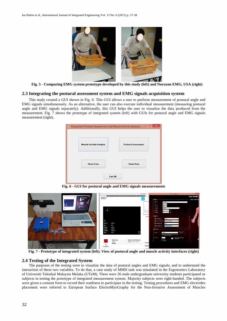
Final stage of validation was a comparison of EMG signals (microvolt) generated by all four electrodes/ amplifiers
of EMG system developed by this study and four EMG electrodes/ amplifiers of a commercial EMG system (Noraxon
EMG, USA) – as the reference system. Three volunteers were assigned to perform a simple load lifting experiment as
shown in Fig. 5. Two loads (5 kg and 10 kg) were lifted by subjects for both EMG systems. Also, the experiment
procedures and electrodes placement were standardised based on The European Surface ElectroMyoGraphy for the
Non-Invasive Assessment of Muscles (SENIAM) [26]. A ratio of microvolt values between lifting load of 10 kg and 5
kg was calculated for each EMG system. Then, the ratio from both EMG systems was compared by calculating the
percentage difference. To get the percentage difference, the microvolt ratio of EMG system developed by this study
minus the microvolt ratio of commercial EMG system, and divided by the average of these 2 ratios. Then multiply the
result by 100 to yield a final value in percent, as simplified by this formula:
(1)
Whereby:
V1 = Microvolt ratio of EMG system developed by this study
V2 = Microvolt ratio of commercial EMG system (Noraxon EMG, USA)
This study paid a high attention to minimize the noise level in the EMG signals for aiming a percentage difference
below than 5 percent [27]. This is important to ensure the accuracy of EMG data produced by the developed system. To
achieve this target, the researchers made three efforts. The first effort was related to the software such as enhancement
on the algorithm and program codes for better noise filtration. The second effort was given to the hardware – securing
the electrode cables with a minimal tension. This is important to minimize artifacts due to cable movement and to avoid
the electrodes detach from skin during the experiment. In this study, the researchers utilized a regular tape to secure the
electrode cables. The third effort concentrated on the measurement procedures. This includes skin preparation and
attachment of the electrodes. The quality of EMG signals mainly depends on a proper skin preparation. The purposes of
skin preparation are to stable the electrodes attachment and allow a low skin impedance. This study applied the
following skin preparation steps before attaching the electrodes:
i. Removing the hair – the hair was shaved with a disposable razor. This is needed to ensure the area where the
electrodes would be attached must be cleaned and free from hairs.
ii. Cleaning of the skin – the skin was cleaned and dried by using alcohol and a soft textile towel.
To confirm the skin was prepared properly, it receives a light red color indicating a good skin impedance state [25].
Furthermore, this study attached the electrodes to the skin with the guidance of The European Surface
ElectroMyoGraphy for the Non-Invasive Assessment of Muscles (SENIAM) [26].
Isa Halim et al., International Journal of Integrated Engineering Vol. 13 No. 6 (2021) p. 27-38
32
Fig. 5 - Comparing EMG system prototype developed by this study (left) and Noraxon EMG, USA (right)
2.3 Integrating the postural assessment system and EMG signals acquisition system
This study created a GUI shown in Fig. 6. This GUI allows a user to perform measurement of postural angle and
EMG signals simultaneously. As an alternative, the user can also execute individual measurement (measuring postural
angle and EMG signals separately). Additionally, this GUI helps the user to visualize the data produced from the
measurement. Fig. 7 shows the prototype of integrated system (left) with GUIs for postural angle and EMG signals
measurement (right).
Fig. 6 - GUI for postural angle and EMG signals measurements
Fig. 7 - Prototype of integrated system (left). View of postural angle and muscle activity interfaces (right)
2.4 Testing of the Integrated System The purposes of the testing were to visualize the data of postural angles and EMG signals, and to understand the
interaction of these two variables. To do that, a case study of MMH task was simulated in the Ergonomics Laboratory
of Universiti Teknikal Malaysia Melaka (UTeM). There were 26 male undergraduate university students participated as
subjects in testing the prototype of integrated measurement system. Majority subjects were right-handed. The subjects
were given a consent form to record their readiness to participate in the testing. Testing procedures and EMG electrodes
placement were referred to European Surface ElectroMyoGraphy for the Non-Invasive Assessment of Muscles

Isa Halim et al., International Journal of Integrated Engineering Vol. 13 No. 6 (2021) p. 27-38
33
(SENIAM) [26]. The EMG signals were measured from four muscles, right and left biceps and right and left
brachioradialis. A 3D sensor (Microsoft KinectTM) was positioned 2.3 - 3.5 meters away from the subjects to record and
measure their postural angles.
In the simulated case study, the subjects were required to lift a load of 5 kg in a plastic crate. As shown in Fig. 8,
the sequence of lifting process was: 1. Hold and lift the plastic crate from the floor (Activity A); 2. Stand up, hold and
carry the plastic crate (Activity B); 3. Put the plastic crate onto a table (Activity C). The distance from the start point to
the destination was 3 meters. The subjects performed this lifting process in one cycle with a duration less than 2
minutes. Before doing the experiment, all subjects were provided with a consent form. The consent form consists of
research information and experiment procedures so that they can make a firm decision to participate in the study. The
subjects were asked to sign the form and informed taking part in this experiment is voluntary basis – they can leave the
experiment at any time without prejudice. A few trials were given to the subjects prior to actual testing to familiarize
themselves with the experiment procedures. Each subject took around 20-30 minutes for the experiment preparation
and execution. At the end of testing session, all subjects were given a reward to appreciate their participation. The
experiment procedures were reviewed and approved by the Research Ethics Committee of Universiti Teknikal
Malaysia Melaka (Reference no.: UTeM,11.02/500-25/1/4(21)). Data of postural angles and EMG signals generated
from the experiment were compiled continuously to represent continuous activities of the simulated MMH task.
Fig. 8 - Lifting process of a 5 kg load
3. Results
Table 2 tabulates data of postural angles (deg.) and EMG signals (µV) of biceps (upper arm) and brachioradialis
(forearm) from all subjects. These data represent the postures and muscle activity of subjects while performing a
simulated case study of MMH task (holding, lifting and carrying of 5 kg load) as mentioned in 2.2. In general, the
integrated measurement system developed by this study was capable to measure and generate data of postural angles
and EMG signals. Specifically, Table 2 presents the maximum postural angles (deg.) between both left and right
forearms and upper arms, and the maximum values of EMG signals (µV) measured in the left and right biceps and
brachioradialis muscles. The biceps lie on the front of the upper arm between the shoulder and the elbow and the
brachioradialis is located in the forearm. The EMG signals represent electrical voltage (measured in microvolts, µV)
generated in the muscles during their contraction [28]. Higher EMG signals indicate greater muscle contraction, and
hence larger force generated by the muscle. Additionally, muscle contraction varies among individuals [29], and this
was proved by the current study whereby the magnitude of EMG signals is differ among the subjects (Table 2). One of
direct factors influencing the EMG signals is intrinsic or inherent individual factors [28]. These factors include joint
angle, number of active motor units, blood flow, muscle cross-sectional area, muscle fiber composition, muscle fiber
diameter, depth and location of active muscle fibers and amount of tissue between muscle surface and EMG electrodes
[28]. Furthermore, this study observed that majority subjects exerted higher contraction in the left biceps and left
brachioradialis muscles than the right muscles (Table 2). This indicates the subjects paid greater effort on the left upper
and lower arms to grip, lift and carry the load. The higher EMG values for the left muscles compared to the right in the
present study reveal the right-handed subjects tend to exert more forces in left hand muscles, in line with the findings of
previous study [30].
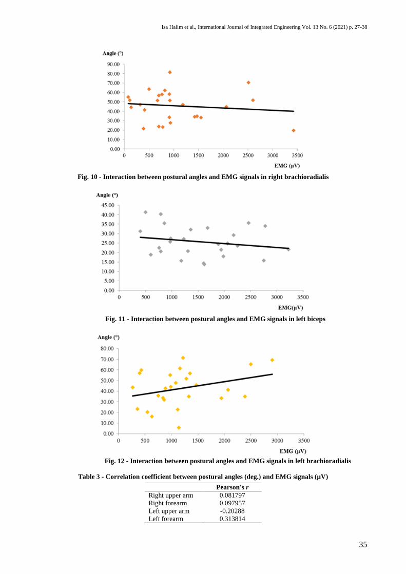
Line charts in Fig. 9 to Fig. 12 illustrates the interaction between the postural angles and the EMG signals when the
subjects performed the simulated case study of MMH task. The EMG signals (µV) and postural angles (º) are plotted in
horizontal (x) axis and vertical (y) axis respectively. Based on the charts, this study observed that the EMG signals in
right biceps showed a marginally increase when the postural angle increase. However, the left biceps illustrated an
opposite trend. Meanwhile the right brachioradialis muscle showed increment of EMG signals when the postural angles
decrease. In contrast, the left brachioradialis muscle indicated the EMG signals increase when the postural angles
increase.

Isa Halim et al., International Journal of Integrated Engineering Vol. 13 No. 6 (2021) p. 27-38
34
Statistical analysis associated with Pearson correlation coefficient (Pearson's r) revealed that there is a low
coefficient of correlation between the postural angles (deg.) and EMG signals (µV), as shown in Table 3. Generally,
this indicates the correlation is exist but weak. In other words, the magnitude of EMG signals generated from the
muscles were not change significantly regardless the increment of upper arm and forearm angles. This is due to
handling the 5 kg load was not forceful enough to contract the muscles. The correlation coefficients of right upper arm,
right forearm and left forearm show a positive correlation. This means, as the postural angles of these parts increase,
the EMG signals are also rise. However, the correlation coefficient of left upper arm indicates negative correlation,
which means there is a steady decrement of EMG signals as the postural angles of this part increase.
Table 2 - Postural angle (deg.) and EMG signals (µV) of biceps (upper arm) and brachioradialis (forearm)
Right upper arm Right forearm Left upper arm Left forearm
Subject
Postural
angles
(deg.)
EMG
signals
(µV)
Postural
angles
(deg.)
EMG
signals
(µV)
Postural
angles
(deg.)
EMG
signals
(µV)
Postural
angles
(deg.)
EMG
signals
(µV)
1 23.192 2070 33.460 1550 15.783 2750 34.990 1330
2 27.819 600 34.684 1470 40.318 790 56.858 400
3 31.907 390 34.298 1420 33.043 1680 35.083 2390
4 35.166 770 23.406 780 35.456 860 20.374 550
5 24.164 940 21.742 390 25.696 970 5.770 1140
6 21.157 1070 44.798 2060 21.528 1940 33.509 1940
7 17.986 660 47.019 320 27.121 1230 47.935 1080
8 29.872 1910 19.742 3420 31.339 400 16.188 630
9 21.073 600 23.884 700 20.692 790 23.257 360
10 17.241 220 55.133 80 18.803 600 43.759 270
11 34.276 420 41.447 410 41.323 500 33.843 840
12 28.058 1250 33.517 910 27.455 980 31.851 860
13 17.861 1560 51.917 2600 20.757 1310 45.734 1470
14 25.424 1460 44.145 140 35.610 2460 35.920 750
15 20.684 3210 70.624 2510 23.589 2250 69.292 2900
16 25.329 1200 27.696 930 24.407 1890 22.846 1120
17 13.634 1930 51.915 110 13.736 1620 42.610 890
18 19.061 500 63.466 500 22.520 760 59.871 430
19 26.765 820 58.040 760 32.158 1360 51.873 1280
20 26.951 1120 58.238 910 15.655 1180 55.089 980
21 36.602 2000 62.100 820 18.002 1980 65.246 2500
22 33.193 1780 56.821 700 29.245 2180 61.421 1160
23 41.234 2300 81.534 920 34.021 2780 71.291 1220
24 23.811 1020 51.642 670 14.330 1610 44.186 990
25 27.318 760 47.067 1180 24.849 2060 41.312 2070
26 22.681 1900 51.485 920 21.664 3220 56.606 1350
Fig. 9 - Interaction between postural angles and EMG signals in right biceps
Isa Halim et al., International Journal of Integrated Engineering Vol. 13 No. 6 (2021) p. 27-38
35
Fig. 10 - Interaction between postural angles and EMG signals in right brachioradialis
Fig. 11 - Interaction between postural angles and EMG signals in left biceps
Fig. 12 - Interaction between postural angles and EMG signals in left brachioradialis
Table 3 - Correlation coefficient between postural angles (deg.) and EMG signals (µV)
Pearson's r
Right upper arm 0.081797
Right forearm 0.097957
Left upper arm -0.20288
Left forearm 0.313814

Isa Halim et al., International Journal of Integrated Engineering Vol. 13 No. 6 (2021) p. 27-38
36
4. Discussion
Based on the aforementioned results, this study observed that the patterns of EMG signals are depending on hand
postural angles when subjects performing a simulated case study of MMH task (holding, lifting and carrying of 5 kg
load). Findings presented in this study showed a good agreement with the previous studies which reported the hand
postural angles are important factors for the hand muscles contraction. Lee [31] identified that the hand posture has a
great influence on the forearm muscle activities when lifting cylindrical and rectangular objects. Amar and company
[32] in their study revealed that the postural angles determined muscle activities in one-handed lifting of 1, 1.5, 2, 2.5,
and 3 kg loads. Lee [33] found that the hand postures resulted in significant differences of EMG activity of serratus
anterior muscle during push-up plus exercise. Roman and Bartuzi [34] concluded that wrist posture influenced EMG
signals of extensor digitorum communis and flexor carpi ulnaris muscles. Cudlip [35] detected that wrist posture
affected over two thirds of muscle contraction levels. The back muscle activities increased as the upper trunk and pelvic
angles exceeded 0° [36]. Shair [37] identified an interaction (but not significant) between trunk postural angle and
EMG activity of erector spinae muscle during squat core-lifting task. One explanation on this postural angle and EMG
signal interaction is that muscle shortening (concentric contractions) due to changes in postural angle stimulates the
EMG signal and its amplitude [34].
Another plausible explanation for the interaction between the postural angle and EMG signal might be due to the
different effects of gravity or mechanical load in different body postures. The above explanation was supported by a
study conducted by Edmondston [38] whereby the authors identified that neck flexion increased mechanical load and
extensor muscle activity compared to other neck positions. The above theory and explanation regarding the relationship
between the postural angles and EMG activity had been applied by several researchers to develop an effective postural
assessment tool. For example, Hignett and McAtamney [39] had developed Rapid Entre Body Assessment (REBA) to
evaluate work postures due to postural angle, load and coupling. The postural scores of REBA were given based on the
angle measured from the neutral position (0 degree), in which the scores will be higher when the postural angle
deviates from the neutral position [39]. However, the above mentioned observational tools had its own limitation in
terms of precision and clarity as they were developed based on semi-quantitative method. Therefore, it is necessary to
develop an integrated measurement system that allows simultaneous quantification and accurate readings of postural
angles and EMG signals.
The application of integrated measurement system developed by this study is to assist ergonomics practitioners and
engineers to objectively measure postural angles and muscle exertion during MMH task. They can analyze the postural
angles and EMG signals to justify risk level of work posture and muscle exertion. For example, if the torso in forward
bending, 0°–20° is considered low risk, however, 20°–60° and greater than 60° are defined as medium and high risks
respectively [40]. Meanwhile, based on EMG signals, ergonomics practitioners and engineers may determine the onset
of muscle fatigue when holding load greater than 15% of maximum voluntary contraction (MVC) [41]. Additionally,
they can utilize this integrated measurement system to analyze the interaction of work postures and muscles exertion so
that MMH task can be designed ergonomically to avoid postural stress and muscle sprain.
With the advancement of digital technology and computational intelligence nowadays, many postural angle
measurement system [42-44] and EMG signal acquisition system [45-47] were designed and built using a cost effective
hardware. This study was also utilized a low cost 3D sensor for postural angle measurement and low cost EMG
hardware (e.g. electrodes/ amplifiers and cables). However, dissimilar to the previous works, this study made an extra
effort to develop and testing an integrated measurement system for measuring postural angles and EMG signals in
MMH tasks. Advanced commercial human motion tracking and EMG signal acquisition systems available in the
market might be provide an extensive feature and function for acquiring high quality postural angle and EMG data. The
commercial ones employed multi high-speed and high-definition cameras, and state-of-the-art cables and amplifiers
with adjustable gains which can offer highly accurate values; hence their performance is definitely higher than the low
cost systems. For instance, EMG cable technology such as built-in miniaturized amplifier and PS/2 connector type play
a significant role to avoid motion artefacts and noise during signals acquisition process. On the other hand, the
advantages of low cost postural angle and EMG measurement systems are acceptable accuracy and inexpensive, thus
making affordable to small-scale research laboratories and industry practitioners.
5. Conclusion
The contribution of this study was a prototype of an integrated measurement system for measuring postural angles
and EMG signals. The application of this integrated measurement system is to provide a simultaneous assessment of
work posture and muscle activity for ergonomics practitioners and engineers aiming to design a safe MMH task in
industry workplaces. The system was developed using a cost effective hardware. Based on laboratory testing of manual
lifting task, this study found that the developed system was able to generate postural angles which correlate to the
patterns of EMG signals. Interestingly, this finding showed a good agreement with the established studies. Further
study is required to enhance the reliability and usability of the system so that it may facilitate ergonomics practitioners
and engineers to assess work posture and muscle effort in MMH task.

Isa Halim et al., International Journal of Integrated Engineering Vol. 13 No. 6 (2021) p. 27-38
37
Acknowledgement
The researchers would like to thank the Faculty of Manufacturing Engineering and the Centre for Research and
Innovation Management, Universiti Teknikal Malaysia Melaka for funding this study by the University High Impact
Short Term Grant (PJP/2017/FKP/H19/S01527) and Mr. Tarek Al Bawab for helping in the experiment set up.
References
[1] Adams, M. A., & Dolan, P. (2007). How to use the spine, pelvis, and legs effectively in lifting. In Movement,
Stability & Lumbopelvic Pain (pp. 167-183). Churchill Livingstone
[2] Das, B. (2015). An evaluation of low back pain among female brick field workers of West Bengal, India.
Environmental Health and Preventive Medicine, 20(5), 360-368
[3] Bakhtiari, N., Dianat, I., & Nedaei, M. (2018). Electromyographic evaluation of different handle shapes of
masons’ trowels. International Journal of Occupational Safety and Ergonomics, 1-6
[4] Gillette, J. C., & Stephenson, M. L. (2019). Electromyographic assessment of a shoulder support exoskeleton
during on-site job tasks. IISE Transactions on Occupational Ergonomics and Human Factors, 1-9
[5] Nasser Alasim, H., & Nimbarte, A. D. (2019). Variability of electromyographic spectral measures in non-fatigued
shoulder muscles and implications for assessing muscle fatigue. IISE Transactions on Occupational Ergonomics
and Human Factors, 7(2), 119-131
[6] Wang, D., Dai, F., Ning, X., Dong, R. G., & Wu, J. Z. (2017). Assessing work-related risk factors on low back
disorders among roofing workers. Journal of Construction Engineering and Management, 143(7), 04017026.
[7] Singh, R. E., Iqbal, K., White, G., & Holtz, J. K. (2019). A Review of EMG Techniques for Detection of Gait
Disorders. In Machine Learning in Medicine and Biology, 1-22
[8] Liu, P., Liu, L., & Clancy, E. A. (2015). Influence of joint angle on EMG-torque model during constant-posture,
torque-varying contractions. IEEE Transactions on Neural Systems and Rehabilitation Engineering, 23(6), 1039-
1046
[9] Beck, T. W., Housh, T. J., Johnson, G. O., Weir, J. P., Cramer, J. T., Coburn, J. W., & Malek, M. H. (2004).
Mechanomyographic and electromyographic amplitude and frequency responses during fatiguing isokinetic
muscle actions of the biceps brachii (Doctoral dissertation, University of Nebraska--Lincoln)
[10] Li, X., Komeili, A., Gül, M., & El-Rich, M. (2017). A framework for evaluating muscle activity during repetitive
manual material handling in construction manufacturing. Automation in Construction, 79, 39-48
[11] Hermanns, I., Raffler, N., Ellegast, R. P., Fischer, S., & Göres, B. (2008). Simultaneous field measuring method
of vibration and body posture for assessment of seated occupational driving tasks. International Journal of
Industrial Ergonomics, 38(3–4), 255–263
[12] Haggag, H., Hossny, M., Nahavandi, S., & Creighton, D. (2013). Real time ergonomic assessment for assembly
operations using kinect. Proceedings - UKSim 15th International Conference on Computer Modelling and
Simulation, UKSim 2013, 495–500
[13] Carvajal, J. C. P., Jaramillo, J. S., & Castaño, A. G. (2015). Guidelines for a Rehabilitation Model for Banana
Packing Plants from the Integration of Environmental Variables and human factors. Procedia Manufacturing,
3(January), 6190–6197. https://doi.org/10.1016/j.promfg.2015.07.916
[14] Plantard, P., Shum, H. P. H., Le Pierres, A. S., & Multon, F. (2017). Validation of an ergonomic assessment
method using Kinect data in real workplace conditions. Applied Ergonomics, 65, 562–569
[15] Manghisi, V. M., Uva, A. E., Fiorentino, M., Bevilacqua, V., Trotta, G. F., & Monno, G. (2017). Real time RULA
assessment using Kinect v2 sensor. Applied Ergonomics, 65, 481–491
[16] Nahavandi, D., & Hossny, M. (2017). Skeleton-free task-specific rapid upper limb ergonomie assessment using
depth imaging sensors. Proceedings of IEEE Sensors, 1–3
[17] Jiang, S., Liu, P., Fu, D., Xue, Y., Luo, W., & Wang, M. (2017). A low-cost rapid upper limb assessment method
in manual assembly line based on somatosensory interaction technology. AIP Conference Proceedings, 1834
[18] Toro, S. F. D., Santos-Cuadros, S., Olmeda, E., Álvarez-Caldas, C., Díaz, V., & San Román, J. L. (2019). Is the
Use of a Low-Cost sEMG Sensor Valid to Measure Muscle Fatigue? Sensors, 19(14), 3204
[19] Brunelli, D., Tadesse, A. M., Vodermayer, B., Nowak, M., & Castellini, C. (2015). Low-cost wearable
multichannel surface EMG acquisition for prosthetic hand control. Proceedings - 2015 6th IEEE International
Workshop on Advances in Sensors and Interfaces, IWASI 2015, 94–99
[20] Fang, Y., Liu, H., Li, G., & Zhu, X. (2015). A Multichannel Surface EMG System for Hand Motion Recognition.
International Journal of Humanoid Robotics, 12(2), 1550011
[21] Poo, T. S., & Sundaraj, K. (2010). Design and development of a low cost EMG signal acquisition system using
surface EMG electrode. In 2010 IEEE Asia Pacific Conference on Circuits and Systems (pp. 24-27)
[22] Aktan, M. E., Göker, İ., Akdoğan, E., & Öztürk, B. (2017, October). Design, implementation and performance
analysis of a microcontroller based wireless electromyography device. In 2017 Medical Technologies National
Congress, 1-4

Isa Halim et al., International Journal of Integrated Engineering Vol. 13 No. 6 (2021) p. 27-38
38
[23] Albawab, T. M. M., Halim, I., Ahmad, N., Umar, R. Z. R., Mohamed, M. S. S., Abullais, F., ... & Saptari, A.
(2018). Upper Limb Joints and motions sampling system using Kinect camera. Journal of Advanced
Manufacturing Technology, 12(2), 147-158
[24] Zschorlich, V. R. (1989). Digital filtering of EMG-signals. Electromyography and Clinical Neurophysiology,
29(2), 81-6
[25] Konrad, P. (2005). The ABC of EMG. A practical introduction to kinesiological electromyography, 1, 30-35
[26] Hermens, H.J.; Freriks, B.; Merletti, R.; Stegeman, D.; Blok, J.; Rau, G.; Disselhorst-Klug, C.; Hägg, G. European
Recommendations for Surface Electromyography. Roessingh Res. Dev. 1999, 8, 13–54
[27] Pizzolato, S., Tagliapietra, L., Cognolato, M., Reggiani, M., Müller, H., & Atzori, M. (2017). Comparison of six
electromyography acquisition setups on hand movement classification tasks. PloS one, 12 (10), e0186132
[28] Reaz, M. B. I., Hussain, M. S., & Mohd-Yasin, F. (2006). Techniques of EMG signal analysis: detection,
processing, classification and applications (Correction). Biological Procedures Online, 8 (1), 163-163
[29] Enoka, R. M., & Duchateau, J. (2008). Muscle fatigue: what, why and how it influences muscle function. The
Journal of Physiology, 586(1), 11-23
[30] Hagberg, C., & Hagberg, M. (1989). Surface EMG amplitude and frequency dependence on exerted force for the
upper trapezius muscle: a comparison between right and left sides. European Journal of Applied Physiology and
Occupational Physiology, 58(6), 641-645
[31] Lee, K. S., & Jung, M. C. (2018). Effect of hand postures and object properties on forearm muscle activities using
surface electromyography. International Journal of Occupational Safety and Ergonomics, 1-10
[32] Amar, M. R., Cochran, D., & Woldstad, J. (2017). The effect of single-handed lifting tasks on the activation of the
neck-shoulder shared musculature. International Biomechanics, 4(1), 1-8
[33] Lee, S., Lee, D., & Park, J. (2013). The effect of hand position changes on electromyographic activity of shoulder
stabilizers during push-up plus exercise on stable and unstable surfaces. Journal of Physical Therapy Science,
25(8), 981-984
[34] Roman-Liu, D., & Bartuzi, P. (2013). The influence of wrist posture on the time and frequency EMG signal
measures of forearm muscles. Gait & Posture, 37(3), 340-344
[35] Cudlip, A. C., Holmes, M. W., Callaghan, J. P., & Dickerson, C. R. (2018). The effects of shoulder abduction
angle and wrist angle on upper extremity muscle activity in unilateral right handed push/pull tasks. International
Journal of Industrial Ergonomics, 64, 102-107
[36] Kamil, N. S. M., & Dawal, S. Z. M. (2015). Effect of postural angle on back muscle activities in aging female
workers performing computer tasks. Journal of Physical Therapy Science, 27(6), 1967-1970
[37] Shair, E. F., Ahmad, S. A., Wada, C., Abdullah, A. R., Marhaban, M. H., & Tamrin, S. M. The relationship
between trunk angle and electromyography (EMG) signals in biceps brachii and erector spinae muscles during
core-lifting task. In: 5th International Symposium on Applied Engineering and Sciences (SAES2017), 14-15 Nov.
2017, Universiti Putra Malaysia. (p. 39)
[38] Edmondston, S. J., Sharp, M., Symes, A., Alhabib, N., & Allison, G. T. (2011). Changes in mechanical load and
extensor muscle activity in the cervico-thoracic spine induced by sitting posture modification. Ergonomics, 54(2),
179-186
[39] Hignett, S., & McAtamney, L. (2000). Rapid entire body assessment (REBA). Applied Ergonomics, 31(2), 201-
205
[40] Chander, D. S., & Cavatorta, M. P. (2017). An observational method for postural ergonomic risk assessment
(PERA). International Journal of Industrial Ergonomics, 57, 32-41
[41] Rohmert, W. (1973). Problems in determining rest allowances: part 1: use of modern methods to evaluate stress
and strain in static muscular work. Applied Ergonomics, 4(2), 91-95
[42] Yang, L., Grooten, W. J., & Forsman, M. (2017). An iPhone application for upper arm posture and movement
measurements. Applied Ergonomics, 65, 492-500
[43] Krishnan, C., Washabaugh, E. P., & Seetharaman, Y. (2015). A low cost real-time motion tracking approach
using webcam technology. Journal of Biomechanics, 48(3), 544-548
[44] Mustapha, G., Razak, M. F. A., Hamzah, M. S. M., & Mohd, N. H. (2016). The Development of a Low Cost
Motion Analysis System: Cekak Visual 3D V1. 0. International Journal GEOMATE, 11(24), 2248-2252
[45] Brunelli, D., Tadesse, A. M., Vodermayer, B., Nowak, M., & Castellini, C. (2015, June). Low-cost wearable
multichannel surface EMG acquisition for prosthetic hand control. In 2015 6th International Workshop on
Advances in Sensors and Interfaces (IWASI) (pp. 94-99). IEEE
[46] Reinvee, M., & Pääsuke, M. (2016, September). Overview of contemporary low-cost sEMG hardware for
applications in human factors and ergonomics. In Proceedings of the Human Factors and Ergonomics Society
Annual Meeting (Vol. 60, No. 1, pp. 408-412). Sage CA: Los Angeles, CA: SAGE Publications
[47] Cheney, P. D., Kenton, J. D., Thompson, R. W., McKiernan, B. J., Lininger, R. E., & Trank, J. W. (1998). A low-
cost, multi-channel, EMG signal processing amplifier. Journal of Neuroscience Methods, 79(1), 123-127