Embed Size (px)
Citation preview
Int. J. Mol. Sci. 2011, 12, 1474-1495; doi:10.3390/ijms12031474
International Journal of
Molecular Sciences ISSN 1422-0067
www.mdpi.com/journal/ijms
Review
Integrated Lipidomics in the Secreted Phospholipase A2 Biology
Makoto Murakami *, Hiroyasu Sato, Yoshitaka Taketomi and Kei Yamamoto
Lipid Metabolism Project, The Tokyo Metropolitan Institute of Medical Science, 2-1-6 Kamikitazawa,
Setagaya-ku, Tokyo 156-8506, Japan; E-Mails: [email protected] (H.S.);
[email protected] (Y.T.); and [email protected] (K.Y.)
* Author to whom correspondence should be addressed; E-Mail: [email protected];
Tel.: +81-3-5316-3228.
Received: 30 January 2011; in revised form: 18 February 2011 / Accepted: 24 February 2011 /
Published: 25 February 2011
Abstract: Mammalian genomes encode genes for more than 30 phospholipase A2s
(PLA2s) or related enzymes, which are subdivided into several subgroups based on their
structures, catalytic mechanisms, localizations and evolutionary relationships. More than
one third of the PLA2 enzymes belong to the secreted PLA2 (sPLA2) family, which consists
of low-molecular-weight, Ca2+
-requiring extracellular enzymes, with a His-Asp catalytic
dyad. Individual sPLA2 isoforms exhibit unique tissue and cellular localizations and
enzymatic properties, suggesting their distinct pathophysiological roles. Recent studies
using transgenic and knockout mice for several sPLA2 isoforms, in combination with
lipidomics approaches, have revealed their distinct contributions to various biological
events. Herein, we will describe several examples of sPLA2-mediated phospholipid
metabolism in vivo, as revealed by integrated analysis of sPLA2 transgenic/knockout mice
and lipid mass spectrometry. Knowledge obtained from this approach greatly contributes to
expanding our understanding of the sPLA2 biology and pathophysiology.
Keywords: phospholipase A2; phospholipid; lipidomics; transgenic mouse;
knockout mouse
Abbreviations: PLA2, phospholipase A2; sPLA2, secreted PLA2; cPLA2, cytosolic PLA2;
iPLA2, Ca2+
-independent PLA2; Tg, transgenic; ARDS, acute respiratory distress
syndrome; BALF, bronchoalveolar fluid; LPS, lipopolysaccharide; ESI-MS, electrospray
ionization mass spectrometry; PC, phosphatidylcholine; LPC, lysophosphatidylcholine;
PG, phosphatidylglycerol; PE, phosphatidylethanolamine; PS, phosphatidylserine;
OPEN ACCESS
Int. J. Mol. Sci. 2011, 12
1475
LDL, low-density lipoprotein; HDL, high-density lipoprotein; VLDL, very low-density
lipoprotein; ABC, ATP-binding cassette; PGD2, prostaglandin D2; PGE2, prostaglandin E2;
LTB4, leukotriene B4; PUFA, polyunsaturated fatty acid; DHA, docosahexaenoic acid;
DPA, docosapentaenoic acid; LXR, liver X receptor; PPAR, peroxisome proliferator-
activated receptor; StAR, steroidogenic acute regulatory protein; DRG, dorsal root
ganglion; COX, cyclooxygenase; LOX, lipoxygenase; CYP450, cytochrome P450; HPLC,
high performance liquid chromatography; WT, wild-type.
1. Introduction
Phospholipase A2 (PLA2) hydrolyzes the sn-2 position of glycerophospholipids to yield fatty acids
and lysophospholipids. In the view of signal transduction, the PLA2 reaction has been considered to be
of particular importance since arachidonic acid, one of the polyunsaturated fatty acids (PUFAs)
released by PLA2, is metabolized to various lipid mediators such as prostaglandins and leukotrienes. In
addition, lysophospholipids or its metabolites, such as lysophosphatidic acid and platelet-activating
factor, also represent another class of lipid mediators. These lipid mediators exert numerous biological
actions through their cognate G protein-coupled receptors on target cells. PLA2 has also been implicated
in membrane glycerophospholipid remodeling, thereby contributing to cellular homeostasis.
Mammalian genomes encode more than 30 PLA2s or related enzymes, which are classified into
several subgroups on the basis of their primary structures and functions. Critical contributions of the
intracellular PLA2 families, namely cytosolic PLA2s (cPLA2s) and Ca2+
-independent PLA2s (iPLA2s),
to aracidnonic acid metabolism and membrane homeostasis, respectively, have been well established
by numerous studies [1,2]. The secreted PLA2 (sPLA2) family represents structurally related,
disulfide-rich, low molecular weight, lipolytic enzymes with a His-Asp catalytic dyad. sPLA2s occur in a
wide variety of vertebrate and invertebrate animals, plants, fungus, bacteria, and viruses, and 11 sPLA2
isozymes (IB, IIA, IIC, IID, IIE, IIF, III, V, X, XIIA and XIIB) have been identified in mammals [2–4].
Of these, sPLA2s belonging to the group I/II/V/X collection (conventional sPLA2s) are closely related,
14–19-kDa secreted enzymes with a highly conserved Ca2+
-binding loop (XCGXGG) and a catalytic
site (DXCCXXHD). In addition to these elements, there are six absolutely conserved disulfide bonds
and up to two additional unique disulfide bonds, which contribute to the high degree of stability of
these enzymes. Group III and group XII sPLA2s (atypical sPLA2s) share homology with the I/II/V/X
collection of sPLA2s only in the Ca2+
-binding loop and catalytic site, thereby representing the group III
and XII collections, respectively. sPLA2 enzymes hydrolyze the ester bond at the sn-2 position of
glycerophospholipids with distinct selectivity toward sn-2 fatty acids and polar head groups in the
presence of mM concentrations of Ca2+
. Since individual sPLA2s display distinct cellular/tissue
distributions and substrate head group specificities, they may play non-redundant, isoform-specific
roles in vivo.
Although many potential functions of sPLA2s have been proposed on the basis of in vitro studies,
the precise biological roles and relevant target membranes of these enzymes in vivo have remained
elusive until recently. Several, if not all, sPLA2s are capable of releasing arachidonic acid from
Int. J. Mol. Sci. 2011, 12
1476
cultured cell membranes when overexpressed or added exogenously at excess amounts in vitro [2–4].
However, it still remains controversial whether this function could indeed be operated by sPLA2s
in vivo. The reason why sPLA2s are secreted is most probably because sPLA2s participate in
pathophysiology by regulating extracellular phospholipid metabolism, which include adjacent cell
membranes (plasma membranes or microvesicles shed from cells), non-cellular lipid components such
as lipoproteins and pulmonary surfactant, and foreign phospholipids such as microbe membranes and
dietary lipids. The in vitro actions of individual sPLA2s on various target membranes are summarized
in Table 1. This target variation may explain the molecular evolution of a number of sPLA2s with
distinct localizations and substrate specificities. Therefore, once some phenotypes appear in
sPLA2-knockout or -transgenic mice, this could be attributable to a combination of these varied actions
rather than only by alterations in lipid mediator levels.
In the past few years, we have analyzed the phenotypes of transgenic or knockout mice for several
sPLA2 isozymes, in combination with a lipid profiling technique by mass spectrometry. This integrated
approach, together with studies using these mice by other research groups, has helped us understand
the potential action of a given sPLA2 on particular target membranes and its impact on pathophysiology
in vivo. In this article, we will give an overview of current analyses on transgenic or knockout mice for
two particular conventional sPLA2s, group V and X, and an atypical sPLA2, group III. Also, we will give
a brief summary of pathophysiological functions of other sPLA2s that have been clarified to date.
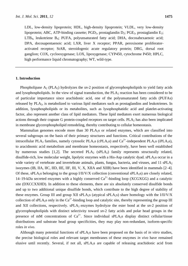
Table 1. In vitro actions of sPLA2s on various membranes.
sPLA2s
resting cell
membrane
activated cell
membrane
lipoprotein
(PC)
surfactant
(PC)
Gram-positive
bacteria
Gram-negative
bacteria
conventional sPLA2s IB weak moderate weak weak none none
IIA none moderate weak weak very high weak*
IID none weak n.d. weak high none
IIE none weak n.d. n.d. moderate none
IIF moderate moderate moderate n.d. none none
V high very high very high very high high none
X very high very high very high high moderate none
atypical sPLA2s III moderate moderate high n.d. n.d. none
XIIA none none n.d. n.d. high moderate
n.d.; not determined. sPLA2-IIC is not included since it is a pseudogene in human.
*sPLA2-IIA kills Gram-negative bacteria only in the presence of bacterial permeability-increasing protein.
For details, please see refs [30,31,41,47,55,79,85,94].
2. Biological Functions of sPLA2s in Vivo
2.1. Group V sPLA2 (sPLA2-V)
Among the conventional sPLA2s, sPLA2-V has the simplest structure. It lacks group I- and
II-specific disulfide bonds, group II-specific C-terminal extension, and group I- or X-specific
N-terminal propeptide [5]. However, sPLA2-V is evolutionally close to group II sPLA2s, since the
Int. J. Mol. Sci. 2011, 12
1477
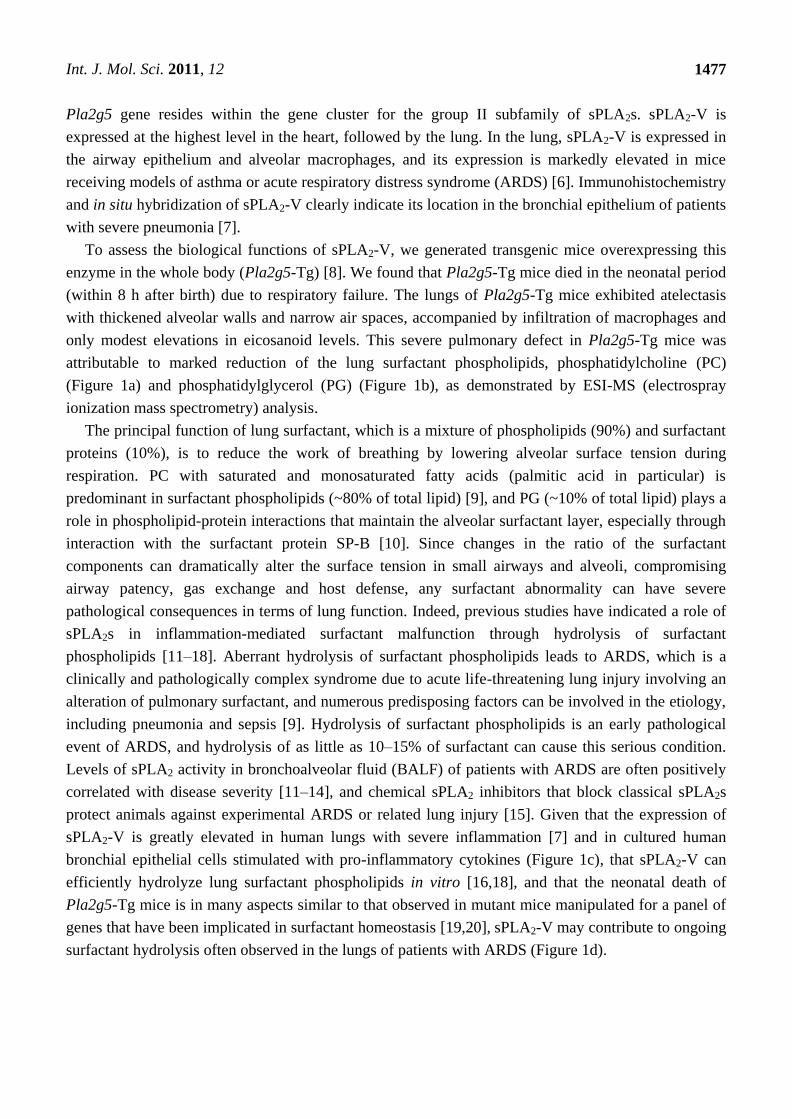
Pla2g5 gene resides within the gene cluster for the group II subfamily of sPLA2s. sPLA2-V is
expressed at the highest level in the heart, followed by the lung. In the lung, sPLA2-V is expressed in
the airway epithelium and alveolar macrophages, and its expression is markedly elevated in mice
receiving models of asthma or acute respiratory distress syndrome (ARDS) [6]. Immunohistochemistry
and in situ hybridization of sPLA2-V clearly indicate its location in the bronchial epithelium of patients
with severe pneumonia [7].
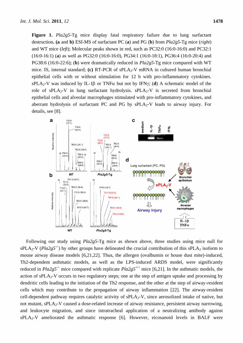
To assess the biological functions of sPLA2-V, we generated transgenic mice overexpressing this
enzyme in the whole body (Pla2g5-Tg) [8]. We found that Pla2g5-Tg mice died in the neonatal period
(within 8 h after birth) due to respiratory failure. The lungs of Pla2g5-Tg mice exhibited atelectasis
with thickened alveolar walls and narrow air spaces, accompanied by infiltration of macrophages and
only modest elevations in eicosanoid levels. This severe pulmonary defect in Pla2g5-Tg mice was
attributable to marked reduction of the lung surfactant phospholipids, phosphatidylcholine (PC)
(Figure 1a) and phosphatidylglycerol (PG) (Figure 1b), as demonstrated by ESI-MS (electrospray
ionization mass spectrometry) analysis.
The principal function of lung surfactant, which is a mixture of phospholipids (90%) and surfactant
proteins (10%), is to reduce the work of breathing by lowering alveolar surface tension during
respiration. PC with saturated and monosaturated fatty acids (palmitic acid in particular) is
predominant in surfactant phospholipids (~80% of total lipid) [9], and PG (~10% of total lipid) plays a
role in phospholipid-protein interactions that maintain the alveolar surfactant layer, especially through
interaction with the surfactant protein SP-B [10]. Since changes in the ratio of the surfactant
components can dramatically alter the surface tension in small airways and alveoli, compromising
airway patency, gas exchange and host defense, any surfactant abnormality can have severe
pathological consequences in terms of lung function. Indeed, previous studies have indicated a role of
sPLA2s in inflammation-mediated surfactant malfunction through hydrolysis of surfactant
phospholipids [11–18]. Aberrant hydrolysis of surfactant phospholipids leads to ARDS, which is a
clinically and pathologically complex syndrome due to acute life-threatening lung injury involving an
alteration of pulmonary surfactant, and numerous predisposing factors can be involved in the etiology,
including pneumonia and sepsis [9]. Hydrolysis of surfactant phospholipids is an early pathological
event of ARDS, and hydrolysis of as little as 10–15% of surfactant can cause this serious condition.
Levels of sPLA2 activity in bronchoalveolar fluid (BALF) of patients with ARDS are often positively
correlated with disease severity [11–14], and chemical sPLA2 inhibitors that block classical sPLA2s
protect animals against experimental ARDS or related lung injury [15]. Given that the expression of
sPLA2-V is greatly elevated in human lungs with severe inflammation [7] and in cultured human
bronchial epithelial cells stimulated with pro-inflammatory cytokines (Figure 1c), that sPLA2-V can
efficiently hydrolyze lung surfactant phospholipids in vitro [16,18], and that the neonatal death of
Pla2g5-Tg mice is in many aspects similar to that observed in mutant mice manipulated for a panel of
genes that have been implicated in surfactant homeostasis [19,20], sPLA2-V may contribute to ongoing
surfactant hydrolysis often observed in the lungs of patients with ARDS (Figure 1d).
Int. J. Mol. Sci. 2011, 12
1478
Figure 1. Pla2g5-Tg mice display fatal respiratory failure due to lung surfactant
destruction. (a and b) ESI-MS of surfactant PC (a) and PG (b) from Pla2g5-Tg mice (right)
and WT mice (left); Molecular peaks shown in red, such as PC32:0 (16:0-16:0) and PC32:1
(16:0-16:1) (a) as well as PG32:0 (16:0-16:0), PG34:1 (16:0-18:1), PG36:4 (16:0-20:4) and
PG38:6 (16:0-22:6); (b) were dramatically reduced in Pla2g5-Tg mice compared with WT
mice. IS, internal standard; (c) RT-PCR of sPLA2-V mRNA in cultured human bronchial
epithelial cells with or without stimulation for 12 h with pro-inflammatory cytokines.
sPLA2-V was induced by IL-1 or TNF but not by IFN; (d) A schematic model of the
role of sPLA2-V in lung surfactant hydrolysis. sPLA2-V is secreted from bronchial
epithelial cells and alveolar macrophages stimulated with pro-inflammatory cytokines, and
aberrant hydrolysis of surfactant PC and PG by sPLA2-V leads to airway injury. For
details, see [8].
Following our study using Pla2g5-Tg mice as shown above, three studies using mice null for
sPLA2-V (Pla2g5-/-
) by other groups have delineated the crucial contribution of this sPLA2 isoform to
mouse airway disease models [6,21,22]. Thus, the allergen (ovalbumin or house dust mite)-induced,
Th2-dependent asthmatic models, as well as the LPS-induced ARDS model, were significantly
reduced in Pla2g5-/-
mice compared with replicate Pla2g5+/+
mice [6,21]. In the asthmatic models, the
action of sPLA2-V occurs in two regulatory steps; one at the step of antigen uptake and processing by
dendritic cells leading to the initiation of the Th2 response, and the other at the step of airway-resident
cells which may contribute to the propagation of airway inflammation [22]. The airway-resident
cell-dependent pathway requires catalytic activity of sPLA2-V, since aerosolized intake of native, but
not mutant, sPLA2-V caused a dose-related increase of airway resistance, persistent airway narrowing,
and leukocyte migration, and since intratracheal application of a neutralizing antibody against
sPLA2-V ameliorated the asthmatic response [6]. However, eicosanoid levels in BALF were
Int. J. Mol. Sci. 2011, 12
1479
unchanged in this model, suggesting that the airway action of sPLA2-V does not profoundly depend on
lipid mediators. Although the molecular mechanism underlying the airway-resident cell-dependent
pathway has not yet been clarified, we speculate that the protection from disease-associated surfactant
hydrolysis by the absence of sPLA2-V may be a likely explanation for this event. Thus, blockade of
endogenous sPLA2-V could provide a potential new therapeutic approach for treating diverse
phenotypes of human asthma.
Studies using Pla2g5-/-
mice have also revealed unique functions of sPLA2-V in inflammation, host
defense, and atherosclerosis. Pla2g5-/-
mice displayed reduced zymosan-induced peritonitis since
peritoneal macrophages produced less eicosanoids [23], were protected from Candida albicans
infection since phagocytic killing of the fungi by macrophage was reduced [24,25], and were more
sensitive to inflammatory arthritis since phagocytosis of the pro-inflammatory immune-complex by
macrophages was reduced in the joints [26]. sPLA2-V can also potently hydrolyze phospholipids in
low-density (LDL) and high-density (HDL) lipoprotein particles, and LDL receptor-deficient mice
transplanted with Pla2g5-/-
bone marrow cells are partially protected from atherosclerosis
development [27]. Furthermore, a recent single nucleotide polymorphism analysis has revealed an
association of the human sPLA2-V gene haplotype with plasma LDL levels in patients with type 2
diabetes [28], suggesting its metabolic role.
2.2. Group X sPLA2 (sPLA2-X)
Structurally, sPLA2-X has both the group I- and II-specific properties. Unlike sPLA2-V, which is
constitutively active once synthesized, sPLA2-X is synthesized as an inactive zymogen and converted
to an active enzyme by proteolytic removal of the N-terminal propeptide [29]. Amongst the sPLA2
members, sPLA2-X shows the highest affinity for PC and thereby for the PC-rich outer leaflet in the
plasma membrane of mammalian cells [30,31]. Accordingly, supplementation or forcible transfection
of exogenous sPLA2-X results in increased release of arachidonic acid and its oxygenated metabolites
in many cell types. However, these results should be carefully interpreted, because unlike cPLA2,
which is ubiquitously expressed and is a central player of arachidonic acid release [1], the expression
of sPLA2-X is tissue- or cell-specific. In fact, sPLA2-X is constitutively expressed at high levels in the
genital and digestive organs, where they play roles in sperm activation and gastrointestinal
phospholipid digestion, respectively, independently of lipid mediator production [32,33].
In the lung, sPLA2-X is focally expressed in airway epithelial cells, and its expression is elevated in
the epithelium as well as in alveolar macrophages following asthmatic challenge in both mice and
humans [34,35]. The contribution of sPLA2-X to airway inflammation was confirmed by a study using
mice lacking this enzyme (Pla2g10-/-
), in which the ovalbumin-induced, Th2-dependent asthmatic
responses in the airway, including infiltrations of CD4+ and CD8
+ T cells and eosinophils, mucus
secretion, elevation of Th2 cytokines, and production of pro-asthmatic lipid mediators such as
cysteinyl leukotrienes and prostaglandin D2 (PGD2), were markedly reduced [34]. Taken together with
the evidence from Pla2g5-/-
mice (see above), it has become obvious that the two particular sPLA2s,
sPLA2-V and -X, participate in the asthma pathology. In addition, Pla2g10-/-
mice are protected from
neutrophil-induced myocardial damage following ischemia-reperfusion, where sPLA2-X is involved in
the production of leukotriene B4 (LTB4) by neutrophils [36].
Int. J. Mol. Sci. 2011, 12
1480
In order to address the in vivo action of sPLA2-X, we produced transgenic mice overexpressing this
enzyme in the whole body (Pla2g10-Tg) [8]. Unexpectedly, in contrast to Pla2g5-Tg neonates that
exhibited fatal respiratory failure (see above), systemic Pla2g10-Tg mice displayed no apparent
abnormality of the respiratory tract with normal alveolar architecture and surfactant composition [8],
despite the fact that sPLA2-X can potently hydrolyze surfactant PC in vitro [18]. This surprising result
turned out to be because sPLA2-X protein existed as an inactive zymogen in most tissues. The active
form of sPLA2-X was produced at inflamed sites in Pla2g10-Tg mice [8]. These results suggest that
sPLA2-X mostly exists as an inactive zymogen under physiological conditions and that its proteolytic
activation occurs during inflammation. In contrast, macrophage-specific Pla2g10-Tg mice developed
severe lung inflammation which led to early death by 2~3-weeks of age [37]. Although the
discrepancy between systemic and macrophage-specific Pla2g10-Tg mice is unclear, sPLA2-X
expressed in alveolar macrophages might be efficiently converted by proteolytic processing to an
active form.
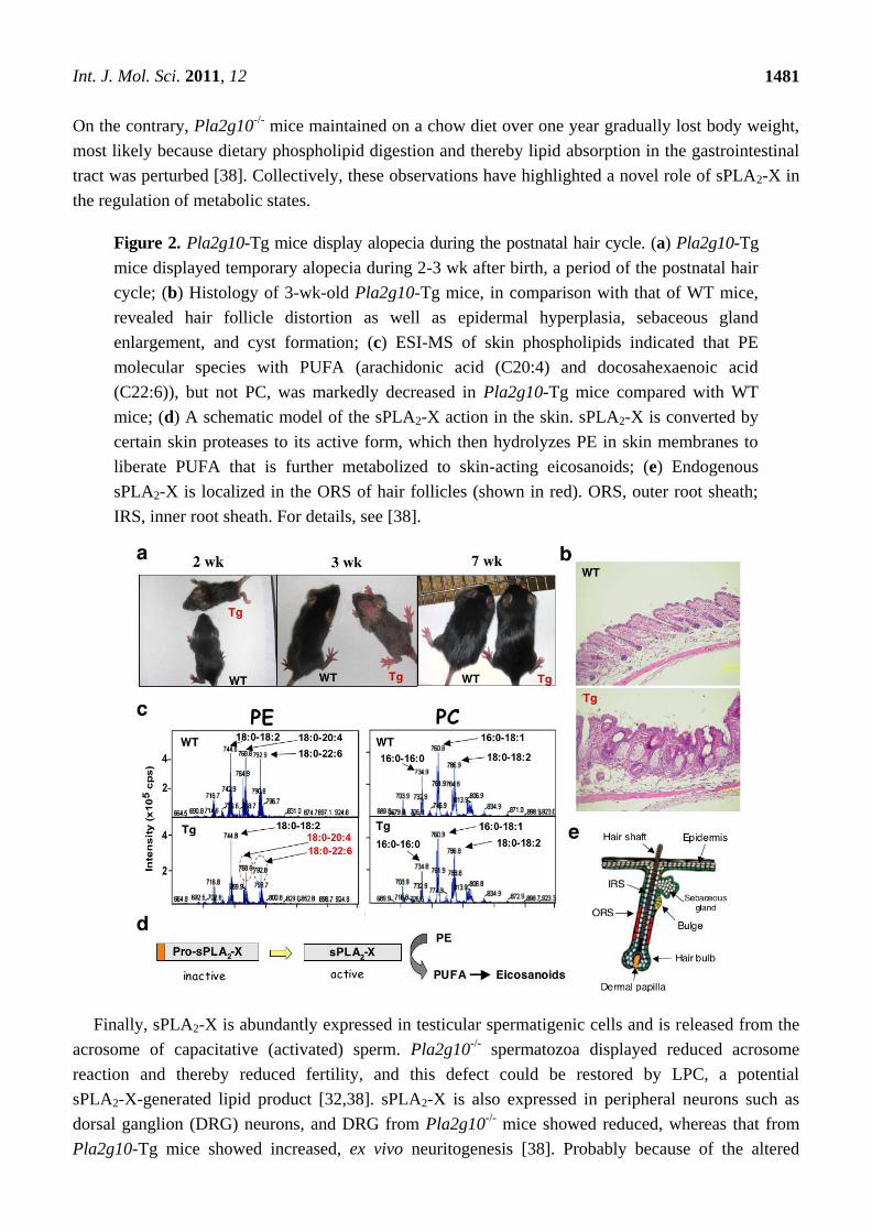
Although systemic Pla2g10-Tg mice did not have any alveolar injury, we found a remarkable
phenotype in these mice before weaning: they developed alopecia [38]. Although pelage hairs of
Pla2g10-Tg mice initially grew, complete but transient hair loss was observed at 3–4 weeks of age, a
period corresponding to the late stage of the initial hair cycle (Figure 2a). Proteolytic activation of
sPLA2-X in Pla2g10-Tg skin temporally preceded hair loss. Histological analyses of the alopecic
Pla2g10-Tg skin revealed hair follicle distortion, hyperkeratosis and sebaceous gland hyperplasia
(Figure 2b), which were accompanied by increased expression of genes related to terminal
differentiation of epidermis and reduced expression of genes related to hair development. ESI-MS
analysis of Pla2g10-Tg skin revealed that sPLA2-X hydrolyzed phosphatidylethanolamine (PE), but
not PC, molecular species to yield PUFAs (Figure 2c), which were further converted to some if not all
eicosanoids. A schematic model for the action of sPLA2-X in Pla2g10-Tg skin is illustrated in Figure 2d.
These results, together with the finding that endogenous sPLA2-X shows a hair cycle-dependent
periodic expression in the outer root sheath of hair follicles in mouse skin (Figure 2e) [38], suggest a
potential functional link between sPLA2-X and skin biology, and may provide a molecular explanation
for the skin abnormality induced by aberrant expression of other sPLA2s such as sPLA2-IIA, whose
transgenic mice also developed alopecia [39]. Importantly, in Pla2g10-/-
mice, hair growth in the
anagen phase was significantly delayed, and this was caused by growth retardation of the outer root
sheath in hair follicles [40]. Thus, sPLA2-X intrinsically functions in the hair quality control.
The ability of sPLA2-X to potently hydrolyze phospholipids in LDL and HDL in vitro has led to the
hypothesis that, as in the case of sPLA2-V (see above), sPLA2-X may also participate in atherosclerosis.
Indeed, sPLA2-X-hydrolyzed LDL particles promote foam cell formation from mouse peritoneal
macrophages [41]. These in vitro observations may be relevant to cardiovascular pathology, since
Pla2g10-/-
mice are protected from angiotensin-II-induced aortic aneurysm and atherosclerosis [42].
sPLA2-X-released PUFAs negatively regulates liver X receptor (LXR), and accordingly, deficiency of
sPLA2-X results in augmented LXR activation leading to increased expression of LXR-target genes.
Thus, in Pla2g10-/-
mice, elevated expression of the ATB-binding cassette (ABC) transporters ABCA1
and ABCG1 led to increased cholesterol efflux by macrophages [43], that of the steroidogenesis acute
regulatory protein StAR resulted in increased corticosterone production by adrenal cells [44], and that
of PPAR (peroxisome proliferator-activated receptor ) facilitated adipogenesis and adiposity [45].
Int. J. Mol. Sci. 2011, 12
1481
On the contrary, Pla2g10-/-
mice maintained on a chow diet over one year gradually lost body weight,
most likely because dietary phospholipid digestion and thereby lipid absorption in the gastrointestinal
tract was perturbed [38]. Collectively, these observations have highlighted a novel role of sPLA2-X in
the regulation of metabolic states.
Figure 2. Pla2g10-Tg mice display alopecia during the postnatal hair cycle. (a) Pla2g10-Tg
mice displayed temporary alopecia during 2-3 wk after birth, a period of the postnatal hair
cycle; (b) Histology of 3-wk-old Pla2g10-Tg mice, in comparison with that of WT mice,
revealed hair follicle distortion as well as epidermal hyperplasia, sebaceous gland
enlargement, and cyst formation; (c) ESI-MS of skin phospholipids indicated that PE
molecular species with PUFA (arachidonic acid (C20:4) and docosahexaenoic acid
(C22:6)), but not PC, was markedly decreased in Pla2g10-Tg mice compared with WT
mice; (d) A schematic model of the sPLA2-X action in the skin. sPLA2-X is converted by
certain skin proteases to its active form, which then hydrolyzes PE in skin membranes to
liberate PUFA that is further metabolized to skin-acting eicosanoids; (e) Endogenous
sPLA2-X is localized in the ORS of hair follicles (shown in red). ORS, outer root sheath;
IRS, inner root sheath. For details, see [38].
Finally, sPLA2-X is abundantly expressed in testicular spermatigenic cells and is released from the
acrosome of capacitative (activated) sperm. Pla2g10-/-
spermatozoa displayed reduced acrosome
reaction and thereby reduced fertility, and this defect could be restored by LPC, a potential
sPLA2-X-generated lipid product [32,38]. sPLA2-X is also expressed in peripheral neurons such as
dorsal ganglion (DRG) neurons, and DRG from Pla2g10-/-
mice showed reduced, whereas that from
Pla2g10-Tg mice showed increased, ex vivo neuritogenesis [38]. Probably because of the altered
Int. J. Mol. Sci. 2011, 12
1482
neuritogenesis, pain nociception in the acetic acid writhing test was partially ameliorated in
Pla2g10-/-
mice, whereas it was augmented in Pla2g10-Tg mice, compared with that in littermate
control mice [38].
2.3. Group III sPLA2 (sPLA2-III)
sPLA2-III is the only enzyme belonging to the group III collection. It is an unusually large protein
(55 kDa) among the sPLA2 family and consists of three domains, in which a central sPLA2 domain
displaying all the features of group III bee venom sPLA2, including 10 cysteines and the key residues
of the Ca2+
loop and catalytic site, is flanked by large and unique N- and C-terminal region [46].
sPLA2-III is processed to a sPLA2 domain-only form (devoid of the N- and C-terminal domains),
which is sufficient for its catalytic function [47,48]. sPLA2-III undergoes N-glycosylation and can
hydrolyze PC and PE equally and augment arachidonate release from cell membranes more efficiently
than sPLA2-IIA, and less efficiently than sPLA2-X and sPLA2-V. sPLA2-III is immunohistochemically
detected in the vascular endothelium of various tissues, peripheral and central nervous systems, male
reproductive tracts, and several types of cancer [48,49]. Implantation of sPLA2-III-transfected
colorectal adenocarcinoma cells into nude mice promotes the growth of tumor xenografts [48].
Expression profiling of the full set of sPLA2s in human colon suggests that sPLA2-III might be a good
candidate as a novel biomarker for colon cancers [50]. In the central nervous system, Pla2g3 mRNA is
localized in DRG neurons in mice, and overexpression of human sPLA2-III in cultured neuronal cells
facilitates neurite outgrowth and survival in correlation with the production of LPC, whereas
knockdown of endogenous sPLA2-III by siRNA partially suppresses these processes [49].
To address the potential in vivo action of sPLA2-III, we produced transgenic mice overexpressing
this enzyme in the whole body (Pla2g3-Tg). Unlike Pla2g5-Tg mice, which die shortly after birth due
to a lung disorder resulting from aberrant hydrolysis of the lung surfactant phospholipids (see above),
Pla2g3-Tg mice showed no respiratory disorder, and lung surfactant phospholipids did not show
appreciable difference between control and Pla2g3-Tg mice [51]. Furthermore, although Pla2g10-Tg
mice show alopecia (see above), Pla2g3-Tg mice had normal pelage hairs up to nine months of age.
Later on, however, Pla2g3-Tg mice spontaneously developed inflammation such as dermatitis,
lymphocytic sialadenitis and splenomegaly [51]. The dermatitis was accompanied by hyperkeratosis,
acanthosis, parakeratosis, erosion, ulcer, neutrophil infiltration, and increased production of
proinflammatory cytokines, chemokines and prostaglandin E2 (PGE2). It is thus likely that
overexpression of sPLA2-III facilitates the production of pro-inflammatory lipid mediators in the
whole body, leading to systemic inflammation.
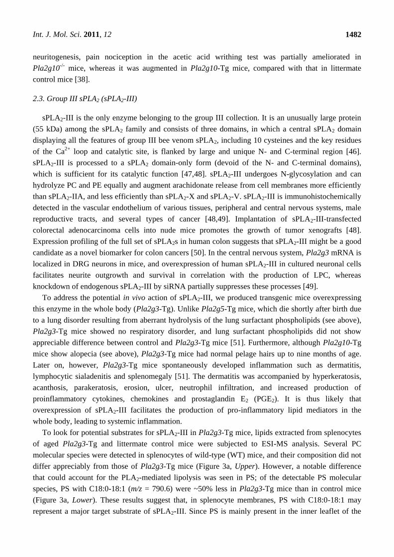
To look for potential substrates for sPLA2-III in Pla2g3-Tg mice, lipids extracted from splenocytes
of aged Pla2g3-Tg and littermate control mice were subjected to ESI-MS analysis. Several PC
molecular species were detected in splenocytes of wild-type (WT) mice, and their composition did not
differ appreciably from those of Pla2g3-Tg mice (Figure 3a, Upper). However, a notable difference
that could account for the PLA2-mediated lipolysis was seen in PS; of the detectable PS molecular
species, PS with C18:0-18:1 (m/z = 790.6) were ~50% less in Pla2g3-Tg mice than in control mice
(Figure 3a, Lower). These results suggest that, in splenocyte membranes, PS with C18:0-18:1 may
represent a major target substrate of sPLA2-III. Since PS is mainly present in the inner leaflet of the
Int. J. Mol. Sci. 2011, 12
1483
plasma membrane of live cells and exposed on apoptotic cell surfaces [52,53], extracellular sPLA2-III
might preferentially hydrolyze PS with C18:0-18:1 on apoptotic cells and thereby modulate the
life-span of inflammatory cells. In support of this idea, susceptibility of cell membranes to sPLA2s
increases in apoptotic cells [54].
As in the case of sPLA2-V and -X, sPLA2-III can potently hydrolyze phospholipids in plasma
lipoprtein particles [55]. Indeed, the decreased level of plasma lipoproteins, HDL in particular, was
obvious in Pla2g3-Tg mice in comparison with WT mice (Figure 3b), suggesting HDL hydrolysis by
overexpressed sPLA2-III. LDL treated with sPLA2-III in vitro was pro-atherogenic, promoting foam
cell formation from macrophages. When Pla2g3-Tg mice that had been crossed with ApoE-/-
mice
(Pla2g3tg
/ApoE-/-
) were fed a high-cholesterol diet, lipid accumulation in the aortic walls was markedly
increased as compared with replicate ApoE-/-
mice (Figure 3c). Immunohistochemistry and in situ
hybridization revealed the presence of sPLA2-III in human atherosclerotic plaques, particularly in
macrophages and smooth muscle cells [55,56]. These results suggest that sPLA2-III may have a role in
acceleration of atherosclerosis development [55].
Figure 3. Pla2g3-Tg mice display systemic inflammatory and atherosclerotic phenotypes.
(a) ESI-MS of PC and PS in splenocytes from Pla2g3-Tg (III-Tg) and wild-type (WT)
mice. Major peaks are indicated by arrows. Peaks altered in III-Tg mice relative to WT
mice are shown in red. SM, sphingomyelin; (b) HPLC profile of plasma lipoproteins in
III-Tg and WT mice; (c) Increased atherosclerosis in III-Tg mice on the ApoE-/-
background (male, 24-wk-old). Atheroslcerotic lesions were visualized by oil red
O staining. Areas positive for the staining were quantified. For details, see [51,55].
Int. J. Mol. Sci. 2011, 12
1484
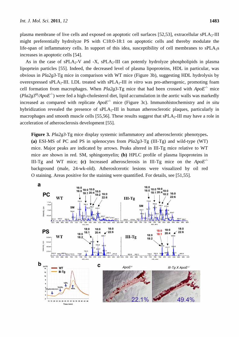
Figure 4. Pla2g3-/-
mice have multiple defects in epididymal sperm maturation. (a) Altered
eicosanoid levels in Pla2g3-/-
mice. The levels of individual eicosanoids in the epididymis
were determined by LC-ESI-MS/MS. sPLA2-III is selectively coupled with 12/15-LOX
and CYP450 pathways; (b) Unusual accumulation of PC molecular species in the
epididymal fluids from Pla2g3-/-
mice relative to Pla2g3+/+
mice, as assessed by ESI-MS;
(c) The roles of sPLA2-III in epididymal sperm maturation are summarized. For details,
see [57].
sPLA2-III is expressed in the testis and epididymis, and in the latter tissue the mature form of
sPLA2-III is secreted from the proximal epididymal epithelium into the lumen [57]. We have recently
succeeded in generating Pla2g3-/-
mice and found that they displayed male infertility [57]. Although
testicular spermatogenesis in Pla2g3-/-
mice was grossly normal, spermatozoa from the cauda (tail)
epididymidis displayed hypomotility, and their ability to fertilize intact eggs was markedly impaired.
Epididymal spermatozoa in Pla2g3-/-
mice had aberrant acrosomal structures and flagella with
abnormal axonemes. These results revealed an unexplored role of this atypical sPLA2 in epididymal
lipid homeostasis, whose perturbation led to sperm dysfunction.
After the complex differentiation process of male germ cells, spermatozoa exit the seminiferous
tubules of the testis through the efferent ducts toward the epididymis. During their transit from the
caput (head) to the cauda (tail) epididymidis, sperm cells undergo significant morphological and
biochemical modifications, which lead to acquisition of their forward motility and ability to recognize
and fertilize oocytes [58]. Unique to mammalian sperm cells is the abundance of phospholipid species
with C22-PUFAs, particularly docosahexaenoic acid (DHA) and docosapentaenoic acid (DPA), whose
proportion in membrane phospholipids appears to correlate with sperm maturity, motility and

Int. J. Mol. Sci. 2011, 12
1485
fertility [59–62]. The percentage of DHA relative to total fatty acids is correlated with the normal
morphology of sperm cells [61], and sperm from subfertile men with low sperm motility or counts
contain a percentage of DHA lower than that from normal men [63]. Sperm maturation involves the
remodeling of membrane phospholipids toward the acquisition of motility and fertility during sperm
migration through the epididymis. Indeed, the increase in C22-PUFAs such as DHA and DPA and the
reciprocal decrease in arachidonic acid (C20:4) favor an increase in the unsaturation degree of fatty
acids in mouse sperm membrane during epididymal transit [59], which could consequently contribute
to increasing the mouse sperm membranous fluidity [64,65]. Interestingly, ESI-MS analysis of sperm
membrane phospholipids revealed that, during epididymal transit, PC in WT sperm underwent a
dramatic shift in its acyl groups from oleic, linoleic and arachidonic acids to DPA and DHA, whereas
this membrane lipid remodeling was compromised in Pla2g3-/-
sperm [57]. Accordingly, cauda
epididymal spermatozoa in Pla2g3-/-
mice had PC species containing more oleate and less DHA/DPA
than did those in Pla2g3+/+
mice, a finding that appears to be consistent with the aforementioned
notion that sperm with higher DHA percentages have better motility and fertility. Thus, sPLA2-III may
participate in the hydrolysis of PC with oleic, linoleic and arachidonic acids in the sperm membrane
during epididymal transit and that this event may be followed by reacylation of LPC, a PLA2 reaction
product, with DHA and DPA, leading to an increase of PC with DPA/DHA in mature spermatozoa. In
the Pla2g3-/-
epididymis, impairment of the deacylation step may eventually perturb the subsequent
reconstitution of DPA/DHA in the sperm membrane, culminating in the asthenozoospermia phenotype.
We also found a notable change in the ESI-MS/MS profile of lipid mediators in the epididymis of
Pla2g3-/-
mice [57]. Thus, arachidonate/linoleate metabolites of the 12/15-lipoxygenase (LOX) and
cytochrome P450 (CYP450) pathways, but not those of the cyclooxygenase (COX) and 5-LOX
pathways, were substantially reduced in the epididymis of Pla2g3-/-
mice compared with that of
Pla2g3+/+
mice (Figure 4a). Although the role of 12/15-LOX or CYP450 metabolites in male fertility
has not yet been fully established, expression of 12/15-LOX in spermatogenic cells has led to the
suggestion that it may participate in sperm maturation [62]. In this context, the possibility that certain
12/15-LOX- or CYP450-derived lipid mediator(s) may be at least partly responsible for the regulation
of sperm maturation by sPLA2-III should be taken into account.
Additionally, sPLA2-III may also affect lipid transport between sperm and epididymal epithelial
cells. Several lipoprotein components are secreted by epididymal epithelial cells [67] and associate
with and dissociate from sperm membranes scheduled for endocytosis by epididymal principal
cells [68]. Male fertility can be impaired to various degrees by inactivation of the genes involved in
lipoprotein metabolism [69–71]. In fact, membrane transport by epididymosomes, a particular
lipoprotein membrane particle emitted from caput epididymal principal cells into the lumen, is
fundamental for the process of sperm cell maturation in the epididymis [59,72]. Our speculation that
sPLA2-III may also affect this epididymal lipid transport is supported by the finding that, as assessed
by ESI-MS, the epididymal fluid from Pla2g3-/-
mice contained PC more abundantly than that from
Pla2g3+/+
mice (Figure 4b). Taken together, we conclude that sPLA2-III may regulate epididymal
sperm maturation through (i) regulation of phospholipid remodeling in sperm membranes,
(ii) production of 12/15-LOX and CYP450 metabolites, and (iii) modification of lipid transport
between sperm and epididymal epithelial cells (Figure 4c).
Int. J. Mol. Sci. 2011, 12
1486
2.4. Other sPLA2s: Classical and Novel Enzymes
Group IB pancreatic sPLA2 (sPLA2-IB) is synthesized in the pancreatic acinar cells, and after
secretion into the pancreatic juice, an N-terminal heptapeptide of the inactive zymogen is cleaved by
trypsin to yield an active enzyme in the duodenum [74]. The main role of sPLA2-IB is digestion of
dietary and biliary phospholipids. Thus, perturbation of this process by gene disruption (Pla2g1b-/-
) or
pharmacological inhibition of sPLA2-IB led to protection from diet-induced obesity and insulin
resistance due to decreased lipid digestion and absorption in the gut [75,76]. In agreement, the
PLA2G1B gene resides within a locus for obesity susceptibility in humans [77].
Group IIA sPLA2 (sPLA2-IIA) is often referred to as an inflammatory sPLA2, since its expression is
markedly induced during inflammation, cardiovascular diseases, and tissue damages [78]. When
overexpressed, sPLA2-IIA is capable of augmenting arachidonic acid release in cytokine-stimulated
cells, albeit more weakly than sPLA2-V, -X and -III [79]. Despite these facts, the contribution of
sPLA2-IIA to inflammation has remained a subject of debate until recently, since a natural mutation of
its gene in C57BL/6 and 129Sv mice [80] prevents the proper assessment of its functions by a classical
gene targeting strategy. Intrinsic deficiency of sPLA2-IIA in these mouse strains is associated with
increased incidence of intestinal polyposis and tumorigengesis [80], a phenotype that is reversed by
transgenic expression of the Pla2g2a gene [81]. A recent study using Pla2g2a-deficient BALB/c mice
as well as Pla2g2a-Tg mice has provided compelling evidence that the enzyme plays an exacerbating
role in inflammatory arthritis [26]. The best-recognized physiologic function of sPLA2-IIA is the
degradation of Gram-positive bacterial membrane, thereby providing the first line of antimicrobial defense
of the host [82–85]. The serum level of sPLA2-IIA also shows correlation with the risk of cardiovascular
diseases [86], and Pla2g2a-Tg mice fed an atherogenic diet developed atherosclerosis [87,88]. This effect
is probably because sPLA2-mediated hydrolysis of LDL phospholipids leads to generation of small-
dense, pro-atherogenic LDL particles that facilitate macrophage foam cell formation, even though the
hydrolytic activity of sPLA2-IIA toward lipoportein particles is much weaker than that of sPLA2-V, -X
and -III. Pla2g2a-Tg mice also displayed permanent alopecia and were susceptible to carcinogen-
induced skin tumorigenesis [39,40].
The roles of other group II subfamily sPLA2 isoforms remain elusive, since knockout or transgenic
mice for these enzymes have not yet been reported. sPLA2-IIC is expressed in rodent testis, but not in
humans [89]. sPLA2-IID is structurally most similar to sPLA2-IIA, and its transcript is constitutively
detected in the lymphoid organs [90]. This enzyme may have immuno-suppressive functions, since it is
expressed in regulatory T cells and its fusion protein has the ability to suppress inflammatory bowel
disease and experimental autoimmune encephalomyelitis in mice [91]. sPLA2-IIE, another group
IIA-related enzyme, is expressed constitutively in several tissues at low levels and has a lower
catalytic activity than other group II sPLA2s [92]. sPLA2-IIF possesses a unique 30-amino acid
C-terminal extension that contains an additional Cys residue, which might contribute to formation of a
homodimer or a heterodimer with a second protein [93,94]. This enzyme is expressed most abundantly
in the skin [100].
Lastly, group XII sPLA2s (sPLA2-XIIA and -XIIB) represent a unique collection of the sPLA2
family. sPLA2-XIIA has the central catalytic domain with a His/Asp catalytic dyad, yet the location of
Cys residues outside the catalytic domain is rather distinct from that of other sPLA2s [95]. High
Int. J. Mol. Sci. 2011, 12
1487
expression of this enzyme is found in many tissues, although its enzymatic activity is very weak. A
study using Xenopus suggests the role of this enzyme in early neuronal development [96]. sPLA2-XIIB
is structurally related to sPLA2-XIIA and is expressed in liver and intestine [97]. A recent study has
demonstrated that the transcription of Pla2g12b was regulated by the transcription factor HNF-4 and
its co-activator PGC-1, and deletion of the Pla2g12b gene resulted in increased fat accumulation in
the liver leading to steatohepatitis, a phenotype similar to that seen in Hnf4a-/-
mice [98]. The aberrant fat
accumulation in Pla2g12b-/-
liver was ascribed to impaired hepatic secretion of VLDL. However, because
sPLA2-XIIB lacks the catalytic activity since the catalytic center His is replaced with Leu [97], the
molecular mechanism whereby this sPLA2 isoform regulates VLDL secretion remains unknown.
3. Conclusions
During the past decade, the biological functions of several sPLA2s and their target substrates have
been clarified by studies using transgenic and knockout mice in combination with lipidomics.
Nevertheless, full understanding of the biological roles of all sPLA2 isoforms is still a challenging area
of research. The control of particular sPLA2s, alone or in combination of multiple isoforms, should
have advantages over the inhibition of selective lipid metabolic pathways in the treatment of various
diseases. Interestingly, the pan-sPLA2 inhibitor A-002 (varespladib), which inhibits the conventional
class of sPLA2s, can markedly reduce the atherosclerotic lesion area in experimental animals and even
in humans in early-phase clinical studies [99]. This fact points to the sPLA2 family as a potential
therapeutic target for atherosclerosis, and probably other diseases in which one or more sPLA2s are
involved, such as asthma, arthritis, and metabolic syndrome.
Acknowledgements
This work was supported by grants-in aid for scientific research from the Ministry of Education,
Science, Culture, Sports and Technology of Japan.
References
1. Uozumi, N.; Kume, K.; Nagase, T.; Nakatani, N.; Ishii, S.; Tashiro, F.; Komagata, Y.; Maki, K.;
Ikuta, K.; Ouchi, Y.; Miyazaki, J.; Shimizu, T. Role of cytosolic phospholipase A2 in allergic
response and parturition. Nature 1997, 390, 618–622.
2. Kudo, I.; Murakami, M. Phospholipase A2 enzymes. Prostag. Other Lipid Mediat. 2002, 68–69,
3–58.
3. Lambeau, G.; Gelb, M.H. Biochemistry and physiology of mammalian secreted phospholipases
A2. Annu. Rev. Biochem. 2008, 77, 495–520.
4. Murakami, M.; Taketomi, Y.; Girard, C.; Yamamoto, K.; Lambeau, G. Emerging roles of
secreted phospholipase A2 enzymes: Lessons from transgenic and knockout mice. Biochimie
2010, 92, 561–582.
5. Chen, J.; Engle, S.J.; Seilhamer, J.J.; Tischfield, J.A. Cloning and recombinant expression of a
novel human low molecular weight Ca2+
-dependent phospholipase A2. J. Biol. Chem. 1994, 269,
2365–2368.
Int. J. Mol. Sci. 2011, 12
1488
6. Munoz, N.M.; Meliton, A.Y.; Arm, J.P.; Bonventre, J.V.; Cho, W.; Leff, A.R. Deletion of
secretory group V phospholipase A2 attenuates cell migration and airway hyperresponsiveness in
immunosensitized mice. J. Immunol. 2007, 179, 4800–4807.
7. Masuda, S.; Murakami, M.; Mitsuishi, M.; Komiyama, K.; Ishikawa, Y.; Ishii, T.; Kudo, I.
Expression of secretory phospholipase A2 enzymes in lungs of humans with pneumonia and their
potential prostaglandin-synthetic function in human lung-derived cells. Biochem. J. 2005, 387,
27–38.
8. Ohtsuki, M.; Taketomi, Y.; Arata, S.; Masuda, S.; Ishikawa, Y.; Ishii, T.; Takanezawa, Y.; Aoki, J.;
Arai, H.; Yamamoto, K.; Kudo, I.; Murakami, M. Transgenic expression of group V, but not
group X, secreted phospholipase A2 in mice leads to neonatal lethality because of lung
dysfunction. J. Biol. Chem. 2006, 281, 36420–36433.
9. Touqi, L.; Arbibe, L. A role for phospholipase A2 in ARDS pathogenesis. Mol. Med. Today
1999, 5, 244–249.
10. Nag, K.; Munro, J.G.; Inchley, K.; Schürch, S.; Petersen, N.O.; Possmayer, F. SP-B refining of
pulmonary surfactant phospholipid films. Am. J. Physiol. 1999, 277, L1179–L1189.
11. Arbibe, L.; Koumanov, K.; Vial, D.; Rougeot, C.; Faure, G.; Havet, N.; Longacre, S.; Vargaftig, B.B.;
Béréziat, G.; Voelker, D.R.; Wolf, C.; Touqui, L. Generation of lyso-phospholipids from
surfactant in acute lung injury is mediated by type-II phospholipase A2 and inhibited by a direct
surfactant protein A-phospholipase A2 protein interaction. J. Clin. Invest 1998, 102, 1152–1160
12. Wu, Y.; Singer, M.; Thouron, F.; Alaoui-El-Azher, M.; Touqui, L. Effect of surfactant on
pulmonary expression of type IIA PLA2 in an animal model of acute lung injury. Am. J. Physiol.
Lung. Cell Mol. Physiol. 2002, 282, L743–L750.
13. Wu, Y.Z.; Medjane, S.; Chabot, S.; Kubrusly, F.S.; Raw, I.; Chignard, M.; Touqui, L. Surfactant
protein-A and phosphatidylglycerol suppress type IIA phospholipase A2 synthesis via nuclear
factor-B. Am. J. Respir. Crit. Care Med. 2003, 168, 692–699.
14. Wang, Z.; Schwan, A.L.; Lairson, L.L.; O’Donnell, J.S.; Byrne, G.F.; Foye, A.; Holm, B.A.;
Notter, R.H. Surface activity of a synthetic lung surfactant containing a phospholipase-resistant
phosphonolipid analog of dipalmitoyl phosphatidylcholine. Am. J. Physiol. Lung Cell Mol.
Physiol. 2003, 285, L550–L559.
15. Furue, S.; Kuwabara, K.; Mikawa, K.; Nishina, K.; Shiga, M.; Maekawa, N.; Ueno, M.;
Chikazawa, Y.; Ono, T.; Hori, Y.; Matsukawa, A.; Yoshinaga, M.; Obara, H. Crucial role of
group IIA phospholipase A2 in oleic acid-induced acute lung injury in rabbits. Crit. Care Med.
2001, 29, 719–727.
16. Chabot, S.; Koumanov, K.; Lambeau, G.; Gelb, M.H.; Balloy, V.; Chignard, M.; Whitsett, J.A.;
Touqui, L. Inhibitory effects of surfactant protein A on surfactant phospholipid hydrolysis by
secreted phospholipases A2. J. Immunol. 2003, 171, 995–1000.
17. Seeds, M.C.; Jones, K.A.; Duncan, H.R.; Willingham, M.C.; Borgerink, H.M.; Woodruff, R.D.;
Bowton, D.L.; Bass, D.A. Cell-specific expression of group X and group V secretory
phospholipases A2 in human lung airway epithelial cells. Am. J. Respir. Cell Mol. Biol. 2000, 23,
37–44.
Int. J. Mol. Sci. 2011, 12
1489
18. Hite, R.D.; Seeds, M.C.; Safta, A.M.; Jacinto, R.B.; Gyves, J.I.; Bass, D.A.; Waite, B.M.
Lysophospholipid generation and phosphatidylglycerol depletion in phospholipase A2-mediated
surfactant dysfunction. Am. J. Physiol. Lung Cell Mol. Physiol. 2005, 288, L618–L624.
19. Shulenin, S.; Nogee, L.M.; Annilo, T.; Wert, S.E.; Whitsett, J.A.; Dean, M. ABCA3 gene
mutations in newborns with fatal surfactant deficiency. N. Engl. J. Med. 2004, 350, 1296–1303.
20. Bridges, J.P.; Ikegami, M.; Brilli, L.L.; Chen, X.; Mason, R.J.; Shannon, J.M. LPCAT1 regulates
surfactant phospholipid synthesis and is required for transitioning to air breathing in mice. J. Clin.
Invest. 2010, 120, 1736–1748.
21. Munoz, N.M.; Meliton, A.Y.; Meliton, L.N.; Dudek, S.M.; Leff, A.R. Secretory group V
phospholipase A2 regulates acute lung injury and neutrophilic inflammation caused by LPS in
mice. Am. J. Physiol. Lung Cell Mol. Physiol. 2009, 296, L879–L887.
22. Giannattasio, G.; Fujioka, D.; Xing, W.; Katz, H.R.; Boyce, J.A.; Balestrieri, B. Group V
secretory phospholipase A2 reveals its role in house dust mite-induced allergic pulmonary
inflammation by regulation of dendritic cell function. J. Immunol. 2010, 185, 4430–4438.
23. Satake, Y.; Diaz, B.L.; Balestrieri, B.; Lam, B.K.; Kanaoka, Y.; Grusby, M.J.; Arm, J.P. Role of
group V phospholipase A2 in zymosan-induced eicosanoid generation and vascular permeability
revealed by targeted gene disruption. J. Biol. Chem. 2004, 279, 16488–16494.
24. Balestrieri, B.; Hsu, V.W.; Gilbert, H.; Leslie, C.C.; Han, W.K.; Bonventre, J.V.; Arm, J.P.
Group V secretory phospholipase A2 translocates to the phagosome after zymosan stimulation of
mouse peritoneal macrophages and regulates phagocytosis. J. Biol. Chem. 2006, 281,
6691–6698.
25. Balestrieri, B.; Maekawa, A.; Xing, W.; Gelb, M.H.; Katz, H.R.; Arm, J.P. Group V secretory
phospholipase A2 modulates phagosome maturation and regulates the innate immune response
against Candida albicans. J. Immunol. 2009, 182, 4891–4898.
26. Boilard, E.; Lai, Y.; Larabee, K.; Balestrieri, B.; Ghomashchi, F.; Fujioka, D.; Gobezie, R.;
Coblyn, J.S.; Weinblatt, M.E.; Massarotti, E.M.; Thornhill, T.S.; Divangahi, M.; Remold, H.;
Lambeau, G.; Gelb, M.H.; Arm, J.P.; Lee, D.M. A novel anti-inflammatory role for secretory
phospholipase A2 in immune complex-mediated arthritis. EMBO Mol. Med. 2010, 2, 172-187.
27. Bostrom, M.A.; Boyanovsky, B.B.; Jordan, C.T.; Wadsworth, M.P.; Taatjes, D.J.; De Beer, F.C.;
Webb, N.R. Group V secretory phospholipase A2 promotes atherosclerosis: Evidence from
genetically altered mice. Arterioscler. Thromb. Vasc. Biol. 2007, 27, 600–606.
28. Wootton, P.T.; Arora, N.L.; Drenos, F.; Thompson, S.R.; Cooper, J.A.; Stephens, J.W.; Hurel,
S.J.; Hurt-Camejo, E.; Wiklund, O.; Humphries, S.E.; Talmud, P.J. Tagging SNP haplotype
analysis of the secretory PLA2-V gene, PLA2G5, shows strong association with LDL and oxLDL
levels, suggesting functional distinction from sPLA2-IIA: Results from the UDACS study. Hum.
Mol. Genet. 2007, 16, 1437–1444.
29. Cupillard, L.; Koumanov, K.; Mattéi, M.G.; Lazdunski, M.; Lambeau, G. Cloning, chromosomal
mapping, and expression of a novel human secretory phospholipase A2. J. Biol. Chem. 1997,
272, 15745–15752.
30. Murakami, M.; Koduri, R.S.; Enomoto, A.; Shimbara, S.; Seki, M.; Yoshihara, K.; Singer, A.;
Valentin, E.; Ghomashchi, F.; Lambeau, G.; Gelb, M.H.; Kudo, I. Distinct arachidonate-releasing
functions of mammalian secreted phospholipases A2 in fibroblastic and mastocytoma cells
Int. J. Mol. Sci. 2011, 12
1490
through heparan sulfate shuttling and external plasma membrane mechanisms. J. Biol. Chem.
2001, 276, 10083–10096.
31. Bezzine, S.; Koduri, R.S.; Valentin, E.; Murakami, M.; Kudo, I.; Ghomashchi, F.; Sadilek, M.;
Lambeau, G.; Gelb, M.H. Exogenously added human group X secreted phospholipase A2 but not
the group IB, IIA, and V enzymes efficiently release arachidonic acid from adherent mammalian
cells. J. Biol. Chem. 2000, 275, 3179–3191.
32. Escoffier, J.; Jemel, I.; Tanemoto, A.; Taketomi, Y.; Payre, C.; Coatrieux, C.; Sato, H.;
Yamamoto, K.; Masuda, S.; Pernet-Gallay, K.; Pierre, V.; Hara, S.; Murakami, M.; De Waard,
M.; Lambeau, G.; Arnoult, C. Group X phospholipase A2 is released during sperm acrosome
reaction and controls fertility outcome in mice. J. Clin. Invest 2010, 120, 1415–1428.
33. Sato, H.; Isogai, Y.; Masuda, S.; Taketomi, Y.; Miki, Y.; Kamei, D.; Hara, S.; Kobayashi, T.;
Ishikawa, Y.; Ishii, T.; Ikeda, K.; Taguchi, R.; Ishimoto, Y.; Suzuki, N.; Yokota, Y.; Hanasaki, K.;
Suzuki-Yamamoto, T.; Yamamoto, K.; Murakami, M. Physiological roles of group X secreted
phospholipase A2 in reproduction, gastrointestinal phospholipid digestion, and neuronal function.
J. Biol. Chem. 2011, in press.
34. Henderson, W.R., Jr.; Chi, E.Y.; Bollinger, J.G.; Tien, Y.T.; Ye, X.; Castelli, L.; Rubtsov, Y.P.;
Singer, A.G.; Chiang, G.K.; Nevalainen, T.; Rudensky, A.Y.; Gelb, M.H. Importance of group
X-secreted phospholipase A2 in allergen-induced airway inflammation and remodeling in a
mouse asthma model. J. Exp. Med. 2007, 204, 865–877.
35. Hallstrand, T.S.; Chi, E.Y.; Singer, A.G.; Gelb, M.H.; Henderson, W.R., Jr. Secreted
phospholipase A2 group X overexpression in asthma and bronchial hyperresponsiveness. Am. J.
Respir. Crit. Care Med. 2007, 176, 1072–1078.
36. Fujioka, D.; Saito, Y.; Kobayashi, T.; Yano, T.; Tezuka, H.; Ishimoto, Y.; Suzuki, N.; Yokota,
Y.; Nakamura, T.; Obata, J.E.; Kanazawa, M.; Kawabata, K.; Hanasaki, K.; Kugiyama, K.
Reduction in myocardial ischemia/reperfusion injury in group X secretory phospholipase A2-
deficient mice. Circulation 2008, 117, 2977–2985.
37. Curfs, D.M.; Ghesquiere, S.A.; Vergouwe, M.N.; Van der Made, I.; Gijbels, MJ.; Greaves, D.R.;
Verbeek, J.S.; Hofker, M.H.; De Winther, M.P. Macrophage secretory phospholipase A2 group X
enhances anti-inflammatory responses, promotes lipid accumulation, and contributes to aberrant
lung pathology. J. Biol. Chem. 2008, 283, 21640–21648.
38. Yamamoto, K.; Taketomi, T.; Isogai, Y.; Miki, Y.; Sato, H.; Masuda, S.; Nishito, Y.; Morioka, K.;
Ishimoto, Y.; Suzuki, N.; Yokoya, Y.; Hanasaki, K.; Ishikawa, Y.; Ishii, T.; Kobayashi, T.;
Fukami, K.; Ikeda, K.; Nakanishi, H.; Taguchi, R.; Murakami, M. Hair follicular expression and
function of group X secreted phospholipase A2 in mouse skin. J. Biol. Chem. 2011, in press.
39. Grass, D.S.; Felkner, R.H.; Chiang, M.Y.; Wallace, R.E.; Nevalainen, T.J.; Bennett, C.F.;
Swanson, M.E. Expression of human group II PLA2 in transgenic mice results in epidermal
hyperplasia in the absence of inflammatory infiltrate. J. Clin. Invest 1996, 97, 2233–2241.
40. Mulherkar, R.; Kirtane, B.M.; Ramchandani, A.; Mansukhani, N.P.; Kannan, S.; Naresh, K.N.
Expression of enhancing factor/phospholipase A2 in skin results in abnormal epidermis and
increased sensitivity to chemical carcinogenesis. Oncogene 2003, 22, 1936–1944.
Int. J. Mol. Sci. 2011, 12
1491
41. Hanasaki, K.; Yamada, K.; Yamamoto, S.; Ishimoto, Y.; Saiga, A.; Ono, T.; Ikeda, M.; Notoya,
M.; Kamitani, S.; Arita, H. Potent modification of low density lipoprotein by group X secretory
phospholipase A2 is linked to macrophage foam cell formation. J. Biol. Chem. 2002, 277,
29116–29124.
42. Zack, M.; Boyanovsky, B.B.; Shridas, P.; Bailey, W.; Forrest, K.; Howatt, D.A.; Gelb, M.H.;
De Beer, F.C.; Daugherty, A.; Webb, N.R. Group X secretory phospholipase A2 augments
angiotensin II-induced inflammatory responses and abdominal aortic aneurysm formation in
apoE-deficient mice. Atherosclerosis 2010, in press.
43. Shridas, P.; Bailey, W.M.; Gizard, F.; Oslund, R.C.; Gelb, M.H.; Bruemmer, D.; Webb, N.R.
Group X secretory phospholipase A2 negatively regulates ABCA1 and ABCG1 expression and
cholesterol efflux in macrophages. Arterioscler Thromb. Vasc. Biol. 2010, 30, 2014–2021.
44. Shridas, P.; Bailey, W.M.; Boyanovsky, B.B.; Oslund, R.C.; Gelb, M.H.; Webb, N.R. Group X
secretory phospholipase A2 regulates the expression of steroidogenic acute regulatory protein
(StAR) in mouse adrenal glands. J. Biol. Chem. 2010, 285, 20031–20039.
45. Li, X.; Shridas, P.; Forrest, K.; Bailey, W.; Webb, N.R. Group X secretory phospholipase A2
negatively regulates adipogenesis in murine models. FASEB J. 2010, 24, 4313–4324.
46. Valentin, E.; Ghomashchi, F.; Gelb, M.H.; Lazdunski, M.; Lambeau, G. Novel human secreted
phospholipase A2 with homology to the group III bee venom enzyme. J. Biol. Chem. 2000, 275,
7492–7496.
47. Murakami, M.; Masuda, S.; Shimbara, S.; Bezzine, S.; Lazdunski, M.; Lambeau, G.; Gelb, M.H.;
Matsukura, S.; Kokubu, F.; Adachi, M.; Kudo, I. Cellular arachidonate-releasing function of
novel classes of secretory phospholipase A2s (groups III and XII). J. Biol. Chem. 2003, 278,
10657–10667.
48. Murakami, M.; Masuda, S.; Shimbara, S.; Ishikawa, Y.; Ishii, T.; Kudo, I. Cellular distribution,
post-translational modification, and tumorigenic potential of human group III secreted
phospholipase A2. J. Biol. Chem. 2005, 280, 24987–24998.
49. Masuda, S.; Yamamoto, K.; Hirabayashi, T.; Ishikawa, Y.; Ishii, T.; Kudo, I.; Murakami, M.
Human group III secreted phospholipase A2 promotes neuronal outgrowth and survival.
Biochem. J. 2008, 409, 429–438.
50. Mounier, C.M.; Wendum, D.; Greenspan, E.; Flejou, J.F.; Rosenberg, D.W.; Lambeau, G.
Distinct expression pattern of the full set of secreted phospholipases A2 in human colorectal
adenocarcinomas: sPLA2-III as a biomarker candidate. Br. J. Cancer 2008, 98, 587–595.
51. Sato, H.; Taketomi, Y.; Isogai, Y.; Masuda, S.; Kobayashi, T.; Yamamoto, K.; Murakami, M.
Group III secreted phospholipase A2 transgenic mice spontaneously develop inflammation.
Biochem. J. 2009, 421, 17–27.
52. Emoto, K.; Inadome, H.; Kanaho, Y.; Narumiya, S.; Umeda, M. Local change in phospholipid
composition at the cleavage furrow is essential for completion of cytokinesis. J. Biol. Chem.
2005, 280, 37901–37907.
53. Miyanishi, M.; Tada, K.; Koike, M.; Uchiyama, Y.; Kitamura, T.; Nagata, S. Identification of
Tim4 as a phosphatidylserine receptor. Nature 2007, 450, 435–439.
Int. J. Mol. Sci. 2011, 12
1492
54. Olson, E.D.; Nelson, J.; Griffith, K.; Nguyen, T.; Streeter, M.; Wilson-Ashworth, H.A.; Gelb,
M.H.; Judd, A.M.; Bell, J.D. Kinetic evaluation of cell membrane hydrolysis during apoptosis by
human isoforms of secretory phospholipase A2. J. Biol. Chem. 2010, 285, 10993–11002.
55. Sato, H.; Kato, R.; Isogai, Y.; Saka, G.; Ohtsuki, M.; Taketomi, Y.; Yamamoto, K.; Tsutsumi,
K.; Yamada, J.; Masuda, S.; Ishikawa, Y.; Ishii, T.; Kobayashi, T.; Ikeda, K.; Taguchi, R.;
Hatakeyama, S.; Hara, S.; Kudo, I.; Itabe, H.; Murakami, M. Analyses of group III secreted
phospholipase A2 transgenic mice reveal potential participation of this enzyme in plasma
lipoprotein modification, macrophage foam cell formation, and atherosclerosis. J. Biol. Chem.
2008, 283, 33483–33497.
56. Kimura-Matsumoto, M.; Ishikawa, Y.; Komiyama, K.; Tsuruta, T.; Murakami, M.; Masuda, S.;
Akasaka, Y.; Ito, K.; Ishiguro, S.; Morita, H.; Sato, S.; Ishii, T. Expression of secretory
phospholipase A2s in human atherosclerosis development. Atherosclerosis 2008, 196, 81–91.
57. Sato, H.; Taketomi, Y.; Isogai, Y.; Miki, Y.; Yamamoto, K.; Masuda, S.; Hosono, T.; Arata, S.;
Ishikawa, Y.; Ishii, T.; Kobayashi, T.; Nakanishi, H.; Ikeda, K.; Taguchi, R.; Hara, S.; Kudo, I.;
Murakami, M. Group III secreted phospholipase A2 regulates epididymal sperm maturation and
fertility in mice. J. Clin. Invest 2010, 120, 1400–1414.
58. Cooper, T.G. Role of the epididymis in mediating changes in the male gamete during maturation.
Adv. Exp. Med. Biol. 1995, 377, 87–101.
59. Rejraji, H.; Sion, B.; Prensier, G.; Carreras, M.; Motta, C.; Frenoux, J.M.; Vericel, E.; Grizard, G.;
Vernet, P.; Drevet, J.R. Lipid remodeling of murine epididymosomes and spermatozoa during
epididymal maturation. Biol. Reprod. 2006, 74, 1104–1113.
60. Lenzi, A.; Picardo, M.; Gandini, L.; Dondero, F. Lipids of the sperm plasma membrane: from
polyunsaturated fatty acids considered as markers of sperm function to possible scavenger
therapy. Hum. Reprod. 1996, 2, 246–256.
61. Lenzi, A.; Gandini, L.; Maresca, V.; Rago, R.; Sgrò, P.; Dondero, F.; Picardo, M. Fatty acid
composition of spermatozoa and immature germ cells. Mol. Hum. Reprod. 2000, 6, 226–231.
62. Furimsky, A.; Vuong, N.; Xu, H.; Kumarathasan, P.; Xu, M.; Weerachatyanukul, W.; Bou, K.M.;
Kates, M.; Tanphaichitr, N. Percoll gradient-centrifuged capacitated mouse sperm have increased
fertilizing ability and higher contents of sulfogalactosylglycerolipid and docosahexaenoic
acid-containing phosphatidylcholine compared to washed capacitated mouse sperm. Biol.
Reprod. 2005, 72, 574–583.
63. Aksoy, Y.; Aksoy, H.; Altinkaynak, K.; Aydin, H.R.; Ozkan, A. Sperm fatty acid composition in
subfertile men. Prostag. Leuk. Essent. Fatty 2006, 75, 75–79.
64. Hall, J.C.; Hadley, J.; Doman, T. Correlation between changes in rat sperm membrane lipids,
protein, and the membrane physical state during epididymal maturation. J. Androl. 1991, 12,
76–87.
65. Haidl, G.; Opper, C. Changes in lipids and membrane anisotropy in human spermatozoa during
epididymal maturation. Hum. Reprod. 1997, 12, 2720–2723.
66. Fischer, K.A.; Van Leyen, K.; Lovercamp, K.W.; Manandhar, G.; Sutovsky, M.; Feng, D.;
Safranski, T.; Sutovsky, P. 15-Lipoxygenase is a component of the mammalian sperm
cytoplasmic droplet. Reproduction 2005, 130, 213–222.
Int. J. Mol. Sci. 2011, 12
1493
67. Law, G.L.; McGuinness, M.P.; Linder, C.C.; Griswold, M.D. Expression of apolipoprotein E
mRNA in the epithelium and interstitium of the testis and the epididymis. J. Androl. 1997, 18,
32–42.
68. Hermo, L.; Wright, J.; Oko, R.; Morales, C.R. Role of epithelial cells of the male excurrent duct
system of the rat in the endocytosis or secretion of sulfated glycoprotein-2 (clusterin). Biol.
Reprod. 1991, 44, 1113–1131.
69. Huang, L.S.; Voyiaziakis, E.; Chen, H.L.; Rubin, E.M.; Gordon, J.W. A novel functional role for
apolipoprotein B in male infertility in heterozygous apolipoprotein B knockout mice. Proc. Natl.
Acad. Sci. USA 1996, 93, 10903–10907.
70. Moghadasian, M.H.; Nguyen, L.B.; Shefer, S.; McManus, B.M.; Frohlich, J.J. Histologic,
hematologic, and biochemical characteristics of apo E-deficient mice: effects of dietary
cholesterol and phytosterols. Lab. Invest 1999, 79, 355–364.
71. Andersen, O.M.; Yeung, C.H.; Vorum, H.; Wellner, M.; Andreassen, T.K.; Erdmann, B.;
Mueller, E.C.; Herz, J.; Otto, A.; Cooper, T.G.; Willnow, T.E. Essential role of the
apolipoprotein E receptor-2 in sperm development. J. Biol. Chem. 2003, 278, 23989–23995.
72. Saez, F.; Frenette, G.; Sullivan, R. Epididymosomes and prostasomes: their roles in posttesticular
maturation of the sperm cells. J. Androl 2003, 24, 149–154.
73. Masuda, S.; Murakami, M.; Matsumoto, S.; Eguchi, N.; Urade, Y.; Lambeau, G.; Gelb, M.H.;
Ishikawa, Y.; Ishii, T.; Kudo, I. Localization of various secretory phospholipase A2 enzymes in
male reproductive organs. Biochim. Biophys. Acta 2004, 1686, 61–76.
74. Seilhamer, J.J.; Randall, T.L.; Yamanaka, M.; Johnson, L.K. Pancreatic phospholipase A2:
Isolation of the human gene and cDNAs from porcine pancreas and human lung. DNA 1986, 5,
519–527.
75. Labonte, E.D.; Kirby, R.J.; Schildmeyer, N.M.; Cannon, A.M.; Huggins, K.W.; Hui, D.Y. Group
IB phospholipase A2-mediated lysophospholipid absorption directly contributes to postprandial
hyperglycemia. Diabetes 2006, 55, 935–941.
76. Hui, D.Y.; Cope, M.J.; Labonte, E.D.; Chang, H.T.; Shao, J.; Goka, E.; Abousalham, A.;
Charmot, D.; Buysse, J. The phospholipase A2 inhibitor methyl indoxam suppresses diet-induced
obesity and glucose intolerance in mice. Br. J. Pharmacol. 2009, 157, 1263–1269.
77. Wilson, S.G.; Adam, G.; Langdown, M.; Reneland, R.; Braun, A.; Andrew, T.; Surdulescu, G.L.;
Norberg, M.; Dudbridge, F.; Reed, P.W.; Sambrook, P.N.; Kleyn, P.W.; Spector, T.D. Linkage
and potential association of obesity-related phenotypes with two genes on chromosome 12q24 in
a female dizygous twin cohort. Eur. J. Hum Genet 2006, 14, 340–348.
78. Pruzanski, W.; Vadas, P. Phospholipase A2—A mediator between proximal and distal effectors
of inflammation. Immunol. Today 1991, 12, 143-146.
79. Murakami, M.; Kambe, T.; Shimbara, S.; Yamamoto S, Kuwata H, Kudo I. Functional association
of type IIA secretory phospholipase A2 with the glycosylphosphatidylinositol-anchored heparan
sulfate proteoglycan in the cyclooxygenase-2-mediated delayed prostanoid-biosynthetic
pathway. J. Biol. Chem. 1999, 274, 29927-29936.
80. MacPhee, M.; Chepenik, P.K.; Liddel, A.R.; Nelson, K.K.; Siracusa, D.L.; Buchberg, M.A. The
secretory phospholipase A2 gene is a candidate for the Mom1 locus, a major modifier of
Apcmin
-induced intestinal neoplasia. Cell 1995, 81, 957–966.
Int. J. Mol. Sci. 2011, 12
1494
81. Cormier, R.T.; Hong, K.H.; Halberg, R.B.; Hawkins, T.L.; Richardson, P.; Mulherkar, R.;
Dove, W.F.; Lander, E.S. Secretory phospholipase Pla2g2a confers resistance to intestinal
tumorigenesis. Nat. Genet 1997, 17, 88–91.
82. Weinrauch, Y.; Elsbach, P.; Madsen, L.M.; Foreman, A.; Weiss, J. The potent anti-Staphylococcus
aureus activity of a sterile rabbit inflammatory fluid is due to a 14-kD phospholipase A2. J. Clin.
Invest 1996, 97, 250–257.
83. Weinrauch, Y.; Abad, C.; Liang, N.S.; Lowry, S.F.; Weiss, J. Mobilization of potent plasma
bactericidal activity during systemic bacterial challenge. Role of group IIA phospholipase A2.
J. Clin. Invest 1998, 102, 633–638.
84. Laine, V.J.; Grass, D.S.; Nevalainen, T.J. Protection by group II phospholipase A2 against
Staphylococcus aureus. J. Immunol. 1999, 162, 7402–7408.
85. Koduri, R.S.; Grönroos, J.O.; Laine, V.J.; Le Calvez, C.; Lambeau, G.; Nevalainen, T.J.; Gelb, M.H.
Bactericidal properties of human and murine groups I, II, V, X, and XII secreted phospholipases
A2. J. Biol. Chem. 2002, 277, 5849–5857.
86. Kugiyama, K.; Ota, Y.; Takazoe, K.; Moriyama, Y.; Kawano, H.; Miyao, Y.; Sakamoto, T.;
Soejima, H.; Ogawa, H.; Doi, H.; Sugiyama, S.; Yasue, H. Circulating levels of secretory type II
phospholipase A2 predict coronary events in patients with coronary artery disease. Circulation
1999, 100, 1280–1284.
87. Ivandic, B.; Castellani, L.W.; Wang, X.P.; Qiao, J.H.; Mehrabian, M.; Navab, M.; Fogelman,
A.M.; Grass, D.S.; Swanson, M.E.; De Beer, M.C.; De Beer, F.; Lusis, A.J. Role of group II
secretory phospholipase A2 in atherosclerosis: 1. Increased atherogenesis and altered lipoproteins
in transgenic mice expressing group IIa phospholipase A2. Arterioscler. Thromb. Vasc. Biol.
1999, 19, 1284–1290.
88. Webb, N.R.; Bostrom, M.A.; Szilvassy, S.J.; Van der Westhuyzen, D.R.; Daugherty, A.; De
Beer, F.C. Macrophage-expressed group IIA secretory phospholipase A2 increases
atherosclerotic lesion formation in LDL receptor-deficient mice. Arterioscler. Thromb. Vasc.
Biol. 2003, 23, 263–268.
89. Chen, J.; Shao, C.; Lazar, V.; Srivastava, C.H.; Lee, W.H.; Tischfield, J.A. Localization of group
IIc low molecular weight phospholipase A2 mRNA to meiotic cells in the mouse. J. Cell
Biochem. 1997, 64, 369–375.
90. Shakhov, A.N.; Rubtsov, A.V.; Lyakhov, I.G.; Tumanov, A.V.; Nedospasov, S.A. SPLASH
(PLA2IID), a novel member of phospholipase A2 family, is associated with lymphotoxin
deficiency. Genes Immun. 2000, 1, 191–199.
91. Von Allmen, C.E.; Schmitz, N.; Bauer, M.; Hinton, H.J.; Kurrer, M.O.; Buser, R.B.; Gwerder,
M.; Muntwiler, S.; Sparwasser, T.; Beerli, R.R.; Bachmann, M.F. Secretory phospholipase
A2-IID is an effector molecule of CD4+CD25+ regulatory T cells. Proc. Natl. Acad. Sci. USA
2009, 106, 11673–11678.
92. Suzuki, N.; Ishizaki, J.; Yokota, Y.; Higashino, K.; Ono, T.; Ikeda, M.; Fujii, N.; Kawamoto, K.;
Hanasaki, K. Structures, enzymatic properties, and expression of novel human and mouse
secretory phospholipases A2. J. Biol. Chem. 2000, 275, 5785–5793.
Int. J. Mol. Sci. 2011, 12
1495
93. Valentin, E.; Ghomashchi, F.; Gelb, MH.; Lazdunski, M.; Lambeau, G. On the diversity of
secreted phospholipases A2. Cloning, tissue distribution, and functional expression of two novel
mouse group II enzymes. J. Biol. Chem. 1999, 274, 31195–31202.
94. Murakami, M.; Yoshihara, K.; Shimbara, S.; Lambeau, G.; Gelb, M.H.; Singer, A.G.; Sawada, M.;
Inagaki, N.; Nagai, H.; Ishihara, M.; Ishikawa, Y.; Ishii, T.; Kudo, I. Cellular arachidonate-releasing
function and inflammation-associated expression of group IIF secretory phospholipase A2. J.
Biol. Chem. 2002, 277, 19145–19155.
95. Gelb, M.H.; Valentin, E.; Ghomashchi, F.; Lazdunski, M.; Lambeau, G. Cloning and
recombinant expression of a structurally novel human secreted phospholipase A2. J. Biol. Chem.
2000, 275, 39823–39826.
96. Munoz-Sanjuan, I.; Brivanlou, A.H. Induction of ectopic olfactory structures and bone
morphogenetic protein inhibition by Rossy, a group XII secreted phospholipase A2. Mol. Cell
Biol. 2005, 25, 3608–3619.
97. Rouault, M.; Bollinger, J.G.; Lazdunski, M.; Gelb, M.H.; Lambeau, G. Novel mammalian group
XII secreted phospholipase A2 lacking enzymatic activity. Biochemistry 2003, 42, 11494–11503.
98. Guan, M.; Qu, L.; Tan, W.; Chen, L.; Wong, C.W. Hepatocyte nuclear factor-4 regulates liver
triglyceride metabolism in part through secreted phospholipase A2 GXIIB. Hepatology 2011,
in press.
99. Rosenson, R.S. Phospholipase A2 inhibition and atherosclerotic vascular disease: prospects for
targeting secretory and lipoprotein-associated phospholipase A2 enzymes. Curr. Opin. Lipidol.
2010, 21, 473–480.
100. Yamamoto, K.; Takemomi, Y.; Miki, Y.; Shimo, K.; Nakanishi, H.; Ikeda, K.; Taguchi, R.;
Gelb, M.H.; Murakami, M. The Tokyo Metropolitan Institute of Medical Science, Tokyo, Japan,
2010, unpublished results.
© 2011 by the authors; licensee MDPI, Basel, Switzerland. This article is an open access article
distributed under the terms and conditions of the Creative Commons Attribution license
(http://creativecommons.org/licenses/by/3.0/).