Embed Size (px)
Citation preview
Murakami et al. Anatomy-CT Integrated Education.
Integrated education of gross anatomy and CT radiology for current advances in medicine
Authors
Tohru Murakami *1, Yuki Tajika *1, Hitoshi Ueno *1, Sachiko Awata *2,
Satoshi Hirasawa *2, Maki Sugimoto *3, Yoshihiko Kominato *4, Yoshito Tsushima *2,
Keigo Endo *2, 5, Hiroshi Yorifuji *1
Affiliations
*1 Anatomy, Gunma University Graduate School of Medicine
*2 Diagnostic Radiology and Nuclear Medicine, Gunma University Graduate School of
Medicine
*3 Gastroenterology, Kobe University Graduate School of Medicine
*4 Legal Medicine, Gunma University Graduate School of Medicine
*5 Kyoto College of Medical Science
Running Title
Anatomy-CT Integrated Education
Corresponding Address
Tohru Murakami, M.D., Ph.D.Anatomy, Gunma University Graduate School of Medicine 39-22, Showa-machi 3-chome, Maebashi, Gunma 371-8511, JapanPhone 81-27-220-7912, Fax 81-27-220-7916 Email [email protected]
Grant Information
Grant-sponsor: MEXT, Japan. Grant number: A13006.
!��� /���1 37
This is the pre-peer reviewed version of the following article: Tohru Murakami, Yuki Tajika, Hitoshi Ueno, Sachiko Awata, Satoshi Hirasawa, Maki Sugimoto, Yoshihiko Kominato, Yoshito Tsushima, Keigo Endo, and Hiroshi Yorifuji. An integrated teaching method of gross anatomy and computed tomography radiology. Anat Sci Educ, 2014, which has been published in final form at http://onlinelibrary.wiley.com/doi/10.1002/ase.1430/abstract.
Murakami et al. Anatomy-CT Integrated Education.
Abstract
It is essential to learn human anatomy in 3D for advanced medicine. We designed such
an education system by integrating anatomy dissection with diagnostic CT radiology.
Cadavers were scanned by CT, and students consulted the postmortem CT images while
dissecting the cadaver to gain a better understanding of 3D human anatomy and
diagnostic radiology. Students used handheld DICOM viewers at the bench-side (OsiriX
on iPod touch). Students had lectures and workshops on diagnostic radiology, and study
assignments where they discussed findings in anatomy labs in comparison with CT
radiology. This teaching method for gross anatomy was used from 2009, and yielded
positive students’ perspectives, and significant improvements in radiology skills at
clinical courses.
Keywords
CT, DICOM, anatomy, dissection, iOS, iPod touch, iPad, OsiriX
!
!��� /���2 37
Murakami et al. Anatomy-CT Integrated Education.
Abbreviations
• 3D = three Dimension(al)
• CT = (X-ray) Computer Tomography
• DICOM = Digital Imaging and Communications in Medicine
• MPR = Multiplannar Reconstruction
• MRI = Magnetic Resonance Imaging
• PACS = Picture Archiving and Communication System
• PET = Positron Emission Tomography
!
!��� /���3 37
Murakami et al. Anatomy-CT Integrated Education.
Introduction
In-depth knowledge regarding the 3D anatomy of human structures is crucial in
advanced medicine with the rapid development of high-definition CT (Mayo Clinic,
2009), vascular catheterization (Tang et al., 2010), endoscopic or robot surgeries
(Kaouk et al., 2009; Sugimoto et al., 2009; Irwin et al., 2010; Auyang et al., 2011; Kroh
et al., 2011; Sodergren et al., 2011; Sugimoto et al., 2011; Volonté et al., 2012), fine-
focused radiation oncology (Wowra et al., 2009; Alberti, 2011; Wowra et al., 2012),
pre-operational 3D simulations (Fabian et al., 2008; Tang et al., 2008; Fang et al., 2010;
Sugimoto et al., 2010a; Chen et al., 2012; Osawa, 2013), and 3D image-guided
surgical operations (Sugimoto et al., 2010b; Volonté et al., 2011; Mazeron et al., 2013).
Human gross anatomy courses with dissection labs provide the best possible
opportunity for learning 3D anatomy, although this may not always be the case —
traditionally, dissection progresses from the superficial to deep structures, viewed from
a few planar views, such as frontal, dorsal, or lateral. Students often fail to appreciate
arbitrary planes and structural relations in the course of such lab work. Dozens of trials
have been described previously, where 3D models were incorporated into traditional
dissections (Hisley et al., 2007; Petersson et al., 2009; Hopkins et al., 2011; Vuchkova
et al., 2011; Brown et al., 2012), or radiology was integrated into anatomy education to
various extents (McNiesh et al., 1983; Erkonen et al., 1990; Erkonen et al., 1992; Lanier
and Kaude, 1993; Hisley et al., 2007; Petersson et al., 2009; Rengier et al., 2009; de
Notaris et al., 2010; Lufler et al., 2010; Bohl et al., 2011; Cabrera et al., 2011;
Griksaitis et al., 2012; Knobe et al., 2012; Kotzé et al., 2012; Machado et al., 2013).
These studies indicated moderate to positive effects on student’s perspectives or
academic performance, although none provided methods to directly relate 3D CT to
the cadavers.
!��� /���4 37
Murakami et al. Anatomy-CT Integrated Education.
Here, we report an education method for integration of CT radiology with cadaver
dissections, which is referred to as “Anatomy-CT.” This was made possible fur to the use
of a handheld device, iPod touch, with the inexpensive DICOM viewer software OsiriX
(Rosset et al., 2004; Trelease and Rosset, 2008; Melissano et al., 2009; Choudhri and
Radvany, 2010; Tam, 2010; Yamauchi et al., 2010). All cadavers were scanned with a
CT scanner, and students used the device at the bench next to the cadaver to directly
compare the postmortem CT and the cadaveric anatomy. This project was implemented
and evaluated from 2009 to 2011.
!
!��� /���5 37
Murakami et al. Anatomy-CT Integrated Education.
Materials and Methods
Postmortem CT scanning
All cadavers were obtained by donation. Gunma University contracts with the
prospective donors for use of their postmortem CT data for education. Cadavers with
such contracts were scanned using Toshiba Asterion single-slice (Anatomy Classes of
2009 and 2010) or 4-slice (2010 and 2011) CT scanners (Toshiba, Tokyo, Japan) at the
Postmortem Imaging Facility of Gunma University (Awata and Endo, 2009; Sano et al.,
2011). Most cadavers were scanned within 1 day after death, but some were stored for
up to 3 days at 4°C when the facility was not available.
Cadavers were scanned from the neck to the knee at a slice thickness of 5 mm (single-
slice scanner) or 2 mm (4-slice) first without fixation. After the initial scan, cadavers
were injected with Gastrografin (Bayer, Leverkusen, Germany) radioopaque solution
diluted 1:20 in unbuffered 3.7% formaldehyde through the left radial artery, and
scanned at a thickness of 5 mm (single-slice) or 1 mm (4-slice). The postmortem CT data
were interpreted by the radiologists at Gunma University Hospital, and the radiology
reports were provided to the students for their reference (discussed later).
Curriculum and laboratory setup
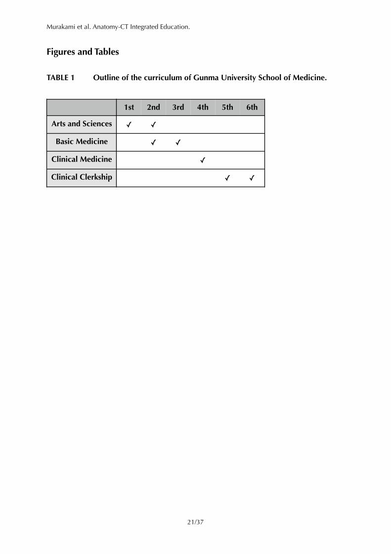
The curriculum at Gunma University School of Medicine includes one and a half years
of arts and sciences, one and a half years of basic medical sciences, and 3 years of
clinical medicine and clerkship (TABLE 1). The gross anatomy course is taught for
second-year students during the second semester (the academic year consists of two
semesters).
!��� /���6 37
Murakami et al. Anatomy-CT Integrated Education.
The gross anatomy course spans over 14 weeks, and consists of 6 hours of systems
lectures (neural, motor, and cardiovascular systems), 7 hours of regional anatomy
lectures, 3 hours of radiology lectures (FIGURE 1D), 10 hours of embryology lectures,
11 hours of osteology labs, and 162 hours of dissection labs (these numbers are for
Anatomy Class 2011). Students learn dissection using the textbook “Laboratory Manual
of Dissection” (Terada and Fujita, 2004) in teams of 4 persons (3 for surplus) (FIGURE
1C). The teams were made up by students in their own way. Students were also required to
take a 2-hour DICOM viewer workshop using DICOM software on their own computers
(FIGURE 1E). In the radiology lectures and workshop, students learn the Hounsfield
units, sectional anatomy of the thorax and abdomen, pulmonary and hepatic segmental
anatomy, DICOM operations (queries and retrieval), and 3D reconstructions. Class
guides, notes, and PDF copies of handouts were distributed and archived at a weblog
site <http://anatomy.med.gunma-u.ac.jp/> . 1
DICOM systems
Students were rented an iPod touch (one par person for Class of 2010 and 2011)
(FIGURE 1A). For the Class of 2009, students were advised to use their own laptops for
viewing CT images in the lab because we could not purchase iPod touches for the
class. Apps installed on the iPod touch include OsiriX for iOS devices (iPhone, iPod
touch, and iPad), GoodReader (PDF viewer), some reference apps, and utility apps.
OsiriX was pre-loaded with CT data of the cadaver assigned to the team, and those of
representative healthy living individuals of different ages and genders.
!��� /���7 37
��� Note for editors and reviewers: This address is valid since late May, 2013. The former address 1
is <http://anatomy.dept.med.gunma-u.ac.jp/>.
Murakami et al. Anatomy-CT Integrated Education.
Two iMac computers running the Mac version of OsiriX with 64-bit extension (Pixmeo,
Bernex, Switzerland) were loaded with all the cadaver scans as well as sample datasets
of different modalities (CT, MRI, PET, ultrasonography, and X-ray). The students
voluntarily used the iMac at the lab for MPR and 3D renderings (OsiriX for iOS lacks
these capabilities) (FIGURE 1B).
Students were advised to install OsiriX on their own Macs for assignments (discussed
below). Those with Windows computers were directed to use another freeware DICOM
viewer, INTAGE Realia (Cybernet, Tokyo, Japan; the product has been discontinued) as
an alternative to OsiriX despite its limited 2D and networking features. Students were
provided with DVDs containing CT images in DICOM format of all the cadavers for the
class, and of healthy living individuals.
All the DICOM datasets were archived on a PACS server, Apple Xserve with dcm4chee
(dcm4chee.org, 2013) (open-source PACS software), for local queries and retrieval. The
anatomy lab was equipped with an IEEE802.11b/g/n WiFi network for DICOM transfers.
The 3D reconstructions for the lectures and the exams were made with OsiriX and
HDVR plug-in (Fovia, Palo Alto, CA) on a Mac Pro with 2 × 6-core processors.
Assignments designed for integration of anatomy and CT
To facilitate integration of CT radiology to anatomy dissection, we gave the students
study assignments of two short papers, one each for the thorax and the abdomen. The
students were provided with the certificate of death, where the cause of death is
summarized, and a radiology report of the cadaver’s postmortem CT. Prior to actual
dissection, they were advised to learn interpretations of the respective regions in the
!��� /���8 37
Murakami et al. Anatomy-CT Integrated Education.
report, and to possibly add their own findings by examining the CT images for
themselves. The findings may include effusions, inflammation, tumors, injuries,
postoperative changes, artifactual materials, postmortem changes, personal differences,
and “no significant pathologies.” The students were instructed to verify such
radiological interpretations on the actual cadaver during dissection consulting the
cadaver’s CT images on the iPod touches, lab iMacs, or their own laptops.
For each of the thoracic and abdominal regions, each student should compile a two-
page paper of anatomical verifications of the radiological findings comparing CT
images and sketches of the corresponding structures of the cadaver (photography in the
lab was prohibited for students to avoid possible privacy problems). The aim of the
paper was provisionally set to provide the radiologists who wrote the radiology reports
with materials to self-evaluate their interpretations. This way, the students focused on
their own observations and not simply on reproducing the radiology reports.
Exams
The anatomy course was divided into three periods, and an exam was given at the end
of each period. The exam featured written tests (short essays and multiple-choice
questions) and practical tests. The students were divided into two groups at random.
Each group took either written or practical test in the first 90 minutes, and took the
other test in a subsequent 90 minutes. For the practical tests, students were to move
from one question station to another (50 stations per exam) at timed intervals (90 s) and
write answers using anatomical terms on an answer sheet (FIGURE 1F). The questions
were made from dissected cadavers, photographs of dissected cadavers, and images of
!��� /���9 37
Murakami et al. Anatomy-CT Integrated Education.
different modalities (~10 of 50 stations), i.e., X-ray, ultrasonography, CT, and MRI (slices
and 3D) (FIGURE 2).
Surveys and analyses
The exam scores of Anatomy Classes 2008 – 2011 were analyzed according to the
types of questions (written tests, radiological questions in practical tests, and non-
radiological questions in practical tests). Class 2008 was included as a pre-project
control. In the second year, students take neuroanatomy, histology, general physiology,
and neurophysiology as well as gross anatomy. The scores of these basic sciences were
also analyzed to standardize yearly differences in students’ academic abilities.
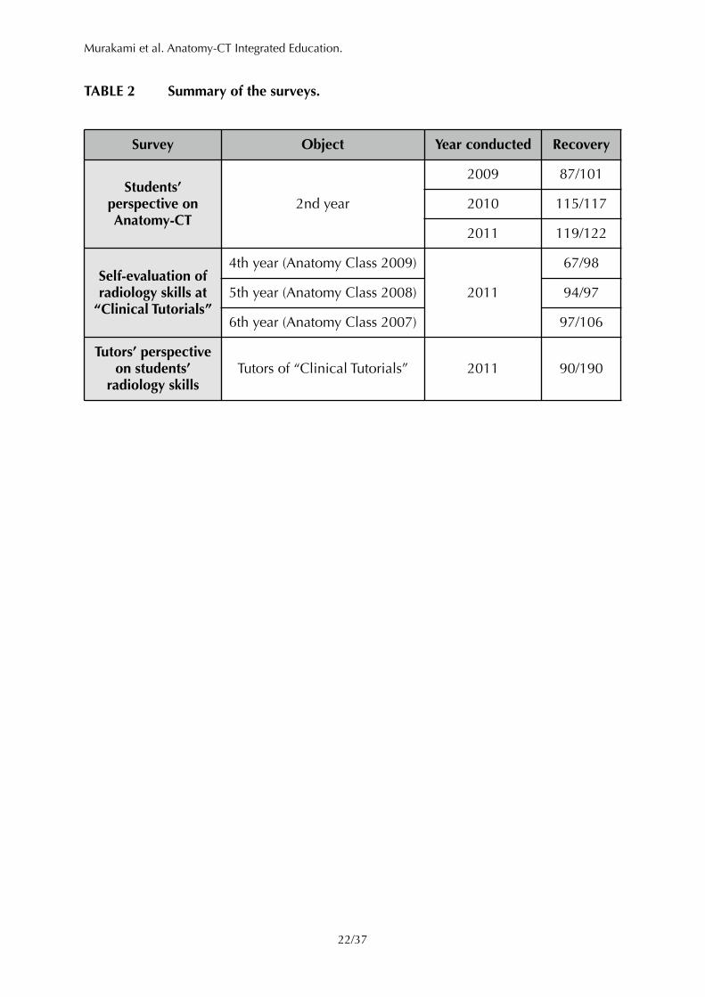
Questionnaires of semantic differential scales were taken at the end of the anatomy
course of Classes 2009 – 2011 to survey the students’ perspectives on the Anatomy-CT
project (TABLE 2).
In the fourth year, students take small-team (5 – 6 students), case-based learning of
clinical medicine (Clinical Tutorials). To evaluate the effects of our Anatomy-CT project
on the clinical classes, we surveyed the fourth (Anatomy Class 2009), fifth (2008, pre-
project), and sixth (2007, pre-project) year students at the end of academic year 2011,
asking self-evaluation of their abilities in diagnostic radiology at Clinical Tutorials
(TABLE 2). Thus, for the fifth and sixth year students, the survey was retrospective back
to 2010 and 2009, respectively. We also surveyed the tutors of the Clinical Tutorials for
radiological abilities of Anatomy Class 2009 compared to those of 2008 (TABLE 2).
Statistical data were analyzed with Microsoft Excel and the Statcel3 add-in (Yanai,
2011).
!��� /���10 37
Murakami et al. Anatomy-CT Integrated Education.
The project was assessed at the end of each academic year by a committee consisting
of five professors of Gunma University of basic and clinical medicine, and four
independent anatomy and radiology professionals.
!
!��� /���11 37
Murakami et al. Anatomy-CT Integrated Education.
Results and Discussions
Participants to the study
The numbers of students enrolled in the Anatomy Class were 102, 101, 117, and 122 in
the academic years 2008, 2009, 2010, and 2011, respectively (the changes in numbers
were due to changes in complements and a few holdovers). In the Japanese educational
system, high school graduates and prospective graduates are eligible to apply for
medical schools, and no previous degrees are required, although Gunma University
School of Medicine takes 15 students with previous experience for each class.
Assignments
Students used CT images on iPod touches at the bench and for the dissections and the
assignments (FIGURE 1C). When they found issues — pathologies, anomalies, distinct
anatomy, etc. — on the cadaver, CT, or radiology report, they tried to identify and
discuss these on both the cadaver and the CT images (examples shown in FIGURE 3).
The papers of assignments were compiled into a book as a reference for the radiologists
who examined the postmortem CT images.
Some pitfalls were indicated in the reports. There were different degrees of postmortem
or near-death changes in most cadavers, such as pulmonary edema, pleural effusion,
ascites, hypostasis, and ectopic gas in the vessels. Many of these might be insignificant
but tend to attract students’ attention. Also, artifacts occurred in the process of fixation
after the CT scan. This produces significant discrepancies between the CT images and
the cadaver anatomy, which might distract students’ interpretations. Pulmonary
!��� /���12 37
Murakami et al. Anatomy-CT Integrated Education.
infiltration of fixative, for example, often overlays preexistent edema and pneumonia
and makes it difficult to match pathologies in the cadaver and CT images.
Academic performance
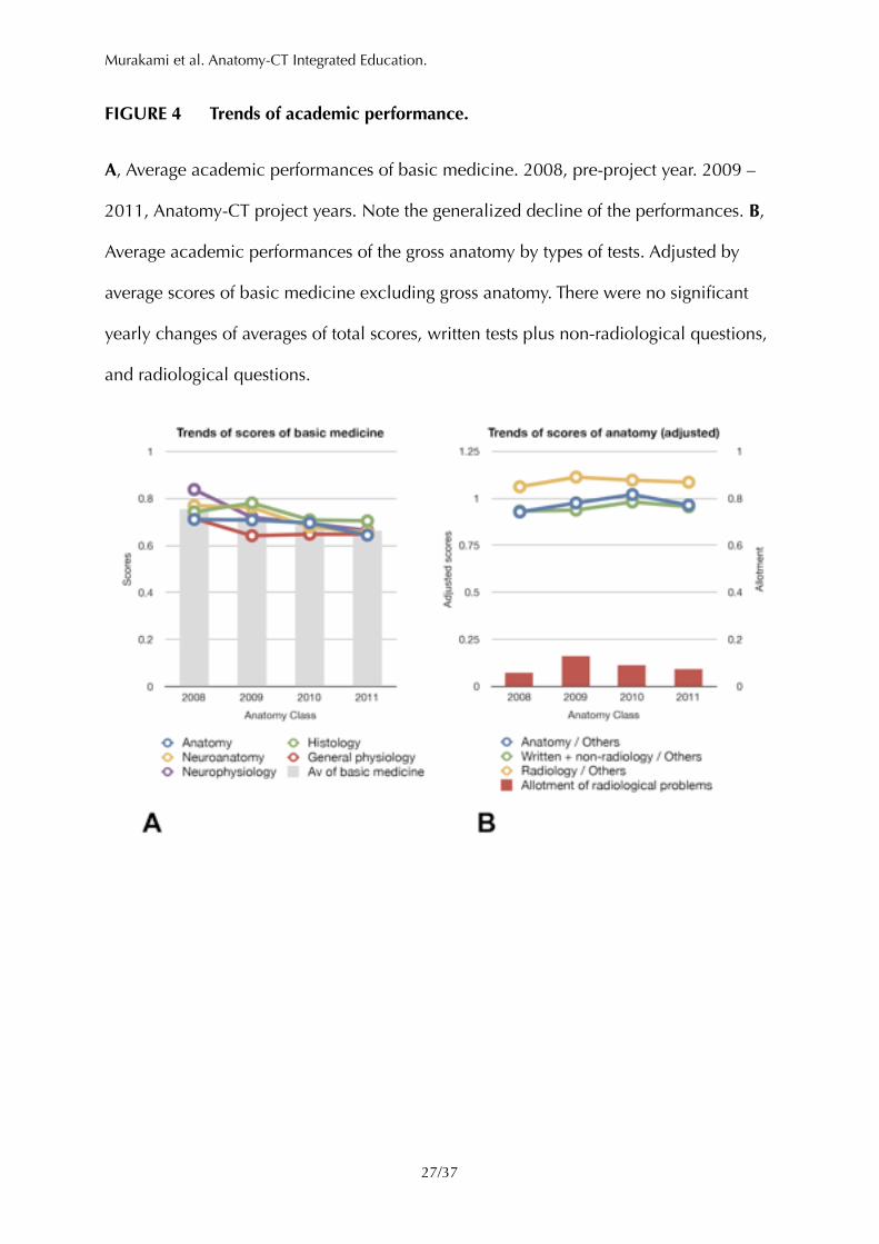
There have been significant yearly decreases in academic performance since Anatomy
Class 2008 not only in gross anatomy but also in other basic sciences, including
neuroanatomy, histology, general physiology, and neurophysiology (FIGURE 4A). This
trend causes issues in other medical schools in Japan, and was discussed at the
Association of Japanese Medical Colleges (Association of Japanese Medical Colleges,
2012). Thus, to evaluate the effects of our Anatomy-CT project on academic
performances in anatomy and radiology, the scores of gross anatomy were standardized
by the average score of all basic sciences except gross anatomy. With such
standardization, there were no significant yearly alterations since 2008 in the total
scores of gross anatomy, and the sum of written tests and non-radiological questions of
practical tests (FIGURE 4B).
In contrast to our expectations, there were no significant increases in the performances
of the radiological questions on the practical tests (FIGURE 4B). This may be partly
attributed to the exams themselves. The questions were mostly different every year, and
radiological questions in Anatomy Class 2008 were rather simpler than those in Class
2009 – 2011. The questions in 2008 were basically identification of structures in CT
slices and 3D reconstructions, while those in 2009 – 2011 required more advanced
knowledge and skills in radiology, such as window level/width settings, segmental
anatomy of the lung and the liver, and different radiology modalities (X-ray,
ultrasonography, MRI, and CT).
!��� /���13 37
Murakami et al. Anatomy-CT Integrated Education.
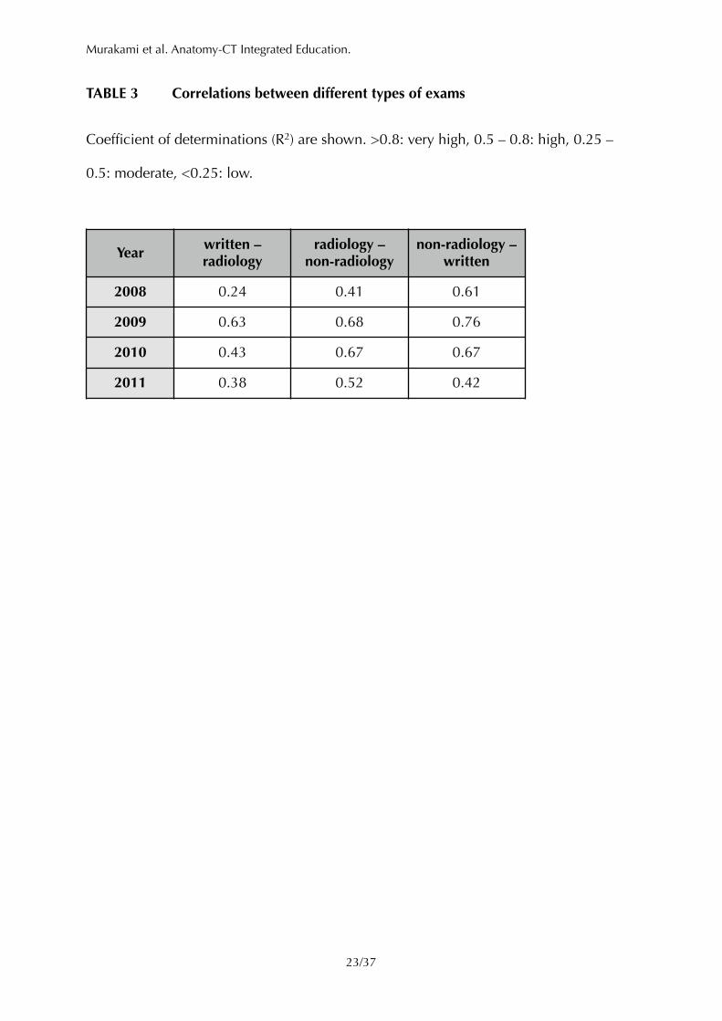
To evaluate the state of integration of radiology to anatomy education, correlations
were analyzed between scores of radiological and non-radiological questions of
practical tests, and written tests (TABLE 3). Anatomy Class 2008 showed high
correlations (R2 = 0.61) between written exams and non-radiological questions, but
low to moderate correlations (0.24 and 0.41) between radiological and other types of
questions. In contrast, Class 2009 showed high correlations (0.62 – 0.63) between
radiological questions and other types of questions, suggesting that radiology was
successfully integrated into traditional anatomical studies. Classes 2010 and 2011 also
showed moderate to high correlations between radiological questions and other types
of questions, although the values (0.38 – 0.67) were somewhat lower than those of
Class 2009. This might be because of qualitative variations of students’ abilities as
mentioned above (FIGURE 4A).
Students’ perspectives about the project
The surveys performed at the end of the anatomy classes showed very positive student
perspectives regarding our project (TABLE 2 and FIGURE 5). Anatomy Classes of 2010
and 2011 were combined for the summary here, although Class 2009 was omitted
because iPod touches were not available for the class. About 70% of students thought
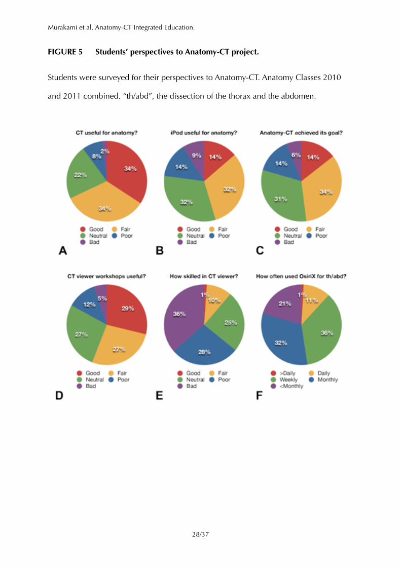
that CT radiology was useful for better understanding of anatomy (FIGURE 5A), ~46%
thought the iPod touch with OsiriX was useful for dissection (5B), and ~48% thought
Anatomy-CT achieved its goal (5C). Although ~56% of students thought the CT software
workshops were useful (5D), only ~14% gave positive self-evaluations about their
proficiency in CT radiology (5E), suggesting some room for improvement in the
workshops. There seemed to be some discrepancy between students’ perspective and
their actual usage of the iPod touch (5B and F). This might be because a team often
!��� /���14 37
Murakami et al. Anatomy-CT Integrated Education.
shared the iPod touch of a spontaneous leader of discussions, and we had anatomy labs
2 – 4 days a week, which fit the choice of “weekly” in the questionnaire.
In summary, the concept of the Anatomy-CT project was well understood and
appreciated by the students, although there is some room for further development to
improve students’ radiological accomplishments, consistent with the academic
performances discussed above.
The positive comments to the question for appreciations in Anatomy-CT in Classes
2009–2011 included:
• Good understanding of 3D structures of the human body.
• Early learning CT radiology.
• Early exposure to clinical medicine through radiology.
• Practical usage of CT data using DICOM viewers.
• Experience of problem-based learning through the assignments.
The negative responses included:
• The iPod touch screen is too small for CT viewing.
• Too few lectures and labs.
• Too few CT software workshops.
• Too little guidance regarding iPod touch usage.
By adjusting schedules and contents for lectures, workshops, and labs, negative
comments on lectures and workshops disappeared by Class 2011. To address the screen
issue, we introduced 10-inch iPads for Class 2012 for future evaluations.
!��� /���15 37
Murakami et al. Anatomy-CT Integrated Education.
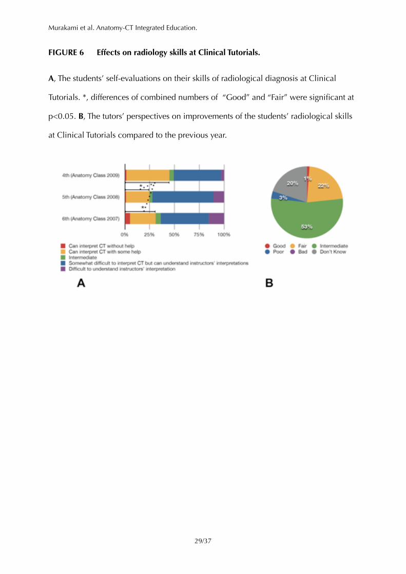
Effects on clinical classes
In the academic year 2011, we conducted another survey to the fourth to sixth year
students for effects of Anatomy-CT to clinical classes in the fourth year (TABLE 2 and
FIGURE 6A). The questionnaire asked the students’ skills in CT radiology at their
“Clinical Tutorials”. The Clinical Tutorials are small-team, case-based learning of clinical
medicine, where students are provided clinical cases often with radiological data. The
fourth year students in 2011 were the first to take Anatomy-CT as Anatomy Class 2009.
The fourth year students gave significantly better self-estimation than the fifth or sixth
year students (Anatomy Classes 2008 and 2007, respectively). Although the survey was
retrospective for the fifth and the sixth year students, this suggested that Anatomy-CT
improved radiology skills in clinical classes.
We also surveyed teachers (“tutors”) of Clinical Tutorials in 2011, asking if there were
any improvements in students’ radiological skills compared to the previous year (TABLE
2 and FIGURE 6B). Although three quarters of the responses were neutral or
inconclusive, one quarter were positive and only a few were negative, again suggesting
improvement of radiological skills of students who took Anatomy-CT.
Future Plans
The Anatomy-CT project concluded with very positive student perspectives. Although
there were no immediate effects in academic performance in the gross anatomy classes,
prospective improvements of radiology skills were indicated in clinical classes. As we
had requests from the students for larger screens, we introduced the iPad, one per team,
for Anatomy Class 2012 as the successor to the iPod touch. Preliminarily, we observed
!��� /���16 37
Murakami et al. Anatomy-CT Integrated Education.
positive perspectives on the use of iPad (with requests for more iPads), increased
discussions within teams using CT images on the iPad screen with decreased demands
for the lab iMacs. We will continue the project with the iPad and assess the effects of
the Anatomy-CT project. We hope our trials will contribute to fulfillment of anatomical
and radiological education.
!
!��� /���17 37
Murakami et al. Anatomy-CT Integrated Education.
Acknowledgments
The authors are grateful to Mr. Jiro Kayama, Mr. Hiroyuki Takei, Mr. Yoshihiro Morimura
and Mr. Mitsuaki Shikata at Gunma University for their support for postmortem
imaging. This study was supported by a Grant-in-Aid of “Program for Promoting
University Education and Student Support Theme A: Program for Promoting University
Education Reform, 2009–2011” awarded to Gunma University from MEXT, Japan
(A13006) , and approved by Gunma University Ethics Committee.
!
!��� /���18 37
Murakami et al. Anatomy-CT Integrated Education.
Notes on Contributors
Tohru Murakami, M.D., Ph.D. is an associate professor of anatomy, and a researcher of
developmental biology at Gunma University (GU). He is responsible to the
system designs of Anatomy-CT.
Yuki Tajika, Ph.D. is an assistant professor of anatomy, and a researcher of cell biology
at GU. He has taught anatomy and cell biology since 2005.
Hitoshi Ueno, Ph.D. is an assistant professor of anatomy at GU. He has taught anatomy
and cell biology since 2010. His research interests are intracellular transport,
cell differentiation, and development.
Sachiko Awata, M.D. is a radiologist at GU since 2008. She is involved in postmortem
image interpretations of Anatomy-CT project.
Satoshi Hirasawa, M.D., Ph.D. is a research associate of radiology at GU. He has been
involved in postmortem image interpretations since 2008.
Maki Sugimoto, M.D., Ph.D. is a professor of medicine at Kobe University. He is
involved in projects of advanced medicine as 3D printing of human organ
models and OsiriX-based surgical technologies, and has contributed to
Anatomy-CT project.
Yoshihiko Kominato, M.D., Ph.D. is a professer of legal medicine at GU since 2006. He
is responsible for the Postmortem Imaging Facility.
!��� /���19 37
Murakami et al. Anatomy-CT Integrated Education.
Yoshito Tsushima, M.D., Ph.D. is a professor of radiology. He has taught diagnostic
imaging since 2011 at GU.
Keigo Endo, M.D., Ph.D. was a professor of radiology at GU, and is the president of
Kyoto College of Medical Science. He has taught diagnostic imaging from 1991
to 2010 at GU.
Hiroshi Yorifuji, M.D., Ph.D. is a professor of anatomy at GU since 2002. He is
responsible to the general management of Anatomy-CT project.
!
!��� /���20 37
Murakami et al. Anatomy-CT Integrated Education.
Figures and Tables
TABLE 1 Outline of the curriculum of Gunma University School of Medicine.
!
1st 2nd 3rd 4th 5th 6th
Arts and Sciences ✓ ✓
Basic Medicine ✓ ✓
Clinical Medicine ✓
Clinical Clerkship ✓ ✓
!��� /���21 37
Murakami et al. Anatomy-CT Integrated Education.
TABLE 2 Summary of the surveys.
!
Survey Object Year conducted Recovery
Students’ perspective on Anatomy-CT
2nd year
2009 87/101
2010 115/117
2011 119/122
Self-evaluation of radiology skills at
“Clinical Tutorials”
4th year (Anatomy Class 2009)
2011
67/98
5th year (Anatomy Class 2008) 94/97
6th year (Anatomy Class 2007) 97/106
Tutors’ perspective on students’
radiology skillsTutors of “Clinical Tutorials” 2011 90/190
!��� /���22 37
Murakami et al. Anatomy-CT Integrated Education.
TABLE 3 Correlations between different types of exams
Coefficient of determinations (R2) are shown. >0.8: very high, 0.5 – 0.8: high, 0.25 –
0.5: moderate, <0.25: low.
!
Year written – radiology
radiology – non-radiology
non-radiology – written
2008 0.24 0.41 0.61
2009 0.63 0.68 0.76
2010 0.43 0.67 0.67
2011 0.38 0.52 0.42
!��� /���23 37
Murakami et al. Anatomy-CT Integrated Education.
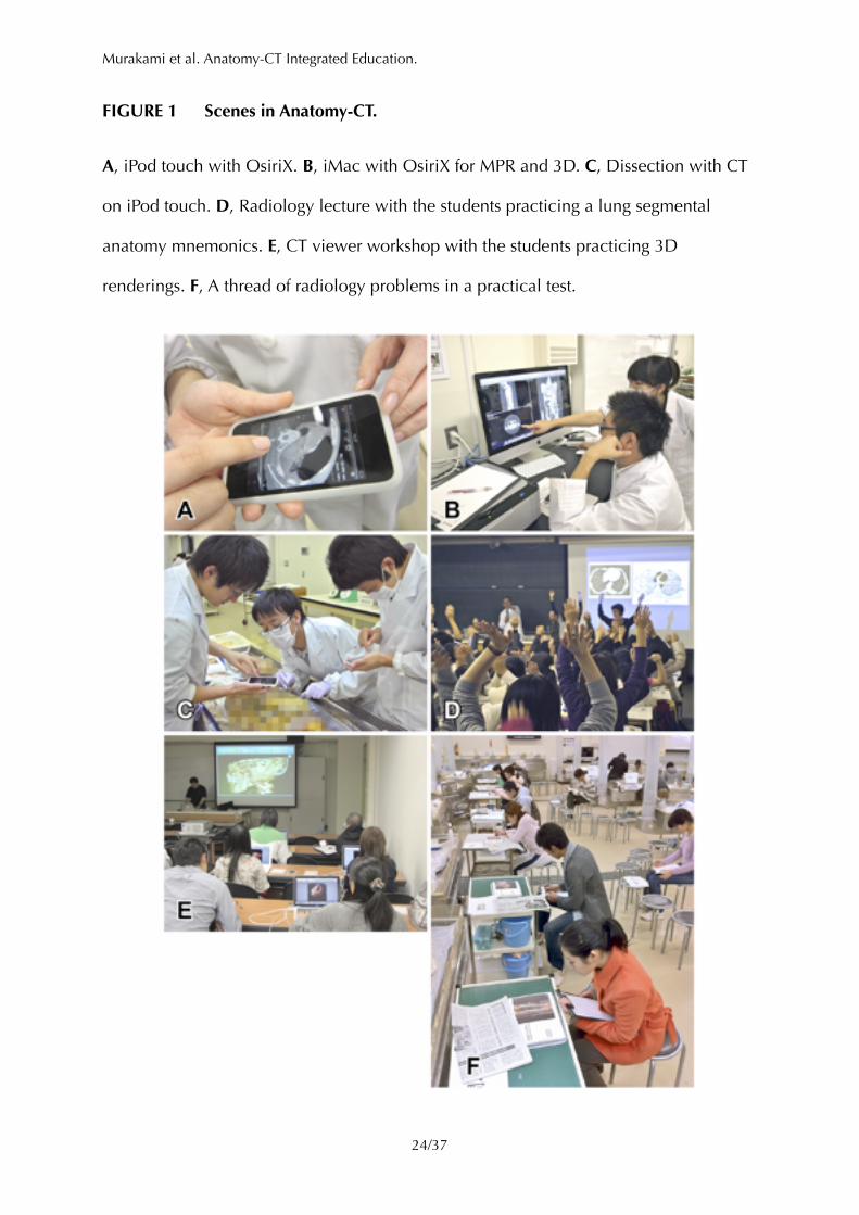
FIGURE 1 Scenes in Anatomy-CT.
A, iPod touch with OsiriX. B, iMac with OsiriX for MPR and 3D. C, Dissection with CT
on iPod touch. D, Radiology lecture with the students practicing a lung segmental
anatomy mnemonics. E, CT viewer workshop with the students practicing 3D
renderings. F, A thread of radiology problems in a practical test.
!
!
!��� /���24 37
Murakami et al. Anatomy-CT Integrated Education.
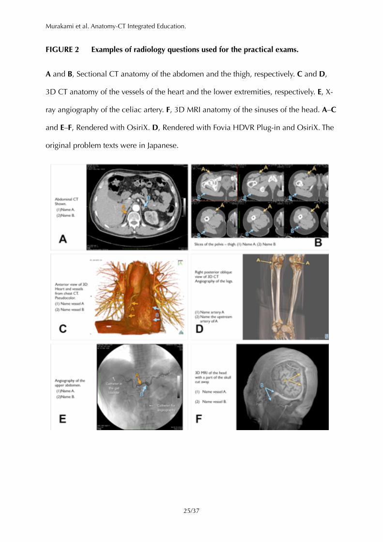
FIGURE 2 Examples of radiology questions used for the practical exams.
A and B, Sectional CT anatomy of the abdomen and the thigh, respectively. C and D,
3D CT anatomy of the vessels of the heart and the lower extremities, respectively. E, X-
ray angiography of the celiac artery. F, 3D MRI anatomy of the sinuses of the head. A–C
and E–F, Rendered with OsiriX. D, Rendered with Fovia HDVR Plug-in and OsiriX. The
original problem texts were in Japanese.
!
!��� /���25 37
Murakami et al. Anatomy-CT Integrated Education.
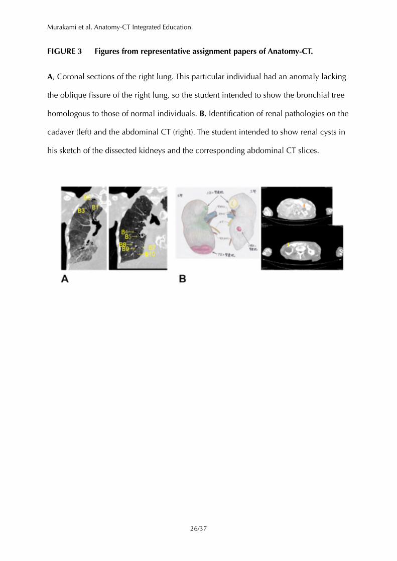
FIGURE 3 Figures from representative assignment papers of Anatomy-CT.
A, Coronal sections of the right lung. This particular individual had an anomaly lacking
the oblique fissure of the right lung, so the student intended to show the bronchial tree
homologous to those of normal individuals. B, Identification of renal pathologies on the
cadaver (left) and the abdominal CT (right). The student intended to show renal cysts in
his sketch of the dissected kidneys and the corresponding abdominal CT slices.
!
!��� /���26 37
Murakami et al. Anatomy-CT Integrated Education.
FIGURE 4 Trends of academic performance.
A, Average academic performances of basic medicine. 2008, pre-project year. 2009 –
2011, Anatomy-CT project years. Note the generalized decline of the performances. B,
Average academic performances of the gross anatomy by types of tests. Adjusted by
average scores of basic medicine excluding gross anatomy. There were no significant
yearly changes of averages of total scores, written tests plus non-radiological questions,
and radiological questions.
!
!��� /���27 37
Murakami et al. Anatomy-CT Integrated Education.
FIGURE 5 Students’ perspectives to Anatomy-CT project.
Students were surveyed for their perspectives to Anatomy-CT. Anatomy Classes 2010
and 2011 combined. “th/abd”, the dissection of the thorax and the abdomen.
!
!��� /���28 37
Murakami et al. Anatomy-CT Integrated Education.
FIGURE 6 Effects on radiology skills at Clinical Tutorials.
A, The students’ self-evaluations on their skills of radiological diagnosis at Clinical
Tutorials. *, differences of combined numbers of “Good” and “Fair” were significant at
p<0.05. B, The tutors’ perspectives on improvements of the students’ radiological skills
at Clinical Tutorials compared to the previous year.
!
!��� /���29 37
Murakami et al. Anatomy-CT Integrated Education.
Literatures cited
Alberti C. 2011. Organ-confined prostate carcinoma radiation brachytherapy compared
with external either photon- or hadron-beam radiation therapy. Just a short up-
to-date. Eur Rev Med Pharmacol Sci 15:769–774.
Association of Japanese Medical Colleges. 2012. Surveys of the decrease of academic
performances of medical students in Japan (excerpt). ajmcuminjp. http://
www.ajmc.umin.jp/m-kaiken.html.
Auyang ED, Santos BF, Enter DH, Hungness ES, Soper NJ. 2011. Natural orifice
translumenal endoscopic surgery (NOTES®): a technical review. Surg Endosc
25:3135–3148.
Awata S, Endo K. 2009. One year experience of autopsy imaging center of Gunma
University. Igaku no Ayumi 231:902–905.
Bohl M, Francois W, Gest T. 2011. Self-guided clinical cases for medical students based
on postmortem CT scans of cadavers. Clin Anat 24:655–663.
Brown PM, Hamilton NM, Denison AR. 2012. A novel 3D stereoscopic anatomy
tutorial. Clin Teach 9:50–53.
Cabrera AR, Lee WR, Madden R, Sims E, Hoang JK, White LE, Marks LB, Chino JP.
2011. Incorporating gross anatomy education into radiation oncology residency:
a 2-year curriculum with evaluation of resident satisfaction. J Am Coll Radiol
8:335–340.
!��� /���30 37
Murakami et al. Anatomy-CT Integrated Education.
Chen S-I, Wang S-C, Hsiao H-T, Lee L-C, Lee M-H, Yao C-M. 2012. A pre-operation
planning system and biomechanical evaluation of the cranial flap. In:. 2012 IEEE
Symposium on Robotics and Applications (ISRA). IEEE, pp. 714–716.
Choudhri AF, Radvany MG. 2010. Initial Experience with a Handheld Device Digital
Imaging and Communications in Medicine Viewer: OsiriX Mobile on the
iPhone. J Digit Imaging 24:184–189.
de Notaris M, Prats-Galino A, Cavallo L, Esposito F, Iaconetta G, Gonzalez J,
Montagnani S, Ferrer E, Cappabianca P. 2010. Preliminary experience with a
new three-dimensional computer-based model for the study and the analysis of
skull base approaches. Childs Nerv Syst 26:621–626.
Erkonen WE, Albanese MA, Smith WL, Pantazis NJ. 1990. Gross Anatomy Instruction
with Diagnostic Images. Investigative Radiology 25:292.
Erkonen WE, Albanese MA, Smith WL, Pantazis NJ. 1992. Effectiveness of Teaching
Radiologic Image Interpretation in Gross Anatomy: A Long-Term Follow-up.
Investigative Radiology 27:264.
Fabian R, Tengg-Kobligk von H, Zechmann C, Kauczor H-U, Giesel FL. 2008. Beyond
the Eye – Medical Applications of 3D Rapid Prototyping Objects. European
Medical Imaging Review. http://www.touchbriefings.com/pdf/3225/giesel.pdf.
!��� /���31 37
Murakami et al. Anatomy-CT Integrated Education.
Fang CH, Xie AW, Chen ML, Huang YP, Lu CM, Li XF, Pan JH, Peng FP. 2010.
Application of a visible simulation surgery technique in preoperation planning
for intrahepatic calculi. World J Surg 34:327–335.
Griksaitis MJ, Sawdon MA, Finn GM. 2012. Ultrasound and cadaveric prosections as
methods for teaching cardiac anatomy: a comparative study. Anat Sci Educ 5:20–
26.
Hisley KC, Anderson LD, Smith SE, Kavic SM, Tracy JK. 2007. Coupled physical and
digital cadaver dissection followed by a visual test protocol provides insights into
the nature of anatomical knowledge and its evaluation. Anat Sci Ed 1:27–40.
Hopkins R, Regehr G, Wilson TD. 2011. Exploring the changing learning environment
of the gross anatomy lab. Acad Med 86:883–888.
Irwin BH, Rao PP, Stein MJ, Desai RM. 2010. New Advances in Urologic
Laparoendoscopic Single Site (LESS) Surgery. In:. linkspringercom. London:
Springer London, pp. 197–208.
Kaouk JH, Goel RK, Haber G-P, Crouzet S, Stein RJ. 2009. Robotic single-port
transumbilical surgery in humans: initial report. BJU Int 103:366–369.
Knobe M, Carow JB, Ruesseler M, Leu BM, Simon M, Beckers SK, Ghassemi A, Sönmez
TT, Pape H-C. 2012. Arthroscopy or ultrasound in undergraduate anatomy
education: a randomized cross-over controlled trial. BMC Med Educ 12:85.
!��� /���32 37
Murakami et al. Anatomy-CT Integrated Education.
Kotzé SH, Mole CG, Greyling LM. 2012. The translucent cadaver: an evaluation of the
use of full body digital X-ray images and drawings in surface anatomy education.
Anat Sci Educ 5:287–294.
Kroh M, El-Hayek K, Rosenblatt S, Chand B, Escobar P, Kaouk J, Chalikonda S. 2011.
First human surgery with a novel single-port robotic system: cholecystectomy
using the da Vinci Single-Site platform. Surg Endosc 25:3566–3573.
Lanier L, Kaude JV. 1993. Radiologic Anatomy - A Credit Course for First-Year Medical
Students. Acta Radiologica 34:414–416.
Lufler RS, Zumwalt AC, Romney CA, Hoagland TM. 2010. Incorporating radiology into
medical gross anatomy: does the use of cadaver CT scans improve students'
academic performance in anatomy? Anat Sci Educ 3:56–63.
Machado JAD, Barbosa JMP, Ferreira MAD. 2013. Student perspectives of imaging
anatomy in undergraduate medical education. Anat Sci Educ 6:163–169.
Mayo Clinic. 2009. Hi Def CT Scanner | Mayo Clinic Podcasts. podcastsmayoclinicorg.
http://podcasts.mayoclinic.org/2009/04/24/hi-def-ct-scanner/.
Mazeron R, Gilmore J, Dumas I, Champoudry J, Goulart J, Vanneste B, Tailleur A,
Morice P, Haie-Meder C. 2013. Adaptive 3D Image-Guided Brachytherapy: A
Strong Argument in the Debate on Systematic Radical Hysterectomy for Locally
Advanced Cervical Cancer. Oncologist.
!��� /���33 37
Murakami et al. Anatomy-CT Integrated Education.
McNiesh LM, Madewell JE, Allman RM. 1983. Cadaver radiography in the teaching of
gross anatomy. Radiology 148:73–74.
Melissano G, Bertoglio L, Civelli V, Moraes Amato AC, Coppi G, Civilini E, Calori G, De
Cobelli F, Del Maschio A, Chiesa R. 2009. Demonstration of the Adamkiewicz
Artery by Multidetector Computed Tomography Angiography Analysed with the
Open-Source Software OsiriX. European Journal of Vascular and Endovascular
Surgery 37:395–400.
Osawa J. 2013. 3-D Printing Is Ready for Surgery. The Wall Street Journal. http://
online.wsj.com/article/
SB10001424127887324504704578410764264855512.html.
Petersson H, Sinkvist D, Wang C, Smedby O. 2009. Web-based interactive 3D
visualization as a tool for improved anatomy learning. Anat Sci Educ 2:61–68.
Rengier F, Doll S, Tengg-Kobligk von H, Kirsch J, Kauczor H-U, Giesel FL. 2009.
Integrated teaching of anatomy and radiology using three-dimensional image
post-processing. Eur Radiol 19:2870–2877.
Rosset A, Spadola L, Ratib O. 2004. OsiriX: An Open-Source Software for Navigating in
Multidimensional DICOM Images. J Digit Imaging 17:205–216.
Sano R, Hirawasa S, Kobayashi S, Shimada T, Awata S, Takei H, Otake H, Takahashi K,
Takahashi Y, Kominato Y. 2011. Use of postmortem computed tomography to
!��� /���34 37
Murakami et al. Anatomy-CT Integrated Education.
reveal an intraoral gunshot injuries in a charred body. Leg Med (Tokyo) 13:286–
288.
Sodergren M, Clark J, Beardsley J, Bryant T, Horton K, Darzi A, Teare J. 2011. A novel
flexible endoluminal stapling device for use in NOTES colotomy closure: a
feasibility study using an ex vivo porcine model. Surg Endosc 25:3266–3272.
Sugimoto M, Tanaka K, Matsuoka Y, Man-i M, Morita Y, Tanaka S, Fujiwara S, Azuma T.
2011. da Vinci robotic single-incision cholecystectomy and hepatectomy using
single-channel GelPort access. J Hepatobiliary Pancreat Sci 18:493–498.
Sugimoto M, Yasuda H, Koda K, Suzuki M, Yamazaki M, Tezuka T, Kosugi C, Higuchi R,
Watayo Y, Yagawa Y, Uemura S, Tsuchiya H, Azuma T. 2010a. Carbon dioxide-
enhanced virtual MDCT cholangiopancreatography. J Hepatobiliary Pancreat Sci
17:601–610.
Sugimoto M, Yasuda H, Koda K, Suzuki M, Yamazaki M, Tezuka T, Kosugi C, Higuchi R,
Watayo Y, Yagawa Y, Uemura S, Tsuchiya H, Azuma T. 2010b. Image overlay
navigation by markerless surface registration in gastrointestinal, hepatobiliary
and pancreatic surgery. J Hepatobiliary Pancreat Sci 17:629–636.
Sugimoto M, Yasuda H, Koda K, Suzuki M, Yamazaki M, Tezuka T, Kosugi C, Higuchi R,
Watayo Y, Yagawa Y, Uemura S, Tsuchiya H, Hirano A, Ro S. 2009. Evaluation for
transvaginal and transgastric NOTES cholecystectomy in human and animal
natural orifice translumenal endoscopic surgery. Journal of hepato-biliary-
pancreatic surgery 16:255–260.
!��� /���35 37
Murakami et al. Anatomy-CT Integrated Education.
Tam M. 2010. Building virtual models by postprocessing radiology images: A guide for
anatomy faculty. Anat Sci Educ 3:261–266.
Tang D, Yang C, Geva T, Del Nido PJ. 2008. Patient-specific MRI-based 3D FSI RV/LV/
patch models for pulmonary valve replacement surgery and patch optimization.
J Biomech Eng 130:041010.
Tang GL, Chin J, Kibbe MR. 2010. Advances in diagnostic imaging for peripheral
arterial disease. Expert Rev Cardiovasc Ther 8:1447–1455.
Terada H, Fujita T. 2004. Laboratoy Manual of Dissection 11 ed. Tokyo, Japan:
Nanzando 430 pp.
Trelease R, Rosset A. 2008. Transforming clinical imaging data for virtual reality
learning objects. Anat Sci Educ 1:50–55.
Volonté F, Pugin F, Bucher P, Sugimoto M, Ratib O, Morel P. 2011. Augmented reality
and image overlay navigation with OsiriX in laparoscopic and robotic surgery:
not only a matter of fashion. J Hepatobiliary Pancreat Sci 18:506–509.
Volonté F, Pugin F, Buchs NC, Spaltenstein J, Hagen M, Ratib O, Morel P. 2012.
Console-Integrated Stereoscopic OsiriX 3D Volume-Rendered Images for da
Vinci Colorectal Robotic Surgery. Surg Innov.
!��� /���36 37
Murakami et al. Anatomy-CT Integrated Education.
Vuchkova J, Maybury T, Farah C. 2011. Testing the educational potential of 3D
visualization software in oral radiographic interpretation. J Dent Educ 75:1417–
1425.
Wowra B, Muacevic A, Tonn J-C. 2012. CyberKnife radiosurgery for brain metastases.
Prog Neurol Surg 25:201–209.
Wowra B, Muacevic A, Zausinger S, Tonn J-C. 2009. Radiosurgery for spinal malignant
tumors. Dtsch Arztebl Int 106:106–112.
Yamauchi T, Yamazaki M, Okawa A, Furuya T, Hayashi K, Sakuma T, Takahashi H,
Yanagawa N, Koda M. 2010. Efficacy and reliability of highly functional open
source DICOM software (OsiriX) in spine surgery. Journal of Clinical
Neuroscience 17:756–759.
Yanai H. 2011. 4 Steps Excel Statistics, 3rd ed. Saitama, Japan: OMS Publishing 296 pp.
!��� /���37 37