Embed Size (px)
DESCRIPTION
mikrobiologi
Citation preview

24
The Salmonella Pathogenicity Island-1 and -2 Encoded Type III Secretion Systems
Amanda Wisner, Taseen Desin, Aaron White, Andrew Potter and Wolfgang Köster Vaccine and Infectious Disease Organization
University of Saskatchewan Canada
1. Introduction
1.1 General aspects
Salmonellae are motile, facultatively anaerobic, Gram-negative rods measuring 0.3-1.5 by 1.0-2.5 m in size. The genus Salmonella was named for Dr. Daniel Salmon, a veterinary bacteriologist at the United States Department of Agriculture (USDA) (Gast, 2003, Salyers & Whitt, 2002). The Salmonella species are closely related to Escherichia, Yersinia, and Shigella, and contain a circular chromosome approximately 4.7 Mbp in size with an overall GC content of 52% (Marcus, et al., 2000, Salyers & Whitt, 2002, Thomson, et al., 2008). The genus Salmonella lies within the kingdom Eubacteria, class Gammaproteobacteria, order Enterobacteriales, and family Enterobacteriaceae. Salmonella is divided into two species, Salmonella bongori and Salmonella enterica. Within Salmonella enterica there are 6 subspecies: salamae, arizonae, diarizonae, houtenae, indica, and enterica (Tindall, et al., 2005). These subspecies can be further classified into approximately 50 serogroups based on their lipopolysaccharide (LPS) O antigen component (Sabbagh, et al., 2010). Salmonella enterica
subspecies enterica finds its niche in warm-blooded animals and is the primary species associated with human infections. S. bongori and other S. enterica subspecies are more commonly associated with cold-blooded animals, and in some cases can cause disease in these animals (Brenner, et al., 2000).
Salmonella enterica subspecies enterica can be further divided into over 2500 serovars based on their flagellar (H) antigen and LPS O antigen structures (Brenner, et al., 2000, Coburn, et
al., 2007, Sabbagh, et al., 2010, Tindall, et al., 2005). For the purposes of this document, serovars within Salmonella enterica subspecies enterica (e.g., Enteritidis, Typhimurium, and Typhi) will be identified by an italicized S, followed by the serovar name (e.g., S. Enteritidis, S. Typhimurium, and S. Typhi). Many serovars are host-adapted, and tend to cause life-threatening systemic disease in their host. For example, S. Typhi and S. Paratyphi cause systemic disease in humans and some primates, while S. Gallinarum and S. Pullorum produce systemic disease in chickens, S. Dublin causes systemic disease in cattle, and S.
Choleraesuis in pigs. In contrast, many serovars are non host-adapted and tend to cause
www.intechopen.com

Salmonella – A Diversified Superbug
470
gastroenteritis in many different host species. S. Typhimurium and S. Enteritidis are the most well-known examples and are able to cause different disease outcomes in various host species (Barrow, 2007, Boyle, et al., 2007, Lax, et al., 1995, Spreng, et al., 2006, Zhang & Mosser, 2008). S. Typhimurium and S. Enteritidis are able to induce a systemic infection in mice, young calves, chicks, and piglets. However, they are also able to colonize poultry and adult cattle without symptoms (Barrow, 2007, Boyle, et al., 2007, Lax, et al., 1995, Spreng, et
al., 2006, Zhang & Mosser, 2008). In humans, infection with either of these serovars results in a self-limiting gastroenteritis (salmonellosis) involving fever, diarrhea, and abdominal pain. In rare cases, typically in the very young or immunocompromised, the infection can become systemic and lead to hospitalization and even death. A very small proportion of humans with salmonellosis can develop reactive arthritis (previously referred to as Reiter’s syndrome), which is initially characterized by joint pain, eye irritation, and pain during urination (Boyle, et al., 2007, Cogan & Humphrey, 2003, Townes, 2010).
1.2 Human disease, animal reservoirs, and modes of transmission
Infections by S. enterica are one of the most common causes of bacterial food-borne gastroenteritis (food poisoning) in the world, along with E. coli and Campylobacter infections
(WHO, 2007). Of the S. enterica serovars, S. Enteritidis and S. Typhimurium are the leading cause of salmonellosis in humans in most countries. S. Enteritidis and S. Typhimurium are passed to humans primarily via consumption of contaminated poultry meat, water, and eggs. S. Enteritidis is more often associated with salmonellosis acquired from eggs, as it has a greater tendency to colonize eggs and reproductive organs of poultry than S. Typhimurium (Gantois, et al., 2008). Because chickens mostly do not show symptoms of disease, entire flocks can become colonized quite quickly and shed bacteria in their feces for extended periods of time (Catarame, et al., 2005, Clavijo, et al., 2006, Penha Filho, et al., 2009, Van Immerseel, et al., 2005). Loss of consumer confidence in products because of Salmonella contamination can result in substantial economic loss to the poultry industry. Additionally, human cases of salmonellosis place a significant burden on the health care system (Boyle, et
al., 2007). There are approximately 1.4 million cases of salmonellosis per year resulting in about 15,000 hospitalizations and 400 deaths per year in the United States of America (USA) (USDA-ERS, 2009). Around 95% of these cases are caused by consumption of contaminated food products, and S. Enteritidis is responsible for at least 15% of these cases. S. Enteritidis is the second most commonly isolated serovar in North America after S. Typhimurium, while S. Enteritidis is number one in the European Union (EU) (Barrow, 2007, Callaway, et al., 2008, Cogan & Humphrey, 2003, Foley & Lynne, 2008, Vieira, 2009).
1.3 Virulence factors
1.3.1 Flagella
Flagella are complex motility structures found in members of Prokarya, Archaea, and Eukarya (Gophna, et al., 2003). The presence of flagella has been associated with virulence in many pathogens, including Salmonella, which usually expresses between five and ten flagella at random positions on the cell surface (Parker & Guard-Petter, 2001, van Asten & van Dijk, 2005). However, there is conflicting evidence for the contribution of flagella to
www.intechopen.com
The Salmonella Pathogenicity Island-1 and -2 Encoded Type III Secretion Systems
471
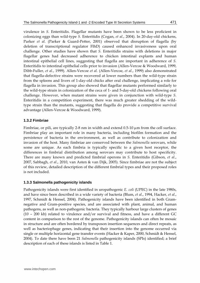
virulence in S. Enteritidis. Flagellar mutants have been shown to be less proficient in colonizing eggs than wild-type S. Enteritidis (Cogan, et al., 2004). In 20-day-old chickens, Parker et al. (Parker & Guard-Petter, 2001) observed that disruption of flagella (by deletion of transcriptional regulator FlhD) caused enhanced invasiveness upon oral challenge. Other studies have shown that S. Enteritidis strains with deletions in major flagellar genes had decreased adherence to chicken intestinal explants and human intestinal epithelial cell lines, suggesting that flagella are important in adherence of S. Enteritidis to intestinal epithelial cells prior to invasion (Allen-Vercoe & Woodward, 1999, Dibb-Fuller, et al., 1999). Allen-Vercoe et al. (Allen-Vercoe, et al., 1999) also demonstrated that flagella-defective strains were recovered at lower numbers than the wild-type strain from the spleens and livers of 1-day-old chicks after oral challenge, implicating a role for flagella in invasion. This group also showed that flagellar mutants performed similarly to the wild-type strain in colonization of the ceca of 1- and 5-day-old chickens following oral challenge. However, when mutant strains were given in conjunction with wild-type S. Enteritidis in a competition experiment, there was much greater shedding of the wild-type strain than the mutants, suggesting that flagella do provide a competitive survival advantage (Allen-Vercoe & Woodward, 1999).
1.3.2 Fimbriae
Fimbriae, or pili, are typically 2-8 nm in width and extend 0.5-10 m from the cell surface. Fimbriae play an important role in many bacteria, including biofilm formation and the persistence of bacteria in the environment, as well as contribute to colonization and invasion of the host. Many fimbriae are conserved between the Salmonella serovars, while some are unique. As each fimbria is typically specific to a given host receptor, the differences in fimbrial distribution among serovars may contribute to host specificity. There are many known and predicted fimbrial operons in S. Enteritidis (Gibson, et al., 2007, Sabbagh, et al., 2010, van Asten & van Dijk, 2005). Since fimbriae are not the subject of this review, detailed description of the different fimbrial types and their proposed roles is not included.
1.3.3 Salmonella pathogenicity islands
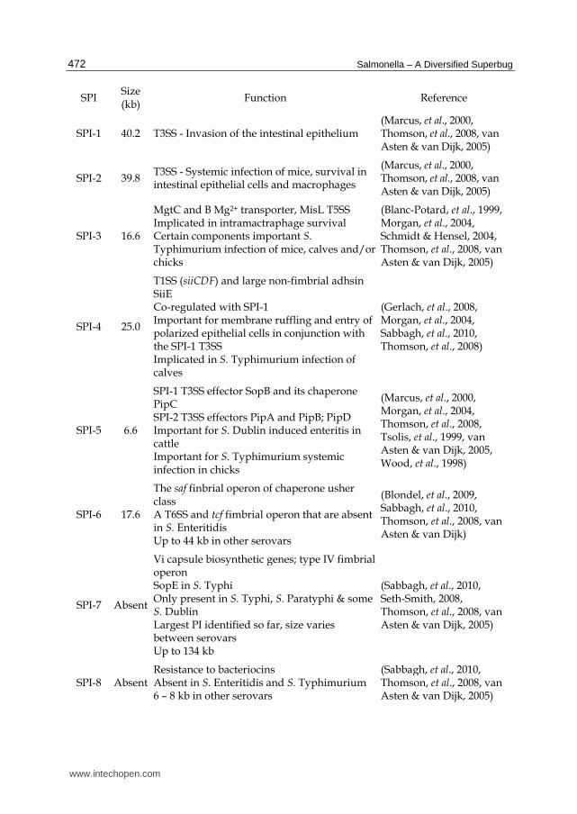
Pathogenicity islands were first identified in uropathogenic E. coli (UPEC) in the late 1980s, and have since been described in a wide variety of bacteria (Blum, et al., 1994, Hacker, et al., 1997, Schmidt & Hensel, 2004). Pathogenicity islands have been identified in both Gram-negative and Gram-positive species, and are associated with plant, animal, and human pathogens, as well as non-pathogenic bacteria. They typically harbour large clusters of genes (10 – 200 kb) related to virulence and/or survival and fitness, and have a different GC content in comparison to the rest of the genome. Pathogenicity islands can often be mosaic in structure and are often bordered by transposon insertion sequences and direct repeats, as well as bacteriophage genes, indicating that their insertion into the genome occurred via single or multiple horizontal gene transfer events (Hacker & Kaper, 2000, Schmidt & Hensel, 2004). To date there have been 21 Salmonella pathogenicity islands (SPIs) identified; a brief description of each of these islands is listed in Table 1.
www.intechopen.com
Salmonella – A Diversified Superbug
472
SPI Size (kb) Function Reference
SPI-1 40.2 T3SS - Invasion of the intestinal epithelium (Marcus, et al., 2000, Thomson, et al., 2008, van Asten & van Dijk, 2005)
SPI-2 39.8 T3SS - Systemic infection of mice, survival in intestinal epithelial cells and macrophages
(Marcus, et al., 2000, Thomson, et al., 2008, van Asten & van Dijk, 2005)
SPI-3 16.6
MgtC and B Mg2+ transporter, MisL T5SS Implicated in intramactraphage survival Certain components important S. Typhimurium infection of mice, calves and/or chicks
(Blanc-Potard, et al., 1999, Morgan, et al., 2004, Schmidt & Hensel, 2004, Thomson, et al., 2008, van Asten & van Dijk, 2005)
SPI-4 25.0
T1SS (siiCDF) and large non-fimbrial adhsin SiiE Co-regulated with SPI-1 Important for membrane ruffling and entry of polarized epithelial cells in conjunction with the SPI-1 T3SS Implicated in S. Typhimurium infection of calves
(Gerlach, et al., 2008, Morgan, et al., 2004, Sabbagh, et al., 2010, Thomson, et al., 2008)
SPI-5 6.6
SPI-1 T3SS effector SopB and its chaperone PipC SPI-2 T3SS effectors PipA and PipB; PipD Important for S. Dublin induced enteritis in cattle Important for S. Typhimurium systemic infection in chicks
(Marcus, et al., 2000, Morgan, et al., 2004, Thomson, et al., 2008, Tsolis, et al., 1999, van Asten & van Dijk, 2005, Wood, et al., 1998)
SPI-6 17.6
The saf finbrial operon of chaperone usher class A T6SS and tcf fimbrial operon that are absent in S. Enteritidis Up to 44 kb in other serovars
(Blondel, et al., 2009, Sabbagh, et al., 2010, Thomson, et al., 2008, van Asten & van Dijk)
SPI-7 Absent
Vi capsule biosynthetic genes; type IV fimbrial operon SopE in S. Typhi Only present in S. Typhi, S. Paratyphi & some S. Dublin Largest PI identified so far, size varies between serovars Up to 134 kb
(Sabbagh, et al., 2010, Seth-Smith, 2008, Thomson, et al., 2008, van Asten & van Dijk, 2005)
SPI-8 Absent Resistance to bacteriocins Absent in S. Enteritidis and S. Typhimurium 6 – 8 kb in other serovars
(Sabbagh, et al., 2010, Thomson, et al., 2008, van Asten & van Dijk, 2005)
www.intechopen.com

The Salmonella Pathogenicity Island-1 and -2 Encoded Type III Secretion Systems
473
SPI Size (kb) Function Reference
SPI-9 16.3 T1SS, and a RTX-like protein The RTX protein is complete in S. Enteritidis, but not S. Typhimurium
(Thomson, et al., 2008, van Asten & van Dijk, 2005)
SPI-10 10.0 Sef fimbrial operon in S. Enteritidis Larger in other serovars (up to 33 kb)
(Sabbagh, et al., 2010, Thomson, et al., 2008, van Asten & van Dijk, 2005)
SPI-11 6.7 PagC, PagD and MsgA important for survival of S. Typhimurium in macropahges
(Sabbagh, et al., 2010, Thomson, et al., 2008)
SPI-12 5.8 SPI-2 T3SS effector sspH2 Important for full virulence of S. Typhimurium in mice
(Haneda, et al., 2009, Sabbagh, et al., 2010, Thomson, et al., 2008)
SPI-13 25.3 Important for systemic infection in mice by S. Typhimurium
(Haneda, et al., 2009, Shi, et al., 2006, Thomson, et al., 2008)
SPI-14 6.8 Electron transfer and putative regulatory genes
(Sabbagh, et al., 2010, Thomson, et al., 2008)
SPI-15 Absent 5 hypothetical proteins Not present in either S. Enteritidis or S. Typhimurium
(Sabbagh, et al., 2010, Thomson, et al., 2008)
SPI-16 3.3 LPS modification High homology to SPI-17
(Sabbagh, et al., 2010, Thomson, et al., 2008)
SPI-17 3.6 LPS modification; high homology to SPI-16 Present in S. Enteritidis and S. Typhi, but not S. Typhimurium
(Sabbagh, et al., 2010, Thomson, et al., 2008)
SPI-18 Absent
In S. Typhi encodes 2 genes for the cytolysin HlyE and the invasion TaiE Not present in either S. Enteritidis or S. Typhimurium
(Sabbagh, et al., 2010, Thomson, et al., 2008)
SPI-19 14.1 T6SS likely non-functional in S. Enteritidis as most of island has been deleted Up to 45 kb in other serovars
(Blondel, et al., 2009, Thomson, et al., 2008)
SPI-20 Absent T6SS Only identified in Salmonella enterica subsp. arizonae 34 kb
(Blondel, et al., 2009, Thomson, et al., 2008)
SPI-21 Absent T6SS Only identified in Salmonella enterica subsp. arizonae 55 kb
(Blondel, et al., 2009, Thomson, et al., 2008)
Table 1. Salmonella pathogenicity islands
www.intechopen.com
Salmonella – A Diversified Superbug
474
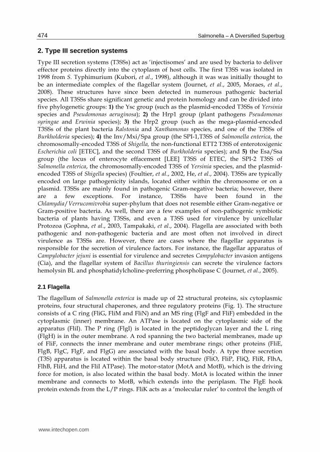
2. Type III secretion systems
Type III secretion systems (T3SSs) act as ‘injectisomes’ and are used by bacteria to deliver effector proteins directly into the cytoplasm of host cells. The first T3SS was isolated in 1998 from S. Typhimurium (Kubori, et al., 1998), although it was was initially thought to be an intermediate complex of the flagellar system (Journet, et al., 2005, Moraes, et al., 2008). These structures have since been detected in numerous pathogenic bacterial species. All T3SSs share significant genetic and protein homology and can be divided into five phylogenetic groups: 1) the Ysc group (such as the plasmid-encoded T3SSs of Yersinia species and Pseudomonas aeruginosa); 2) the Hrp1 group (plant pathogens Pseudomonas syringae and Erwinia species); 3) the Hrp2 group (such as the mega-plasmid-encoded T3SSs of the plant bacteria Ralstonia and Xanthamonas species, and one of the T3SSs of Burkholderia species); 4) the Inv/Mxi/Spa group (the SPI-1,T3SS of Salmonella enterica, the chromosomally-encoded T3SS of Shigella, the non-functional ETT2 T3SS of enterotoxigenic Escherichia coli [ETEC], and the second T3SS of Burkholderia species); and 5) the Esa/Ssa group (the locus of enterocyte effacement [LEE] T3SS of ETEC, the SPI-2 T3SS of Salmonella enterica, the chromosomally-encoded T3SS of Yersinia species, and the plasmid-encoded T3SS of Shigella species) (Foultier, et al., 2002, He, et al., 2004). T3SSs are typically encoded on large pathogenicity islands, located either within the chromosome or on a plasmid. T3SSs are mainly found in pathogenic Gram-negative bacteria; however, there are a few exceptions. For instance, T3SSs have been found in the Chlamydia/Verrucomivrobia super-phylum that does not resemble either Gram-negative or Gram-positive bacteria. As well, there are a few examples of non-pathogenic symbiotic bacteria of plants having T3SSs, and even a T3SS used for virulence by unicellular Protozoa (Gophna, et al., 2003, Tampakaki, et al., 2004). Flagella are associated with both pathogenic and non-pathogenic bacteria and are most often not involved in direct virulence as T3SSs are. However, there are cases where the flagellar apparatus is responsible for the secretion of virulence factors. For instance, the flagellar apparatus of Campylobacter jejuni is essential for virulence and secretes Campylobacter invasion antigens (Cia), and the flagellar system of Bacillus thuringiensis can secrete the virulence factors hemolysin BL and phosphatidylcholine-preferring phospholipase C (Journet, et al., 2005).
2.1 Flagella
The flagellum of Salmonella enterica is made up of 22 structural proteins, six cytoplasmic proteins, four structural chaperones, and three regulatory proteins (Fig. 1). The structure consists of a C ring (FliG, FliM and FliN) and an MS ring (FlgF and FliF) embedded in the cytoplasmic (inner) membrane. An ATPase is located on the cytoplasmic side of the apparatus (FliI). The P ring (FlgI) is located in the peptidoglycan layer and the L ring (FlgH) is in the outer membrane. A rod spanning the two bacterial membranes, made up of FliF, connects the inner membrane and outer membrane rings; other proteins (FliE, FlgB, FlgC, FlgF, and FlgG) are associated with the basal body. A type three secretion (T3S) apparatus is located within the basal body structure (FliO, FliP, FliQ, FliR, FlhA, FlhB, FliH, and the FliI ATPase). The motor-stator (MotA and MotB), which is the driving force for motion, is also located within the basal body. MotA is located within the inner membrane and connects to MotB, which extends into the periplasm. The FlgE hook protein extends from the L/P rings. FliK acts as a ‘molecular ruler’ to control the length of
www.intechopen.com

The Salmonella Pathogenicity Island-1 and -2 Encoded Type III Secretion Systems
475
the hook. The hook is followed by the hook-filament junction (FlgK and FlgL) and the long filament is comprised of either FliC or FljB flagellin; Salmonella encodes both proteins, but they are never expressed at the same time. This differential expression may aid Salmonella in escaping the host immune defenses by antigenic variation, and/or contribute to host specificity. Finally, the filament is topped off by the FlgD cap (Aizawa, 2001, Liu & Ochman, 2007, Macnab, 2004, McCann & Guttman, 2008, Morgan, et al., 2004, Pallen & Matzke, 2006, van Asten & van Dijk, 2005).
Fig. 1. Schematic representation of the flagellar apparatus, SPI-1, and SPI-2 T3SSs of Salmonella. The molecular organization of the flagellar system is depicted above on the left, the SPI-1 T3SS in the middle, and the SPI-2 T3SS on the right. Stoichiometry of proteins was followed where known. Adapted and modified from (Moraes, et al., 2008, Pallen, et al., 2005, Tampakaki, et al., 2004).
2.2 Salmonella pathogenicity island-1 type III secretion system
In S. Enteritidis, SPI-1 is 40.2 kb in length and has a GC content of 47% (Marcus, et al., 2000, van Asten & van Dijk, 2005). SPI-1 contains 41 genes encoding a T3SS, T3SS regulatory genes, T3SS effectors, and a metal transport system (Fig. 2) (Schmidt & Hensel, 2004, Thomson, et al., 2008). SPI-1 is important for cell invasion of intestinal epithelial cells as well as apoptosis of macrophages (Galán, 2001, Mills, et al., 1995, van der Velden, et al., 2000). S. Typhimurium strains defective for InvC (a major structural component of the SPI-1 T3SS) have a 50% higher lethal dose when given orally to Balb/c mice, but perform similarly to wild-type strains when given intraperitoneally, indicating a role for SPI-1 in colonization and invasion during the initial phase of infection, but not during the systemic phase (Galán & Curtiss, 1989).
www.intechopen.com

Salmonella – A Diversified Superbug
476
Fig. 2. Genetic organization of Salmonella pathogenicity islands 1 and 2. The organization of the ~40kb regions of the S. Enteritidis chromosome harbouring SPI-1 and SPI-2 is shown above. Gray arrows represent known or predicted transcriptional units and genes are coloured based on the function of the encoded protein. Based on the published S. Enteritidis genome sequence (Thomson, et al., 2008).
2.2.1 Structural components and effectors of the Salmonella pathogenicity island-1 type III secretion system
The basal body of the SPI-1 T3SS (Fig. 1) is composed of an inner membrane ring formed by PrgH and PrgK, many inner membrane proteins (SpaP, SpaQ, SpaR, SpaS, and InvA), an ATPase (InvC) and an outer membrane secretin (InvG). Extending from the outer membrane secretin is the needle formed by PrgI, topped by the translocon made up of SipB and SipC (Moraes, et al., 2008). The SPI-1 T3SS is responsible for the secretion of a specific set of effectors. AvrA, SipA, SipB, SipC, SipD, and SptP are all encoded on SPI-1, while the genes encoding GogB, SopE, SopE2, and SspH1 are located on lysogenic bacteriophages in the genome. The gene for SopB is located on SPI-5, and the genes for SopA, SopD, SlrP, SteA, and SteB are located elsewhere within the chromosome. GogB, SlrP, SspH1, SteA, and SteB are also secreted by the SPI-2 T3SS (Abrahams & Hensel, 2006, Bernal-Bayard & Ramos-Morales, 2009, Salyers & Whitt, 2002, Thomson, et al., 2008).
2.2.2 Assembly and regulation of the Salmonella pathogenicity island-1 type III secretion system
The assembly of the SPI-1 T3SS proceeds in a similar manner to the assembly of the flagella. The inner membrane and outer membrane rings are formed first in a sec-dependent manner, followed by the association of the rings and formation of the remaining basal body components, including the ATPase. Formation of the needle and translocon is T3S-dependent, and needle length is controlled by InvJ, which acts as a ‘molecular ruler’ (Deane, et al., 2010, He, et al., 2004, Moraes, et al., 2008).
Expression of the SPI-1 T3SS is regulated by many environmental and genetic signals. Environmental signals include pH, osmolarity, the presence of bile, magnesium concentration, and the presence of short chain fatty acids (Altier, 2005). The preferred
www.intechopen.com

The Salmonella Pathogenicity Island-1 and -2 Encoded Type III Secretion Systems
477
invasion site of Salmonella is the M-cells of the distal small intestine. When bile is present, indicating the beginning of the small intestine, or when short-chain fatty acids are present, which are produced by microflora of the large intestine, SPI-1 expression is repressed. These environmental signals indicate that the bacterium is not near its preferred site of entry. SPI-1 expression is induced at near neutral pH, and high osmolarity (Altier, 2005, Garmendia, et al., 2003). In the presence of high iron, the ferric uptake regulator (Fur) acts to increase the expression of HilD (a SPI-1 regulator, discussed further in the following text) in an unknown manner. Once in the Salmonella containing vacuole, SCV, where there is limited iron, this indirect activation of HilD by Fur is stopped (Altier, 2005, Ellermeier, J. R. & Slauch, 2008). See Fig. 3 for a diagram of the interaction of the regulation pathways outlined below.
Fig. 3. Regulation of Salmonella pathogenicity islands 1 and 2. The major modes of SPI-1 and SPI-2 regulation are depicted above; see text for details (Sections 2.2.2, 2.3.2, and 2.4).
www.intechopen.com

Salmonella – A Diversified Superbug
478
Nucleoid associated proteins (NAPs) affect supercoiling of deoxyribonucleic acid (DNA), and are thus able to alter gene expression. The NAPs Hha and H-NS both repress transcription of many genes, including rtsA and the SPI-1 gene hilA under conditions of low osmolarity (Altier, 2005, Olekhnovich & Kadner, 2007, Rhen & Dorman, 2005). Hu, IHF, and Fis are also NAPs, and are important for expression of SPI-1 genes (Altier, 2005, Fass & Groisman, 2009).
PhoP/PhoQ and BarA/SirA belong to two-component global regulatory systems that respond to environmental conditions. In low magnesium conditions, for example within the SCV, PhoP can act to negatively regulate HilA, leading to down regulation of the SPI-1 T3SS. SirA positively regulates HilA, by regulating the expression of HilD (Altier, 2005, Ellermeier, J. R. & Slauch, 2007, Hacker & Kaper, 2000, Hueck, 1998). BarA/SirA also controls the csr system. CsrA can bind messenger ribonucleic acid (mRNA) at their ribosomal binding site, thus stabilizing, or alternatively, reducing, translation of SPI-1 T3SS proteins, likely at the level of HilD. CsrB and C are small RNA molecules that bind and stop the action of CsrA. BarA/SirA activate CsrB and C, keeping CsrA levels in check. Optimal levels of all three molecules are needed for proper expression of SPI-1. The EnvZ/OmpR system senses osmolarity and has been proposed to regulate HilD post-translationally. The PhoP/PhoQ and PhoR/PhoB systems can activate expression of HilE, which then acts to repress expression of SPI-1 genes, through direct binding to HilD. The type 1 fimbriae regulators FimZ and FimY have also been shown to negatively regulate transcription of SPI-1 genes, likely through activation of hilE, while the flagella regulator FliZ positively regulates expression of HilA post-transcriptionally (Altier, 2005, Ellermeier, J. R. & Slauch, 2007). Mlc is a global regulator that detects the presence of sugars such as glucose and mannose, whereby Mlc can repress expression of hilE when sugars are readily available, such as in the small intestine (Lim, et al., 2007). The Lon protease (controlled by DnaK and 32), negatively regulates SPI-1 by degrading HilD in response to the stress of the SCV environment (Matsui, et al., 2008).
HilA belongs to the OmpR/ToxR family of transcriptional regulators, while InvF, HilC, and HilD are in the AraC/XylS family. Each of these genes are located on SPI-1 (Fig. 2) (Hacker & Kaper, 2000, Schmidt & Hensel, 2004). Expression of HilD is likely induced by environmental conditions, and leads to expression of HilC and RtsA. RtsA and HilC can also activate expression of themselves, and each other. RtsA activates hilA expression directly, as well as the expression of slrP, a SPI-1 T3SS effector, and dsbA, which is required for assembly of the T3SS. HilC and D act to derepress transcription of hilA and rtsA by relieving silencing by H-NS and Hha. HilA is then free to activate transcription of the prg/org and inv/spa operons (including invF). RtsA, HilD, and HilC can also activate transcription of the inv/spa operon independently of HilA, but to a lower degree than HilA. InvF activates transcription of the sic/sip (including sicA) operon of SPI-1, as well as genes within SPI-4 and SPI-5 (Altier, 2005, Ellermeier, J. R. & Slauch, 2007, Hacker & Kaper, 2000, Olekhnovich & Kadner, 2007, Rhen & Dorman, 2005, Schmidt & Hensel, 2004). SicA is the chaperone for the translocator proteins SipB and C. Once the translocon components have been secreted, SicA is free and can activate expression of invF, creating a positive feedback loop of secreted effector gene expression once the SPI-1 T3SS is fully formed (He, et al., 2004, Rhen & Dorman, 2005).
www.intechopen.com
The Salmonella Pathogenicity Island-1 and -2 Encoded Type III Secretion Systems
479
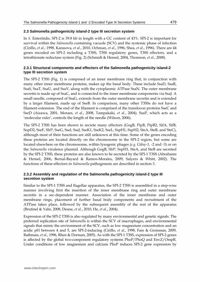
2.3 Salmonella pathogenicity island-2 type III secretion system
In S. Enteritidis, SPI-2 is 39.8 kb in length with a GC content of 43%. SPI-2 is important for survival within the Salmonella containing vacuole (SCV) and the systemic phase of infection (Cirillo, et al., 1998, Karasova, et al., 2010, Ochman, et al., 1996, Shea, et al., 1996). There are 44 genes encoded on SPI-2 including a T3SS, T3SS regulatory genes, T3SS effectors, and a tetrathionate reductase system (Fig. 2) (Schmidt & Hensel, 2004, Thomson, et al., 2008).
2.3.1 Structural components and effectors of the Salmonella pathogenicity island-2 type III secretion system
The SPI-2 T3SS (Fig. 1) is composed of an inner membrane ring that, in conjunction with many other inner membrane proteins, makes up the basal body. These include SsaD, SsaR, SsaS, SsaT, SsaU, and SsaV, along with the cytoplasmic ATPase SsaN. The outer membrane secretin is made up of SsaC, and is connected to the inner membrane components via SsaJ. A small needle, composed of SsaG, extends from the outer membrane secretin and is extended by a larger filament, made up of SseB. In comparison, many other T3SSs do not have a filament extension. The end of the filament is comprised of the translocon proteins SseC and SseD (Aizawa, 2001, Moraes, et al., 2008, Tampakaki, et al., 2004). SsaP, which acts as a ‘molecular ruler’, controls the length of the needle (Wilson, 2006).
The SPI-2 T3SS has been shown to secrete many effectors (GogB, PipB, PipB2, SifA, SifB, SopD2, SseF, SlrP, SseG, SseI, SseJ, SseK1, SseK2, SseL, SspH1, SspH2, SteA, SteB, and SteC), although most of their functions are still unknown at this time. Some of the genes encoding these proteins are located directly on the chromosome in the SPI-2 region, but some are located elsewhere on the chromosome, within lysogenic phages (e.g. Gifsy-1, -2 and -3) or on the Salmonella virulence plasmid. Although GogB, SlrP, SspH1, SteA, and SteB are secreted by the SPI-2 T3SS, these proteins are also known to be secreted by the SPI-1 T3SS (Abrahams & Hensel, 2006, Bernal-Bayard & Ramos-Morales, 2009, Salyers & Whitt, 2002). The functions of these effectors in Salmonella pathogenesis are described in section 3.
2.3.2 Assembly and regulation of the Salmonella pathogenicity island-2 type III secretion system
Similar to the SPI-1 T3SS and flagellar apparatus, the SPI-2 T3SS is assembled in a step-wise manner involving first the insertion of the inner membrane ring and outer membrane secretin in a sec-dependent manner. Association of the inner membrane and outer membrane rings, placement of further basal body components and recruitment of the ATPase takes place, followed by the subsequent assembly of the rest of the apparatus (Brutinel & Yahr, 2008, Deane, et al., 2010, He, et al., 2004).
Expression of the SPI-2 T3SS is also regulated by many environmental and genetic signals. The preferred replication site of Salmonella is within the SCV of macrophages, and environmental signals that mimic the environment of the SCV, such as low magnesium concentration and an acidic pH between 4 and 5, are SPI-2-inducing (Cirillo, et al., 1998, Fass & Groisman, 2009, Rathman, et al., 1996, Rhen & Dorman, 2005). As with the SPI-1 T3SS, expression of SPI-2 genes is affected by the global two-component regulatory systems PhoP/PhoQ and EnvZ/OmpR. Under conditions of low magnesium and calcium PhoP induces SPI-2 gene expression by
www.intechopen.com

Salmonella – A Diversified Superbug
480
direct interaction with the ssrB gene, and post-transcriptional action on SsrA. In the presence of low osmolarity and acidic pH, OmpR can directly bind both the ssrA and ssrB promoters, activating transcription. OmpR can also act in conjunction with SsrB to activate transcription of the non-SPI-2-encoded effector SseI (Deiwick, et al., 1998, Fass & Groisman, 2009, Feng, et al., 2003, Garmendia, et al., 2003, Walthers, et al., 2007).
SPI-2 encodes its own two-component regulatory system, SsrA/SsrB. SsrB is able to bind to all SPI-2 promoters, including those of ssrA, ssrB, and many effectors located outside of SPI-2 (Fass & Groisman, 2009, Walthers, et al., 2007). As with SPI-1, H-NS silences the expression of SPI-2 genes by binding directly to many SPI-2 promoters. This binding can be relieved by the SPI-1 protein HilD under certain conditions, such as stationary phase growth in LB, and may also be relieved by SsrB and/or SlyA (Bustamante, et al., 2008, Fass & Groisman, 2009, Walthers, et al., 2007). The NAPs Hha and YdgT can also repress transcription of SPI-2 genes. Fis, a NAP that is able to bind the promoter regions of ssr and ssa operons, is also important for expression of SPI-2 as well as SPI-1 genes. Proper levels of Fis are important for activation of ssrA, and Fis may also induce SPI-2 gene expression indirectly through controlling expression of PhoP. IHF, another NAP, is also important for expression of both SPI-2 and SPI-1 genes (Fass & Groisman, 2009). Some of the mechanisms controlling regulation of SPI-2 are outlined in Fig. 3.
2.4 Cross-talk between the Salmonella flagellar and pathogenicity island-1 and -2 type III secretion systems
The complex regulation of the T3SSs ensures that each system is only expressed under the correct conditions. Expression of multiple versions of each T3SS simultaneously would be energetically expensive, so coordinated expression of the three systems under the specific conditions where each system is required is desirable. Global regulation by two-component regulatory systems that sense divalent cation concentrations, osmolarity, and pH are, in part, responsible for the changes in expression between the flagellar, the SPI-1, and the SPI-2 T3SSs. The SPI-1 T3SS is preferentially expressed within the distal small intestine, which has low oxygen, high osmolarity, a pH of 8, a high concentration of divalent cations, and is rich in nutrients. The environment of the SCV is much different, having low osmolarity, a low concentration of divalent cation concentration, a pH between 4 and 5, and is nutrient poor. In these conditions, the SPI-2 T3SS is preferentially expressed (He, et al., 2004).
The BarA/SirA system positively regulates expression of SPI-1 genes, but negatively regulates expression of flagellar genes. Therefore, in environmental conditions that activate BarA/SirA, the SPI-1 T3SS will be expressed while the flagellar system is downregulated. RtsA and RtsB have also been proposed to be involved in the switch from expression of flagella to expression of the SPI-1 T3SS. RtsA is important for SPI-1 expression, while RtsB represses expression of flagellar genes by interfering with the flhDC promoter (Ellermeier, C. D. & Slauch, 2003). In conditions of low divalent cation concentration, PhoP suppresses expression of SPI-1 genes while activating expression of SPI-2 genes. This ensures that once in the SCV, when the SPI-1 T3SS is no longer needed for invasion of non-phagocytic cells, the SPI-2 T3SS expression is induced while the SPI-1 T3SS is downregulated (Rhen & Dorman, 2005).
www.intechopen.com
The Salmonella Pathogenicity Island-1 and -2 Encoded Type III Secretion Systems
481
Interspecies and interkingdom quorum sensing may also be involved in regulating expression of these three systems. In the presence of host norepinephrine there is an upregulation of flagellar genes in S. Typhimurium (Bearson & Bearson, 2008). S. Typhimurium encodes a putative regulatory protein, YhcS, which has high amino acid similarity to QseA of E. coli. QseA activates expression of the LEE T3SS by E. coli in response to autoinducer 3 (AI-3) quorum sensing molecules produced by intestinal flora, as well as epinephrine and norepinephrine produced by the host. YhcS may act similarly to QseA in E. coli by activating expression of either (or both of) the SPI-1 or SPI-2 T3SSs (Bearson & Bearson, 2008, Choi, et al., 2007, Karavolos, et al., 2008). As mentioned previously, under certain growth conditions HilD can relieve H-NS-mediated repression of SPI-2 genes (Bustamante, et al., 2008). This may account for the fact that SPI-2 is expressed to some extent along with SPI-1 in the intestinal lumen, and that SPI-1 is expressed for a short time in macrophages before the complete switch to SPI-2 expression. The expression of the SPI-2 T3SS before invasion of intestinal epithelial cells would allow the bacteria to ready itself for the SCV environment. Furthermore, the expression of the SPI-1 T3SS is important for inducing macrophage apoptosis during the initial stage of infection while the bacteria is replicating, and before spread to the rest of the body. Some of the interplay between regulation of SPI-1 and SPI-2 can be visualized in Fig. 3.
2.5 Evolution of the type III secretion system
The flagellar systems of Prokarya are completely different from those of Archaea and Eukarya, suggesting that they evolved convergently into structures serving the same function (Gophna, et al., 2003, Liu & Ochman, 2007). However, prokaryotic flagellar systems with a chemotaxis apparatus that controls changes in the direction of motion share their chemotaxis system with archaeal flagellar systems (Liu & Ochman, 2007, Pallen & Matzke, 2006). As some members of Prokarya do not have this chemotaxis system, it may have been acquired by horizontal transfer from a member of Archaea or may have been present for sensing environmental signals before the diversification of Prokarya and Archaea, and has since been lost in some prokaryotic families. While the flagellar systems of Prokarya maintain many of the same genes and proteins among members, they can be quite diverse in their function. For instance, the flagella of Spirochaetes are located in the periplasm, while Vibrio species express both polar and lateral flagellar systems that share a chemotaxis transduction system but use different motive forces (Na+ or H+). Most of the flagellar proteins that serve the same function are homologous, however, not all flagellar system proteins are conserved among all bacterial species. For example, the flagellar structures of Gram-positive bacteria do not have the L and P rings (which would be located in the outer membrane of Gram-negative bacteria). Spirochaetes do not have the L and P ring either, as their flagella are located in the periplasm. Some of the structural genes (flgH, flgI, fliD, fliE, and fliH specifically) are missing in some bacteria; this could indicate a later evolution of these genes combined with limited horizontal transfer, or be an example of sporadic loss of genes from some bacterial families. The latter explanation seems more likely in this case as there are many families of bacteria that contain these genes, and only a few who are lacking (Liu & Ochman, 2007).
The flagella phylogenetic tree is directly related to that of the bacterial speciation genetic tree based on 16S ribosomal RNA. This suggests that flagella have been in existence since
www.intechopen.com

Salmonella – A Diversified Superbug
482
before the diversification of bacteria, and have been maintained throughout vertical evolution (McCann & Guttman, 2008). Liu and Ochman propose that the entire flagellar system is actually evolved from a single gene. They suggest, based on sequence similarities, that all of the flagellar genes arose from random duplications and reassortments of a single precursor gene in the ancestor of modern bacteria (Liu & Ochman, 2007). This seems quite unlikely; although there may be sequence similarities between an inner membrane component and an outer membrane component, this does not mean that they are related on an evolutionary scale. Convergent evolution is a more likely explanation for this, in which two different proteins have evolved to serve a similar function, in this case to be embedded in the bacterial membrane.
Unlike flagellar systems, the T3SS phylogenetic tree is not related to that of 16S ribosomal RNA, suggesting that T3SSs were acquired at some point after the diversification of bacteria, and evolved via horizontal transfer events (Foultier, et al., 2002, Gophna, et al., 2003, Liu & Ochman, 2007, Nguyen, et al., 2000). T3SSs are encoded on large pathogenicity islands, while flagellar genes are encoded on the chromosome (Hueck, 1998, Macnab, 2004, van Asten & van Dijk, 2005). It is thought that SPI-2 may have arrived in two separate events, with the ttr operon arriving first, followed by the rest of SPI-2 (Marcus, et al., 2000).
The effectors of T3SSs are highly variable between species of bacteria, and are quite often encoded on different regions of the chromosome than the pathogenicity island-encoded T3SSs. The effectors and their evolution will not be discussed here, but information on this topic can be found in a recent review (Stavrinides, et al., 2008). In general, there are about ten core proteins of the flagellar T3S apparatus and the injectisome T3SSs that are highly similar in gene sequence, amino acid sequence, and function (Fig. 1.). For the purposes of this discussion, the flagellar system will be compared only with the two Salmonella T3SSs, with homologous proteins given in the order flagella/SPI-1/SPI-2. These homologous proteins are: the cytoplasmic ATPase (FliI/InvC/SsaN), the T3S apparatus (FliH/PrgH/SsaK, FliN/SpaO/SsaQ, FliP/SpaP/SsaR, FliQ/?/SsaS, FliR/SpaR/SsaT, FlhB/SpaS/SsaU and FlhA/InvA/SsaV), part of the connecting rod (FliF/PrgK/SsaJ), and the needle/hook ‘molecular ruler’ (FliK/InvJ/SsaP) (Blocker, et al., 2003, Desvaux, et al., 2006, He, et al., 2004, Tampakaki, et al., 2004, Wilson, 2006).
The structure of the flagellar apparatus and T3SSs begin to differ more markedly starting at the outer membrane (besides the motor-stator which is only present in the basal body of the flagellar system). The MS ring of the flagellar system is larger than that of the outer membrane secretin of the T3SS (Aizawa, 2001). The secretin of the T3SS belongs to the same family of proteins that make up the T2SS and T4SS secretins, and the pore used by filamentous phages, suggesting that filamentous phages either introduced this type of protein to bacteria, or acquired it from them (Nguyen, et al., 2000). The T3SS needle is straight and thin, as is its filament, although the filament is slightly larger, and notably rigid. The flagellar hook apparatus is larger and curved, and its filament is quite long and flexible. These structures lack significant amino acid and genetic homology, but do share helical symmetry, and overall assembly mechanisms. The flagellum contains approximately 5.6 subunits of flagellin per turn, with an axial rise of 4.7 Å. To compare, the filament of the LEE T3SS in E. coli contains 5.5 subunits of EspA per turn, and has an axial rise 4.6 Å (Aizawa, 2001, Journet, et al., 2005, Snyder, et al., 2009, Tampakaki, et al., 2004). The inner diameter of the T3SS filament is between 2 and 3 nm, similar to the inner
www.intechopen.com

The Salmonella Pathogenicity Island-1 and -2 Encoded Type III Secretion Systems
483
channel of the flagellum (Blocker, et al., 2003, Journet, et al., 2005, Tampakaki, et al., 2004). The action of the ‘molecular rulers’ is likely different as well. It has been proposed that the method for measuring hook length in flagella is more ‘measuring cup’-like than ‘molecular ruler’-like. Journet suggests that the motor-stator switch area of the flagellum acts as a measuring cup, filling with FliK. FliK acts as an accessory to the hook protein, and is secreted at the same time. Once the ‘cup’ empties of FliK, the apparatus switches its secretion preference from the hook protein (FlgE) to the flagellin protein (FliC or FljB), completing assembly of the flagellum. In contrast, the InvJ and SsaP proteins (similar to the ‘molecular rulers’ of other bacterial T3SSs) act more like a ruler. It has been suggested that dimers of these proteins are located outside the cell, with one attached to the outer membrane, and the second extending from that. Once the needle complex (PrgI/SsaG) reaches the same height as the InvJ/SsaP dimer, the T3SS switches to secretion and assembly of the translocon of SPI-1 (SipB, C, and D) or filament of SPI-2 (SseB) (Journet, et al., 2005).
Another key area in which the T3SSs and flagellar systems differ is in their chaperones. Although both systems tend to have specific chaperones for specific proteins, the T3SS proteins are recognized by their chaperones at an N-terminal region, while flagellar system chaperones bind at the C-terminal region (Liu & Ochman, 2007). Although the flagellar and T3SS chaperones are different, in some cases the three systems can secrete each other’s proteins. For example, both the SPI-1 and SPI-2 T3SSs can secrete the flagellar protein FliC, while the flagellar system can secrete the SPI-1 T3SS effector proteins SptP and SopE, if the SptP and SopE chaperones are absent, and in some instances effectors from T3SSs of other bacterial species (Journet, et al., 2005, Tampakaki, et al., 2004).
3. Pathogenesis of Salmonella
Salmonella can enter host cells in at least two ways. The first involves uptake into phagocytic cells (macrophages), while the second is more complicated and involves the action of the SPI-1 T3SS on non-phagocytic cells. After attachment to epithelial cells, the SPI-1 T3SS induces membrane ruffling by secreting effectors into the host cell to trigger cytoskeleton rearrangement. Once inside the epithelial cell, some of these same effectors ‘switch off’ the membrane ruffling, returning the host cell membrane to its original state (Ibarra & Steele-Mortimer, 2009, Ly & Casanova, 2007, Salyers & Whitt, 2002, Waterman & Holden, 2003). Entry into the host cell (epithelial or macrophage) results in the bacteria being encased within an SCV. While the goal of many intracellular pathogens would be to escape this vacuolar space into the cell cytoplasm, Salmonella takes advantage of this space and remains in the SCV (Bhavsar, et al., 2007, Ibarra & Steele-Mortimer, 2009, Salyers & Whitt, 2002).
Once inside the SCV, the SPI-2 T3SS is expressed and begins secreting effector proteins, which are used to manipulate the intracellular environment (Cirillo, et al., 1998, Ibarra & Steele-Mortimer, 2009, Ramsden, et al., 2007). Approximately one hour after entry into the host cell, the SCV switches from early endosomal markers, such as early endosome marker 1 (EE-1), to late endosomal/lysosomal markers, such as lysosomal-associated membrane protein-1 (LAMP-1) and lysosomal glycoproteins (lgps). One important factor that the SCV acquires during this switch is the V-ATPase, which facilitates the acidification of the SCV.
www.intechopen.com

Salmonella – A Diversified Superbug
484
This acidification is an important factor for the induction of Salmonella virulence/survival genes (Abrahams & Hensel, 2006, Bhavsar, et al., 2007, Ibarra & Steele-Mortimer, 2009, Kuhle & Hensel, 2004, Ramsden, et al., 2007, Salyers & Whitt, 2002). Another important factor for Salmonella survival within host cells is iron acquisition. Salmonella releases two siderophores for sequestering Fe2+ from the host cell, enterobactin and salmochelin (Ibarra & Steele-Mortimer, 2009). As the SCV matures, it moves along host cell microtubules towards the Golgi apparatus. This process is dependent on many effectors, including SifA, SifB, SopD2, SseF, SseG, SseI, SseJ, SseL, PipB, and PipB2 (Abrahams & Hensel, 2006, Ibarra & Steele-Mortimer, 2009, Ramsden, et al., 2007). SsaB is also important in blocking the fusion of the SCV with lysosomes during this process, which would result in bacterial killing (Kuhle & Hensel, 2004). Movement along the microtubules involves recruitment of a dynein-dynactin motor complex by SifA, SseF, SseG, and PipB2. PipB2 interacts with the motor protein kinesin, while the other three proteins have also been shown to be responsible for keeping the SCV localized to the Golgi apparatus in an unknown manner. These proteins are also very important in Salmonella-induced filament (sif) formation, which will be discussed in the following paragraph. SCV membrane integrity is important, and is controlled by a number of SPI-2 T3SS effectors, including SspH2, SseI, SteC, and the Salmonella virulence plasmid-encoded protein SpvB. The interaction of these proteins with host filamen and actin causes the formation of an actin-mesh around the SCV (Abrahams & Hensel, 2006, Kuhle & Hensel, 2004, Ramsden, et al., 2007). Another function of the SPI-2 T3SS may be to stop the formation of the NADPH phagocytic oxidase (phox) and inducible nitric oxide synthase (iNOS) on the SCV membrane, ultimately resulting in protection of Salmonella from reactive oxygen and nitrogen species (ROS and RNS, respectively) (Abrahams & Hensel, 2006, Coburn, et al., 2005, Salyers & Whitt, 2002). A superoxide dismutase encoded by the Gifsy-2 lysogenic phage helps Salmonella survive the oxidative burst, which involves production of ROS and RNS by phagocytic cells that can damage bacteria, and is therefore important for bacterial survival within the SCV (Ibarra & Steele-Mortimer, 2009, Salyers & Whitt, 2002).
The maturation/movement process of the SCV can take around 4 to 6 hours. At this point, when the SCV has been altered to suit the bacteria, Salmonella begin to replicate (Abrahams & Hensel, 2006, Finlay & Brumell, 2000). Replication of Salmonella is associated with the formation of sifs. Sifs have similar markers to the SCV, and many of the same proteins are responsible for their formation/membrane integrity (SifA, SifB, SseF, SseG, SseJ, SseL, SspH2, SpvB, PipB, and PipB2). The SPI-1 effector SipA has also been shown to be important in sif formation. These sifs extend from the SCV towards the host cell membrane, and other SCVs, if there are multiple SCVs in one cell (Ibarra & Steele-Mortimer, 2009, Kuhle & Hensel, 2004, Ramsden, et al., 2007). The AvrA effector secreted by the SPI-1 T3SS deubiquitinates both IB- and β-catenin, which stabilizes the proteins and results in the continued repression of NFB-mediated gene transcription. This delays apoptosis of intestinal epithelial cells, thereby allowing Salmonella to survive within them for longer (Bernal-Bayard & Ramos-Morales, 2009, Bhavsar, et al., 2007, Grassl & Finlay, 2008, Ibarra & Steele-Mortimer, 2009). SlrP also mediates ubiquitination of certain host proteins including Thioredoxin-1 (Trx1). Trx1 can activate the NFB transcription factor, and has functions among other host cell proteins as well. Binding of SlrP to Trx1 stops its action, which under some conditions can lead to apoptotic cell death, although the exact mechanisms of this need to be studied further (Bernal-Bayard & Ramos-Morales, 2009, Bhavsar, et al., 2007,
www.intechopen.com
The Salmonella Pathogenicity Island-1 and -2 Encoded Type III Secretion Systems
485
Ramsden, et al., 2007). SspH1 can also inhibit NFB transcription (Ibarra & Steele-Mortimer, 2009, Kuhle & Hensel, 2004, Ramsden, et al., 2007).
3.1 Role of the Salmonella pathogenicity island-1 and -2 type III secretion systems in the chicken model of infection
Contaminated poultry and eggs remain a major source of food poisoning caused by Salmonella. As the majority of work on Salmonella to date has been done in mice, we examined the role of this bacterium in a chicken model of S. Enteritidis infection developed using select strains isolated from chickens. We were particularly interested in the role of the SPI-1 and SPI-2 encoded T3SSs of S. Enteritidis in the various stages of infection and colonization of birds, as the SPI-1 encoded T3SS has been found to be important for entry into intestinal epithelial cells, and the SPI-2 encoded T3SS has been found to be highly important in the later stages of infection in mice, particularly after Salmonella has been vacuolised. To examine this, chickens were challenged orally with 106-109 Salmonella bacteria. In co-challenge trials using 35-day-old chickens we showed that although a S. Enteriditis wild-type strain was only slightly more competitive in colonizing the ceca than mutants defective in SPI-1 and SPI-2, the systemic spread of both of these mutant strains to the liver and spleen was significantly less successful than that of the wild-type strain (Desin, et al., 2009, Wisner, et al., 2010). Colonization of the gut was nearly 100% for both wild-type and mutants, whereas Salmonella was detected in the liver and spleen in approximately 30% of the birds in these trials. In order to acheive a higher percentage of chickens that were systemically infected, we performed colonization experiments with younger leghorn chickens hatched from specific pathogen-free (SPF) eggs and orally challenged 7 days post-hatch. With respect to colonization of the ceca we found no statistically relevant differences between the wild-type and SPI-1 or SPI-2 mutant strains. However, although 100% of the younger birds developed a systemic infection when challenged with the wild-type strain, challenges with mutant strains devoid of functional SPI-1 and/or SPI-2 T3SSs resulted in a clearly delayed, and less severe, systemic infection. Interestingly, at the end of the test period, 4 days post-challenge, the systemic presence of both the mutant and the wild-type strains was found to be decreasing. From these findings, it is evident that both the SPI-1 and SPI-2 pathogenicity islands are important for the fast and efficient invasion and systemic spread of Salmonella in chickens. However, the data obtained 3 and 4 days post-challenge indicates that SPI-1 and SPI-2 are not the only factors needed for systemic dissemination, and that other virulence factors may compensate for the loss of SPI-1 and SPI-2.
4. Concluding remarks
Most of the animal studies in the context of bacterial T3SS-related pathogenicity have been performed with S. Typhimurium in mice. Based on our findings, however, we see that not all the results obtained from those experiments can be directly transposed to other hosts. Our in
vitro and in vivo results based on a chicken model of S. Enteritidis infection demonstrated that both SPI-1 and SPI-2 play an important role in invasion and systemic spread, although they do not seem to be essential. Further studies will be necessary to understand the full spectrum of virulence mechanisms of different Salmonella strains in various host organisms, including humans.
www.intechopen.com
Salmonella – A Diversified Superbug
486
5. Acknowledgements
Projects in the authors laboratories related to this review were supported through the Industrial Research Chair (IRC) program of the Natural Sciences and Engineering Research Council of Canada (NSERC), the Saskatchewan Health Research Foundation (SHRF) and the Poultry Industry Council (PIC).
6. References
Abrahams, G. L. & Hensel, M. (2006). Manipulating cellular transport and immune responses: dynamic interactions between intracellular Salmonella enterica and its host cells. Cellular Microbiology, Vol. 8, No. 5, (May), pp. 728-737, ISSN 1462-5814
Aizawa, S. I. (2001). Bacterial flagella and type III secretion systems. FEMS Microbiology
Letters, Vol. 202, No. 2, (Aug 21), pp. 157-164, ISSN 0378-1097 Allen-Vercoe, E., Sayers, A. R. & Woodward, M. J. (1999). Virulence of Salmonella enterica
serotype Enteritidis aflagellate and afimbriate mutants in a day-old chick model. Epidemiology and Infection, Vol. 122, No. 3, (Jun), pp. 395-402, ISSN 0950-2688
Allen-Vercoe, E. & Woodward, M. J. (1999). Colonisation of the chicken caecum by afimbriate and aflagellate derivatives of Salmonella enterica serotype Enteritidis. Veterinary Microbiology, Vol. 69, No. 4, (Sep 29), pp. 265-275, ISSN 0378-1135
Allen-Vercoe, E. & Woodward, M. J. (1999). The role of flagella, but not fimbriae, in the adherence of Salmonella enterica serotype Enteritidis to chick gut explant. Journal
of Medical Microbiology, Vol. 48, No. 8, (Aug), pp. 771-780, ISSN 0022-2615 Altier, C. (2005). Genetic and environmental control of Salmonella invasion. Journal of
microbiology, Vol. 43, No. Special, (Feb), pp. 85-92, ISSN 1225-8873 Barrow, P. A. (2007). Salmonella infections: immune and non-immune protection with
vaccines. Avian Pathology, Vol. 36, No. 1, (Feb), pp. 1-13, ISSN 0307-9457 Bearson, B. L. & Bearson, S. M. (2008). The role of the QseC quorum-sensing sensor kinase
in colonization and norepinephrine-enhanced motility of Salmonella enterica serovar Typhimurium. Microbial Pathogenesis, Vol. 44, No. 4, (Apr), pp. 271-278, ISSN 0882-4010
Bernal-Bayard, J. & Ramos-Morales, F. (2009). Salmonella type III secretion effector SlrP is an E3 ubiquitin ligase for mammalian thioredoxin. Journal of Biological Chemistry, Vol. 284, No. 40, (Oct 2), pp. 27587-27595, ISSN 0021-9258
Bhavsar, A. P., Guttman, J. A. & Finlay, B. B. (2007). Manipulation of host-cell pathways by bacterial pathogens. Nature, Vol. 449, No. 7164, (Oct 18), pp. 827-834, ISSN 1476-4687
Blanc-Potard, A. B., Solomon, F., Kayser, J. & Groisman, E. A. (1999). The SPI-3 pathogenicity island of Salmonella enterica. Journal of Bacteriology, Vol. 181, No. 3, (Feb), pp. 998-1004, ISSN 0021-9193
Blocker, A., Komoriya, K. & Aizawa, S. (2003). Type III secretion systems and bacterial flagella: insights into their function from structural similarities. Proceedings of the
National Acadamy of Sciences, Vol. 100, No. 6, (Mar 18), pp. 3027-3030, ISSN 0027-8424
www.intechopen.com

The Salmonella Pathogenicity Island-1 and -2 Encoded Type III Secretion Systems
487
Blondel, C. J., Jimenez, J. C., Contreras, I. & Santiviago, C. A. (2009). Comparative genomic analysis uncovers 3 novel loci encoding type six secretion systems differentially distributed in Salmonella serotypes. BMC Genomics, Vol. 10, No. pp. 354, ISSN 1471-2164
Blum, G., Ott, M., Lischewski, A., Ritter, A., Imrich, H., Tschape, H. & Hacker, J. (1994). Excision of large DNA regions termed pathogenicity islands from tRNA-specific loci in the chromosome of an Escherichia coli wild-type pathogen. Infection and
Immunity, Vol. 62, No. 2, (Feb), pp. 606-614, ISSN 0019-9567 Boyle, E. C., Bishop, J. L., Grassl, G. A. & Finlay, B. B. (2007). Salmonella: from
pathogenesis to therapeutics. Journal of Bacteriology, Vol. 189, No. 5, (Mar), pp. 1489-1495, ISSN 0021-9193
Brenner, F. W., Villar, R. G., Angulo, F. J., Tauxe, R. & Swaminathan, B. (2000). Salmonella nomenclature. Journal of Clinical Microbiology, Vol. 38, No. 7, (Jul), pp. 2465-2467, ISSN 0095-1137
Brutinel, E. D. & Yahr, T. L. (2008). Control of gene expression by type III secretory activity. Current Opinion in Microbiology, Vol. 11, No. 2, (Apr), pp. 128-133, ISSN 1369-5274
Bustamante, V. H., Martinez, L. C., Santana, F. J., Knodler, L. A., Steele-Mortimer, O. & Puente, J. L. (2008). HilD-mediated transcriptional cross-talk between SPI-1 and SPI-2. Proceedings of the National Acadamy of Sciences, Vol. 105, No. 38, (Sep 23), pp. 14591-14596, ISSN 0027-8424
Callaway, T. R., Edrington, T. S., Anderson, R. C., Byrd, J. A. & Nisbet, D. J. (2008). Gastrointestinal microbial ecology and the safety of our food supply as related to Salmonella. Journal of Animal Science, Vol. 86, No. 14 Suppl, (Apr), pp. E163-172, ISSN 1525-3163
Catarame, T. M. G., O'Hanlon, K. A., McDowell, D. A., Blair, I. S. & Duffy, G. (2005). Comparison of a real-time polymerase chain reaction assay with a culture method for the detection of Salmonella in retail meat samples. Journal of Food
Safety, Vol. 26, No. 5, pp. 1 - 15, ISSN 1745-4565 Choi, J., Shin, D. & Ryu, S. (2007). Implication of quorum sensing in Salmonella enterica
serovar Typhimurium virulence: the luxS gene is necessary for expression of genes in pathogenicity island 1. Infection and Immunity, Vol. 75, No. 10, (Oct), pp. 4885-4890, ISSN 0019-9567
Cirillo, D. M., Valdivia, R. H., Monack, D. M. & Falkow, S. (1998). Macrophage-dependent induction of the Salmonella pathogenicity island 2 type III secretion system and its role in intracellular survival. Molecular Microbiology, Vol. 30, No. 1, (Oct), pp. 175-188, ISSN 0950-382X
Clavijo, R. I., Loui, C., Andersen, G. L., Riley, L. W. & Lu, S. (2006). Identification of genes associated with survival of Salmonella enterica serovar Enteritidis in chicken egg albumen. Applied and Environmental Microbiology, Vol. 72, No. 2, (Feb), pp. 1055-1064, ISSN 0099-2240
Coburn, B., Grassl, G. A. & Finlay, B. B. (2007). Salmonella, the host and disease: a brief review. Immunology and cell biology, Vol. 85, No. 2, (Feb-Mar), pp. 112-118, ISSN 0818-9641
www.intechopen.com
Salmonella – A Diversified Superbug
488
Coburn, B., Li, Y., Owen, D., Vallance, B. A. & Finlay, B. B. (2005). Salmonella enterica serovar Typhimurium pathogenicity island 2 is necessary for complete virulence in a mouse model of infectious enterocolitis. Infection and Immunity, Vol. 73, No. 6, (Jun), pp. 3219-3227, ISSN 0019-9567
Cogan, T. A. & Humphrey, T. J. (2003). The rise and fall of Salmonella Enteritidis in the UK. Journal of Applied Microbiology, Vol. 94, No. Supplement S1, pp. 114S-119S, ISSN 1364-5072
Cogan, T. A., Jorgensen, F., Lappin-Scott, H. M., Benson, C. E., Woodward, M. J. & Humphrey, T. J. (2004). Flagella and curli fimbriae are important for the growth of Salmonella enterica serovars in hen eggs. Microbiology, Vol. 150, No. 4, (Apr), pp. 1063-1071, ISSN 1350-0872
Deane, J. E., Abrusci, P., Johnson, S. & Lea, S. M. (2010). Timing is everything: the regulation of type III secretion. Cellular and Molecular Life Sciences, Vol. 67, No. 7, (Apr), pp. 1065-1075, ISSN 1420-9071
Deiwick, J., Nikolaus, T., Shea, J. E., Gleeson, C., Holden, D. W. & Hensel, M. (1998). Mutations in Salmonella pathogenicity island 2 (SPI2) genes affecting transcription of SPI1 genes and resistance to antimicrobial agents. Journal of
Bacteriology, Vol. 180, No. 18, (Sep), pp. 4775-4780, ISSN 0021-9193 Desin, T. S., Lam, P. K., Koch, B., Mickael, C., Berberov, E., Wisner, A. L., Townsend, H. G.,
Potter, A. A. & Köster, W. (2009). Salmonella enterica serovar Enteritidis pathogenicity island 1 is not essential for but facilitates rapid systemic spread in chickens. Infection and Immunity, Vol. 77, No. 7, (Jul), pp. 2866-2875, ISSN 0019-9567
Desvaux, M., Hebraud, M., Henderson, I. R. & Pallen, M. J. (2006). Type III secretion: what's in a name? Trends in Microbiology, Vol. No. (Mar 11), pp. ISSN 0966-842X
Dibb-Fuller, M. P., Allen-Vercoe, E., Thorns, C. J. & Woodward, M. J. (1999). Fimbriae- and flagella-mediated association with and invasion of cultured epithelial cells by Salmonella enteritidis. Microbiology, Vol. 145 ( Pt 5), No. (May), pp. 1023-1031, ISSN 1350-0872
Ellermeier, C. D. & Slauch, J. M. (2003). RtsA and RtsB coordinately regulate expression of the invasion and flagellar genes in Salmonella enterica serovar Typhimurium. Journal of Bacteriology, Vol. 185, No. 17, (Sep), pp. 5096-5108, ISSN 0021-9193
Ellermeier, J. R. & Slauch, J. M. (2007). Adaptation to the host environment: regulation of the SPI1 type III secretion system in Salmonella enterica serovar Typhimurium. Current Opinion in Microbiology, Vol. 10, No. 1, (Feb), pp. 24-29, ISSN 1369-5274
Ellermeier, J. R. & Slauch, J. M. (2008). Fur regulates expression of the Salmonella pathogenicity island 1 type III secretion system through HilD. Journal of
Bacteriology, Vol. 190, No. 2, (Jan), pp. 476-486, ISSN 1098-5530 Fass, E. & Groisman, E. A. (2009). Control of Salmonella pathogenicity island-2 gene
expression. Current Opinion in Microbiology, Vol. 12, No. 2, (Apr), pp. 199-204, ISSN 1369-5274
Feng, X., Oropeza, R. & Kenney, L. J. (2003). Dual regulation by phospho-OmpR of ssrA/B gene expression in Salmonella pathogenicity island 2. Molecular Microbiology, Vol. 48, No. 4, (May), pp. 1131-1143, ISSN 0950-382X
www.intechopen.com

The Salmonella Pathogenicity Island-1 and -2 Encoded Type III Secretion Systems
489
Finlay, B. B. & Brumell, J. H. (2000). Salmonella interactions with host cells: in vitro to in vivo. Philosophical Transactions of the Royal Society B: Biological Sciences, Vol. 355, No. 1397, (May 29), pp. 623-631, ISSN 0962-8436
Foley, S. L. & Lynne, A. M. (2008). Food animal-associated Salmonella challenges: pathogenicity and antimicrobial resistance. Journal of Animal Science, Vol. 86, No. 14 Supplemental, (Apr), pp. E173-187, ISSN 1525-3163
Foultier, B., Troisfontaines, P., Muller, S., Opperdoes, F. R. & Cornelis, G. R. (2002). Characterization of the ysa pathogenicity locus in the chromosome of Yersinia enterocolitica and phylogeny analysis of type III secretion systems. Journal of
Molecular Evolution, Vol. 55, No. 1, (Jul), pp. 37-51, ISSN 0022-2844 Galán, J. E. (2001). Salmonella interactions with host cells: type III secretion at work.
Annual Review of Cell and Developmental Biology, Vol. 17, No. pp. 53-86, ISSN 0022-2844
Galán, J. E. & Curtiss, R., 3rd. (1989). Cloning and molecular characterization of genes whose products allow Salmonella typhimurium to penetrate tissue culture cells. Proceedings of the National Acadamy of Sciences, Vol. 86, No. 16, (Aug), pp. 6383-6387, ISSN 0027-8424
Gantois, I., Eeckhaut, V., Pasmans, F., Haesebrouck, F., Ducatelle, R. & Van Immerseel, F. (2008). A comparative study on the pathogenesis of egg contamination by different serotypes of Salmonella. Avian Pathology, Vol. 37, No. 4, (Aug), pp. 399-406, ISSN 0307-9457
Garmendia, J., Beuzon, C. R., Ruiz-Albert, J. & Holden, D. W. (2003). The roles of SsrA-SsrB and OmpR-EnvZ in the regulation of genes encoding the Salmonella
typhimurium SPI-2 type III secretion system. Microbiology, Vol. 149, No. 9, (Sep), pp. 2385-2396, ISSN 1350-0872
Gast, R. K. (2003). Chapter 16: Salmonella Infections, In: Diseases of Poultry, Saif, Y. M., pp. 567-614, Iowa State Press, 0-8138-0423-X, Ames, Iowa
Gerlach, R. G., Claudio, N., Rohde, M., Jackel, D., Wagner, C. & Hensel, M. (2008). Cooperation of Salmonella pathogenicity islands 1 and 4 is required to breach epithelial barriers. Cellular Microbiology, Vol. 10, No. 11, (Nov), pp. 2364-2376, ISSN 1462-5814
Gibson, D. L., White, A. P., Rajotte, C. M. & Kay, W. W. (2007). AgfC and AgfE facilitate extracellular thin aggregative fimbriae synthesis in Salmonella enteritidis. Microbiology, Vol. 153, No. Pt 4, (Apr), pp. 1131-1140, ISSN 1350-0872
Gophna, U., Ron, E. Z. & Graur, D. (2003). Bacterial type III secretion systems are ancient and evolved by multiple horizontal-transfer events. Gene, Vol. 312, No. (Jul 17), pp. 151-163, ISSN 0378-1119
Grassl, G. A. & Finlay, B. B. (2008). Pathogenesis of enteric Salmonella infections. Current
Opinion in Gastroenterology, Vol. 24, No. 1, (Jan), pp. 22-26, ISSN 1531-7056 Hacker, J., Blum-Oehler, G., Muhldorfer, I. & Tschape, H. (1997). Pathogenicity islands of
virulent bacteria: structure, function and impact on microbial evolution. Molecular Microbiology, Vol. 23, No. 6, (Mar), pp. 1089-1097, ISSN 0950-382X
Hacker, J. & Kaper, J. B. (2000). Pathogenicity islands and the evolution of microbes. Annual Review of Microbiology, Vol. 54, No. pp. 641-679, ISSN 0066-4227
www.intechopen.com
Salmonella – A Diversified Superbug
490
Haneda, T., Ishii, Y., Danbara, H. & Okada, N. (2009). Genome-wide identification of novel genomic islands that contribute to Salmonella virulence in mouse systemic infection. FEMS Microbiology Letters, Vol. 297, No. 2, (Aug), pp. 241-249, ISSN 0378-1097
He, S. Y., Nomura, K. & Whittam, T. S. (2004). Type III protein secretion mechanism in mammalian and plant pathogens. Biochimica et Biophysica Acta, Vol. 1694, No. 1-3, (Nov 11), pp. 181-206, ISSN 0006-3002
Hueck, C. J. (1998). Type III protein secretion systems in bacterial pathogens of animals and plants. Microbiology and Molecular Biology Reviews, Vol. 62, No. 2, (Jun), pp. 379-433, ISSN 1092-2172
Ibarra, J. A. & Steele-Mortimer, O. (2009). Salmonella--the ultimate insider. Salmonella virulence factors that modulate intracellular survival. Cellular Microbiology, Vol. 11, No. 11, (Nov), pp. 1579-1586, ISSN 1462-5814
Journet, L., Hughes, K. T. & Cornelis, G. R. (2005). Type III secretion: a secretory pathway serving both motility and virulence (review). Molecular Membrane Biology, Vol. 22, No. 1-2, (Jan-Apr), pp. 41-50, ISSN 0968-7688
Karasova, D., Sebkova, A., Havlickova, H., Sisak, F., Volf, J., Faldyna, M., Ondrackova, P., Kummer, V. & Rychlik, I. (2010). Influence of 5 major Salmonella pathogenicity islands on NK cell depletion in mice infected with Salmonella enterica serovar Enteritidis. BMC Microbiology, Vol. 10, No. pp. 75, ISSN 1471-2180
Karavolos, M. H., Bulmer, D. M., Winzer, K., Wilson, M., Mastroeni, P., Williams, P. & Khan, C. M. (2008). LuxS affects flagellar phase variation independently of quorum sensing in Salmonella enterica serovar typhimurium. Journal of
Bacteriology, Vol. 190, No. 2, (Jan), pp. 769-771, ISSN 1098-5530 Kuhle, V. & Hensel, M. (2004). Cellular microbiology of intracellular Salmonella enterica:
functions of the type III secretion system encoded by Salmonella pathogenicity island 2. Cellular and Molecular Life Sciences, Vol. 61, No. 22, (Nov), pp. 2812-2826, ISSN 1420-682X
Lax, A. J., Barrow, P. A., Jones, P. W. & Wallis, T. S. (1995). Current perspectives in salmonellosis. British Veterinary Journal, Vol. 151, No. 4, (Jul-Aug), pp. 351-377, ISSN 0007-1935
Lim, S., Yun, J., Yoon, H., Park, C., Kim, B., Jeon, B., Kim, D. & Ryu, S. (2007). Mlc regulation of Salmonella pathogenicity island I gene expression via hilE repression. Nucleic acids research, Vol. 35, No. 6, pp. 1822-1832, ISSN 0305-1048
Liu, R. & Ochman, H. (2007). Origins of flagellar gene operons and secondary flagellar systems. Journal of Bacteriology, Vol. 189, No. 19, (Oct), pp. 7098-7104, ISSN 0021-9193
Liu, R. & Ochman, H. (2007). Stepwise formation of the bacterial flagellar system. Proceedings of the National Acadamy of Sciences, Vol. 104, No. 17, (Apr 24), pp. 7116-7121, ISSN 0027-8424
Ly, K. T. & Casanova, J. E. (2007). Mechanisms of Salmonella entry into host cells. Cellular
Microbiology, Vol. 9, No. 9, (Sep), pp. 2103-2111, ISSN 1462-5814 Macnab, R. M. (2004). Type III flagellar protein export and flagellar assembly. Biochimica et
Biophysica Acta, Vol. 1694, No. 1-3, (Nov 11), pp. 207-217, ISSN 0006-3002
www.intechopen.com

The Salmonella Pathogenicity Island-1 and -2 Encoded Type III Secretion Systems
491
Marcus, S. L., Brumell, J. H., Pfeifer, C. G. & Finlay, B. B. (2000). Salmonella pathogenicity islands: big virulence in small packages. Microbes and Infection, Vol. 2, No. 2, (Feb), pp. 145-156, ISSN 1286-4579
Matsui, M., Takaya, A. & Yamamoto, T. (2008). Sigma32-mediated negative regulation of Salmonella pathogenicity island 1 expression. Journal of Bacteriology, Vol. 190, No. 20, (Oct), pp. 6636-6645, ISSN 0021-9193
McCann, H. C. & Guttman, D. S. (2008). Evolution of the type III secretion system and its effectors in plant-microbe interactions. New Phytologist, Vol. 177, No. 1, pp. 33-47, ISSN 0028-646X
Mills, D. M., Bajaj, V. & Lee, C. A. (1995). A 40 kb chromosomal fragment encoding Salmonella typhimurium invasion genes is absent from the corresponding region of the Escherichia coli K-12 chromosome. Molecular Microbiology, Vol. 15, No. 4, (Feb), pp. 749-759, ISSN 0950-382X
Moraes, T. F., Spreter, T. & Strynadka, N. C. (2008). Piecing together the type III injectisome of bacterial pathogens. Current Opinion in Structural Biology, Vol. 18, No. 2, (Apr), pp. 258-266, ISSN 0959-440X
Morgan, E., Campbell, J. D., Rowe, S. C., Bispham, J., Stevens, M. P., Bowen, A. J., Barrow, P. A., Maskell, D. J. & Wallis, T. S. (2004). Identification of host-specific colonization factors of Salmonella enterica serovar Typhimurium. Molecular
Microbiology, Vol. 54, No. 4, (Nov), pp. 994-1010, ISSN 0950-382X Nguyen, L., Paulsen, I. T., Tchieu, J., Hueck, C. J. & Saier, M. H., Jr. (2000). Phylogenetic
analyses of the constituents of Type III protein secretion systems. Journal of
Molecular Microbiology and Biotechnology, Vol. 2, No. 2, (Apr), pp. 125-144, ISSN 1464-1801
Ochman, H., Soncini, F. C., Solomon, F. & Groisman, E. A. (1996). Identification of a pathogenicity island required for Salmonella survival in host cells. Proceedings of
the National Acadamy of Sciences, Vol. 93, No. 15, (Jul 23), pp. 7800-7804, ISSN 0027-8424
Olekhnovich, I. N. & Kadner, R. J. (2007). Role of nucleoid-associated proteins Hha and H-NS in expression of Salmonella enterica activators HilD, HilC, and RtsA required for cell invasion. Journal of Bacteriology, Vol. 189, No. 19, (Oct), pp. 6882-6890, ISSN 0021-9193
Pallen, M. J. & Matzke, N. J. (2006). From The Origin of Species to the origin of bacterial flagella. Nature Reviews Immunology, Vol. 4, No. 10, (Oct), pp. 784-790, ISSN 1740-1526
Pallen, M. J., Penn, C. W. & Chaudhuri, R. R. (2005). Bacterial flagellar diversity in the post-genomic era. Trends in Microbiology, Vol. 13, No. 4, (Apr), pp. 143-149, ISSN 0966-842X
Parker, C. T. & Guard-Petter, J. (2001). Contribution of flagella and invasion proteins to pathogenesis of Salmonella enterica serovar enteritidis in chicks. FEMS
Microbiology Letters, Vol. 204, No. 2, (Nov 13), pp. 287-291, ISSN 1574-6968 Penha Filho, R. A., de Paiva, J. B., Arguello, Y. M., da Silva, M. D., Gardin, Y., Resende, F.,
Berchieri Junior, A. B. & Sesti, L. (2009). Efficacy of several vaccination programmes in commercial layer and broiler breeder hens against experimental
www.intechopen.com
Salmonella – A Diversified Superbug
492
challenge with Salmonella enterica serovar Enteritidis. Avian Pathology, Vol. 38, No. 5, (Oct), pp. 367-375, ISSN 0307-9457
Ramsden, A. E., Holden, D. W. & Mota, L. J. (2007). Membrane dynamics and spatial distribution of Salmonella-containing vacuoles. Trends in Microbiology, Vol. 15, No. 11, (Nov), pp. 516-524, ISSN 0966-842X
Rathman, M., Sjaastad, M. D. & Falkow, S. (1996). Acidification of phagosomes containing Salmonella typhimurium in murine macrophages. Infection and Immunity, Vol. 64, No. 7, (Jul), pp. 2765-2773, ISSN 0019-9567
Rhen, M. & Dorman, C. J. (2005). Hierarchical gene regulators adapt Salmonella enterica to its host milieus. International Journal of Medical Microbiology, Vol. 294, No. 8, (Mar), pp. 487-502, ISSN 1438-4221
Sabbagh, S. C., Forest, C. G., Lepage, C., Leclerc, J. M. & Daigle, F. (2010). So similar, yet so different: uncovering distinctive features in the genomes of Salmonella enterica serovars Typhimurium and Typhi. FEMS Microbiology Letters, Vol. 305, No. 1, (Apr), pp. 1-13, ISSN 0378-109
Salyers, A. & Whitt, D. (2002). Bacterial Pathogenesis: A Molecular Approach (2nd), ASM Press, 978-1-55581-418-2, Herndon, Virginia
Schmidt, H. & Hensel, M. (2004). Pathogenicity islands in bacterial pathogenesis. Clinical
Microbiology Reviews, Vol. 17, No. 1, (Jan), pp. 14-56, ISSN 0893-8512 Seth-Smith, H. M. (2008). SPI-7: Salmonella's Vi-encoding Pathogenicity Island. Journal of
infection in developing countries, Vol. 2, No. 4, pp. 267-271, ISSN 1972-2680 Shea, J. E., Hensel, M., Gleeson, C. & Holden, D. W. (1996). Identification of a virulence
locus encoding a second type III secretion system in Salmonella typhimurium. Proceedings of the National Acadamy of Sciences, Vol. 93, No. 6, (Mar 19), pp. 2593-2597, ISSN 0027-8424
Shi, L., Adkins, J. N., Coleman, J. R., Schepmoes, A. A., Dohnkova, A., Mottaz, H. M., Norbeck, A. D., Purvine, S. O., Manes, N. P., Smallwood, H. S., Wang, H., Forbes, J., Gros, P., Uzzau, S., Rodland, K. D., Heffron, F., Smith, R. D. & Squier, T. C. (2006). Proteomic analysis of Salmonella enterica serovar typhimurium isolated from RAW 264.7 macrophages: identification of a novel protein that contributes to the replication of serovar typhimurium inside macrophages. Journal of
Biological Chemistry, Vol. 281, No. 39, (Sep 29), pp. 29131-29140, ISSN 0021-9258 Snyder, L. A., Loman, N. J., Futterer, K. & Pallen, M. J. (2009). Bacterial flagellar diversity
and evolution: seek simplicity and distrust it? Trends in Microbiology, Vol. 17, No. 1, (Jan), pp. 1-5, ISSN 0966-842X
Spreng, S., Dietrich, G. & Weidinger, G. (2006). Rational design of Salmonella-based vaccination strategies. Methods, Vol. 38, No. 2, (Feb), pp. 133-143, ISSN 1548-7091
Stavrinides, J., McCann, H. C. & Guttman, D. S. (2008). Host-pathogen interplay and the evolution of bacterial effectors. Cellular Microbiology, Vol. 10, No. 2, (Feb), pp. 285-292, ISSN 1462-5814
Tampakaki, A. P., Fadouloglou, V. E., Gazi, A. D., Panopoulos, N. J. & Kokkinidis, M. (2004). Conserved features of type III secretion. Cellular Microbiology, Vol. 6, No. 9, (Sep), pp. 805-816, ISSN 1462-5822
www.intechopen.com

The Salmonella Pathogenicity Island-1 and -2 Encoded Type III Secretion Systems
493
Thomson, N. R., Clayton, D. J., Windhorst, D., Vernikos, G., Davidson, S., Churcher, C., Quail, M. A., Stevens, M., Jones, M. A., Watson, M., Barron, A., Layton, A., Pickard, D., Kingsley, R. A., Bignell, A., Clark, L., Harris, B., Ormond, D., Abdellah, Z., Brooks, K., Cherevach, I., Chillingworth, T., Woodward, J., Norberczak, H., Lord, A., Arrowsmith, C., Jagels, K., Moule, S., Mungall, K., Sanders, M., Whitehead, S., Chabalgoity, J. A., Maskell, D., Humphrey, T., Roberts, M., Barrow, P. A., Dougan, G. & Parkhill, J. (2008). Comparative genome analysis of Salmonella Enteritidis PT4 and Salmonella Gallinarum 287/91 provides insights into evolutionary and host adaptation pathways. Genome Research, Vol. 18, No. 10, (Oct), pp. 1624-1637, ISSN 1088-9051
Tindall, B. J., Grimont, P. A., Garrity, G. M. & Euzeby, J. P. (2005). Nomenclature and taxonomy of the genus Salmonella. International Journal of Systematic and
Evolutionary Microbiology, Vol. 55, No. 1, (Jan), pp. 521-524, ISSN 1466-5026 Townes, J. M. (2010). Reactive arthritis after enteric infections in the United States: the
problem of definition. Clinical Infectious Diseases, Vol. 50, No. 2, (Jan 15), pp. 247-254, ISSN 1058-4838
Tsolis, R. M., Adams, L. G., Ficht, T. A. & Baumler, A. J. (1999). Contribution of Salmonella
typhimurium virulence factors to diarrheal disease in calves. Infection and
Immunity, Vol. 67, No. 9, (Sep), pp. 4879-4885, ISSN 0019-9567 USDA-ERS. (May 22, 2009). Foodborne Illness Cost Calculator: Salmonella, In: Data Sets,
December 2009, Available from: http://www.ers.usda.gov/data/foodborneillness/salm_Intro.asp van Asten, A. J. & van Dijk, J. E. (2005). Distribution of "classic" virulence factors among
Salmonella spp. FEMS Immunology and Medical Microbiology, Vol. 44, No. 3, (Jun 1), pp. 251-259, ISSN 0928-8244
van der Velden, A. W., Lindgren, S. W., Worley, M. J. & Heffron, F. (2000). Salmonella
pathogenicity island 1-independent induction of apoptosis in infected macrophages by Salmonella enterica serotype typhimurium. Infection and
Immunity, Vol. 68, No. 10, (Oct), pp. 5702-5709, ISSN 0019-9567 Van Immerseel, F., Methner, U., Rychlik, I., Nagy, B., Velge, P., Martin, G., Foster, N.,
Ducatelle, R. & Barrow, P. A. (2005). Vaccination and early protection against non-host-specific Salmonella serotypes in poultry: exploitation of innate immunity and microbial activity. Epidemiology and Infection, Vol. 133, No. 6, (Dec), pp. 959-978, ISSN 0950-2688
Vieira, A. (Year). WHO Global Foodborne Infection Network Country Databank - A resource to link human and non-human sources of Salmonella, ISVEE Conference, Durban, 2009
Walthers, D., Carroll, R. K., Navarre, W. W., Libby, S. J., Fang, F. C. & Kenney, L. J. (2007). The response regulator SsrB activates expression of diverse Salmonella pathogenicity island 2 promoters and counters silencing by the nucleoid-associated protein H-NS. Molecular Microbiology, Vol. 65, No. 2, (Jul), pp. 477-493, ISSN 0950-382X
www.intechopen.com
Salmonella – A Diversified Superbug
494
Waterman, S. R. & Holden, D. W. (2003). Functions and effectors of the Salmonella pathogenicity island 2 type III secretion system. Cellular Microbiology, Vol. 5, No. 8, (Aug), pp. 501-511, ISSN 1462-5822
WHO. (March 2007). Food safety and foodborne illness, In: World Health Organization:
Media Centre, 2010, Available from: http://www.who.int/mediacentre/factsheets/fs237/en/ Wilson, J. W. (2006). Chapter 9: Bacterial protein secretion mechanisms, In: Molecular
Paradigms of Infectious Disease: a Bacterial Perspective, Nickerson, C. A. & Schurr, M. J., pp. 274 - 320, Springer, 978-0-387-30917-0, Detroit, Michigan
Wisner, A. L., Desin, T. S., Koch, B., Lam, P. K., Berberov, E. M., Mickael, C. S., Potter, A. A. & Köster, W. (2010). Salmonella enterica subspecies enterica serovar Enteritidis SPI-2 T3SS: Role in Intestinal Colonization of Chickens and Systemic Spread. Microbiology, Vol. 156, No. 9, (May 20), pp. 2770-2781, ISSN 1350-0872
Wood, M. W., Jones, M. A., Watson, P. R., Hedges, S., Wallis, T. S. & Galyov, E. E. (1998). Identification of a pathogenicity island required for Salmonella enteropathogenicity. Molecular Microbiology, Vol. 29, No. 3, (Aug), pp. 883-891, ISSN 0950-382X
Zhang, X. & Mosser, D. M. (2008). Macrophage activation by endogenous danger signals. Journal of Pathology, Vol. 214, No. 2, (Jan), pp. 161-178, ISSN 0022-3417
www.intechopen.com

Salmonella - A Diversified SuperbugEdited by Mr. Yashwant Kumar
ISBN 978-953-307-781-9Hard cover, 576 pagesPublisher InTechPublished online 20, January, 2012Published in print edition January, 2012
InTech EuropeUniversity Campus STeP Ri Slavka Krautzeka 83/A 51000 Rijeka, Croatia Phone: +385 (51) 770 447 Fax: +385 (51) 686 166www.intechopen.com
InTech ChinaUnit 405, Office Block, Hotel Equatorial Shanghai No.65, Yan An Road (West), Shanghai, 200040, China
Phone: +86-21-62489820 Fax: +86-21-62489821
Salmonella is an extremely diversified genus, infecting a range of hosts, and comprised of two species:enterica and bongori. This group is made up of 2579 serovars, making it versatile and fascinating forresearchers drawing their attention towards different properties of this microorganism. Salmonella relateddiseases are a major problem in developed and developing countries resulting in economic losses, as well asproblems of zoonoses and food borne illness. Moreover, the emergence of an ever increasing problem ofantimicrobial resistance in salmonella makes it prudent to unveil different mechanisms involved. This book isthe outcome of a collaboration between various researchers from all over the world. The recent advancementsin the field of salmonella research are compiled and presented.
How to referenceIn order to correctly reference this scholarly work, feel free to copy and paste the following:
Amanda Wisner, Taseen Desin, Aaron White, Andrew Potter and Wolfgang Köster (2012). The SalmonellaPathogenicity Island-1 and -2 Encoded Type III Secretion Systems, Salmonella - A Diversified Superbug, Mr.Yashwant Kumar (Ed.), ISBN: 978-953-307-781-9, InTech, Available from:http://www.intechopen.com/books/salmonella-a-diversified-superbug/the-salmonella-pathogenicity-island-1-and-2-encoded-type-iii-secretion-systems