Embed Size (px)
Citation preview

© 2013 National Marrow Donor Program ® and The Medical College of Wisconsin
Document Title: CIBMTR Forms Manual: Plasma Cell Disorders Pre-HCT Data Form 2016 Version 2.5 (08/22/2014) Page 1 of 52
Instructions for Plasma Cell Disorders (PCD) Pre-HCT Data (Form 2016 – Revision 3)
This section of the CIBMTR Forms Instruction Manual is intended to be a resource for completing the Plasma Cell Disorders (PCD) Pre-HCT Data Form. E-mail comments regarding the content of the CIBMTR Forms Instruction Manual to: [email protected]. Comments will be considered for future manual updates and revisions. For questions that require an immediate response, please contact your transplant center’s CIBMTR CRC.
TABLE OF CONTENTS Key Fields ....................................................................................................................... 4 Subsequent Transplant ................................................................................................... 4
Disease Assessment at Diagnosis .................................................................................. 5 Laboratory Studies at Diagnosis ..................................................................................... 9
Amyloidosis Organ Involvement at Diagnosis ............................................................... 18 Pre-HCT Therapy .......................................................................................................... 26
Laboratory Studies at Last Evaluation Prior to the Start of the Preparative Regimen ... 34 Amyloidosis Organ Involvement at Last Evaluation Prior to the Start of the Preparative Regimen ..................................................................................................................... 40
Disease Status at Last Evaluation Prior to the Start of the Preparative Regimen ......... 43
Plasma Cell Disorders (PCD) Pre-HCT Data The blood is composed of platelets, red blood cells, and several kinds of white blood cells. One kind of white blood cells, the plasma cells (also called plasma B cells, plasmocytes, or effector B cells) produce proteins called antibodies or immunoglobulins (Igs) that are part of our defense system against foreign substances (called antigens). Antibodies are produced in response to such things as viruses, bacteria, and other infectious agents.

Instructions for Plasma Cell Disorders (PCD) Pre-HCT Data CIBMTR Form 2016
© 2013 National Marrow Donor Program ® and The Medical College of Wisconsin
Document Title: CIBMTR Forms Manual: Plasma Cell Disorders Pre-HCT Data Form 2016 Version 2.5 (08/22/2014) Page 2 of 52
Multiple myeloma is a cancer that leads to the proliferation of malignant plasma cells (myeloma cells). Myeloma cells usually proliferate in the bone marrow. When myeloma cells grow into isolated masses in other sites, these masses are called plasmacytomas. Health problems caused by multiple myeloma can affect the bones, immune system, kidneys, and red blood cell count. The immunoglobulins (antibodies) produced by healthy plasma cells are composed of pairs of heavy chains and light chains (see Graphic 1 below). Healthy plasma cells create many different kinds of immunoglobulins that are classified by their heavy chain type into five categories (IgG, IgA, IgM, IgD, or IgE). The light chain types are designated kappa (κ) or lambda (λ). The whole Ig molecule is then labeled IgG kappa, IgG lambda, IgA kappa, IgA lambda, etc. These protein levels can be measured in blood serum and/or urine. Graphic 1: Structure of an Immunoglobulin (Antibody)
Secretory Multiple Myeloma: Healthy plasma cells make immunoglobulins (antibodies) of all types. With the proliferation of malignant plasma cells, the level of one immunoglobulin type increases in the blood and/or urine. This abnormal immunoglobulin type is called the monoclonal immunoglobulin, monoclonal protein (M-protein/M-spike/M-component), or paraprotein. In most cases, the normal immunoglobulins are reciprocally depressed. Patients with this condition are said to have secretory myeloma. Some myeloma patients make only an excess of the light chain portion of the immunoglobulin molecule (i.e., only monoclonal kappa or lambda light chains). The light chain is also called Bence Jones protein. In most patients whose myeloma cells only make light chains, this paraprotein may not be detectable in the blood, but only in the urine. These patients are said to have light chain only disease. Ninety-seven percent of patients diagnosed with multiple myeloma have a detectable paraprotein in the blood serum and/or urine.

Instructions for Plasma Cell Disorders (PCD) Pre-HCT Data CIBMTR Form 2016
© 2013 National Marrow Donor Program ® and The Medical College of Wisconsin
Document Title: CIBMTR Forms Manual: Plasma Cell Disorders Pre-HCT Data Form 2016 Version 2.5 (08/22/2014) Page 3 of 52
Table 1. Distribution of Monoclonal Proteins in Secretory Multiple Myeloma
Monoclonal Proteins at Diagnosis Percent Source of monoclonal proteins
Serum monoclonal proteins 80%
Urine monoclonal proteins 75% Type of monoclonal proteins
IgG 50-54%
IgA 20%
Monoclonal light chain (light chain only disease)
20%
IgD 2%
Kyle RA, et al. Review of 1027 patients with newly diagnosed multiple myeloma. Mayo Clin Proc. 2003;78(1):21-33.
International Myeloma Working Group. Criteria for the classification of monoclonal gammopathies, multiple myeloma and related disorders: a report of the International Myeloma Working Group. Br J Haem. 2003;121(5):749-757.
Nonsecretory Multiple Myeloma: In some myeloma patients, the malignant plasma cells do not produce an excess of the heavy chain or light chain portion of the immunoglobulin molecule; therefore, a paraprotein is not detectable in the serum or urine. These patients are said to have nonsecretory myeloma (i.e., the absence of a paraprotein on immunofixation). Immunofixation detects the specific immunoglobulins after separating the proteins into bands on an electrophoresis gel. Nonsecretory myeloma accounts for 3% of myeloma cases. Table 2. Epidemiology of Multiple Myeloma in the United States
Cases diagnosed per year ~21,700
US Prevalence (2009) ~71,213
Median Age at Diagnosis 69 yrs
Sex Higher incidence in men
Race Higher incidence in African Americans
5-year survival rate 40%
National Cancer Institute. Surveillance Epidemiology and End Results (SEER) Stat Fact Sheets: Myeloma. Acessed at: http://seer.cancer.gov/statfacts/html/mulmy.html. Accessibility verified on August 8, 2013.
Amyloidosis: Amyloidosis is a disease in which abnormally folded proteins build up in different tissues of the body. In the most common amyloidosis, AL amyloidosis, the abnormally folded protein is the light chain component of an immunoglobulin. These light chains may build up in a variety of tissues, but the most common sites of build-up are the heart, kidneys, liver, and nerves. According to the Amyloidosis Foundation, AL Amyloidosis is a relatively rare disorder, with 1200-3200 new cases reported each year in the United States. The disease mostly impacts men over 40.1 1Amyloidosis Foundation. Amyloidosis - Primary AL. Accessed at: http://www.amyloidosis.org/TreatmentInformation/primaryAL.html. Accessibility verified on August 8, 2013.

Instructions for Plasma Cell Disorders (PCD) Pre-HCT Data CIBMTR Form 2016
© 2013 National Marrow Donor Program ® and The Medical College of Wisconsin
Document Title: CIBMTR Forms Manual: Plasma Cell Disorders Pre-HCT Data Form 2016 Version 2.5 (08/22/2014) Page 4 of 52
The Plasma Cell Disorder Pre-HCT Data Form is one of the Comprehensive Report Forms. This form captures plasma cell disorder pre-HCT data such as: disease status at diagnosis, laboratory studies at diagnosis, amyloidosis organ involvement at diagnosis, pre-HCT therapy, laboratory studies at last evaluation prior to the start of the preparative regimen, amyloidosis organ involvement at last evaluation prior to the start of the preparative regimen, and disease status at the last evaluation prior to the start of the preparative regimen. This form must be completed for all recipients whose primary disease reported on Form 2400, question 357, is “Multiple myeloma/plasma cell disorder (PCD) (170).”
Key Fields Accuracy of the Key Fields is essential for ensuring that:
Data are being reported for the correct recipient.
Outcomes data accurately reflects appropriate transplant type and product for each transplant center.
Data are being shared with the correct donor center, cord blood bank, cooperative registry, or other agency.
The Key Fields precede the form body and are automatically populated in the FormsNet3SM application based on information provided on the CRID Assignment Form 2804. If errors are noted in the key fields, correct Form 2804 and then review it for accuracy. After Form 2804 has been corrected, verify data has been updated on all completed forms. If the data has not been updated automatically, centers will need to reprocess the completed forms to correct the key field data. If errors are noted in key fields for second or subsequent transplants, contact your CRC to make any necessary corrections to the transplant or product type. Transplant and product type will not be automatically populated on product or donor specific forms (Forms 2004, 2005, and 2006) and will need to be manually reported.
Subsequent Transplant If this is a report of a second or subsequent transplant for the same disease subtype, and this baseline insert was not completed for the previous transplant (e.g., patient was on TED track for the prior HCT, prior HCT was autologous with no consent), begin the form at question 1. Is this the report of a second or subsequent transplant for the same disease? If this is a report of a second or subsequent transplant for the same disease, select “yes” and continue with the following question (Is the second or subsequent transplant for relapse or progression of the same disease?). If this is a report of a second or subsequent transplant for a different disease, select “no” and begin the form at question 1.
Instructions for Plasma Cell Disorders (PCD) Pre-HCT Data CIBMTR Form 2016
© 2013 National Marrow Donor Program ® and The Medical College of Wisconsin
Document Title: CIBMTR Forms Manual: Plasma Cell Disorders Pre-HCT Data Form 2016 Version 2.5 (08/22/2014) Page 5 of 52
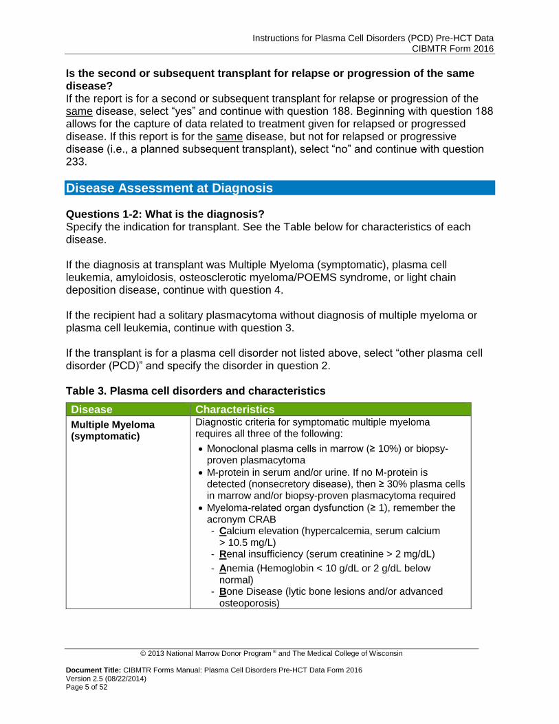
Is the second or subsequent transplant for relapse or progression of the same disease? If the report is for a second or subsequent transplant for relapse or progression of the same disease, select “yes” and continue with question 188. Beginning with question 188 allows for the capture of data related to treatment given for relapsed or progressed disease. If this report is for the same disease, but not for relapsed or progressive disease (i.e., a planned subsequent transplant), select “no” and continue with question 233.
Disease Assessment at Diagnosis Questions 1-2: What is the diagnosis? Specify the indication for transplant. See the Table below for characteristics of each disease. If the diagnosis at transplant was Multiple Myeloma (symptomatic), plasma cell leukemia, amyloidosis, osteosclerotic myeloma/POEMS syndrome, or light chain deposition disease, continue with question 4. If the recipient had a solitary plasmacytoma without diagnosis of multiple myeloma or plasma cell leukemia, continue with question 3. If the transplant is for a plasma cell disorder not listed above, select “other plasma cell disorder (PCD)” and specify the disorder in question 2. Table 3. Plasma cell disorders and characteristics
Disease Characteristics
Multiple Myeloma (symptomatic)
Diagnostic criteria for symptomatic multiple myeloma requires all three of the following:
Monoclonal plasma cells in marrow (≥ 10%) or biopsy-proven plasmacytoma
M-protein in serum and/or urine. If no M-protein is detected (nonsecretory disease), then ≥ 30% plasma cells in marrow and/or biopsy-proven plasmacytoma required
Myeloma-related organ dysfunction (≥ 1), remember the acronym CRAB - Calcium elevation (hypercalcemia, serum calcium
> 10.5 mg/L) - Renal insufficiency (serum creatinine > 2 mg/dL)
- Anemia (Hemoglobin < 10 g/dL or 2 g/dL below normal)
- Bone Disease (lytic bone lesions and/or advanced osteoporosis)

Instructions for Plasma Cell Disorders (PCD) Pre-HCT Data CIBMTR Form 2016
© 2013 National Marrow Donor Program ® and The Medical College of Wisconsin
Document Title: CIBMTR Forms Manual: Plasma Cell Disorders Pre-HCT Data Form 2016 Version 2.5 (08/22/2014) Page 6 of 52
Table 3. Plasma cell disorders and characteristics (cont.)
Disease Characteristics
Plasma Cell Leukemia Peripheral blood absolute plasma cell count of at least 2.0 x 10⁹/L (2,000 cells/mm3)
More than 20% plasma cells in the peripheral differential white blood cell count.1
Solitary Plasmacytoma (in absence of bone marrow findings diagnostic for multiple myeloma or plasma cell leukemia)
Extramedullary:
No M-protein in serum and/or urine
Extramedullary tumor of clonal plasma cells
Normal bone marrow
Normal skeletal survey
No related organ or tissue impairment (end organ damage including bone lesions)
Bone Derived
No M-protein in serum and/or urine
Single area of bone destruction due to clonal plasma cells
Bone marrow not consistent with multiple myeloma
Normal skeletal survey (and MRI of spine and pelvis if done)
No related organ or tissue impairment (no end organ damage other than solitary bone lesion)1
Note: if the recipient has greater than one plasmacytoma, but has not been diagnosed with another plasma cell disorder, select “other plasma cell disorder” and specify how many plasmacytomas are present and if each is bone derived or extramedullary.
Amyloidosis Amyloidosis is the buildup of abnormally folded proteins in various tissues of the body. Affected tissues may include the kidneys, heart, liver, gastrointestinal tract, etc. In the most common type of amyloidosis, “AL amyloidosis,” light chains from antibodies function as the amyloid protein, building up within organs and disrupting organ function. Serum and urine tests are useful for evaluating amyloidosis, but a tissue biopsy is the best way to diagnose the condition.

Instructions for Plasma Cell Disorders (PCD) Pre-HCT Data CIBMTR Form 2016
© 2013 National Marrow Donor Program ® and The Medical College of Wisconsin
Document Title: CIBMTR Forms Manual: Plasma Cell Disorders Pre-HCT Data Form 2016 Version 2.5 (08/22/2014) Page 7 of 52
Table 3. Plasma cell disorders and characteristics (cont.)
Disease Characteristics
Osteosclerotic myeloma/ POEMS Syndrome
POEMS syndrome is poorly understood, but generally refers to Polyneuropathy, Organomegaly, Endocrinopathy, M-protein, and Skin changes. Diagnosis may be made using the presence of the major criteria and one minor criteria below:
Major Criteria (both of the following):
Polyneuropathy
Monoclonal plasmaproliferative disorder
Minor Criteria (at least one of the following):
Sclerotic bone lesions†
Castleman disease†
Organomegaly (splenomegaly, hepatomegaly, lymphadenopathy)
Edema (edema, pleural effusion, or ascites)
Endocrinopathy (adrenal, thyroid‡, pituitary, gonadal, parathyroid, pancreatic‡)
Skin changes (hyperpigmentation, hypertrichosis, plethora, hemangiomata, white nails)
Papilledema †Osteosclerotic lesion or Castleman disease is usually present. ‡Because of the high prevalence of diabetes mellitus and thyroid abnormalities, this diagnosis alone is not sufficient to meet this minor criterion.2
Light Chain Deposition Disease
Similar to amyloidosis, light chain deposition disease is characterized by the overproduction and deposition of light chains in organs throughout the body; however, the organ most often affected is the kidneys. Under microscopy, the pattern of deposition and the use of staining techniques help pathologists differentiate between amyloidosis and light chain deposition disease.3
1The International Myeloma Working Group. Criteria for the classification of monoclonal gammopathies, multiple myeloma, and related disorders: a report of the International Myeloma Working Group. Brit J Haematol. 2003;121(5):749-757.
2Dispenzieri A, Kyle RA, Lacy MQ, Rajkumar SV, Therneau TM, Larson DR, Greipp PR, Witzig TE, Basu R, Suarez GA, Fonseca R, Lust JA, Gertz MA. POEMS syndrome: definitions and long-term outcome. Blood. 2003;101(7), 2496-2506.
3University of North Carolina Kidney Center. Light Chain Deposition Disease. Accessed at: http://www.unckidneycenter.org/kidneyhealthlibrary/lightchain.html Accessibility verified on April 5, 2013.
Question 3: Solitary plasmacytoma was: Indicate if the solitary plasmacytoma was “bone derived” or “extramedullary.” Refer to Table 3 above for additional information regarding the characteristics of each type.

Instructions for Plasma Cell Disorders (PCD) Pre-HCT Data CIBMTR Form 2016
© 2013 National Marrow Donor Program ® and The Medical College of Wisconsin
Document Title: CIBMTR Forms Manual: Plasma Cell Disorders Pre-HCT Data Form 2016 Version 2.5 (08/22/2014) Page 8 of 52
Question 4: What was the date of diagnosis? Report the date the recipient was first diagnosed with the plasma cell disorder indicated for transplant. Enter the date the blood/urine was collected for the laboratory evaluations (e.g., serum/urine protein electrophoresis [SPEP/UPEP, respectively], or serum/urine immunofixation) or enter the date of the first pathological diagnosis (e.g., bone marrow biopsy, plasmacytoma). Enter the date the sample was collected for examination. If the exact date is not known, use the process described for reporting partial or unknown dates in General Instructions, Guidelines for Completing Forms. Question 5: Did the recipient have a preceding or concurrent plasma cell disorder? Indicate if the recipient had a concurrent or preceding plasma cell disorder. Many recipients progress to symptomatic myeloma from a preceding condition or have a concurrent plasma cell disorder, such as amyloidosis.
Example 1. If a recipient has smoldering myeloma (asymptomatic) and then develops symptomatic multiple myeloma, “multiple myeloma (symptomatic)” should be reported as the primary diagnosis in question 1 and “smoldering myeloma (asymptomatic)” should be reported in question 6. Example 2. If a recipient has smoldering myeloma (asymptomatic) and amyloidosis, “amyloidosis” should be reported as the primary diagnosis in question 1 and “smoldering myeloma (asymptomatic)” should be reported in question 6. Example 3. If the recipient has symptomatic multiple myeloma and amyloidosis, “multiple myeloma (symptomatic)” should be reported as the primary diagnosis in question 1 and “amyloidosis” should be reported as a concurrent diagnosis is question 6.
Questions 6-7: Specify preceding/concurrent disorder: Indicate the preceding or concurrent disorder. See Table 3 for descriptions of disease and the previous question for examples of situations with preceding or concurrent disorders. If the recipient has a preceding or concurrent plasma cell disorder that is not listed, select “other plasma cell disorder (PCD)” and specify the type in question 7. Question 8: Date of diagnosis of preceding/concurrent disorder: Report the date the recipient was first diagnosed with the preceding or concurrent plasma cell disorder. Enter the date the blood/urine was collected for the laboratory evaluations (e.g., serum/urine protein electrophoresis [SPEP/UPEP, respectively], or serum/urine immunofixation) or enter the date of the first pathological diagnosis (e.g., bone marrow biopsy, plasmacytoma, tissue). Enter the date the sample was collected for examination.

Instructions for Plasma Cell Disorders (PCD) Pre-HCT Data CIBMTR Form 2016
© 2013 National Marrow Donor Program ® and The Medical College of Wisconsin
Document Title: CIBMTR Forms Manual: Plasma Cell Disorders Pre-HCT Data Form 2016 Version 2.5 (08/22/2014) Page 9 of 52
If the exact date is not known, use the process described for reporting partial or unknown dates in General Instructions, Guidelines for Completing Forms. Copy questions 6-8 to report more than one concurrent or preceding disorder.
Laboratory Studies at Diagnosis For questions 9-118, report values obtained at diagnosis or prior to the first treatment for the plasma cell disorder for which the transplant was performed. If testing is performed multiple times prior to the start of the first treatment, report the last test before the start of treatment. If the recipient has a plasma cells disorder other than plasma cell leukemia, do not answer questions 15-18, as they apply only to the diagnosis of PCL. Questions 9-10: WBC: Indicate if the WBC was “known” or “unknown” at the time of plasma cell disorder diagnosis. If “known,” report the value and unit of measure documented on the laboratory report in question 10. If “unknown,” continue with question 11. Questions 11-12: Hemoglobin: Indicate whether the hemoglobin was “known” or “unknown” at the time of plasma cell disorder diagnosis. If “known,” report the value and unit of measure documented on the laboratory report in question 12. If “unknown” continue with question 13. Question 13-14: Platelets: Indicate whether the platelet count was “known” or “unknown” at the time of plasma cell disorder diagnosis. If “unknown” continue with question 15. Questions 15-16: Absolute number of plasma cells in blood: (for PCL only) Indicate if the absolute number of plasma cells in the blood was “known” or “unknown” at the time of plasma cell leukemia (PCL) diagnosis. If “known,” report the absolute number of plasma cells in the blood documented on the laboratory report in question 16. If “unknown” continue with question 17. If only the percentage of plasma cells is available, multiply the percentage of plasma cells by the white blood cell count (WBC) to determine the absolute number of plasma cells. Questions 17-18: Plasma cells in blood: (for PCL only) Indicate if the percentage of plasma cells in the blood was “known” or “unknown” at the time of PCL diagnosis. If “known,” report the percentage documented on the laboratory report in question 18. If “unknown” continue with question 19.

Instructions for Plasma Cell Disorders (PCD) Pre-HCT Data CIBMTR Form 2016
© 2013 National Marrow Donor Program ® and The Medical College of Wisconsin
Document Title: CIBMTR Forms Manual: Plasma Cell Disorders Pre-HCT Data Form 2016 Version 2.5 (08/22/2014) Page 10 of 52
Questions 19-20: Serum albumin: Indicate whether the serum albumin was “known” or “unknown” at the time of plasma cell disorder diagnosis. If “known,” report the value and unit of measure documented on the laboratory report. If “unknown,” continue with question 21. Questions 21-22: Serum calcium: Indicate whether the serum calcium was “known” or “unknown” at the time of plasma cell disorder diagnosis. If “known,” report the value and unit of measure documented on the laboratory report in question 22. If “unknown,” continue with question 23. Questions 23-24: Serum creatinine: Indicate whether the serum creatinine was “known” or “unknown” at the time of plasma cell disorder diagnosis. If “known,” report the laboratory value and unit of measure documented on the laboratory report in question 24 and continue with question 25. If “unknown,” continue with question 26. Question 25: Upper limit of serum creatinine: Indicate the upper limit of normal for serum creatinine value used at your institution. Questions 26-27: LDH Indicate whether the LDH (lactate dehydrogenase) level was “known” or “unknown” at the time of plasma cell disorder diagnosis. If “known,” report the value and unit of measure documented on the laboratory report in question 27 and continue with question 28. If “unknown,” continue with question 29. Question 28: Upper limit of normal for LDH: Indicate the upper limit of normal for LDH value and the unit of measure used at your institution. Questions 29-30: Serum β2 microglobulin: At the time of plasma cell disorder diagnosis, an elevated serum β2 microglobulin protein may indicate a poorer prognosis. If this value was “known,” report the value and unit of measure documented on the laboratory report in question 30. If “unknown,” continue with question 31. Question 31: What was the Durie-Salmon staging? Indicate the Durie-Salmon staging at diagnosis and continue with question 32. If the Durie-Salmon stage is not documented in the medical record, use Table 4 below to determine the appropriate stage. If the Durie-Salmon stage is unknown and cannot be determined using the table below, select “unknown” and continue with question 33.

Instructions for Plasma Cell Disorders (PCD) Pre-HCT Data CIBMTR Form 2016
© 2013 National Marrow Donor Program ® and The Medical College of Wisconsin
Document Title: CIBMTR Forms Manual: Plasma Cell Disorders Pre-HCT Data Form 2016 Version 2.5 (08/22/2014) Page 11 of 52
Table 4. Durie-Salmon Staging System for Multiple Myeloma
Stage Criteria
I All of the following:
Hemoglobin > 10 g/dL
Serum calcium normal (< 10.5 mg/dL)
On radiograph, normal bone structure or solitary bone plasmacytoma only
Low M-component production rate (IgG < 5 g/dL, IgA < 3 g/dL), Urinary light chain M-component on electrophoresis (<4 g/24 hr)
II Fitting neither stage I nor stage III
III One or more of the following:
Hemoglobin < 8.5 g/dL
Serum calcium > 12 mg/dL
Advanced lytic bone lesions (three or more lytic lesions)
High M-component product rate (IgG > 7 g/dL, IgA > 5 g/dL), Urinary light chain M-component on electrophoresis (> 12 g/24 hr)
Adapted from Durie BG, Salmon SE. A clinical staging system for multiple myeloma: Correlation of measured myeloma cell mass with presenting clinical features, response to treatment, and survival. Cancer. 1975;36(3):842-854.
Question 32: What was the Durie-Salmon sub classification? Indicate the Durie-Salmon sub classification at diagnosis and continue with question 33. If the Durie-Salmon sub classification is not documented in the medical record, use the criteria below to determine the appropriate sub classification.
A: Relatively normal renal function (serum creatinine < 2.0 mg/dL)
B: Abnormal renal function (serum creatinine ≥ 2.0 mg/dL) Question 33: Immunochemical type: Indicate whether the plasma cell disorder is secretory or non-secretory. Secretory plasma cell disorders are characterized by the presence of clonal paraproteins (M-protein) in the blood and serum, detected by immunofixation. Non-secretory refers to the absence of clonal paraprotein (M-protein) in the serum or urine. If the plasma cell disorder is secretory, continue with question 34. If the plasma cell disorder is non-secretory, continue with question 54.
NOTE: Paraprotein identification and quantification
Specify the paraprotein(s) (commonly called the M-spike or monoclonal protein) identified based on immunofixation (IFE) results. Report the quantity of paraprotein using questions 40-53 based on serum protein electrophoresis (SPEP) or urine protein electrophoresis (UPEP), or using questions 54-58 based on serum free light chains.

Instructions for Plasma Cell Disorders (PCD) Pre-HCT Data CIBMTR Form 2016
© 2013 National Marrow Donor Program ® and The Medical College of Wisconsin
Document Title: CIBMTR Forms Manual: Plasma Cell Disorders Pre-HCT Data Form 2016 Version 2.5 (08/22/2014) Page 12 of 52
Questions 34-35: Serum heavy chain: Indicate the involved heavy chain(s) in the plasma cell disorder detected on serum immunofixation. The involved heavy chain, also called M-spike or monoclonal protein, can be identified but not quantified using this test. If only one heavy chain is involved, select “IgG,” “IgA,” “IgM,” “IgD, or “IgE” and continue with question 36. If the heavy chain is IgM, ensure that the disease subtype is not Waldenstrom’s Macroglobulinemia. If the disease subtype is Waldenstrom’s Macroglobulinemia, complete Form 2019 and update the Pre-TED form to indicate that Waldenstrom’s is the transplant indication. If two heavy chains are involved in the plasma cell disorder, select “Biclonal” and specify which two heavy chains in question 35. If the recipient has light chain only disease, meaning that no monoclonal heavy chain was present at any time during the disease history, select “Not applicable (light chain only disease)” and continue with question 36. Table 5. Concept of Clonality in Multiple Myeloma
Type of Myeloma M-proteins Expressed
Plasma Cell Myeloma One heavy chain (IgG, IgA, etc.) and one light chain (either kappa or lambda)
Biclonal Myeloma Two different M-proteins (e.g., IgG Kappa and IgA Lambda)
Question 36: Serum light chain: Indicate the involved light chain in the plasma cell disorder detected on serum immunofixation. The involved light chain can be identified, but not quantified, using this test. Select “κ” if the kappa light chain is involved or “λ” if the lambda light chain is involved. Questions 37-38: Urine heavy chain: Indicate the involved heavy chain(s) in the plasma cell disorder detected on urine immunofixation. The involved heavy chain, also called M-spike or monoclonal protein, can be identified but not quantified using this test. If only one heavy chain is involved, select “IgG,” “IgA,” “IgM,” “IgD, or “IgE” and continue with question 39. If the heavy chain is IgM, ensure that the disease subtype is not Waldenstrom’s Macroglobulinemia. If the disease subtype is Waldenstrom’s Macroglobulinemia, complete Form 2019 and update the Pre-TED form to indicate that Waldenstrom’s is the transplant indication. If two heavy chains were involved in the plasma cell disorder, select “Biclonal” and specify which two heavy chains in question 38. If the recipient has light chain only disease, meaning that no monoclonal heavy chain was present at any time during the disease history, select “Not applicable (light chain only disease)” and continue with question 39. Question 39: Urine light chain: Indicate the involved light chain in the plasma cell disorder detected on urine immunofixation. The involved light chain can be identified, but not quantified, using this test. Select “κ” if the kappa light chain is involved or “λ” if the lambda light chain is involved.

Instructions for Plasma Cell Disorders (PCD) Pre-HCT Data CIBMTR Form 2016
© 2013 National Marrow Donor Program ® and The Medical College of Wisconsin
Document Title: CIBMTR Forms Manual: Plasma Cell Disorders Pre-HCT Data Form 2016 Version 2.5 (08/22/2014) Page 13 of 52
Questions 40-41: Serum monoclonal protein (M-spike): (only from electrophoresis) Monoclonal gammopathy is defined as the increased production of abnormal immunoglobulins. The abnormal protein produced is called paraprotein or M-protein. Indicate whether the serum monoclonal immunoglobulin was “known” or “unknown” at the time of the plasma cell disorder diagnosis. If “known,” report the value and unit of measure documented on the laboratory report in question 41. If “unknown,” continue with question 42. For recipients with biclonal myeloma, report the serum monoclonal immunoglobulin with the largest quantity.
NOTE: Urinary Monoclonal Protein Questions 42-43 are intended to capture the 24-hour urine monoclonal protein results, not the 24-hour protein excretion (questions 44-45 capture the total protein secretion/24 hours). The results will be reported as XX g or XX g/dL. If the value is reported in XX g/dL, it can be multiplied by the volume of the urine to determine the 24-hour urine monoclonal protein. For example: (total in g/dL of monoclonal protein) x (total urine volume) = urinary M-protein/24 hours (0.145 g/dL of monoclonal protein) x (1500 mL total urine) = 2.175 g/24 hours. Do not report immunofixation results here. Questions 42-43: Urinary monoclonal protein (M-spike): Indicate whether the amount of urinary monoclonal protein was “known” or “unknown” at the time of plasma cell disorder diagnosis. The value reported here should be based on a 24 hour urine collection. If “known,” report the laboratory value in question 43. If “unknown,” continue with question 44. Questions 44-45: Total urinary protein secretion: Indicate whether the amount of urinary protein was “known” or “unknown” at the time of plasma cell disorder diagnosis. The value reported here should be based on a 24-hour urine collection. If “known,” report the laboratory value in question 45. If “unknown,” continue with question 46. Questions 46-47: 24-hour creatinine clearance: Indicate whether the amount of urinary protein was “known” or “unknown” at the time of plasma cell disorder diagnosis. The value reported here should be based on a 24-hour urine collection in milliliters per minute (mL/minute). If “known,” report the laboratory value in question 47. If “unknown,” continue with question 48.
Instructions for Plasma Cell Disorders (PCD) Pre-HCT Data CIBMTR Form 2016
© 2013 National Marrow Donor Program ® and The Medical College of Wisconsin
Document Title: CIBMTR Forms Manual: Plasma Cell Disorders Pre-HCT Data Form 2016 Version 2.5 (08/22/2014) Page 14 of 52
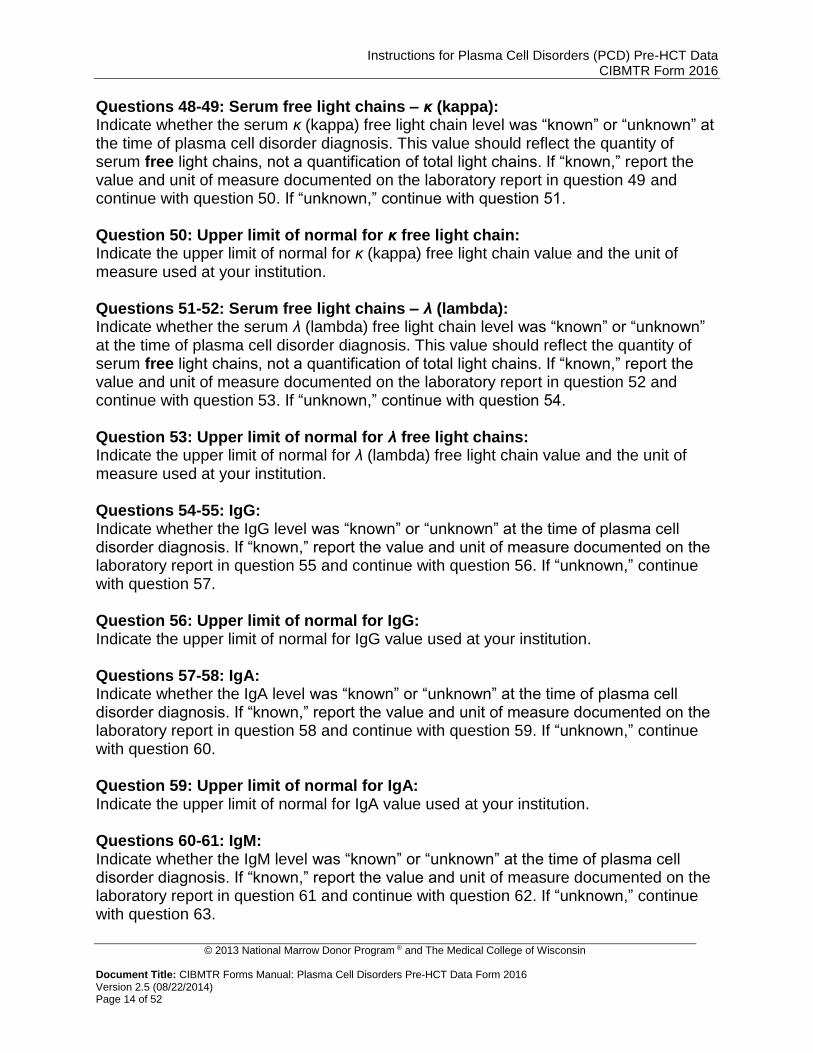
Questions 48-49: Serum free light chains – κ (kappa): Indicate whether the serum κ (kappa) free light chain level was “known” or “unknown” at the time of plasma cell disorder diagnosis. This value should reflect the quantity of serum free light chains, not a quantification of total light chains. If “known,” report the value and unit of measure documented on the laboratory report in question 49 and continue with question 50. If “unknown,” continue with question 51. Question 50: Upper limit of normal for κ free light chain: Indicate the upper limit of normal for κ (kappa) free light chain value and the unit of measure used at your institution. Questions 51-52: Serum free light chains – λ (lambda): Indicate whether the serum λ (lambda) free light chain level was “known” or “unknown” at the time of plasma cell disorder diagnosis. This value should reflect the quantity of serum free light chains, not a quantification of total light chains. If “known,” report the value and unit of measure documented on the laboratory report in question 52 and continue with question 53. If “unknown,” continue with question 54. Question 53: Upper limit of normal for λ free light chains: Indicate the upper limit of normal for λ (lambda) free light chain value and the unit of measure used at your institution. Questions 54-55: IgG: Indicate whether the IgG level was “known” or “unknown” at the time of plasma cell disorder diagnosis. If “known,” report the value and unit of measure documented on the laboratory report in question 55 and continue with question 56. If “unknown,” continue with question 57. Question 56: Upper limit of normal for IgG: Indicate the upper limit of normal for IgG value used at your institution. Questions 57-58: IgA: Indicate whether the IgA level was “known” or “unknown” at the time of plasma cell disorder diagnosis. If “known,” report the value and unit of measure documented on the laboratory report in question 58 and continue with question 59. If “unknown,” continue with question 60. Question 59: Upper limit of normal for IgA: Indicate the upper limit of normal for IgA value used at your institution. Questions 60-61: IgM: Indicate whether the IgM level was “known” or “unknown” at the time of plasma cell disorder diagnosis. If “known,” report the value and unit of measure documented on the laboratory report in question 61 and continue with question 62. If “unknown,” continue with question 63.

Instructions for Plasma Cell Disorders (PCD) Pre-HCT Data CIBMTR Form 2016
© 2013 National Marrow Donor Program ® and The Medical College of Wisconsin
Document Title: CIBMTR Forms Manual: Plasma Cell Disorders Pre-HCT Data Form 2016 Version 2.5 (08/22/2014) Page 15 of 52
Question 62: Upper limit of normal for IgM: Indicate the upper limit of normal for IgM value used at your institution. Questions 63-64: IgD: Indicate whether the IgD level was “known” or “unknown” at the time of plasma cell disorder diagnosis. If “known,” report the value and unit of measure documented on the laboratory report in question 64 and continue with question 65. If “unknown,” continue with question 66. Question 65: Upper limit of normal for IgD: Indicate the upper limit of normal for IgD value used at your institution. Questions 66-67: IgE: Indicate whether the IgE level was “known” or “unknown” at the time of plasma cell disorder diagnosis. If “known,” report the value and unit of measure documented on the laboratory report in question 67 and continue with question 68. If “unknown,” continue with question 69. Question 68: Upper limit of normal for IgE: Indicate the upper limit of normal for IgE value used at your institution.
NOTE:
Under normal circumstances, a marrow aspirate is used to obtain the differential cell count, review morphology of the cells, and perform cytogenetic studies, flow cytometry, etc. A biopsy is obtained to evaluate the overall cellularity of the marrow. In the case of myeloma, the marrow plasma cells tend to be a patchy infiltrate rather than a diffuse infiltrate as in the case of acute leukemia. Therefore, it is possible that the plasma cell numbers may vary between the aspirate and the biopsy.
The percentage of plasma cells in the bone marrow aspirate and/or biopsy may also be identified on a flow cytometry report. A flow cytometry report may NOT be used as source documentation when reporting the data for questions 69-72.
If the bone marrow pathology report states a range for plasma cells, enter the average of the range rounded to the nearest whole number (e.g., if 0-5%, enter 3%).
If the report states > 90% plasma cells, enter 91% on the form.
If the report states a marrow packed with plasma cells or sheets of plasma cells, report 99% on the form.
If the report states < 5% plasma cells, enter 4% on the form. Questions 69-70: Plasma cells in bone marrow aspirate: Indicate whether the percentage of plasma cells in the bone marrow aspirate was “known” or “unknown” at the time of plasma cell disorder diagnosis. If “known,” report the percentage of plasma cells in the bone marrow aspirate documented on the pathology report in question 70. If “unknown,” continue with question 71.

Instructions for Plasma Cell Disorders (PCD) Pre-HCT Data CIBMTR Form 2016
© 2013 National Marrow Donor Program ® and The Medical College of Wisconsin
Document Title: CIBMTR Forms Manual: Plasma Cell Disorders Pre-HCT Data Form 2016 Version 2.5 (08/22/2014) Page 16 of 52
Questions 71-72: Plasma cells in bone marrow biopsy: Indicate whether the percentage of plasma cells in the bone marrow biopsy was “known” or “unknown” at the time of plasma cell disorder diagnosis. If “known,” report the percentage of plasma cells in the bone marrow biopsy documented on the pathology report in question 72. If “unknown,” continue with question 73. Question 73: Were conventional cytogenetics tested? Cytogenetics is the study of chromosomes. Cytogenetic assessment involves testing blood or bone marrow for the presence of a known chromosomal abnormality that reflects the recipient’s disease. Cytogenetics may also be referred to as karyotyping or g-banding. Indicate if cytogenetic studies were obtained at the time the recipient was diagnosed with a plasma cell disorder or prior to the start of treatment. If cytogenetic studies were obtained, select “yes” and continue with question 74. If no cytogenetic studies were obtained or it is unknown if chromosome studies were performed, select “no” or “unknown” and continue with question 96. Question 74: Results of test: If cytogenetic studies identified abnormalities, indicate “abnormalities identified” and continue with question 75. If cytogenetic studies yielded no evaluable metaphases or there were no abnormalities identified, indicate this and continue with question 96. Questions 75-94: Specify cytogenetic abnormalities identified via conventional cytogenetics at diagnosis: Report all abnormalities identified by all methods of cytogenetic assessment prior to the start of first therapy by selecting “yes” or “no” for each question. Do not leave any responses blank. If one or more abnormality is best classified as “other abnormality,” select “yes” in question 93 and specify in question 94. Question 95: Was documentation submitted to the CIBMTR (e.g., cytogenetic report)? Indicate if a copy of the cytogenetic report is attached. Use the Log of Appended Documents (Form 2800) to attach a copy of the cytogenetic report. Attaching a copy of the report may prevent additional queries. Question 96: Were cytogenetics tested via FISH? FISH, fluorescence in situ hybridization, is a sensitive technique that assesses a large number of cells. This technique uses special probes that recognize and bind to fragments of DNA commonly found in plasma cell disorders. These probes are mixed with cells from the recipient’s blood. A fluorescent “tag” is then used to visualize the binding of the probe to the diseased cells.

Instructions for Plasma Cell Disorders (PCD) Pre-HCT Data CIBMTR Form 2016
© 2013 National Marrow Donor Program ® and The Medical College of Wisconsin
Document Title: CIBMTR Forms Manual: Plasma Cell Disorders Pre-HCT Data Form 2016 Version 2.5 (08/22/2014) Page 17 of 52
Indicate if FISH studies were obtained at the time the recipient was diagnosed with a plasma cell disorder or prior to the start of treatment. If FISH studies were obtained, select “yes” and continue with question 97. If no FISH studies were obtained or it is unknown if FISH studies were performed, select “no” or “unknown” and continue with question 117. Question 97: Results of test: If FISH studies identified abnormalities, indicate “abnormalities identified” and continue with question 98. If there were no abnormalities identified, indicate “no abnormalities” and continue with question 117. Questions 98-115: Specify cytogenetic abnormalities identified via FISH at diagnosis: Report all abnormalities identified by all methods of FISH assessment prior to the start of first therapy by selecting “yes” or “no” for each question. Do not leave any responses blank. If one or more abnormality is best classified as “other abnormality,” select “yes” in question 114 and specify in question 115. Question 116: Was documentation submitted to the CIBMTR (e.g., FISH report)? Indicate if a copy of the FISH report is attached. Use the Log of Appended Documents (Form 2800) to attach a copy of the FISH report. Attaching a copy of the report may prevent additional queries. Question 117: Was a gene expression profile performed? Gene expression profiling (GEP) allows for the analysis of thousands of genes at once, creating a global picture of cell function. GEP can distinguish cells that are actively dividing and show how cells react to specific treatments.1
If gene expression profiling was performed at the time of plasma cell disorder diagnosis or prior to the start of first therapy, indicate “yes” and continue with question 118. If gene expression was not performed, select “no” and continue with question 119. 1Brunner J, Dispenzieri A. “Forms Revision: Myeloma Changes.” 2013 Tandem Meetings: CIBMTR Clinical Research Professionals/Data Manager Annual Meeting. Accessed at http://www.cibmtr.org/Meetings/Materials/CRPDMC/Documents/2013/Feb2013/Feb13FormRevMM-6up.pdf.
Question 118: Were results considered high-risk myeloma? Based on the opinion of a physician, indicate if the results of the gene expression profile are considered high-risk myeloma. Indicate “yes” or “no.”

Instructions for Plasma Cell Disorders (PCD) Pre-HCT Data CIBMTR Form 2016
© 2013 National Marrow Donor Program ® and The Medical College of Wisconsin
Document Title: CIBMTR Forms Manual: Plasma Cell Disorders Pre-HCT Data Form 2016 Version 2.5 (08/22/2014) Page 18 of 52
Amyloidosis Organ Involvement at Diagnosis Complete questions 119-187 for amyloidosis patients only. If diagnosis was other than amyloidosis (question 1), or there is no evidence or history of it (question 6), skip to question 188. Question 119: Was an abdominal fat aspirate performed? Abdominal fat is the most common initial biopsy site when amyloidosis is suspected. A sample of abdominal fat is taken by needle aspiration and stained with Congo red dye. Indicate if an abdominal fat biopsy was performed at diagnosis or prior to the first therapy. If “yes,” continue with question 120. If “no,” continue with question 121. Question 120: Specify the aspirate results: Samples that are positive have their normal architecture disrupted by amyloid deposits, which appear red under microscopy due to Congo red stain. Under polarized light microscopy, amyloid deposits stained with Congo red appear green (birefringence). Indicate the results of the abdominal fat aspirate. Select “positive (for amyloid involvement)” if the aspirate showed characteristics of amyloid involvement. Select “negative” if the aspirate was negative. Select “unknown” if the results of the evaluation were unknown or inconclusive. Continue with question 121. Question 121: Was a renal biopsy performed? Kidney involvement in amyloidosis is common, and evaluation of renal tissue may be necessary to determine the extent of the disease. Indicate if a renal biopsy was performed at diagnosis or prior to the first therapy. If “yes,” continue with question 122. If “no,” continue with question 123. Question 122: Specify the renal biopsy results: Samples that are positive have their normal architecture disrupted by amyloid deposits, which appear red under microscopy due to Congo red stain. Under polarized light microscopy, amyloid deposits stained with Congo red appear green (birefringence). Indicate the results of the renal biopsy. Select “positive (for amyloid involvement)” if the biopsy showed characteristics of amyloid involvement. Select “negative” if the biopsy was negative. Select “unknown” if the results of the evaluation were unknown or inconclusive. Continue with question 123. Question 123: Was a cardiographic imaging procedure performed? Cardiographic imaging may show amyloid infiltration in heart tissue. Cardiac MRI, echocardiogram [(sometimes referred to as an echo) (do not report an ECG or EKG, which refer to electrocardiogram)], or Multiple Gate Acquisition (MUGA) scans may be performed to assess heart involvement. Indicate if cardiographic imaging was performed at diagnosis or prior to the first therapy. If “yes,” continue with question 124. If “no,” continue with question 130.

Instructions for Plasma Cell Disorders (PCD) Pre-HCT Data CIBMTR Form 2016
© 2013 National Marrow Donor Program ® and The Medical College of Wisconsin
Document Title: CIBMTR Forms Manual: Plasma Cell Disorders Pre-HCT Data Form 2016 Version 2.5 (08/22/2014) Page 19 of 52
Question 124: Was a cardiac MRI done? Cardiac MRI may be used to differentiate amyloid involvement from other cardiopathologies. Indicate if a cardiac MRI was performed at diagnosis or prior to the first therapy. If “yes,” continue with question 125. If “no,” continue with question 126. Question 125: Specify cardiac MRI results: Characteristics of amyloid involvement in cardiac tissue include impaired ventricular systolic function, thickened valves, increased atrial septal thickness and left ventricular mass, pleural and pericardial effusions, and subendocardial hyperenhancement. Indicate if the results of cardiac MRI were “normal,” “abnormal,” or “unknown,” and continue with question 126.
Penugonda N. Cardiac MRI in infiltrative disorders: A concise review. Curr Cardiol Rev.2010;6(2):134-136.
Question 126: Was the left ventricular ejection fraction measured? The left ventricular ejection fraction (LVEF) is a percentage that represents the volume of blood pumped from the left ventricle into the aorta (also known as stroke volume) compared to the volume of blood in the ventricle just prior to the heart contraction (also known as end diastolic volume). Indicate if the left ventricular ejection fraction (LVEF) was measured. If “yes,” continue with question 127. If “no,” continue with question 129. Question 127: Specify the left ventricular ejection fraction: Indicate the left ventricular ejection fraction at diagnosis or prior to the first therapy. Most imaging reports will report the LVEF, but if not, the LVEF may be determined by dividing the stroke volume (SV, the volume of blood pumped into the aorta from the left ventricle) by the end diastolic volume (EDV, the volume of blood in the left ventricle just prior to contraction) of the left ventricle. For example, if the stroke volume was 75 ml and the end diastolic volume was 150ml, the ejection fraction would be 50%. If the recipient had multiple assessments using different methods, report the most recent assessment prior to the initiation of treatment. Question 128: Specify the method used to determine the left ventricular ejection fraction: Indicate the method used to determine the LVEF reported in question 127. Question 129: Was diastolic dysfunction present? Diastole is the period in which chambers of the heart fill with blood. Diastolic dysfunction may be characterized by the difficulty of the ventricles to expand and contract appropriately due to stiffening of the heart walls by amyloid deposits. Indicate if diastolic dysfunction was present. Specify “yes,” “no,” or “unknown,” and continue with question 130. Questions 130-131: Specify the intraventricular septal wall thickness measured by echocardiogram: The heart is divided into the right and left sides by the septum. The area between the left and right ventricles is the intraventricular septum. Indicate if the intraventricular

Instructions for Plasma Cell Disorders (PCD) Pre-HCT Data CIBMTR Form 2016
© 2013 National Marrow Donor Program ® and The Medical College of Wisconsin
Document Title: CIBMTR Forms Manual: Plasma Cell Disorders Pre-HCT Data Form 2016 Version 2.5 (08/22/2014) Page 20 of 52
septal thickness is “known” or “unknown.” If known, based on evaluation by echocardiogram, indicate the thickness of the intraventricular septal wall in question 131. If unknown or not measured by echocardiogram, continue with question 132. Question 132: Was a cardiac biopsy performed? Heart involvement in amyloidosis is common and evaluation of cardiac tissue may be necessary to determine the extent of the disease. Indicate if a cardiac biopsy was performed at diagnosis or prior to the first therapy. If “yes,” continue with question 133. If “no,” continue with question 134. Question 133: Specify the cardiac biopsy results: Samples that are positive have their normal architecture disrupted by amyloid deposits, which appear red under microscopy due to Congo red stain. Under polarized light microscopy, amyloid deposits stained with Congo red appear green (birefringence). Indicate the results of the cardiac biopsy. Select “positive (for amyloid involvement)” if the biopsy showed characteristics of amyloid involvement. Select “negative” if the biopsy was negative. Select “unknown” if the results of the evaluation were unknown or inconclusive. Continue with question 134. Question 134: Were any serum cardiac biomarkers assessed? Assessment of cardiac biomarkers helps determine if injury to cardiac tissue has occurred. Cardiac biomarkers include brain natriuretic peptide (BNP), N-terminal prohormone brain natriuretic peptide (NT-proBNP), troponin I, troponin T, and high-sensitivity troponin T. Indicate if serum cardiac biomarkers were assessed at diagnosis or prior to first therapy. If “yes,” continue with question 135. If “no” or “unknown” continue with question 150. Questions 135-136: Brain natriuretic peptide (BNP): Indicate if the BNP was assessed at the time of amyloidosis diagnosis. If “yes,” report the value and unit of measure documented on the laboratory report in question 136 and continue with question 137. If “no,” continue with question 138. Question 137: Upper limit of normal for BNP: Indicate the upper limit of normal for BNP used at your institution. Questions 138-139: N-terminal prohormone brain natriuretic peptide (NT-proBNP): Indicate if the NT-proBNP was assessed at the time of amyloidosis diagnosis. If “yes,” report the value and unit of measure documented on the laboratory report in question 139 and continue with question 140. If “no,” continue with question 141. Question 140: Upper limit of normal for NT-proBNP: Indicate the upper limit of normal for NT-proBNP used at your institution.

Instructions for Plasma Cell Disorders (PCD) Pre-HCT Data CIBMTR Form 2016
© 2013 National Marrow Donor Program ® and The Medical College of Wisconsin
Document Title: CIBMTR Forms Manual: Plasma Cell Disorders Pre-HCT Data Form 2016 Version 2.5 (08/22/2014) Page 21 of 52
Questions 141-142: Troponin I: Indicate if the Troponin I was assessed at the time of amyloidosis diagnosis. If “yes,” report the value and unit of measure documented on the laboratory report in question 142 and continue with question 143. If “no,” continue with question 144. Question 143: Upper limit of normal for troponin I: Indicate the upper limit of normal for Troponin I used at your institution. Questions 144-145: Troponin T: Indicate if the Troponin T was assessed at the time of amyloidosis diagnosis. If “yes,” report the value and unit of measure documented on the laboratory report in question 145 and continue with question 146. If “no,” continue with question 147. Question 146: Upper limit of normal for Troponin T: Indicate the upper limit of normal for Troponin T used at your institution. Questions 147-148: High-sensitivity troponin T: Indicate if the high-sensitivity troponin T was assessed at the time of amyloidosis diagnosis. If “yes,” report the value and unit of measure documented on the laboratory report in question 145 and continue with question 146. If “no,” continue with question 147. Question 149: Upper limit of normal for high-sensitivity troponin T: Indicate the upper limit of normal for high-sensitivity troponin T used at your institution. Question 150: Specify the recipient’s New York Heart Association functional classification of heart failure: (Symptoms may include dyspnea, chest pain, fatigue, and palpitations; activity level should be assessed with consideration for patient’s age group) Indicate the recipient’s New York Heart Association (NYHA) functional classification. If the recipient’s NYHA functional classification it not known, select “unknown.” Table 6. New York Heart Association Function Classification
Class Description
Class I Able to perform ordinary activities without symptoms; no limitation of physical activity
Class II Ordinary physical activity produces symptoms; slight limitation of physical activity
Class III Less-than-ordinary physical activity produces symptoms; moderate limitation of physical activity
Class IV Symptoms present even at rest; severe limitation of physical activity.

Instructions for Plasma Cell Disorders (PCD) Pre-HCT Data CIBMTR Form 2016
© 2013 National Marrow Donor Program ® and The Medical College of Wisconsin
Document Title: CIBMTR Forms Manual: Plasma Cell Disorders Pre-HCT Data Form 2016 Version 2.5 (08/22/2014) Page 22 of 52
Question 151: Was there clinical suspicion of gastrointestinal (GI) involvement? GI involvement by amyloidosis is usually proven by biopsy; however, clinical symptoms of gastrointestinal involvement may include esophageal reflux, constipation, nausea and abdominal pain, diarrhea, weight loss, or early satiety (fullness).1 Indicate if there was any clinical suspicion of GI involvement at diagnosis or prior to the first therapy. If “yes,” continue with question 152. If “no” or “unknown,” continue with question 154. 1Cowan AJ, Skinner M, Seldin DC, Berk JL, Lichtenstein DR, O'Hara CJ, Doros G, Sanchorawala V. Amyloidosis of the gastrointestinal tract: A 13-year, single-center, referral experience. Haematologica. 2013;98(1):141-146.
Question 152: Upper GI tract: Symptoms of upper GI tract involvement may include esophageal reflux, nausea, and abdominal pain. Indicate if the recipient had upper GI involvement. Select “yes” or “no” and continue with question 153. Question 153: Lower GI tract: Symptoms of lower GI tract involvement may include abdominal pain, nausea, weight loss, constipation, or diarrhea. Indicate if there was any suspicion of lower GI involvement. Select “yes” or “no” and continue with question 154. Question 154: Was a gastrointestinal biopsy performed? GI involvement in amyloidosis is less common than cardiac or renal involvement, and evaluation of GI tissue may be necessary to determine the extent of the disease. Indicate if a GI biopsy was performed at diagnosis or prior to the first therapy. If “yes,” continue with question 155. If “no,” continue with question 160. Question 155: Rectal Indicate if the recipient had a rectal biopsy to confirm amyloid involvement at the time of diagnosis or before first therapy. If “yes,” continue with question 156. If “no,” continue with question 157. Question 156: Specify the biopsy results: Samples that are positive have their normal architecture disrupted by amyloid deposits, which appear red under microscopy due to Congo red stain. Under polarized light microscopy, amyloid deposits stained with Congo red appear green (birefringence). Indicate the results of the rectal biopsy. Select “positive (for amyloid involvement)” if the biopsy showed characteristics of amyloid involvement. Select “negative” if the biopsy was negative. Select “unknown” if the results of the evaluation were unknown or inconclusive. Continue with question 157. Questions 157-158: Other site: Indicate if the recipient had a gastrointestinal site other than the rectum biopsied at diagnosis or prior to the first therapy. If “yes,” specify the site in question 158 and continue with question 159. If “no,” continue with question 160.

Instructions for Plasma Cell Disorders (PCD) Pre-HCT Data CIBMTR Form 2016
© 2013 National Marrow Donor Program ® and The Medical College of Wisconsin
Document Title: CIBMTR Forms Manual: Plasma Cell Disorders Pre-HCT Data Form 2016 Version 2.5 (08/22/2014) Page 23 of 52
Question 159: Specify the biopsy results: Samples that are positive have their normal architecture disrupted by amyloid deposits, which appear red under microscopy due to Congo red stain. Under polarized light microscopy, amyloid deposits stained with Congo red appear green (birefringence). Indicate the results of the GI biopsy. Select “positive (for amyloid involvement)” if the biopsy showed characteristics of amyloid involvement. Select “negative” if the biopsy was negative. Select “unknown” if the results of the evaluation were unknown or inconclusive. Continue with question 160. Question 160: Was hepatomegaly present on radiographic imaging (liver span > 15 cm) or on examination (liver edge palpable > 3 cm below right costal margin)? At the time of diagnosis or prior to first therapy, indicate if the liver spanned more than 15 cm or the edge of the liver was palpable more than 3 cm below the right costal margin by radiographic imaging. Indicate “yes” if hepatomegaly was present at diagnosis or prior to first therapy. Indicate “no” if hepatomegaly was not present at diagnosis or prior to first therapy. Indicate “unknown” if it was not possible to determine the presence or absence of hepatomegaly at diagnosis or prior to first therapy. Questions 161-162: Specify the level of serum alkaline phosphatase: Indicate whether the alkaline phosphatase (ALP) level at the time of amyloidosis diagnosis or prior to first treatment is “known” or “unknown.” If “known,” report the laboratory count and unit of measure documented on the laboratory report in question 162 and continue with question 163. If “unknown,” continue with question 164. Question 163: Upper limit of normal for alkaline phosphatase: Report the upper limit of normal for ALP at your institution. Question 164: Was a liver biopsy performed? Evaluation of liver tissue may be necessary to determine the extent of the disease. Indicate if a liver biopsy was performed at diagnosis or prior to the first therapy. If “yes,” continue with question 165. If “no,” continue with question 166. Question 165: Specify the liver biopsy results: Samples that are positive have their normal architecture disrupted by amyloid deposits, which appear red under microscopy due to Congo red stain. Under polarized light microscopy, amyloid deposits stained with Congo red appear green (birefringence). Indicate the results of the liver biopsy. Select “positive (for amyloid involvement)” if the biopsy showed characteristics of amyloid involvement. Select “negative” if the biopsy was negative. Select “unknown” if the results of the evaluation were unknown or inconclusive. Continue with question 166. Question 166: Was a sensory/motor exam performed? Indicate if a sensory/motor exam was performed. This exam evaluates the neurological status of the recipient and consists of assessing the recipient’s body positioning, involuntary movements, muscle tone, muscle strength, ability to sense pain and light

Instructions for Plasma Cell Disorders (PCD) Pre-HCT Data CIBMTR Form 2016
© 2013 National Marrow Donor Program ® and The Medical College of Wisconsin
Document Title: CIBMTR Forms Manual: Plasma Cell Disorders Pre-HCT Data Form 2016 Version 2.5 (08/22/2014) Page 24 of 52
touch, position sense (proprioception), stereognosia (ability to discern an object with eyes closed, such as a coin), graphesthesia (ability to identify number of letter drawn on skin with eyes closed), and extinction (ability to discern multiple simultaneous stimuli). If a sensory/motor exam was performed, indicate “yes,” and continue with question 167. If “no” or “unknown,” continue with question 168.
NYU School of Medicine; Russell S, Triola M. The Precise Neurological Exam. Available at: http://informatics.med.nyu.edu/modules/pub/neurosurgery/ Accessibility verified April 7, 2013.
Question 167: Specify the exam results: Indicate the results of the sensory/motor exam. If the recipient’s sensory/motor exam was within normal limits, select “normal.” If the recipient displayed neurologic impairment, select “abnormal.” If the results of the test are unknown or inconclusive, select “unknown.” Continue with question 168. Question 168: Was a nerve biopsy performed? Evaluation of nerve tissue may be necessary to determine the extent of the disease. Indicate if a nerve biopsy was performed at diagnosis or prior to the first therapy. If “yes,” continue with question 169. If “no,” continue with question 174. Question 169: Sural The most common site for a nerve biopsy is the sural nerve in the ankle. Indicate if the nerve biopsy was taken from the sural nerve. If “yes,” continue with question 170. If “no,” continue with question 171. Question 170: Specify the sural nerve biopsy results: Samples that are positive have their normal architecture disrupted by amyloid deposits, which appear red under microscopy due to Congo red stain. Under polarized light microscopy, amyloid deposits stained with Congo red appear green (birefringence). Indicate the results of the nerve biopsy. Select “positive (for amyloid involvement)” if the biopsy showed characteristics of amyloid involvement. Select “negative” if the biopsy was negative. Select “unknown” if the results of the evaluation were unknown or inconclusive. Continue with question 171. Questions 171-172: Other site Indicate if a nerve biopsy was taken from a site other than the sural nerve. If “yes,” specify the nerve biopsy site in question 172 and continue with question 173. If “no,” continue with question 174. Question 173: Specify other nerve biopsy results: Samples that are positive have their normal architecture disrupted by amyloid deposits, which appear red under microscopy due to Congo red stain. Under polarized light microscopy, amyloid deposits stained with Congo red appear green (birefringence). Indicate the results of the nerve biopsy. Select “positive (for amyloid involvement)” if the biopsy showed characteristics of amyloid involvement. Select “negative” if the biopsy

Instructions for Plasma Cell Disorders (PCD) Pre-HCT Data CIBMTR Form 2016
© 2013 National Marrow Donor Program ® and The Medical College of Wisconsin
Document Title: CIBMTR Forms Manual: Plasma Cell Disorders Pre-HCT Data Form 2016 Version 2.5 (08/22/2014) Page 25 of 52
was negative. Select “unknown” if the results of the evaluation were unknown or inconclusive. Continue with question 174. Copy questions 172-173 to report more than one other site. Questions 174-175: Did the recipient display any other evidence of peripheral nerve involvement for amyloidosis? Indicate if the recipient displayed any other evidence of peripheral nerve involvement (other than displayed on sensory/motor examination and nerve biopsy). If “yes,” specify the other evidence in question 175 and continue with question 176. If “no,” continue with question 176. Question 176: Did the recipient display symptomatic orthostatic hypotension (not attributable to medications or volume depletion)? Orthostatic hypotension is a decrease in blood pressure (systolic by 20 mmHg or diastolic by 10 mmHg) within 3 minutes of standing from a sitting or lying down position. Symptoms include “dizziness, lightheadedness, blurred vision, weakness, fatigue, nausea, palpitation and headache.”1 Indicate if the recipient had evidence of orthostatic hypotension that was not attributable to medications or volume depletion. 1Lanier JB, Mote MB, Clay EC. Evaluation and management of orthostatic hypotension. Am Fam Physician. 2011;84(5):527-536.
Questions 177-178: Did the recipient display any other evidence of autonomic neuropathy (e.g., pseudo-obstruction or intractable diarrhea)? Pseudo-obstruction is a condition in which food does not pass through the intestines as if the intestines were blocked; however, rather than a blockage, it is caused by nerve damage within the intestinal tract. Intractable diarrhea is diarrhea that cannot be stopped by medication. Indicate if the recipient had any other evidence of autonomic neuropathy, such as pseudo-obstruction or intractable diarrhea, at the time of diagnosis or prior to first therapy. If “yes,” specify the other evidence in question 178. If “no,” continue with question 179. Question 179: Did the recipient display any other clinical involvement? Indicate if the recipient displayed any other clinical manifestations at the time of diagnosis or prior to first therapy. Please review the preceding section starting from question 119 and ensure that any manifestations reported here do not already have a specific place for reporting. If the recipient displayed clinical involvement not already reported elsewhere, select “yes” and continue with question 180. If “no,” continue with question 188. Questions 180-185: Specify the evidence of other organ involvement: For each option, indicate if there was evidence of other organ involvement. Select “yes” or “no” for each, and do not leave any questions blank. If there was other organ

Instructions for Plasma Cell Disorders (PCD) Pre-HCT Data CIBMTR Form 2016
© 2013 National Marrow Donor Program ® and The Medical College of Wisconsin
Document Title: CIBMTR Forms Manual: Plasma Cell Disorders Pre-HCT Data Form 2016 Version 2.5 (08/22/2014) Page 26 of 52
involvement not listed in this section, select “yes” in question 184 and specify the other organ in question 185. Arthropathy is a disease of the joints. An example of a common athropathy in patients with amyloidosis is carpal tunnel-like symptoms. Amyloid deposits may be found in the lung, impairing their function. Examples of lung involvement may be alveolar-septal disease, nodular disease, intra- and extra-thoracic adenopathy, pleural disease, and diaphragm deposition.1 Soft tissue involvement, other than those already listed, may include glandular involvement (such as submandibular glands). Involvement of the tongue by amyloidosis is characterized by macroglossia, or the enlargement of the tongue. 1Berk JL, O'Regan A, Skinner M. Pulmonary and tracheobronchial amyloidosis. Semin Respir Crit Care Med. 2002;23(2):155-65.
Question 186: Was a biopsy performed? Indicate if a biopsy was performed on one of the sites above. If “yes,” continue with question 187. If “no,” continue with question 188. Question 187: Specify the biopsy results: Samples that are positive have their normal architecture disrupted by amyloid deposits, which appear red under microscopy due to Congo red stain. Under polarized light microscopy, amyloid deposits stained with Congo red appear green (birefringence). Indicate the results of the biopsy. Select “positive (for amyloid involvement)” if the biopsy showed characteristics of amyloid involvement. Select “negative” if the biopsy was negative. Select “unknown” if the results of the evaluation were unknown or inconclusive. Continue with question 188.
Pre-HCT Therapy Question 188: Was therapy given? Indicate if the recipient received treatment for the plasma cell disorder between the time of diagnosis and the preparative regimen. If “yes,” continue with question 189. If “no,” continue with question 233. Copy questions 189-232 to report more than one line of therapy Question 189: Systemic therapy: Systemic therapy (e.g., chemotherapy) may be injected into a vein or given orally and is delivered to the whole body via the bloodstream. If “yes,” continue with question 190. If “no,” continue with question 224.

Instructions for Plasma Cell Disorders (PCD) Pre-HCT Data CIBMTR Form 2016
© 2013 National Marrow Donor Program ® and The Medical College of Wisconsin
Document Title: CIBMTR Forms Manual: Plasma Cell Disorders Pre-HCT Data Form 2016 Version 2.5 (08/22/2014) Page 27 of 52
Questions 190-191: Date therapy started: Indicate if the therapy start date is “known” or “unknown.” If the therapy start date is known, enter the date the recipient began this line of therapy in question 191. If the start date is partially known (i.e., the recipient started treatment in mid-July 2010), use the process described for reporting partial or unknown dates in General Instructions, Guidelines for Completing Forms. Questions 192-193: Date therapy stopped: Indicate if therapy stop date is “known” or “unknown.” If the therapy stop date is known and the recipient received therapy administered in cycles, report the date the recipient started the last cycle for this line of therapy in question 193. If the recipient received therapy administered on a daily basis (e.g., lenalidomide therapy at 10 mg/day) report the last date the recipient received the line of therapy. Questions 194-195: Number of cycles Systemic therapy (e.g., chemotherapy, monoclonal Abs) is usually administered in cycles with rest periods between the cycles. This enables cancer cells to be attacked at vulnerable times and provides healthy cells adequate time to recover from the damage. A cycle can last one or more days and may repeat weekly, bi-weekly, or monthly. A systemic therapy course may consist of multiple cycles. Indicate if the number of cycles is “known” or “unknown.” If “known,” enter the number of cycles the recipient received during the line of therapy being reported in question 195. If “unknown,” continue with question 196. Question 196-222: Treatment Systemic treatments vary based on protocol and in most cases are administered in the outpatient setting. A treatment may consist of a single drug or a combination of drugs. Additionally, the drugs may be administered on one day, over consecutive days, or continuously. Indicate “yes” or “no” for each chemotherapy treatment regimen or drug administered for the line of therapy being reported. Do not leave any yes/no responses blank. If the recipient received a chemotherapy treatment that is not listed, check “yes” for “other systemic therapy” and specify the treatment in question 222. Report the generic name of the agent, not the brand name. Common regimens such as VCD (Bortezomib [Velcade], cyclophosphamide, and dexamethasone) and RD (Lenalidomide [Revlimed] and dexamethasone) are combined options available for selection. Select the appropriate regimen option, if applicable. If a regimen (such as RD) is selected, the individual drugs (corticosteroids and lenalidomide) should not be selected. However, if a drug is given in addition to a common regimen, specify both the regimen and the individual drug. Question 223: Was this line of therapy given for stem cell priming? Indicate “yes” if this line of therapy was given for stem cell priming. For example, high dose cyclophosphamide (Cytoxan) may be used in a myeloma patient to collect their

Instructions for Plasma Cell Disorders (PCD) Pre-HCT Data CIBMTR Form 2016
© 2013 National Marrow Donor Program ® and The Medical College of Wisconsin
Document Title: CIBMTR Forms Manual: Plasma Cell Disorders Pre-HCT Data Form 2016 Version 2.5 (08/22/2014) Page 28 of 52
peripheral blood stem cells (PBSCs) as they recover their white blood count. Answer “no” if this line of therapy was not given for stem cell priming. Question 224: Radiation therapy: Radiation therapy uses high-energy radiation to kill cancer cells. For multiple myeloma, external beam radiation is the type of radiation used most frequently. In this method, a beam of radiation is delivered to a specific part of the body, such as a lytic lesion or plasmacytoma. Indicate if the recipient received radiation therapy between the time of diagnosis and the start of the preparative regimen. If “yes,” continue with question 225. If “no,” continue with question 229. Questions 225-226: Date therapy started: Indicate if the start date for radiation therapy is “known” or “unknown.” If known, enter the date the line of radiation therapy began in question 226. If unknown, continue with question 227. Questions 227-228: Date therapy stopped: Indicate if the stop date for radiation therapy is “known” or “unknown.” If known, enter the date the line of radiation therapy ended in question 228. If unknown, continue with question 229. Question 229: Best response to line of therapy: Indicate the best response to the line of therapy. For more information on determining what baseline values to use to establish best response, see Appendix V.
NOTE: Currently there is an issue on Form 2016 regarding the number of plasma cells required for CR. CR requires less than (but not equal to) 5 % plasma cells in the bone marrow.

Instructions for Plasma Cell Disorders (PCD) Pre-HCT Data CIBMTR Form 2016
© 2013 National Marrow Donor Program ® and The Medical College of Wisconsin
Document Title: CIBMTR Forms Manual: Plasma Cell Disorders Pre-HCT Data Form 2016 Version 2.5 (08/22/2014) Page 29 of 52
Table 7. Best Response
Best Response Definition
Stringent Complete Remission (sCR)
Follows criteria for CR as defined below, plus all of the following:
Normal free light chain ratio
Absence of clonal cells in the bone marrow by immunohistochemistry or immunofluorescence (confirmation with repeat bone marrow biopsy not needed). (Presence and/or absence of clonal cells is based upon the κ/λ ratio. An abnormal κ/λ ratio by immunohistochemistry and/or immunofluorescence requires a minimum of 100 plasma cells for analysis. An abnormal ratio reflecting the presence of an abnormal clone is κ/λ of > 4:1 or < 1:2.)
sCR requires two consecutive assessments (by the same method) made at any time before the institution of any new therapy. If radiographic studies were performed, there must be no known evidence of new or progressive bone lesions. Radiographic studies are not required to satisfy sCR requirements.
Complete Remission (CR)
A treatment response where all of the following criteria are met:
Negative immunofixation on serum and urine samples
Disappearance of any soft tissue plasmacytomas
< 5% plasma cells in the bone marrow (confirmation with repeat bone marrow biopsy not needed)
NOTE: CR Requirements For recipients with light chain only myeloma, all of the following criteria must be met:
Normal serum free light chain ratio
Negative immunofixation on urine samples
Disappearance of any soft tissue plasmacytomas
< 5% plasma cells in the bone marrow (confirmation with repeat bone marrow biopsy not needed)
For recipients with non-secretory myeloma, all of the following criteria must be met:
Disappearance of all soft tissue plasmacytomas
< 5% plasma cells in the bone marrow (confirmation with repeat bone marrow biopsy not needed)
CR requires two consecutive assessments (by the same method) made at any time before the institution of any new therapy. If radiographic studies were performed, there must be no known evidence of new or progressive bone lesions. Radiographic studies are not required to satisfy CR requirements.

Instructions for Plasma Cell Disorders (PCD) Pre-HCT Data CIBMTR Form 2016
© 2013 National Marrow Donor Program ® and The Medical College of Wisconsin
Document Title: CIBMTR Forms Manual: Plasma Cell Disorders Pre-HCT Data Form 2016 Version 2.5 (08/22/2014) Page 30 of 52
Table 7. Best Response (cont.)
Best Response Definition
Near Complete Remission (nCR)
A treatment response where all of the following criteria met:
Serum and Urine M-protein detectable by immunoelectrophoresis (immunofixation, IFE) but not on electrophoresis (SPEP and UPEP)
≤ 5% plasma cells in bone marrow.
nCR requires two consecutive assessments (by the same method) made at any time prior to the initiation of any new therapy. If radiographic studies were performed, there must be no known evidence of new or progressive bone lesions. Radiographic studies are not required to satisfy nCR requirements.
Very Good Partial Response (VGPR)
One or more of the following must be present:
Serum and urine M-protein detectable by immunofixation but not on electrophoresis
≥ 90% reduction in serum M-protein and urine M-protein level < 100 mg/24 hours.
If the serum and urine M-protein are not measurable (i.e., do not meet the following criteria at time of diagnosis):
Serum M-protein ≥ 1 g/dL
Urine M-protein ≥ 200 mg/24 hours;
then a ≥ 90% decrease in the difference between involved and uninvolved free light chain levels is required in place of the M-protein criteria.
VGPR requires two consecutive assessments (by the same method) made at any time before the institution of any new therapy. If radiographic studies were performed, there must be no known evidence of new or progressive bone lesions. Radiographic studies are not required to satisfy VGPR requirements.
Partial Response (PR)
Both of the following must be present:
≥ 50% reduction in serum M-protein
Reduction in 24-hour urinary M-protein by ≥ 90% or to < 200 mg/24 hours.
If the serum and urine M-protein are not measurable (i.e., do not meet the following criteria):
Serum M-protein ≥ 1 g/dL
Urine M-protein ≥ 200 mg/24 hours; then a ≥ 50% decrease in the difference between involved and uninvolved free light chain levels is required in place of the M-protein criteria (provided the serum free light chain assay shows involved level > 10 mg/dL and the serum free light chain is abnormal). If serum and urine M-protein and serum-free light assay are not measurable, a ≥ 50% reduction in bone marrow plasma cells is required in place of M-protein, provided the baseline bone marrow plasma cell percentage was ≥ 30%.

Instructions for Plasma Cell Disorders (PCD) Pre-HCT Data CIBMTR Form 2016
© 2013 National Marrow Donor Program ® and The Medical College of Wisconsin
Document Title: CIBMTR Forms Manual: Plasma Cell Disorders Pre-HCT Data Form 2016 Version 2.5 (08/22/2014) Page 31 of 52
Table 7. Best Response (cont.)
Best Response Definition
Partial Response (PR) (cont.)
In addition to the above-listed criteria, if soft tissue plasmacytomas were present at baseline, a ≥ 50% reduction in their size is also required.
PR requires two consecutive assessments (by the same method) made at any time before the institution of any new therapy. If radiographic studies were performed, there must be no known evidence of new or progressive bone lesions. Radiographic studies are not required to satisfy PR requirements.
Stable Disease (SD) Does not meet the criteria for CR, VGPR, PR, or PD.
SD requires two consecutive assessments (by the same method) made at any time before the institution of any new therapy. If radiographic studies were performed, there must be no known evidence of new or progressive bone lesions. Radiographic studies are not required to satisfy SD requirements.
Progressive Disease (PD)
Requires one or more of the following:
Increase of ≥ 25% from the lowest response value achieved in:
Serum M-component with an absolute increase ≥ 0.5 g/dL (for progressive disease, serum M-component increases of ≥ 1 g/dL are sufficient if the starting M-component is ≥ 5 g/dL); and/or
Urine M-component with an absolute increase ≥ 200 mg/24 hours; and/or
For recipients without measurable serum and urine M-protein levels, the difference between involved and uninvolved free light chain levels with an absolute increase > 10 mg/dL; and/or
Bone marrow plasma cell percentage with absolute percentage ≥ 10%; and/or
Definite development of new bone lesions or soft tissue plasmacytomas, or definite increase in the size of any existing bone lesions or soft tissue plasmacytomas; and/or
Development of hypercalcemia (corrected serum calcium > 11.5 mg/dL or 2.65 mmol) that can be attributed solely to the plasma cell proliferative disorder.
PD requires two consecutive assessments (by the same method) made at any time before classification as disease progression and/or the start of any new therapy.

Instructions for Plasma Cell Disorders (PCD) Pre-HCT Data CIBMTR Form 2016
© 2013 National Marrow Donor Program ® and The Medical College of Wisconsin
Document Title: CIBMTR Forms Manual: Plasma Cell Disorders Pre-HCT Data Form 2016 Version 2.5 (08/22/2014) Page 32 of 52
Table 7. Best Response (cont.)
Best Response Definition
Relapse from CR (untreated)
Requires one or more of the following:
Reappearance of serum or urine M-protein by immunofixation or electrophoresis; and/or
Development of ≥ 5% plasma cells in the bone marrow; and/or
Appearance of any other sign of progression (e.g., new plasmacytoma, lytic bone lesion, hypercalcemia).
Relapse requires two consecutive assessments (by the same method) made at any time before classification as relapse and/or the start of any new therapy.
If, at any response level, some but not all criteria are met, the best response should be downgraded to next lower level of response. The percentage of plasma cells in the bone marrow aspirate and/or biopsy may also be identified on a flow cytometry report. A flow cytometry report may NOT be used to confirm CR (e.g., < 5% plasma cells in the bone marrow). If the disease response to this line of therapy is unknown, select “unknown” and continue with question 231. If the recipient had amyloidosis, but no evidence of myeloma, select “Not Applicable (Amyloidosis with no evidence of myeloma)” and continue with question 231. Question 230: Date assessed: Enter the date the best response was assessed. Report the date of the first assessment, not the date of the second confirmatory assessment. Report the date the blood/urine was collected for the laboratory evaluations (e.g., SPEP/UPEP, serum/urine immunofixation) or report the date the bone marrow was collected for pathologic examination. If the exact date is not known, use the process described for reporting partial or unknown dates in General Instructions, Guidelines for Completing Forms. Question 231: Did disease relapse/progress following this line of therapy? Indicate “yes” if a relapse or progression occurred following the line of therapy being reported and continue with question 232. Indicate “no” if the recipient did not relapse or progress following this line of therapy and continue with question 233.

Instructions for Plasma Cell Disorders (PCD) Pre-HCT Data CIBMTR Form 2016
© 2013 National Marrow Donor Program ® and The Medical College of Wisconsin
Document Title: CIBMTR Forms Manual: Plasma Cell Disorders Pre-HCT Data Form 2016 Version 2.5 (08/22/2014) Page 33 of 52
Table 8. Progression or Relapse
Best Response Definition
Progressive Disease (PD)
Requires one or more of the following:
Increase of ≥ 25% from the lowest response value achieved in:
Serum M-component with an absolute increase ≥ 0.5 g/dL (for progressive disease, serum M-component increases of ≥ 1 g/dL are sufficient if the starting M-component is ≥ 5 g/dL); and/or
Urine M-component with an absolute increase ≥ 200 mg/24 hours; and/or
For recipients without measurable serum and urine M-protein levels, the difference between involved and uninvolved free light chain levels with an absolute increase > 10 mg/dL; and/or
Bone marrow plasma cell percentage with absolute percentage ≥ 10%; and/or
Definite development of new bone lesions or soft tissue plasmacytomas, or definite increase in the size of any existing bone lesions or soft tissue plasmacytomas; and/or
Development of hypercalcemia (corrected serum calcium > 11.5 mg/dL or 2.65 mmol) that can be attributed solely to the plasma cell proliferative disorder.
PD requires two consecutive assessments (by the same method) made at any time before classification as disease progression, and/or the start of any new therapy.
Relapse from CR (untreated)
Requires one or more of the following:
Reappearance of serum or urine M-protein by immunofixation or electrophoresis; and/or
Development of ≥ 5% plasma cells in the bone marrow; and/or
Appearance of any other sign of progression (e.g., new plasmacytoma, lytic bone lesion, hypercalcemia).
Relapse requires two consecutive assessments (by the same method) made at any time before classification as relapse, and/or the start of any new therapy.
Question 232: Date of relapse/progression: Enter the date the relapse or progression was established following the line of therapy. Report the date the blood/urine was collected for the laboratory evaluations (e.g., SPEP/UPEP, serum/urine immunofixation) or report the date the bone marrow was collected for pathological evaluation. However, if there was not a second assessment obtained prior to the start of the new therapy, report the date the new therapy started as the date of relapse/progression.

Instructions for Plasma Cell Disorders (PCD) Pre-HCT Data CIBMTR Form 2016
© 2013 National Marrow Donor Program ® and The Medical College of Wisconsin
Document Title: CIBMTR Forms Manual: Plasma Cell Disorders Pre-HCT Data Form 2016 Version 2.5 (08/22/2014) Page 34 of 52
If the exact date is not known, use the process described for reporting partial or unknown dates in General Instructions, Guidelines for Completing Forms.
Laboratory Studies at Last Evaluation Prior to the Start of the Preparative Regimen Questions 233-234: Absolute number of plasma cells in blood: (for PCL only) Indicate if the absolute number of plasma cells in the blood at the last evaluation prior to the start of the preparative regimen is “known” or “unknown.” If “known,” report the absolute number of plasma cells in the blood as documented on the laboratory report in question 234. If “unknown” continue with question 235. If only the percentage of plasma cells is available, multiply the percentage of plasma cells by the white blood cell count (WBC) to determine the absolute number of plasma cells. Questions 235-236: Plasma cells in blood: (for PCL only) Indicate if the percentage of plasma cells in the blood at the last evaluation prior to the start of the preparative regimen is “known” or “unknown.” If “known,” report the percentage as documented on the laboratory report in question 236. If “unknown” continue with question 237. Questions 237-238: Serum albumin: Indicate whether the serum albumin at the last evaluation prior to the start of the preparative regimen is “known” or “unknown.” If “known,” report the value and unit of measure documented on the laboratory report in question 238. If “unknown,” continue with question 239. Question 239-240: Serum calcium: Indicate whether the serum calcium at the last evaluation prior to the start of the preparative regimen is “known” or “unknown.” If “known,” report the value and unit of measure documented on the laboratory report in question 240. If “unknown,” continue with question 241. Questions 241-242: Serum β2 microglobulin: An elevated serum β2 microglobulin protein at the last evaluation prior to the start of the preparative regimen may indicate a poorer prognosis. If this value is “known,” report the value and unit of measure documented on the laboratory report in question 242. If “unknown,” continue with question 243. Questions 243-244: Serum monoclonal protein (M-spike): (only from electrophoresis) Monoclonal gammopathy is defined as the increased production of abnormal immunoglobulins. The abnormal protein produced is called paraprotein or M-protein. Indicate whether the quantity serum monoclonal immunoglobulin at the last evaluation

Instructions for Plasma Cell Disorders (PCD) Pre-HCT Data CIBMTR Form 2016
© 2013 National Marrow Donor Program ® and The Medical College of Wisconsin
Document Title: CIBMTR Forms Manual: Plasma Cell Disorders Pre-HCT Data Form 2016 Version 2.5 (08/22/2014) Page 35 of 52
prior to the start of the preparative regimen is “known” or “unknown.” If “known,” report the value and unit of measure documented on the laboratory report in question 244. If “unknown,” continue with question 245. For recipients with biclonal myeloma, report the serum monoclonal immunoglobulin with the largest quantity. Question 245: Serum immunofixation: Serum immunofixation is a laboratory technique that detects and types monoclonal antibodies or immunoglobulins in the blood. If “known,” continue with question 246. If “unknown,” continue with question 249. Question 246: Specify monoclonal immunoglobulin result: If monoclonal immunoglobulin was “present,” continue with question 247. If “absent,” continue with question 249. Question 247: Original monoclonal bands: Indicate “yes” if the original monoclonal band was present or “no” if it was not present. Question 248: New monoclonal (or oligoclonal) bands: Indicate “yes” if a new monoclonal band (or oligoclonal) was present or “no” if it was not present.
NOTE: Urinary Monoclonal Protein Questions 249-250 are intended to capture the 24-hour urine monoclonal protein results, not the 24-hour protein excretion. The results will be reported as XX g or XX g/dL. If the value is reported in XX g/dL, it can be multiplied by the volume of the urine to determine the 24-hour urine monoclonal protein. For example: (total in g/dL of monoclonal protein) x (total urine volume) = urinary M-protein/24 hours (0.145 g/dL of monoclonal protein) x (1500 mL total urine) = 2.175 g/24 hours. Do not report immunofixation results here. Questions 249-250: Urinary monoclonal protein (M-spike): Indicate whether the amount of urinary monoclonal protein at the last evaluation prior to the start of the preparative regimen is “known” or “unknown.” The value reported here should be based on a 24-hour urine collection. If “known,” report the laboratory value in question 250. If “unknown,” continue with question 251. Question 251: Urinary immunofixation Urine immunofixation is a laboratory technique that detects and types monoclonal antibodies or immunoglobulins in the urine. Indicate if the results of urinary immunofixation at the last evaluation prior to the start of the preparative regimen are

Instructions for Plasma Cell Disorders (PCD) Pre-HCT Data CIBMTR Form 2016
© 2013 National Marrow Donor Program ® and The Medical College of Wisconsin
Document Title: CIBMTR Forms Manual: Plasma Cell Disorders Pre-HCT Data Form 2016 Version 2.5 (08/22/2014) Page 36 of 52
“known” or “unknown.” If “known,” continue with question 252. If “unknown,” continue with question 255. Question 252: Specify monoclonal immunoglobulin result: If monoclonal immunoglobulin was “present,” continue with question 253. If “absent,” continue with question 255. Question 253: Original monoclonal bands: Indicate “yes” if the original monoclonal band was present or “no” if it was not present. Question 254: New monoclonal (or oligoclonal) bands: Indicate “yes” if a new monoclonal (or oligoclonal) band was present or “no” if it was not present. Questions 255-256: Total urinary protein excretion: Indicate whether the amount of urinary protein at the last evaluation prior to the start of the preparative regimen was “known” or “unknown.” The value reported here should be based on a 24-hour urine collection. If “known,” report the laboratory value in question 256. If “unknown,” continue with question 257. Questions 257-258: 24-hour creatinine clearance: Indicate whether the amount of urinary protein at the last evaluation prior to the start of the preparative regimen was “known” or “unknown.” The value reported here should be based on a 24-hour urine collection in milliliters per minute (mL/minute). If “known,” report the laboratory value in question 258. If “unknown,” continue with question 259. Questions 259-260: Serum free light chains – κ (kappa): Indicate whether the serum κ (kappa) free light chain level at the last evaluation prior to the start of the preparative regimen is “known” or “unknown.” This value should reflect the quantity of serum free light chains, not a quantification of total light chains. If “known,” report the value and unit of measure documented on the laboratory report in question 260 and continue with question 261. If “unknown,” continue with question 262. Question 261: Upper limit of normal for κ free light chain: Indicate the upper limit of normal for κ (kappa) free light chains value and the unit of measure used at your institution. Questions 262-263: Serum free light chains – λ (lambda): Indicate whether the serum λ (lambda) free light chain level at the last evaluation prior to the start of the preparative regimen is “known” or “unknown.” This value should reflect the quantity of serum free light chains, not a quantification of total light chains. If “known,” use question 263 to record the value and unit of measure documented on the laboratory report and continue with question 264. If “unknown,” continue with question 265.

Instructions for Plasma Cell Disorders (PCD) Pre-HCT Data CIBMTR Form 2016
© 2013 National Marrow Donor Program ® and The Medical College of Wisconsin
Document Title: CIBMTR Forms Manual: Plasma Cell Disorders Pre-HCT Data Form 2016 Version 2.5 (08/22/2014) Page 37 of 52
Question 264: Upper limit of normal for λ free light chains: Indicate the upper limit of normal for λ (lambda) free light chains value and the unit of measure used at your institution. Questions 265-266: IgG: Indicate whether the IgG level at the last evaluation prior to the start of the preparative regimen is “known” or “unknown.” If “known,” report the value and unit of measure documented on the laboratory report in question 266 and continue with question 267. If “unknown,” continue with question 268. Question 267: Upper limit of normal for IgG: Indicate the upper limit of normal for IgG value used at your institution. Questions 268-269: IgA: Indicate whether the IgA level at the last evaluation prior to the start of the preparative regimen is “known” or “unknown.” If “known,” use question 269 to record the value and unit of measure documented on the laboratory report and continue with question 270. If “unknown,” continue with question 271. Question 270: Upper limit of normal for IgA: Indicate the upper limit of normal for IgA value used at your institution. Questions 271-272: IgM: Indicate whether the IgM level at the last evaluation prior to the start of the preparative regimen is “known” or “unknown.” If “known,” report the value and unit of measure documented on the laboratory report in question 272 and continue with question 273. If “unknown,” continue with question 274. Question 273: Upper limit of normal for IgM: Indicate the upper limit of normal for IgM value used at your institution. Questions 274-275: IgD: Indicate whether the IgD level at the last evaluation prior to the start of the preparative regimen is “known” or “unknown.” If “known,” report the value and unit of measure documented on the laboratory report in question 275 and continue with question 276. If “unknown,” continue with question 277. Question 276: Upper limit of normal for IgD: Indicate the upper limit of normal for IgD value used at your institution. Questions 277-278: IgE: Indicate whether the IgE level at the last evaluation prior to the start of the preparative regimen is “known” or “unknown.” If “known,” report the value and unit of measure documented on the laboratory report in question 278 and continue with question 279. If “unknown,” continue with question 280.

Instructions for Plasma Cell Disorders (PCD) Pre-HCT Data CIBMTR Form 2016
© 2013 National Marrow Donor Program ® and The Medical College of Wisconsin
Document Title: CIBMTR Forms Manual: Plasma Cell Disorders Pre-HCT Data Form 2016 Version 2.5 (08/22/2014) Page 38 of 52
Question 279: Upper limit of normal for IgE: Indicate the upper limit of normal for IgE value used at your institution.
NOTE:
Under normal circumstances, the marrow aspirate is used to obtain the differential cell count, review morphology of the cells, and perform cytogenetic studies, flow cytometry, etc. The biopsy is obtained to evaluate the overall cellularity of the marrow. In the case of myeloma, the marrow plasma cells tend to be a patchy infiltrate rather than a diffuse infiltrate as in the case of acute leukemia. Therefore, it is possible that the plasma cell numbers may vary between the aspirate and biopsy.
The percentage of plasma cells in the bone marrow aspirate and/or biopsy may also be identified on a flow cytometry report. A flow cytometry report may NOT be used as source documentation when reporting the data for questions 280-283.
If the bone marrow pathology report states a range for plasma cells, enter the average of the range rounded to the nearest whole number (e.g., if 0-5%, enter 3%).
If the report states > 90% plasma cells, enter 91% on the form.
If the report states a marrow packed with plasma cells or sheets of plasma cells, report 99% on the form.
If the report states < 5% plasma cells, enter 4% on the form. Questions 280-281: Plasma cells in bone marrow aspirate: Indicate whether the percentage of plasma cells in the bone marrow aspirate at the last evaluation prior to the start of the preparative regimen is “known” or “unknown.” If “known,” report the percentage of plasma cells in the bone marrow aspirate documented on the pathology report in question 281. If “unknown,” continue with question 282. Questions 282-283: Plasma cells in bone marrow biopsy: Indicate whether the percentage of plasma cells in the bone marrow biopsy at the last evaluation prior to the start of the preparative regimen is “known” or “unknown.” If “known,” report the percentage of plasma cells in the bone marrow biopsy documented on the pathology report in question 283. If “unknown,” continue with question 284. Question 284: Were conventional cytogenetics tested? Cytogenetics is the study of chromosomes. Cytogenetic assessment involves testing blood or bone marrow for the presence of a known chromosomal abnormality that reflects the recipient’s disease. Cytogenetics may also be referred to as karyotyping or g-banding. Indicate if cytogenetic studies were obtained at the last evaluation prior to the start of the preparative regimen. If cytogenetic studies were obtained, select “yes” and continue with question 285.

Instructions for Plasma Cell Disorders (PCD) Pre-HCT Data CIBMTR Form 2016
© 2013 National Marrow Donor Program ® and The Medical College of Wisconsin
Document Title: CIBMTR Forms Manual: Plasma Cell Disorders Pre-HCT Data Form 2016 Version 2.5 (08/22/2014) Page 39 of 52
If no cytogenetic studies were obtained or it is unknown if chromosome studies were performed, select “no” or “unknown” and continue with question 306. Question 285: Results of test: If cytogenetic studies identified abnormalities, indicate “abnormalities identified” and continue with question 286. If cytogenetic studies yielded no evaluable metaphases or there were no abnormalities identified, indicate this and continue with question 306. Questions 286-305: Specify cytogenetic abnormalities identified via conventional cytogenetics at the last evaluation prior to the start of the preparative regimen: Report all abnormalities identified by all methods of cytogenetic assessment at the last evaluation prior to the start of the preparative regimen by selecting “yes” or “no” for each question. Do not leave any responses blank. If one or more abnormality is best classified as “other abnormality,” select “yes” in question 304 and specify in question 305. Question 306: Were cytogenetics tested via FISH? FISH, fluorescence in situ hybridization, is a sensitive technique that assesses a large number of cells. This technique utilizes special probes that recognize and bind to fragments of DNA commonly found in plasma cell disorders. These probes are mixed with cells from the recipient’s blood. A fluorescent “tag” is then used to visualize the binding of the probe to the diseased cells. Indicate if FISH studies were obtained at the last evaluation prior to the start of the preparative regimen. If FISH studies were obtained, select “yes” and continue with question 307. If no FISH studies were obtained or it is unknown if FISH studies were performed, select “no” or “unknown” and continue with question 326. Question 307: Results of test: If FISH studies identified abnormalities, indicate “abnormalities identified” and continue with question 308. If there were no abnormalities identified, indicate this and continue with question 326. Questions 308-325: Specify cytogenetic abnormalities identified via FISH at last evaluation prior to the start of the preparative regimen: Report all abnormalities identified by all methods of FISH assessment at the last evaluation prior to the start of the preparative regimen by selecting “yes” or “no” for each question. Do not leave any responses blank. If one or more abnormality is best classified as “other abnormality,” select “yes” in question 324 and specify in question 325.

Instructions for Plasma Cell Disorders (PCD) Pre-HCT Data CIBMTR Form 2016
© 2013 National Marrow Donor Program ® and The Medical College of Wisconsin
Document Title: CIBMTR Forms Manual: Plasma Cell Disorders Pre-HCT Data Form 2016 Version 2.5 (08/22/2014) Page 40 of 52
Amyloidosis Organ Involvement at Last Evaluation Prior to the Start of the Preparative Regimen
Questions 326–362 are for amyloid patients only. If diagnosis was other than amyloidosis (question 1), or there is no evidence or history of it (question 6), skip to question 363. Question 326: Was a cardiographic imaging procedure performed? Cardiographic imaging may show amyloid infiltration in heart tissue. Cardiac MRI, echocardiogram [(sometimes referred to as an echo) (do not report an ECG or EKG, which refer to electrocardiogram)], or Multiple Gate Acquisition (MUGA) scans may be performed to assess heart involvement. Indicate if cardiographic imaging was performed at the last evaluation prior to the start of the preparative regimen. If “yes,” continue with question 327. If “no” or “unknown,” continue with question 333. Question 327: Was a cardiac MRI done? Cardiac MRI may be used to differentiate amyloid involvement from other cardiopathologies. Indicate if a cardiac MRI was performed at the last evaluation prior to the start of the preparative regimen. If “yes,” continue with question 328. If “no,” continue with question 329. Question 328: Specify cardiac MRI results: Characteristics of amyloid involvement in cardiac tissue include impaired ventricular systolic function, thickened valves, increased atrial septal thickness, and left ventricular mass, pleural and pericardial effusions, and subendocardial hyperenhancement1. Indicate if the results of cardiac MRI were “normal,” “abnormal,” or “unknown,” and continue with question 329. 1Penugonda N. Cardiac MRI in infiltrative disorders: A concise review. Curr Cardiol Rev. 2010;6(2):134-136.
Question 329: Was the left ventricular ejection fraction measured? The left ventricular ejection fraction (LVEF) is a percentage that represents the volume of blood pumped from the left ventricle into the aorta (also known as stroke volume) compared to the volume of blood in the ventricle just prior to the heart contraction (also known as end diastolic volume). Indicate if the left ventricular ejection fraction (LVEF) was measured at the last evaluation prior to the start of the preparative regimen. If “yes,” continue with question 330. If “no,” continue with question 332. Question 330: Specify results of left ventricular fraction: Indicate the left ventricular ejection fraction at the last evaluation prior to the start of the preparative regimen. Most imaging reports will report the LVEF, but if not, the LVEF may be determined by dividing the stroke volume (SV, the volume of blood pumped into the aorta from the left ventricle) by the end diastolic volume (EDV, the volume of blood in the left ventricle just prior to contraction) of the left ventricle. For example, if the stroke volume was 75 ml and the end diastolic volume was 150 ml, the ejection fraction would be 50%.

Instructions for Plasma Cell Disorders (PCD) Pre-HCT Data CIBMTR Form 2016
© 2013 National Marrow Donor Program ® and The Medical College of Wisconsin
Document Title: CIBMTR Forms Manual: Plasma Cell Disorders Pre-HCT Data Form 2016 Version 2.5 (08/22/2014) Page 41 of 52
Question 331: Specify the method used to determine the left ventricular ejection fraction: Indicate the method used to determine the LVEF reported in question 330. Question 332: Was diastolic dysfunction present? Diastole is the period in which chambers of the heart fill with blood. Diastolic dysfunction may be characterized by the difficulty of the ventricles to expand and contract appropriately due to stiffening of the heart walls by amyloid deposits. Indicate if diastolic dysfunction was present at the last evaluation prior to the start of the preparative regimen. Specify “yes,” “no,” or “unknown,” and continue with question 333. Questions 333-334: Specify the intraventricular septal wall thickness measured by echocardiogram: The heart is divided into the right and left sides by the septum. The area between the left and right ventricles is the intraventricular septum. Indicate if the intraventricular septal thickness is “known” or “unknown.” If “known,” based on evaluation by echocardiogram, indicate the thickness of the intraventricular septal wall in question 334. If unknown or not measured by echocardiogram, continue with question 335. Question 335: Were any serum cardiac biomarkers assessed? Assessment of cardiac biomarkers helps determine if injury to cardiac tissue has occurred. Cardiac biomarkers include brain natriuretic peptide (BNP), N-terminal prohormone brain natriuretic peptide (NT-proBNP), Troponin I, and Troponin T. Indicate if serum cardiac biomarkers were assessed at the last evaluation prior to the start of the preparative regimen. If “yes,” continue with question 336. If “no” or “unknown” continue with question 351. Questions 336-350: Specify the cardiac biomarkers assessed: Indicate if each biomarker was assessed, and if so, specify the biomarker level and the upper limit of normal for the assessment. Do not leave any yes/no responses blank. Question 351: Specify the recipient’s New York Heart Association functional classification of heart failure: (Symptoms may include dyspnea, chest pain, fatigue, and palpitations; activity level should be assessed with consideration for patient’s age group) Indicate the recipient’s New York Heart Association functional classification at the last evaluation prior to the start of the preparative regimen. If the recipient’s NYHA functional classification it not known, select “unknown.”

Instructions for Plasma Cell Disorders (PCD) Pre-HCT Data CIBMTR Form 2016
© 2013 National Marrow Donor Program ® and The Medical College of Wisconsin
Document Title: CIBMTR Forms Manual: Plasma Cell Disorders Pre-HCT Data Form 2016 Version 2.5 (08/22/2014) Page 42 of 52
Table 9. New York Heart Association Function Classification
Class Description
Class I Able to perform ordinary activities without symptoms; no limitation of physical activity
Class II Ordinary physical activity produces symptoms; slight limitation of physical activity
Class III Less-than-ordinary physical activity produces symptoms; moderate limitation of physical activity
Class IV Symptoms present even at rest; severe limitation of physical activity
Question 352: Was hepatomegaly present on radiographic imaging (liver span > 15 cm) or on examination (liver edge palpable > 3 cm below right costal margin)? At the last evaluation prior to the start of the preparative regimen, indicate if the liver spanned more than 15 cm or the edge of the liver was palpable more than 3 cm below the right costal margin by radiographic imaging. Indicate “yes” if hepatomegaly was present. Indicate “no” if hepatomegaly was not present. Indicate “unknown” if it was not possible to determine the presence or absence of hepatomegaly. Questions 353-354: Specify the level of serum alkaline phosphatase: Indicate whether the alkaline phosphatase (ALP) level at the last evaluation prior to the start of the preparative regimen is “known” or “unknown.” If “known,” report the laboratory count and unit of measure documented on the laboratory report in question 354 and continue with question 355. If “unknown,” continue with question 356. Question 355: Upper limit of normal for alkaline phosphatase: Report the upper limit of normal for ALP for the evaluation. Question 356: Was a sensory/motor exam performed? Indicate if a sensory/motor exam was performed. This exam evaluates the neurological status of the recipient and assesses the recipient’s body positioning, involuntary movements, muscle tone, muscle strength, ability to sense pain and light touch, position sense (proprioception), stereognosia (ability to discern an object with eyes closed, such as a coin), graphesthesia (ability to identify number of letter drawn on skin with eyes closed), and extinction (ability to discern multiple simultaneous stimuli).1 If a sensory/motor exam was performed, indicate “yes,” and continue with question 357. If “no” or “unknown,” continue with question 358. 1NYU School of Medicine; Russell S, Triola M. The Precise Neurological Exam. Available at: http://informatics.med.nyu.edu/modules/pub/neurosurgery/ Accessibility verified April 7, 2013.

Instructions for Plasma Cell Disorders (PCD) Pre-HCT Data CIBMTR Form 2016
© 2013 National Marrow Donor Program ® and The Medical College of Wisconsin
Document Title: CIBMTR Forms Manual: Plasma Cell Disorders Pre-HCT Data Form 2016 Version 2.5 (08/22/2014) Page 43 of 52
Question 357: Specify the exam results: Indicate the results of the sensory/motor exam. If the recipient’s results were within normal limits, select “normal.” If the recipient displayed neurologic impairment, select “abnormal.” If the results of the test are unknown or inconclusive, select “unknown.” Continue with question 358. Questions 358-359: Did the recipient display any other evidence of peripheral nerve involvement for amyloidosis? Indicate if the recipient displayed any other evidence of peripheral nerve involvement (other than displayed on sensory/motor examination). If “yes,” specify the other evidence in question 359 and continue with question 360. If “no” or “unknown,” continue with question 360. Question 360: Did the recipient display symptomatic orthostatic hypotension (not attributable to medications or volume depletion)? Orthostatic hypertension is 1) a decrease in systolic blood pressure of 20 mmHg or diastolic blood pressure of 10 mmHg within 3 minutes of standing, compared to sitting or laying down. Symptoms include “dizziness, lightheadedness, blurred vision, weakness, fatigue, nausea, palpitation and headache.” Indicate if the recipient had evidence of orthostatic hypotension that was not attributable to medications or volume depletion.
Lanier JB, Mote MB, Clay EC. Evaluation and management of orthostatic hypotension. Am Fam Physician. 2011;84(5):527-536.
Questions 361-362: Did the recipient display any other evidence of autonomic neuropathy (e.g., pseudo-obstruction or intractable diarrhea)? Pseudo-obstruction is a condition in which food does not pass through the intestines as if the intestines were blocked; however, rather than a blockage, it is caused by nerve damage within the intestinal tract. Intractable diarrhea is diarrhea that cannot be stopped by medication. Indicate if the recipient had any other evidence of autonomic neuropathy, such as pseudo-obstruction or intractable diarrhea, at the time prior to the start of the preparative regimen. If “yes,” specify the other evidence in question 362. If “no” or “unknown,” continue with question 363.
Disease Status at Last Evaluation Prior to the Start of the Preparative Regimen Question 363: What was the disease status? (Report the most recent assessment prior to the preparative regimen) Indicate the disease status of the plasma cell disorder (PCD) at the last evaluation prior to the start of the preparative regimen.

Instructions for Plasma Cell Disorders (PCD) Pre-HCT Data CIBMTR Form 2016
© 2013 National Marrow Donor Program ® and The Medical College of Wisconsin
Document Title: CIBMTR Forms Manual: Plasma Cell Disorders Pre-HCT Data Form 2016 Version 2.5 (08/22/2014) Page 44 of 52
NOTE: Currently there is an issue on Form 2016 regarding the number of plasma cells required for CR. CR requires less than (but not equal to) 5 % plasma cells in the bone marrow. Table 10. Disease Response
Disease Status Definition
Stringent Complete Remission (sCR)
Follows criteria for CR as defined below, plus all of the following:
Normal free light chain ratio,
Absence of clonal cells in the bone marrow by immunohistochemistry or immunofluorescence (confirmation with repeat bone marrow biopsy not needed). (Presence and/or absence of clonal cells is based upon the κ/λ ratio. An abnormal κ/λ ratio by immunohistochemistry and/or immunofluorescence requires a minimum of 100 plasma cells for analysis. An abnormal ratio reflecting the presence of an abnormal clone is κ/λ of > 4:1 or < 1:2.)
sCR requires two consecutive assessments (by the same method) made at any time before the institution of any new therapy. If radiographic studies were performed, there must be no known evidence of new or progressive bone lesions. Radiographic studies are not required to satisfy sCR requirements.
Complete Remission (CR)
A treatment response where all of the following criteria are met:
Negative immunofixation on serum and urine samples
Disappearance of any soft tissue plasmacytomas
< 5% plasma cells in the bone marrow (confirmation with repeat bone marrow biopsy not needed)
NOTE: CR Requirements For recipients with light chain only myeloma, all of the following criteria must be met:
Normal serum free light chain ratio
Negative immunofixation on urine samples
Disappearance of any soft tissue plasmacytomas
< 5% plasma cells in the bone marrow (confirmation with repeat bone marrow biopsy not needed)
For recipients with non-secretory myeloma, all of the following criteria must be met:
Disappearance of all soft tissue plasmacytomas
< 5% plasma cells in the bone marrow (confirmation with repeat bone marrow biopsy not needed)
CR requires two consecutive assessments (by the same method) made at any time before the institution of any new therapy. If radiographic studies were performed, there must be no known evidence of new or progressive bone lesions. Radiographic studies are not required to satisfy CR requirements.

Instructions for Plasma Cell Disorders (PCD) Pre-HCT Data CIBMTR Form 2016
© 2013 National Marrow Donor Program ® and The Medical College of Wisconsin
Document Title: CIBMTR Forms Manual: Plasma Cell Disorders Pre-HCT Data Form 2016 Version 2.5 (08/22/2014) Page 45 of 52
Table 10. Disease Response (cont.)
Disease Status Definition
Near Complete Remission (nCR)
A treatment where all of the following criteria met:
Serum and Urine M-protein detectable by immunoelectrophoresis (immunofixation, IFE) but not on electrophoresis (SPEP and UPEP)
≤ 5% plasma cells in bone marrow. nCR requires two consecutive assessments (by the same method) made at any time prior to the initiation of any new therapy, and no known evidence of new or progressive bone lesions if radiographic studies were performed; radiographic studies are not required to satisfy nCR requirements.
Very Good Partial Response (VGPR)
One or more of the following must be present:
Serum and urine M-protein detectable by immunofixation but not on electrophoresis
≥ 90% reduction in serum M-protein and urine M-protein level < 100 mg/24 hours.
If the serum and urine M-protein are not measurable (i.e., do not meet the following criteria at time of diagnosis):
Serum M-protein ≥ 1 g/dL
Urine M-protein ≥ 200 mg/24 hours;
then a ≥ 90% decrease in the difference between involved and uninvolved free light chain levels is required in place of the M-protein criteria.
VGPR requires two consecutive assessments (by the same method) made at any time before the institution of any new therapy. If radiographic studies were performed, there must be no known evidence of new or progressive bone lesions. Radiographic studies are not required to satisfy VGPR requirements.

Instructions for Plasma Cell Disorders (PCD) Pre-HCT Data CIBMTR Form 2016
© 2013 National Marrow Donor Program ® and The Medical College of Wisconsin
Document Title: CIBMTR Forms Manual: Plasma Cell Disorders Pre-HCT Data Form 2016 Version 2.5 (08/22/2014) Page 46 of 52
Table 10. Disease Response (cont.)
Disease Status Definition
Partial Response (PR)
Both of the following must be present:
≥ 50% reduction in serum M-protein
Reduction in 24-hour urinary M-protein by ≥ 90% or to < 200 mg/24 hours.
If the serum and urine M-protein are not measurable (i.e., do not meet the following criteria):
Serum M-protein ≥ 1 g/dL
Urine M-protein ≥ 200 mg/24 hours; then a ≥ 50% decrease in the difference between involved and uninvolved free light chain levels is required in place of the M-protein criteria (provided the serum free light chain assay shows involved level > 10 mg/dL and the serum free light chain is abnormal). If serum and urine M-protein and serum-free light assay are not measurable, a ≥ 50% reduction in bone marrow plasma cells is required in place of M-protein, provided the baseline bone marrow plasma cell percentage was ≥ 30%. In addition to the above-listed criteria, if soft tissue plasmacytomas were present at baseline, a ≥ 50% reduction in their size is also required. PR requires two consecutive assessments (by the same method) made at any time before the institution of any new therapy. If radiographic studies were performed, there must be no known evidence of new or progressive bone lesions. Radiographic studies are not required to satisfy PR requirements.
Stable Disease (SD) Does not meet the criteria for CR, VGPR, PR, or PD. SD requires two consecutive assessments (by the same method) made at any time before the institution of any new therapy. If radiographic studies were performed, there must be no known evidence of new or progressive bone lesions. Radiographic studies are not required to satisfy SD requirements.

Instructions for Plasma Cell Disorders (PCD) Pre-HCT Data CIBMTR Form 2016
© 2013 National Marrow Donor Program ® and The Medical College of Wisconsin
Document Title: CIBMTR Forms Manual: Plasma Cell Disorders Pre-HCT Data Form 2016 Version 2.5 (08/22/2014) Page 47 of 52
Table 10. Disease Response (cont.)
Disease Status Definition
Progressive Disease (PD)
Requires one or more of the following: Increase of ≥ 25% from the lowest response value achieved in:
Serum M-component with an absolute increase ≥ 0.5 g/dL (for progressive disease, serum M-component increases of ≥ 1 g/dL are sufficient if the starting M-component is ≥ 5 g/dL); and/or
Urine M-component with an absolute increase ≥ 200 mg/24 hours; and/or
For recipients without measurable serum and urine M-protein levels, the difference between involved and uninvolved free light chain levels with an absolute increase > 10 mg/dL; and/or
Bone marrow plasma cell percentage with absolute percentage ≥ 10%; and/or
Definite development of new bone lesions or soft tissue plasmacytomas, or definite increase in the size of any existing bone lesions or soft tissue plasmacytomas; and/or
Development of hypercalcemia (corrected serum calcium > 11.5 mg/dL or 2.65 mmol) that can be attributed solely to the plasma cell proliferative disorder.
PD requires two consecutive assessments (by the same method) made at any time before classification as disease progression, and/or the start of any new therapy.
Relapse from CR (untreated)
Requires one or more of the following:
Reappearance of serum or urine M-protein by immunofixation or electrophoresis; and/or
Development of ≥ 5% plasma cells in the bone marrow; and/or
Appearance of any other sign of progression (e.g., new plasmacytoma, lytic bone lesion, hypercalcemia).
Relapse requires two consecutive assessments (by the same method) made at any time before classification as relapse, and/or the start of any new therapy.
At any response level, if some but not all criteria met, the disease status should be downgraded to next lower level of response. The percentage of plasma cells in the bone marrow aspirate and/or biopsy may also be identified on a flow cytometry report. A flow cytometry report may NOT be used to confirm CR (e.g., < 5% plasma cells in the bone marrow). For more information on determining how to report disease status prior to the preparative regimen, see Appendix V.

Instructions for Plasma Cell Disorders (PCD) Pre-HCT Data CIBMTR Form 2016
© 2013 National Marrow Donor Program ® and The Medical College of Wisconsin
Document Title: CIBMTR Forms Manual: Plasma Cell Disorders Pre-HCT Data Form 2016 Version 2.5 (08/22/2014) Page 48 of 52
If the disease response prior to transplant is unknown, select “unknown” and continue with the signature lines. If the recipient had amyloidosis, but no evidence of myeloma, select “Not Applicable (Amyloidosis with no evidence of myeloma)” and continue with the signature lines.
Example A: A 62 year old man is diagnosed with IgG Kappa multiple myeloma. He receives initial therapy with 6 cycles of bortezomib and lenalidomide/dexamethasone; and achieves a very good partial response (VGPR). The values used to determine disease status at transplant are the values obtained at diagnosis.
Time Point
BMBX SPEP SIFE
UPEP UIFE Skeletal Survey
Treatment Disease Status
10/31/2008
27% plasma cells
3.3 g/dL + 336
mg/24 hours
+ Negativ
e
Bortezomib/ lenalidomide/de
x
Diagnosis: IgG Kappa
4/3/2009 3%
plasma cells
4/17/2009 Negativ
e +
Negative
Negative
VGPR
5/13/2009 Negativ
e +
Negative
Negative
VGPR
(confirmatory)
5/17/2009 Autologous
HSCT
Example B: A 59 year old woman is diagnosed with IgA Lambda multiple myeloma. She receives bortezomib and thalidomide/dexamethasone as initial treatment and achieves a CR. A few months later she has evidence of relapse. She is then treated with lenalidomide/dexamethasone and achieves a PR. The patient receives high-dose cyclophosphamide as part of an autologous stem cell harvest. The values used to determine disease status at transplant would be the values obtained at the time of relapse.

Instructions for Plasma Cell Disorders (PCD) Pre-HCT Data CIBMTR Form 2016
© 2013 National Marrow Donor Program ® and The Medical College of Wisconsin
Document Title: CIBMTR Forms Manual: Plasma Cell Disorders Pre-HCT Data Form 2016 Version 2.5 (08/22/2014) Page 49 of 52
Time Point BMBX SPEP SIFE UPEP UIFE Skeletal Survey
Treatment Disease Status
01/27/2010 4.5 g/dL + Negative Negative
02/01/2010
Aspirate=18%
plasma cells;
biopsy= sheets of plasma
cells
Diagnosis: IgA lambda
02/05/2010 Negative Bortezomib/
thalidomide/dex
03/05/2010 2.6 g/dL +
4/5/2010 1.7 g/dL +
5/5/2010 0.5 g/dL +
6/4/2010 0.03 g/dL + Negative Negative
8/18/2010 1%
plasma cells
0.01 g/dl +
9/15/2010 Not
detected +
10/15/2010 Not
detected Neg CR
11/15/2010 Not
detected Neg
(no treatment given)
CR (confirmatory)
12/15/2011 Not
detected Neg
1/15/2011 1.9 g/dL + Negative Negative Relapse
2/15/2011 7%
plasma cells
2.2 g/dL + Negative lenalidomide/ dexamethaso
ne
Relapse (confirmatory)
3/15/3011 1.4 g/dL +
4/15/2011 0.9 g/dL + PR
5/15/2011 0.7 g/dL + PR
(confirmatory)
6/15/2011 3%
plasma cells
0.5 g/dL +
7/31/2011 Autologous
HSCT
Question 364: Date Assessed: Enter the date of the most recent assessment of disease status prior to the start of the preparative regimen. Report the date the blood/urine was collected for the laboratory evaluations (e.g., SPEP/UPEP, serum/urine immunofixation) or report the date the bone marrow was collected for pathological evaluation. A PET scan may be used if a previous PET scan had been obtained and only in limited circumstances (e.g., plasmacytomas, lytic lesions). If the exact date is not known, use the process described for reporting partial or unknown dates in General Instructions, Guidelines for Completing Forms.

Instructions for Plasma Cell Disorders (PCD) Pre-HCT Data CIBMTR Form 2016
© 2013 National Marrow Donor Program ® and The Medical College of Wisconsin
Document Title: CIBMTR Forms Manual: Plasma Cell Disorders Pre-HCT Data Form 2016 Version 2.5 (08/22/2014) Page 50 of 52
Signature: The FormsNet3SM application will automatically populate the signature data fields, including name and email address of person completing the form and date upon submission of the form.

Instructions for Plasma Cell Disorders (PCD) Pre-HCT Data CIBMTR Form 2016
© 2013 National Marrow Donor Program ® and The Medical College of Wisconsin
Document Title: CIBMTR Forms Manual: Plasma Cell Disorders Pre-HCT Data Form 2016 Version 2.5 (08/22/2014) Page 51 of 52
Manual Change History
Version Number
Date of Change
Type of Change (Add / Remove / Modify)
Description of Change
2.1 11/21/2013 Modify Formatted Table 3.: Indented “Bone Disease (lytic bone lesions and/or advanced osteoporosis)” to match rest of text.
2.1 11/21/2013 Add Added “(Revision 3)” to title of document
2.2 03/28/2014 Add Added a Note to Questions 229 and 363:
NOTE: Currently there is an issue on Form 2016 regarding the number of plasma cells required for CR. CR requires less than (but not equal to) 5 % plasma cells in the bone marrow.
2.2 03/28/2014 Modify Questions 9-118 – modified explanatory text preceding questions:
For questions 9-118, report values obtained at diagnosis or prior to the first treatment for the plasma cell disorder for which the transplant was performed. If testing is performed multiple times prior to the start of the first treatment, report the last test before the start of treatment. If the recipient has a plasma cells disorder other than plasma cell leukemia, do not answer questions 15-18, as they apply only to the diagnosis of PCL.
2.3 06/01/2014 Modify Updated formatting to match CIBMTR brand standards

Instructions for Plasma Cell Disorders (PCD) Pre-HCT Data CIBMTR Form 2016
© 2013 National Marrow Donor Program ® and The Medical College of Wisconsin
Document Title: CIBMTR Forms Manual: Plasma Cell Disorders Pre-HCT Data Form 2016 Version 2.5 (08/22/2014) Page 52 of 52
Version Number
Date of Change
Type of Change (Add / Remove / Modify)
Description of Change
2.4 07/11/2014 Add Added text to Multiple Myeloma Disease Reponse Criteria for VGPR:
One or more of the following must be present:
Serum and urine M-protein detectable by immunofixation but not on electrophoresis
≥ 90% reduction in serum M-protein and urine M-protein level < 100 mg/24 hours.
If the serum and urine M-protein are not measurable (i.e., do not meet the following criteria at time of diagnosis):
Serum M-protein ≥ 1 g/dL
Urine M-protein ≥ 200 mg/24 hours;
then a ≥ 90% decrease in the difference between involved and uninvolved free light chain levels is required in place of the M-protein criteria.
VGPR requires two consecutive assessments (by the same method) made at any time before the institution of any new therapy. If radiographic studies were performed, there must be no known evidence of new or progressive bone lesions. Radiographic studies are not required to satisfy VGPR requirements.
2.5 08/22/2014 Modify In Question 31, the calcium criteria for Durie-Salmon was changed from ≤12 to <10.5 mg/dL to match the case report form.
Serum calcium normal (< 10.5 mg/dL)