Embed Size (px)
Citation preview

Institute of Surgical Research
„B” Module – Basic Medical SkillsMonitoring Skills in Medicine
B1-2 Practicals – Perioperative volume therapy
B3-4 Practicals – Cardiovascular monitoring
B5-6 Practicals – Complex monitoring - respiratory system,- microcirculation,- gastrointestinal system,- excretion – urinary tract monitoring
Respiratory System Monitoring
1. Observations: respiratory movements; type, depth andfrequency of breathing; skin colour (cyanosis).
2. Methods for monitoring the respiratory system:• Securing open airways – intubation• Mechanical ventilation• Monitoring of respiratory gases

Endotracheal intubation
Definition: introducing a tube through the mouth ornose to secure open airways.

Advantages of endotracheal intubation
- decreased anatomical dead space, increased efficiency ofalveolar ventilation
- possibility for positive pressure ventilation; air/anesthetic gasmixture could enter only the lungs
- aspiration (vomiting, regurgitation) can be avoided
- enables easy suctioning of the mucus
- atelectasis (micro or macro) can be eliminated/avoided
- easy patient positioning
- giving drugs can be easy intratracheally.
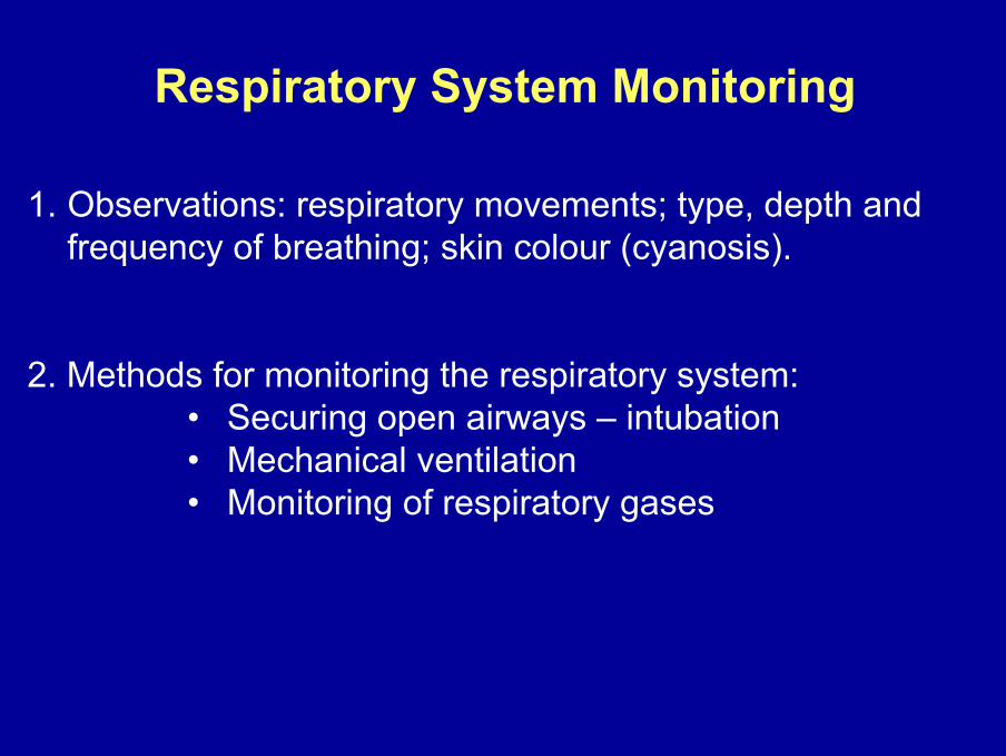
Technique of endotracheal intubation
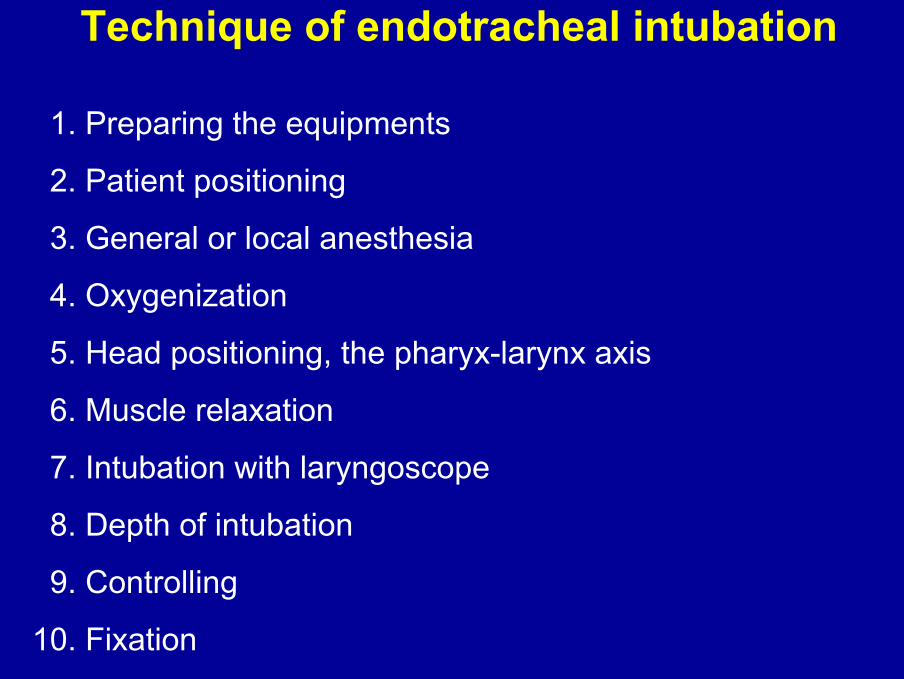
1. Preparing the equipments
2. Patient positioning
3. General or local anesthesia
4. Oxygenization
5. Head positioning, the pharyx-larynx axis
6. Muscle relaxation
7. Intubation with laryngoscope
8. Depth of intubation
9. Controlling
10. Fixation
1. Preparing the equipments
suction catheter, suction pump
Ruben-ballon with valve and mask,tube adaptors, adhesive tape
laryngoscope
endotracheal tubes
Guedel-tube
Magill-forceps
syringelaryngeal mask

2. Patient positioningThe most preferred position: the patient is laid in supine position, the head is toward the person performing the procedure.

3. General or local anesthesiaGeneral anesthesia or local anesthesia and sedationshould be induced.
4. OxygenizationDeliver oxygen with a face mask (for 3 min at least).

5. Head positioning I.
Possibilities for the alignment of the oral and pharyngeal axis:
1. Classical Jackson position: the patient is laid in supineposition without pillow, the head should be tilted backward atthe atlanto-occipital joint, so the cervical spine is retroflected.

5. Head positioning II.
2. Modified Jackson position (short-necked, obese patient, torticollis): a 10-15 cm pillow is placed under the nape, and thehead is tilted so that the mouth can be opened (sniffing position).

6. Relaxation
Paralyze the patient using muscle relaxation afteroxygenation and anesthesia, and wait for the effect.

7. Intubation with laryngoscope I.1. Grasp the laryngoscope by the left hand and insert the curved blade intothe mouth along with the median line of the tongue. If necessary push thetongue to the left side with the „Z” blade.2. The end of the blade should be between the base of tongue and theepiglottis, in the plica glossoepiglottica. By lifting the base of tongue theepiglottis will be elevated...

3. … and the triangular glottis with its peak will be visible upward.
7. Intubation with laryngoscope II.
4. If the epiglottis and the trachea cannot be seen, the assistant may press down the base of thyroidand cricoid cartilage (Sellick-maneuver).

8. Depth of intubationThe distal end of tube has to be positioned in the trachea 1 to3 cm above the bifurcation. Depth of intubation can be readon the tube.
Blowing the cuff: inhibits the inspirated air to escape andtrickling of saliva, blood, or gastric content into the lung; allows suctioning.
The balloon has an automatic valve which shuts down afterremoval of inflating syringe.
Avoid overinflation!

9. Controlling
1. Auscultation at both axillary lines. If the tube is toodeep, it may get into the right bronchus and ventilation is weak or can not be heard on the left side.
2. Knock the wall of the upper thorax and listen at the endof the tube – the outflow of air can be heard.
3. Insufflation through the tube induces a symmetricalmoving of the chest.
4. Using capnography, the CO2 waveform indicates theright position of the tube (EtCO2 is zero, if the tube is inthe oesophagus).

10. Fixation
A bite protector (Guedel-tube or a wet roller bandage) in themouth can be used to avoid biting of the tube.
The tube and the bite protector are fixed with a strip ofadhesive tape.

Monitoring of respiratory gases
Measurement of arterial pCO2 Measurement of arterial pO2 andoxygen saturation
Capnometry PulseoximetryNon-invasive
Invasive: blood gas analysis
Measurement of end-tidal CO2– Infrared absorption photometry
Measurement of O2 saturationin arterial bloodDifference in red and infraredlight absorption between oxy-and deoxyhemoglobin
- highly accurate- intermittent

Capnographycapnograph
Capnometrycapnometer
Capnography vs Capnometry
Measurement and display of bothEtCO2 value and CO2 waveform - capnogram
Measurement and display of EtCO2 value(no waveform)

Capnograph / Pulse-oximeter
OxiMax N-85
Measured parameters:
EtCO2 end-tidal CO2RR respiratory rateFiCO2 inspirated CO2SaO2 O2 saturationP heart rate
continuously monitored.
Sensors

Invasive monitoring of respiratory gasesBlood gas analysis
Goals: to determine• the patient’s blood gas values
(O2 uptake and CO2 elimination in the lung, blood pH)• the function of lungs and kidneys, and their role in acid-
base balance• respiratory diseases.
Steps:- taking blood sample (inhibitor of blood coagulation, air bubbles!),
- measurement using blood gas analyzer,- data interpretation, treatment of the patient.
AVL Compact 2(AVL Medical Instruments)

Improper tools, orincorrect sample manipulation inaccuracy !
Before taking blood sample, draw/discard about 4-times of cannulavolume with a 5 ml syringe.
• Fill the conus of a 2 ml syringe with heparine (50-100 µl/ml blood).
• Take blood sample (max. 1 ml) into the syringe.
• Remove air bubbles from the syringe.
• Close the syringe with a cap.
• Flush the cannula with saline.
Blood gas analysis – Taking blood sample
Directly before the measurement, remove some drops from thesample.

Blood gas and acid-base parameters
Measured parameters:pO2, pCO2, pH
Calculated parameters:BE, HCO3
¯, cHCO3¯, SaO2, ctO2
Metabolites:cLactate, cGlucose
Electrolytes:cK+, cNa+, cCl-, cCa2+

Goals of mechanical ventilation:• to increase oxygenation,• to prevent/to eliminate atelectasis,• to ensure optimal ventilation,• to improve ventilation/perfusion ratio,• to decrease work of breathing.
Mechanical ventilationIndications of mechanical ventilation:• insufficient breathing or in absence of spontaneous breathing,• in severe hypoxemia or hypercapnia/in respiratory failure,• increased work of breathing,• stabilization of chest wall.
Respirator
The most widely used technique ofmechanical ventilation
with positive pressure: during inspiration air is forced intothe lungs with higher pressure than that in the alveoli,
with volume-controlled respirator: inspiration with presettidal volume and ventilator rate or with preset minuteventilation, with inspiration/expiration ratio (pressurized valveto prevent extremely high pressure overload).
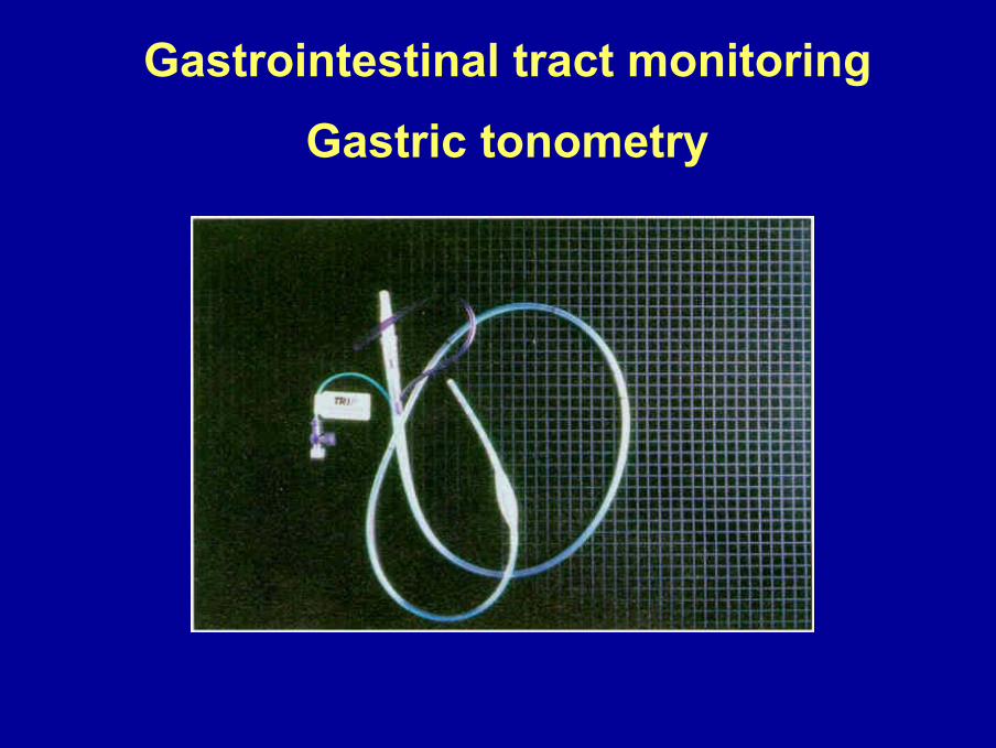
Gastrointestinal tract monitoringGastric tonometry

Indirect tonometry: the basics

pCO2 HCO3
[ ]
pHi= pKD + lg ————————————
0,03 x
Determination of intramucosal pH (pHi)
Tonomitor sample: measurement of mucosal pCO2
Arterial blood sample: HCO3 is determined by pCO2 andpH

CO2-gap
CO2-gap = pgCO2 - pACO2
pACO2 = arterial pCO2
pgCOpgCO2 2 = = locallocal ((measuredmeasured byby tonomitortonomitor) ) tissuetissue pCOpCO22

2.5 ml
Sigmoid Tonomitor(with balloon)
Saline
Bicarbonate buffer
Equilibration time: min. 30 min„Static” device
0.2-0.3 ml
Equilibration time: 5-6 min„Dynamic” device
Capillary Tonomitor
Catheters of gastrotonometry
by Boda et al. 2006

Intestinal and sublingual mucosal CO2-gapin hemorrhagic shock
Idő (perc)-30 0 30 60 90 120 150 180 210 240
%
50
100
150
200
250
300
350
vékonybél mucosa co2 gap Nyelv alatti mucosa co2 gap
Hemorrhagic shock Colloid resuscitation
Time (min)
Intestinal mucosal CO2-gapSublingual mucosal CO2-gap

Microcirculation of sublingual mucosa usingintravital videomicroscopy or orthogonal polarization
spectral (OPS) imaging.
Natural contrast agent: Hgb in the capillaries;Visibility: approx. 1 mm depth;Measured parameters:
Red blood cell velocityCapillary perfusion ratio (perfused/nonperfused capillaries ratio)
Monitoring Monitoring ofof thethe microcirculationmicrocirculation

Jugular vein and carotid arterycannulation
Blood sampleBlood gas analysis
Mechanical ventilation
Sublingual capillarytonomitor
OPS imaging ofmicrocirculation
CAPNOGRAPH
pCO2-gap determination

All you want to know aboutCatheterization
(but never dare to ask)

Urinary system monitoring
Catheterization of the bladderDefinition: artificial emptying of the urinary bladder.
Aims: therapeutic (urine retention, incontinence, preoperative preparation)diagnostic (monitoring fluid status, urologic/microbiologic tests)
Principles of catheterization
- catheterize only if it is necessary- avoid catheterization in case of urethral injuries- catheterize in accordance with the rules of asepsis!

Catheters
Material: synthetic, latex or silicone.
Size: external diameter is given in Charriére (1 Ch) or1 French (1 F) (=0.33 mm)
The most widely used: 14-22 Ch Foley-catheter (withballoon, easy fixation).

Tools for catheterization
- catheter in appropriate size- urine container sack and tube- sponges for cleaning of genital area- disinfectant- saline (in syringe) to fill the balloon- sterile lubricant (Instillagel)- sterile gloves

Male catheterization
- Lift the penis (about 60 degrees) with left hand and retractthe foreskin
- Clean the urethral meatus with disinfectant 3 times- Inject some Instillagel to the urethra- Insert the catheter into the urethra with
sterile forceps- Fill the balloon with 10 ml saline- Pull back the catheter until the balloon
allows- Connect the urine container sack to the
catheter.

Male catheterization

Removing the catheter in males

Female catheterization
- Spread the labia gently with left hand- Clean the introitus with disinfectant 3 times- Grasp the catheter with sterile forceps at some cm-s from
the end- Put Instillagel onto the first some cm-s of the catheter- Insert the catheter gently into the
urethra- Connect the urine container sack
to the catheter- Fill the catheter with 10 ml saline- Pull the catheter back.
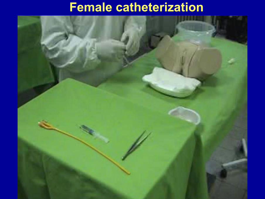
Female catheterization

Removing catheter in females

The execution of the practicalEndotracheal intubation in vivo (ext. operating room)
Capnography → EtCO2 end-tidal CO2Pulse oximetry → SaO2 O2 saturation
Sublingual gastrotonometry
max. 4 studentsScrub preparation (ext. op.
room)Cannulation of jugular vein
and carotid artery;Surgical team: surgeon, 2 assistants, 1 scrub nurse;
max. 6 studentsEndotracheal
intubation - model(int. op. room)
Taking blood samples → Blood gas analysisMechanical ventilation - settings
Sublingual microcirculation - OPS technique
max. 6 studentsBladder
catheterization -model
(int. op. room)
male female

Have a good practice!