Embed Size (px)
Citation preview
ORIGINAL RESEARCHpublished: 28 August 2017
doi: 10.3389/fnmol.2017.00274
Frontiers in Molecular Neuroscience | www.frontiersin.org 1 August 2017 | Volume 10 | Article 274
Edited by:
Chen Zhang,
Peking University, China
Reviewed by:
Zhiqiang Yan,
Fudan University, China
Jie He,
Institute of Neuroscience, Shanghai
Institutes for Biological Sciences
(CAS), China
*Correspondence:
Qingshun Zhao
Renjie Chai
Dong Liu
†These authors have contributed
equally to this work.
Received: 01 July 2017
Accepted: 14 August 2017
Published: 28 August 2017
Citation:
Gong J, Wang X, Zhu C, Dong X,
Zhang Q, Wang X, Duan X, Qian F,
Shi Y, Gao Y, Zhao Q, Chai R and
Liu D (2017) Insm1a Regulates Motor
Neuron Development in Zebrafish.
Front. Mol. Neurosci. 10:274.
doi: 10.3389/fnmol.2017.00274
Insm1a Regulates Motor NeuronDevelopment in ZebrafishJie Gong 1†, Xin Wang 2†, Chenwen Zhu 2, Xiaohua Dong 3, Qinxin Zhang 3, Xiaoning Wang 2,
Xuchu Duan 1, Fuping Qian 2, 4, Yunwei Shi 2, Yu Gao 2, Qingshun Zhao 3*, Renjie Chai 2, 4* and
Dong Liu 2*
1 School of Life Science, Nantong University, Nantong, China, 2Co-innovation Center of Neuroregeneration, Key Laboratory
of Neuroregeneration of Jiangsu and Ministry of Education, Nantong University, Nantong, China, 3MOE Key Laboratory of
Model Animal for Disease Study, Model Animal Research Center, Nanjing University, Nanjing, China, 4 Key Laboratory for
Developmental Genes and Human Disease, Ministry of Education, Institute of Life Sciences, Southeast University, Nanjing,
China
Insulinoma-associated1a (insm1a) is a zinc-finger transcription factor playing a series of
functions in cell formation and differentiation of vertebrate central and peripheral nervous
systems and neuroendocrine system. However, its roles on the development of motor
neuron have still remained uncovered. Here, we provided evidences that insm1a was a
vital regulator of motor neuron development, and provided a mechanistic understanding
of how it contributes to this process. Firstly, we showed the localization of insm1a in spinal
cord, and primary motor neurons (PMNs) of zebrafish embryos by in situ hybridization,
and imaging analysis of transgenic reporter line Tg(insm1a: mCherry)ntu805. Then we
demonstrated that the deficiency of insm1a in zebrafish larvae lead to the defects of
PMNs development, including the reduction of caudal primary motor neurons (CaP),
and middle primary motor neurons (MiP), the excessive branching of motor axons,
and the disorganized distance between adjacent CaPs. Additionally, knockout of insm1
impairedmotor neuron differentiation in the spinal cord. Locomotion analysis showed that
swimming activity was significantly reduced in the insm1a-null zebrafish. Furthermore,
we showed that the insm1a loss of function significantly decreased the transcript levels
of both olig2 and nkx6.1. Microinjection of olig2 and nkx6.1 mRNA rescued the motor
neuron defects in insm1a deficient embryos. Taken together, these data indicated that
insm1a regulated the motor neuron development, at least in part, through modulation of
the expressions of olig2 and nkx6.1.
Keywords: insm1a, motor neuron, differentiation, zebrafish, development
INTRODUCTION
In vertebrates, motor neurons have precise subtype identities that characterized by a number ofmorphological criteria, such as soma location, and shape, axon path, and target muscle innervation(Shirasaki and Pfaff, 2002; Lewis and Eisen, 2003). Meanwhile, motor neurons generally extendtheir axonal trajectory with a highly stereotyped manner during the nervous system development(Eisen, 1991; Palaisa and Granato, 2007). It has been reported that in chick and bullfrog, theirmotor neuron axons always followed the conservative pathways in order to project to appropriateregions of target musculatures (Landmesser, 1980; Farel and Bemelmans, 1985). In the embryo andlarva of zebrafish, there are two different kinds of spinal motor neurons, which are called primary
Gong et al. Insm1a Regulates Motor Neuron Development
motor neurons (PMNs), and secondary motor neuron (SMNs)(Myers, 1985; Myers et al., 1986). The PMNs can be furtherclassified into three groups, caudal primary motor neurons(CaP), middle primarymotor neurons (MiP), and rostral primarymotor neurons (RoP), by the positions of somata in the spinalcord, and the trajectory of neuron axons (Myers et al., 1986;Westerfield et al., 1986). CaPs, whose somata locate in themiddle of each spinal cord hemisegment, can innervate ventralaxial muscle, and have been well-studied because of theireasy observation and distinct axon projection (Myers et al.,1986; Rodino-Klapac and Beattie, 2004). MiPs project axons toinnervate the dorsal axial muscle, while RoPs project axons tocontrol the middle muscle (Rodino-Klapac and Beattie, 2004).Although the somata of the three identifiable PMNs are localizedin different position in the spinal cord, their axons pioneer to themyoseptum through a mutual exit point (Eisen et al., 1986). Dueto the identifiability of the three kinds of PMNs, they have alreadybecome an excellent system to study motor axon guidance andtheir intraspinal navigation (Beattie et al., 2002).
The insulinoma-associated 1 (insm1) gene, which is firstisolated from an subtraction cDNA library of insulinoma tumorcells, encodes a DNA-binding zinc finger transcription factorwith SNAG repressor motifs in N-terminal as well as Cys2-His2 Zn finger motifs in C-terminal, and widely expresses inthe developing nervous system, endocrine cells, pancreatic cells,and related neuroendocrine tumor cells (Goto et al., 1992;Xie et al., 2002; Jacob et al., 2009; Lan and Breslin, 2009;Jia et al., 2015b). Consequently, extensive studies focused onthe biological function of insm1 in nervous, and endocrinecell proliferation, differentiation, and transformation have beenreported in the model organisms (Farkas et al., 2008; Wildneret al., 2008; Lan and Breslin, 2009; Ramachandran et al.,2012; Jia et al., 2015a,b). For example, in the insm1 knockoutmice, its endocrine progenitor in the developing pancreas wereless differentiated, meanwhile hormone production, and cellmigration also exhibited seriously defects (Osipovich et al.,2014). Farkas et al. reported that compared to the wild typeand heterozygous mice, the number of basal progenitors in theinsm1 null dorsal telencephalon (dTel) was decreased almosthalf, and the radial thickness of dTel cortical plate as well as theneurogenesis in the neocortex were also predominantly reducedafter lacking insm1 gene (Farkas et al., 2008). In the zebrafish,insm1a can regulate a series of related genes, which are necessaryfor the Müller glia (MG) formation, and differentiation as wellas the zone definition of injury-responsive MG to participatein the retina regeneration (Ramachandran et al., 2012).Moreover, it was also reported that during the developmentof zebrafish retina, insm1 could regulate cell cycle kinetics anddifferentiation of the progenitor cells by acting the upstreamof the basic helix-loop-helix (bHLH) transcription factors, andthe photoreceptor specification genes (Forbes-Osborne et al.,2013). Although insm1a is widely detected in the nervoussystem and its necessity in the brain and retina developmenthave been also illuminated well, little is known about thefunction and molecular mechanisms of insm1a on the formationand development of other neuronal types, especially in thezebrafish.
The zebrafish has become an excellent model system toinvestigate the mechanisms of the neuron formation andits axonal pathfinding due to the accessible observation ofmotor neurons from the initial stages of embryo development(Zelenchuk and Bruses, 2011). Here, we examined the function ofinsm1a in the primary motor neurons development by CRISPR/Cas9-mediated knockout in the Tg(mnx1:GFP)ml2 transgeniczebrafish and investigated the possible transcriptional networkduring this process.
MATERIALS AND METHODS
Zebrafish Line and BreedingThe zebrafish embryos and adults were maintained in zebrafishCenter of Nantong University under conditions in accordancewith our previous protocols (Xu et al., 2014; Wang et al.,2016). The transgenic zebrafish line, Tg(mnx1:GFP)ml2, has beendescribed in the previous work (Zelenchuk and Bruses, 2011).
RNA Isolation, Reverse Transcription andQuantitative PCRTotal RNA was extracted from zebrafish embryos by TRIzolreagent according to the manufacturer’s instructions (Invitrogen,USA). Genomic contaminations were removed by DNaseI, andthen 2 µg total RNA was reversely transcribed using a reversedfirst strand cDNA synthesis kit (Fermentas, USA) and storedat −20◦C. qRT-PCR was performed using the correspondingprimers (Supplementary Table 1) in a 20 µl reaction volume with10µl SYBR premix (Takara, Japan) and elongation factor 1a (ef1a)was used as the internal control. All samples were analyzed intriplicate.
Whole Mount In situ HybridizationA 501 bp cDNA fragment of insm1a was amplified fromthe cDNA library that established from wild type (WT) ABembryos using the specific primers of insm1a F1 and insm1aR1 (Supplementary Table 1). Digoxigenin-labeled sense andantisense probes were synthesized using linearized pGEM-T-easy vector subcloned with this insm1a fragment by in vitrotranscription with DIG-RNA labeling Kit (Roche, Switzerland).Zebrafish embryos and larvae were collected and fixed with 4%paraformaldehyde (PFA) in phosphate-buffered saline (PBS) forone night. The fixed samples were then dehydrated througha series of increasing concentrations of methanol and storedat −20◦C in 100% methanol eventually. Whole mount in situhybridization was subsequently performed as described in theprevious study (Huang et al., 2013).
Establishment of Tg(insm1a: EGFP)ntu804
and Tg(insm1a: mCherry)ntu805 TransgenicLineTransgenic zebrafish were created using the Tol2kit transgenesissystem and Gateway vectors. The insm1a promoter wascloned and insert into the p5E-MCS entry vector. A multiSiteGateway vector construction reaction (Invitrogen, USA) wasconducted with the resulting p5E-insm1a together with pME-EGFP (or mCherry) and p3E-polyA subcloned into the
Frontiers in Molecular Neuroscience | www.frontiersin.org 2 August 2017 | Volume 10 | Article 274
Gong et al. Insm1a Regulates Motor Neuron Development
pDestTol2pA2 to produce insm1a: EGFP or insm1a: mCherryconstruct. Subsequently, this construct was co-injected withtol2-transposase mRNAs into zebrafish one to two-cell-stageembryos to create the Tg(insm1a: EGFP)ntu804 and Tg(insm1a:mCherry)ntu805 transgenic line.
sgRNA/ Cas9 mRNA Synthesis andInjectionCas9 mRNA was obtained by in vitro transcription with thelinearized plasmid pXT7-Cas9 according to the procedurepreviously described. The sgRNAs were transcribed from theDNA templates that amplified by PCR with a pT7 plasmid asthe template, a specific forward primer and a universal reverseprimer (Supplementary Table 1) (Chang et al., 2013; Qi et al.,2016). Transgenic zebrafish lines Tg(mnx1:GFP)ml2, were naturalmated to obtain embryos for microinjection. One to two-cellstage zebrafish embryos were injected with 2–3 nl of a solutioncontaining 250 ng/µl Cas9 mRNA and 15 ng/µl sgRNA. At24 h post fertilization (hpf), zebrafish embryos were randomlysampled for genomic DNA extraction according to the previousmethods to determine the indel mutations by sequencing.
Morpholino and mRNAs InjectionsTranslation blocking antisense Morpholino (MOs; Gene Tools)against the ATG-containing sequence was designed (5′-AAATCCTCTGGGCATCTTCGCCAGC-3′) to target the translationstart site according to the manufacturer’s instruction and theother MO oligo (5′-CCTCTTACCTCAGTTACAATTTATA-3′)was used as standard control. The MOs were diluted to 0.3mM with RNase-free water and injected into the yolk of one totwo-cell stage embryos and then raised in E3 medium at 28.5◦C.
The cDNAs containing the open reading frame of the targetgenes were cloned into PCS2+ vector respectively and thenwere transcribed in vitro using the mMESSAGE mMACHIN Kit(Ambion, USA) after the recombinant plasmids linearized withNotI Restriction Enzyme (NEB, England), and then the cappedmRNAs were purified by RNeasy Mini Kit (Qiagen, Germany). 2nl target genes and mCherry mRNA mixture (1:1) were injectedat 20 ng/µl into 1/2-cell stage embryos.
Locomotion Analysis in Zebrafish LarvaeTo determine whether the deficiency of insm1a affectspontaneous movement, knockout, and normal larvae wereraised in a 24-well-culture plate with one larva in each well-filled with 1 ml E3 medium. The 24-well-culture plate wastransferred to the Zebralab Video-Track system (Zebrabox,France) equipped with a sealed opaque plastic box that keptinsulated from laboratory environment, an infrared filter anda monochrome camera. After adapting for 30 min, traveleddistances of the larvae were videotaped with every 5 minsforming a movement distance and trajectory by the linkedsoftware.
Microscopy and Statistical AnalysisZebrafish embryos were anesthetized with E3/0.16 mg/mLtricaine/1% 1-phenyl-2-thiourea (Sigma, USA) and embedded in0.8% low melt agarose, and then were examined with a Leica
TCS-SP5 LSM confocal imaging system. For the results of insitu hybridization, Photographs were taken using an Olympusstereomicroscope MVX10. Statistical comparisons of the datawere carried out by student’s t-test or one-way analysis ofvariance (ANOVA) followed by Duncan’s test, and P < 0.05were considered statistically significant. All statistical analysiswas performed using the SPSS 13.0 software (SPSS, USA).
RESULT
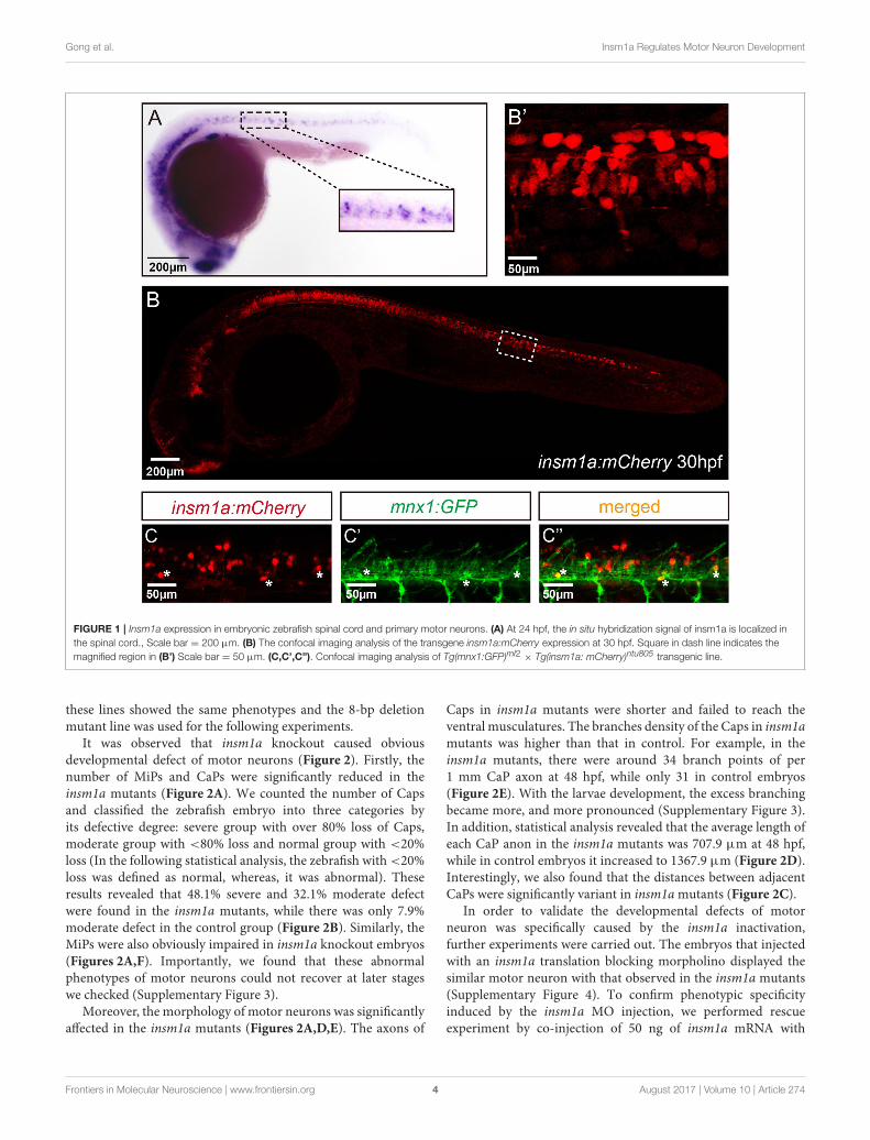
Insm1a is Expressed in Spinal Cord andPMNs of ZebrafishTo analyze the expression of insm1a in zebrafish nervous system,we performed the whole amount in situ hybridization (WISH)analysis with a digoxigenin-labeled insm1a probe. Similar to theprevious study (Lukowski et al., 2006), at late somitogenesis (24hpf) insm1a transcripts were apparently localized in ventral partof the neurons in the spinal cord, where most of the motorneurons located at this stage (Figure 1A).
To further determine the localization of insm1a, we generatedthe Tg(insm1a: EGFP)ntu804 and Tg(insm1a: mCherry)ntu805
transgenic zebrafish lines, in which the insm1a promoterdirected expression of EGFP or mCherry respectively. It wasshown that at 30 hpf the insm1a:mCherry and insm1a:EGFPexpression was observed in the spinal cord, retina and brain,which was similar with the results of in situ hybridization(Figures 1B,B’; Supplementary Figures 1A,A’) (Lukowski et al.,2006). In addition, we found that insm1a:EGFP expression washighly activated in Müller glia of injury sites in adult zebrafishretina (Supplementary Figure 1B), which was consistent with theISH data carried out by other researchers (Ramachandran et al.,2012). These results suggested that the transgenes recapitulatedthe endogenous insm1a expression.
To investigate whether insm1a is expressed in motor neurons,we outcrossed Tg(insm1a: mCherry)ntu805 transgenic line withTg(mnx1:GFP)ml2 line, in whichmotor neurons were labeled withGFP (Zelenchuk and Bruses, 2011). We found that the GFP+
motor neurons were also labeled with mCherry fluorescence(Figures 1C–C”), suggesting insm1a was expressed in motorneurons.
Knockout of insm1a Caused Primary MotorNeurons Developmental DefectIn order to examine whether insm1a is required for thedevelopment of motor neuron, the CRISPR/Cas9 system wasutilized to knockout insm1a in Tg(mnx1:GFP)ml2 transgeniczebrafish line. To ensure complete disruption of functionalproteins, we chose the target sites near the translation startcodon (ATG) in the exon1 of zebrafish insm1a (SupplementaryFigure 2A). The selected gRNA-Cas9 system efficiently inducedmutations in the targeting site with four types of mutationswere identified (Supplementary Figure 2B). The mutated allelesincluded a 5-bp deletion, an 8-bp deletion and two 10-bpdeletions, which all resulted in reading frame shift and prematuretranslation termination (Supplementary Figure 2C). In addition,
Frontiers in Molecular Neuroscience | www.frontiersin.org 3 August 2017 | Volume 10 | Article 274
Gong et al. Insm1a Regulates Motor Neuron Development
FIGURE 1 | Insm1a expression in embryonic zebrafish spinal cord and primary motor neurons. (A) At 24 hpf, the in situ hybridization signal of insm1a is localized in
the spinal cord., Scale bar = 200 µm. (B) The confocal imaging analysis of the transgene insm1a:mCherry expression at 30 hpf. Square in dash line indicates the
magnified region in (B’) Scale bar = 50 µm. (C,C’,C”). Confocal imaging analysis of Tg(mnx1:GFP)ml2 × Tg(insm1a: mCherry)ntu805 transgenic line.
these lines showed the same phenotypes and the 8-bp deletionmutant line was used for the following experiments.
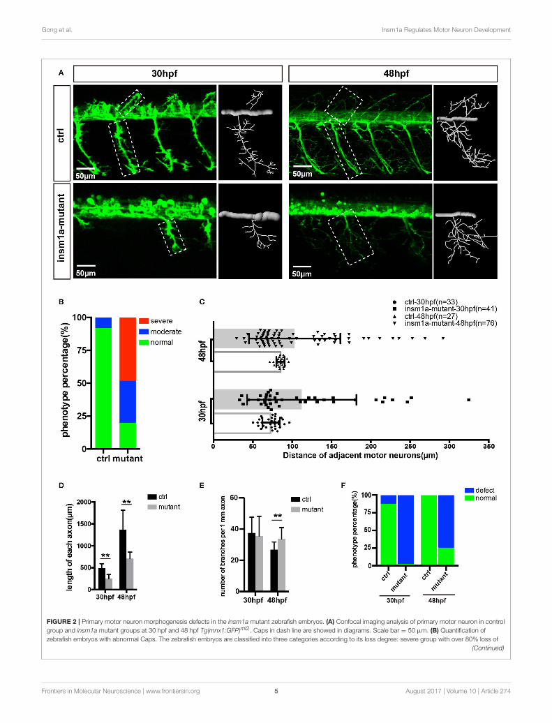
It was observed that insm1a knockout caused obviousdevelopmental defect of motor neurons (Figure 2). Firstly, thenumber of MiPs and CaPs were significantly reduced in theinsm1a mutants (Figure 2A). We counted the number of Capsand classified the zebrafish embryo into three categories byits defective degree: severe group with over 80% loss of Caps,moderate group with <80% loss and normal group with <20%loss (In the following statistical analysis, the zebrafish with <20%loss was defined as normal, whereas, it was abnormal). Theseresults revealed that 48.1% severe and 32.1% moderate defectwere found in the insm1a mutants, while there was only 7.9%moderate defect in the control group (Figure 2B). Similarly, theMiPs were also obviously impaired in insm1a knockout embryos(Figures 2A,F). Importantly, we found that these abnormalphenotypes of motor neurons could not recover at later stageswe checked (Supplementary Figure 3).
Moreover, the morphology of motor neurons was significantlyaffected in the insm1a mutants (Figures 2A,D,E). The axons of
Caps in insm1a mutants were shorter and failed to reach theventral musculatures. The branches density of the Caps in insm1amutants was higher than that in control. For example, in theinsm1a mutants, there were around 34 branch points of per1 mm CaP axon at 48 hpf, while only 31 in control embryos(Figure 2E). With the larvae development, the excess branchingbecame more, and more pronounced (Supplementary Figure 3).In addition, statistical analysis revealed that the average length ofeach CaP anon in the insm1a mutants was 707.9 µm at 48 hpf,while in control embryos it increased to 1367.9 µm (Figure 2D).Interestingly, we also found that the distances between adjacentCaPs were significantly variant in insm1amutants (Figure 2C).
In order to validate the developmental defects of motorneuron was specifically caused by the insm1a inactivation,further experiments were carried out. The embryos that injectedwith an insm1a translation blocking morpholino displayed thesimilar motor neuron with that observed in the insm1a mutants(Supplementary Figure 4). To confirm phenotypic specificityinduced by the insm1a MO injection, we performed rescueexperiment by co-injection of 50 ng of insm1a mRNA with
Frontiers in Molecular Neuroscience | www.frontiersin.org 4 August 2017 | Volume 10 | Article 274
Gong et al. Insm1a Regulates Motor Neuron Development
FIGURE 2 | Primary motor neuron morphogenesis defects in the insm1a mutant zebrafish embryos. (A) Confocal imaging analysis of primary motor neuron in control
group and insm1a mutant groups at 30 hpf and 48 hpf Tg(mnx1:GFP)ml2. Caps in dash line are showed in diagrams. Scale bar = 50 µm. (B) Quantification of
zebrafish embryos with abnormal Caps. The zebrafish embryos are classified into three categories according to its loss degree: severe group with over 80% loss of
(Continued)
Frontiers in Molecular Neuroscience | www.frontiersin.org 5 August 2017 | Volume 10 | Article 274

Gong et al. Insm1a Regulates Motor Neuron Development
FIGURE 2 | Continued
Cap primary motor neuron, moderate group with <80% loss, and normal group with <20% loss. (C) Quantification of distance between adjacent motor neurons (µm)
in control group and insm1a mutant groups at 30 hpf (n = 33 and 41 respectively) and 48 hpf (n = 27 and 76 respectively). (D,E) The length and branching number of
Cap axons in control group and insm1a mutant groups at 30 and 48 hpf. Asterisks above the bars indicate significant differences (**P < 0.01). (F) Quantification of
zebrafish embryos with abnormal Caps at 30 and 48 hpf.
insm1a MO into zebrafish embryos, and the results showed thatthe co-injection significantly decreased the loss, and prematurebranching of PMNs (Supplementary Figure 4E). Taken together,these results indicated thosemotor neuron developmental defectswere caused by loss of insm1a.
Insm1a Deficiency Suppressed NeuronalCells DifferentiationThe confocal imaging analysis discovered that there were anumber of round and not well-differentiated GFP positive cellsin Tg(mnx1:GFP)ml2 insm1a mutants (Figure 3A). Statisticalanalysis showed that at 30 and 48 hpf the number of theseundifferentiated cell in the insm1a deficiency zebrafish wassignificantly higher than that in the control fish (Figure 3B).We also observed these undifferentiated cells in insm1amorphants, however the number was less than that in mutants(Figures 3A,B).
To further investigate the cellular mechanism underlyingthe motor neuronal defects in insm1a deficient embryos, weperformed confocal time-lapse imaging analysis. It was foundthat in control embryos the axon of CaP sprouted from the spinalcord, and extended toward to the ventral muscle (Figure 3C). Incontrol embryos the axon of CaP started to branch when it passedthrough the midline, while in insm1a mutants the axon initiatedto branch once it came out from the spinal cord (Figures 3A,C,Supplementary Movies 1, 2). In addition, we found that thoseround GFP positive cells did not develop neuronal projections(Figures 3A,C, Supplementary Movie 1).
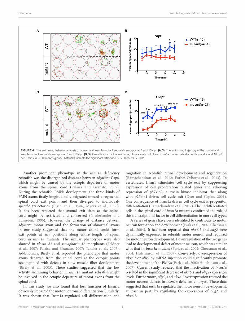
Knockout of insm1a Reduced theZebrafish Swimming ActivityIn order to investigate whether the motor neuron defects affectsthe motor ability, insm1a mutant zebrafish larvae were furtherperformed for 40-min free-swimming activity test independentof any stimuli at 7 and 10 dpf. It demonstrated that themovementtrajectory and swimming distance per 5 mins, which couldreflect the swimming speed, of insm1a mutant zebrafish larvaewere significantly decreased compared to that in the control(Figure 4). The movie in the Supplementary Material showedthat swimming behavior could be easily discovered in the controlgroup, while the zebrafish inmutant group kept involuntomotory(Supplementary Movie 3). Additionally, we also discovered thatunder the stereoscopic microscope the mutant zebrafish becameinsensitive to the touch stimuli (data not shown).
The insm1a Deficiency Caused Alterationof Gene Expression Involved in MotorNeuron DevelopmentSince insm1a is a transcription factor, we supposed that motorneuron developmental defects in insm1a deficient embryos
were associated with altered expression of downstream genesof insm1a or the genes participating in the motor neurondevelopment. Based on the previous studies, NNR2a, NNR2b,islet2, Asci1a, Asci1b, shh, Ngn2, Nkx6.1, and olig2 were selectedto do the qRT-PCR analysis in wild-type (WT) and insm1adeficiency zebrafish embryos (Park et al., 2002; Hutchinson et al.,2007; Davis-Dusenbery et al., 2014; Barreiro-Iglesias et al., 2015).The results showed that expressions of NNR2a, NNR2b, islet2,Asci1a, and Asci1b were significantly influenced in the insm1a
deficiency zebrafish compared to the control (SupplementaryFigure 5). We also found that the expression of shh was obviouslyelevated in insm1amutants at 19, 24, and 36 hpf (SupplementaryFigure 5). Interestingly, olig2 and nkx6.1 transcripts dramaticallydecreased in insm1a deficient embryos (Figures 5A,B).
Olig2 and nkx6.1 Over Expression Rescuedthe Motor Neuron Defects in insm1a
Deficient EmbryosAs the downregulation of olig2 and nkx6.1 in insm1a loss offunction embryos, we reasoned that insm1a might bind thetranscriptional regulatory elements of these two genes. Basedon the JASPAR 2016 database (Mathelier et al., 2016) analysis,we found that both olig2 and nkx6.1 contained the putativebinding sites of Insm1a, suggesting Insm1a directly regulatesthe expression of olig2, and nkx6.1 during PMNs development.To investigate whether the motor neuronal defects in insm1adeficient embryos were caused by reduced expression of olig2 andnkx6.1, we tried to rescue the phenotype with olig2 and nkx6.1gain of function in insm1a deficient embryos. It was shownthat co-injection both olig2 and nkx6.1 mRNA respectivelywith insm1a MO significantly reduced the motor neuronaldefects caused by loss of insm1a (Figures 5C,D). 69.6% zebrafishembryos injected with insm1a MO at 48 hpf had the motorneuron developmental defects, while only 42.1% had the motorneuronal phenotype in the olig2 mRNA and insm1a MO co-injection group (Figure 5D). Similarly, after nkx6.1 mRNA andinsm1aMO co-injection, the ratio of motor neuronal phenotypedecreased to 38.6% (Figure 5D).
DISCUSSION
As one of the most conserved zinc-finger transcriptionalfactor, insm1a plays important roles in various biologicalprocesses in vertebrates (Wildner et al., 2008; Jacob et al., 2009;Forbes-Osborne et al., 2013; Osipovich et al., 2014; Jia et al.,2015b; Lorenzen et al., 2015). Previous studies have identified itsrole in regulating the endocrine cells divisions of the pancreas,the neuroendocrine development, the differentiation of retinaprogenitors and neurogenesis of nervous system (Gierl et al.,2006; Duggan et al., 2008; Farkas et al., 2008; Jacob et al., 2009;
Frontiers in Molecular Neuroscience | www.frontiersin.org 6 August 2017 | Volume 10 | Article 274

Gong et al. Insm1a Regulates Motor Neuron Development
FIGURE 3 | Insm1a deficiency suppressed neuronal cells differentiation. (A) Confocal imaging analysis of primary motor neuron in control group, insm1a mutant group
and morphant group at 30 and 48 hpf Tg(mnx1:GFP)ml2. Phenotypes of neuronal cells in the spinal cord in control group, morphant group, and insm1a mutant
groups at 30 hpf and 48 hpf. Asterisks indicate undifferentiated neuronal cells. Scale bar = 50 µm. (B) Quantification of the undifferentiated neuronal cell in the insm1a
different treatment zebrafish. Asterisks above the bars indicate significant differences (*P < 0.05, **P < 0.01). (C) Time-lapse imaging analysis of the primary motor
neuron in control group and insm1a mutant groups. Asterisks represent undifferentiated neuronal cells. Scale bar = 50 µm
Lan and Breslin, 2009; Ramachandran et al., 2012). Currently,our data in this study provided with new insights into the roleof insm1a in motor neuron development.
Our WISH data and previous study (Ramachandran et al.,2012) demonstrated that insm1a transcripts were detected inretina and spinal cord at 24 hpf. Furthermore, imaging analysis ofour established transgenic reporter line Tg(insm1a: EGFP)ntu804
and Tg(insm1a: mCherry)ntu805 verified the results of in situhybridization, and revealed the expression of EGFP or mCherrythat were driven by insm1a promoter in the PMNs. It is well-known that the spinal cord contains PMNs which project theiraxons out of the spinal cord to the terminal musculature withthe embryo development (Davis-Dusenbery et al., 2014). Takentogether, the localization data of insm1a from both in situhybridization analysis and the study based on transgenic reporterline suggested that insm1a might participate in the regulation ofPMNs development.
To test whether insm1a was required for formation ofPMNs, we generated CRISPR/Cas9-mediated insm1a mutants,
and showed that obvious motor neuron loss and defects of thePMNs axons. Moreover, we also performed insm1a knockdown,and the results showed similar PMNs defects as the onesproduced by the insm1a knockout. In wild embryos, exuberantside branches developed at around 72 hpf, and then invadedinto myotome to form distributed neuromuscular synapses(Liu and Westerfield, 1990; Downes and Granato, 2004). Theseresults suggested that insm1a was pivotal for the primarymotor axon development to block precociously extending intomuscle territories. Additionally, locomotion analysis displayeda typical low activity swimming behavior in insm1a mutantzebrafish. It was known motor neuron was a major kind ofcell type that regulated swimming behavior in zebrafish duringearly development (Brustein et al., 2003). Previous studies alsoshowed a significant involvement of motor neuron in the overalllocomotor behavior (Flanagan-Steet et al., 2005; Levin et al.,2009). Currently, the decrease of swimming activity in this studywas consistent with the motor neuron defects in the insm1aknockout zebrafish.
Frontiers in Molecular Neuroscience | www.frontiersin.org 7 August 2017 | Volume 10 | Article 274
Gong et al. Insm1a Regulates Motor Neuron Development
FIGURE 4 | The swimming behavior analysis of control and insm1a mutant zebrafish embryos at 7 and 10 dpf. (A,C). The swimming trajectory of the control and
insm1a mutant zebrafish embryos at 7 and 10 dpf. (B,D). Quantification of the swimming distance of control and insm1a mutant zebrafish embryos at 7 and 10 dpf
per 5 mins (n = 36 in each group). Asterisks indicate the significant difference (*P < 0.05, **P < 0.01).
Another prominent phenotype in the insm1a deficiencyzebrafish was the disorganized distance between adjacent Caps,which might be caused by the ectopic departure of motoraxons from the spinal cord (Palaisa and Granato, 2007).During the zebrafish PMNs development, the three kinds ofPMN axons firstly longitudinally migrated toward a segmentalspinal cord exit point, and then diverged to individual-specific trajectories (Eisen et al., 1986; Myers et al., 1986).It has been reported that axonal exit sites at the spinalcord might be restricted and conserved (Niederlander andLumsden, 1996). However, the change of distance betweenadjacent motor axon and the formation of abnormal axonsin our study suggested that the motor axons could formexit points at any positions along entire length of spinalcord in insm1a mutants. The similar phenotypes were alsoshowed in plexin A3 and semaphorin 3A morphants (Feldneret al., 2007; Palaisa and Granato, 2007; Tanaka et al., 2007).Additionally, Birely et al. reported the phenotype that motoraxons departed from the spinal cord at the ectopic pointsaccompanied with defects in slow muscle fiber development(Birely et al., 2005). These studies suggested that the lowactivity swimming behavior in insm1a mutant zebrafish mightbe involved in the ectopic departure of motor axons from thespinal cord.
In this study we also found that loss function of Insm1aobviously impaired the motor neuronal differentiation. Similarly,It was shown that Insm1a regulated cell differentiation and
migration in zebrafish retinal development and regeneration(Ramachandran et al., 2012; Forbes-Osborne et al., 2013). Invertebrates, Insm1 stimulates cell cycle exit by suppressingexpression of cell proliferation related genes and relievingrepression of p57kip2, a cyclin kinase inhibitor that alongwith p27kip1 drives cell cycle exit (Dyer and Cepko, 2001).One consequence of insm1a driven cell cycle exit is progenitordifferentiation (Ramachandran et al., 2012). The undifferentiatedcells in the spinal cord of insm1a mutants confirmed the role ofthis transcriptional factor in cell differentiation inmore cell types.
A series of genes have been identified to contribute to motorneuron formation and development (Park et al., 2002; Cheesmanet al., 2004). It has been reported that nkx6.1 and olig2 weredynamically expressed in zebrafih motor neuron and requiredformotor neuron development. Downregulation of the two geneslead to developmental defect of motor neuron, which was similarwith that in insm1a mutant (Park et al., 2002; Cheesman et al.,2004; Hutchinson et al., 2007). Conversely, overexpression ofnkx6.1 or olig2 by mRNA injection could significantly promotethe development of the PMNs (Park et al., 2002; Hutchinson et al.,2007). Current study revealed that the inactivation of insm1aresulted in the significant decrease of nkx6.1 and olig2 expressionlevels. Furthermore, olig2, and nkx6.1 overexpression rescued themotor neuron defects in insm1a deficient embryos. These datasuggested that insm1a regulated the motor neuron development,at least in part, by regulating the expressions of olig2, andnkx6.1.
Frontiers in Molecular Neuroscience | www.frontiersin.org 8 August 2017 | Volume 10 | Article 274

Gong et al. Insm1a Regulates Motor Neuron Development
FIGURE 5 | Over expressions of nkx6.1 and olig2 rescued the motor neuron defects in insm1a deficient embryos. (A,B). Effects of insm1a knockdown on the
expressions of nkx6.1 and olig2 at 19, 24, 36, and 48 hpf. Asterisks indicate significant differences (*P < 0.05). (C) Abnormal Caps in insm1a knockdown zebrafish
embryos were restored by co-injection of nkx6.1 or olig2 mRNA. Diagrams of Caps in dash line are displayed near the corresponding confocal image. Scale bar = 50
µm. (D) Quantification of zebrafish embryos with abnormal Cap primary motor neuron. Asterisks indicate significant differences (**P < 0.01).
ETHICS STATEMENT
All animal experimentation was carried out in accordancewith the NIH Guidelines for the care and use of laboratoryanimals (http://oacu.od.nih.gov/regs/index.htm) and ethicallyapproved by the Administration Committee of ExperimentalAnimals, Jiangsu Province, China [Approval ID: SYXK (SU)2007–0021].
AUTHOR CONTRIBUTIONS
DL, RC, and QSZ conceived the project. JG, XW, CZ, XNW,XCD, FQ, YS, and YG performed most of the experiments. XHDand QXZ generated the mutants. DL, JG, and XW analyzed thedata and prepared the manuscript. All authors commented andapproved the manuscript.
ACKNOWLEDGMENTS
This study was supported by grants from the NationalNatural Science Foundation of China (31400918, 41606169,81570447, 81622013, 81470692, 31500852); Natural ScienceFoundation of Jiangsu Province (BK20160418, BK20150022,BK20140620); the National Key Research Development Programof China (2017YFA0103900, 2015CB965000, 2017YFA0103903);the Yingdong Huo Education Foundation; and the FundamentalResearch Funds for the Central Universities.
SUPPLEMENTARY MATERIAL
The Supplementary Material for this article can be foundonline at: http://journal.frontiersin.org/article/10.3389/fnmol.2017.00274/full#supplementary-material
Frontiers in Molecular Neuroscience | www.frontiersin.org 9 August 2017 | Volume 10 | Article 274

Gong et al. Insm1a Regulates Motor Neuron Development
REFERENCES
Barreiro-Iglesias, A., Mysiak, K. S., Scott, A. L., Reimer, M. M., Yang,
Y., Becker, C. G., et al. (2015). Serotonin promotes development and
regeneration of spinal motor neurons in Zebrafish. Cell Rep. 13, 924–932.
doi: 10.1016/j.celrep.2015.09.050
Beattie, C. E., Granato, M., and Kuwada, J. Y. (2002). Cellular, genetic and
molecular mechanisms of axonal guidance in the zebrafish. Results Probl. Cell
Differ. 40, 252–269. doi: 10.1007/978-3-540-46041-1_13
Birely, J., Schneider, V. A., Santana, E., Dosch, R., Wagner, D. S., Mullins,
M. C., et al. (2005). Genetic screens for genes controlling motor
nerve-muscle development and interactions. Dev. Biol. 280, 162–176.
doi: 10.1016/j.ydbio.2005.01.012
Brustein, E., Saint-Amant, L., Buss, R. R., Chong, M., McDearmid, J. R., and
Drapeau, P. (2003). Steps during the development of the zebrafish locomotor
network. J. Physiol. Paris 97, 77–86. doi: 10.1016/j.jphysparis.2003.10.009
Chang, N., Sun, C., Gao, L., Zhu, D., Xu, X., Zhu, X., et al. (2013). Genome editing
with RNA-guided Cas9 nuclease in zebrafish embryos. Cell Res. 23, 465–472.
doi: 10.1038/cr.2013.45
Cheesman, S. E., Layden, M. J., Von Ohlen, T., Doe, C. Q., and Eisen, J.
S. (2004). Zebrafish and fly Nkx6 proteins have similar CNS expression
patterns and regulate motoneuron formation. Development 131, 5221–5232.
doi: 10.1242/dev.01397
Davis-Dusenbery, B. N., Williams, L. A., Klim, J. R., and Eggan, K.
(2014). How to make spinal motor neurons. Development 141, 491–501.
doi: 10.1242/dev.097410
Downes, G. B., and Granato, M. (2004). Acetylcholinesterase function is
dispensable for sensory neurite growth but is critical for neuromuscular
synapse stability. Dev. Biol. 270, 232–245. doi: 10.1016/j.ydbio.2004.02.027
Duggan, A., Madathany, T., de Castro, S. C., Gerrelli, D., Guddati, K., and Garcia-
Anoveros, J. (2008). Transient expression of the conserved zinc finger gene
INSM1 in progenitors and nascent neurons throughout embryonic and adult
neurogenesis. J. Comp. Neurol. 507, 1497–1520. doi: 10.1002/cne.21629
Dyer, M. A., and Cepko, C. L. (2001). p27Kip1 and p57Kip2 regulate proliferation
in distinct retinal progenitor cell populations. J. Neurosci. 21, 4259–4271.
Available online at: http://www.jneurosci.org/content/21/12/4259.long
Eisen, J. S. (1991). Determination of primary motoneuron identity in developing
zebrafish embryos. Science 252, 569–572. doi: 10.1126/science.1708527
Eisen, J. S., Myers, P. Z., and Westerfield, M. (1986). Pathway selection by growth
cones of identified motoneurones in live zebra fish embryos. Nature 320,
269–271. doi: 10.1038/320269a0
Farel, P. B., and Bemelmans, S. E. (1985). Specificity of motoneuron projection
patterns during development of the bullfrog tadpole (Rana catesbeiana). J.
Comp. Neurol. 238, 128–134. doi: 10.1002/cne.902380112
Farkas, L. M., Haffner, C., Giger, T., Khaitovich, P., Nowick, K., Birchmeier, C.,
et al. (2008). Insulinoma-associated 1 has a panneurogenic role and promotes
the generation and expansion of basal progenitors in the developing mouse
neocortex. Neuron 60, 40–55. doi: 10.1016/j.neuron.2008.09.020
Feldner, J., Reimer, M. M., Schweitzer, J., Wendik, B., Meyer, D., Becker,
T., et al. (2007). PlexinA3 restricts spinal exit points and branching of
trunk motor nerves in embryonic zebrafish. J. Neurosci. 27, 4978–4983.
doi: 10.1523/JNEUROSCI.1132-07.2007
Flanagan-Steet, H., Fox, M. A., Meyer, D., and Sanes, J. R. (2005). Neuromuscular
synapses can form in vivo by incorporation of initially a neural postsynaptic
specializations. Development 132, 4471–4481. doi: 10.1242/dev.02044
Forbes-Osborne, M. A., Wilson, S. G., and Morris, A. C. (2013). Insulinoma-
associated 1a (Insm1a) is required for photoreceptor differentiation in the
zebrafish retina. Dev. Biol. 380, 157–171. doi: 10.1016/j.ydbio.2013.05.021
Gierl, M. S., Karoulias, N., Wende, H., Strehle, M., and Birchmeier, C. (2006).
The zinc-finger factor Insm1 (IA-1) is essential for the development of
pancreatic beta cells and intestinal endocrine cells. Genes Dev. 20, 2465–2478.
doi: 10.1101/gad.381806
Goto, Y., De Silva, M. G., Toscani, A., Prabhakar, B. S., Notkins, A. L., and Lan, M.
S. (1992). A novel human insulinoma-associated cDNA, IA-1, encodes a protein
with zinc-finger DNA-binding motifs. J. Biol. Chem. 267, 15252–15257.
Huang, Y., Wang, X., Wang, X., Xu, M., Liu, M., and Liu, D. (2013). Nonmuscle
myosin II-B (myh10) expression analysis during zebrafish embryonic
development. Gene Expr. Patterns 13, 265–270. doi: 10.1016/j.gep.2013.
04.005
Hutchinson, S. A., Cheesman, S. E., Hale, L. A., Boone, J. Q., and Eisen, J. S. (2007).
Nkx6 proteins specify one zebrafish primarymotoneuron subtype by regulating
late islet1 expression. Development 134, 1671–1677. doi: 10.1242/dev.02826
Jacob, J., Storm, R., Castro, D. S., Milton, C., Pla, P., Guillemot, F., et al. (2009).
Insm1 (IA-1) is an essential component of the regulatory network that specifies
monoaminergic neuronal phenotypes in the vertebrate hindbrain.Development
136, 2477–2485. doi: 10.1242/dev.034546
Jia, S., Ivanov, A., Blasevic, D.,Muller, T., Purfurst, B., Sun,W., et al. (2015a). Insm1
cooperates with Neurod1 and Foxa2 to maintain mature pancreatic beta-cell
function. EMBO J. 34, 1417–1433. doi: 10.15252/embj.201490819
Jia, S., Wildner, H., and Birchmeier, C. (2015b). Insm1 controls the differentiation
of pulmonary neuroendocrine cells by repressing Hes1. Dev. Biol. 408, 90–98.
doi: 10.1016/j.ydbio.2015.10.009
Lan, M. S., and Breslin, M. B. (2009). Structure, expression, and biological function
of INSM1 transcription factor in neuroendocrine differentiation. FASEB J. 23,
2024–2033. doi: 10.1096/fj.08-125971
Landmesser, L. T. (1980). The generation of neuromuscular specificity. Annu. Rev.
Neurosci. 3, 279–302. doi: 10.1146/annurev.ne.03.030180.001431
Levin, E. D., Aschner, M., Heberlein, U., Ruden, D., Welsh-Bohmer, K. A., Bartlett,
S., et al. (2009). Genetic aspects of behavioral neurotoxicology. Neurotoxicology
30, 741–753. doi: 10.1016/j.neuro.2009.07.014
Lewis, K. E., and Eisen, J. S. (2003). From cells to circuits: development
of the zebrafish spinal cord. Prog. Neurobiol. 69, 419–449.
doi: 10.1016/S0301-0082(03)00052-2
Liu, D. W., and Westerfield, M. (1990). The formation of terminal fields in
the absence of competitive interactions among primary motoneurons in the
zebrafish. J. Neurosci. 10, 3947–3959.
Lorenzen, S. M., Duggan, A., Osipovich, A. B., Magnuson, M. A., and
Garcia-Anoveros, J. (2015). Insm1 promotes neurogenic proliferation
in delaminated otic progenitors. Mech. Dev. 138(Pt 3), 233–245.
doi: 10.1016/j.mod.2015.11.001
Lukowski, C. M., Ritzel, R. G., and Waskiewicz, A. J. (2006). Expression of
two insm1-like genes in the developing zebrafish nervous system. Gene Expr.
Patterns 6, 711–718. doi: 10.1016/j.modgep.2005.12.008
Mathelier, A., Fornes, O., Arenillas, D. J., Chen, C. Y., Denay, G., Lee, J., et al.
(2016). JASPAR 2016: a major expansion and update of the open-access
database of transcription factor binding profiles. Nucleic Acids Res. 44, D110–
D115. doi: 10.1093/nar/gkv1176
Myers, P. Z. (1985). Spinal motoneurons of the larval zebrafish. J. Comp. Neurol.
236, 555–561. doi: 10.1002/cne.902360411
Myers, P. Z., Eisen, J. S., and Westerfield, M. (1986). Development and axonal
outgrowth of identifiedmotoneurons in the zebrafish. J. Neurosci. 6, 2278–2289.
Niederlander, C., and Lumsden, A. (1996). Late emigrating neural crest cells
migrate specifically to the exit points of cranial branchiomotor nerves.
Development 122, 2367–2374.
Osipovich, A. B., Long, Q., Manduchi, E., Gangula, R., Hipkens, S. B., Schneider,
J., et al. (2014). Insm1 promotes endocrine cell differentiation by modulating
the expression of a network of genes that includes Neurog3 and Ripply3.
Development 141, 2939–2949. doi: 10.1242/dev.104810
Palaisa, K. A., and Granato, M. (2007). Analysis of zebrafish sidetracked mutants
reveals a novel role for Plexin A3 in intraspinal motor axon guidance.
Development 134, 3251–3257. doi: 10.1242/dev.007112
Park, H. C., Mehta, A., Richardson, J. S., and Appel, B. (2002). olig2 is required for
zebrafish primary motor neuron and oligodendrocyte development. Dev. Biol.
248, 356–368. doi: 10.1006/dbio.2002.0738
Qi, J., Dong, Z., Shi, Y., Wang, X., Qin, Y., Wang, Y., et al. (2016). NgAgo-based
fabp11a gene knockdown causes eye developmental defects in zebrafish. Cell
Res. 26, 1349–1352. doi: 10.1038/cr.2016.134
Ramachandran, R., Zhao, X. F., and Goldman, D. (2012). Insm1a-mediated
gene repression is essential for the formation and differentiation of Muller
glia-derived progenitors in the injured retina. Nat. Cell Biol. 14, 1013–1023.
doi: 10.1038/ncb2586
Rodino-Klapac, L. R., and Beattie, C. E. (2004). Zebrafish topped is
required for ventral motor axon guidance. Dev. Biol. 273, 308–320.
doi: 10.1016/j.ydbio.2004.06.007
Frontiers in Molecular Neuroscience | www.frontiersin.org 10 August 2017 | Volume 10 | Article 274

Gong et al. Insm1a Regulates Motor Neuron Development
Shirasaki, R., and Pfaff, S. L. (2002). Transcriptional
codes and the control of neuronal identity. Annu. Rev.
Neurosci. 25, 251–281. doi: 10.1146/annurev.neuro.25.112701.
142916
Tanaka, H., Maeda, R., Shoji, W., Wada, H., Masai, I., Shiraki, T., et al. (2007).
Novel mutations affecting axon guidance in zebrafish and a role for plexin
signalling in the guidance of trigeminal and facial nerve axons. Development
134, 3259–3269. doi: 10.1242/dev.004267
Wang, X., Ling, C. C., Li, L., Qin, Y., Qi, J., Liu, X., et al. (2016).MicroRNA-10a/10b
represses a novel target gene mib1 to regulate angiogenesis. Cardiovasc. Res.
110, 140–150. doi: 10.1093/cvr/cvw023
Westerfield, M., McMurray, J. V., and Eisen, J. S. (1986). Identified motoneurons
and their innervation of axial muscles in the zebrafish. J. Neurosci. 6,
2267–2277.
Wildner, H., Gierl, M. S., Strehle, M., Pla, P., and Birchmeier, C. (2008). Insm1
(IA-1) is a crucial component of the transcriptional network that controls
differentiation of the sympatho-adrenal lineage. Development 135, 473–481.
doi: 10.1242/dev.011783
Xie, J., Cai, T., Zhang, H., Lan, M. S., and Notkins, A. L. (2002).
The zinc-finger transcription factor INSM1 is expressed during embryo
development and interacts with the Cbl-associated protein. Genomics 80,
54–61. doi: 10.1006/geno.2002.6800
Xu, M., Liu, D., Dong, Z., Wang, X., Wang, X., Liu, Y., et al. (2014). Kinesin-12
influences axonal growth during zebrafish neural development. Cytoskeleton
71, 555–563. doi: 10.1002/cm.21193
Zelenchuk, T. A., and Bruses, J. L. (2011). In vivo labeling of zebrafish motor
neurons using an mnx1 enhancer and Gal4/UAS. Genesis 49, 546–554.
doi: 10.1002/dvg.20766
Conflict of Interest Statement: The authors declare that the research was
conducted in the absence of any commercial or financial relationships that could
be construed as a potential conflict of interest.
Copyright © 2017 Gong, Wang, Zhu, Dong, Zhang, Wang, Duan, Qian, Shi, Gao,
Zhao, Chai and Liu. This is an open-access article distributed under the terms
of the Creative Commons Attribution License (CC BY). The use, distribution or
reproduction in other forums is permitted, provided the original author(s) or licensor
are credited and that the original publication in this journal is cited, in accordance
with accepted academic practice. No use, distribution or reproduction is permitted
which does not comply with these terms.
Frontiers in Molecular Neuroscience | www.frontiersin.org 11 August 2017 | Volume 10 | Article 274


![Locomotion [2014]](https://img.pdfslide.us/doc/110x75/5564e3eed8b42ad3488b4e94/locomotion-2014.jpg)