Embed Size (px)
Citation preview

toxins
Article
Insights into the Evolution of a Snake VenomMulti-Gene Family from the Genomic Organizationof Echis ocellatus SVMP GenesLibia Sanz * and Juan J. Calvete *
Laboratorio de Venómica Estructural y Funcional, Instituto de Biomedicina de Valencia,Consejo Superior de Investigaciones Científicas, Jaume Roig 11, 46010 València, Spain* Correspondence: [email protected] (L.S.); [email protected] (J.J.C.); Tel.: +34-96-339-1760 (L.S.);
+34-96-339-1778 (J.J.C.)
Academic Editors: Jay Fox and José María GutiérrezReceived: 12 June 2016; Accepted: 6 July 2016; Published: 12 July 2016
Abstract: The molecular events underlying the evolution of the Snake Venom Metalloproteinase(SVMP) family from an A Disintegrin And Metalloproteinase (ADAM) ancestor remain poorlyunderstood. Comparative genomics may provide decisive information to reconstruct the evolutionaryhistory of this multi-locus toxin family. Here, we report the genomic organization of Echis ocellatusgenes encoding SVMPs from the PII and PI classes. Comparisons between them and between thesegenes and the genomic structures of Anolis carolinensis ADAM28 and E. ocellatus PIII-SVMP EOC00089suggest that insertions and deletions of intronic regions played key roles along the evolutionarypathway that shaped the current diversity within the multi-locus SVMP gene family. In particular,our data suggest that emergence of EOC00028-like PI-SVMP from an ancestral PII(e/d)-type SVMPinvolved splicing site mutations that abolished both the 31 splice AG acceptor site of intron 12* and the51 splice GT donor site of intron 13*, and resulted in the intronization of exon 13* and the consequentdestruction of the structural integrity of the PII-SVMP characteristic disintegrin domain.
Keywords: Snake venom toxin multi-gene family; snake venom metalloproteinase;genomic organization of SVMP genes; PII-SVMP; PI-SVMP; gene duplication; intronicretroelements; intronization
1. Introduction
The ADAM (A Disintegrin-like And Metalloproteinase) family of transmembrane type 1 proteinsbelongs to the MEROP database M12 family of Zn2+-dependent metalloendopeptidases [1] and PFAMfamily PF01421 [2]. Members of the ADAM family play important roles in cell signaling and inregulating cell-cell and cell-matrix interactions [3,4]. The ADAM family comprises ancient proteinswhose origin extends back >750 My [5,6]. To date, close to 40 ADAM genes have been identifiedin vertebrate and invertebrate bilaterian animals, both in deuterostomes, from the basal chordate,Ciona intestinalis, to higher vertebrates, and in protostome, such as arthropods, nematodes,platyhelminths, rotifers, molluscs, and annelids. The evolutionary history of vertebrate ADAMgenes is punctuated by gene duplication and retroposition events [7,8], followed by neo- orsubfunctionalization [7]. Gene duplications are an essential source of genetic novelty that can lead toevolutionary innovation if the new function has no deleterious effects to its host organism or providesselective advantages. For example, in mammalian species, including marsupials and monotremes,except the platypus, ADAM28, ADAMDEC1 (decysin, a soluble ADAM-like protein), and ADAM7form a cluster, likely as a result of tandem duplication of ADAM28 [9]. Instead, in most non-mammalianvertebrate genomes investigated, including those of aves, reptiles, and fishes, a single ADAM28 locusis present in this region [7,10]. The data suggest that ADAM7 and ADAMDEC1 were duplicated from
Toxins 2016, 8, 216; doi:10.3390/toxins8070216 www.mdpi.com/journal/toxins

Toxins 2016, 8, 216 2 of 32
ADAM28, probably only in mammals [7]. On the other hand, as described below in more detail, itis thought that ADAM28 played a starring role in the emergence of toxic metalloproteinases in thesuperfamily Colubroidea of Caenophidian snakes (viperids, elapids, and colubrids).
The concept that gene duplication plays a major role in evolution has been around for over acentury [11]. In his classic and influential book “Evolution by Gene Duplication” [12] Susumo Ohnoargued that gene duplication is the most important evolutionary force since the emergence of theuniversal common ancestor. Common sources of gene duplications include ectopic homologousrecombination, retrotransposition event, aneuploidy, polyploidy, and replication slippage [13].Duplication creates genetic redundancy, where the second copy of the gene is often free from selectivepressure. Thus, over generations of the organism, duplicate genes accumulate mutations faster thana functional single-copy gene, making it possible for one of the two copies to develop a new anddifferent function. Duplicated genes may switch their transcription to other tissues by localizingclosely to, and utilizing the regulatory elements of, a neighboring gene [14–16]. Examples of this are(i) the formation of toxin gene families during the evolution of the venom system of advanced snakesby co-option, multiplication, and weaponization in the venom gland of paralogs of genes encodingfor normal body proteins [17–20], and (ii) the finding of 309 distinct widow spider genes exhibitingvenom gland biased expression [21], suggesting that the switching of genes to venom gland expressionin numerous unrelated gene families has been a dominant mode of evolution [21–23].
Because of its functional importance for prey capture, predator defense, and competitordeterrence, venom represented a key innovation that has underpinned the explosive radiation oftoxicoferan reptiles in the Late Jurassic period of the Mesozoic era, ~150 million years before present(MYBP) [24–28]. Toxicofera [18] (Greek for “those who bear toxins”) is the term coined for theclade of squamate reptiles that includes the Serpentes (snakes), Anguimorpha (monitor lizards, gilamonster, and alligator lizards) ,and Iguania (iguanas, agamas, and chameleons) lizards. One of thefounding families of advanced snake venom comprises the Zn2+-dependent metalloendopeptidases(SVMPs) [17–19,29–32]. SVMPs are key enzymes contributing to toxicity of vipers and pitvipersvenoms. Hemorrhage is one of the most significant effects in envenomings induced by viperidand crotalid snakebites. Damage to the microvasculature, induced by SVMPs, is the main eventresponsible for this effect. In addition to hemorrhagic activity, members of the SVMP family alsohave fibrin(ogen)olytic activity, act as prothrombin activators, activate blood coagulation factor X,possess apoptotic activity, inhibit platelet aggregation, are proinflammatory, and inactivate bloodserine proteinase inhibitors [33–36].
The closest non-venom ancestors of SVMPs was likely an ADAM28 precursor gene [37]. The originof SVMPs has been inferred to have occurred following the split of the Pareatidae from the remainingCaenophidians, approximately 60 MYBP around the Cretaceous–Paleocene boundary of the CenozoicEra [18,19,29,31,38]. SVMPs are found in the venoms of all advanced snakes and are classified intodifferent classes depending upon their domain structure [39–41]. The ancestral multidomain PIII form,which is found in all snake venoms, derives from the extracellular region (metalloproteinase domainwith disintegrin-like and cysteine-rich domains at the C-terminus) of a duplicated ADAM28 precursorgene that lost the C-terminal epidermal-growth-factor (EGF-)-like, transmembrane, and cytoplasmicdomains [31,32,41–43]. On the other hand, the derived PII-SVMPs, comprising the metalloproteinaseand C-terminal disintegrin domain, have been only found in venoms of vipers and rattlesnakes(Viperidae). This strongly suggests that they emerged, subsequently to the separation of Viperidae andElapidae, ~37 million years ago, in the Eocene epoch of the Cenozoic era, but before the separation ofthe Viperidae subfamilies Viperinae and Crotalinae 12–20 MYBP, from a duplicated PIII-SVMP genethat lost its cysteine-rich domain (see Figures 1 and 8 in [43] and Figure 18.1 in [44]). The disintegrindomain has been lost from the PII-SVMP structure on multiple occasions, resulting in the formation ofthe PI class of SVMPs [45] made only by the catalytic Zn2+-metalloproteinase domain [39–41].
Details on the mechanisms of co-option and the molecular events underlying the transformationof an ADAM28 precursor gene copy into the SVMP multi-gene family of extant snake venoms

Toxins 2016, 8, 216 3 of 32
remain elusive. In previous works, we described a family of RPTLN genes that exhibit a broad andreptile-specific distribution, for which we hypothesize may have played a key role in the recruitmentand restricted expression of SVMP genes in the venom gland of Caenophidian snakes [46]. We havealso reported the genomic organization of Echis ocellatus PIII-SVMP gene EOC00089, and comparedit to those of its closest orthologs from Homo sapiens and the lizard, Anolis carolinensis [47]. Now, wefit two new pieces in the puzzle: the genomic structures of E. ocellatus PII—(EOC00006-like) andPI—(EOC00028-like) SVMP genes. Insights into post-duplication events gained from the structuralcomparison of the three classes of SVMP genes are discussed.
2. Results and Discussion
2.1. The Genomic Structure of Pre-Pro EOC00006-Like PII-SVMP and Pre-Pro EOC00028-Like Genes
Genomic sequences encoding full-length pre-pro EOC00006-like PII-SVMP (17828 nt) [KX219964](Figure A1) and EOC00028-like PI-SVMP (21605 nt) [KX219965] (Figure A2) genes were assembledfrom overlapping PCR-amplified fragments (Appendix A, Figures A1 and A2). The pre-pro PII-SVMPgene consists of 15 exons interrupted by 14 introns (Figure 1A), whereas the pre-pro PI-SVMP genecontains 13 exons and 12 introns (Figure 1B).
Toxins 2016, 8, 216 3 of 29
Details on the mechanisms of co‐option and the molecular events underlying the transformation
of an ADAM28 precursor gene copy into the SVMP multi‐gene family of extant snake venoms remain
elusive. In previous works, we described a family of RPTLN genes that exhibit a broad and reptile‐
specific distribution, for which we hypothesize may have played a key role in the recruitment and
restricted expression of SVMP genes in the venom gland of Caenophidian snakes [46]. We have also
reported the genomic organization of Echis ocellatus PIII‐SVMP gene EOC00089, and compared it to
those of its closest orthologs from Homo sapiens and the lizard, Anolis carolinensis [47]. Now, we fit
two new pieces in the puzzle: the genomic structures of E. ocellatus PII—(EOC00006‐like) and PI—
(EOC00028‐like) SVMP genes. Insights into post‐duplication events gained from the structural
comparison of the three classes of SVMP genes are discussed.
2. Results and Discussion
2.1. The Genomic Structure of Pre‐Pro EOC00006‐Like PII‐SVMP and Pre‐Pro EOC00028‐Like Genes
Genomic sequences encoding full‐length pre‐pro EOC00006‐like PII‐SVMP (17828 nt) [KX219964]
(Figure A1) and EOC00028‐like PI‐SVMP (21605 nt) [KX219965] (Figure A2) genes were assembled
from overlapping PCR‐amplified fragments (Appendix A, Figures A1 and A2). The pre‐pro PII‐
SVMP gene consists of 15 exons interrupted by 14 introns (Figure 1A), whereas the pre‐pro PI‐SVMP
gene contains 13 exons and 12 introns (Figure 1B).
Figure 1. Scheme of the genomic organization of pre‐pro EOC00006‐like PII‐SVMP (A) and pre‐pro
EOC00028‐like PI‐SVMP (B) genes. The distribution, phase, and size of the 14 (PII) and 12 (PI) introns
and the boundaries of the protein‐coding regions are highlighted. SP, signal peptide. Homologous
exons and introns have identical numbering. Intron 12 of the PI‐SVMP gene corresponds to the fusion
of the genomic segment spanning intron12*‐exon13*‐intron13*. Mature PII‐ and PI‐SVMP amino acid
sequences span 299 and 263 amino acid residues, respectively. Zn2+, relative location of the catalytic
Zn2+‐binding environment; RGD, integrin‐binding arginine‐glycine‐aspartic acid tripeptide motif.
The translated 494 (PII) and 457 (PI) pre‐pro‐SVMP amino acid sequences exhibit identical
distribution and features (in terms of codon location and phase) for their first 11 introns and 12 exons,
which code for the signal peptide (SP), prodomain (PD), metalloproteinase (MP) domain, and the
short tetrapeptide (ELLQ) “spacer” sequence (Appendix A, Figures A1 and A2). These 413 (PII)/414
(PI) amino acid sequences show 85% identity, strongly suggesting that both SVMPs have a shared
Figure 1. Scheme of the genomic organization of pre-pro EOC00006-like PII-SVMP (A) and pre-proEOC00028-like PI-SVMP (B) genes. The distribution, phase, and size of the 14 (PII) and 12 (PI) intronsand the boundaries of the protein-coding regions are highlighted. SP, signal peptide. Homologousexons and introns have identical numbering. Intron 12 of the PI-SVMP gene corresponds to the fusionof the genomic segment spanning intron12*-exon13*-intron13*. Mature PII- and PI-SVMP amino acidsequences span 299 and 263 amino acid residues, respectively. Zn2+, relative location of the catalyticZn2+-binding environment; RGD, integrin-binding arginine-glycine-aspartic acid tripeptide motif.
The translated 494 (PII) and 457 (PI) pre-pro-SVMP amino acid sequences exhibit identicaldistribution and features (in terms of codon location and phase) for their first 11 introns and 12 exons,which code for the signal peptide (SP), prodomain (PD), metalloproteinase (MP) domain, and theshort tetrapeptide (ELLQ) “spacer” sequence (Appendix A, Figures A1 and A2). These 413 (PII)/414(PI) amino acid sequences show 85% identity, strongly suggesting that both SVMPs have a sharedancestry. It is also worth noting that the protein-coding positions interrupted by each of the introns

Toxins 2016, 8, 216 4 of 32
of the PII- and PI-SVMP genes are entirely conserved in Anolis carolinensis [XP_008118058] (and alsoin human [NG_029394]) ADAM28 gene. Introns are inserted after or between secondary structureelements, supporting the “introns-added-late” model, which proposes that during the evolution ofthe eukaryotic branch, introns were added at the boundaries of structural modules coded for byancestral continuous genes [48]. In addition, as will be analyzed in detail below, pairwise alignmentof topologically equivalent PII- and PI-SVMP introns show that homologous intronic nucleic acidsequences share 88%–99% identity (Figure 2). This clearly indicates that EOC00006-like PII-SVMP andEOC00028-like PI-SVMP represent paralog genes.
Toxins 2016, 8, 216 4 of 29
ancestry. It is also worth noting that the protein‐coding positions interrupted by each of the introns
of the PII‐ and PI‐SVMP genes are entirely conserved in Anolis carolinensis [XP_008118058] (and also
in human [NG_029394]) ADAM28 gene. Introns are inserted after or between secondary structure
elements, supporting the “introns‐added‐late” model, which proposes that during the evolution of
the eukaryotic branch, introns were added at the boundaries of structural modules coded for by
ancestral continuous genes [48]. In addition, as will be analyzed in detail below, pairwise alignment
of topologically equivalent PII‐ and PI‐SVMP introns show that homologous intronic nucleic acid
sequences share 88%–99% identity (Figure 2). This clearly indicates that EOC00006‐like PII‐SVMP
and EOC00028‐like PI‐SVMP represent paralog genes.
Figure 2. Pairwise comparisons of the sequence identities between the exonic and intronic nucleic
acid sequences of pre‐pro EOC00089‐like PIII‐SVMP, EOC00006‐like PII‐SVMP, and EOC00028‐like
PI‐SVMP genes.
New genes can arise through four mechanisms: gene duplication, retroposition, horizontal gene
transfer, and de novo origination from non‐coding sequences [49]. Available evidence strongly
suggests that gene duplication has played a pivotal role in the origin of venom multi‐gene families
[20–23,50,51]. Although the fate of many new genes may be to lose their function and become
pseudogenes, some can be fixed through evolution of redundancy, subfunctionalization, or
neofunctionlization. Several models have been proposed to explain functional divergence following
venom toxin gene duplications [52–55]. However, this issue remains controversial and is the subject
of vivid debates. The family portrait of SVMPs shows a complicated picture. SVMPs belong to
different “generations”, that in the canonical model for the evolutionary expansion of this multi‐gene
family are hierarchically related, being PIII‐SVMPs the most ancient and the PII‐ and PI‐ SVMPs the
succesively most recently derived family members [31,32,42]. However, due to the limited genomic
information available, this model can be confounded by high rates of protein amino acid sequence
divergence [56], and the occurrence of alternative routes (e.g., PIII > PI) can not be presently ruled
out. The only other full‐length viperid SVMP gene sequenced to date is E. ocellatus EOC00089‐like
PIII‐SVMP [47] [KX219963]. The ORF encoding the pre‐pro‐metalloproteinase domains of this gene
exhibits 63% amino acid sequence identity with the homologous coding regions of the PII‐ and PI‐
SVMPs here reported, and 72%–83% nucleotide sequence identity between topologically equivalent
PIII‐, PII‐, and PI‐SVMP introns (Figure 2). Although these figures clearly point to a common origin,
it is not possible to infer whether they belong to the same or to a different PIII > PII > PI hierarchical
Figure 2. Pairwise comparisons of the sequence identities between the exonic and intronic nucleicacid sequences of pre-pro EOC00089-like PIII-SVMP, EOC00006-like PII-SVMP, and EOC00028-likePI-SVMP genes.
New genes can arise through four mechanisms: gene duplication, retroposition, horizontal genetransfer, and de novo origination from non-coding sequences [49]. Available evidence strongly suggeststhat gene duplication has played a pivotal role in the origin of venom multi-gene families [20–23,50,51].Although the fate of many new genes may be to lose their function and become pseudogenes, some canbe fixed through evolution of redundancy, subfunctionalization, or neofunctionlization. Several modelshave been proposed to explain functional divergence following venom toxin gene duplications [52–55].However, this issue remains controversial and is the subject of vivid debates. The family portrait ofSVMPs shows a complicated picture. SVMPs belong to different “generations”, that in the canonicalmodel for the evolutionary expansion of this multi-gene family are hierarchically related, beingPIII-SVMPs the most ancient and the PII- and PI- SVMPs the succesively most recently derived familymembers [31,32,42]. However, due to the limited genomic information available, this model canbe confounded by high rates of protein amino acid sequence divergence [56], and the occurrenceof alternative routes (e.g., PIII > PI) can not be presently ruled out. The only other full-lengthviperid SVMP gene sequenced to date is E. ocellatus EOC00089-like PIII-SVMP [47] [KX219963].The ORF encoding the pre-pro-metalloproteinase domains of this gene exhibits 63% amino acidsequence identity with the homologous coding regions of the PII- and PI-SVMPs here reported, and72%–83% nucleotide sequence identity between topologically equivalent PIII-, PII-, and PI-SVMPintrons (Figure 2). Although these figures clearly point to a common origin, it is not possible to inferwhether they belong to the same or to a different PIII > PII > PI hierarchical lineage. Nonetheless, the
Toxins 2016, 8, 216 5 of 32
fact that the PIII-SVMP gene has lost introns 5 and 6 (ADAM28 numbering), with the consequence thatexons 4, 5, and 6 have merged into a single exon, suggests that either these events occurred after theduplication that gave rise to the PII-SVMP ancestor, or that the PIII-SVMP EOC00089-like gene doesnot lay in the direct line of descent of the EOC00006-like PII-SVMP and EOC00028-like PI-SVMP genes.Refinement of the family tree of the multi-gene family of E. ocellatus SVMPs will surely emerge fromfuture comparative genomic analysis of the carpet viper and other viperid species.
2.2. Role of Introns in the Evolution of the SVMP Multi-Gene Family
Since their discovery in 1977 [57,58], introns have been the subject of considerable debate. Itis now generally accepted that introns represent more than merely junk DNA that must be prunedfrom pre-mRNAs to yield mature, functional mRNAs prior to their translation. Mounting evidenceindicates that while introns do not encode protein products, they play essential roles in a wide range ofgene expression regulatory functions such as non-sense mediated decay [59], mRNA export [60], andregulation of the amount of recombination between the flanking exons [61], or they serve as locationsfor nonhomologous recombination that would allow for exon shuffling [62,63]. As discussed below,most of the structural divergence between the EOC00006-like PII-SVMP and EOC00028-like PI-SVMPgenes is due to the different size of their topologically equivalent eleven (1–11) introns (SupplementaryFigure S1). The role of introns in the evolution of snake venom gene families remains elusive. However,in other biological systems, i.e., Arabidopsis and Drosophila, intron features, such as sequence andlength, have been shown to function in maintaining pre-mRNA secondary structure, thus influencingtemporal and spatial patterns of gene expression by modulating transcription efficiency and splicingaccuracy [64–67].
Most PII- and PI-SVMP introns belong to phase 0, followed by phase 2; and, in both genes, onlyintron 1, separating the monoexonic signal peptide from the start of the prodomain, is a phase 1 intron(Figure 1). Analysis of the exon–intron structures of a large number of human genes has revealed astatistically highly significant enrichment of phase 1 introns flanking signal peptide cleavage sites [68].Phase 1 introns most frequently split the four GGN codons encoding glycine. A plausible explanationfor the correlation between signal peptide domains and the intron phase is that the base preferences ofproto-splice sites [69,70] mirrors the amino acid preference for glycine in the signal peptidase consensuscleavage site [71].
The signal peptide is the most conserved structural element between pre-pro EOC00006-likePII-SVMP and EOC00028-like PI-SVMP is (Figure 2). In both genes, it is encoded by identical exon 1amino acid sequences (Figures A1 and A2), which is also highly conserved in present-day SVMPs [46].These findings support the view that co-option of this signal peptide may have played a role in therestricted expression of SVMP genes in the venom gland of Caenophidian snakes, some 60–50 Mya [46].
Nucleotide sequence comparison of the topologically equivalent introns of the E. ocellatus PII- andPI-SVMPs (Supplementary Figure S1) provide insights into the events underlying the conversion ofa PII-SVMP into a PI-SVMP gene. In this regard, some introns differ in the number and location ofintronic retroelements (Table 1). Thus, insertions in introns PI-SVMP 1 and 9 introduced complete andtruncated SINE/Sauria elements in positions 1764–2101 (Figure S1, panel A) and 321–502 (Figure S1,panel I), respectively. The inserted nucleic acid sequence in intron 9 retains the GT-AG splicingsites, indicating that this insertion event created a twintron, an intron within an intron. PII-SVMPintron 6 (Figure S1, panel F) and PI-SVMP introns 11 (Figure S1, panel K) and 12 (Figure S1, panel L)are also twintrons. Compared to its topologically equivalent PII-SVMP intron, a large insertion inintron 11 of the EOC00028-like PI-SVMP gene replaced the first 66 nucleotides for a longer stretchof 3281 nucleotides; region 2461–2561 of the inserted nucleic acid sequence is 97% identical toHyla tsinlingensis Hts-35 [KP204922], a microsatellite sequence that is also partly present in intron 61of Podarcis reelin (RELN) genes [GU181006-13] (positions 554–623) [72]. Microsatellites are simplenucleotide sequence repeats (SSR) ranging in length from two to five base pairs that are tandemlyrepeated, typically 5–50 times (reviewed in [73]). These non-coding elements are abundant in major

Toxins 2016, 8, 216 6 of 32
lineages of vertebrates. Mammalian, fish, and squamate reptile genomes appear to be relativelymicrosatellite rich [74]. However, besides Hts35, RepeatMasker only identified few SSR tracks inintrons 1 (5ˆ GTTT; 28ˆ TC) and 2 (13ˆ ATTT; 4ˆ TAA) of the PII-SVMP gene (Figure A1), and introns1 (11ˆ GTTT; 21ˆ AG) and 2 (9ˆ GTTT; 4ˆ TAA) of the PI-SVMP gene (Figure A2).
Table 1. Comparison of type and location of retroelements identified in introns of E. ocellatus PII-SVMPEOC00006-like and PI-SVMP EOC00028-like genes.
IntronPII-SVMP PI-SVMP
Inserted Retroelement
1 SINE/Sauria 2 SINE/Sauria, LTR/ERV1,DNA/hAT-Ac
3 LINE/L2/CR1 LINE/L2/CR15 LINE/L2/CR1 LINE/L2/CR16 SINE/Sauria -8 LINE/L2/CR1 -9 - SINE/Sauria
10 DNA transposon DNA transposon
Growing evidence supports that repetitive intronic elements, such as the long interspersedelements (LINEs) and the short interspersed elements (SINEs) contained in several introns of bothPII- and PI-SVMP genes (Table 1) can influence genome stability and gene expression (reviewedin [75]). Thus, these interspersed repeats may alter genome recombination structure and rates, througha number of mechanisms, including replication slippage and unequal crossover [76,77], potentiallyimpacting regulation of gene expression [78], recombination events leading to tandem duplication ofsegments of the genome [79,80], gene conversion [81], and chromosomal organization [79]. Moreover,the insertion of interspersed repeats into a new genomic position may introduce promoter orenhancer sequence motifs for transcription of nearby genes [82,83], and alternative splicing sites orpolyadenylation sites [84], thereby resulting in a change of overall level of gene expression. Interspersedrepetitive elements have also played an important role in expanding the repertoire of transcriptionfactor binding sites in eukaryotic genomes [85]. However, whether these elements have contributed tothe genomic context that facilitated the evolution and radiation of venom loci in snakes deserves futuredetailed comparative genomic studies.
2.3. A Fusion Event Led to the Conversion of a PII(e/d)-Type SVMP into EOC00028-like PI-SVMP
PI-SVMP intron 12 is a twintron resulting from the fusion of the genomic region spanning ancestralintrons 12* and 13* and exon 13* (homologous to identical numbered elements in the genomic structurePII) (Figures 1 and 3A). Splicing site mutations affecting both the 31 splice AG acceptor site of intron12* and the 51 splice GT donor site of intron 13* led to the retention, and subsequent intronization, ofexon 13* within a fused (12* + 13*) twintron (Figure 3A). Intronization of exon-coding nucleic acidsequences has been proposed as a major contributor to intron creation [86]. Intron 13* encoded part ofthe N-terminal region of a disintegrin domain, most likely, as discussed below, an eventual subunit ofdimeric disintegrin. In addition to the disruption of the structural integrity of the disintegrin domain, astop codon after exon 14 removed intron 14 and exon 15 from the PII(e/d)-type SVMP (Fox & Serrano’snomenclature [40]) precursor gene structure, thereby completing the conversion of the PII-SVMP intopresent EOC00028-like PI-SVMP gene (Figure 3A).

Toxins 2016, 8, 216 7 of 32Toxins 2016, 8, 216 7 of 29
Figure 3. Panel A, cartoon comparing the 3′ regions of the PII‐SVMP and PI‐SVMP genes and
highlighting the processes (intronization of ancestral exon 13* inside twintron 12 resulting from the
fusion of introns 12* and 13*, and creation of a stop codon after exon 14) that destroyed the integrity
of the disintegrin domain, converting an ancestral PII(e/d)‐type SVMP into extant EOC00028‐like PI‐
SVMP. Panel B, alignment of the amino acid sequences encoded by exon 14 of EOC00028‐like PI‐
SVMP and exon 2 of the dimeric disintegrin subunit ML‐G1 [AM261811] [87]. Degeneration of PI‐
SVMP’s conserved functional and structural amino acid residues in dimeric disintegrins are
highlighted in boldface and grey background.
Region 1013–2134 of PI‐SVMP intron 12 exhibits 91% nucleotide sequence identity with range
14 to 1135 of Macrovipera lebetina gene encoding part of exon 1 and full‐length intron 1 of the VGD‐
containing dimeric disintegrin subunit precursor, ML‐G1 [AM261811] [87]. PI‐SVMP exon 14 (mature
protein amino acid residues 221–263, Figure A2) exhibits strong homology (79% identity) to exon 2
of the same VGD‐bearing dimeric disintegrin subunit. The PI‐SVMP exon 14 shows the consequences
of genetic drift (Figure 3B): the conseved α5β1 integrin‐inhibitory VGD tripeptide motif [44] of the PII‐
SVMP precursor gene has been replaced by a VSD motif (generated by a G > A mutation: GTG AGT
GAT > GTG GGT GAT), and the absolutely conserved tenth cysteine residue of dimeric disintegrin
subunits has degenerated (TGC) to a serine residue (AGC) (Figure 3B).
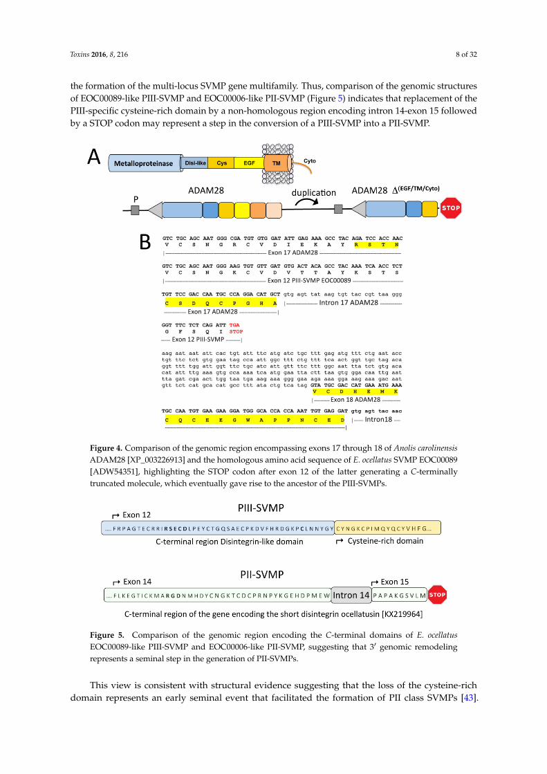
3. Concluding Remarks and Perspectives
The event that gave birth to the family of SVMPs was the generation of a STOP codon at the 3′
end of exon 16 of a duplicated ADAM28 gene (Figure 4). This mutation produced an ORF truncated
at the N‐terminal part of the EGF‐like domain, which encoded a precursor of an ancestral PIII‐SVMP
lacking this domain and the C‐terminal membrane anchoring and cytoplasmic polypeptides (Figure
4). On the other hand, our results comparing the available genomic structures of SVMP genes, e.g.,
EOC00089‐like PIII‐SVMP [47] [KX219963], EOC00006‐like PII‐SVMP [KX219964], and EOC00028‐
like PI‐SVMP [KX219965] (this work), suggest that the evolutionary history of SVMPs is marked with
events of insertions and deletions of intronic regions. This scenario points to introns as key players
in the formation of the multi‐locus SVMP gene multifamily. Thus, comparison of the genomic
Figure 3. Panel A, cartoon comparing the 31 regions of the PII-SVMP and PI-SVMP genes andhighlighting the processes (intronization of ancestral exon 13* inside twintron 12 resulting fromthe fusion of introns 12* and 13*, and creation of a stop codon after exon 14) that destroyed the integrityof the disintegrin domain, converting an ancestral PII(e/d)-type SVMP into extant EOC00028-likePI-SVMP. Panel B, alignment of the amino acid sequences encoded by exon 14 of EOC00028-likePI-SVMP and exon 2 of the dimeric disintegrin subunit ML-G1 [AM261811] [87]. Degenerationof PI-SVMP’s conserved functional and structural amino acid residues in dimeric disintegrins arehighlighted in boldface and grey background.
Region 1013–2134 of PI-SVMP intron 12 exhibits 91% nucleotide sequence identity with range 14 to1135 of Macrovipera lebetina gene encoding part of exon 1 and full-length intron 1 of the VGD-containingdimeric disintegrin subunit precursor, ML-G1 [AM261811] [87]. PI-SVMP exon 14 (mature proteinamino acid residues 221–263, Figure A2) exhibits strong homology (79% identity) to exon 2 of the sameVGD-bearing dimeric disintegrin subunit. The PI-SVMP exon 14 shows the consequences of geneticdrift (Figure 3B): the conseved α5β1 integrin-inhibitory VGD tripeptide motif [44] of the PII-SVMPprecursor gene has been replaced by a VSD motif (generated by a G > A mutation: GTG AGT GAT >GTG GGT GAT), and the absolutely conserved tenth cysteine residue of dimeric disintegrin subunitshas degenerated (TGC) to a serine residue (AGC) (Figure 3B).
3. Concluding Remarks and Perspectives
The event that gave birth to the family of SVMPs was the generation of a STOP codon at the 31
end of exon 16 of a duplicated ADAM28 gene (Figure 4). This mutation produced an ORF truncated atthe N-terminal part of the EGF-like domain, which encoded a precursor of an ancestral PIII-SVMPlacking this domain and the C-terminal membrane anchoring and cytoplasmic polypeptides (Figure 4).On the other hand, our results comparing the available genomic structures of SVMP genes, e.g.,EOC00089-like PIII-SVMP [47] [KX219963], EOC00006-like PII-SVMP [KX219964], and EOC00028-likePI-SVMP [KX219965] (this work), suggest that the evolutionary history of SVMPs is marked withevents of insertions and deletions of intronic regions. This scenario points to introns as key players in
Toxins 2016, 8, 216 8 of 32
the formation of the multi-locus SVMP gene multifamily. Thus, comparison of the genomic structuresof EOC00089-like PIII-SVMP and EOC00006-like PII-SVMP (Figure 5) indicates that replacement of thePIII-specific cysteine-rich domain by a non-homologous region encoding intron 14-exon 15 followedby a STOP codon may represent a step in the conversion of a PIII-SVMP into a PII-SVMP.
Toxins 2016, 8, 216 8 of 29
structures of EOC00089‐like PIII‐SVMP and EOC00006‐like PII‐SVMP (Figure 5) indicates that
replacement of the PIII‐specific cysteine‐rich domain by a non‐homologous region encoding intron
14‐exon 15 followed by a STOP codon may represent a step in the conversion of a PIII‐SVMP into a
PII‐SVMP.
This view is consistent with structural evidence suggesting that the loss of the cysteine‐rich
domain represents an early seminal event that facilitated the formation of PII class SVMPs [43]. The
PII subfamily of SVMPs is characterized by the diversity of disintegrin domains exhibited by different
family members [39,40], ranging from the more ancestral long disintegrin domains (~84 amino‐acid‐
residue polypeptide cross‐linked by 7 disulfide linkages) to the more recently evolved short
disintegrin (41–51 amino‐acid‐residues crosslinked by 4 disulfide bonds) [42]; for a scheme of the
evolutionary path of the disintegrin domains, see Figure 1 in [43]. EOC00006‐like is an example of a
PII‐SVMP with short disintegrin domain. Given the structural diversity of PII‐SVMPs, genomic
sequences from the different members of the subfamily are required for a more accurate glimpse of
the genomic mechanisms operating in the generation and subsequent diversification of PII‐SVMPs.
Figure 4. Comparison of the genomic region encompassing exons 17 through 18 of Anolis carolinensis
ADAM28 [XP_003226913] and the homologous amino acid sequence of E. ocellatus SVMP EOC00089
[ADW54351], highlighting the STOP codon after exon 12 of the latter generating a C‐terminally
truncated molecule, which eventually gave rise to the ancestor of the PIII‐SVMPs.
Figure 4. Comparison of the genomic region encompassing exons 17 through 18 of Anolis carolinensisADAM28 [XP_003226913] and the homologous amino acid sequence of E. ocellatus SVMP EOC00089[ADW54351], highlighting the STOP codon after exon 12 of the latter generating a C-terminallytruncated molecule, which eventually gave rise to the ancestor of the PIII-SVMPs.Toxins 2016, 8, 216 9 of 29
Figure 5. Comparison of the genomic region encoding the C‐terminal domains of E. ocellatus
EOC00089‐like PIII‐SVMP and EOC00006‐like PII‐SVMP, suggesting that 3′ genomic remodeling
represents a seminal step in the generation of PII‐SVMPs.
Comparison of the EOC00006‐like PII‐SVMP and EOC00028‐like PI‐SVMP gene structures also
points to genomic remodeling of the 3′ region of a PII(e/d)‐type SVMP precursor gene [39,40] as the
EOC00028‐like PI‐SVMP gene generator mechanism. The PII > PI conversion involved the generation
of twintron 12 (by fusion of introns 12* and 13*) and the loss, by intronization, of exon 13*, thereby
destroying the consistency of the region coding for the disintegrin domain. This elaborated
mechanism indicates that the structural diversification of SVMPs is not due to a random mutation
generating a STOP codon before the disintegrin domain, but follows a well orchestrated sequence of
events imprinted in the genome of snake species sometime after the split of Viperidae and Elapidae,
37 million years ago, but before the separation of the Viperidae subfamilies Viperinae and Crotalinae
12–20 MYBP. The mechanisms underlying loss or gain of spliceosomal introns are still poorly
understood. The most widely accepted hypothesis is that intron insertion may occur via a process
similar to group II intron retrotransposition [88,89]. According to this view, the spliceosomal
components remain transiently associated with a recently excised intron and then attach at a potential
splice site of a non‐homologous pre‐mRNA, where they catalyze the reverse reaction [90,91]. The
modified pre‐mRNA is reverse‐transcribed and the resulting cDNA participates in a recombination
with its parent gene, thereby inserting a novel intron into the target gene [90–93]. An attractive feature
of this mechanism is that it ensures that the inserted nucleic acid sequence has the full complement
of intron signature sequences required for efficient splicing [94].
Studies of multi‐gene protein families are crucial for understanding the role of gene duplication
and genomic exon‐intron organization in generating protein diversity. For example, full‐length
genomic sequences of Crotalinae group II PLA2 isogenes from P. flavoviridis (Tokunoshima and
Amami‐Oshima islands, Japan) [95], and T. gramineus (Taiwan) [96] have been reported. All these
genes exhibit four coding regions and conserved exon‐intron structures spanning about 1.9 kb. A
cluster of five tandemly arranged PLA2 genes have been located in a 25 kb 3′ segment of a 31 kb
fragment of the Amami‐Oshima P. flavoviridis genome [97], which in addition harbors a PLA2
pseudogene in its 6 kb 5′ region [98]. Genomic sequence comparisons between the pancreatic PLA2
gene of P. elegans, group IB pancreatic PLA2 gene of L. semifasciata, and the L. semifasciata group IA
venom PLA2 gene, suggest that Crotalinae group II venom PLA2 genes emerged before the divergence
of Elapinae and Crotalinae, whereas groups of IB and IA PLA2 genes appeared after Elapinae was
established as a taxonomic lineage [99].
Duplicated structures found in eukaryotic genomes may result from complex interplays
between different mechanisms [100]. Mitotic and meiotic non‐allelic homologous recombination
(NAHR) events, resolved as unequal crossing‐over, have been traditionally invoked to account for
segmental duplications within genomes [101,102]. Duplicated regions can be organized as direct
tandems (e.g., the cluster of tandem snake venom PLA2 genes), but also be separated by hundreds of
kb [100]. Our present and previous work [47] inaugurate a line of research that will allow the
depiction of a more precise characterization of the genomic context in which the SVMP multi‐gene
Figure 5. Comparison of the genomic region encoding the C-terminal domains of E. ocellatusEOC00089-like PIII-SVMP and EOC00006-like PII-SVMP, suggesting that 31 genomic remodelingrepresents a seminal step in the generation of PII-SVMPs.
This view is consistent with structural evidence suggesting that the loss of the cysteine-richdomain represents an early seminal event that facilitated the formation of PII class SVMPs [43].
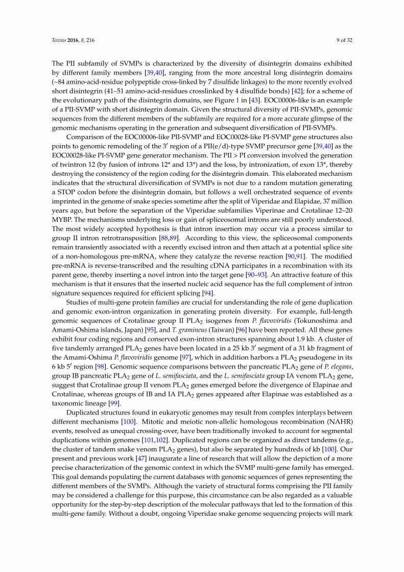
Toxins 2016, 8, 216 9 of 32
The PII subfamily of SVMPs is characterized by the diversity of disintegrin domains exhibitedby different family members [39,40], ranging from the more ancestral long disintegrin domains(~84 amino-acid-residue polypeptide cross-linked by 7 disulfide linkages) to the more recently evolvedshort disintegrin (41–51 amino-acid-residues crosslinked by 4 disulfide bonds) [42]; for a scheme ofthe evolutionary path of the disintegrin domains, see Figure 1 in [43]. EOC00006-like is an exampleof a PII-SVMP with short disintegrin domain. Given the structural diversity of PII-SVMPs, genomicsequences from the different members of the subfamily are required for a more accurate glimpse of thegenomic mechanisms operating in the generation and subsequent diversification of PII-SVMPs.
Comparison of the EOC00006-like PII-SVMP and EOC00028-like PI-SVMP gene structures alsopoints to genomic remodeling of the 31 region of a PII(e/d)-type SVMP precursor gene [39,40] as theEOC00028-like PI-SVMP gene generator mechanism. The PII > PI conversion involved the generationof twintron 12 (by fusion of introns 12* and 13*) and the loss, by intronization, of exon 13*, therebydestroying the consistency of the region coding for the disintegrin domain. This elaborated mechanismindicates that the structural diversification of SVMPs is not due to a random mutation generatinga STOP codon before the disintegrin domain, but follows a well orchestrated sequence of eventsimprinted in the genome of snake species sometime after the split of Viperidae and Elapidae, 37 millionyears ago, but before the separation of the Viperidae subfamilies Viperinae and Crotalinae 12–20MYBP. The mechanisms underlying loss or gain of spliceosomal introns are still poorly understood.The most widely accepted hypothesis is that intron insertion may occur via a process similar togroup II intron retrotransposition [88,89]. According to this view, the spliceosomal componentsremain transiently associated with a recently excised intron and then attach at a potential splice siteof a non-homologous pre-mRNA, where they catalyze the reverse reaction [90,91]. The modifiedpre-mRNA is reverse-transcribed and the resulting cDNA participates in a recombination with itsparent gene, thereby inserting a novel intron into the target gene [90–93]. An attractive feature of thismechanism is that it ensures that the inserted nucleic acid sequence has the full complement of intronsignature sequences required for efficient splicing [94].
Studies of multi-gene protein families are crucial for understanding the role of gene duplicationand genomic exon-intron organization in generating protein diversity. For example, full-lengthgenomic sequences of Crotalinae group II PLA2 isogenes from P. flavoviridis (Tokunoshima andAmami-Oshima islands, Japan) [95], and T. gramineus (Taiwan) [96] have been reported. All these genesexhibit four coding regions and conserved exon-intron structures spanning about 1.9 kb. A cluster offive tandemly arranged PLA2 genes have been located in a 25 kb 31 segment of a 31 kb fragment ofthe Amami-Oshima P. flavoviridis genome [97], which in addition harbors a PLA2 pseudogene in its6 kb 51 region [98]. Genomic sequence comparisons between the pancreatic PLA2 gene of P. elegans,group IB pancreatic PLA2 gene of L. semifasciata, and the L. semifasciata group IA venom PLA2 gene,suggest that Crotalinae group II venom PLA2 genes emerged before the divergence of Elapinae andCrotalinae, whereas groups of IB and IA PLA2 genes appeared after Elapinae was established as ataxonomic lineage [99].
Duplicated structures found in eukaryotic genomes may result from complex interplays betweendifferent mechanisms [100]. Mitotic and meiotic non-allelic homologous recombination (NAHR)events, resolved as unequal crossing-over, have been traditionally invoked to account for segmentalduplications within genomes [101,102]. Duplicated regions can be organized as direct tandems (e.g.,the cluster of tandem snake venom PLA2 genes), but also be separated by hundreds of kb [100]. Ourpresent and previous work [47] inaugurate a line of research that will allow the depiction of a moreprecise characterization of the genomic context in which the SVMP multi-gene family has emerged.This goal demands populating the current databases with genomic sequences of genes representing thedifferent members of the SVMPs. Although the variety of structural forms comprising the PII familymay be considered a challenge for this purpose, this circumstance can be also regarded as a valuableopportunity for the step-by-step description of the molecular pathways that led to the formation of thismulti-gene family. Without a doubt, ongoing Viperidae snake genome sequencing projects will mark
Toxins 2016, 8, 216 10 of 32
the beginning of comparative snake genomics, and will be key to revealing not only the topology andcopy number of the genes encoding SVMPs, but also to provide decisive information to reconstructthe evolutionary history of this multilocus gene family.
4. Materials and Methods
4.1. Genomic DNA
Genomic DNA was extracted from the fresh liver of E. ocellatus (Kaltungo, Nigeria) maintainedat the herpetarium of the Liverpool School of Tropical Medicine. Echis ocellatus liver was ground toa fine powder under liquid nitrogen and the genomic DNA extracted using a Roche DNA isolationkit for cells and tissue containing SDS (2% final concentration) and proteinase K (400 µg/mL finalconcentration). The homogenates were incubated at 55 ˝C overnight. Thereafter, 300 µL of 6 M NaCl(NaCl-saturated H2O) was added to each sample, and the mixture was vortexed for 30 s at maximumspeed and centrifuged for 30 min at 10,000 g. An equal volume of isopropanol was added to eachsupernatant, and the sample mixed, incubated at ´20 ˝C for 1 h, and centrifuged for 20 min at 4 ˝Cand 10,000 g. The resulting pellets were washed with 70% ethanol, dried, and, finally, resuspended in300–500 µL sterile distilled H2O.
4.2. Strategy for PCR Amplification of Overlapping Genomic DNA Fragments
For sequencing E. ocellatus genes encoding PII-SVMP EOC00006 [Q14FJ4] and PI-SVMPEOC00028 [Q2UXQ3] we employed a similar iterative process as described in [47]. Full-lengthcDNA-deduced amino acid sequences of disintegrin domains [103] and of the genomic organization ofdimeric disintegrin domains [AM286800] [87] and PIII-SVMP EOC00089 [47] from the same specieswere used as templates to design primers for the PCR-amplification of protein-specific genomicsequences (Table 2).
PI-SVMP stretch 72AREILNS.....QRWNDLQ263 was amplified on an Eppendorf Mastercycle®
epgradient S instrument in a 50 µL reaction mixture containing 17.5 µL of H2O, 25 µL Master-Mix(Thermo Scientific, Waltham, MA USA) including buffer, dNTPs, and Phusion High-Fidelity DNApolymerase, 2.5 µL of each primer (10 µM) Met1PIRv and Met5PIFw, 1.5 µL of DMSO (100%), and1 µL of genomic DNA (50 ng/µL). PCR conditions included an initial denaturation step at 98 ˝C for30 s followed by 35 cycles of denaturation (20 s at 98 ˝C), annealing (15 s at 63 ˝C), extension (300 s at72 ˝C), and a final extension for 5 min at 72 ˝C. All other PCR amplifications were carried out in thesame thermocycler using iProof High Fidelity polymerase (BioRad, Hercules, CA, USA). The 50 µLreaction mixture contained 10 µL of 5ˆbuffer, 1 µL of 10 mM (each) dNTPs, 2 µL of MgCl2 50 mM,1.5 µL of DMSO (100%), 1 µL of each Fw and Rv primer (10 µM), 1 µL of genomic DNA (50 ng/µL),and 32.5 µL of water. PCR conditions included an initial denaturation step at 98 ˝C for 120 s followedby 35 cycles of denaturation (10 s at 98 ˝C), annealing (15 s at the lower melting temperature of theprimers), extension (60 s per Kb at 72 ˝C), and a final extension for 5 min at 72 ˝C.
4.3. Purification and Cloning of PCR Products
PCR-amplified DNA fragments were purified from agarose electrophoretic bands using theGENECLEAN Turbo kit (MP Biomedicals). The purified fragments were inserted into pJET_1.2(Thermo Scientific, Waltham, MA USA) using phage T4 ligase and cloned into E. coli DH5α byelectroporation at 1700 V. Transformed cells, resuspended in 200 µL LB medium, were incubatedat 37 ˝C for 1 h, and were subsequently plated on LB agar/ampicilline to select positive clones.The presence of the inserted DNA fragments was verified by PCR amplification or digestion of theexpression vector with the restriction enzyme Bgl II. The inserted DNA fragments were sequencedin-house on an Applied Biosystems model 377 DNA sequencing system (Foster City, CA, USA) usingpJETFw and pJETRv primers.
Toxins 2016, 8, 216 11 of 32
Table 2. Forward (Fw) and reverse (Rv) primers used to PCR-amplify genomic DNA stretches fromE. ocellatus PII-SVMP EOC00006-like (right) and PI-SVMP EOC00028-like (left) genes.
Primer DNA sequence Primer DNA sequence
Sp35_Eo Fw ATGATCCAAGTTCTCTTGGTAACTATATGCTTAGC 5’ PS-Disi Fw ATGATCCAAGTTCTCTTGGMet14PI Fw CTATATGCTTAGCAGTTTTTCCATATC Intr4 Fw ATGACACTGACCTCTAGAGTTGGIntr1F1PI Fw CTAGTCATTCCGGCCATATGAC IntrB9_4-2 Fw AAGCTTGCTTGCTAGTAGGTGGIntr2F1PI Fw ATCAGTCTGAGAGGATGCATTTCC Intr4 Rv TGGACATTGTATGGTCACCTGIntr3F1PI Fw GTGACCATGCAATGTCCATATG Prodom 3 Fw GGAGCTTTTAAGCAGCCAGAGMet15PI Fw GTTGCCTGTAGGAGCTGTTAAG Prodom 3 Rv CTCTGGCTGCTTAAAAGCTCC
Prodom 2 Fw GACGCTGTGCAATATGAATTTG Prodom 2 Fw GACGCTGTGCAATATGAATTTGProdom 2 Rv CAAATTCATATTGCACAGCGTC Prodom 2 Rv CAAATTCATATTGCACAGCGTC
Intr3 Rv GCACCAACTCTGTATCTCAGTC Intr3 Fw CACAGGTAAATAAGCCACAAACACCPro2 Fw CAGTGAGACTCATTATTCCCCTGATGGCAG Intr3 Rv GCACCAACTCTGTATCTCAGTC
Pro3 Rv CTGCCATCAGGGGAATAATGAGTCTCACTG Pro2-SVMP_Fw CAGAAGATTACAGTGAGACTCATTATTCCCWGATGG
IntrB13-1 Fw CTTGCCTCCCTATAGGATCACTGC Pro3-SVMP_Rv CTGCCATCAGGGGAATAATGAGTCTCACTMet16PI Rv GATGCGTCCATAATAATAGCAGTG IntrB13-1 Fw CTTGCCTCCCTATAGGATCACTGC
Prodom 1 Fw GATGCCAAAAAAAAGGATGAGG Prodom 1 Fw GATGCCAAAAAAAAGGATGAGGProdom 1 Rv CCTCATCCTTTTTTTTGGCATC Prodom 1 Rv CCTCATCCTTTTTTTTGGCATC
IntronB7PI Fw TGGAACAACAGCTGTTGTTATGACG Intr2 Fw ACAATGGGAAACTGAGGAACAGIntronB7PI Rv TGAGAGACATGCTGATGTGGTC Intr2 Rv GGGAACTCTGACTTAGAGAAAGTC
Met4 PI Fw GACCCAAGATACATTCAGCTTGTC Met1PII Fw CAACAGCATTTTCACCCAAGATACMet4 PI Rv GACAAGCTGAATGTATCTTGGGTC Met1PII Rv GTATCTTGGGTGAAAATGCTGTTGMet8PI Rv TATCCATGTTGTTATAGCAGTTAAATC Met 1-2 Fw CATGGATACATCAAATTGTCAACG
Intron B16 Fw TGTGCTTACCCAACACTGAGCC Met 1-3 Rv TGTACATCTGTCAGGTGGACATGMet5 PI Fw GCACGTGAAATTTTGAACTCA Met2PII Fw GCCGTTCACCTTGATAACCTTATAGGMet5PI Rv GAGTTCAAAATTTCACGTGCTG Met2PII Rv CCTATAAGGTTATcAAGGTGAACGGCMet9PI Rv AGCATTATCATGCGTTATGCG Met 6 PII Fw CCACAATCGTCTGTAGCAATTACTGAMet3 PI Fw GGAAGAGCTTACATGGAGAG Met 6 PII Rv TCAGTAATTGCTACAGACGATTGTGGMet3PI Rv CTCTCCATGTAAGCTCTTCC Met3 PII Fw GATCATAGCACAGATCATCTTTGGMet2PI Rv GCTCCCCAGACATAACGCATC Met3PII Rv CCAAAGATGATCTGTGCTATGATCc
IntrB23PI Fw CTGACTATGACTCACTTAACAACTGG Met 4 Fw ATGATCCAGGTTCTCTTGGTAACTATATGIntrF2PI Fw GGCCGCGTGAATGCATCTGCTTC Met 4 Rv TGAACTGATAGGAACGGTATTGTGIntr2F2PI Fw GCATCAGTTTGTTCGCACTCAATAAAG Fw_Ocella NcoI ATCCATGGTAGACTGTGAATCTGGACCIntr3F2PI Fw GAGCATAATCTGGAACTAAGATCAAG IntrDis1 Rv ATACGGCTAGTATGGAGCAGGMet7PI Fw GCACAAGATTCCTATCACTTCAG Dis PII Rv TCACATCAACACACTGCCTTTTGCMet13PI Rv TCCTACCTGCAAAAGTTCATTTTC - -
Intron B10PI Rv CTGACTCAGGGCACCAATCTC - -Met1PI Rv CTACTGCAGATCGTTCCATCTCTG - -
4.4. Sequence Analysis
Exon-intron boundaries were localized by visual inspection and corroborated using Wise2 [104].Amino acid and nucleotide sequence similarity searches were done using BLAST [105]. Multiplesequence alignments were performed using ClustalW2 [106]. The occurrence of retrotransposableelements and simple nucleotide sequence repeats (SSRs) were assessed using RepeatMasker (versionrm-20110920) [107], a program that screens DNA sequences for interspersed repeats and low complexityDNA sequences included in the Repbase database [108].
4.5. Sequence Availability
Pre-pro EOC00006-like PII-SVMP and EOC00028-like PI-SVMP gene sequences have beendeposited with the NCBI GeneBank [109] and are accessible under accession codes KX219964 andKX219965, respectively.
Supplementary Materials: The following are available online at www.mdpi.com/2072-6651/8/7/216/s1,Figure S1: Pairwise nucleotide sequence alignments of topologically equivalent paralog introns 1–12 fromPre-pro EOC00006-like PII-SVMP and 1–13 from Pre-pro EOC00028-like PI-SVMP gene sequences.
Acknowledgments: This work has been financed by Grant BFU2013-42833-P from the Ministerio de Economía yCompetitividad, Madrid (Spain).
Author Contributions: J.J.C. and L.S. conceived and designed the experiments; L.S. performed the experiments;J.J.C. and L.S. analyzed the data; J.J.C. wrote the paper.
Conflicts of Interest: The authors declare no conflict of interest.
Toxins 2016, 8, 216 12 of 32
Appendix A
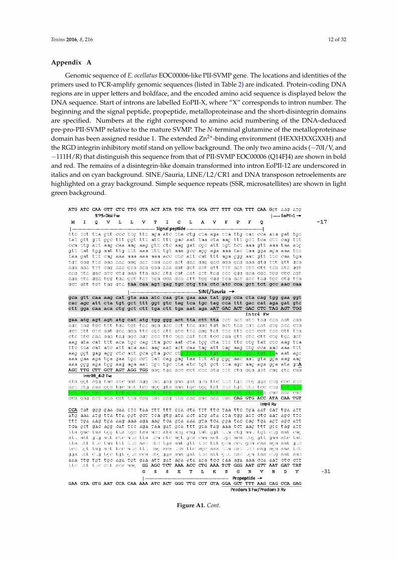
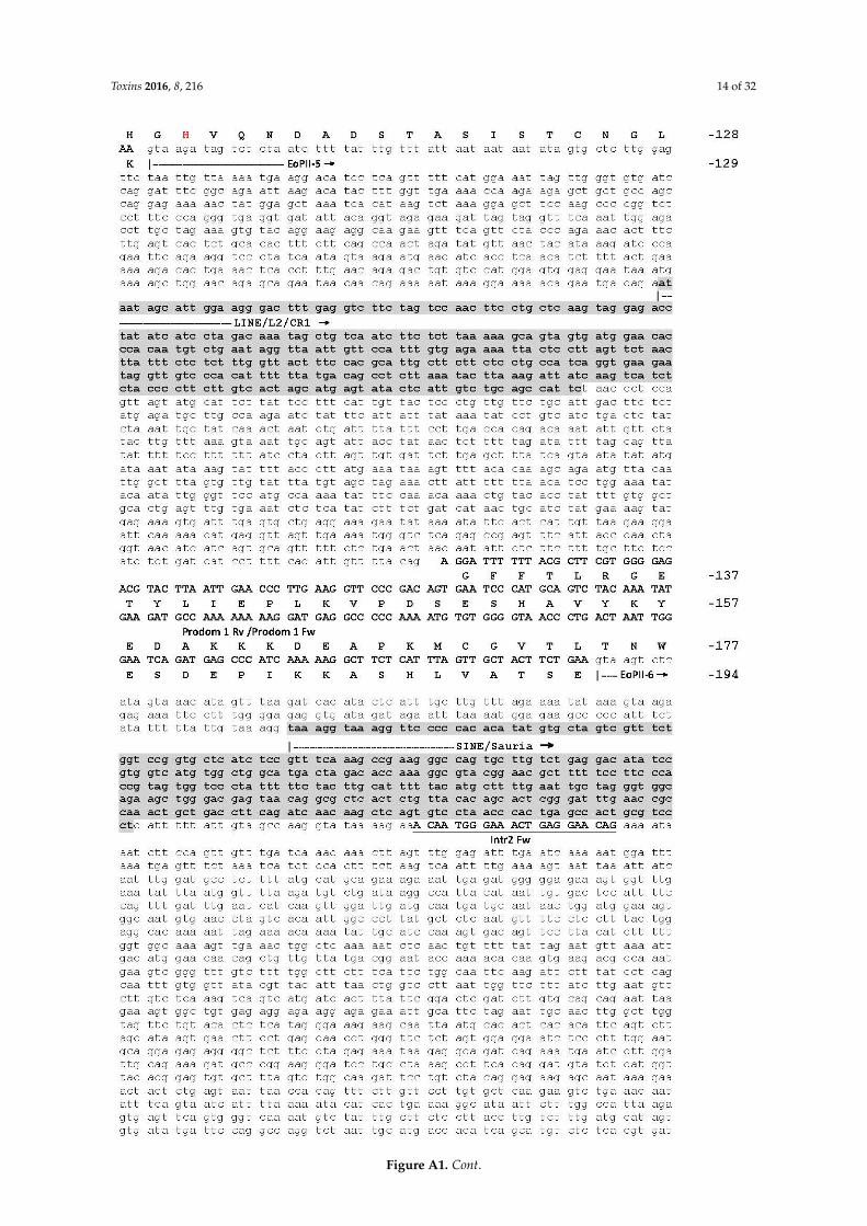
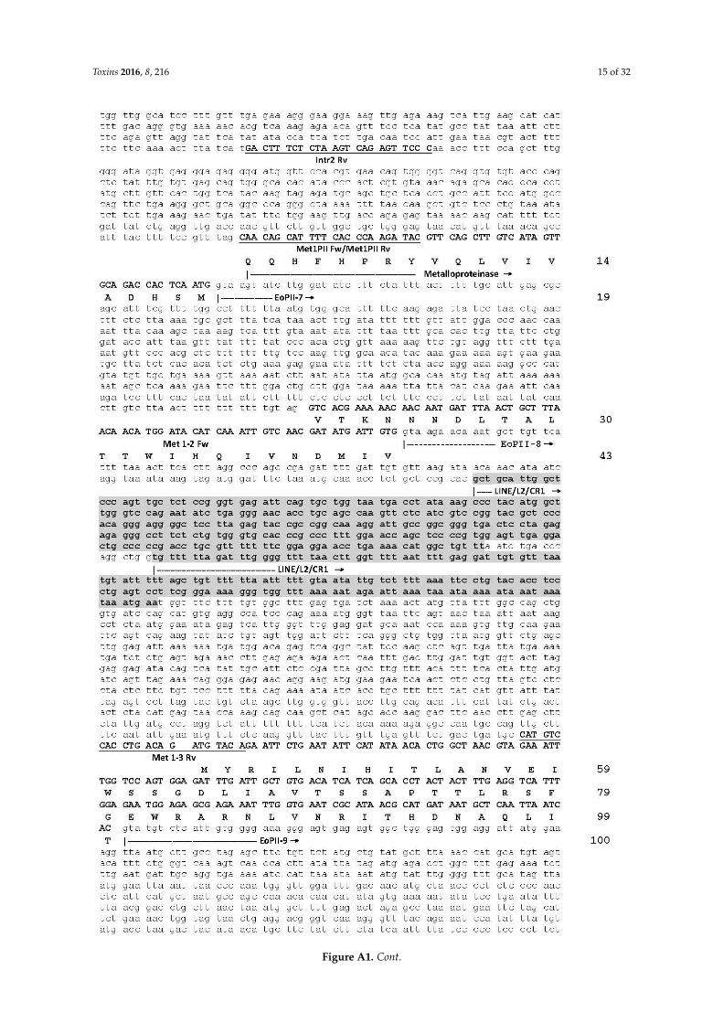
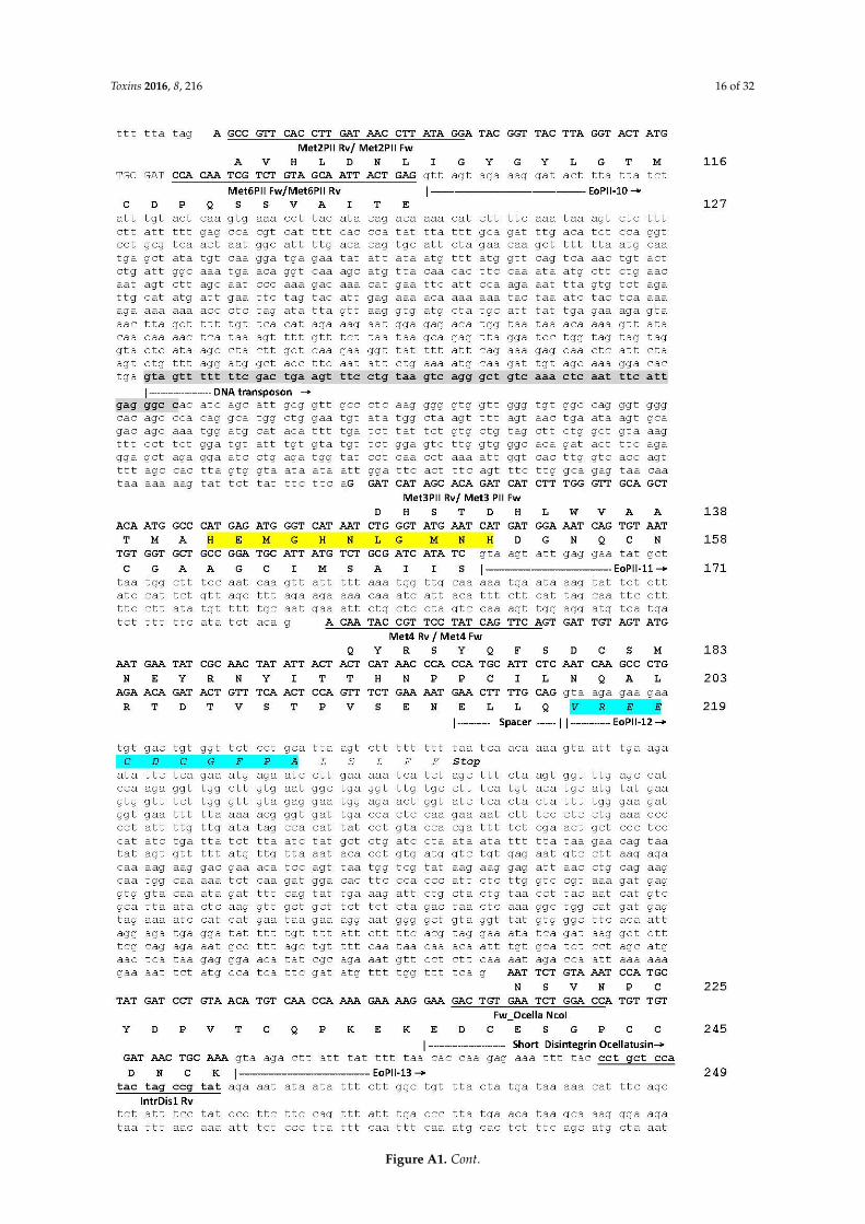
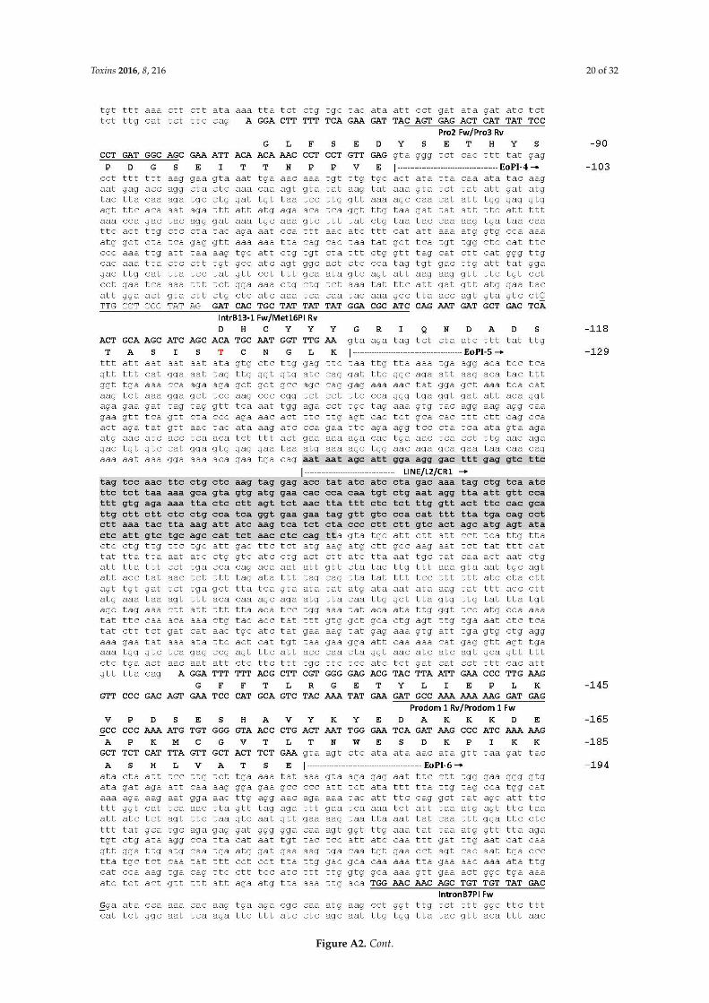
Genomic sequence of E. ocellatus EOC00006-like PII-SVMP gene. The locations and identities of theprimers used to PCR-amplify genomic sequences (listed in Table 2) are indicated. Protein-coding DNAregions are in upper letters and boldface, and the encoded amino acid sequence is displayed below theDNA sequence. Start of introns are labelled EoPII-X, where “X” corresponds to intron number. Thebeginning and the signal peptide, propeptide, metalloproteinase and the short-disintegrin domainsare specified. Numbers at the right correspond to amino acid numbering of the DNA-deducedpre-pro-PII-SVMP relative to the mature SVMP. The N-terminal glutamine of the metalloproteinasedomain has been assigned residue 1. The extended Zn2+-binding environment (HEXXHXXGXXH) andthe RGD integrin inhibitory motif stand on yellow background. The only two amino acids (´70I/V, and´111H/R) that distinguish this sequence from that of PII-SVMP EOC00006 (Q14FJ4) are shown in boldand red. The remains of a disintegrin-like domain transformed into intron EoPII-12 are underscored initalics and on cyan background. SINE/Sauria, LINE/L2/CR1 and DNA transposon retroelements arehighlighted on a gray background. Simple sequence repeats (SSR, microsatellites) are shown in lightgreen background.Toxins 2016, 8, 216 13 of 29
Figure A1. Cont.

Toxins 2016, 8, 216 13 of 32Toxins 2016, 8, 216 14 of 29
Figure A1. Cont.
Toxins 2016, 8, 216 14 of 32Toxins 2016, 8, 216 15 of 29
Figure A1. Cont.
Toxins 2016, 8, 216 15 of 32
Toxins 2016, 8, 216 16 of 29
Figure A1. Cont.
Toxins 2016, 8, 216 16 of 32Toxins 2016, 8, 216 17 of 29
Figure A1. Cont.

Toxins 2016, 8, 216 17 of 32Toxins 2016, 8, 216 18 of 29
Figure A1. Genomic organization of E. ocellatus EOC00006‐like PII‐SVMP gene.
Figure A1. Genomic organization of E. ocellatus EOC00006-like PII-SVMP gene.
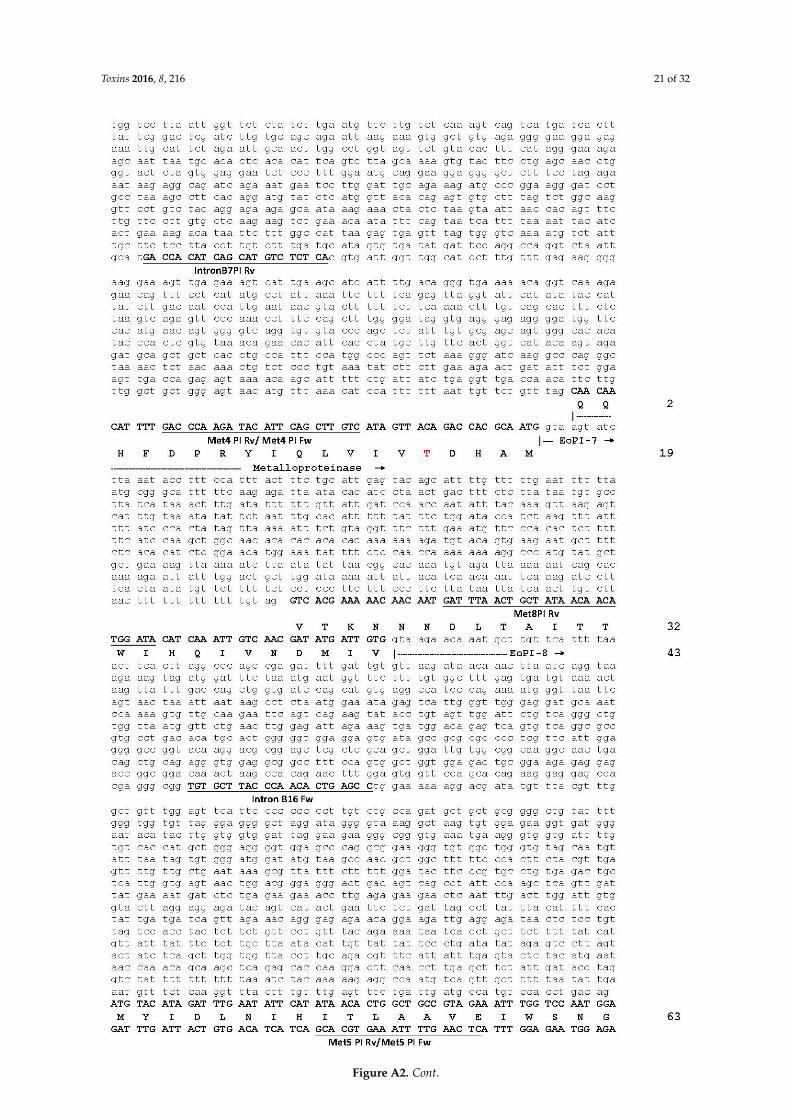
Genomic sequence of E. ocellatus EOC00028-like PI-SVMP gene. The locations and identities ofthe primers used to PCR-amplify genomic sequences (listed in Table 2) are indicated. Protein-codingDNA regions are in upper letters and boldface, and the encoded amino acid sequence is displayedbelow the DNA sequence. Start of introns are labelled EoPI-X, where “X” corresponds to intronnumber. The beginning and the signal peptide, propeptide, metalloproteinase domains and theC-terminal extension are specified. Numbers at the right correspond to amino acid numberingof the DNA-deduced pre-pro-PII-SVMP relative to the mature SVMP. The extended Zn2+-bindingenvironment (HEXXHXXGXXH) stands on yellow background. The only two amino acids (-124T/Aand 15T/A) that distinguish this sequence from that of PI-SVMP EOC00028 (Q2UXQ3) are shownin bold and red. The remains of a disintegrin-like domain and a dimeric disintegrin domaintransformed into intron EoPI-12 are underscored in italics and on cyan background. The N-terminalglutamine of the metalloproteinase domain has been assigned residue 1. SINE/Sauria, LINE/L2/CR1,LTR/ERV1, DNA/hAT-Ac and DNA transposon retroelements are highlighted on a gray background.Inserted nucleotide sequences in introns 1 (582 nucleotides between positions 1534–1582, including aSINE/Sauria element); 9 (between nucleotides 194–195 of the topologically equivalent intron of PII); 11(replacing nucleotides 1-66 of PII intron 11 for a stretch of 3281 nucleotides); and 12 (after nucleotide999 of the homologous PII intron) are underlined. Simple sequence repeats (SSR, microsatellites) areshown in light green background.

Toxins 2016, 8, 216 18 of 32
Toxins 2016, 8, 216 19 of 29
Genomic sequence of E. ocellatus EOC00028‐like PI‐SVMP gene. The locations and identities of
the primers used to PCR‐amplify genomic sequences (listed in Table 2) are indicated. Protein‐coding
DNA regions are in upper letters and boldface, and the encoded amino acid sequence is displayed
below the DNA sequence. Start of introns are labelled EoPI‐X, where “X” corresponds to intron
number. The beginning and the signal peptide, propeptide, metalloproteinase domains and the C‐
terminal extension are specified. Numbers at the right correspond to amino acid numbering of the
DNA‐deduced pre‐pro‐PII‐SVMP relative to the mature SVMP. The extended Zn2+‐binding
environment (HEXXHXXGXXH) stands on yellow background. The only two amino acids (‐124T/A
and 15T/A) that distinguish this sequence from that of PI‐SVMP EOC00028 (Q2UXQ3) are shown in
bold and red. The remains of a disintegrin‐like domain and a dimeric disintegrin domain transformed
into intron EoPI‐12 are underscored in italics and on cyan background. The N‐terminal glutamine of
the metalloproteinase domain has been assigned residue 1. SINE/Sauria, LINE/L2/CR1, LTR/ERV1,
DNA/hAT‐Ac and DNA transposon retroelements are highlighted on a gray background. Inserted
nucleotide sequences in introns 1 (582 nucleotides between positions 1534–1582, including a
SINE/Sauria element); 9 (between nucleotides 194–195 of the topologically equivalent intron of PII);
11 (replacing nucleotides 1‐66 of PII intron 11 for a stretch of 3281 nucleotides); and 12 (after
nucleotide 999 of the homologous PII intron) are underlined. Simple sequence repeats (SSR,
microsatellites) are shown in light green background.
Figure A2. Cont.

Toxins 2016, 8, 216 19 of 32Toxins 2016, 8, 216 20 of 29
Figure A2. Cont.
Toxins 2016, 8, 216 20 of 32Toxins 2016, 8, 216 21 of 29
Figure A2. Cont.
Toxins 2016, 8, 216 21 of 32
Toxins 2016, 8, 216 22 of 29
Figure A2. Cont.

Toxins 2016, 8, 216 22 of 32Toxins 2016, 8, 216 21 of 29
Figure A2. Cont.

Toxins 2016, 8, 216 23 of 32Toxins 2016, 8, 216 23 of 29
Figure A2. Cont.

Toxins 2016, 8, 216 24 of 32Toxins 2016, 8, 216 21 of 29
Figure A2. Cont.

Toxins 2016, 8, 216 25 of 32Toxins 2016, 8, 216 24 of 29
Figure A2. Cont.

Toxins 2016, 8, 216 26 of 32Toxins 2016, 8, 216 21 of 29
Figure A2. Cont.

Toxins 2016, 8, 216 27 of 32Toxins 2016, 8, 216 25 of 29
Figure A2. Genomic organization of E. ocellatus EOC00028‐like PI‐SVMP gene.
References
1. MEROPS‐ the Peptidase Database: Family M72. Available online: https://merops.sanger.ac.uk/cgi‐
bin/famsum?family=M12 (accessed on 8 May 2016).
2. PFAM Family: REprolysin. Available online: http://pfam.xfam.org/family/PF01421 (accessed on 8 May
2016).
3. Seals, D.F.; Courtneidge, S.A. The ADAMs family of metalloproteases: Multidomain proteins with multiple
functions. Genes Dev. 2003, 17, 7–30.
4. Giebeler, N.; Zigrino, P. A disintegrin and metalloprotease (ADAM): Historical overview of their functions.
Toxins 2016, 8, doi:10.3390/toxins8040122.
5. Arendt, D.; Technau, U.; Wittbrodt, J. Evolution of the bilaterian larval foregut. Nature 2001, 409, 81–85.
6. Tucker, R.P.; Adams, J.C. Adhesion Networks of Cnidarians: A Postgenomic View. Int. Rev. Cell Mol. Biol.
2014, 308, 323–377.
7. Bahudhanapati, H.; Bhattacharya, S.; Wei, S. Evolution of Vertebrate Adam Genes; Duplication of
Testicular Adams from Ancient Adam9/9‐like Loci. PLoS ONE 2015, 10, e0136281.
8. Cho, C. Testicular and epididymal ADAMs: Expression and function during fertilization. Nat. Rev. Urol.
2012, 9, 550–560.
9. Bates, E.E.; Fridman, W.H.; Mueller, C.G. The ADAMDEC1 (decysin) gene structure: Evolution by
duplication in a metalloprotease gene cluster on chromosome 8p12. Immunogenetics 2002, 54, 96–105.
10. Wei, S.; Whittaker, C.A.; Xu, G.; Bridges, L.C.; Shah, A.; White, J.M.; Desimone, D.W. Conservation and
divergence of ADAM family proteins in the Xenopus genome. BMC Evol. Biol. 2010, 10, doi:10.1186/1471‐
2148‐10‐211.
11. Taylor, J.S.; Raes, J. Duplication and divergence: The evolution of new genes and old ideas. Annu. Rev.
Genet. 2004, 38, 615–643.
12. Ohno, S. Evolution by Gene Duplication; Springer‐Verlag: Berlin‐Heidelberg, Germany, 1970.
13. Zhang, J. Evolution by gene duplication: An update. Trends Ecol. Evol. 2003, 18, 292–298.
14. True, J.R.; Carroll, S.B. Gene co‐option in physiological and morphological evolution. Annu. Rev. Cell Dev.
Biol. 2002, 18, 53–80.
15. Kaessmann, H.; Vinckenbosch, N.; Long, M. RNA‐based gene duplication: Mechanistic and evolutionary
insights. Nat. Rev. Genet. 2009, 10, 19–31.
16. Vinckenbosch, N.; Dupanloup, I.; Kaessmann, H. Evolutionary fate of retroposed gene copies in the human
genome. Proc. Natl. Acad. Sci. USA 2006, 103, 3220–3225.
Figure A2. Genomic organization of E. ocellatus EOC00028-like PI-SVMP gene.
References
1. MEROPS- the Peptidase Database: Family M72. Available online: https://merops.sanger.ac.uk/cgi-bin/famsum?family=M12 (accessed on 8 May 2016).
2. PFAM Family: REprolysin. Available online: http://pfam.xfam.org/family/PF01421 (accessed on8 May 2016).
3. Seals, D.F.; Courtneidge, S.A. The ADAMs family of metalloproteases: Multidomain proteins with multiplefunctions. Genes Dev. 2003, 17, 7–30. [CrossRef] [PubMed]
4. Giebeler, N.; Zigrino, P. A disintegrin and metalloprotease (ADAM): Historical overview of their functions.Toxins 2016, 8. [CrossRef] [PubMed]
5. Arendt, D.; Technau, U.; Wittbrodt, J. Evolution of the bilaterian larval foregut. Nature 2001, 409, 81–85.[CrossRef] [PubMed]
6. Tucker, R.P.; Adams, J.C. Adhesion Networks of Cnidarians: A Postgenomic View. Int. Rev. Cell Mol. Biol.2014, 308, 323–377. [PubMed]
7. Bahudhanapati, H.; Bhattacharya, S.; Wei, S. Evolution of Vertebrate Adam Genes; Duplication of TesticularAdams from Ancient Adam9/9-like Loci. PLoS ONE 2015, 10, e0136281. [CrossRef] [PubMed]
8. Cho, C. Testicular and epididymal ADAMs: Expression and function during fertilization. Nat. Rev. Urol.2012, 9, 550–560. [CrossRef] [PubMed]
9. Bates, E.E.; Fridman, W.H.; Mueller, C.G. The ADAMDEC1 (decysin) gene structure: Evolution by duplicationin a metalloprotease gene cluster on chromosome 8p12. Immunogenetics 2002, 54, 96–105. [CrossRef][PubMed]
10. Wei, S.; Whittaker, C.A.; Xu, G.; Bridges, L.C.; Shah, A.; White, J.M.; Desimone, D.W. Conservation anddivergence of ADAM family proteins in the Xenopus genome. BMC Evol. Biol. 2010, 10. [CrossRef] [PubMed]
11. Taylor, J.S.; Raes, J. Duplication and divergence: The evolution of new genes and old ideas. Annu. Rev. Genet.2004, 38, 615–643. [CrossRef] [PubMed]
12. Ohno, S. Evolution by Gene Duplication; Springer-Verlag: Berlin-Heidelberg, Germany, 1970.13. Zhang, J. Evolution by gene duplication: An update. Trends Ecol. Evol. 2003, 18, 292–298. [CrossRef]14. True, J.R.; Carroll, S.B. Gene co-option in physiological and morphological evolution. Annu. Rev. Cell Dev.
Biol. 2002, 18, 53–80. [CrossRef] [PubMed]
Toxins 2016, 8, 216 28 of 32
15. Kaessmann, H.; Vinckenbosch, N.; Long, M. RNA-based gene duplication: Mechanistic and evolutionaryinsights. Nat. Rev. Genet. 2009, 10, 19–31. [CrossRef] [PubMed]
16. Vinckenbosch, N.; Dupanloup, I.; Kaessmann, H. Evolutionary fate of retroposed gene copies in the humangenome. Proc. Natl. Acad. Sci. USA 2006, 103, 3220–3225. [CrossRef] [PubMed]
17. Fry, B.; Wüster, W. Assembling an Arsenal: Origin and evolution of the snake venom proteome inferredfrom phylogenetic analysis of toxin sequences. Mol. Biol. Evol. 2004, 21, 870–883. [CrossRef] [PubMed]
18. Fry, B.G.; Vidal, N.; Norman, J.A.; Vonk, F.J.; Scheib, H.; Ramjan, S.F.; Kuruppu, S.; Fung, K.; Hedges, S.B.;Richardson, M.K.; et al. Early evolution of the venom system in lizards and snakes. Nature 2006, 439, 584–588.[CrossRef] [PubMed]
19. Fry, B.G.; Casewell, N.R.; Wüster, W.; Vidal, N.; Young, B.; Jackson, N. The structural and functionaldiversification of the Toxicofera reptile venom system. Toxicon 2012, 60, 434–448. [CrossRef] [PubMed]
20. Casewell, N.R.; Wüster, W.; Vonk, F.J.; Harrison, R.A.; Fry, B.G. Complex cocktails: The evolutionary noveltyof venoms. Trends Ecol. Evol. 2013, 28, 219–229. [CrossRef] [PubMed]
21. Haney, R.A.; Clarke, T.H.; Gadgil, R.; Fitzpatrick, R.; Hayashi, C.Y.; Ayoub, N.A.; Garb, J.E. Effects ofgene duplication, positive selection, and shifts in gene expression on the evolution of the venom glandtranscriptome in widow spiders. Genome Biol. Evol. 2016, 8, 228–242. [CrossRef] [PubMed]
22. Wong, E.S.; Belov, K. Venom evolution through gene duplications. Gene 2012, 496, 1–7. [CrossRef] [PubMed]23. Vonk, F.J.; Casewell, N.R.; Henkel, C.V.; Heimberg, A.M.; Jansen, H.J.; McCleary, R.J.; Kerkkamp, H.M.;
Vos, R.A.; Guerreiro, I.; Calvete, J.J.; et al. The king cobra genome reveals dynamic gene evolution andadaptation in the snake venom system. Proc. Natl. Acad. Sci. USA 2013, 110, 20651–20656. [CrossRef][PubMed]
24. Hedges, S.B.; Vidal, N. Lizards, snakes, and amphisbaenians (Squamata). In The Timetree of Life; Hedges, S.B.,Kumar, S., Eds.; Oxford University Press: Oxford, UK, 2009; pp. 383–389.
25. Jones, M.E.; Anderson, C.L.; Hipsley, C.A.; Müller, J.; Evans, S.E.; Schoch, R.R. Integration of molecules andnew fossils supports a Triassic origin for Lepidosauria (lizards, snakes, and tuatara). BMC Evol. Biol. 2013,13. [CrossRef] [PubMed]
26. Pyron, R.A.; Burbrink, F.T.; Wiens, J.J. A phylogeny and revised classification of Squamata, including 4161species of lizards and snakes. BMC Evol. Biol. 2013, 13. [CrossRef] [PubMed]
27. Reeder, T.W.; Townsend, T.M.; Mulcahy, D.G.; Noonan, B.P.; Wood, P.L., Jr.; Sites, J.W., Jr.; Wiens, J.J. Integratedanalyses resolve conflicts over squamate reptile phylogeny and reveal unexpected placements for fossil.PLoS ONE 2015, 10, e0118199. [CrossRef] [PubMed]
28. Hsiang, A.Y.; Field, D.J.; Webster, T.H.; Behlke, A.D.; Davis, M.B.; Racicot, R.A.; Gauthier, J.A. The origin ofsnakes: Revealing the ecology, behavior, and evolutionary history of early snakes using genomics, phenomics,and the fossil record. BMC Evol. Biol. 2015, 15. [CrossRef] [PubMed]
29. Reeks, T.A.; Fry, B.G.; Alewood, P.F. Privileged frameworks from snake venom. Cell. Mol. Life Sci. 2015, 72,1939–1958. [CrossRef] [PubMed]
30. Hite, L.A.; Jia, L.G.; Bjarnason, J.B.; Fox, J.W. cDNA sequences for four snake venom metalloproteinases:Structure, classification, and their relationship to mammalian reproductive proteins. Arch. Biochem. Biophys.1994, 308, 182–191. [CrossRef] [PubMed]
31. Moura-da-Silva, A.M.; Theakston, R.D.; Crampton, J.M. Evolution of disintegrin cysteine-rich andmammalian matrix-degrading metalloproteinases: Gene duplication and divergence of a common ancestorrather than convergent evolution. J. Mol. Evol. 1996, 43, 263–269. [CrossRef] [PubMed]
32. Casewell, N.R. On the ancestral recruitment of metalloproteinases into the venom of snakes. Toxicon 2012,60, 449–454. [CrossRef] [PubMed]
33. Escalante, T.; Rucavado, A.; Fox, J.W.; Gutiérrez, J.M. Key events in microvascular damage induced by snakevenom hemorrhagic metalloproteinases. J. Proteomics 2011, 74, 1781–1794. [CrossRef] [PubMed]
34. Markland, F.S., Jr.; Swenson, S. Snake venom metalloproteinases. Toxicon 2013, 62, 3–18. [CrossRef] [PubMed]35. Herrera, C.; Escalante, T.; Voisin, M.B.; Rucavado, A.; Morazán, D.; Macêdo, J.K.; Calvete, J.J.; Sanz, L.;
Nourshargh, S.; Gutiérrez, J.M.; et al. Tissue localization and extracellular matrix degradation by PI, PII andPIII snake venom metalloproteinases: Clues on the mechanisms of venom-induced hemorrhage. PLoS Negl.Trop. Dis. 2015, 9, e0003731. [CrossRef] [PubMed]
36. Gutiérrez, J.M.; Escalante, T.; Rucavado, A.; Herrera, C. Hemorrhage Caused by Snake VenomMetalloproteinases: A Journey of Discovery and Understanding. Toxins 2016, 8. [CrossRef] [PubMed]
Toxins 2016, 8, 216 29 of 32
37. Jia, L.G.; Shimokawa, K.; Bjarnason, J.B.; Fox, J.W. Snake venom metalloproteinases: Structure, function andrelationship to the ADAMs family of proteins. Toxicon 1996, 34, 1269–1276. [CrossRef]
38. Pyron, R.A.; Burnbrink, F.T. Extinction ecological opportunity and the origins of global snake diversity.Evolution 2012, 66, 163–178. [CrossRef] [PubMed]
39. Fox, J.W.; Serrano, S.M. Structural considerations of the snake venom metalloproteinases, key members ofthe M12 reprolysin family of metalloproteinases. Toxicon 2005, 45, 969–985. [CrossRef] [PubMed]
40. Fox, J.W.; Serrano, S.M. Insights into and speculations about snake venom metalloproteinase (SVMP)synthesis, folding and disulfide bond formation and their contribution to venom complexity. FEBS J. 2008,275, 3016–3030. [CrossRef] [PubMed]
41. Casewell, N.R.; Sunagar, K.; Takacs, Z.; Calvete, J.J.; Jackson, T.N.W.; Fry, B.G. Snake venom metalloproteaseenzymes. In Venomous Reptiles and Their Toxins: Evolution, Pathophysiology and Biodiscovery; ISBN:978-0-19-930939-9. Fry, B.G., Ed.; Oxford University Press: Oxford, UK, 2015; Chapter 23; pp. 347–363.
42. Juárez, P.; Comas, I.; González-Candelas, F.; Calvete, J.J. Evolution of snake venom disintegrins by positiveDarwinian selection. Mol. Biol. Evol. 2008, 25, 2391–2407. [CrossRef] [PubMed]
43. Carbajo, R.J.; Sanz, L.; Pérez, A.; Calvete, J.J. NMR structure of bitistatin—A missing piece in the evolutionarypathway of snake venom disintegrins. FEBS J. 2015, 282, 341–360. [CrossRef] [PubMed]
44. Calvete, J.J. Brief History and Molecular Determinants of Snake Venom Disintegrin Evolution. In Toxinsand Hemostasis. From Bench to Bedside; Kini, R.M., Markland, F., McLane, M.A., Morita, T., Eds.; SpringerScience+Business Media B.V.: Amsterdam, The Netherlands, 2010; pp. 285–300.
45. Casewell, N.R.; Wagstaff, S.C.; Harrison, R.A.; Renjifo, C.; Wüster, W. Domain loss facilitates acceleratedevolution and neofunctionalization of duplicate snake venom metalloproteinase toxin genes. Mol. Biol. Evol.2011, 28, 2637–2649. [CrossRef] [PubMed]
46. Sanz-Soler, R.; Sanz, L.; Calvete, J.J. Distribution of RPTLN genes across Reptilia. Hypothesized role forRPTLN in the evolution of SVMPs. Integr. Compar. Biol. 2016. [CrossRef] [PubMed]
47. Sanz, L.; Harrison, R.A.; Calvete, J.J. First draft of the genomic organization of a PIII-SVMP gene. Toxicon2012, 60, 455–469. [CrossRef] [PubMed]
48. Endo, T.; Fedorov, A.; de Souza, S.J.; Gilbert, W. Do Introns Favor or Avoid Regions of Amino AcidConservation? Mol. Biol. Evol. 2002, 19, 521–525. [CrossRef] [PubMed]
49. Zhou, Q.; Wang, W. On the origin and evolution of new genes-a genomic and experimental perspective.J. Genet. Genomics 2008, 35, 639–648. [CrossRef]
50. Kordis, D.; Gubensek, F. Adaptive evolution of animal toxin multigene families. Gene 2000, 261, 43–52.[CrossRef]
51. Cao, Z.; Yu, Y.; Wu, Y.; Hao, P.; Di, Z.; He, Y.; Chen, Z.; Yang, W.; Shen, Z.; He, X.; et al. The genome ofMesobuthus martensii reveals a unique adaptation model of arthropods. Nat. Commun. 2013, 4. [CrossRef][PubMed]
52. Fry, B.G.; Wüster, W.; Kini, R.M.; Brusic, V.; Khan, A.; Venkataraman, D.; Rooney, A.P. Molecular evolutionand phylogeny of elapid snake venom three-finger toxins. J. Mol. Evol. 2003, 57, 110–129. [CrossRef][PubMed]
53. Reyes-Velasco, J.; Card, D.C.; Andrew, A.L.; Shaney, K.J.; Adams, R.H.; Schield, D.R.; Casewell, N.R.;Mackessy, S.P.; Castoe, T.A. Expression of venom gene homologs in diverse python tissues suggests a newmodel for the evolution of snake venom. Mol. Biol. Evol. 2015, 32, 173–183. [CrossRef] [PubMed]
54. Hargreaves, A.D.; Swain, M.T.; Logan, D.W.; Mulley, J.F. Testing the Toxicofera: Comparative transcriptomicscasts doubt on the single, early evolution of the reptile venom system. Toxicon 2014, 92, 140–156. [CrossRef][PubMed]
55. Sunagar, K.; Jackson, T.N.; Undheim, E.A.; Ali, S.A.; Antunes, A.; Fry, B.G. Three-fingered RAVERs: RapidAccumulation of Variations in Exposed Residues of snake venom toxins. Toxins 2013, 5, 2172–2208. [CrossRef][PubMed]
56. Chang, D.; Duda, T.F. Extensive and continuous duplication facilitates rapid evolution and diversification ofgene families. Mol. Biol. Evol. 2012, 29, 2019–2029. [CrossRef] [PubMed]
57. Chow, L.T.; Gelinas, R.E.; Broker, T.R.; Roberts, R.J. An amazing sequence arrangement at the 51 ends ofadenovirus 2 messenger RNA. Cell 1977, 12, 1–8. [CrossRef]
58. Berget, S.M.; Moore, C.; Sharp, P.A. Spliced segments at the 51 terminus of adenovirus 2 late mRNA.Proc. Natl. Acad. Sci. USA 1977, 74, 3171–3175. [CrossRef] [PubMed]

Toxins 2016, 8, 216 30 of 32
59. Bicknell, A.A.; Cenik, C.; Chua, H.N.; Roth, F.P.; Moore, M.J. Introns in UTRs: Why we should stop ignoringthem. BioEssays 2012, 34, 1025–1034. [CrossRef] [PubMed]
60. Cenik, C.; Chua, H.N.; Zhang, H.; Tarnawsky, S.P.; Akef, A.; Derti, A.; Tasan, M.; Moore, M.J.; Palazzo, A.F.;Roth, F.P. Genome analysis reveals interplay between 51-UTR introns and nuclear mRNA export for secretoryand mitochondrial genes. PLoS Genet. 2011, 7, e1001366. [CrossRef] [PubMed]
61. Comeron, J.M.; Kreitman, M. The correlation between intron length and recombination in Drosophila:Dynamic equilibrium between mutational and selective forces. Genetics 2000, 156, 1175–1190. [PubMed]
62. De Souza, S.J.; Long, M.; Gilbert, W. Introns and gene evolution. Genes Cells 1996, 1, 493–505. [CrossRef][PubMed]
63. Patthy, L. Exon shuffling and other ways of module exchange. Matrix Biol. 1996, 15, 301–310. [CrossRef]64. Hughes, A.L.; Hughes, M.K. Small genomes for better flyers. Nature 1995, 377, 391. [CrossRef] [PubMed]65. Lynch, M. Intron evolution as a population-genetic process. Proc. Natl. Acad. Sci. USA 2002, 99, 6118–6123.
[CrossRef] [PubMed]66. Haddrill, P.R.; Charlesworth, B.; Halligan, D.L.; Andolfatto, P. Patterns of intron sequence evolution in
Drosophila are dependent upon length and GC content. Genome Biol. 2005, 6, R67. [CrossRef] [PubMed]67. Zhao, M.; He, L.; Gu, Y.; Wang, Y.; Chen, Q.; He, C. Genome-wide analyses of a plant-specific LIM-domain
gene family implicate its evolutionary role in plant diversification. Genome Biol. Evol. 2014, 6, 1000–1012.[CrossRef] [PubMed]
68. Tordai, H.; Patthy, L. Insertion of spliceosomal introns in proto-splice sites: The case of secretory signalpeptides. FEBS Lett. 2004, 575, 109–111. [CrossRef] [PubMed]
69. Tomita, M.; Shimizu, N.; Brutlag, D.L. Introns and reading frames: Correlation between splicing sites andtheir codon positions. Mol. Biol. Evol. 1996, 13, 1219–1223. [CrossRef] [PubMed]
70. Long, M.; de Souza, S.J.; Rosenberg, C.; Gilbert, W. Relationship between proto-splice sites and intron phases:Evidence from dicodon analysis. Proc. Natl. Acad. Sci. USA 1998, 95, 219–223. [CrossRef] [PubMed]
71. Von Heijne, G. Patterns of amino acids near signal-sequence cleavage sites. Eur. J. Biochem. 1983, 133, 17–21.[CrossRef] [PubMed]
72. Pinho, C.; Rocha, S.; Carvalho, B.M.; Lopes, S.; Mourão, S.; Vallinoto, M.; Brunes, T.O.; Haddad, C.F.B.;Gonçalves, H.; Sequeira, F.; et al. New primers for the amplification and sequencing of nuclear loci in ataxonomically wide set of reptiles and amphibians. Conserv. Genet. Resour. 2010, 2, 181–185. [CrossRef]
73. Ellegren, H. Microsatellites: Simple sequences with complex evolution. Nat. Rev. Genet. 2004, 5, 435–445.[CrossRef] [PubMed]
74. Adams, R.H.; Blackmon, H.; Reyes-Velasco, J.; Schield, D.R.; Card, D.C.; Andrew, A.L.; Waynewood, N.;Castoe, T.A. Microsatellite landscape evolutionary dynamics across 450 million years of vertebrate genomeevolution. Genome 2016, 59, 295–310. [CrossRef] [PubMed]
75. Liang, K.C.; Tseng, J.T.; Tsai, S.J.; Sun, H.S. Characterization and distribution of repetitive elements inassociation with genes in the human genome. Comput. Biol. Chem. 2015, 57, 29–38. [CrossRef] [PubMed]
76. Schlötterer, C.; Tautz, D. Slippage synthesis of simple sequence DNA. Nucleic Acids Res. 1992, 20, 211–215.[CrossRef] [PubMed]
77. Charlesworth, B.; Sniegowski, P.; Stephan, W. The evolutionary dynamics of repetitive DNA in eukaryotes.Nature 1994, 371, 215–220. [CrossRef] [PubMed]
78. Martin, P.; Makepeace, K.; Hill, S.A.; Hood, D.W.; Moxon, E.R. Microsatellite instability regulatestranscription factor binding and gene expression. Proc. Natl. Acad. Sci. USA 2005, 102, 3800–3804. [CrossRef][PubMed]
79. Li, Y.C.; Korol, A.B.; Fahima, T.; Beiles, A.; Nevo, E. Microsatellites: Genomic distribution, putative functionsand mutational mechanisms: A review. Mol. Ecol. 2002, 11, 2453–2465. [CrossRef] [PubMed]
80. Shaney, K.J.; Schield, D.R.; Card, D.C.; Ruggiero, R.P.; Pollock, D.D.; Mackessy, S.P.; Castoe, T.A.Squamate reptile genomics and evolution. In Handbook. of Toxinology: Venom Genomics and Proteomics;Gopalakrishnakone, P., Calvete, J.J., Eds.; Springer Science+Business Media: Dordrecht, The Netherlands,2016; pp. 29–49.
81. Balaresque, P.; King, T.E.; Parkin, E.J.; Heyer, E.; Carvalho-Silva, D.; Kraaijenbrink, T.; de Knijff, P.;Tyler-Smith, C.; Jobling, M.A. Gene conversion violates the stepwise mutation model for microsatellites inY-chromosomal palindromic repeats. Hum. Mutat. 2014, 35, 609–617. [CrossRef] [PubMed]

Toxins 2016, 8, 216 31 of 32
82. Eller, C.D.; Regelson, M.; Merriman, B.; Nelson, S.; Horvath, S.; Marahrens, Y. Repetitive sequenceenvironment distinguishes housekeeping genes. Gene 2007, 390, 153–165. [CrossRef] [PubMed]
83. Sverdlov, E.D. Perpetually mobile footprints of ancient infections in human genome. FEBS Lett. 1998, 428,1–6. [CrossRef]
84. Makalowski, W. Genomic scrap yard: How genomes utilize all that junk. Gene 2000, 259, 61–67. [CrossRef]85. Bourque, G.; Leong, B.; Vega, V.B.; Chen, X.; Lee, Y.L.; Srinivasan, K.G.; Chew, J.L.; Ruan, Y.; Wei, C.L.;
Ng, H.H.; et al. Evolution of the mammalian transcription factor binding repertoire via transposable elements.Genome. Res. 2008, 18, 1752–1762. [CrossRef] [PubMed]
86. Irimía, M.; Rukov, J.L.; Penny, D.; Vinther, J.; García-Fernández, J.; Roy, S.W. Origin of introns by ‘intronization’of exonic sequences. Trends Genet. 2008, 24, 378–381. [CrossRef] [PubMed]
87. Bazaa, A.; Juarez, P.; Marrakchi, N.; Bel Lasfer, Z.; El Ayeb, M.; Harrison, R.A.; Calvete, J.J.; Sanz, L. Loss ofintrons along the evolutionary diversification pathway of snake venom disintegrins evidenced by sequenceanalysis of genomic DNA from Macrovipera lebetina transmediterranea and Echis ocellatus. J. Mol. Evol. 2007,64, 261–271. [CrossRef] [PubMed]
88. Bonen, L.; Vogel, J. The ins and outs of group II introns. Trends Genet. 2001, 17, 322–331. [CrossRef]89. Dibb, N.J.; Newman, A.J. Evidence that introns arose at proto-splice sites. EMBO J. 1989, 8, 2015–2021.
[PubMed]90. Logsdon, J.M., Jr. The recent origins of spliceosomal introns revisited. Curr. Opin. Genet. Dev. 1998, 8,
637–648. [CrossRef]91. Lynch, M.; Richardson, A.O. The evolution of spliceosomal introns. Curr. Opin. Genet. Dev. 2002, 12, 701–710.
[CrossRef]92. Patel, A.A.; Steitz, J.A. Splicing double: Insights from the second spliceosome. Nat. Rev. Mol. Cell Biol. 2003,
4, 960–970. [CrossRef] [PubMed]93. Rodríguez-Trelles, F.; Tarrío, R.; Ayala, F.J. Origins and evolution of spliceosomal introns. Annu. Rev. Genet.
2006, 40, 47–76. [CrossRef] [PubMed]94. Luo, Y.; Li, C.; Gong, X.; Wang, Y.; Zhang, K.; Cui, Y.; Sun, Y.E.; Li, S. Splicing-related features of introns
serve to propel evolution. PLoS ONE 2013, 8, e58547. [CrossRef] [PubMed]95. Nakashima, K.; Ogawa, T.; Oda, N.; Hattori, M.; Sakaki, Y.; Kihara, H.; Ohno, M. Accelerated evolution
of Trimeresurus. flavoviridis venom gland phospholipase A2 isozymes. Proc. Natl. Acad. Sci. USA 1993, 90,5964–5968. [CrossRef] [PubMed]
96. Nakashima, K.; Nobuhisa, I.; Deshimaru, M.; Nakai, M.; Ogawa, T.; Shimohigashi, Y.; Fukumaki, Y.;Hattori, M.; Sakaki, Y.; Hattori, S.; et al. Accelerated evolution in the protein-coding regions is universalin crotalinae snake venom gland phospholipase A2 isozyme genes. Proc. Natl. Acad. Sci. USA 1995, 92,5605–5609. [CrossRef] [PubMed]
97. Ikeda, N.; Chijiwa, T.; Matsubara, K.; Oda-Ueda, N.; Hattori, S.; Matsuda, Y.; Ohno, M. Unique structuralcharacteristics and evolution of a cluster of venom phospholipase A2 isozyme genes of Protobothropsflavoviridis snake. Gene 2010, 461, 15–25. [CrossRef] [PubMed]
98. Chijiwa, T.; Ikeda, N.; Masuda, H.; Hara, H.; Oda-Ueda, N.; Hattori, S.; Ohno, M. Structural characteristicsand evolution of a novel venom phospholipase A2 gene from Protobothrops flavoviridis. Biosci. Biotechnol.Biochem. 2012, 76, 551–558. [CrossRef] [PubMed]
99. Chijiwa, T.; Nakasone, H.; Irie, S.; Ikeda, N.; Tomoda, K.; Oda-Ueda, N.; Hattori, S.; Ohno, M. Structuralcharacteristics and evolution of the Protobothrops elegans pancreatic phospholipase A2 gene in contrast withthose of Protobothrops genus venom phospholipase A2 genes. Biosci. Biotechnol. Biochem. 2013, 77, 97–102.[CrossRef] [PubMed]
100. Koszul, R.; Fischer, G. A prominent role for segmental duplications in modeling eukaryotic genomes. C. R.Biol. 2009, 332, 254–266. [CrossRef] [PubMed]
101. Ohta, T. Simple model for treating evolution of multigene families. Nature 1976, 263, 74–76. [CrossRef][PubMed]
102. Smith, G.P. Evolution of repeated DNA sequences by unequal crossover. Science 1976, 191, 528–535.[CrossRef] [PubMed]
103. Juárez, P.; Wagstaff, S.C.; Sanz, L.; Harrison, R.A.; Calvete, J.J. Molecular cloning of Echis ocellatus disintegrinsreveals non-venom-secreted proteins and a pathway for the evolution of ocellatusin. J. Mol. Evol. 2006, 63,183–193. [CrossRef] [PubMed]
Toxins 2016, 8, 216 32 of 32
104. GeneWise. Available online: http://www.ebi.ac.uk/Tools/Wise2/index.html (accessed on 31 May 2016).105. Basic Local Alignment Search Tool (BLAST). Available online: http://blast.ncbi.nlm.nih.gov/Blast.cgi
(accessed on 5 June 2016).106. ClustalW2-Multiple Sequence Alignment. Available online: http://www.ebi.ac.uk/Tools/msa/clustalw2
(accessed on 5 June 2016).107. RepeatMasker. Available on: http://www.repeatmasker.org (accessed on 30 December 2015).108. Genetic Information Research Institute. Available on: http://www.girinst.org (accessed on 7 January 2016).109. National Center for Biotechnology Information (NCBI) Database. Available on: http://www.ncbi.nlm.nih.
gov (accessed on 10 January 2016).
© 2016 by the authors; licensee MDPI, Basel, Switzerland. This article is an open accessarticle distributed under the terms and conditions of the Creative Commons Attribution(CC-BY) license (http://creativecommons.org/licenses/by/4.0/).