Embed Size (px)
Citation preview

Edith Cowan University Edith Cowan University
Research Online Research Online
Research outputs 2014 to 2021
2021
Insights into the cultured bacterial fraction of corals Insights into the cultured bacterial fraction of corals
Michael Sweet
Helena Villela
Tina Keller-Costa
Rodrigo Costa
Stefano Romano
See next page for additional authors
Follow this and additional works at: https://ro.ecu.edu.au/ecuworkspost2013
Part of the Terrestrial and Aquatic Ecology Commons
10.1128/mSystems.01249-20 Sweet, M., Villela, H., Keller-Costa, T., Costa, R., Romano, S., Bourne, D. G., ... Peixoto, R. (2020). Insights into the cultured bacterial fraction of corals. mSystems, 6(3), article e01249-20. https://doi.org/10.1128/mSystems.01249-20 This Journal Article is posted at Research Online. https://ro.ecu.edu.au/ecuworkspost2013/10713

Authors Authors Michael Sweet, Helena Villela, Tina Keller-Costa, Rodrigo Costa, Stefano Romano, David G. Bourne, Anny Cárdenas, Megan J. Huggett, Allison H. Kerwin, Felicity Kuek, Mónica Medina, Julie L. Meyer, Moritz Müller, F. Joseph Pollock, Michael S. Rappé, Mathieu Sere, Koty H. Sharp, Christian R. Voolstra, Nathan Zaccardi, Maren Ziegler, and Raquel Peixoto
This journal article is available at Research Online: https://ro.ecu.edu.au/ecuworkspost2013/10713

Insights into the Cultured Bacterial Fraction of Corals
Michael Sweet,a Helena Villela,b Tina Keller-Costa,c,d Rodrigo Costa,c,d,e Stefano Romano,f David G. Bourne,g,h Anny Cárdenas,i
Megan J. Huggett,j,k Allison H. Kerwin,l Felicity Kuek,h,m Mónica Medina,n Julie L. Meyer,o Moritz Müller,p F. Joseph Pollock,n,q
Michael S. Rappé,r Mathieu Sere,a Koty H. Sharp,s Christian R. Voolstra,i Nathan Zaccardi,s Maren Ziegler,t Raquel Peixotob,u
aAquatic Research Facility, Environmental Sustainability Research Centre, University of Derby, Derby, United KingdombFederal University of Rio de Janeiro, Rio de Janeiro, BrazilcInstitute for Bioengineering and Biosciences (iBB), University of Lisbon, Lisbon, PortugaldInstituto Superior Técnico (IST), University of Lisbon, Lisbon, PortugaleDepartment of Energy, Joint Genome Institute and Lawrence Berkeley National Laboratory, Berkeley, California, USAfGut Microbes and Health, Quadram Institute Bioscience, Norwich, United KingdomgCollege of Science and Engineering, James Cook University, Townsville, AustraliahAustralian Institute of Marine Science, Townsville, AustraliaiDepartment of Biology, University of Konstanz, Konstanz, GermanyjSchool of Environmental and Life Sciences, The University of Newcastle, Ourimbah, NSW, AustraliakCentre for Marine Ecosystems Research, Edith Cowan University, Joondalup, WA, AustralialDepartment of Biology, McDaniel College, Westminster, Maryland, USAmCollege of Public Health, Medical and Veterinary Sciences, James Cook University, Townsville, AustralianDepartment of Biology, Pennsylvania State University, University Park, Pennsylvania, USAoSoil and Water Sciences Department, Genetics Institute, University of Florida, Gainesville, Florida, USApFaculty of Engineering, Computing and Science, Swinburne University of Technology Sarawak Campus, Kuching, Sarawak, MalaysiaqHawaii and Palmyra Programs, The Nature Conservancy, Honolulu, Hawaii, USArHawaii Institute of Marine Biology, University of Hawaii, Kaneohe, Hawaii, USAsDepartment of Biology and Marine Biology, Roger Williams University, Bristol, Rhode Island, USAtDepartment of Animal Ecology and Systematics, Justus Liebig University Giessen, Giessen, GermanyuRed Sea Research Center (RSRC), Division of Biological and Environmental Science and Engineering (BESE), King Abdullah University of Science and Technology
(KAUST), Thuwal, Saudi Arabia
Michael Sweet, Helena Villela, Tina Keller-Costa, Rodrigo Costa, Stefano Romano, and Raquel Peixoto contributed equally. Author order was determined by mutual agreement.
ABSTRACT Bacteria associated with coral hosts are diverse and abundant, withrecent studies suggesting involvement of these symbionts in host resilience toanthropogenic stress. Despite their putative importance, the work dedicated to cultur-ing coral-associated bacteria has received little attention. Combining published andunpublished data, here we report a comprehensive overview of the diversity and func-tion of culturable bacteria isolated from corals originating from tropical, temperate, andcold-water habitats. A total of 3,055 isolates from 52 studies were considered by ourmetasurvey. Of these, 1,045 had full-length 16S rRNA gene sequences, spanning 138 for-mally described and 12 putatively novel bacterial genera across the Proteobacteria,Firmicutes, Bacteroidetes, and Actinobacteria phyla. We performed comparative genomicanalysis using the available genomes of 74 strains and identified potential signatures ofbeneficial bacterium-coral symbioses among the strains. Our analysis revealed .400 bio-synthetic gene clusters that underlie the biosynthesis of antioxidant, antimicrobial, cyto-toxic, and other secondary metabolites. Moreover, we uncovered genomic features—notpreviously described for coral-bacterium symbioses—potentially involved in host coloni-zation and host-symbiont recognition, antiviral defense mechanisms, and/or integratedmetabolic interactions, which we suggest as novel targets for the screening of coralprobiotics. Our results highlight the importance of bacterial cultures to elucidatecoral holobiont functioning and guide the selection of probiotic candidates to
Citation Sweet M, Villela H, Keller-Costa T, CostaR, Romano S, Bourne DG, Cárdenas A, HuggettMJ, Kerwin AH, Kuek F, Medina M, Meyer JL,Müller M, Pollock FJ, Rappé MS, Sere M, Sharp KH,Voolstra CR, Zaccardi N, Ziegler M, Peixoto R.2021. Insights into the cultured bacterial fractionof corals. mSystems 6:e01249-20. https://doi.org/10.1128/mSystems.01249-20.
Editor Nick Bouskill, Lawrence BerkeleyNational Laboratory
Ad Hoc Peer Reviewers Lauren Speare,University of North Carolina at Chapel Hill;
Amanda Shore, Farmingdale State College
Copyright © 2021 Sweet et al. This is an open-access article distributed under the terms ofthe Creative Commons Attribution 4.0International license.
Address correspondence to Michael Sweet,[email protected], or Raquel Peixoto,[email protected].
Received 1 December 2020Accepted 13 May 2021Published 22 June 2021
May/June 2021 Volume 6 Issue 3 e01249-20 msystems.asm.org 1
RESEARCH ARTICLE
Dow
nloa
ded
from
http
s://j
ourn
als.
asm
.org
/jour
nal/m
syst
ems
on 0
8 A
ugus
t 202
1 by
139
.230
.253
.14.

promote coral resilience and improve holistic and customized reef restoration andrehabilitation efforts.
IMPORTANCE Our paper is the first study to synthesize currently available but decen-tralized data of cultured microbes associated with corals. We were able to collate3,055 isolates across a number of published studies and unpublished collectionsfrom various laboratories and researchers around the world. This equated to 1,045individual isolates which had full-length 16S rRNA gene sequences, after filtering ofthe original 3,055. We also explored which of these had genomes available.Originally, only 36 were available, and as part of this study, we added a further 38—equating to 74 in total. From this, we investigated potential genetic signatures thatmay facilitate a host-associated lifestyle. Further, such a resource is an important stepin the selection of probiotic candidates, which are being investigated for promotingcoral resilience and potentially applied as a novel strategy in reef restoration and reha-bilitation efforts. In the spirit of open access, we have ensured this collection is availableto the wider research community through the web site http://isolates.reefgenomics.org/with the hope many scientists across the globe will ask for access to these cultures forfuture studies.
KEYWORDS symbiosis, holobiont, metaorganism, cultured microorganisms, coral,probiotics, beneficial microbes, genomes, symbiosis
In recent years, the concept of the metaorganism or holobiont, which defines theassociations formed by a host organism and its microbiome (1–3), has become a cor-
nerstone of biology (4). Scleractinian corals are an excellent example of host-microbeassociations, as they build reefs through close symbiotic interactions between the hostmodular animal, its endosymbiotic dinoflagellates (Symbiodiniaceae), and an array ofother microbial partners, including bacteria, archaea, and fungi (3, 4). The bacterialtaxa associated with corals can vary between coral species and geographical origin,though often there are patterns in the community structure that link microbial andcoral taxa (5, 6). Many original discoveries on the importance of coral-associated bacte-ria and their interactions with the coral host were made using culture-based methods(7, 8). However, the majority of recent studies exploring the importance of coral-associ-ated microbes have focused on the use of cultivation-independent approaches, basedon 16S rRNA gene amplicon sequencing (9) and, more recently, shotgun metagenom-ics (10, 11). Such methods are central in identifying what bacteria are associated withcorals and how their metabolic and functional potential contribute to holobiont healthand response to environmental conditions (9, 12–14). However, the bacterial metabolicpathways that interact with the host and respond to environmental changes are oftenbest understood using culture-based approaches (15). This is particularly relevantbecause metagenomic information gives insights into potential functional traits andother cellular traits only, and often environmental changes have pleiotropic effects onholobiont physiology that are impossible to grasp using metagenomics alone (16–19).
Inherently, culture-based approaches retrieve only a small fraction of the total bac-terial diversity within any given environment, a phenomenon known as the “greatplate anomaly” (20–22). Often however, it is not a case of being “unculturable” but ofnot yet knowing the (range of) conditions needed to culture specific microorganisms(23). Cultivating host-associated microorganisms can be challenging, as their nutrientrequirements and cross-feeding networks are often unknown (24). In addition, many“environmental” microorganisms grow very slowly (in contrast to clinical isolates), andare not adapted to or capable of growing on commonly used nutrient-rich media, andare outcompeted by copiotrophic bacteria (25, 26). To counter this, at least to somedegree, recent studies have implemented novel and alternative culture-based methodsto retrieve a higher proportion of the bacterial diversity present in any given sample(24, 27, 28), and these approaches have also been applied to corals (29–31).
Organismal, growth form, and tissue complexity create unique microenvironments
Sweet et al.
May/June 2021 Volume 6 Issue 3 e01249-20 msystems.asm.org 2
Dow
nloa
ded
from
http
s://j
ourn
als.
asm
.org
/jour
nal/m
syst
ems
on 0
8 A
ugus
t 202
1 by
139
.230
.253
.14.

that are thought to contribute to the high bacterial diversity often seen in corals(32–34). The diverse coral bacteriome plays an integral role in the balance betweenhealth and disease of the coral holobiont (35, 36) and represents a valuable source ofbiotechnological products (37, 38). Disalvo (39) was perhaps the first to isolate bacteriafrom coral in 1969, recovering strains from the skeletal regions of Porites lobata, fol-lowed by Ducklow and Mitchell (40) who reported on bacteria isolated from mucus ofPorites astreoides and two octocoral species 10 years later. Microbe-mediated diseaseshave also been well documented as driving declines in reef health, especially through-out the Caribbean for example (41). This has fostered a great interest in understandingcoral disease causative agents, stimulating cultivation efforts of coral-associated bacte-ria (42–44). For example, Kushmaro et al. (45) isolated a bacterium that caused bleach-ing of the coral Oculina patagonica, and many subsequent studies have implicatedvibrios in coral disease causation (46–48)—although it should be noted that coralbleaching is not typically considered a disease and is ascribed to dysbiosis of the coralhost and associated Symbiodiniaceae (49). Regardless, many of these studies focusedon targeted isolation and conducted reinfection studies to satisfy Koch’s postulates,with varying success (reviewed in reference 50).
Counter to the notion of pathogenicity of certain bacteria, growing evidence under-lines the key role secondary metabolites produced by (beneficial) bacteria have onhost health (35, 51–54). For instance, Ritchie (55) was among the first to demonstratethat mucus-associated bacteria from healthy colonies inhibit the growth of potentialpathogens. Subsequent studies revealed high antimicrobial activity among culturablecoral-associated bacteria, with up to 25% of the isolates producing antimicrobial com-pounds (56). Kuek et al. (57) showed a strong link between observed antibiotic activityin well diffusion assays and existence of polyketide synthase (PKS) and/or nonriboso-mal peptide synthetase (NRPS) genes in the bacterial isolates. More recently, Raina etal. (17) found that the antimicrobial compound tropodithietic acid (TDA) was producedby the coral-associated bacterium Pseudovibrio sp. and subsequent studies found thatPseudovibrio species harbor several biosynthetic gene clusters for the synthesis of bio-active compounds (58, 59).
Bacterial isolates from corals represent an invaluable resource for assessing the viru-lence of potential pathogens, and for applying classical clinical approaches to elucidatedisease etiology (60). Beneficial traits that bacteria may provide to coral holobiontfunctioning can also be elucidated using pure bacterial cultures (10, 18). Bacteria iso-lated from corals can also be used as probiotics to facilitate host health (61, 62), andsuch approaches have been proposed to promote coral resilience in the face of envi-ronmental stress. For example, Rosado et al. (53) showed that application of so-called“beneficial microorganisms for corals” (or BMCs) increases the resilience of the coral totemperature stress and pathogen challenge. However, despite the demonstrated im-portance of BMCs (63), a centralized and curated collection of isolates obtained fromcorals and their associated genetic information does not currently exist. Moreover,many culture-based studies often focus on relatively few bacteria (targeted for patho-genic agents for example), meaning a large-scale comparison of which bacterial iso-lates can be cultured and their genetic information is currently missing. Here, wesought to centralize and curate the current cultured fraction of coral bacteria by com-bining published data with unpublished collections from around the world (Fig. 1).Without doubt, some studies and culture collections will have been missed in this firstcompilation; however, our aim was to start building a resource that can be built upon.To highlight the importance of such a collection, we explore the relationships betweenthe isolated bacteria, the host origin, and the media utilized for growth. Further, a totalof 74 genomes of cultured coral bacteria, 36 of which are available in public databasesand 38 of which are presented in this study for the first time, were investigated to inferpotential genetic signatures that may facilitate a host-associated lifestyle. Finally, alter-native ways and improvements for the isolation of bacterial groups not yet recoveredfrom corals (including the specific targeting of obligate symbionts) are discussed. This
Insights into the Cultured Bacterial Fraction of Coral
May/June 2021 Volume 6 Issue 3 e01249-20 msystems.asm.org 3
Dow
nloa
ded
from
http
s://j
ourn
als.
asm
.org
/jour
nal/m
syst
ems
on 0
8 A
ugus
t 202
1 by
139
.230
.253
.14.

study provides the most comprehensive synthesis of the cultured bacterial fraction ofthe coral holobiont thus far.
RESULTSPhylogenetic analysis of culturable coral-associated bacteria. To define the rela-
tionships and a taxonomic overview of the groups of coral-associated bacteria isolatedfrom around the world, published and unpublished data sets were interrogated, identi-fying 3,055 cultured coral-associated bacteria, for which 1,045 high-quality full-length16S rRNA gene sequences are available (Fig. 2a). Altogether, these data indicate thatbacteria from at least 138 genera can be cultured from corals using a variety of differ-ent media (12 defined commercial media and various bespoke custom media). Whilemost isolates belong to the phylum Proteobacteria (72% of those cultured), strainsfrom Firmicutes (14%), Actinobacteria (10%), and Bacteroidetes (5%) were also recov-ered. The genera Ruegeria, Photobacterium, Pseudomonas, Pseudoalteromonas, Vibrio,Pseudovibrio, and Alteromonas were commonly isolated across studies (see Tables S1,S3, and S4 in the supplemental material). Of 43 genera identified as putative beneficialmicrobes (proposed in current literature; see examples and references in Table S4),
FIG 1 Overview of the data detailed in this article. (a) Sampling sites of the coral species used asisolation sources. Map data © 2020 Google. (b) Data summary recovered from the publications andaccession numbers available in data banks. (c) Overview of the analyses performed in the currentarticle using the available isolates.
Sweet et al.
May/June 2021 Volume 6 Issue 3 e01249-20 msystems.asm.org 4
Dow
nloa
ded
from
http
s://j
ourn
als.
asm
.org
/jour
nal/m
syst
ems
on 0
8 A
ugus
t 202
1 by
139
.230
.253
.14.

FIG 2 Phylogenetic trees of bacterial strains and coral species. (a) 16S rRNA gene-based phylogenetic inference of 1,045 coral-associatedbacterial isolates, plus eight type strains (marked with red arrowheads) representing the species Vibrio alginolyticus, Vibrio bivalvicida,
(Continued on next page)
Insights into the Cultured Bacterial Fraction of Coral
May/June 2021 Volume 6 Issue 3 e01249-20 msystems.asm.org 5
Dow
nloa
ded
from
http
s://j
ourn
als.
asm
.org
/jour
nal/m
syst
ems
on 0
8 A
ugus
t 202
1 by
139
.230
.253
.14.

58% (i.e., 25 isolates) have been shown to be culturable and are represented in this col-lection (Table S4). Most of the isolates that have been cultured from diseased coralsbelong to the family Vibrionaceae (Proteobacteria). However, it should be noted thatmany of the studies reporting Vibrionaceae focused on a targeted approach to isolatethese bacteria. Among the isolates from the phylum Proteobacteria, 25.5% were associ-ated with diseased coral colonies, as were 7.4% of the isolates belonging to the phy-lum Bacteroidetes. Firmicutes and Actinobacteria had the lowest cultivation successfrom diseased corals, with 5.0% and 0.7%, respectively. Although the majority of theisolates were matched with other representatives in GenBank, 12 were highly diver-gent with low similarity to known isolates, suggesting that they may be novel genera.
Taxonomic composition of bacterial isolates by culture medium. The taxonomicpatterns of the cultured bacterial strains at the phylum, order, and genus levels variedaccording to the type of medium used to isolate them (Fig. 3). Marine agar (MA) (includingits diluted versions) was the most commonly utilized medium across studies and sup-ported the growth of 715 distinct isolates collectively. Bacterial isolates belonging to thefamilies Vibrionaceae, Alteromonadaceae, Pseudoalteromonadaceae, Rhodobacteraceae,Flavobacteriaceae, and Micrococcaceae could all be isolated from MA from a diverse set ofcoral species. The next most productive nonselective medium was glycerol artificial sea-water agar (GASWA), which supported the growth of 572 distinct isolates, while a varietyof “custom” media from different laboratories supported the growth of 523 isolates.Interestingly, the latter collection of media, i.e., the custom variants (along with blood agarspecifically), favored the retrieval of Firmicutes (46.8% of isolates) and Proteobacteria repre-sentatives (35.2%) at the expense of Actinobacteria species (17.6%). In contrast, media com-monly deployed to sample a wider bacterial diversity, such as marine agar, favored thegrowth of several Proteobacteria species, usually affiliated with diverse clades within theAlphaproteobacteria and Gammaproteobacteria classes (Fig. 2 and 3). Curiously, use of thio-sulfate-citrate-bile salts-sucrose medium (TCBS) supported the growth of manifold bacte-rial lineages across the four phyla documented in this study, including Micrococcus andPhotobacterium for example, despite its presumed selectivity for Vibrio species.
Bacteria belonging to the phylum Proteobacteria (the dominant isolates captured inthis study, 72%) could be retrieved from nearly all cultivation media and conditionsexamined, according to the design and scope of the study (Fig. 2). Members of otherabundant phyla, i.e., Firmicutes, Actinobacteria, and Bacteroidetes, also appeared to becultured on most media (Fig. 3a). Orange serum agar seemed to be selective forActinobacteria (Table S1). The media MA, R2A, and minimal basal agar shared a very simi-lar pattern at the order level, all yielding similar proportions of members from the ordersVibrionales, Rhodobacterales, Pseudomonadales, Flavobacteriales, and Actinomycetales(Fig. 3). Likewise, LB, blood agar, and the “custom” media shared similar patterns, whichincluded the orders Vibrionales, Pseudomonadales, Bacillales, Alteromonadales, andActinomycetales (Fig. 3b). At the genus level, no immediate patterns seemed to beshared among the media (Fig. 3c). The highest number of unique isolates identified togenus level was obtained from MA, which had 115 unique isolates, followed by 55 iso-lates from custom media, 48 from minimal basal media, and 47 from GASWA (Table S1).However, when dividing the number of different genera by the total number of isolatesin each medium, the normalized ratios show that nutrient agar (0.64), followed by dime-thylsulfoniopropionate (DMSP)-enriched media (0.54) and R2A (0.4), supported thegrowth of higher bacterial diversity. Conversely, lowest bacterial diversities were foundon TCBS (0.04), Nfb (0.04), and GASWA (0.08). The normalized ratios for each medium(considering all the isolates analyzed here) can be found in Table S1.
FIG 2 Legend (Continued)Pseudoalteromonas aestuariivivens, Pseudomonas guariconensis, Massilia namucuonensis, Vibrionimonas magnilacihabitans, Mycetocolatolaasinivorans, and Bacillus subtilis. The colors on the outer ring refer to the coral genus from which the bacteria were isolated, and thebackground colors in the center refer to the bacteria phyla. (b) Phylogenetic tree of the species of corals used in this study produced via(https://www.ncbi.nlm.nih.gov/Taxonomy/CommonTree/wwwcmt.cgi). The label colors used to identify the genera are linked to the outerring of Fig. 2A.
Sweet et al.
May/June 2021 Volume 6 Issue 3 e01249-20 msystems.asm.org 6
Dow
nloa
ded
from
http
s://j
ourn
als.
asm
.org
/jour
nal/m
syst
ems
on 0
8 A
ugus
t 202
1 by
139
.230
.253
.14.

FIG 3 Phylum (a), order (b), and genus (C) level profiles of coral-associated bacteria isolated from each type of culture medium. Taxa(i.e., orders and genera) representing less than 1% of the total percentage of isolates were pulled together and classified as “Others.”
Insights into the Cultured Bacterial Fraction of Coral
May/June 2021 Volume 6 Issue 3 e01249-20 msystems.asm.org 7
Dow
nloa
ded
from
http
s://j
ourn
als.
asm
.org
/jour
nal/m
syst
ems
on 0
8 A
ugus
t 202
1 by
139
.230
.253
.14.

Functional genomics of coral bacterial isolates. A total of 74 cultured coral-asso-ciated bacteria had full or draft genomes available; 36 genomes were accessible as ofFebruary 2020, with a further 38 genomes now available from this study (Table S2).The genome sizes ranged from 2.71Mb in Erythrobacter sp. strain A06_0 (associatedwith the scleractinian coral Acropora humilis) with only 2,669 coding sequences (CDSs),to 7.28Mb in Labrenzia alba (synonym Roseibium album) EL143 (associated with theoctocoral Eunicella labiata) with 7,593 CDSs (Table S2). The mean and median genomesize was 4.77Mb and 4.71Mb, respectively. The average GC content of these genomeswas 53%, with the lowest GC content (32.9%) found in Aquimarina megaterium strainEL33 (isolated from E. labiata), and the highest GC content (71.4%) found in Janibactercorallicola strain NBRC 107790 (from Acropora gemmifera).
Multivariate analysis, based on protein family (Pfam) profiles (Fig. 4a), unsurprisinglyshowed that the genomes grouped mostly according to their (class level) taxonomicaffiliations (permutational multivariate analysis of variance [PERMANOVA], F=11.55, P =
FIG 4 Functional analysis of 74 genomes of cultured coral bacteria according to their protein family (Pfam) profiles. Principal coordinate analysis (PCoA)was performed on the Pfam profiles using the Bray-Curtis similarity matrix calculated from Hellinger-transformed abundance data (a). The ordination isshown in eigenvalue-scale. Symbol shapes indicate the taxonomic class of each genome and the host origin (filled symbols for scleractinian corals; opensymbols for octocorals). In addition, BMC bacteria are highlighted in cyan blue, while typical coral pathogens are highlighted in dark red. Isolate numbers(as in panel b) are given next to each symbol. The number of CDSs assigned to Pfam entries related to eukaryote-like proteins “ELPs” (i.e., ankyrin-,tetratricopeptide-, WD40- and leucine-rich repeats) and other features involved in host-microbe interactions are highlighted in the table below (b). Thecolor code from dark blue to dark red reflects an increase in the number of CDSs related to each function. ELPs, CRISPR proteins, endonucleases,transposases, and secretion systems were each represented by more than one Pfam entry across the data set. The CDS counts of these functionallybelonging Pfams were summed. The number of Pfams that contributed to each function were as follows: ankyrin repeats, 5 Pfam entries; tetratricopeptiderepeats, 21 Pfam entries; WD40 repeats, 6 Pfam entries; leucine-rich repeats, 8 Pfam entries; CRISPR proteins, 21 Pfam entries; endonucleases, 42 Pfamentries; transposases, 37 Pfam entries; T2SS, 17 Pfam entries; T3SS, 19 Pfam entries; T4SS, 15 Pfam entries; T6SS, 18 Pfam entries (see Table S5 in thesupplemental material for Pfam identifiers [IDs] and names). In the case of taurine and dimethylsulfoniopropionate (DMSP) catabolism, only one Pfam entry(PF02668.16 and PF16867.5) was found, respectively.
Sweet et al.
May/June 2021 Volume 6 Issue 3 e01249-20 msystems.asm.org 8
Dow
nloa
ded
from
http
s://j
ourn
als.
asm
.org
/jour
nal/m
syst
ems
on 0
8 A
ugus
t 202
1 by
139
.230
.253
.14.

0.0001). Exceptions were the two Actinobacteria and two Bacteroidetes genomes, whichclustered with four Alphaproteobacteria genomes of the order Sphingomonadales andCaulobacterales and the Luteimonas sp. strain JM171 (Gammaproteobacteria) genome,respectively. However, this is likely a reflection of the very low number of genomes avail-able from coral-associated Actinobacteria and Bacteroidetes, rather than a significantfunctional overlap between the two phyla. Interestingly, a PERMANOVA analysis per-formed on the Pfam profiles of the Vibrionales genomes revealed that the five Vibriogenomes from known pathogens were significantly different from all nonpathogenicVibrionaceae strains (P = 0.0006, df= 1, F=1.829) (Table S5).
Functions that potentially have a role in host-microbe interactions, such as proteinscontaining eukaryote-like domains involved in host-symbiont recognition (11, 64, 65),secretion systems potentially important for host colonization, and biosynthetic geneclusters encoding secondary metabolites were investigated across the isolates(Fig. 4b). Eukaryote-like repeat proteins (ELPs), such as ankyrin repeats, WD40 repeats,tetratricopeptide repeats, and leucine-rich repeats, are widely existing protein motifswhich mediate protein-protein interactions. They can be found in all domains of lifebut are most common in eukaryotes (66–69). The Endozoicomonas strains G2_1, G2_2,and Acr-14 had the highest number of ankyrin repeats (.789), and high numbers ofWD40 repeats (between 37 and 116). In contrast, ankyrin repeats were absent or onlypresent in low numbers in all Vibrio strains. Alteromonadales strains (including thePseudoalteromonas BMCs), had high numbers of tetratricopeptide (.250) and 29 to142 WD40 repeats. The strain with the overall highest number of eukaryote-like repeatprotein-related entries (1,367 repeats) was Endozoicomonas sp. strain G2_01 fromAcropora cytherea, closely followed by the octocoral associate Aquimarina megateriumEL33 (class Flavobacteria) (1,208 repeats). Endozoicomonas montiporae strain CL-33 dis-played the highest number of domains related to antiviral defense mechanisms, suchas CRISPR proteins and endonucleases, which are known to be enriched in the micro-biomes of marine sponges (65, 70) and healthy octocorals (11). Further, 49 out of the74 genomes assessed harbored the TauD (PF02668) gene. TauD is involved in the deg-radation of host-derived taurine (an amino-sulfonic acid widely distributed in animaltissue) into sulfide which is then assimilated into microbial biomass (71–73). An ele-vated number of TauD-encoding CDSs was found in the two BMC strains Cobetia ma-rina BMC6 and Halomonas tateanensis BMC7, both isolated from Pocillopora damicornis.Further, several isolates (N=11) of the Rhodobacteraceae family (Alphaproteobacteria) con-tained CDSs involved in dimethylsulfoniopropionate (DMSP) degradation, potentially con-tributing to sulfur cycling in corals.
Among secretion systems, type II (T2SS), III (T3SS), IV (T4SS), and VI (T6SS), known tobe involved in host colonization (74), horizontal gene transfer (75), or interbacterial an-tagonism and/or virulence (76), dominated the genomes of coral-associated bacteria.We found a high number of entries related to T2SS in the Gammaproteobacteria associ-ates, particularly in the Endozoicomonas and Vibrio genomes (see reference 77 for rolesof the T2SS in symbiosis and pathogenicity). The Vibrionales genomes were furthercharacterized by an elevated number of T6SS-related Pfam domains, whereby the fivepathogenic Vibrio strains encoded a significantly higher number of T6SS domains (meanof 27 T6SS domains in CDSs) than the six nonpathogenic Vibrio strains (meanof 10 T6SSdomains in CDSs; Mann-Whitney U-test, P = 0.0126).
We also assessed the secondary metabolite coding potential in the 74 genomes.AntiSMASH v.5.0 detected a total of 416 biosynthetic gene clusters (BGCs) across allgenomes, whereby the number of BGCs varied substantially between strains, from noBGCs in Endozoicomonas montiporae CL-33 to 12 BGCs in Pseudoalteromonas luteovio-lacea HI1 (Fig. 5). Bacteriocin clusters (N=75), found in 81% of the strains, were themost frequently detected BGCs, followed by homoserine lactone (N=62; in 43% ofstrains), nonribosomal peptide synthetase (NRPS; N=59; in 51% of strains), beta-lac-tone (N=46; in 53% of strains), terpene (N=34; in 38% of strains), ectoine (N= 28; in35% of strains), and siderophore (N=25; in 28% of strains) clusters. The relatively large
Insights into the Cultured Bacterial Fraction of Coral
May/June 2021 Volume 6 Issue 3 e01249-20 msystems.asm.org 9
Dow
nloa
ded
from
http
s://j
ourn
als.
asm
.org
/jour
nal/m
syst
ems
on 0
8 A
ugus
t 202
1 by
139
.230
.253
.14.
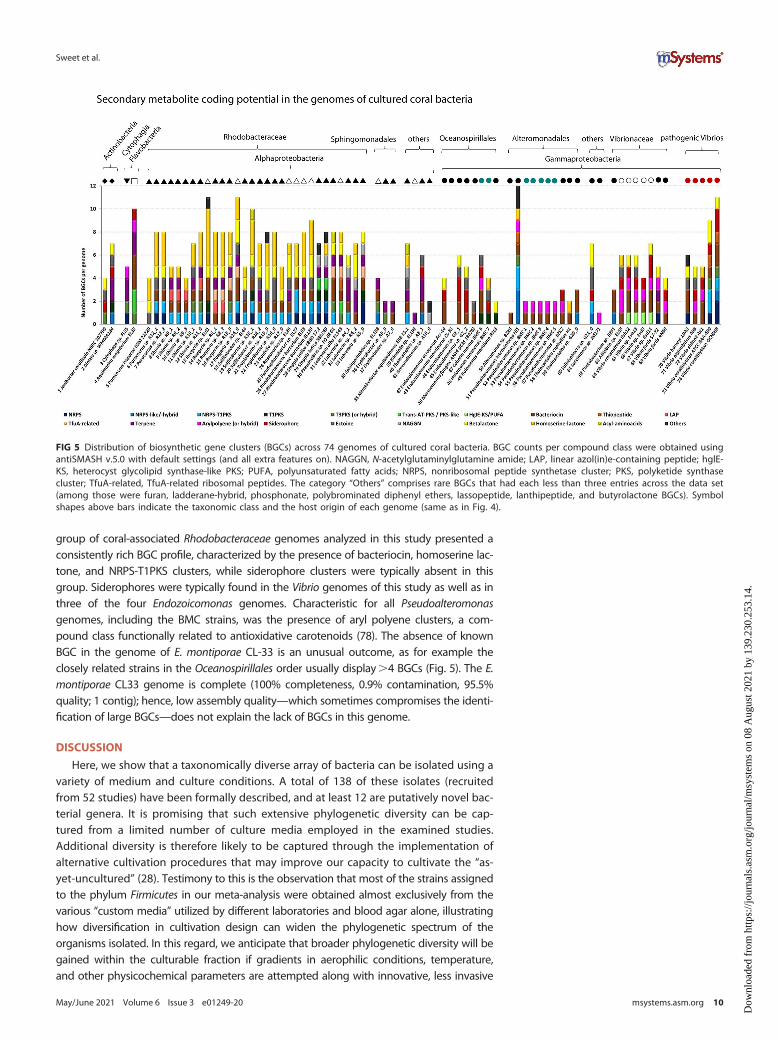
group of coral-associated Rhodobacteraceae genomes analyzed in this study presented aconsistently rich BGC profile, characterized by the presence of bacteriocin, homoserine lac-tone, and NRPS-T1PKS clusters, while siderophore clusters were typically absent in thisgroup. Siderophores were typically found in the Vibrio genomes of this study as well as inthree of the four Endozoicomonas genomes. Characteristic for all Pseudoalteromonasgenomes, including the BMC strains, was the presence of aryl polyene clusters, a com-pound class functionally related to antioxidative carotenoids (78). The absence of knownBGC in the genome of E. montiporae CL-33 is an unusual outcome, as for example theclosely related strains in the Oceanospirillales order usually display.4 BGCs (Fig. 5). The E.montiporae CL33 genome is complete (100% completeness, 0.9% contamination, 95.5%quality; 1 contig); hence, low assembly quality—which sometimes compromises the identi-fication of large BGCs—does not explain the lack of BGCs in this genome.
DISCUSSION
Here, we show that a taxonomically diverse array of bacteria can be isolated using avariety of medium and culture conditions. A total of 138 of these isolates (recruitedfrom 52 studies) have been formally described, and at least 12 are putatively novel bac-terial genera. It is promising that such extensive phylogenetic diversity can be cap-tured from a limited number of culture media employed in the examined studies.Additional diversity is therefore likely to be captured through the implementation ofalternative cultivation procedures that may improve our capacity to cultivate the “as-yet-uncultured” (28). Testimony to this is the observation that most of the strains assignedto the phylum Firmicutes in our meta-analysis were obtained almost exclusively from thevarious “custom media” utilized by different laboratories and blood agar alone, illustratinghow diversification in cultivation design can widen the phylogenetic spectrum of theorganisms isolated. In this regard, we anticipate that broader phylogenetic diversity will begained within the culturable fraction if gradients in aerophilic conditions, temperature,and other physicochemical parameters are attempted along with innovative, less invasive
FIG 5 Distribution of biosynthetic gene clusters (BGCs) across 74 genomes of cultured coral bacteria. BGC counts per compound class were obtained usingantiSMASH v.5.0 with default settings (and all extra features on). NAGGN, N-acetylglutaminylglutamine amide; LAP, linear azol(in)e-containing peptide; hglE-KS, heterocyst glycolipid synthase-like PKS; PUFA, polyunsaturated fatty acids; NRPS, nonribosomal peptide synthetase cluster; PKS, polyketide synthasecluster; TfuA-related, TfuA-related ribosomal peptides. The category “Others” comprises rare BGCs that had each less than three entries across the data set(among those were furan, ladderane-hybrid, phosphonate, polybrominated diphenyl ethers, lassopeptide, lanthipeptide, and butyrolactone BGCs). Symbolshapes above bars indicate the taxonomic class and the host origin of each genome (same as in Fig. 4).
Sweet et al.
May/June 2021 Volume 6 Issue 3 e01249-20 msystems.asm.org 10
Dow
nloa
ded
from
http
s://j
ourn
als.
asm
.org
/jour
nal/m
syst
ems
on 0
8 A
ugus
t 202
1 by
139
.230
.253
.14.
techniques to extract microbial cells from the host matrix. The richness of bacterial phylauncovered in this study corresponds to the phyla more often reported to dominate bacte-rial communities in corals by cultivation-independent studies (12), namely, Proteobacteria,Bacteroidetes, Actinobacteria, and Firmicutes, yet how diversity at lower taxonomic rankswithin each phylum is captured remains to be determined. Another exciting challengeahead is the unveiling of host-microbe and microbe-microbe molecular interdependencenetworks (e.g., cross-kingdom signaling and cross-feeding cascades) (79, 80). Such knowl-edge would likely enable laboratory captivation of so-far “unculturable” coral-specific orenriched lineages. Increasing the diversity of these coral-associated culturable bacteria willlikely help in the identification of genomic features that could underpin the interactionwith the host and its microbiome representing the foundation for experimental validation.
Although one of the initial aims of this study was to ascertain the percentage of cul-turable bacteria from a given coral species, it was deemed too speculative to reportthe findings due to variation in culture effort across the various studies. Indeed, thishighlights the paucity of studies dedicated to determine exactly this, and there is aneed for such mechanistic projects deploying multiple culture media and conditionsto comprehensively sample bacterial associates from a single or a few host species.Collectively, studies aimed at capturing the culturable microbiome will extend ourunderstanding of coral bacterial communities and their putative function in the coralholobiont. A catalog of cultures (as presented here and one which will hopefully beexpanded) provides a means to increase our understanding of host-symbiote relation-ships. The ability to describe, understand, and culture specific symbionts from anygiven organism (like corals) also opens up the potential to utilize them as probiotics torestore degraded habitats (53, 61). For example, specific traits found in certain coral-associated bacteria, such as the presence of the genes nifH (nitrogenase), nirK (nitritereductase), or dmdA (DMSP demethylase) involved in nitrogen and sulfur cycling, orthose known to control pathogens, the enzymatic mitigation of reactive oxygen spe-cies (ROS) or other toxic compounds, may have roles in increasing coral health whenthe host is experiencing stress (53, 63, 81, 82). Identifying these traits via molecularanalyses and laboratory tests using cultured bacteria with defined coral hosts will allowfor the more rapid administration of native bacteria with the potential to help rehabili-tate damaged corals. In addition, such a resource increases the possibility of identifyingnovel compounds of biotechnological interest (83). This seems particularly relevant inthe case of coral-microbe symbioses, which are known to rank as one of the most pro-lific sources of bioactive molecules in the oceans (38).
A search in public databases (National Center for Biotechnology Information [NCBI])found that, despite the 1,045 cultured coral-associated bacterial sequences with full-length 16S rRNA gene sequences, only 36 had genomes available as of February 2020.Clearly, a systematic effort to disclose the genomic features of coral-associated bacteriais needed in order to better understand the holobiont ecology and identify potentiallybeneficial microbes. As part of this study, we were able to add a further 38 to this tally(see Table S2 in the supplemental material). Even with this addition, the number of pub-licly available coral-associated bacterial genomes remains scant, and it is recognized thatto more fully understand the roles of the cultivable fraction of coral bacteria, a thoroughcharacterization of the species kept in culture, including genome sequencing, needs to befostered alongside experimental biology and manipulative approaches. Moreover, a largecollection of coral-associated genomes could also help to identify specific traits that areneeded to thrive in the various niches within the hosts or point to those bacteria whichoffer a specific benefit to their host.
All of the available genomes were screened for an array of functions potentially im-portant in establishing and maintaining interactions between bacterial symbionts andtheir marine invertebrate hosts. Overall, the Endozoicomonas and Pseudoalteromonasstrains displayed high numbers of eukaryote-like protein-encoding genes importantfor host-symbiont recognition in well-studied systems such as marine sponges (65, 84,85). The strain with the second highest number of eukaryote-like repeat protein-related
Insights into the Cultured Bacterial Fraction of Coral
May/June 2021 Volume 6 Issue 3 e01249-20 msystems.asm.org 11
Dow
nloa
ded
from
http
s://j
ourn
als.
asm
.org
/jour
nal/m
syst
ems
on 0
8 A
ugus
t 202
1 by
139
.230
.253
.14.

entries (1,208 CDSs, after Endozoicomonas sp. G2_1 with 1,367 CDSs) was the octocoralassociate Aquimarina sp. strain EL33 (class Flavobacteria). In the current culture collec-tion, 15 additional Aquimarina isolates are reported, from the scleractinian coralsPorites lutea, Pocillopora acuta, Stylophora pistillata, Acropora millepora, Acropora tenuis,and the octocoral E. labiata. Retrieving the genomes from these candidates will allowus to explore these emerging patterns in greater detail. For example, a recent compar-ative genomics survey of host-associated and free-living Aquimarina species revealedcomplex secondary metabolite biosynthesis and polycarbohydrate degradation capaci-ties (86), but further investigation into their mechanisms of interactions with corals iswarranted.
Only eight Endozoicomonas isolates (five of them type species) have so far been cul-tured from corals (according to our collated information). These are from the octoco-rals Eunicea fusca and Plexaura sp. and the scleractinian corals Montipora aequitubercu-lata, Acropora cytherea, Acropora hemprichii, and Acropora sp. To date, only four ofthese (two from this study) have had their genomes sequenced (all from scleractiniancorals) (18, 87). This is surprising given that numerous studies found that this genus ishighly abundant in the healthy coral holobiont and seems to decrease in abundanceupon deteriorating environmental conditions (e.g., reviewed in references 35, 88, and89). Future cultivation efforts should therefore be directed toward the Endozoicomonadaceaefamily in order to increase the representation of their taxonomic and functional diversity inculture collections (29). In this regard, this study finds evidence that supplementing culturemedia with DMSP is an approach worth investing in future attempts to cultivate coral-associated Endozoicomonas, possible in combination with growth at lower temperatures(29). The metabolic data obtained from the comparative analysis of these four strains canbe used, for example, to drive the selection of specific nutrients and conditions required toculture this particular genus of coral symbionts. Furthermore, there are 55 culturedPseudoalteromonas strains in our collection which should also be explored regarding theirsymbiotic properties and their functional gene content (only 6 genomes currently avail-able). Similar to Endozoicomonas, Pseudoalteromonas species are also frequent members ofcoral-associated microbiomes (35). A number of Pseudoalteromonas have been shown todisplay high antimicrobial activity, and many of these bacteria are isolated from coral mu-cus, lending support to the protective role the surface mucous layer has for the host andits importance in the coral holobiont’s defense—against bacterial coral pathogens in par-ticular (90). Indeed, five of the six Pseudoalteromonas (where genomes are available) wereshown to be effective BMCs when corals were challenged with the coral pathogen Vibriocoralliilyticus (53).
Having genomes available from the potential pathogens also allows for greaterinsight into coral biology, especially when interested in ascertaining pathogenicity-related traits (91, 92). For example, from the 11 Vibrio species for which genomic datawere available, we were able to show functional separation (based on Pfam profiles) ofknown pathogenic and nonpathogenic strains. This was further accompanied by a sig-nificantly higher abundance of CDSs encoding for the type VI secretion system, impor-tant for virulence in the pathogenic strains (76). Prevalence of siderophore-encodinggenes was also noted in the Vibrionaceae strains, suggesting that these bacteria likelygain competitive advantages through efficient and extensive iron acquisition, which isa trait often seen in opportunistic and pathogenic bacteria (93, 94). Hypothetically, theselection of beneficial microbes that are also good siderophore producers could add tothe biological control of these pathogens. Indeed, two proposed BMC strains Cobetiamarina BMC6 and Halomonas taenensis BMC7 harbor such siderophore clusters ontheir genomes and so did three of the four Endozoicomonas strains. However, the fivePseudoalteromonas BMC strains and the Endozoicomonas montiporae CL-33 had lownumbers of BGCs, possibly indicating a reduced investment into secondary metabo-lism. Indeed, the low number of BGCs in these Pseudoalteromonas strains is in contrastto the established prevalence of biologically active compounds in many marine host-associated Pseudoalteromonas strains (95). In part, this may reflect a limitation of the
Sweet et al.
May/June 2021 Volume 6 Issue 3 e01249-20 msystems.asm.org 12
Dow
nloa
ded
from
http
s://j
ourn
als.
asm
.org
/jour
nal/m
syst
ems
on 0
8 A
ugus
t 202
1 by
139
.230
.253
.14.

software utilized to detect genes for all secondary metabolites, as genes for commonmetabolites (such as for the production of the antibiotic marinocin and those that pro-duce tetrabromopyrrole coral larval settlement cues by Pseudoalteromonas [96, 97])were not picked up. These bioinformatic limitations emphasize the importance of hav-ing bacterial cultures for the elucidation of the chemical ecology underpinning coralholobiont functioning.
Broader functional traits can also be ascertained from looking at the complete pic-ture of isolates with annotated genomes. For example, 66% (49 out of 74) harboredthe TauD gene, which is involved in taurine utilization (98). Two proposed BMCs, theCobetia marina BMC7 and Halomonas taeanensis BMC7, revealed the highest copynumber of TauD CDSs (seven and eight, respectively), while others range between oneand five TauD copies. Taurine is an organo-sulfur compound widely present in animaltissues, and recent research has shown that obligate symbionts of sponges haveenriched copies of taurine catabolism genes and taurine transporters in comparisonwith free-living bacteria (65, 72, 73). The widespread capability of the isolates studiedhere to potentially utilize host-derived taurine could guide the formulation of novel,taurine-containing cultivation media in the attempt to captivate coral symbionts, par-ticularly from the important, yet underrepresented order Oceanospirillales (TauD wasconsistently present in all Oceanospirillales genomes [N=8] analyzed here). The ubiqui-tous occurrence of bacteriocin clusters among the genomes is another example ofbroad-scale trends which we have identified in our genome meta-analysis. These mayconfer the specific culturable symbionts with particular competitive capacities towardclosely related taxa in highly dense microbiomes (99, 100), as is commonly identifiedacross corals and sponges. Moreover, the widespread presence of NRPS and beta-lac-tone clusters hints toward broad-spectrum antimicrobial and cytotoxic capabilities inmultiple associates. It also corroborates the hypothesis that these marine metaorganismsare promising sources of novel bioactive compounds, representing targets for bioprospec-tion (38). Many strains also possess homoserine lactone-encoding BGCs indicative of so-phisticated, cell-density-dependent chemical communication mechanisms. Antioxidantactivities are likely conferred by the presence of aryl polyene BGCs in the genomes (78,101). These pigment type compounds, functionally related to carotenoids, characterizedmost of the proposed BMC strains. Furthermore, several coral-associated bacteria of differ-ent taxonomic origins are seemingly well equipped to handle osmotic stress as revealedby the occurrence of ectoine- and N-acetylglutaminylglutamine amide (NAGGN)-encodinggenes. Therefore, there is a need to continue the effort in culturing coral-associated bacte-ria to explore new biosynthetic potentials, both for bioprospecting purposes and for betterunderstanding the chemical ecology of the metaorganism.
Identifying likely candidates for symbiosis is one challenge, but once the candidatesare confirmed and characterized, the need to understand how the animal host estab-lishes symbiosis and retains the relationship will also be critical. However, this is a two-way street. Current research in sponges has revealed that bacteria expressing theankyrin genes avoid phagocytosis by sponge amoebocytes, thus becoming residentsof the sponge microbiome by evading the host's immune system (64, 70). Further, asankyrin repeats are enriched in the microbial metagenomes of healthy corals (10, 11),it is expected that commensal coral-associated bacteria also use this aspect of ankyringenes to establish symbiosis. The evolutionary forces shaping the symbiosis are eventrickier here, as bacteriophages encode for ankyrin biosynthesis in their genomes andmight transfer this information across different community members (70). As identifiedabove with siderophore-encoding genes, similar patterns of symbiosis establishmentand energy utilization may be adopted by both commensal and pathogenic bacteria.
To conclude, here we have highlighted that diverse coral-associated bacteria are al-ready cultured, although these are often scattered across collections and rarely col-lated into one easily accessible location. Further, only a few of these have had theirgenomes sequenced. Despite the lack of genomes, we were able to identify a numberof genetic features commonly encoded by these coral bacterial associates. These
Insights into the Cultured Bacterial Fraction of Coral
May/June 2021 Volume 6 Issue 3 e01249-20 msystems.asm.org 13
Dow
nloa
ded
from
http
s://j
ourn
als.
asm
.org
/jour
nal/m
syst
ems
on 0
8 A
ugus
t 202
1 by
139
.230
.253
.14.

features include broad-spectrum antimicrobial, antioxidant, and cytotoxic compound pro-duction capabilities, high abundance of ankyrin repeat entries, tetratricopeptide, andWD40 repeats, and taurine degradation genes. That said, this can only be quantitativelyassessed through comparison of metagenome profiles from corals versus other environ-ments, such as sediments and seawater in a comprehensive fashion (several samples withreplication, etc.). Such metagenome-based analyses should be complemented by (large-scale) marker gene surveys and/or visualization techniques to determine the nature andholobiont site of bacterial association, in particular since any metaorganism (configuration)is specific to a time and place and not static given the temporal (“fluidic”) nature of host-microbe interactions (102). Even though the statistical power, with only part of the repre-sentative genomes available from cultures (as in this study), is limited, we exemplify herethe importance of the cultured bacterial fraction of corals in hypothesis testing andapplied microbiology.
We end by highlighting the importance and need for a global initiative to create anonline catalog of genomic and physiological features of cultured coral-associated bac-teria. Combining the use of these genomic insights with innovative culturing techni-ques (37), aimed at improving the collection of coral-associated bacterial isolates, willsee this field of coral biology move forward. Such an initiative should likely start withthose microbes which have their complete genomes sequenced. This study pioneersthe organization of such a global collection, as part of the efforts from the BeneficialMicrobes for Marine Organisms network (BMMO), through a public invitation toresearchers working in this field. As a result, we have here provided a list of culturedbacteria from corals that are currently available in public databases, plus isolates thatwere kept in collections from all the laboratories that responded to our invitation(Table S1 and available now, open access via http://isolates.reefgenomics.org). Nowother researchers can access this virtual collection and/or contact specific laboratoriesfor collaborations or solicitations of specific microbial strains.
MATERIALS ANDMETHODSLiterature search and data curation. Google Scholar and the National Center for Biotechnology
Information (NCBI) were searched for publicly available 16S rRNA gene sequences of cultured coral-asso-ciated bacteria (as of 2018). Search terms, including coral, bacteria, 16S, and culture, were utilized as wellas combinations of these. The results were supplemented with data from culture collections from labo-ratories around the world through a public invitation to researchers working in this field. In total, wewere able to obtain bacterial isolates originating from 84 coral species (representing tropical, temperate,and cold-water habitats) from all major oceans (Fig. 1; see also Table S1 in the supplemental material).Due to the number and varied nature of the different contributing sources of these isolates, parts of theassociated metadata for certain cultures are missing or incomplete.
Phylogenetic analysis and tree generation. In total, we were able to collate 3,055 individual iso-lates which had (at least) part of the 16S rRNA gene sequenced (see Table S1 for details). We selectedonly high-quality sequences by removing those shorter than 500 bp, or longer than 1,600 bp and con-taining more than one ambiguity. Further, we utilized the mothur (v.1.42.0) commands screen.seqs andfilter.seqs to remove poorly aligned sequences and positions without sequence information, respectively(103). This resulted in 1,045 isolates with near full-length 16S rRNA gene sequences, which were used indownstream phylogenetic analyses. To this end, sequences were aligned using the SILVA 138.1 databaseas a reference (104), and the clear-cut command was used within mothur to generate a phylogenetictree using the relaxed neighbor-joining method (RNJ) (105, 106). To generate the distance matric, thedefault of percent identities (so-called p-distances) was retained.
A phylogenetic tree of coral species was also generated using the Taxonomy Common Tree tool ofNCBI (107). Species names were added manually to create a tree file. Tree features were optimized usingiTOL v4 (108).
Taxonomic composition of bacterial isolates by medium. Bacterial strains listed in Table S1 weresorted by isolation medium and subsequently grouped at phylum, order, and genus levels according tothe current SILVA (138.1) taxonomy (104). Stacked column graphs, showing relative abundances of thecultivated taxa were created thereafter. At the genus level, all groups representing less than 1% of thetotal pool in each medium were included in a group labeled “others.”
Genome analysis. The integrated Microbial Genomes and Microbiomes database (IMG; https://img.jgi.doe.gov/) (109) from the Department of Energy’s Joint Genome Institute (DOE-JGI), and the assemblydatabase from NCBI (https://www.ncbi.nlm.nih.gov/assembly) were searched for publicly availablegenomes from cultured coral bacteria in February 2020. Thirty-six bacterial genome assemblies (21 fromscleractinian coral and 15 from octocoral associates) were downloaded from NCBI and included in thisanalysis. The annotation of genomic features such as genome size, GC content, and number of coding
Sweet et al.
May/June 2021 Volume 6 Issue 3 e01249-20 msystems.asm.org 14
Dow
nloa
ded
from
http
s://j
ourn
als.
asm
.org
/jour
nal/m
syst
ems
on 0
8 A
ugus
t 202
1 by
139
.230
.253
.14.

sequences (CDSs) was performed for all 74 genomes with the RAST server (110) (see Table S2). Proteinfamilies (Pfams) were predicted with the online server WebMGA (default settings) (111) using amino acidsequence files obtained from RAST. The resulting individual Pfam annotation files were then joined using acustomized R script and the resulting count tables were Hellinger transformed for multivariate analyses (seeTable S5). Dissimilarity between genomes based on the Pfam profiles were then calculated using the Bray-Curtis index. Ordination of the genomes based on their functional profiles was carried out using principalcoordinate analysis (PCoA) and plotted in eigenvalue scale (i.e., scaling of each axis using the square root ofthe eigenvalue) with PAST software v3.25 (112). PERMANOVAs (permutational multivariate analyses of var-iance) were performed with 999 permutations to test for overall differences in functional profiles betweenbacterial genomes from different taxonomic classes. Five groups (classes) were used: Alphaproteobacteria,Gammaproteobacteria, Actinobacteria, Cytophagia, and Flavobacteriia. A separate PERMANOVA analysis ofBray-Curtis dissimilarities calculated for the 11 available Vibrio genomes was then performed in order to high-light differences between strains identified as potentially pathogenic (N=5, group 1) and those apparentlynonpathogenic (N=6, group 2) (identification of pathogenicity from available literature—see references).Finally, AntiSMASH version 5.0 (113) was used with default parameters (and extra features “All on”) to identifybiosynthetic gene clusters (BGCs) in all genomes.
Data availability. The newly described genomes associated with this project (38 in total; also seeTable S2) can be found in the following BioProjects on NCBI: accession no. PRJNA698462, PRJNA638634,and PRJNA343499.
SUPPLEMENTAL MATERIAL
Supplemental material is available online only.FIG S1, JPG file, 0.6 MB.TABLE S1, XLSX file, 0.8 MB.TABLE S2, XLSX file, 0.03 MB.TABLE S3, XLSX file, 0.02 MB.TABLE S4, XLSX file, 0.01 MB.TABLE S5, XLSX file, 2.1 MB.
ACKNOWLEDGMENTSPart of this research was carried out in association with the ongoing R&D project
registered as ANP 21005-4, “PROBIO-DEEP - Survey of potential impacts caused byoil and gas exploration on deep-sea marine holobionts and selection of potentialbioindicators and bioremediation processes for these ecosystems” (UFRJ/ShellBrasil/ANP), sponsored by Shell Brasil under the ANP R&D levy as “Compromisso deInvestimentos com Pesquisa e Desenvolvimento.” This research project won the GreatBarrier Reef Foundation’s Out of the Blue Box Reef Innovation Challenge People’s ChoiceAward supported by The Tiffany & Co. Foundation. The Institute of Bioengineering andBiosciences acknowledges funding provided by the Portuguese Foundation for Scienceand Technology (FCT) and the European Regional Development Fund (ERDF) throughgrant UIDB/04565/2020. Part of this work was supported by the research grantFA_05_2017_032 conceded to R.C. and T.K.-C. by the Portuguese Ministry of the Sea(Direção Geral de Política do Mar) under the program “Fundo Azul.” T.K.-C. is the recipientof a Research Scientist contract with FCT (CEECIND/00788/2017). N.Z. and K.H.S. weresupported in part by the INBRE-NIGMS by NIH grant P20GM103430.
M.S. designed the initial study and led the collection, analysis, and write-up of thepaper. H.V. conducted the majority of the analysis on the 16S data, and T.K.-C. and R.C.conducted the majority of the analysis of the genome data. S.R. assisted with the meta-analysis component of the project, and R.P. co-funded the project with M.S. All authorsassisted with data collection, analysis, and writing/editing.
We declare there are no known conflicts of interest associated with this study.
REFERENCES1. Doolittle F, Inkpen AS. 2018. Processes and patterns of interaction as
units of selection: an introduction to ITSNTS thinking. Proc Natl Acad SciU S A 115:400624014. https://doi.org/10.1073/pnas.1722232115.
2. van Oppen MJH, Medina M. 2020. Coral evolutionary responses to micro-bial symbioses. Philos Trans R Soc Lond B Biol Sci 375:20190591. https://doi.org/10.1098/rstb.2019.0591.
3. Bang C, Dagan T, Deines P, Dubilier N, Duschl WJ, Fraune S, Hentschel U, HirtH, Hülter N, Lachnit T, Picazo D, Pita L, Pogoreutz C, Rädecker N, Saad MM,Schmitz RA, Schulenburg H, Voolstra CR, Weiland-Bräuer N, Ziegler M, BoschTCG. 2018. Metaorganisms in extreme environments: do microbes play a rolein organismal adaptation? Zoology (Jena) 127:1–19. https://doi.org/10.1016/j.zool.2018.02.004.
Insights into the Cultured Bacterial Fraction of Coral
May/June 2021 Volume 6 Issue 3 e01249-20 msystems.asm.org 15
Dow
nloa
ded
from
http
s://j
ourn
als.
asm
.org
/jour
nal/m
syst
ems
on 0
8 A
ugus
t 202
1 by
139
.230
.253
.14.

4. Bosch TCG, McFall-Ngai MJ. 2011. Metaorganisms as the new frontier.Zoology (Jena) 114:185–190. https://doi.org/10.1016/j.zool.2011.04.001.
5. Rohwer F, Seguritan V, Azam F, Knowlton N. 2002. Diversity and distribu-tion of coral-associated bacteria. Mar Ecol Prog Ser 243:1–10. https://doi.org/10.3354/meps243001.
6. Littman RA, Willis BL, Pfeffer C, Bourne DG. 2009. Diversities of coral-asso-ciated bacteria differ with location, but not species, for three acroporidcorals on the Great Barrier Reef. FEMS Microbiol Ecol 68:152–163. https://doi.org/10.1111/j.1574-6941.2009.00666.x.
7. Shashar N, Cohen Y, Loya Y, Sar N. 1994. Nitrogen fixation (acetylenereduction) in stony corals: evidence for coral-bacteria interactions. MarEcol Prog Ser 111:259–264. https://doi.org/10.3354/meps111259.
8. Ritchie KB, Smith GW. 1995. Preferential carbon utilization by surfacebacterial communities from water mass, normal, and white-band dis-eased Acropora. Mol Mar Biol Biotechnol 4:345–352.
9. Hernandez-Agreda A, Leggat W, Ainsworth TD. 2019. A place for taxo-nomic profiling in the study of the coral prokaryotic microbiome. FEMSMicrobiol Lett 366:fnz063. https://doi.org/10.1093/femsle/fnz063.
10. Robbins SJ, Singleton CM, Chan CX, Messer LF, Geers AU, Ying H, BakerA, Bell SC, Morrow KM, Ragan MA, Miller DJ, Forêt S, ReFuGe2020 Con-sortium, Voolstra CR, Tyson GW, Bourne DG. 2019. A genomic view ofthe reef-building coral Porites lutea and its microbial symbionts. NatMicrobiol 4:2090–2100. https://doi.org/10.1038/s41564-019-0532-4.
11. Keller-Costa T, Lago-Lestón A, Saraiva JP, Toscan R, Silva SG, Gonçalves J,Cox CJ, Kyrpides N, Nunes da Rocha U, Costa R. 2021. Metagenomicinsights into the taxonomy, function, and dysbiosis of prokaryotic com-munities in octocorals. Microbiome 9:72. https://doi.org/10.1186/s40168-021-01031-y.
12. Huggett MJ, Apprill A. 2019. Coral microbiome database: integration ofsequences reveals high diversity and relatedness of coral-associatedmicrobes. Environ Microbiol Rep 11:372–385. https://doi.org/10.1111/1758-2229.12686.
13. Ziegler M, Seneca FO, Yum LK, Palumbi SR, Voolstra CR. 2017. Bacterialcommunity dynamics are linked to patterns of coral heat tolerance. NatCommun 8:14213. https://doi.org/10.1038/ncomms14213.
14. Neave MJ, Rachmawati R, Xun L, Michell CT, Bourne DG, Apprill A,Voolstra CR. 2017. Differential specificity between closely related coralsand abundant Endozoicomonas endosymbionts across global scales.ISME J 11:186–200. https://doi.org/10.1038/ismej.2016.95.
15. Shibl AA, Isaac A, Ochsenkühn MA, Cárdenas A, Fei C, Behringer G,Arnoux M, Drou N, Santos MP, Gunsalus KC, Voolstra CR, Amin SA. 2020.Diatom modulation of select bacteria through use of two unique sec-ondary metabolites. Proc Natl Acad Sci U S A 117:27445227455. https://doi.org/10.1073/pnas.2012088117.
16. Romano S, Schulz-Vogt HN, González JM, Bondarev V. 2015. Phosphatelimitation induces drastic physiological changes, virulence-related geneexpression, and secondary metabolite production in Pseudovibrio sp.strain FO-BEG1. Appl Environ Microbiol 81:3518–3528. https://doi.org/10.1128/AEM.04167-14.
17. Raina JB, Tapiolas D, Motti CA, Foret S, Seemann T, Tebben J, Willis BL,Bourne DG. 2016. Isolation of an antimicrobial compound produced bybacteria associated with reef-building corals. PeerJ 4:e2275. https://doi.org/10.7717/peerj.2275.
18. Neave MJ, Michell CT, Apprill A, Voolstra CR. 2017. Endozoicomonasgenomes reveal functional adaptation and plasticity in bacterial strainssymbiotically associated with diverse marine hosts. Sci Rep 7:40579.https://doi.org/10.1038/srep40579.
19. Karimi E, Keller-Costa T, Slaby BM, Cox CJ, da Rocha UN, Hentschel U,Costa R. 2019. Genomic blueprints of sponge-prokaryote symbiosis areshared by low abundant and cultivatable Alphaproteobacteria. Sci Rep9:1999. https://doi.org/10.1038/s41598-019-38737-x.
20. Staley JT, Konopka A. 1985. Measurement of in situ activities of nonphotosyn-thetic microorganisms in aquatic and terrestrial habitats. Annu Rev Microbiol39:321–346. https://doi.org/10.1146/annurev.mi.39.100185.001541.
21. Amann RI, Ludwig W, Schleifer KH. 1995. Phylogenetic identification andin situ detection of individual microbial cells without cultivation. Micro-biol Rev 59:1432169. https://doi.org/10.1128/mr.59.1.143-169.1995.
22. Hugenholtz P, Goebel BM, Pace NR. 1998. Impact of culture-independentstudies on the emerging phylogenetic view of bacterial diversity. J Bacteriol180:476524774. https://doi.org/10.1128/JB.180.18.4765-4774.1998.
23. Wang F, Li M, Huang L, Zhang X-H. 2021. Cultivation of uncultured ma-rine microorganisms. Mar Life Sci Technol 3:117–120. https://doi.org/10.1007/s42995-021-00093-z.
24. Stewart EJ. 2012. Growing unculturable bacteria. J Bacteriol 194:4151–4160.https://doi.org/10.1128/JB.00345-12.
25. Suzuki MT, Rappé MS, Haimberger ZW, Winfield H, Adair N, Ströbel J,Giovannoni SJ. 1997. Bacterial diversity among small-subunit rRNA geneclones and cellular isolates from the same seawater sample. Appl EnvironMicrobiol 63:983–989. https://doi.org/10.1128/AEM.63.3.983-989.1997.
26. Lagier JC, Edouard S, Pagnier I, Mediannikov O, Drancourt M, Raoult D.2015. Current and past strategies for bacterial culture in clinical microbiol-ogy. Clin Microbiol Rev 28:208–236. https://doi.org/10.1128/CMR.00110-14.
27. Pham VHT, Kim J. 2012. Cultivation of unculturable soil bacteria. TrendsBiotechnol 30:4752484. https://doi.org/10.1016/j.tibtech.2012.05.007.
28. Lewis WH, Tahon G, Geesink P, Sousa DZ, Ettema TJG. 2021. Innovationsto culturing the uncultured microbial majority. Nat Rev Microbiol19:225–240. https://doi.org/10.1038/s41579-020-00458-8.
29. Pogoreutz C, Voolstra CR. 2018. Isolation, culturing, and cryopreserva-tion of Endozoicomonas (Gammaproteobacteria: Oceanospirillales:Endozoicomonadaceae) from reef-building corals. protocols.io. https://doi.org/10.17504/protocols.io.t2aeqae.
30. Raina JB, Tapiolas D, Willis BL, Bourne DG. 2009. Coral-associated bacte-ria and their role in the biogeochemical cycling of sulfur. Appl EnvironMicrobiol 75:3492–3501. https://doi.org/10.1128/AEM.02567-08.
31. Keller-Costa T, Eriksson D, Gonçalves JMS, Gomes NCM, Lago-Lestón A,Costa R. 2017. The gorgonian coral Eunicella labiata hosts a distinct prokary-otic consortium amenable to cultivation. FEMS Microbiol Ecol 93:143.https://doi.org/10.1093/femsec/fix143.
32. Sweet MJ, Croquer A, Bythell JC. 2011. Bacterial assemblages differ betweencompartments within the coral holobiont. Coral Reefs 30:39–52. https://doi.org/10.1007/s00338-010-0695-1.
33. Pollock FJ, McMinds R, Smith S, Bourne DG, Willis BL, Medina M, ThurberRV, Zaneveld JR. 2018. Coral-associated bacteria demonstrate phylosym-biosis and cophylogeny. Nat Commun 9:4921. https://doi.org/10.1038/s41467-018-07275-x.
34. Pernice M, Raina JB, Rädecker N, Cárdenas A, Pogoreutz C, Voolstra CR.2020. Down to the bone: the role of overlooked endolithic microbiomesin reef coral health. ISME J 14:325–334. https://doi.org/10.1038/s41396-019-0548-z.
35. Sweet MJ, Bulling MT. 2017. On the importance of the microbiome andpathobiome in coral health and disease. Front Mar Sci 4:9.
36. Pogoreutz C, Voolstra CR, Rädecker N, Weis V, Cardenas A, Raina J-B.2021. The coral holobiont highlights the dependence of cnidarian ani-mal hosts on their associated microbes, p 91–118. In Bosch TCG,Hadfield MG (ed), Cellular dialogues in the holobiont. CRC Press, BocaRaton, FL.
37. Modolon F, Barno AR, Villela HDM, Peixoto RS. 2020. Ecological and bio-technological importance of secondary metabolites produced by coral-associated bacteria. J Appl Microbiol 129:1441–1457. https://doi.org/10.1111/jam.14766.
38. Raimundo I, Silva SG, Costa R, Keller-Costa T. 2018. Bioactive secondarymetabolites from octocoral-associated microbes—new chances for bluegrowth. Mar Drugs 16:485. https://doi.org/10.3390/md16120485.
39. Disalvo LH. 1969. Isolation of bacteria from the corallum of Porites lobata(Vaughn) and its possible significance. Integr Comp Biol 9:735–740.https://doi.org/10.1093/icb/9.3.735.
40. Ducklow HW, Mitchell R. 1979. Bacterial populations and adaptations inthe mucus layers on living corals. Limnol Oceanogr 24:715–725. https://doi.org/10.4319/lo.1979.24.4.0715.
41. Aronson RB, Precht WF. 2001. White band diseases and the changingface of Caribbean coral reefs. Hydrobiologia 460:25–38. https://doi.org/10.1023/A:1013103928980.
42. Peters EC, Oprandy JJ, Yevich PP. 1983. Possible causal agent of “whiteband disease” in caribbean acroporid corals. J Invertebr Pathol 41:394–396.https://doi.org/10.1016/0022-2011(83)90260-4.
43. Chet I, Mitchell R. 1976. Ecological aspects of microbial chemotactic behav-ior. Annu Rev Microbiol 30:2212239. https://doi.org/10.1146/annurev.mi.30.100176.001253.
44. Antonius A. 1981. Coral reef pathology: a review, p 3214. In Proceedingsof the 4th International Coral Reef Symposium. University of the Philip-pines, Manila, Philippines.
45. Kushmaro A, Rosenberg E, Fine M, Loya Y. 1997. Bleaching of the coralOculina patagonica by Vibrio AK-1. Mar Ecol Prog Ser 147:159–165.https://doi.org/10.3354/meps147159.
46. Rozenblat YB-H, Rosenberg E. 2004. Temperature-regulated bleachingand tissue lysis of Pocillopora damicornis by the novel pathogen Vibrio
Sweet et al.
May/June 2021 Volume 6 Issue 3 e01249-20 msystems.asm.org 16
Dow
nloa
ded
from
http
s://j
ourn
als.
asm
.org
/jour
nal/m
syst
ems
on 0
8 A
ugus
t 202
1 by
139
.230
.253
.14.
coralliilyticus, p 301–324. In Coral health and disease. Springer, Berlin,Germany.
47. Vidal-Dupiol J, Ladrière O, Meistertzheim AL, Fouré L, Adjeroud M, MittaG. 2011. Physiological responses of the scleractinian coral Pocilloporadamicornis to bacterial stress from Vibrio coralliilyticus. J Exp Biol214:1533–1545. https://doi.org/10.1242/jeb.053165.
48. Ushijima B, Videau P, Burger AH, Shore-Maggio A, Runyon CM, Sudek M,Aeby GS, Callahan SM. 2014. Vibrio coralliilyticus strain OCN008 is an eti-ological agent of acute Montipora white syndrome. Appl Environ Micro-biol 80:2102–2109. https://doi.org/10.1128/AEM.03463-13.
49. Rädecker N, Pogoreutz C, Gegner HM, Cárdenas A, Roth F, Bougoure J,Guagliardo P, Wild C, Pernice M, Raina JB, Meibom A, Voolstra CR. 2021.Heat stress destabilizes symbiotic nutrient cycling in corals. Proc Natl AcadSci U S A 118:e2022653118. https://doi.org/10.1073/pnas.2022653118.
50. Bourne DG, Garren M, Work TM, Rosenberg E, Smith GW, Harvell CD.2009. Microbial disease and the coral holobiont. Trends Microbiol17:5542562. https://doi.org/10.1016/j.tim.2009.09.004.
51. Reshef L, Koren O, Loya Y, Zilber-Rosenberg I, Rosenberg E. 2006. Thecoral probiotic hypothesis. Environ Microbiol 8:2068–2073. https://doi.org/10.1111/j.1462-2920.2006.01148.x.
52. Rosenberg E, Koren O, Reshef L, Efrony R, Zilber-Rosenberg I. 2007. Therole of microorganisms in coral health, disease and evolution. Nat RevMicrobiol 5:355–362. https://doi.org/10.1038/nrmicro1635.
53. Rosado PM, Leite DCA, Duarte GAS, Chaloub RM, Jospin G, Nunes daRocha U, Saraiva JP, Dini-Andreote F, Eisen JA, Bourne DG, Peixoto RS.2019. Marine probiotics: increasing coral resistance to bleaching throughmicrobiome manipulation. ISME J 13:921–936. https://doi.org/10.1038/s41396-018-0323-6.
54. Voolstra CR, Ziegler M. 2020. Adapting with microbial help: microbiomeflexibility facilitates rapid responses to environmental change. BioEssays42:2000004. https://doi.org/10.1002/bies.202000004.
55. Ritchie KB. 2006. Regulation of microbial populations by coral surfacemucus and mucus-associated bacteria. Mar Ecol Prog Ser 322:1–14.https://doi.org/10.3354/meps322001.
56. Shnit-Orland M, Kushmaro A. 2009. Coral mucus-associated bacteria: apossible first line of defense. FEMS Microbiol Ecol 67:371–380. https://doi.org/10.1111/j.1574-6941.2008.00644.x.
57. Kuek FWI, Lim LF, Ngu LH, Mujahid A, Lim PT, Leaw CP, Müller M. 2015.The potential roles of bacterial communities in coral defence: a casestudy at Talang-talang reef. Ocean Sci J 50:269–282. https://doi.org/10.1007/s12601-015-0024-2.
58. Romano S. 2018. Ecology and biotechnological potential of bacteriabelonging to the genus Pseudovibrio. Appl Environ Microbiol 84:e02516-17. https://doi.org/10.1128/AEM.02516-17.
59. Hinger I, Ansorge R, Mussmann M, Romano S. 2020. Phylogenomic analy-ses of members of the widespread marine heterotrophic genus Pseudo-vibrio suggest distinct evolutionary trajectories and a novel genus, Poly-cladidibacter gen. nov. Appl Environ Microbiol 86:e02395-19. https://doi.org/10.1128/AEM.02395-19.
60. Work T, Meteyer C. 2014. To understand coral disease, look at coral cells.Ecohealth 11:610–618. https://doi.org/10.1007/s10393-014-0931-1.
61. Peixoto RS, Sweet M, Bourne DG. 2019. Customized medicine for corals.Front Mar Sci 6:686. https://doi.org/10.3389/fmars.2019.00686.
62. Peixoto RS, Rosado PM, Leite D. C d A, Rosado AS, Bourne DG. 2017. Ben-eficial Microorganisms for Corals (BMC): proposed mechanisms for coralhealth and resilience. Front Microbiol 8:341. https://doi.org/10.3389/fmicb.2017.00341.
63. Peixoto R, Sweet M, Villela HDM, Cardoso PM, Thomas T, Voolstra CR, Hoj L,Bourne DG. 2021. Coral probiotics: premise, promise, prospects. Annu Rev AnimBiosci 9:2652288. https://doi.org/10.1146/annurev-animal-090120-115444.
64. Nguyen MTHD, Liu M, Thomas T. 2014. Ankyrin-repeat proteinsfrom sponge symbionts modulate amoebal phagocytosis. Mol Ecol23:1635–1645. https://doi.org/10.1111/mec.12384.
65. Karimi E, Ramos M, Gonçalves JMS, Xavier JR, Reis MP, Costa R. 2017.Comparative metagenomics reveals the distinctive adaptive features ofthe Spongia officinalis endosymbiotic consortium. Front Microbiol8:2499. https://doi.org/10.3389/fmicb.2017.02499.
66. Li J, Mahajan A, Tsai MD. 2006. Ankyrin repeat: a unique motif mediatingprotein-protein interactions. Biochemistry 45:15168–15178. https://doi.org/10.1021/bi062188q.
67. Lamb JR, Tugendreich S, Hieter P. 1995. Tetratrico peptide repeat inter-actions: to TPR or not to TPR? Trends Biochem Sci 20:257–259. https://doi.org/10.1016/S0968-0004(00)89037-4.
68. Goebl M, Yanagida M. 1991. The TPR snap helix: a novel protein repeatmotif from mitosis to transcription. Trends Biochem Sci 16:173–177.https://doi.org/10.1016/0968-0004(91)90070-c.
69. Li D, Roberts R. 2001. WD-repeat proteins: structure characteristics, bio-logical function, and their involvement in human diseases. Cell Mol LifeSci 58:208522097. https://doi.org/10.1007/pl00000838.
70. Jahn MT, Arkhipova K, Markert SM, Stigloher C, Lachnit T, Pita L, KupczokA, Ribes M, Stengel ST, Rosenstiel P, Dutilh BE, Hentschel U. 2019. Aphage protein aids bacterial symbionts in eukaryote immune evasion.Cell Host Microbe 26:542–550.e5. https://doi.org/10.1016/j.chom.2019.08.019.
71. Karimi E, Slaby BM, Soares AR, Blom J, Hentschel U, Costa R. 2018. Meta-genomic binning reveals versatile nutrient cycling and distinct adaptivefeatures in alphaproteobacterial symbionts of marine sponges. FEMSMicrobiol Ecol https://doi.org/10.1093/femsec/fiy074.
72. Engelberts JP, Robbins SJ, de Goeij JM, Aranda M, Bell SC, Webster NS.2020. Characterization of a sponge microbiome using an integrative ge-nome-centric approach. ISME J 14:1100–1110. https://doi.org/10.1038/s41396-020-0591-9.
73. Botté ES, Nielsen S, Abdul Wahab MA, Webster J, Robbins S, Thomas T,Webster NS. 2019. Changes in the metabolic potential of the spongemicrobiome under ocean acidification. Nat Commun 10:4134. https://doi.org/10.1038/s41467-019-12156-y.
74. Stringlis IA, Zamioudis C, Berendsen RL, Bakker PAHM, Pieterse CMJ.2019. Type III secretion system of beneficial rhizobacteria Pseudomonassimiae WCS417 and Pseudomonas defensor WCS374. Front Microbiol10:1631. https://doi.org/10.3389/fmicb.2019.01631.
75. Juhas M, Crook DW, Hood DW. 2008. Type IV secretion systems: tools ofbacterial horizontal gene transfer and virulence. Cell Microbiol10:237722386. https://doi.org/10.1111/j.1462-5822.2008.01187.x.
76. Lin L, Lezan E, Schmidt A, Basler M. 2019. Abundance of bacterial Type VIsecretion system components measured by targeted proteomics. NatCommun 10:2584. https://doi.org/10.1038/s41467-019-10466-9.
77. Cianciotto NP, White RC. 2017. Expanding role of type II secretion in bac-terial pathogenesis and beyond. Infect Immun 85:e00014-17. https://doi.org/10.1128/IAI.00014-17.
78. Schöner TA, Gassel S, Osawa A, Tobias NJ, Okuno Y, Sakakibara Y, Shindo K,Sandmann G, Bode HB. 2016. Aryl polyenes, a highly abundant class of bac-terial natural products, are functionally related to antioxidative carotenoids.Chembiochem 17:247–253. https://doi.org/10.1002/cbic.201500474.
79. Singh RP, Kothari R, Egan S. 2017. Exploring the complexity of macroal-gal-bacterial interactions through interkingdom signalling system, p301–315. In Systems biology of marine ecosystems. Springer, Berlin,Germany.
80. Sztajer H, Szafranski SP, Tomasch J, Reck M, Nimtz M, Rohde M, Wagner-Döbler I. 2014. Cross-feeding and interkingdom communication in dual-species biofilms of Streptococcus mutans and Candida albicans. ISME J8:2256–2271. https://doi.org/10.1038/ismej.2014.73.
81. Santos H, Carmo FL, Duarte G, Dini-Andreote F, Castro CB, Rosado AS,Van Elsas JD, Peixoto RS. 2014. Climate change affects key nitrogen-fix-ing bacterial populations on coral reefs. ISME J 8:2272–2279. https://doi.org/10.1038/ismej.2014.70.
82. Welsh RM, Rosales SM, Zaneveld JR, Payet JP, Mcminds R, Hubbs SL,Thurber RLV. 2017. Alien vs. predator: bacterial challenge alters coralmicrobiomes unless controlled by Halobacteriovorax predators. PeerJ 5:e3315. https://doi.org/10.7717/peerj.3315.
83. Blockley A, Elliott DR, Roberts AP, Sweet M. 2017. Symbiotic microbesfrommarine invertebrates: driving a new era of natural product drug dis-covery. Diversity 9:49. https://doi.org/10.3390/d9040049.
84. Nguyen-Kim H, Bouvier T, Bouvier C, Doan-Nhu H, Nguyen-Ngoc L,Rochelle-Newall E, Baudoux AC, Desnues C, Reynaud S, Ferrier-Pages C,Bettarel Y. 2014. High occurrence of viruses in the mucus layer of sclerac-tinian corals. Environ Microbiol Rep 6:675–682. https://doi.org/10.1111/1758-2229.12185.
85. Reynolds D, Thomas T. 2016. Evolution and function of eukaryotic-likeproteins from sponge symbionts. Mol Ecol 25:5242–5253. https://doi.org/10.1111/mec.13812.
86. Silva SG, Blom J, Keller-Costa T, Costa R. 2019. Comparative genomicsreveals complex natural product biosynthesis capacities and carbon me-tabolism across host-associated and free-living Aquimarina (Bacteroi-detes, Flavobacteriaceae) species. Environ Microbiol 21:4002–4019.https://doi.org/10.1111/1462-2920.14747.
87. Tandon K, Chiang PW, Chen WM, Tang SL. 2018. Draft genome sequenceof Endozoicomonas acroporae strain Acr-14T, isolated from Acropora
Insights into the Cultured Bacterial Fraction of Coral
May/June 2021 Volume 6 Issue 3 e01249-20 msystems.asm.org 17
Dow
nloa
ded
from
http
s://j
ourn
als.
asm
.org
/jour
nal/m
syst
ems
on 0
8 A
ugus
t 202
1 by
139
.230
.253
.14.

coral. Genome Announc 6:e01576-17. https://doi.org/10.1128/genomeA.01576-17.
88. Roder C, Bayer T, Aranda M, Kruse M, Voolstra CR. 2015. Microbiome struc-ture of the fungid coral Ctenactis echinata aligns with environmental differ-ences. Mol Ecol 24:3501–3511. https://doi.org/10.1111/mec.13251.
89. Ziegler M, Grupstra CGB, Barreto MM, Eaton M, BaOmar J, Zubier K, Al-Sofyani A, Turki AJ, Ormond R, Voolstra CR. 2019. Coral bacterial commu-nity structure responds to environmental change in a host-specific man-ner. Nat Commun 10:3092. https://doi.org/10.1038/s41467-019-10969-5.
90. Shnit-Orland M, Sivan A, Kushmaro A. 2012. Antibacterial activity ofPseudoalteromonas in the coral holobiont. Microb Ecol 64:851–859.https://doi.org/10.1007/s00248-012-0086-y.
91. Ushijima B, Videau P, Poscablo D, Stengel JW, Beurmann S, Burger AH,Aeby GS, Callahan SM. 2016. Mutation of the toxR or mshA genes fromVibrio coralliilyticus strain OCN014 reduces infection of the coral Acrop-ora cytherea. Environ Microbiol 18:4055–4067. https://doi.org/10.1111/1462-2920.13428.
92. Weynberg KD, Voolstra CR, Neave MJ, Buerger P, Van Oppen MJH. 2016.From cholera to corals: viruses as drivers of virulence in a major coralbacterial pathogen. Sci Rep 5:17889. https://doi.org/10.1038/srep17889.
93. Fang Z, Sampson SL, Warren RM, Gey Van Pittius NC, Newton-Foot M. 2015.Iron acquisition strategies in mycobacteria. Tuberculosis 95:123–130. https://doi.org/10.1016/j.tube.2015.01.004.
94. Isaac DT, Laguna RK, Valtz N, Isberg RR. 2015. MavN is a Legionellapneumophila vacuole-associated protein required for efficient ironacquisition during intracellular growth. Proc Natl Acad Sci U S A 112:E5208–E5217. https://doi.org/10.1073/pnas.1511389112.
95. Holmstrom C, Kjelleberg S. 2006. Marine Pseudoalteromonas species areassociated with higher organisms and produce biologically activeextracellular agents. FEMS Microbiol Ecol 30:285–293. https://doi.org/10.1111/j.1574-6941.1999.tb00656.x.
96. Tebben J, Tapiolas DM, Motti CA, Abrego D, Negri AP, Blackall LL,Steinberg PD, Harder T. 2011. Induction of larval metamorphosis of thecoral Acropora millepora by tetrabromopyrrole isolated from a Pseu-doalteromonas bacterium. PLoS One 6:e19082. https://doi.org/10.1371/journal.pone.0019082.
97. Sneed JM, Sharp KH, Ritchie KB, Paul VJ. 2014. The chemical cue tetrabro-mopyrrole from a biofilm bacterium induces settlement of multiple Ca-ribbean corals. Proc R Soc B 281:20133086. https://doi.org/10.1098/rspb.2013.3086.
98. Eichhorn E, Van Der Ploeg JR, Kertesz MA, Leisinger T. 1997. Characteriza-tion of a-ketoglutarate-dependent taurine dioxygenase from Escherichiacoli. J Biol Chem 272:23031–23036. https://doi.org/10.1074/jbc.272.37.23031.
99. Desriac F, Defer D, Bourgougnon N, Brillet B, Le Chevalier P, Fleury Y.2010. Bacteriocin as weapons in the marine animal-associated bacteriawarfare: inventory and potential applications as an aquaculture probi-otic. Mar Drugs 8:115321177. https://doi.org/10.3390/md8041153.
100. Hols P, Ledesma-García L, Gabant P, Mignolet J. 2019. Mobilization ofmicrobiota commensals and their bacteriocins for therapeutics. TrendsMicrobiol 27:6902702. https://doi.org/10.1016/j.tim.2019.03.007.
101. Cimermancic P, Medema MH, Claesen J, Kurita K, Wieland Brown LC,Mavrommatis K, Pati A, Godfrey PA, Koehrsen M, Clardy J, Birren BW,Takano E, Sali A, Linington RG, Fischbach MA. 2014. Insights into
secondary metabolism from a global analysis of prokaryotic biosyntheticgene clusters. Cell 158:412–421. https://doi.org/10.1016/j.cell.2014.06.034.
102. Jaspers C, Fraune S, Arnold AE, Miller DJ, Bosch TCG, Voolstra CR. 2019.Resolving structure and function of metaorganisms through a holisticframework combining reductionist and integrative approaches. Zoology(Jena) 133:81–87. https://doi.org/10.1016/j.zool.2019.02.007.
103. Schloss PD, Westcott SL, Ryabin T, Hall JR, Hartmann M, Hollister EB,Lesniewski RA, Oakley BB, Parks DH, Robinson CJ, Sahl JW, Stres B,Thallinger GG, Van Horn DJ, Weber CF. 2009. Introducing mothur: open-source, platform-independent, community-supported software fordescribing and comparing microbial communities. Appl Environ Micro-biol 75:7537–7541. https://doi.org/10.1128/AEM.01541-09.
104. Quast C, Pruesse E, Yilmaz P, Gerken J, Schweer T, Yarza P, Peplies J,Glöckner FO. 2013. The SILVA ribosomal RNA gene database project:improved data processing and web-based tools. Nucleic Acids Res 41:D590–D596. https://doi.org/10.1093/nar/gks1219.
105. Evans J, Sheneman L, Foster J. 2006. Relaxed neighbor joining: a fast dis-tance-based phylogenetic tree construction method. J Mol Evol62:785–792. https://doi.org/10.1007/s00239-005-0176-2.
106. Saitou N, Nei M. 1987. The neighbor-joining method: a new method forreconstructing phylogenetic trees. Mol Biol Evol 4:406–425. https://doi.org/10.1093/oxfordjournals.molbev.a040454.
107. Sayers EW, Barrett T, Benson DA, Bryant SH, Canese K, Chetvernin V,Church DM, Dicuccio M, Edgar R, Federhen S, Feolo M, Geer LY,Helmberg W, Kapustin Y, Landsman D, Lipman DJ, Madden TL, MaglottDR, Miller V, Mizrachi L, Ostell J, Pruitt KD, Schuler GD, Sequeira E, SherryST, Shumway M, Sirotkin K, Souvorov A, Starchenko G, Tatusova TA,Wagner L, Yaschenko E, Ye J. 2009. Database resources of the NationalCenter for Biotechnology Information. Nucleic Acids Res 37:D52D15.https://doi.org/10.1093/nar/gkn741.
108. Letunic I, Bork P. 2019. Interactive Tree Of Life (iTOL) v4: recent updatesand new developments. Nucleic Acids Res 47:W256–W259. https://doi.org/10.1093/nar/gkz239.
109. Chen IMA, Chu K, Palaniappan K, Pillay M, Ratner A, Huang J,Huntemann M, Varghese N, White JR, Seshadri R, Smirnova T, Kirton E,Jungbluth SP, Woyke T, Eloe-Fadrosh EA, Ivanova NN, Kyrpides NC.2019. IMG/M v.5.0: an integrated data management and comparativeanalysis system for microbial genomes and microbiomes. Nucleic AcidsRes 47:D666–D677. https://doi.org/10.1093/nar/gky901.
110. Overbeek R, Olson R, Pusch GD, Olsen GJ, Davis JJ, Disz T, Edwards RA,Gerdes S, Parrello B, Shukla M, Vonstein V, Wattam AR, Xia F, Stevens R.2014. The SEED and the Rapid Annotation of microbial genomes using Sub-systems Technology (RAST). Nucleic Acids Res 42:D206–D214. https://doi.org/10.1093/nar/gkt1226.
111. Wu S, Zhu Z, Fu L, Niu B, Li W. 2011. WebMGA: a customizable webserver for fast metagenomic sequence analysis. BMC Genomics 12:444.https://doi.org/10.1186/1471-2164-12-444.
112. Hammer Ø, Harper DAT, Ryan PD. 2001. Past: paleontological statisticssoftware package for education and data analysis. Palaeontol Electron4:178.
113. Blin K, Shaw S, Steinke K, Villebro R, Ziemert N, Lee SY, Medema MH,Weber T. 2019. AntiSMASH 5.0: updates to the secondary metabolite ge-nome mining pipeline. Nucleic Acids Res 47:W81–W87. https://doi.org/10.1093/nar/gkz310.
Sweet et al.
May/June 2021 Volume 6 Issue 3 e01249-20 msystems.asm.org 18
Dow
nloa
ded
from
http
s://j
ourn
als.
asm
.org
/jour
nal/m
syst
ems
on 0
8 A
ugus
t 202
1 by
139
.230
.253
.14.