Embed Size (px)
Citation preview
HAL Id: hal-01484978https://hal.archives-ouvertes.fr/hal-01484978
Submitted on 8 Mar 2017
HAL is a multi-disciplinary open accessarchive for the deposit and dissemination of sci-entific research documents, whether they are pub-lished or not. The documents may come fromteaching and research institutions in France orabroad, or from public or private research centers.
L’archive ouverte pluridisciplinaire HAL, estdestinée au dépôt et à la diffusion de documentsscientifiques de niveau recherche, publiés ou non,émanant des établissements d’enseignement et derecherche français ou étrangers, des laboratoirespublics ou privés.
Inside Speech: Multisensory and Modality-specificProcessing of Tongue and Lip Speech Actions
Avril Treille, Coriandre Vilain, Thomas Hueber, Laurent Lamalle, Marc Sato
To cite this version:Avril Treille, Coriandre Vilain, Thomas Hueber, Laurent Lamalle, Marc Sato. Inside Speech: Mul-tisensory and Modality-specific Processing of Tongue and Lip Speech Actions. Journal of Cogni-tive Neuroscience, Massachusetts Institute of Technology Press (MIT Press), 2017, 29, pp.448 - 466.<10.1162/jocn_a_01057>. <hal-01484978>

Inside Speech: Multisensory and Modality-specificProcessing of Tongue and Lip Speech Actions
Avril Treille1, Coriandre Vilain1, Thomas Hueber1,Laurent Lamalle2,3, and Marc Sato4
Abstract
■ Action recognition has been found to rely not only on sen-sory brain areas but also partly on the observer’s motor system.However, whether distinct auditory and visual experiences ofan action modulate sensorimotor activity remains largely un-known. In the present sparse sampling fMRI study, we deter-mined to which extent sensory and motor representationsinteract during the perception of tongue and lip speech actions.Tongue and lip speech actions were selected because tonguemovements of our interlocutor are accessible via their impacton speech acoustics but not visible because of its position insidethe vocal tract, whereas lip movements are both “audible” andvisible. Participants were presented with auditory, visual, andaudiovisual speech actions, with the visual inputs related toeither a sagittal view of the tongue movements or a facial viewof the lip movements of a speaker, previously recorded by anultrasound imaging system and a video camera. Although the
neural networks involved in visual visuo-lingual and visuo-facialperception largely overlapped, stronger motor and somato-sensory activations were observed during visuo-lingual percep-tion. In contrast, stronger activity was found in auditory andvisual cortices during visuo-facial perception. Complementingthese findings, activity in the left premotor cortex and in visualbrain areas was found to correlate with visual recognitionscores observed for visuo-lingual and visuo-facial speech stim-uli, respectively, whereas visual activity correlated with RTs forboth stimuli. These results suggest that unimodal and multi-modal processing of lip and tongue speech actions rely on com-mon sensorimotor brain areas. They also suggest that visualprocessing of audible but not visible movements induces motorand visual mental simulation of the perceived actions to facili-tate recognition and/or to learn the association between audi-tory and visual signals. ■
INTRODUCTION
Through life experiences, we learn about which sensoryfeatures of actions are most behaviorally relevant forsuccessful categorization and recognition. However,one intriguing question is to know what happens whenan action is not accessible to one sensor in the dailyexperience—typically, accessible via their impact onacoustics but not visible. From this question, this fMRIstudy aimed at determining multisensory and modality-specific processing of tongue and lip speech actions,with tongue movements of our interlocutor usually “audi-ble” but not visible and lip movements both “audible” andvisible.
Motor Resonance in Biological Action Recognition
Although information from different sensory modalities,such as sight and/or sound, is processed in unisensoryand multisensory brain areas, several studies have iden-
tified a central role for motor representations in actionrecognition. These results appear in keeping with thelong-standing proposal that perception and action aretwo closely linked processes and with more recent neuro-physiological perspectives based on the existence ofmirror neurons in nonhuman primates and on an action–perception matching system in humans (for reviews,see Rizzolatti & Craighero, 2004; Rizzolatti, Fogassi, &Gallese, 2001). Mirror neurons are polymodal visuo-motor or audio–visuomotor neurons in the ventral pre-motor and posterior parietal cortices (areas F5 and PF)of the macaque monkey, which have been shown to dis-charge both when the monkey performs hand or mouthactions and when it views or listens to similar actionsmade by another individual (e.g., Fogassi et al., 2005;Ferrari, Gallese, Rizzolatti, & Fogassi, 2003; Keysers et al.,2003; Kohler et al., 2002; Gallese, Fadiga, Fogassi, &Rizzolatti, 1996; Rizzolatti, Fadiga, Gallese, & Fogassi,1996; Di Pellegrino, Fadiga, Fogassi, Gallese, & Rizzolatti,1992). The existence of mirror neurons thus suggests thataction observation partly involves the same neural circuitsthat are used in action performance. Since then, auditory–vocal mirror neurons have also been recorded in non-mammalian vertebrates (Prather, Peters, Nowicki, &
1CNRS UMR 5216 & Grenoble Université, 2Université Grenoble-Alpes & CHU de Grenoble, 3CNRS UMS 3552, Grenoble, France,4CNRS UMR 7309 & Aix-Marseille Université
© Massachusetts Institute of Technology Journal of Cognitive Neuroscience X:Y, pp. 1–19doi:10.1162/jocn_a_01057
Mooney, 2008), and numerous neurophysiological andbrain imaging experiments have provided evidence forthe existence of a frontoparietal action–perception match-ing system in humans (Rizzolatti & Craighero, 2004). Alto-gether, these studies demonstrate that sensory informationrelated to biological movements is not only processed insensory regions but also in the observer’s motor systemand partly relies on his or her own motor knowledge.
From that view, a stronger activity in the premotor cor-tex and the posterior parietal cortex is observed duringvisual and audiovisual perception of biological movements,compared with nonbiological movements (e.g., Saygin,2007; Calvert, Campbell, & Brammer, 2000; Howardet al., 1996). Moreover, hearing action-related sounds likeknock on the door or hand clapping or more complexauditory material like a piano piece also activates motorand premotor regions (e.g., Lahav, Saltzman, & Schlaug,2007; Pizzamiglio et al., 2005; Aziz-Zadeh, Iacoboni, Zaidel,Wilson, & Mazziotta, 2004; Haueisen & Knösche, 2001).These results support the long-standing theoretical pro-posal that specific constraints and regularity in biologicalmotion and kinematics are used in action recognition(Viviani & Stucchi, 1992; Johansson, 1973), evenwhen theyare roughly represented by point lights (Loula, Prasad,Harber, & Shiffrar, 2005; Beardsworth & Buckner, 1981).Furthermore, action recognition seems to rely not onlyon biological features per se but also more specificallyon a motor repertoire shared by individuals of the samespecies and related to their relevant physical and/orcommunicative ability for perceptual processing. Forexample, Tai, Scherfler, Brooks, Sawamoto, and Castiello(2004) observed premotor activity during the sight ofhuman hand grasp but not during the sight of the sameaction performed by a robot, which supports the use of ahuman biological motor repertoire in action recognition.On their side, Buccino et al. (2004) showed that the obser-vation of a biting action performed by humans, monkeys,or dogs inducedmotor activity in humans, contrary to whathappens during the observation of dog-specific barkingmovements. Calvo-Merino and colleagues (Calvo-Merino,Grèzes, Glaser, Passingham, & Haggard, 2006; Calvo-Merino, Glaser, Grèzes, Passingham, & Haggard, 2005) alsoshowed that, apart from visual familiarity, the involvementof motor areas during action observation strongly relies onmotor learning. They indeed observed, among other parie-tal and cerebellar regions, stronger premotor cortex activitywhen male dancers viewed dance movements from theirown motor repertoire compared with female dance move-ments that they often saw but never performed. Althougha causal role of the motor system during action recogni-tion is still debated, these fMRI studies suggest a strongcorrelation between motor activity and action observation.
Motor Resonance Extends to Speech Action
Speech is a special type of biological human actions thatinterfaces with the linguistic system and requires an accu-
rate control of our speech articulators (i.e., the lips, thetongue, the jaw, the velum, and the larynx). As with othertype of actions, such as grasping or walking, several neuro-imaging studies suggest that speech recognition is alsopartly mediated by the motor system. Brain areas involvedin the planning and execution of speech actions (i.e., theposterior part of the left inferior frontal gyrus, the pre-motor and primary motor cortices) have indeed shownneural responses during auditory speech perception(e.g., Pulvermuller et al., 2006; Wilson & Iacoboni, 2006;Wilson, Saygin, Sereno, & Iacoboni, 2004). In addition,repetitive and double-pulse TMS studies also suggest thatspeech motor regions are causally recruited during audi-tory speech categorization, especially in case of complexsituations (e.g., the perception of acoustically ambiguoussyllables or when phonological segmentation or workingmemory processes are strongly required; Grabski et al.,2013; d’Ausilio, Bufalari, Salmas, & Fadiga, 2011; d’Ausilioet al., 2009; Möttönen & Watkins, 2009; Sato, Tremblay, &Gracco, 2009; Meister, Wilson, Deblieck, Wu, & Iacoboni,2007). Taken together, these results support the idea thatour motor knowledge used to produce speech soundshelps to partly constraint phonetic decoding of the sen-sory inputs, as proposed in motor and sensorimotortheories of speech perception and language comprehen-sion (Pickering&Garrod, 2013; Schwartz,Ménard, Basirat, &Sato, 2012; Skipper, Van Wassenhove, Nussman, & Small,2007; Liberman & Mattingly, 1985).Importantly, speech provides visual as well as auditory
information. Although humans are proficient to extractphonetic features from the acoustic signal alone and, toa lesser extent, are capable to partly read on lips whenaudition is lacking, interactions between auditory andvisual modalities are beneficial in speech perception.Neuroimaging studies demonstrate the existence of spe-cific brain areas playing a key role in the audiovisual inte-gration of speech. Notably, activity within unisensoryvisual and auditory regions (the visual motion-sensitivecortex, V5/MT, and the Heschl’s gyrus) as well as withinmultisensory regions (the posterior parts of the left supe-rior temporal gyrus/STS [pSTS/pSTG]) is modulated dur-ing audiovisual speech perception, when compared withauditory and visual unimodal conditions (Skipper et al.,2007; Skipper, Nusbaum, & Small, 2005; Callan et al.,2003, 2004; Calvert et al., 2000). Because pSTS/pSTG dis-plays supra-additive and subadditive responses duringcongruent and incongruent stimuli presentation, it hasbeen proposed that both visual and auditory speech in-formation are integrated in these high-level multisensoryintegrative regions and that modulations of neuronalresponses within the sensory-specific cortices wouldthen be due to feedback projections from this multi-sensory region. Such modulations would represent thephysiological correlates of the perceptual changes expe-rienced after multisensory integration (e.g., Beauchamp,2005; Beauchamp, Argall, Bodurka, Duyn, & Martin,2004; Beauchamp, Lee, Argall, & Martin, 2004; Calvert
2 Journal of Cognitive Neuroscience Volume X, Number Y
et al., 2000). In addition, premotor and motor cortices,known to play a crucial role in speech production, mightalso play a key role in audiovisual integration mechanismsin speech perception (e.g., Sato, Buccino, Gentilucci, &Cattaneo, 2010; Watkins & Paus, 2004; Calvert & Campbell,2003; Watkins, Strafella, & Paus, 2003; Campbell et al.,2001). From that view, Skipper and colleagues (2005,2007) observed stronger activation in speech motor re-gions during audiovisual speech perception, comparedwith auditory and visual unimodal conditions. Callan andcolleagues (2003, 2004) also demonstrated increasedmotor activity under adverse listening or viewing con-ditions during bimodal speech presentation. In addition,increased activity or, on the contrary, subadditive re-sponses in the Broca’s area have also been reported duringthe perception of incongruent compared with congruentaudiovisual speech stimuli (Pekkola et al., 2006; Ojanenet al., 2005) or compared with unimodal speech stimuli(Calvert et al., 2000). From these results, multisensory areasand speech motor regions appear as good candidates forbrain areas where acoustic and visual speech signals caninteract, which suggests a possible integration betweenincoming sensory signals and speech motor knowledgespecific to the listener.
Motor Resonance for Audible but Hidden Actions
If the motor system is indeed involved in multisensoryintegration, what happens when an action is not acces-sible to one sensor in the daily experience—typicallyaudible but not visible? We know from the classic studiesby Meltzoff and Moore (1977, 1983) that 3-week-old in-fants, and even newborns, are able to associate from birtha visual action they have never seen, like lip and tongueprotrusion, with motor commands, possibly through theuse of their proprioceptive system. This indirectly sug-gests that, in adults, the sensorimotor network could playa role in the visual processing of audible but not visibleactions by enabling a transfer of motor knowledge towardan inferred visual experience, possibly combined with pastauditory and somatosensory experiences.Lips and tongue are two perfect articulators to
test this specific question. First, we have an excellentsomatosensory–motor control of both articulators,notably during speaking. Second, because of their posi-tion inside the vocal tract, tongue movements of ourinterlocutor are usually “audible” but not visible, whereaslip movements are both “audible” and visible. Inter-estingly, few behavioral studies using virtual tonguemovements or ultrasound images of tongue movementsdemonstrate stronger speech learning with a visualtongue feedback (Katz & Mehta, 2015) and an enhance-ment of auditory stimuli discrimination when they arematched with related visual tongue movements comparedwith auditory-only or incongruent audio-visuo-lingualstimuli (d’Ausilio, Bartoli, Maffongelli, Berry, & Fadiga,2014; Badin, Tarabalka, Elisei, & Bailly, 2010).
To determine the neural networks involved in theperceptual processing of visuo-lingual and visuo-facialactions, an fMRI study on unimodal and multimodalspeech perception was conducted. Participants had torecognize auditory, visual, or audiovisual speech stim-uli, with the visual presentation related to either a sag-ittal view of the tongue movements or a facial view ofthe lip movements of a speaker, with lingual and facialmovements previously recorded by an ultrasound im-aging system and a video camera. Our first goal wasto determine the shared neural correlates of visual andaudiovisual tongue and lip movements as well as theneural specificity of lingual perception compared withfacial perception. We also examined possible similar-ities and differences in the integration between audio-visuo-lingual and audio-visuo-facial modalities and thecorrelation between neural activity and visual syllablerecognition scores.
METHODS
Participants
Fourteen healthy adults (seven women and seven menwith a mean age of 26 years, ranging from 18 to 44 years),who are native French speakers, participated in thestudy after giving their informed consent. Two par-ticipants were removed from the study because ofexcessive head movements or technical problems dur-ing MRI acquisition. All participants were right-handedaccording to standard handedness inventory (Oldfield,1971), had normal or corrected-to-normal vision, andreported no history of speaking, hearing, or motor dis-orders. The protocol was approved by the Grenoble Uni-versity Ethical Committee with all participants screenedfor neurological, psychiatric, and other possible medicalproblems and contraindications to MRI. None of the par-ticipants were experienced with visuo-lingual ultrasoundimages.
Stimuli
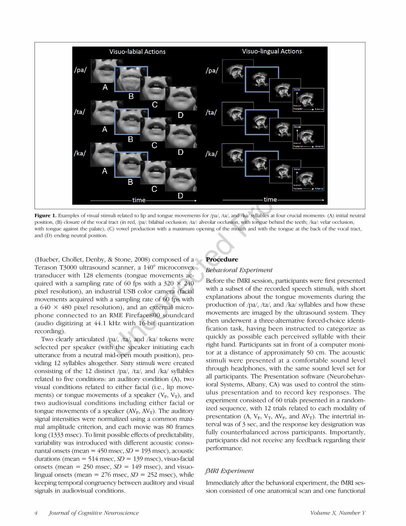
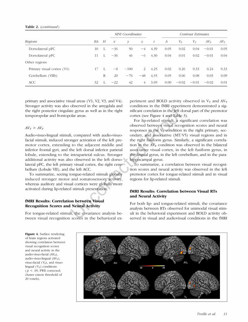
Before the experiment, multiple utterances of /pa/, /ta/,and /ka/ syllables were individually recorded by one maleand one female speakers in a soundproof room. Thesesyllables were selected based on previous studies onaudiovisual speech perception to ensure a gradient ofvisuo-labial saliency (with notably the bilabial /p/ consonantknown to be more visually salient than alveolar /t/ andvelar /k/ consonants). Regarding visuo-lingual saliency, /t/and /k/ consonants have more visible tongue movementthan /p/ because of the involvement of the apex or thedorsum of the tongue during alveolar or velar occlusion(see Figure 1).
Synchronous recordings of auditory, visual, and ultra-sound signals were acquired by the Ultraspeech system
Treille et al. 3
(Hueber, Chollet, Denby, & Stone, 2008) composed of aTerason T3000 ultrasound scanner, a 140° microconvextransducer with 128 elements (tongue movements ac-quired with a sampling rate of 60 fps with a 320 × 240pixel resolution), an industrial USB color camera (facialmovements acquired with a sampling rate of 60 fps witha 640 × 480 pixel resolution), and an external micro-phone connected to an RME Fireface800 soundcard(audio digitizing at 44.1 kHz with 16-bit quantizationrecording).
Two clearly articulated /pa/, /ta/, and /ka/ tokens wereselected per speaker (with the speaker initiating eachutterance from a neutral mid-open mouth position), pro-viding 12 syllables altogether. Sixty stimuli were createdconsisting of the 12 distinct /pa/, /ta/, and /ka/ syllablesrelated to five conditions: an auditory condition (A), twovisual conditions related to either facial (i.e., lip move-ments) or tongue movements of a speaker (VF, VT), andtwo audiovisual conditions including either facial ortongue movements of a speaker (AVF, AVT). The auditorysignal intensities were normalized using a common maxi-mal amplitude criterion, and each movie was 80 frameslong (1333 msec). To limit possible effects of predictability,variability was introduced with different acoustic conso-nantal onsets (mean= 450msec, SD= 193msec), acousticdurations (mean = 514 msec, SD= 139 msec), visuo-facialonsets (mean = 250 msec, SD = 149 msec), and visuo-lingual onsets (mean = 276 msec, SD = 252 msec), whilekeeping temporal congruency between auditory and visualsignals in audiovisual conditions.
Procedure
Behavioral Experiment
Before the fMRI session, participants were first presentedwith a subset of the recorded speech stimuli, with shortexplanations about the tongue movements during theproduction of /pa/, /ta/, and /ka/ syllables and how thesemovements are imaged by the ultrasound system. Theythen underwent a three-alternative forced-choice identi-fication task, having been instructed to categorize asquickly as possible each perceived syllable with theirright hand. Participants sat in front of a computer moni-tor at a distance of approximately 50 cm. The acousticstimuli were presented at a comfortable sound levelthrough headphones, with the same sound level set forall participants. The Presentation software (Neurobehav-ioral Systems, Albany, CA) was used to control the stim-ulus presentation and to record key responses. Theexperiment consisted of 60 trials presented in a random-ized sequence, with 12 trials related to each modality ofpresentation (A, VF, VT, AVF, and AVT). The intertrial in-terval was of 3 sec, and the response key designation wasfully counterbalanced across participants. Importantly,participants did not receive any feedback regarding theirperformance.
fMRI Experiment
Immediately after the behavioral experiment, the fMRI ses-sion consisted of one anatomical scan and one functional
Figure 1. Examples of visual stimuli related to lip and tongue movements for /pa/, /ta/, and /ka/ syllables at four crucial moments: (A) initial neutralposition, (B) closure of the vocal tract (in red, /pa/: bilabial occlusion; /ta/: alveolar occlusion, with tongue behind the teeth; /ka/: velar occlusion,with tongue against the palate), (C) vowel production with a maximum opening of the mouth and with the tongue at the back of the vocal tract,and (D) ending neutral position.
4 Journal of Cognitive Neuroscience Volume X, Number Y
run. During the functional run, participants were in-structed to attentively listen to and/or watch speechstimuli related to /pa/, /ta/, and /ka/ syllables presentedin five different modalities (A, VF, VT, AVF, and AVT). Allstimuli were presented in silent interscanning periodsbecause of sparse sampling acquisition, with the timeinterval between each stimulus onset and the midpointof the following functional scan acquisition being set at5 sec (see below). There were 144 trials, with an 8-secintertrial interval, consisting of 24 trials for each modalityof presentation (with each syllable presented two times)and 24 trials related to a resting condition without anysensory stimulation.
Data Acquisition
Magnetic resonance images were acquired with a 3-Twhole-body MR scanner (Philips Achieva TX). Partici-pants lay in the scanner with head movements minimizedwith a standard birdcage 32-channel head coil and foamcushions. Visual stimuli were presented using the Presen-tation software (Neurobehavioral Systems, Albany, CA)and displayed on a screen situated behind the scannervia a mirror placed above the participant’s eyes. Auditorystimuli were presented through the MR-confon audiosystem (www.mr-confon.de).A high-resolution T1-weighted whole-brain structural
image was acquired for each participant before the func-tional run (magnetization prepared rapid gradient echo,sagittal volume of 256 × 224 × 176 mm3 with a 1-mmisotropic resolution, inversion time = 900 msec, two seg-ments, segment repetition time = 2500 msec, segmentduration = 1795 msec, repetition time [TR]/echo time =16/5 msec with 35% partial echo, flip angle = 30°).Functional images were obtained in a subsequent
functional run using a T2*-weighted EPI sequence withwhole-brain coverage (TR = 8 sec, acquisition time =3000 msec, echo time = 30 msec, flip angle = 90°).Each functional scan was composed of 53 axial slicesparallel to the AC–PC plane acquired in noninterleavedorder (72 × 72 matrix, field of view = 216 mm, 3 ×3 mm2 in-plane resolution with a slice thickness of 3 mmwithout gap). To reduce acoustic noise, a sparse sam-pling acquisition was used (Birn, Bandettini, Cox, &Shaker, 1999; Hall et al., 1999). This acquisition tech-nique is based on neurophysiological properties ofthe slowly rising hemodynamic response, which is esti-mated to occur with a 4- to 6-sec delay in case of speechperception (Grabski et al., 2013; Zaehle et al., 2007). Inthis study, functional scanning therefore occurred onlyduring a fraction of the TR, alternating with silent inter-scanning periods, where stimuli were presented. Allconditions were presented in a pseudorandom se-quence. In addition, three “dummy” scans at the begin-ning of the functional run were added to allow forequilibration of the MRI signal and were removed fromthe analyses.
Data Analyses
Behavioral Analysis
For each participant and modality, the percentage of cor-rect responses and median RTs (from the onset of theacoustic syllables) were computed. For each dependentvariable, a repeated-measures ANOVA was performedwith the modality (A, VF, VT, AVF, and AVT) as thewithin-participant variable. For both analyses, the signifi-cance level was set at p = .05 and Greenhouse–Geissercorrected (for violation of the sphericity assumption)when appropriate. When required, post hoc analyses wereconducted with Newman–Keuls tests.
fMRI Analysis
fMRI data were analyzed using the SPM8 software pack-age (Wellcome Department of Imaging Neuroscience,Institute of Neurology, London, United Kingdom) run-ning on MATLAB (The MathWorks, Natick, MA). Brain-activated regions were labeled using the SPM Anatomytoolbox (Eickhoff et al., 2005) and, if a brain region wasnot assigned or not specified in the SPM Anatomy toolbox,using the Talairach Daemon software (Lancaster et al.,2000). For visualization, activation maps were super-imposed on a standard brain template using the MRICRONsoftware (www.sph.sc.edu/comd/rorden/mricron/).
Data preprocessing steps for each participant includedrigid realignment of functional images, coregistration ofthe structural image to the mean functional image, seg-mentation and normalization of the structural image tocommon subject space using the groupwise DARTEL reg-istration method implemented in SPM8, warping of allrealigned functional images using deformation flow fieldsgenerated from the normalization step, transformationinto the Montreal Neurological Institute (MNI) space,and spatial smoothing using an 8-mm FWHM Gaussiankernel.
For individual analyses, neural activations related tothe perceptual conditions were analyzed using a generallinear model, including five regressors of interest (A, VF,VT, AVF, and AVT) and the six realignment parameters,with the silent trials forming an implicit baseline. TheBOLD response for each event was modeled using asingle-bin finite impulse response basis function span-ning the time of acquisition (3 sec). Before estimation,a high-pass filtering with a cutoff period of 128 sec wasapplied. Beta weights associated with the modeled finiteimpulse responses were then computed to fit the ob-served BOLD signal time course in each voxel for eachcondition. Individual statistical maps were calculated foreach perceptual condition with the related baseline andsubsequently used for group statistics.
To draw population-based inferences, a second-levelrandom effects group analysis was carried out with themodality (A, VF, VT, AVF, and AVT) as the within-participantvariable and the participants treated as a random factor.
Treille et al. 5

First, for each modality, brain activity compared with theresting baseline was evaluated. Second, to determinecommon neural activity across modalities, several con-junction analyses were performed (i.e., VF ∩ VT, AVF ∩AVT, A ∩ VF ∩ AVF, A ∩ VT ∩ AVT, A ∩ VF ∩ VT ∩ AVF ∩AVT). Third, activity differences between visual condi-tions and between audiovisual conditions were eval-uated (i.e., VF > VT, VT > VF, AVF > AVT, AVT > AVF).Fourth, to determine possible correlations betweenperceptual responses observed in the behavioral exper-iment and BOLD responses, covariate analyses wereperformed on the whole brain between neural activityin visual and audiovisual modalities (i.e., VF, AVF, VT,AVT) and visual identification scores as well as RTsrelated to visuo-lingual and visuo-facial speech move-ments (VF, VT). In addition, brain regions showing higheror lower audiovisual responses compared with unimodalauditory and visual responses were identified using themax criterion test (i.e., [AVF > A] ∩ [AVF > VF], [AVF <A] ∩ [AVF < VF], [AVT > A] ∩ [AVT > VT], [AVT < A] ∩[AVT < VT]; see Stevenson et al., 2014). Modality,conjunction, and correlation contrasts were calculatedwith the significance level set at p < .05, family-wiseerror (FWE) corrected at the voxel level with a cluster
extent of at least 20 voxels. All other contrasts werecalculated with a significance level set at p < .001 un-corrected at the voxel level with a cluster extent of atleast 20 voxels.
RESULTS
Behavioral Results
Overall, the mean proportion of correct responses was82%. The main effect of modality was significant (F(4,52) = 37.79, p < .001), with more correct responses inthe A, AVF, and AVT conditions than in the VF conditionand in VF compared with VT conditions (on average, A =98%, AVF = 98%, AVT = 95%, VF = 70%, VT = 49%; allmentioned comparisons significant). The ANOVA onRTs demonstrated a significant effect of the modality(F(4, 52) = 36.25, p < .001), with faster RTs in AVF thanin VF, A, AVT, and VT conditions and slower RTs in VT thanin the other conditions (on average, AVF = 722 msec, VF =774msec, A= 812msec, AVT= 913msec, VT= 1241msec;all mentioned comparisons significant).Importantly, despite slower RTs and lower recognition
scores for visuo-lingual stimuli compared with visuo-facial
Figure 2. Surface rendering of brain regions activated in the auditory (A), visuo-facial (VF), visuo-lingual (VT), audio-visuo-facial (AVF), andaudio-visuo-lingual (AVT) conditions and showing overlapping activity between lip-related conditions (conjunction A ∩ VF ∩ AVF), tongue-relatedconditions (conjunction A ∩ VT ∩ AVT), visual conditions (conjunction VF ∩ VT), and audiovisual conditions (conjunction AVF ∩ AVT) and betweenall modalities (conjunction A ∩ VF ∩ VT ∩ AVF ∩ AVT; p < .05, FWE corrected; cluster extent threshold of 20 voxels).
6 Journal of Cognitive Neuroscience Volume X, Number Y
stimuli (and to the other conditions), recognition scoresfor visuo-lingual stimuli remained above chance level(i.e., 49% vs. 33%). Interestingly, at the syllable level,individual differences were observed between facialand tongue visual recognition (VF: /pa/ 100%, /ta/ 64%,
/ka/ 45%; VT: /pa/ 50%, /ta/ 50%, /ka/ 46%; no statisticalanalyses were performed because of the small numberof trials for each syllable). These differences suggest dif-ferent categorization processes because of the nature ofthe stimuli.
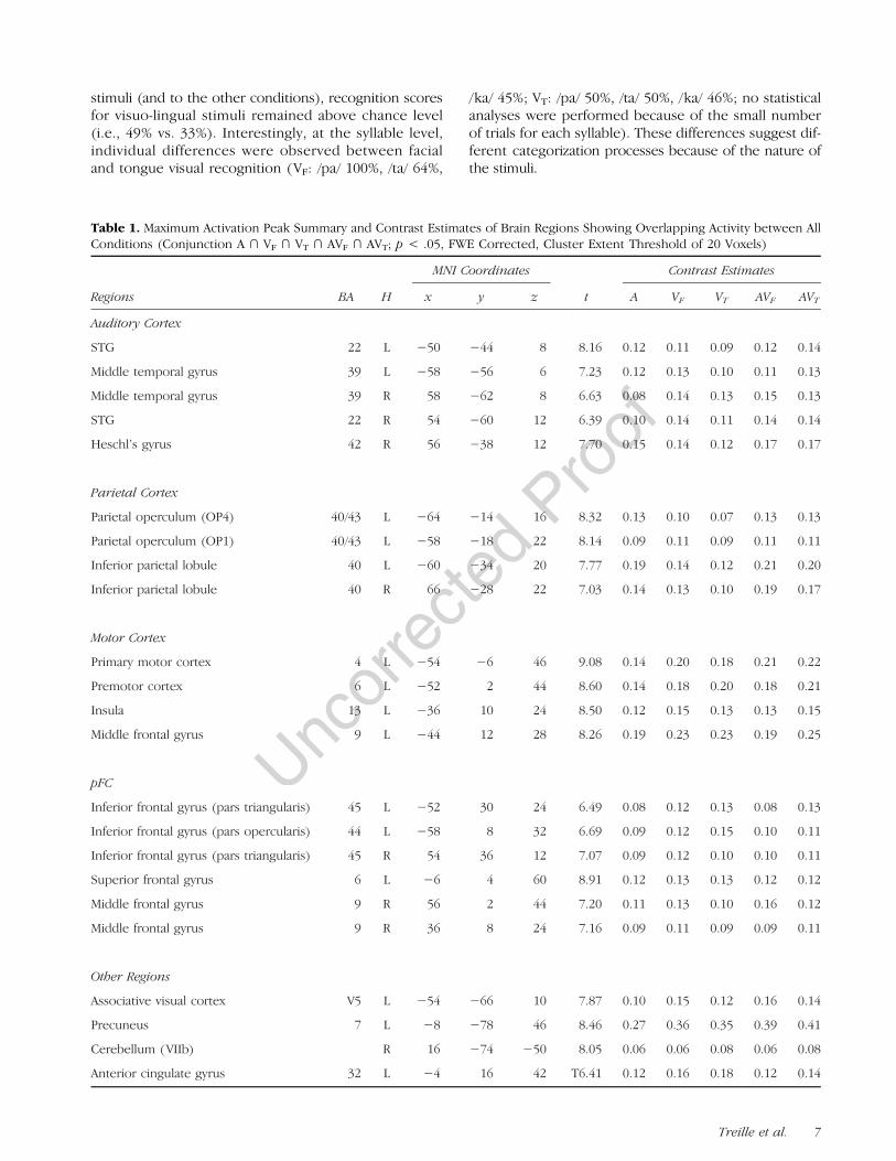
Table 1. Maximum Activation Peak Summary and Contrast Estimates of Brain Regions Showing Overlapping Activity between AllConditions (Conjunction A ∩ VF ∩ VT ∩ AVF ∩ AVT; p < .05, FWE Corrected, Cluster Extent Threshold of 20 Voxels)
Regions BA H
MNI Coordinates
t
Contrast Estimates
x y z A VF VT AVF AVT
Auditory Cortex
STG 22 L −50 −44 8 8.16 0.12 0.11 0.09 0.12 0.14
Middle temporal gyrus 39 L −58 −56 6 7.23 0.12 0.13 0.10 0.11 0.13
Middle temporal gyrus 39 R 58 −62 8 6.63 0.08 0.14 0.13 0.15 0.13
STG 22 R 54 −60 12 6.39 0.10 0.14 0.11 0.14 0.14
Heschl’s gyrus 42 R 56 −38 12 7.70 0.15 0.14 0.12 0.17 0.17
Parietal Cortex
Parietal operculum (OP4) 40/43 L −64 −14 16 8.32 0.13 0.10 0.07 0.13 0.13
Parietal operculum (OP1) 40/43 L −58 −18 22 8.14 0.09 0.11 0.09 0.11 0.11
Inferior parietal lobule 40 L −60 −34 20 7.77 0.19 0.14 0.12 0.21 0.20
Inferior parietal lobule 40 R 66 −28 22 7.03 0.14 0.13 0.10 0.19 0.17
Motor Cortex
Primary motor cortex 4 L −54 −6 46 9.08 0.14 0.20 0.18 0.21 0.22
Premotor cortex 6 L −52 2 44 8.60 0.14 0.18 0.20 0.18 0.21
Insula 13 L −36 10 24 8.50 0.12 0.15 0.13 0.13 0.15
Middle frontal gyrus 9 L −44 12 28 8.26 0.19 0.23 0.23 0.19 0.25
pFC
Inferior frontal gyrus (pars triangularis) 45 L −52 30 24 6.49 0.08 0.12 0.13 0.08 0.13
Inferior frontal gyrus (pars opercularis) 44 L −58 8 32 6.69 0.09 0.12 0.15 0.10 0.11
Inferior frontal gyrus (pars triangularis) 45 R 54 36 12 7.07 0.09 0.12 0.10 0.10 0.11
Superior frontal gyrus 6 L −6 4 60 8.91 0.12 0.13 0.13 0.12 0.12
Middle frontal gyrus 9 R 56 2 44 7.20 0.11 0.13 0.10 0.16 0.12
Middle frontal gyrus 9 R 36 8 24 7.16 0.09 0.11 0.09 0.09 0.11
Other Regions
Associative visual cortex V5 L −54 −66 10 7.87 0.10 0.15 0.12 0.16 0.14
Precuneus 7 L −8 −78 46 8.46 0.27 0.36 0.35 0.39 0.41
Cerebellum (VIIb) R 16 −74 −50 8.05 0.06 0.06 0.08 0.06 0.08
Anterior cingulate gyrus 32 L −4 16 42 T6.41 0.12 0.16 0.18 0.12 0.14
Treille et al. 7
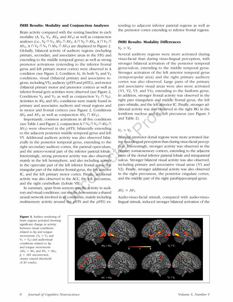
fMRI Results: Modality and Conjunction Analyses
Brain activity compared with the resting baseline in eachmodality (A, VF, VT, AVF, and AVT) as well as conjunctionanalyses (i.e., VF ∩ VT, AVF ∩ AVT, A ∩ VF ∩ AVF, A ∩ VT ∩AVT, A ∩ VF ∩ VT ∩ AVF ∩ AVT) are displayed in Figure 2.Globally, bilateral activity of auditory regions (includingprimary, secondary, and associative areas in the STG andextending to the middle temporal gyrus) as well as strongpremotor activations (extending to the inferior frontalgyrus and left primary motor cortex) were observed in Acondition (see Figure 2, Condition A). In both VF and VTconditions, visual (bilateral primary and associative re-gions, including V5), auditory (pSTS and pSTG), andmotor(bilateral primary motor and premotor cortices as well asinferior frontal gyri) activities were observed (see Figure 2,Conditions VF and VT as well as conjunction VT ∩ VF).Activities in AVF and AVT conditions were mainly found inprimary and associative auditory and visual regions andin motor and frontal cortices (see Figure 2, ConditionsAVF and AVT as well as conjunction AVF ∩ AVT).
Importantly, common activations in all five conditions(see Table 1 and Figure 2, conjunction A∩ VF∩ VT∩ AVF∩AVT) were observed in the pSTS, bilaterally extendingto the adjacent posterior middle temporal gyrus and leftV5. Additional auditory activity was also observed bilat-erally in the posterior temporal gyrus, extending to theright secondary auditory cortex, the parietal operculum,and the antero-ventral part of the inferior parietal lobule.Interestingly, strong premotor activity was also observed,mainly in the left hemisphere, and also including activityin the opercular part of the left inferior frontal gyrus, thetriangular part of the inferior frontal gyrus, the left anteriorIC, and the left primary motor cortex. Finally, additionalactivity was also observed in the ACC, the left precuneus,and the right cerebellum (Lobule VII).
In summary, apart from sensory-specific activity in audi-tory and visual conditions, our results demonstrate a sharedneural network involved in all conditions, mainly includingmultisensory activity around the pSTS and the pSTG ex-
tending to adjacent inferior parietal regions as well asthe premotor cortex extending to inferior frontal regions.
fMRI Results: Modality Differences
VF > VT
Several auditory regions were more activated duringvisuo-facial than during visuo-lingual perception, withstronger bilateral activation of the posterior temporalgyrus/sulcus, extending to the middle temporal gyrus.Stronger activation of the left anterior temporal gyrus(temporopolar area) and the right primary auditorycortex was also observed. Large parts of the primaryand associative visual areas were also more activated(V1, V2, V3, and V4), extending to the fusiform gyrus.In addition, stronger frontal activity was observed in theright pars triangularis and middle frontal gyrus, the leftpars orbitalis, and the left anterior IC. Finally, stronger ad-ditional activity was also observed in the right BG in thelentiform nucleus and the left precuneus (see Figure 3and Table 2).
VT > VF
Bilateral premotor dorsal regions were more activated dur-ing visuo-lingual perception than during visuo-facial percep-tion. Interestingly, stronger activity was observed in theprimary somatosensory cortices, extending to the adjacentparts of the dorsal inferior parietal lobule and intraparietalsulcus. Stronger bilateral visual activity was also observed,including primary and associative visual areas (V1 andV2). Finally, stronger additional activity was also observedin the right precuneus, the posterior cingulate cortex,and the middle part of the right parahippocampal gyrus.
AVF > AVT
Audio-visuo-facial stimuli, compared with audio-visuo-lingual stimuli, induced stronger bilateral activation of the
Figure 3. Surface rendering ofbrain regions activated showingsignificant change in activitybetween visual conditionsrelated to lip and tonguemovements (VF > VT andVT > VF) and audiovisualconditions related to lipand tongue movements(AVF > AVT and AVT > AVF;p < .001 uncorrected;cluster extend thresholdof 20 voxels).
8 Journal of Cognitive Neuroscience Volume X, Number Y
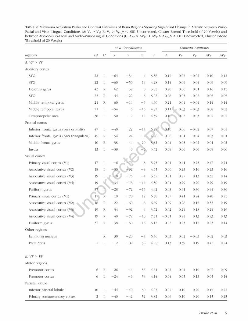
Table 2. Maximum Activation Peaks and Contrast Estimates of Brain Regions Showing Significant Change in Activity between Visuo-Facial and Visuo-Lingual Conditions (A: VF > VT; B: VT > VF; p < .001 Uncorrected, Cluster Extend Threshold of 20 Voxels) andbetween Audio-Visuo-Facial and Audio-Visuo-Lingual Conditions (C: AVF > AVT; D: AVT > AVF; p< .001 Uncorrected, Cluster ExtendThreshold of 20 Voxels)
Regions BA H
MNI Coordinates
t
Contrast Estimates
x y z A VF VT AVF AVT
A. VF > VT
Auditory cortex
STG 22 L −64 −34 4 5.38 0.17 0.05 −0.02 0.10 0.12
STG 22 L −60 −56 14 4.28 0.14 0.09 0.04 0.09 0.09
Heschl’s gyrus 42 R 62 −32 8 3.85 0.20 0.06 0.01 0.16 0.15
STG 22 R 44 −22 −6 5.02 0.08 0.03 −0.02 0.05 0.05
Middle temporal gyrus 21 R 60 −14 −6 4.60 0.21 0.04 −0.04 0.14 0.14
Middle temporal gyrus 21 L −54 6 −16 4.82 0.11 0.03 −0.03 0.08 0.05
Temporopolar area 38 L −50 −2 −12 4.39 0.10 0.02 −0.03 0.07 0.07
Frontal cortex
Inferior frontal gyrus (pars orbitalis) 47 L −40 22 −14 5.29 0.10 0.06 −0.02 0.07 0.05
Inferior frontal gyrus (pars triangularis) 45 R 54 24 −2 4.26 0.06 0.01 −0.04 0.03 0.01
Middle frontal gyrus 10 R 38 44 20 3.82 0.04 0.03 −0.02 0.01 0.02
Insula 13 L −38 0 −6 3.72 0.08 0.06 0.00 0.08 0.06
Visual cortex
Primary visual cortex (V1) 17 L −6 −76 8 5.93 0.04 0.41 0.23 0.47 0.24
Associative visual cortex (V2) 18 L −30 −92 −4 4.65 0.00 0.23 0.16 0.23 0.16
Associative visual cortex (V3) 19 L −10 −76 −4 5.37 0.01 0.27 0.13 0.32 0.14
Associative visual cortex (V4) 19 L −34 −78 −14 4.30 0.01 0.29 0.20 0.29 0.19
Fusiform gyrus 37 L −28 −72 −16 4.42 0.03 0.41 0.30 0.44 0.30
Primary visual cortex (V1) 17 R 10 −70 12 6.38 0.07 0.41 0.24 0.48 0.25
Associative visual cortex (V2) 18 R 22 −60 8 6.89 0.09 0.28 0.15 0.33 0.19
Associative visual cortex (V3) 19 R 34 −92 4 3.72 0.02 0.24 0.18 0.24 0.16
Associative visual cortex (V4) 19 R 40 −72 −10 7.31 −0.01 0.22 0.13 0.23 0.13
Fusiform gyrus 37 R 38 −50 −16 5.12 0.02 0.23 0.15 0.23 0.14
Other regions
Lentiform nucleus R 30 −20 −4 5.46 0.03 0.02 −0.03 0.02 0.03
Precuneus 7 L −2 −82 36 4.65 0.13 0.39 0.19 0.42 0.24
B. VT > VF
Motor regions
Premotor cortex 6 R 26 −4 56 4.61 0.02 0.04 0.10 0.07 0.09
Premotor cortex 6 L −24 −6 54 4.14 0.04 0.05 0.13 0.05 0.14
Parietal lobule
Inferior parietal lobule 40 L −44 −40 50 4.03 0.07 0.10 0.20 0.15 0.22
Primary somatosensory cortex 2 L −40 −42 52 3.82 0.06 0.10 0.20 0.15 0.23
Treille et al. 9
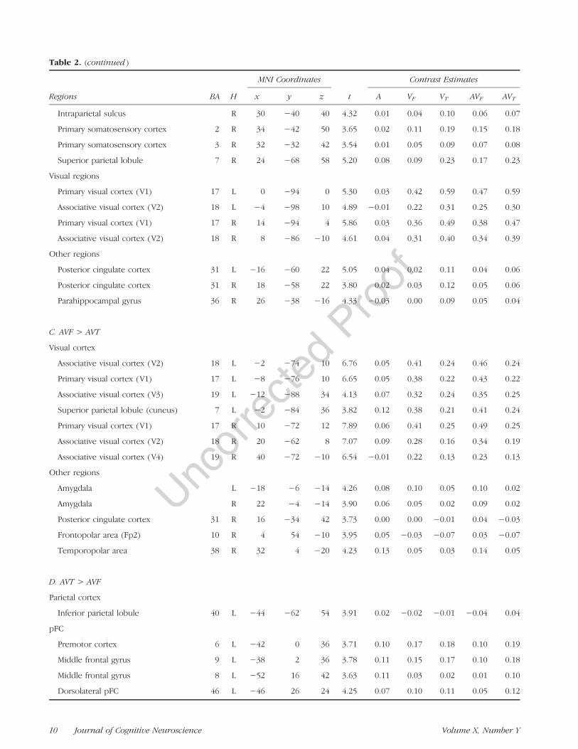
Table 2. (continued )
Regions BA H
MNI Coordinates
t
Contrast Estimates
x y z A VF VT AVF AVT
Intraparietal sulcus R 30 −40 40 4.32 0.01 0.04 0.10 0.06 0.07
Primary somatosensory cortex 2 R 34 −42 50 3.65 0.02 0.11 0.19 0.15 0.18
Primary somatosensory cortex 3 R 32 −32 42 3.54 0.01 0.05 0.09 0.07 0.08
Superior parietal lobule 7 R 24 −68 58 5.20 0.08 0.09 0.23 0.17 0.23
Visual regions
Primary visual cortex (V1) 17 L 0 −94 0 5.30 0.03 0.42 0.59 0.47 0.59
Associative visual cortex (V2) 18 L −4 −98 10 4.89 −0.01 0.22 0.31 0.25 0.30
Primary visual cortex (V1) 17 R 14 −94 4 5.86 0.03 0.36 0.49 0.38 0.47
Associative visual cortex (V2) 18 R 8 −86 −10 4.61 0.04 0.31 0.40 0.34 0.39
Other regions
Posterior cingulate cortex 31 L −16 −60 22 5.05 0.04 0.02 0.11 0.04 0.06
Posterior cingulate cortex 31 R 18 −58 22 3.80 0.02 0.03 0.12 0.05 0.06
Parahippocampal gyrus 36 R 26 −38 −16 4.33 −0.03 0.00 0.09 0.05 0.04
C. AVF > AVT
Visual cortex
Associative visual cortex (V2) 18 L −2 −74 10 6.76 0.05 0.41 0.24 0.46 0.24
Primary visual cortex (V1) 17 L −8 −76 10 6.65 0.05 0.38 0.22 0.43 0.22
Associative visual cortex (V3) 19 L −12 −88 34 4.13 0.07 0.32 0.24 0.35 0.25
Superior parietal lobule (cuneus) 7 L −2 −84 36 3.82 0.12 0.38 0.21 0.41 0.24
Primary visual cortex (V1) 17 R 10 −72 12 7.89 0.06 0.41 0.25 0.49 0.25
Associative visual cortex (V2) 18 R 20 −62 8 7.07 0.09 0.28 0.16 0.34 0.19
Associative visual cortex (V4) 19 R 40 −72 −10 6.54 −0.01 0.22 0.13 0.23 0.13
Other regions
Amygdala L −18 −6 −14 4.26 0.08 0.10 0.05 0.10 0.02
Amygdala R 22 −4 −14 3.90 0.06 0.05 0.02 0.09 0.02
Posterior cingulate cortex 31 R 16 −34 42 3.73 0.00 0.00 −0.01 0.04 −0.03
Frontopolar area (Fp2) 10 R 4 54 −10 3.95 0.05 −0.03 −0.07 0.03 −0.07
Temporopolar area 38 R 32 4 −20 4.23 0.13 0.05 0.03 0.14 0.05
D. AVT > AVF
Parietal cortex
Inferior parietal lobule 40 L −44 −62 54 3.91 0.02 −0.02 −0.01 −0.04 0.04
pFC
Premotor cortex 6 L −42 0 36 3.71 0.10 0.17 0.18 0.10 0.19
Middle frontal gyrus 9 L −38 2 36 3.78 0.11 0.15 0.17 0.10 0.18
Middle frontal gyrus 8 L −52 16 42 3.63 0.11 0.03 0.02 0.01 0.10
Dorsolateral pFC 46 L −46 26 24 4.25 0.07 0.10 0.11 0.05 0.12
10 Journal of Cognitive Neuroscience Volume X, Number Y
primary and associative visual areas (V1, V2, V3, and V4).Stronger activity was also observed in the amygdala andthe right posterior cingulate gyrus as well as in the righttemporopolar and frontopolar areas.
AVT > AVF
Audio-visuo-lingual stimuli, compared with audio-visuo-facial stimuli, induced stronger activation of the left pre-motor cortex, extending to the adjacent middle andinferior frontal gyri, and the left dorsal inferior parietallobule, extending to the intraparietal sulcus. Strongeradditional activity was also observed in the left dorso-lateral pFC, the left primary visual cortex, the right cere-bellum (Lobule VII), and the left ACC.To summarize, seeing tongue-related stimuli globally
induced stronger motor and somatosensory activity,whereas auditory and visual cortices were globally moreactivated during lip-related stimuli presentation.
fMRI Results: Correlation between VisualRecognition Scores and Neural Activity
For tongue-related stimuli, the covariance analysis be-tween visual recognition scores in the behavioral ex-
periment and BOLD activity observed in VT and AVT
conditions in the fMRI experiment demonstrated a sig-nificant correlation in the left dorsal part of the premotorcortex (see Figure 4 and Table 3).
For lip-related stimuli, a significant correlation wasobserved between visual recognition scores and neuralresponses in the VF condition in the right primary, sec-ondary, and associative (MT/V5) visual regions and inthe right fusiform gyrus. Similarly, a significant correla-tion in the AVF condition was observed in the bilateralassociative visual cortex, in the left fusiform gyrus, inthe lingual gyrus, in the left cerebellum, and in the para-hippocampal gyrus.
To summarize, a correlation between visual recogni-tion scores and neural activity was observed in the leftpremotor cortex for tongue-related stimuli and in visualregions for lip-related stimuli.
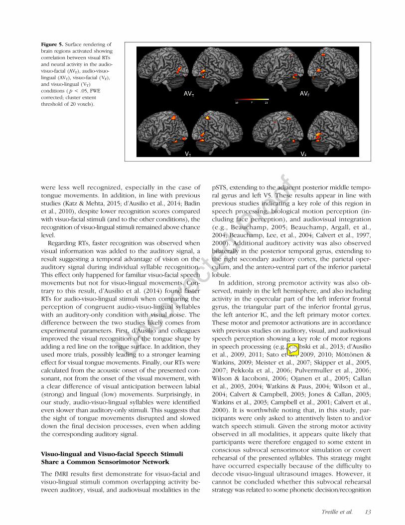
fMRI Results: Correlation between Visual RTsand Neural Activity
For both lip- and tongue-related stimuli, the covarianceanalysis between RTs observed for unimodal visual stim-uli in the behavioral experiment and BOLD activity ob-served in visual and audiovisual conditions in the fMRI
Table 2. (continued )
Regions BA H
MNI Coordinates
t
Contrast Estimates
x y z A VF VT AVF AVT
Dorsolateral pFC 10 L −36 50 −4 4.39 0.05 0.02 0.04 −0.01 0.05
Dorsolateral pFC 11 L −36 46 −6 4.30 0.04 0.01 0.02 −0.01 0.04
Other regions
Primary visual cortex (V1) 17 L −8 −100 2 4.25 0.02 0.26 0.33 0.24 0.33
Cerebellum (VIIb) R 20 −76 −48 4.35 0.05 0.06 0.08 0.03 0.09
ACC 32 L −22 42 4 3.69 0.00 −0.02 −0.01 −0.02 0.01
Figure 4. Surface renderingof brain regions activatedshowing correlation betweenvisual recognition scoresand neural activity in theaudio-visuo-facial (AVF),audio-visuo-lingual (AVT),visuo-facial (VF), and visuo-lingual (VT) conditions( p < .05, FWE corrected;cluster extent threshold of20 voxels).
Treille et al. 11
experiment demonstrated a significant correlation in vi-sual regions (including the primary and associative visualbrain areas and the fusiform gyrus). Other correlationalactivity was found in the superior parietal lobule andadjacent intraparietal sulcus for VT, VF, and AVF condi-
tions as well as in the left premotor cortex for VF (seeFigure 5 and Table 4).To summarize, a correlation between RTs and neural
activity was mainly observed in visual and superior parie-tal regions for both tongue- and lip-related stimuli.
fMRI Results: Different Audiovisual NeuralResponses Compared with Auditory andVisual Modalities
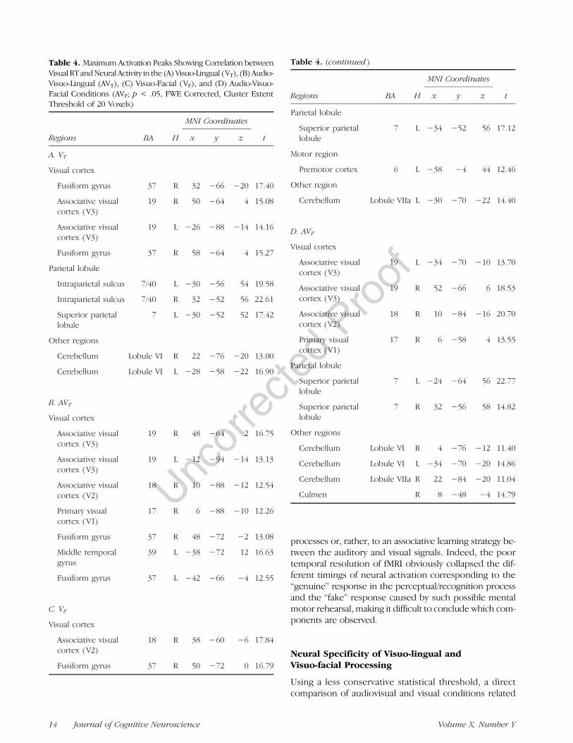
Higher neural responses were only found for audio-visuo-facial stimuli (see Figure 6, condition [AVF > A] ∩[AVF > V]) around the bilateral secondary visual areas, theright cerebellum, and the parahippocampal gyrus and inthe left granular retrosplenial cortex (see Figure 6 andTable 5).
DISCUSSION
Four main results emerged from this fMRI study. First,the neural networks involved in visuo-lingual and visuo-facial perception strongly overlap and share similar sen-sorimotor brain areas. This suggests comparable visualprocessing of lingual and labial movements, both crucialfor the realization of speech sounds. Second, furtheranalyses demonstrate stronger motor and somatosensoryactivations during visuo-lingual perception and strongeractivation of auditory and visual cortices during visuo-facial perception. This result suggests more importantsomatosensory–motor internal simulation of the pre-sented syllables for visuo-lingual speech stimuli that indaily life are clearly audible but not visible, whereasvisible and audible visuo-facial speech stimuli seem tostrongly rely on well-known sensory representations.Third, behavioral results confirm that both visuo-lingualand visuo-facial speech stimuli were correctly recognized,although to a lower extent and slower for visuo-lingualstimuli. Complementing these findings, activity in the leftpremotor cortex and in visual brain areas was found tocorrelate with visual recognition scores observed forvisuo-lingual and visuo-facial speech stimuli, respectively,whereas visual activity correlated with RTs for both stim-uli. Altogether, these results suggest that visual process-ing of audible but not visible movements induce motorand visual mental simulation of the perceived speechactions to facilitate recognition and/or learn the asso-ciation between auditory and visual signals.
Syllable Recognition
The recognition scores replicated a number of well-known effects in auditory, visual, and audiovisual speechperception. As expected, perceptual recognition scoresshow a ceiling effect for auditory and audiovisual modal-ities. Also consistent with previous studies on unimodaland multimodal speech perception, visual-only syllables
Table 3. Maximum Activation Peaks Showing Correlation betweenVisual Recognition Scores and Neural Activity in the (A) Visuo-Lingual (VT), (B) Audio-Visuo-Lingual (AVT), (C) Visuo-Facial (VF), and(D) Audio-Visuo-Facial Conditions (AVF; p< .05, FWE Corrected,Cluster Extent Threshold of 20 Voxels)
Regions BA H
MNI Coordinates
tx y z
A. VT
Premotor cortex 6 L −34 −4 54 16.65
B. AVT
Premotor cortex 6 L −34 0 54 16.34
C. VF
Visual cortex
Associative visualcortex (MT/V5)
19 R 44 −64 0 11.21
Primary visualcortex (V1)
17 R 22 −60 2 12.21
Associative visualcortex (V2)
18 R 22 −90 20 12.75
Fusiform gyrus 37 R 52 −68 −2 11.82
D. AVF
Visual cortex
Fusiform gyrus 37 L −36 −50 −22 19.54
Associative visualcortex (V3)
19 L −34 −76 −12 17.19
Associative visualcortex (V2)
18 R 22 −92 14 14.57
Associative visualcortex (V2)
18 L −8 −88 22 14.96
Associative visualcortex (V3)
19 R 22 −66 −10 12.03
Lingual gyrus 18 R 8 −74 −8 13.14
Other regions
Culmen L −14 −48 −6 18.82
Declive L −30 −58 −16 13.54
Parahippocampalgyrus
19 L −20 −56 −10 13.93
12 Journal of Cognitive Neuroscience Volume X, Number Y
were less well recognized, especially in the case oftongue movements. In addition, in line with previousstudies (Katz & Mehta, 2015; d’Ausilio et al., 2014; Badinet al., 2010), despite lower recognition scores comparedwith visuo-facial stimuli (and to the other conditions), therecognition of visuo-lingual stimuli remained above chancelevel.Regarding RTs, faster recognition was observed when
visual information was added to the auditory signal, aresult suggesting a temporal advantage of vision on theauditory signal during individual syllable recognition.This effect only happened for familiar visuo-facial speechmovements but not for visuo-lingual movements. Con-trary to this result, d’Ausilio et al. (2014) found fasterRTs for audio-visuo-lingual stimuli when comparing theperception of congruent audio-visuo-lingual syllableswith an auditory-only condition with visual noise. Thedifference between the two studies likely comes fromexperimental parameters. First, d’Ausilio and colleaguesimproved the visual recognition of the tongue shape byadding a red line on the tongue surface. In addition, theyused more trials, possibly leading to a stronger learningeffect for visual tongue movements. Finally, our RTs werecalculated from the acoustic onset of the presented con-sonant, not from the onset of the visual movement, witha clear difference of visual anticipation between labial(strong) and lingual (low) movements. Surprisingly, inour study, audio-visuo-lingual syllables were identifiedeven slower than auditory-only stimuli. This suggests thatthe sight of tongue movements disrupted and sloweddown the final decision processes, even when addingthe corresponding auditory signal.
Visuo-lingual and Visuo-facial Speech StimuliShare a Common Sensorimotor Network
The fMRI results first demonstrate for visuo-facial andvisuo-lingual stimuli common overlapping activity be-tween auditory, visual, and audiovisual modalities in the
pSTS, extending to the adjacent posterior middle tempo-ral gyrus and left V5. These results appear in line withprevious studies indicating a key role of this region inspeech processing, biological motion perception (in-cluding face perception), and audiovisual integration(e.g., Beauchamp, 2005; Beauchamp, Argall, et al.,2004; Beauchamp, Lee, et al., 2004; Calvert et al., 1997,2000). Additional auditory activity was also observedbilaterally in the posterior temporal gyrus, extending tothe right secondary auditory cortex, the parietal oper-culum, and the antero-ventral part of the inferior parietallobule.
In addition, strong premotor activity was also ob-served, mainly in the left hemisphere, and also includingactivity in the opercular part of the left inferior frontalgyrus, the triangular part of the inferior frontal gyrus,the left anterior IC, and the left primary motor cortex.These motor and premotor activations are in accordancewith previous studies on auditory, visual, and audiovisualspeech perception showing a key role of motor regionsin speech processing (e.g., Grabski et al., 2013; d’Ausilioet al., 2009, 2011; Sato et al., 2009, 2010; Möttönen &Watkins, 2009; Meister et al., 2007; Skipper et al., 2005,2007; Pekkola et al., 2006; Pulvermuller et al., 2006;Wilson & Iacoboni, 2006; Ojanen et al., 2005; Callanet al., 2003, 2004; Watkins & Paus, 2004; Wilson et al.,2004; Calvert & Campbell, 2003; Jones & Callan, 2003;Watkins et al., 2003; Campbell et al., 2001; Calvert et al.,2000). It is worthwhile noting that, in this study, par-ticipants were only asked to attentively listen to and/orwatch speech stimuli. Given the strong motor activityobserved in all modalities, it appears quite likely thatparticipants were therefore engaged to some extent inconscious subvocal sensorimotor simulation or covertrehearsal of the presented syllables. This strategy mighthave occurred especially because of the difficulty todecode visuo-lingual ultrasound images. However, itcannot be concluded whether this subvocal rehearsalstrategy was related to some phonetic decision/recognition
Figure 5. Surface rendering ofbrain regions activated showingcorrelation between visual RTsand neural activity in the audio-visuo-facial (AVF), audio-visuo-lingual (AVT), visuo-facial (VF),and visuo-lingual (VT)conditions ( p < .05, FWEcorrected; cluster extentthreshold of 20 voxels).
Treille et al. 13
processes or, rather, to an associative learning strategy be-tween the auditory and visual signals. Indeed, the poortemporal resolution of fMRI obviously collapsed the dif-ferent timings of neural activation corresponding to the“genuine” response in the perceptual/recognition processand the “fake” response caused by such possible mentalmotor rehearsal, making it difficult to conclude which com-ponents are observed.
Neural Specificity of Visuo-lingual andVisuo-facial Processing
Using a less conservative statistical threshold, a directcomparison of audiovisual and visual conditions related
Table 4. Maximum Activation Peaks Showing Correlation betweenVisual RT andNeural Activity in the (A)Visuo-Lingual (VT), (B)Audio-Visuo-Lingual (AVT), (C) Visuo-Facial (VF), and (D) Audio-Visuo-Facial Conditions (AVF; p < .05, FWE Corrected, Cluster ExtentThreshold of 20 Voxels)
Regions BA H
MNI Coordinates
tx y z
A. VT
Visual cortex
Fusiform gyrus 37 R 32 −66 −20 17.40
Associative visualcortex (V3)
19 R 50 −64 4 15.08
Associative visualcortex (V3)
19 L −26 −88 −14 14.16
Fusiform gyrus 37 R 58 −64 4 15.27
Parietal lobule
Intraparietal sulcus 7/40 L −30 −56 54 19.58
Intraparietal sulcus 7/40 R 32 −52 56 22.61
Superior parietallobule
7 L −30 −52 52 17.42
Other regions
Cerebellum Lobule VI R 22 −76 −20 13.00
Cerebellum Lobule VI L −28 −58 −22 16.90
B. AVT
Visual cortex
Associative visualcortex (V3)
19 R 48 −64 2 16.75
Associative visualcortex (V3)
19 L −12 −94 −14 13.13
Associative visualcortex (V2)
18 R 10 −88 −12 12.54
Primary visualcortex (V1)
17 R 6 −88 −10 12.26
Fusiform gyrus 37 R 48 −72 −2 13.08
Middle temporalgyrus
39 L −38 −72 12 16.63
Fusiform gyrus 37 L −42 −66 −4 12.55
C. VF
Visual cortex
Associative visualcortex (V2)
18 R 38 −60 −6 17.84
Fusiform gyrus 37 R 50 −72 0 16.79
Table 4. (continued )
Regions BA H
MNI Coordinates
tx y z
Parietal lobule
Superior parietallobule
7 L −34 −52 56 17.12
Motor region
Premotor cortex 6 L −38 −4 44 12.46
Other region
Cerebellum Lobule VIIa L −30 −70 −22 14.40
D. AVF
Visual cortex
Associative visualcortex (V3)
19 L −34 −70 −10 13.70
Associative visualcortex (V3)
19 R 52 −66 6 18.53
Associative visualcortex (V2)
18 R 10 −84 −16 20.70
Primary visualcortex (V1)
17 R 6 −58 4 13.55
Parietal lobule
Superior parietallobule
7 L −24 −64 56 22.77
Superior parietallobule
7 R 32 −56 58 14.82
Other regions
Cerebellum Lobule VI R 4 −76 −12 11.40
Cerebellum Lobule VI L −34 −70 −20 14.86
Cerebellum Lobule VIIa R 22 −84 −20 11.04
Culmen R 8 −48 −4 14.79
14 Journal of Cognitive Neuroscience Volume X, Number Y
to facial or lingual stimuli demonstrates stronger acti-vation of the premotor regions and the primary somato-sensory cortices during the observationof tonguemovements.Because tongue movements are not usually visible andparticipants were not experienced with visuo-lingualultrasound images, this result could be explained by amore important somatosensory–motor covert simulationof tongue movements and the use of both motor andproprioceptive knowledge, to better achieve a phoneticdecoding of the presented visuo-lingual stimuli or to learnthe association between the two signals. Apart from covertsimulation, another explanation could be related to theunusual nature of the lingual stimuli that might implyincreased difficulty and high-level categorization processesin the premotor cortex (Venezia, Saberi, Chubb, & Hickok,2012; Sato et al., 2011).These somatosensory–motor activations appear how-
ever reduced for lip movements. This is likely due tothe fact that visuo-facial speech stimuli are perceived indaily life, with their processing being more automatizedand requiring less motor simulation. In contrast, in bothvisual and audiovisual conditions related to lip move-
ments, stronger visual activity was however observed, ex-tending to a large part of primary and associative visualareas. This result might come from low-level features(contrast, luminance, and motion energy), the facial na-ture as well as stronger visual experience for facial stim-uli. In line with previous studies, our results alsoshowed stronger activity within the auditory cortex dur-ing lip reading condition than in the visuo-lingual con-dition. It was indeed demonstrated that syllables’ visualcues are sufficient to activate auditory cortical sites, nor-mally engaged during the perception of heard speech,in the absence of auditory speech sound (Campbellet al., 2001; Calvert et al., 1997). This result suggestsa direct matching between the visible articulatorymovements and auditory representation of the perceivedsyllables/phonemes. These stronger visual and auditoryactivations during facial perception could be the resultof projections between auditory and visual regions—possibly mediated by the STS. Indeed, studies havedemonstrated direct functional and anatomical pathwaybetween primary sensory areas in nonhuman (Cappe &Barone, 2005) and human (Eckert et al., 2008; Watkins,
Figure 6. Axial views of brainregions showing higher neuralresponses (condition [AVF > A]∩ [AVF > V]) in the audio-visuo-facial condition; p < .001uncorrected, cluster extentthreshold of 20 voxels).
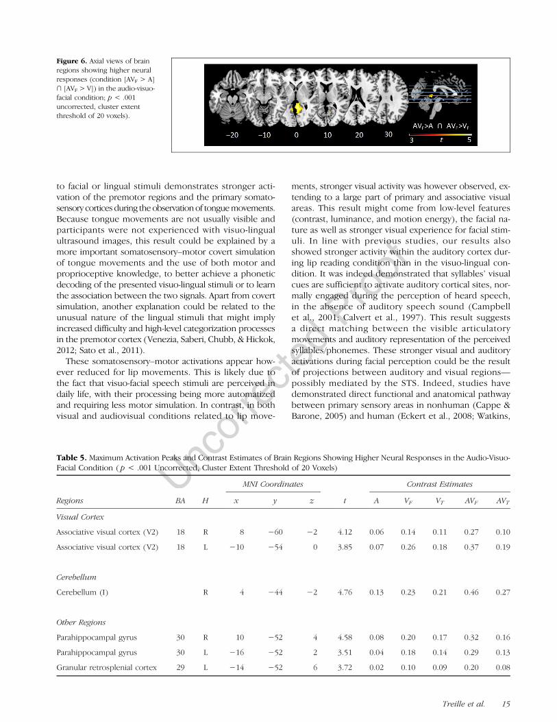
Table 5. Maximum Activation Peaks and Contrast Estimates of Brain Regions Showing Higher Neural Responses in the Audio-Visuo-Facial Condition ( p < .001 Uncorrected, Cluster Extent Threshold of 20 Voxels)
Regions BA H
MNI Coordinates
t
Contrast Estimates
x y z A VF VT AVF AVT
Visual Cortex
Associative visual cortex (V2) 18 R 8 −60 −2 4.12 0.06 0.14 0.11 0.27 0.10
Associative visual cortex (V2) 18 L −10 −54 0 3.85 0.07 0.26 0.18 0.37 0.19
Cerebellum
Cerebellum (I) R 4 −44 −2 4.76 0.13 0.23 0.21 0.46 0.27
Other Regions
Parahippocampal gyrus 30 R 10 −52 4 4.58 0.08 0.20 0.17 0.32 0.16
Parahippocampal gyrus 30 L −16 −52 2 3.51 0.04 0.18 0.14 0.29 0.13
Granular retrosplenial cortex 29 L −14 −52 6 3.72 0.02 0.10 0.09 0.20 0.08
Treille et al. 15
Shams, Tanaka, Haynes, & Rees, 2006) cerebral cortex.From that view, lower activation of the visual cortexduring the sight of tongue movements could also beexplained because such movements are not likely todirectly excite the auditory cortex because of theirunusual characteristics.
Correlation between Behavioral Performanceand Neural Activity
Interestingly, activities in the left premotor cortex andin visual brain areas were found to correlate with visualrecognition scores observed for visuo-lingual and visuo-facial speech stimuli, respectively. Hence, the morethese areas were activated, the better were the visualrecognition scores. These results appear consistent withthose observed from the direct comparison betweenvisuo-lingual and visuo-facial movements. As previouslynoted, given the poor temporal resolution of fMRI, it ishowever impossible to determine whether motor simu-lation is related to some recognition/decision processesor rather to some associative learning effect.
Another result is that activity in visual and superiorparietal brain areas correlated with RTs for both visuo-facial and visuo-lingual stimuli. Given that these brainregions are known to play a role in visual imagery, thislater finding might indicate the use of a visual imagerystrategy by the participants to learn the association be-tween auditory and visual signals.
Integration between Auditory and Visual Signals
As previously noted, fMRI studies have demonstrated theexistence of specific multisensory brain areas involved inthe integration process of auditory and visual signals.More specifically, when compared with auditory andvisual unimodal modalities, the observation of audio-visual stimuli was found to induce supra-additive responsesin pSTS/pSTG (Beauchamp, 2005; Beauchamp, Argall,et al., 2004; Beauchamp, Lee, et al., 2004; Calvert et al.,2000) as well as subadditive responses in Broca’s area(Calvert et al., 2000). Beauchamp (2005) determined twominimal criteria to select brain regions involved in audio-visual speech integration: The region must be activatedduring auditory, visual, and audiovisual modalities andmust display supra-additive audiovisual response. In thisstudy, higher neural responses using the max criterion test([AV > A] ∩ [AV > V]) were only found for audio-visuo-facial stimuli around the bilateral secondary visual areas,the right cerebellum, and the parahippocampal gyrus andin the left granular retrosplenial cortex. Although a pSTS/pSTG activation was observed for all conditions, no higherresponse was found for this region supposed to be aspecific brain area involved in the integration process.Although we do not have a clear explanation for this nullresult, one possibility is that the strong sensorimotor
activity observed in all modalities, including the pSTS/pSTG, might have changed the classical audiovisual inte-gration network.
Concluding Remarks
Taken together, our results provide new evidence for anaction–perception functional coupling in speech pro-cessing. According to a recent neurobiological andperceptuo-motor model of multisensory speech percep-tion by Skipper and colleagues (2007), apart from sensoryprocessing, motor activity during speech perceptionmight partly constrain phonetic interpretation of thesensory inputs through the internal generation of candi-date articulatory categorizations and, in return, auditoryand somatosensory predictions. In this study, becauseof the lack of visual knowledge in the processing of thegenerally hidden tongue movements, a larger motor re-cruitment could have been necessary to infer appropriatemotor speech representations to correctly decode theperceived syllables. This process would have been guidedby the participant’s expertise in speech production, en-abling to transfer procedural motor knowledge into abetter understanding of such unfamiliar visual stimuli.One alternative explanation is that motor activity does notdirectly reflect some phonetic decision processes butrather a learning effect between auditory and visual signals.Visual and motor familiarities have already been com-
pared in the course of action recognition, and previousstudies have shown that the involvement of the motorsystem during action observation strongly relies on motorlearning (e.g., Calvo-Merino et al., 2005, 2006). In linewith previous behavioral studies (Katz & Mehta, 2015;d’Ausilio et al., 2014; Badin et al., 2010), the presentdata demonstrate that, even if participants have no visualfamiliarity with one given human action, they are never-theless able to recognize this action because of theirmotor knowledge and past auditory and somatosensoryexperience. This is in line with the assumption of sensory–motor transfer mechanisms at hand in the visual percep-tion of audible but invisible tongue actions. The situationexperienced by the participants of the present experimentis to a certain extent similar to the one experienced bynewborns and 3-month-old infants, in the classical exper-iments on facial imitation by Meltzoff and Moore (1977,1983). They have shown astonishing capacities to replicateto a certain extent a facial movement they have neverseen done by a caregiver. These abilities are interpretedby the authors in reference to the link between proprio-ceptive and motor information feeding newborns withinformation about their own unseen movements in rela-tion with the visual representation of the perceived move-ment of the caregiver and enabling the required actionmatching. Despite the correlational approach used in thisstudy, our results suggest that, even if we have no visualbut auditory and somatosensory experiences of an action,the connection between our motor abilities and the visual
16 Journal of Cognitive Neuroscience Volume X, Number Y
incoming signal exists and enables adequate processingand performance.
UNCITED REFERENCES
Grabski, Schwartz, et al., 2013Grabski, Tremblay, Gracco, Girin, & Sato, 2013
Acknowledgments
This study was supported by research grants from CNRS(Centre National de la Recherche Scientifique), from AgenceNationale de la Recherche (ANR SPIM, “Imitation in Speech:From Sensorimotor Integration to the Dynamics of Conversa-tional Interaction”), and from the European Research Council(FP7/2007-2013 Grant agreement no. 339152, “Speech Unit(e)s”).Any opinions, findings, and conclusions or recommendationsexpressed in this material are those of the authors and do notnecessarily reflect the views of the funding agencies. We thankJean-Luc Schwartz for helpful discussions.
Reprint requests should be sent to Avril Treille, GIPSA-lab, UMR5216, Université Stendhal, 1180, Avenue Centrale, BP25, 38031Grenoble Cedex 9, France, or via e-mail: [email protected].
REFERENCES
Aziz-Zadeh, L., Iacoboni, M., Zaidel, E., Wilson, S., & Mazziotta,J. (2004). Left hemisphere motor facilitation in responseto manual action sounds. European Journal of Neuroscience,19, 2609–2612.
Badin, P., Tarabalka, Y., Elisei, F., & Bailly, G. (2010). Can you“read” tongue movements? Evaluation of the contributionof tongue display to speech understanding. SpeechCommunication, 52, 493–503.
Beardsworth, T., & Buckner, T. (1981). The ability to recognizeoneself from a video recording of one’s movements withoutseeing one’s body. Bulletin of the Psychonomic Society,18, 19–22.
Beauchamp, M. S. (2005). Statistical criteria in fMRIstudies of multisensory integration. Neuroinformatics,3, 93–114.
Beauchamp, M. S., Argall, B. D., Bodurka, J., Duyn, J. H., &Martin, A. (2004). Unraveling multisensory integration:Patchy organization within human STS multisensory cortex.Nature Neuroscience, 7, 1190–1192.
Beauchamp, M. S., Lee, K. E., Argall, B. D., & Martin, A.(2004). Integration of auditory and visual informationsabout objects in superior temporal sulcus. Neuron, 41,809–823.
Birn, R. M., Bandettini, P. A., Cox, R. W., & Shaker, R. (1999).Event-related fMRI of tasks involving brief motion. HumanBrain Mapping, 7, 106–114.
Buccino, G., Lui, F., Canessa, N., Patteri, I., Lagravinese, G.,Benuzzi, F., et al. (2004). Neural circuit involved in therecognition of actions performed by nonconspecifics: AnfMRI study. Journal of Cognitive Neurosciences, 16,114–126.
Callan, D. E., Jones, J. A., Munhall, K. G., Callan, A. M., Kroos, C., &Vatikiotis-Bateson, E. (2003). Neural processes underlyingperceptual enhancement by visual speech gestures.NeuroReport, 14, 2213–2217.
Callan, D. E., Jones, J. A., Munhall, K. G., Callan, A. M., Kroos, C.,& Vatikiotis-Bateson, E. (2004). Multisensory integrationsites identified by perception of spatial wavelet filtered
visual speech gesture information. Journal of CognitiveNeuroscience, 16, 805–816.
Calvert, G. A., Bullmore, E., Brammer, M. J., Campbell, R.,Iversen, S. D., Woodruff, P., et al. (1997). Silent lip readingactivates the auditory cortex. Science, 276, 593–596.
Calvert, G. A., & Campbell, R. (2003). Reading speech from stilland moving faces: The neural substrates of visible speech.Journal of Cognitive Neuroscience, 15, 57–70.
Calvert, G. A., Campbell, R., & Brammer, M. J. (2000). Evidencefrom functional magnetic resonance imaging of crossmodalbinding in the human heteromodal cortex. Current Biology,10, 649–657.
Calvo-Merino, B., Glaser, D. E., Grèzes, J., Passingham, R. E., &Haggard, P. (2005). Action observation and acquired motorskills: An fMRI study with expert dancers. Cerebral Cortex,15, 1243–1249.
Calvo-Merino, B., Grèzes, J., Glaser, D. E., Passingham, R. E., &Haggard, P. (2006). Seeing or doing? Influence of visualand motor familiarity in action observation. Current Biology,16, 1905–1910.
Campbell, R., MacSweeney, M., Surguladze, S., Calvert, G.,McGuire, P., Suckling, J., et al. (2001). Cortical substratesfor the perception of face actions: An fMRI study of thespecificity of activation for seen speech and for meaninglesslower-face acts (gurning). Cognitive Brain Research, 12,233–243.
Cappe, C., & Barone, P. (2005). Heteromodal connectionssupporting multisensory integration at low levels of corticalprocessing in the monkey. European Journal of Neuroscience,22, 2886–2902.
d’Ausilio, A., Bartoli, E., Maffongelli, L., Berry, J. J., & Fadiga, L.(2014). Vision of tongue movements bias auditory speechperception. Neuropsychologia, 63, 85–91.
d’Ausilio, A., Bufalari, I., Salmas, P., & Fadiga, L. (2011). The roleof the motor system in discriminating degraded speechsounds. Cortex, 48, 882–887.
d’Ausilio, A., Pulvermüller, F., Salmas, P., Bufalari, I., Begliomini,C., & Fadiga, L. (2009). The motor somatotopy of speechperception. Current Biology, 19, 381–385.
Di Pellegrino, G., Fadiga, L., Fogassi, L., Gallese, V., & Rizzolatti, G.(1992). Understanding motor events: A neurophysiologicalstudy. Experimental Brain Research, 91, 176–180.
Eckert, M. A., Kamdar, N. V., Chang, C. E., Beckmann, C. F.,Greicius, M. D., & Menon, V. (2008). A crossmodal systemlinking primary auditory and visual cortices: Evidencefrom intrinsic fMRI connectivity analysis. Human BrainMapping, 29, 848–885.
Eickhoff, S. B., Stephan, K. E., Mohlberg, H., Grefkes, C.,Fink, G. R., Amunts, K., et al. (2005). A new SPM toolboxfor combining probabilistic cytoarchitectonic maps andfunctional imaging data. Neuroimage, 25, 1325–1335.
Ferrari, P. F., Gallese, V., Rizzolatti, G., & Fogassi, L. (2003).Mirror neurons responding to the observation of ingestiveand communicative mouth actions in the monkey ventralpremotor cortex. European Journal of Neuroscience, 17,1703–1714.
Fogassi, L., Ferrari, P. F., Gesierich, B., Rozzi, S., Chersi, F., &Rizzolatti, G. (2005). Parietal lobe: From action organizationto intention understanding. Science, 308, 662–667.
Gallese, V., Fadiga, L., Fogassi, L., & Rizzolatti, G. (1996).Action recognition in the premotor cortex. Brain, 119,593–609.
Grabski, K., Schwartz, J. L., Lamalle, L., Vilain, C., Vallée, N.,Baciu, M., et al. (2013). Shared and distinct neuralcorrelates of vowel perception and production. Journal ofNeurolinguistics, 26, 384–408.
Grabski, K., Tremblay, P., Gracco, V., Girin, L., & Sato, M.(2013). A mediating role of the auditory dorsal pathway
Treille et al. 17
in selective adaptation to speech: A state-dependenttranscranial magnetic stimulation study. Brain Research,1515, 55–65.
Hall, D. A., Haggard, M. P., Akeroyd, M. A., Palmer, A. R.,Summerfield, A. Q., Elliott, M. R., et al. (1999). Sparsetemporal sampling in auditory fMRI. Human Brain Mapping,7, 213–223.
Haueisen, J., & Knösche, T. R. (2001). Involuntary motoractivity in pianists evoked by music perception. Journal ofCognitive Neuroscience, 13, 786–792.
Howard, R. J., Brammer, M., Wright, I., Woodruff, P. W.,Bullmore, E. T., & Zeki, S. (1996). A direct demonstration offunctional specialization within motion-related visual andauditory cortex of the human brain. Current Biology, 6,1015–1019.
Hueber, T., Chollet, G., Denby, B., & Stone, M. (2008).Acquisition of ultrasound, video and acoustic speech data fora silent-speech interface application. In Proceedings ofInternational Seminar on Speech Production (Strasbourg,France) (pp. 365–369).
Johansson, R. (1973). Visual perception of biological motionand a model for its analysis. Perception & Psychophysics, 14,201–211.
Jones, J., & Callan, D. E. (2003). Brain activity during audio-visualspeech perception: An fMRI study of the McGurk effect.NeuroReport, 14, 1129–1133.
Katz, W. F., & Mehta, S. (2015). Visual feedback of tonguemovements for novel speech sound learning. Frontiers inHuman Neuroscience, 9, 612.
Keysers, C., Kholer, E., Umilta, M. A., Fogassi, L., Gallese, V., &Rizzolatti, G. (2003). Audiovisual mirror neurons andaction recognition. Experimental Brain Research, 153,628–636.
Kohler, E., Keysers, C., Umilta, M. A., Fogassi, L., Gallese, V.,& Rizzolatti, G. (2002). Hearing sounds, understandingactions: Action representation in mirror neurons. Science,297, 846–848.
Lahav, A., Saltzman, E., & Schlaug, G. (2007). Actionrepresentation of sound: Audiomotor recognition networkwhile listening to newly acquired actions. Journal ofNeuroscience, 27, 3008–3014.
Lancaster, J. L., Woldorff, M. G., Parsons, L. M., Liotti, M.,Freitas, C. S., Rainey, L., et al. (2000). Automated Talairachatlas labels for functional brain mapping. Human BrainMapping, 10, 120–131.
Liberman, A. M., & Mattingly, I. G. (1985). The motor theoryof speech perception revised. Cognition, 21, 1–36.
Loula, F., Prasad, S., Harber, K., & Shiffrar, M. (2005).Recognizing people from their movements. Journal ofExperimental Psychology: Human Perception andPerformance, 31, 210–220.
Meister, I. G., Wilson, S. M., Deblieck, C., Wu, A. D., & Iacoboni,M. (2007). The essential role of premotor cortex in speechperception. Current Biology, 17, 1692–1696.
Meltzoff, A. N., & Moore, M. K. (1977). Imitation of facialand manual gestures by human neonates. Science, 198,75–78.
Meltzoff, A. N., & Moore, M. K. (1983). Newborn infants imitateadult facial gestures. Child Development, 54, 702–709.
Möttönen, R., & Watkins, K. E. (2009). Motor representations ofarticulators contribute to categorical perception of speechsounds. Journal of Neuroscience, 29, 9819–9825.
Ojanen, V., Möttönen, R., Pekkola, J., Jääskeläinen, I. P., Joensuu,R., Autti, T., et al. (2005). Processing of audio-visual speechin Broca’s area. Neuroimage, 25, 333–338.
Oldfield, R. C. (1971). The assessment and analysis ofhandedness: The Edinburgh inventory. Neuropsychologia,9, 97–114.
Pekkola, J., Laasonen, M., Ojanen, V., Autti, T., Jaaskelainen,L. P., Kujala, T., et al. (2006). Perception of matchingand conflicting audio-visual speech in dyslexic andfluent readers: An fMRI study at 3T. Neuroimage, 29,797–807.
Pickering, M. J., & Garrod, S. (2013). An integrated theory oflanguage production and comprehension. Behavioral andBrain Sciences, 36, 329–347.
Pizzamiglio, L., Aprile, T., Spitoni, G., Pitzalis, S., Bates, E.,D’Amico, S., et al. (2005). Separate neural systems forprocessing action- or non-action related sounds. Neuroimage,24, 852–861.
Prather, J. F., Peters, S., Nowicki, S., & Mooney, R. (2008). Preciseauditory-vocal mirroring in neurons for learned vocalcommunication. Nature, 451, 305–310.
Pulvermuller, F., Huss, M., Kherif, F., Moscosodel PradoMartin, F., Hauk, O., & Shtyrov, Y. (2006). Motor cortexmaps articulatory features of speech sounds. Proceedingsof the National Academy of Sciences, U.S.A., 103,7865–7870.
Rizzolatti, G., & Craighero, L. (2004). The mirror-neuron system.Annual Review of Neuroscience, 27, 169–192.
Rizzolatti, G., Fadiga, L., Gallese, V., & Fogassi, L. (1996).Premotor cortex and the recognition of motor actions.Cognitive Brain Research, 3, 131–142.
Rizzolatti, G., Fogassi, L., & Gallese, V. (2001). Neurophysiologicalmechanisms underlying the understanding and imitation ofaction. Nature Review Neuroscience, 2, 661–670.
Sato, M., Buccino, G., Gentilucci, M., & Cattaneo, L. (2010).On the tip of the tongue: Modulation of the primary motorcortex during audio-visual speech perception. SpeechCommunication, 52, 533–541.
Sato, M., Grabski, K., Glenberg, A., Brisebois, A., Basirat, A.,Ménard, L., et al. (2011). Articulatory bias in speechcategorization: Evidence from use-induced motor plasticity.Cortex, 47, 1001–1003.
Sato, M., Tremblay, P., & Gracco, V. (2009). A mediating role ofthe premotor cortex in phoneme segmentation. Brain andLanguage, 111, 1–7.
Saygin, A. P. (2007). Superior temporal and premotor brainareas necessary for biological motion perception. Brain, 130,2452–2461.
Schwartz, J. L., Ménard, L., Basirat, A., & Sato, M. (2012).The Perception for Action Control Theory (PACT): Aperceptuo-motor theory of speech perception. Journal ofNeurolinguistics, 25, 336–354.
Skipper, J., Van Wassenhove, V., Nussman, H., & Small,S. L. (2007). Hearing lips and seeing voices: Howcortical areas supporting speech production meditateaudio-visual speech perception. Cerebral Cortex, 17,2387–2399.
Skipper, J. I., Nusbaum, H. C., & Small, S. L. (2005). Listeningto talking faces: Motor cortical activation during speechperception. Neuroimage, 25, 76–89.
Stevenson, R. A., Ghose, D., Krueger Fister, J., Sarko, D. K.,Altieri, N. A., Nidiffer, A. R., et al. (2014). Identifying andquantifying multisensory integration: A tutorial review. BrainTopography, 27, 707–730.
Tai, Y. F., Scherfler, C., Brooks, D. J., Sawamoto, N., &Castiello, U. (2004). The human premotor cortex is“mirror” only for biological actions. Current Biology, 14,117–120.
Venezia, J. H., Saberi, K., Chubb, C., & Hickok, G. (2012).Response bias modulates the speech motor systemduring syllable discrimination. Frontiers in Psychology,3, 157.
Viviani, P., & Stucchi, N. (1992). Biological movements lookuniform: Evidence of motor perceptual interactions. Journal
18 Journal of Cognitive Neuroscience Volume X, Number Y
of Experimental Psychology: Human Perception andPerformance, 18, 603–623.
Watkins, K. E., & Paus, T. (2004). Modulation of motorexcitability during speech perception: The role of Broca’sarea. Journal of Cognitive Neuroscience, 16, 978–987.
Watkins, K. E., Strafella, A. P., & Paus, T. (2003). Seeing andhearing speech excites the motor system involved inspeech production. Neuropsychologia, 41, 989–994.
Watkins, S., Shams, L., Tanaka, S., Haynes, J. D., & Rees, G.(2006). Sound alters activity in human V1 in associationwith illusory visual perception. Neuroimage, 31, 1247–1256.
Wilson, S., & Iacoboni, M. (2006). Neural responses to non-native phonemes varying in producibility: Evidence for thesensorimotor nature of speech perception. Neuroimage, 33,316–325.
Wilson, S. M., Saygin, A. P., Sereno, M. I., & Iacoboni, M. (2004).Listening to speech activates motor areas involved in speechproduction. Nature Neuroscience, 7, 701–702.
Zaehle, T., Schmidt, C. F., Meyer, M., Baumann, S., Baltes, C.,Boesiger, P., et al. (2007). Comparison of “silent” clusteredand sparse temporal fMRI acquisitions in tonal and speechperception tasks. Neuroimage, 37, 1195–1204.
Treille et al. 19
AUTHOR QUERIES
AUTHOR PLEASE ANSWER ALL QUERIES
During the preparation of your manuscript, the questions listed below arose. Kindly supply the necessaryinformation.
1. Please check if affiliations were correctly captured/structured.2. Please specify which of the two reference items is being referred to in all occurrences of this citation
(Grabski et al., 2013).3. Please provide city and state/country location for this product name (Philips Achieva TX).4. Please insert citations of uncited references in the body.
END OF ALL QUERIES


![Fast Object Class Labelling via Speechopenaccess.thecvf.com/.../Gygli_Fast_Object_Class_Labelling_via_Sp… · Using speech as an input modality has a long history [2] and is recently](https://img.pdfslide.us/doc/110x75/5eb44b7148538638254d8ffd/fast-object-class-labelling-via-using-speech-as-an-input-modality-has-a-long-history.jpg)