Embed Size (px)
Citation preview
INSECT FLIGHT CONTROL BY NEURAL STIMULATION OF PUPAE-IMPLANTED FLEXIBLE
MULTISITE ELECTRODES W.M. Tsang1, Z. Aldworth2, A. Stone3, A. Permar3, R. Levine3,
J. G. Hildebrand3, T. Daniel2, A.I. Akinwande1 and J. Voldman1 1Electrical Engineering and Computer Science, Massachusetts Institute of
Technology, USA 2Department of Biology, University of Washington, USA
3Arizona Research Laboratories, Division of Neurobiology, University of Arizona, USA
ABSTRACT
We describe the demonstration of moth Manduca sexta flight control via pupae-implanted MEMS-based electrodes that directly interface with the central nervous system (CNS). We have developed a flexible electrode array that provides multisite electrical stimulation of an interganglionic bundle of nerve fibers in the moth's ab-dominal nerve cord (analogous to a vertebrate's spinal cord). These electrodes were able to stimulate the abdomen motion of pupae and adult moths leading to a change in flight direction of tethered adult moths.
KEYWORDS: flexible stimulation electrode, insect flight control, polyimide, moth
INTRODUCTION
Significant interest exists in creating insect-based Micro-Air-Vehicles (MAVs) that would combine advantageous features of insects—small size, relatively large payload capacity, navigation ability—with the benefits of MEMS and electronics—sensing, actuation and information processing. Researchers have reported the use of MEMS-based electrodes, implanted into the muscle of pupae, to control the wing motion of the moth Manduca sexta, leading to rudimentary turning behavior in a moth hung by wires [1]. Alternatively, another group has implanted conventional silver wires into the muscle of adult beetles to enable flight control [2].
In this work, we have developed a flexible electrode array that provides mul-tisite stimulation of an interganglionic bundle of nerve fibers in the moth's abdomi-nal nerve cord. These flexible multisite electrodes (FMEs) are implanted into moth pupae and directly interface with the CNS of the moth (rather than muscle). CNS-based flight control is likely to be more robust in real-world applications as it allows the moth’s brain to process the MEMS stimulation inputs, thus minimally perturbing the moth’s flight control system and allowing the moth, for instance, to avoid obsta-cles en route to a destination.
EXPERIMENTAL
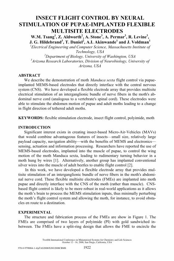
The structure and fabrication process of the FMEs are show in Figure 1. The FMEs are comprised of two layers of polyimide (PI) with gold sandwiched in-between. The FMEs have a split-ring design that allows the FME to encircle the
978-0-9798064-1-4/µTAS2008/$20©2008CBMS 1922
Twelfth International Conference on Miniaturized Systems for Chemistry and Life SciencesOctober 12 - 16, 2008, San Diego, California, USA

nerve cord, and the electrodes on the FME are on flexible tabs that protrude into the split ring and can bend back to make good contact with the nerve cord. The split-ring and tab design makes the FME adaptable to a wide range of nerve cord diame-ters, maintaining good contact as animals undergo metamorphosis and the nerve cord diameter increases.
Figure 1. (a) Structure and (b) fabrication process of the FME.
(a) (b)
RESULTS AND DISCUSSION We have developed surgical techniques (Figure 2) to insert FMEs into pupae at the
ventral fourth abdominal position (Figure 3a) as early as 7 days before the adult moth emerges. At this position, the FME stimulation causes the abdomen to flex, providing ruddering of the moth during flight. Implanted pupae were able to develop into adult moths with normal flight characteristics, and dissected adult moths showed apparent growth of the dorsal pad connective tissue on the polyimide surface (Figure 3d) and around the side of the electrode, providing evidence both of the biocompatibility of the electrodes and their ability to integrate into the nervous system. Figure 2. The surgical process: (a) Open window in pupal cuticle; (b) Createincision in adult cuticle; (c) Isolate nerve cord on glass probe; (d) place split-ringportion of FME onto the nerve cord; (e) Insert FME into incision and (f) Usesurgical glue to seal incision.
(d) (e) (f) ( a) ( b) (c)
We were able to stimulate pupae and adult moths. In pupae, we observed ab-
dominal flexion using square wave pulses of ≥ 4 volts at various pairs of the stimu-lation sites, and similar behavior was observed in tethered adult moths (Figure 4). Finally, in loosely tethered flight, we have used this abdominal ruddering to cause the normally hovering moth to change its abdominal angle, leading to a change in flight direction (Figure 5). CONCLUSIONS
We demonstrate the ability to create MEMS-based electrodes that can be im-planted in pupae, directly interface with the CNS, and enable insect flight control.
1923
Twelfth International Conference on Miniaturized Systems for Chemistry and Life SciencesOctober 12 - 16, 2008, San Diego, California, USA

(a) (b) (c) (d)
ganglion FME
nerve cord
FME
FME
tissue
Figure 3. Images show (a) the electrode implantation site in the pupa; (b) an FME inserted around the abdominal nerve cord; (c) an adult moth with a pupae-implanted FME protruding from its abdomen, also shown in the inset; and (d) growth of dorsal pad connective tissue around an implanted FME.
ACKNOWLEDGEMENTS
This work was supported by the Air Force Research Laboratory as part of the DARPA HI-MEMS program REFERENCES [1] A. Bozkurt, R. Gilmour, D. Stern and A Lal., IEEE MEMS 2008, pp. 160-163
(2008). [2] H. Sato, C. W. Berry, B. E. Casey, G. Lavella, Y. Yao, J. M. V. Brooks and M.
M. Maharbiz, IEEE MEMS 2008, pp. 164-167(2008).
( a ) Stimulatio On Stimulatio Off
( b )
(a
abdominal angle
(b
(c
Figure 4. Images show abdominal mo-tion of (a) a pupa and (b) an adult moth before and during electrical stimulation.
Figure 5. Flight control of a loosely tethered adult moth: (a) definitions of flight direction (colored circle) and ab-dominal angle (θ) of the moth; (b) varia-tion of θ and (c) the magnitude and di-rection of the moth’s velocity (the colors indicate the direction) with stimulation.
1924
Twelfth International Conference on Miniaturized Systems for Chemistry and Life SciencesOctober 12 - 16, 2008, San Diego, California, USA