Embed Size (px)
Citation preview
at SciVerse ScienceDirect
Insect Biochemistry and Molecular Biology 43 (2013) 612e625
Contents lists available
Insect Biochemistry and Molecular Biology
journal homepage: www.elsevier .com/locate/ ibmb
Gloverins of the silkworm Bombyx mori: Structural and bindingproperties and activities
Hui-Yu Yi a,b, Xiao-Juan Deng a, Wan-Ying Yang a, Cong-Zhao Zhou c, Yang Cao a,**,Xiao-Qiang Yu b,*
a Laboratory of Insect Molecular Biology and Biotechnology, Guangdong Provincial Key Laboratory of Agro-animal Genomics and Molecular Breeding,College of Animal Science, South China Agricultural University, Guangzhou 510642, ChinabDivision of Cell Biology and Biophysics, School of Biological Sciences, University of Missouri-Kansas City, 5007 Rockhill Road, Kansas City, MO 64110, USAc School of Life Sciences, University of Science and Technology of China, Hefei, Anhui 230026, China
a r t i c l e i n f o
Article history:Received 15 January 2013Received in revised form21 March 2013Accepted 26 March 2013
Keywords:GloverinLipopolysaccharideCircular dichroismAntibacterialRandom coila-helixBombyx mori
* Corresponding author. Tel.: þ1 816 235 6379; fax** Corresponding author.
E-mail addresses: [email protected] (Y. Cao)gmail.com (X.-Q. Yu).
0965-1748/$ e see front matter � 2013 Elsevier Ltd.http://dx.doi.org/10.1016/j.ibmb.2013.03.013
a b s t r a c t
Gloverins are basic, glycine-rich and heat-stable antibacterial proteins (w14- kDa) in lepidopteran in-sects with activity against Escherichia coli, Gram-positive bacteria, fungi and a virus. Hyalophora gloverigloverin adopts a random coil structure in aqueous solution but has a-helical structure in membrane-likeenvironment, and it may interact with the lipid A moiety of lipopolysaccharide (LPS). Manduca sextagloverin binds to the O-specific antigen and outer core carbohydrate of LPS. In the silkworm Bombyxmori, there are four gloverins with slightly acidic to neutral isoelectric points. In this study, we investigatestructural and binding properties and activities of B. mori gloverins (BmGlvs), as well as correlationsbetween structure, binding property and activity. Recombinant BmGlv1-4 were expressed in bacteria andpurified. Circular dichroism (CD) spectra showed that all four BmGlvs mainly adopted random colistructure (>50%) in aqueous solution in regardless of pH, but contained a-helical structure in thepresence of 1,1,1,3,3,3-hexafluoro-2-propanol (HFIP), smooth and rough mutants (Ra, Rc and Re) of LPSand lipid A. Plate ELISA assay showed that BmGlvs at pH 5.0 bound to rough mutants of LPS and lipid Abut not to smooth LPS. Antibacterial activity assay showed that positively charged BmGlvs (at pH 5.0)were active against E. coli mutant strains containing rough LPS but inactive against E. coli with smoothLPS. Our results suggest that binding to rough LPS is the prerequisite for the activity of BmGlvs againstE. coli.
� 2013 Elsevier Ltd. All rights reserved.
1. Introduction
Insects have the largest numbers and species on earth, and theycombat a variety of pathogens mainly relying on sophisticatedinnate immune system. Insect defense system consists of threemajor parts: structural barriers, cellular and humoral immune re-sponses (Lemaitre and Hoffmann, 2007). Structural barriers, thefirst protective lines, refer to cuticle, midgut epithelium and tra-chea. Cellular immune responses, including phagocytosis, nodula-tion and encapsulation, are mediated by several types of hemocytes(Lavine and Strand, 2002; Sideri et al., 2007). Humoral immuneresponses include melanization of hemolymph and secretion of
: þ1 816 235 1503.
, [email protected], xyu3113@
All rights reserved.
antimicrobial peptides (AMPs) (Hoffmann, 1995). AMPs, the majorand best known immune effectors induced by infection, are syn-thesized by fat body, hemocytes and other tissues, and regulated bythe Toll and immune deficiency (IMD) pathways (Bulet et al., 1999;Lemaitre and Hoffmann, 2007).
At least 150 insect AMPs have been purified or identified. Mostinsect AMPs are small and cationic, and they show activities againstbacteria and/or fungi (Hoffmann, 1995; Bulet and Stocklin, 2005).Based on the sequences, structures and activities, insect AMPs canbe classified into four families, the a-helical peptides (e.g., cecropin,moricin and sarcotoxin), cysteine-rich peptides (e.g., insect defen-sin, drosomycin and heliomicin), proline-rich peptides (e.g., api-daecin, drosocin and lebocin), and glycine-rich peptides (e.g.,attacin and gloverin) (Laszlo-Otvos, 2000; Bulet and Stocklin,2005). Glycine-rich peptides with molecular masses of more than10 kDa have become a large family of insect AMPs, including atta-cin, sarcotoxin II, gloverin, hymenoptaecin, coleoptericin, hemi-ptericin and tenecin 4 (Hultmark et al., 1983; Ando et al.,1987; Axen
H.-Y. Yi et al. / Insect Biochemistry and Molecular Biology 43 (2013) 612e625 613
et al., 1997; Casteels et al., 1993; Bulet et al., 1991; Cociancich et al.,1994; Chae et al., 2012).
So far, small cationic AMPs (w4 kDa) have been the focus ofstructural and activity study. Cysteine-rich peptides, which arecyclic by formation of disulfide bonds, can form stable structures inaqueous solution. However, linear AMPs do not form stable struc-tures in aqueous solution but can change to stable structures inmembrane mimic environment (Nguyen et al., 2008; Haney andVogel, 2009). Cysteine-rich AMPs, such as insect defensin A, dro-somycin, termicin and heliomicin, contain “cysteine stabilized abmotif” (CSab) structure with antiparallel b-sheet connected to asingle a-helix by two disulfide bridges (Cornet et al., 1995; Landonet al., 1997; Silva et al., 2003; Lamberty et al., 2001). But nearly allthe structures of a-helical linear AMPs are obtained by usingmicelle suspensions or in the presence of organic solvents. Forexample, Hyalophora cecropia cecropin A exists as random coli inaqueous solution but forms an amphipathic helical structure in1,1,1,3,3,3-hexafluoro-2-propanol (HFIP)/water solution (Holaket al., 1988). The a-helical structure of moricin is also obtained inmethanol and in solution containing 2,2,2-trifluoroethanol (TFE) orsodium dodecylsulphate (SDS) (Dai et al., 2008).
Since the first purification and characterization of gloverin fromHyalophora gloveri pupal hemolymph (Axen et al., 1997), a gloverinhas been isolated from Helicoverpa armigera (Mackintosh et al.,1998), and two gloverins have been detected in hemolymph ofseptic injured Diatraea saccharialis larvae (Silva et al., 2010). Glo-verin genes have also been identified in Antheraea mylitta (Gandhe-Archana et al., 2006), Galleria mellonella (Seitz et al., 2003; Brownet al., 2009), Manduca sexta (Abdel-Latief and Hilker, 2008; Xuet al., 2012; Zhu et al., 2003), Plutella xylostella (Eum et al., 2007;Etebari et al., 2011), Spodoptera exigua (Hwang and Kim, 2011), andTrichoplusia ni (Lundstrom et al., 2002). In the silkworm Bombyxmori, four gloverin genes (Bmglv1-4) have been identified, andBmglv2-4 genes are derived from duplication of Bmglv1 (Chenget al., 2006; Kaneko et al., 2007; Kawaoka et al., 2008; Mrinal andNagaraju, 2008). Among the gloverins with known activities,H. gloveri gloverin is active against Escherichia coli D21f2 and D21mutant strains with rough LPS (Axen et al., 1997), H. armigera glo-verin is active against E. coli strains with smooth LPS and D22 strainthat is defective in lipid A (Mackintosh et al., 1998), T. ni gloverin 1and 2 have activity against E. coli D21f2 and D22 strains and a virus(Lundstrom et al., 2002; Moreno-Habel et al., 2012). However,S. exigua gloverin is active against a Gram-positive bacterium(Flavobacterium sp.) but inactive against E. coli strain with smoothLPS (Hwang and Kim, 2011),M. sexta gloverin shows activity againsta Gram-positive Bacillus cereus and two fungi (Saccharomyces cer-evisiae and Cryptococcus neoformans) but inactive against E. colistrain with smooth LPS (Xu et al., 2012).
The majority of gloverins is basic or highly basic (pI w8.3 forH. gloveri gloverin, T. ni gloverin 1 and S. exigua gloverin, pI> 9.0 formost other gloverins) and heat-stable with high content (>18%) ofglycine residues (Xu et al., 2012).H. gloveri gloverin (HgGlv, pIw8.3)can inhibit the growth of E. coli by inhibiting synthesis of bacterialouter membrane proteins and increasing permeability of themembrane (Axen et al., 1997). Basic gloverins may interact withlipopolysaccharide (LPS) via chargeecharge interaction withnegatively charged lipid A (Axen et al., 1997). But direct binding ofgloverin to microbial components including LPS has only been re-ported for M. sexta gloverin (MsGlv, pI w9.3) (Xu et al., 2012). Re-combinant MsGlv can bind to the O-specific antigen and outer corecarbohydrate moieties of LPS, Gram-positive lipoteichoic acid (LTA)and peptidoglycan (PG), and laminarin, but does not bind to lipid A(Xu et al., 2012). Known gloverins with acidic or neutral pI includeHeliothis virescens gloverin (pI w7.2) (Genbank accession number:ACR78446), A. mylitta gloverin 2 (pI w6.8) (Genbank accession
number: ABG72700), and four B. mori gloverins (BmGlvs) (pI w5.5,7.0, 6.3 and 7.0 for BmGlv1-4, respectively) (Kawaoka et al., 2008).Recombinant BmGlvs show activity against E. coli strains withsmooth LPS (Kawaoka et al., 2008; Mrinal and Nagaraju, 2008).However, it is not clear whether BmGlvs can interact with LPS andwhether they also adopt random coil structures in aqueous solutionand undergo conformational transitions in the hydrophobic envi-ronment. In this study, we investigate structural transitions ofBmGlvs in the hydrophobic environment (organic solvent, deter-gent micelles and LPS), binding properties of BmGlvs to LPS andother microbial cell wall components, and antibacterial activities ofBmGlvs against E. coli strains with smooth and rough mutant formsof LPS.
2. Materials and methods
2.1. Microorganisms and microbial components
E. coli DH5a (TIANGEN, China) and E. coli Rosetta� (DE3)(Transgen, China) strains were used to clone and express recom-binant BmGlvs. E. coli (ATCC 25922), Pichia pastoris, Serratia mar-cescens and Bacillus thuringiensiswere from American Type CultureCollection (ATCC). Staphylococcus aureus and B. cereus were kindlyprovided by Professor Brian Geisbrecht, S. cerevisiae (BY4741) andC. neoformans (alpha) were provided by Professor AlexanderIdnurm, Bacillus subtilis was provided by Professor MichaelO’Connor, School of Biological Sciences at University of Missouri eKansas City. E. coli D21, D21e7, D21f1 and D21f2 strains with roughmutants of LPS were purchased from E. coli Genetic Resources atYale CGSC, The coli Genetic stock center (USA).
Smooth LPS from Salmonella enterica, S. marcescens, E. coli055:B5, E. coli 026:B6 and E. coli 0111:B4, roughmutants of LPS fromE. coli EH100 (Ra mutant), E. coli J5 (Rc mutant), E. coli F583 (Rdmutant) and S. enterica serotype minnesota Re 595 (Re mutant), aswell as lipid A monophosphoryl from E. coli F583 (Rd mutant),laminarin, mannan, and zymosan were from SigmaeAldrich (MO,USA) and used for binding assay. TLRgrade LPS and PG from E. coliK12 (LPS-K12 and PG-K12), TLRgrade peptidoglycan (PG) and lip-oteichoic acid (LTA) from B. subtilis (LTA-BS and PG-BS) andS. aureus (LTA-SA and PG-SA) were from Invivogen (CA, USA) andalso used for binding assay. LPS from E. coli serotype 055:B5(smooth LPS), E. coli serotype EH100 (Ra), E. coli serotype J5 (Rc) andE. coli serotype R515 (Re), and monophosphoryl lipid A from E. coliserotype R515 were purchased from Alexis (Alexis, Switzerland)and used for circular dichroism (CD) experiments.
2.2. Construction of expression vectors for recombinant B. morigloverins
Total RNAs were isolated from the fat body of day-3 fifth instarB. mori larvae collected at 24 h after injection of E. coli (ATCC 25922)(1 � 105 cells per larva) using TRIzol Reagent (Invitrogen), and thefirst strand cDNA was synthesized using M-MLV Reverse Tran-scriptase (TOYOBO, Japan). RT-PCR was performed to obtain cDNAsequences encoding B. mori mature gloverins using the followingprimer pairs: 50-CAT GCC ATG GAT ATT CAC GAC TTT GTC AC-30 and50-CGC CTC GAG CCA CTC GTG AGT AAT CTG-30 (for mature BmGlv1,residues 44e178), 50-CAT GCC ATG GAC GTC ACT TGG GAC AAACAA-30 and 50-CAG CTC GAG CCA ATC ATG GCG GAT CTC TG-30 (formature BmGlv2, residues 43e173), 50-CGA TCC ATG GAC GTC ACGTGG GAC ACG-30 and 50-CCG CTC GAG CCA CTC ATG CCG GAT CTC-30
(for mature BmGlv3, residues 43e173), 50-CAT GCC ATG GAC GTCACC TGG GAC AAA CAAG-30 and 50-CCG CTC GAG CCA ATC ATG GCGGAA CTC T-30 (for mature BmGlv4, residues 41e171). PCR productswere purified using the EZNA cycle-pure kit (Omega, USA) and
H.-Y. Yi et al. / Insect Biochemistry and Molecular Biology 43 (2013) 612e625614
digestedwith Nco I and Xho I enzymes. After purification by agarosegel electrophoresis, these DNA fragments were ligated into the NcoI/Xho I-digested expression vector pET-21d (þ) (Novagen), and theligated products were then transformed into competent E. coliDH5a cells. The recombinant expression vectors containing targetgenes from positive bacterial colonies were extracted andconfirmed by restriction enzyme digestion and DNA sequencing.
2.3. Expression and purification of recombinant B. mori gloverinsand production of polyclonal antibody against BmGlv2
To express soluble recombinant BmGlvs, recombinant plasmidswere transformed into E. coli Rosetta� (DE3) cells. Positive bacte-rial colonies were inoculated into LB medium containing ampicillin(100 mg/ml), chloramphenicol (34 mg/ml) and incubated overnightat 37 �C. The overnight cultures were diluted 1:100 into fresh LBmedium containing ampicillin (100 mg/ml), chloramphenicol(34 mg/ml) and incubated at 37 �C to OD600 ¼ 0.6e0.8, then iso-propyl-D-thiogalactoside (IPTG) (1 mM final concentration) wasadded to induce protein expression. After incubation for another4 h at 37 �C, bacterial cells were harvested by centrifugation at8,000g for 10 min at 4 �C.
The bacterial pellets were re-suspended in 10 mM phosphate,200 mM NaCl, pH 8.0 (10 ml/g) and sonicated for 3 min. Aftercentrifugation at 16,000g for 30 min at 4 �C, the supernatant wascollected and applied to Ni2þ-NTA column (Amersham Bio-sciences). The column was washed sequentially with 10, 25 and50 mM imidazole in 10 mM phosphate, 200 mM NaCl, pH 8.0. Re-combinant proteins were eluted with 500 mM imidazole in 10 mMphosphate, 200 mMNaCl, pH 8.0, and then loaded to a Superdex 75column (16/60 mm) pre-equilibrated with 10 mM phosphate,200 mM NaCl, pH 8.0. Fractions containing recombinant BmGlvswere pooled, desalted, and concentrated to 1 mg/ml in 10 mMphosphate (pH 5.0 or 8.0) or 10 mM phosphate, 100 mM NaCl (pH5.0 or 8.0). Protein concentrations were determined by absorbanceat 280 nmwith the theoretical molar extinction coefficients (http://www.expasy.org).
To produce polyclonal antibody against BmGlv2 in a rabbit,purified recombinant BmGlv2 (600 mg) was applied to a preparativeSDS-PAGE, and the gel slice containing recombinant BmGlv2 wascut out and used as an antigen to inject a rabbit for polyclonalantibody production (Cocalico Biologicals, Inc., Reamstown, PA,USA).
2.4. SDS-PAGE and western blot analyses
Day 3 fifth instar B. mori Dazao larvae were injected with saline,E. coli (ATCC 25922) (1 � 105 cells/larva), S. aureus (ATCC 27217)(1�105 cells/larva), or yeast (Pichia pastoris) (1�105 cells/larva), orwithout treatment (naïve) and hemolymph was collected at 24 hpost-injection. Hemolymph from at least four larvae of each groupwas combined, hemocytes were removed by centrifugation andcell-free hemolymph samples were used for Western blot analysis.Recombinant BmGlv1-4 purified from E. coli (1 mg each for SDS-PAGE analysis, 0.1 mg each for Western blot) and cell-free hemo-lymph samples (2 ml each) were separated on 15% SDS-PAGE andproteins were stained with Coomassie Brilliant Blue or transferredto nitrocellulose membranes (162-0097, Bio-Rad). The membranewas blocked with 5% BSA in Tris-buffered saline (100 mM TriseHCl,pH 7.6, 150 mM NaCl) containing 0.1% Tween-20 (TBS-T) at roomtemperature for at least 3 h and then incubated overnight withrabbit anti-BmGlv2 antibody (1:2000) at 4 �C in TBS-T containing5% BSA with gentle rocking. Then, the membrane was washed fourtimes with TBS-T and incubated with goat anti-rabbit IgG conju-gated to alkaline phosphatase (1:10,000) in TBS-T containing 5%
BSA for 2 h at room temperature. After washing four times withTBS-T (10 min each time), the signal was developed by usingalkaline phosphatase (AP) conjugate color development Kit (170-6432, Bio-Rad).
2.5. CD spectroscopy
CD experiments were performed on a Jasco-810 spec-tropolarimeter (Jasco, Tokyo, Japan) at 25 �C using a quartz cell witha path length of 0.1 cm. Spectra were recorded over awavelength of190e260 nm. Each spectrum was obtained after subtracting thesignal from protein-free solution. Proteins were dissolved to0.15 mg/ml in 10 mM phosphate, pH 5.0 or 8.0, in the presence orabsence of 40% HFIP, 10 mM SDS, 10 mM dodecylphosphocholine(DPC), smooth LPS, Ra-LPS, Rc-LPS, Re-LPS, or lipid A (w/w¼ 1/1), orin the presence of increasing concentrations of HFIP (10e40%), SDS(0.5e100 mM) or DPC (1e20 mM). Percentages of secondarystructures were estimated using the Jasco protein secondarystructure estimation program by the method of Yang et al. (1986).
2.6. Binding of B. mori gloverins to LPS
To test binding of BmGlvs to LPS and other microbial compo-nents, plate ELISA assays were performed. Briefly, wells of a flatbottom 96-well plate (Polysorp, Nunc) were coated with differentforms of LPS, LTA, PG, laminarin, mannan, zymosan, or lipid A (2 mg/well) as described previously (Yu and Kanost, 2000; Yu et al., 2005).The plates were placed overnight at room temperature until thewater evaporated completely, heated to 60 �C for 30 min, and thenblocked with 1 mg/ml BSA in Tris buffer (TB) (50 mM TriseHCl,50 mM NaCl, pH 8.0) for 2 h at 37 �C. Then, plates were rinsed fourtimes with TB, purified BmGlv1, BmGlv2, BmGlv3, BmGlv4, or CP36(a recombinant cuticle protein from M. sexta as a control protein)was diluted to 1 mg/ml in 10mMphosphate,100mMNaCl, pH 5.0 or8.0, containing 0.1mg/ml BSA and added to the coated plates (50 ml/well). Binding was allowed to occur for 3 h at room temperature,and the plates were rinsed four times with TB. Then monoclonalanti-polyhistidine antibody (SigmaeAldrich, USA) (1:2000 in TBcontaining 0.1 mg/ml BSA) was added (100 ml/well) and incubatedovernight at 4 �C. The plates were rinsed four times with TB, andalkaline phosphatase-conjugated goat anti-mouse IgG (SigmaeAldrich, USA) (1:3000 in TB containing 0.1 mg/ml BSA) was added(100 ml/well) and incubated for 2 h at 37 �C. The plates were rinsed,p-nitro-phenyl phosphate (1 mg/ml in 10 mM diethanolamine,0.5 mM MgCl2) was added (50 ml/well), and absorbance at 405 nmof each well was determined every minute for 30 min period usinga microtiter plate reader (Bio-Tek Instrument, Inc.). Specific bindingof BmGlvs to each microbial component was obtained by sub-tracting the total binding of the control CP36 protein from the totalbinding of each BmGlv. These experiments were repeated at leastthree times. Figures were made with the GraphPad Prism software(GraphPad, CA, USA) with one representative set of data. Signifi-cance of difference was determined by an unpaired t-test or by oneway ANOVA followed by a Tukey’s multiple comparison test usingthe same software (GraphPad, CA, USA).
2.7. Antimicrobial activity assays
Antimicrobial activity of purified BmGlvs was tested against twoGram-negative and four Gram-positive bacteria (E. coli DH5a,S. marcescens, S. aureus, B. subtilis, B. cereus, and B. thuringiensis),two fungal strains (S. cerevisiae (BY4741) and C. neoformans(alpha)), and four isogenic E. coli K-12 strains with different roughmutants of LPS: E. coli D21 (with Ra-LPS), E. coli D21e7 (with Rc-LPS), E. coli D21f1 (with Rd-LPS), and E. coli D21f2 (with Re-LPS).
H.-Y. Yi et al. / Insect Biochemistry and Molecular Biology 43 (2013) 612e625 615
The activities were determined using bacterial clearance assays asdescribed byMrinal and Nagaraju (2008) with slight modifications.Briefly, overnight bacterial or fungal cultures were subcultured inLB or YPD medium (1% yeast extract, 2% peptone and 2% dextrose)until mid-log phase. The bacterial and fungal cultures werecentrifuged at 1000 g for 10 min at 4 �C and washed once with10 mM phosphate, 100 mM NaCl, pH 5.0 or 8.0. The bacterial andfungal cells were adjusted to OD600 ¼ 0.4 and 0.2, respectively, in10 mM phosphate, 100 mM NaCl, pH 5.0 or 8.0. Then the preparedcell cultures (85 ml each) were mixed with purified BmGlvs (15 ml of1 mg/ml in 10 mM phosphate, 100 mM NaCl, pH 5.0 or 8.0) (finalconcentrations of BmGlvs were 150 mg/ml or w10 mM) or bufferalone (10mMphosphate, 100mMNaCl, pH 5.0 or 8.0) as controls in96-well plates. Bacterial cells were incubated at 37 �Cwith 220 rpmshaking and fungal cells were cultured at 30 �C with 220 rpmshaking. OD600 was measured every hour by Powerwave XS platereader (BioTek, VT, US). The activity assay was also performed withincreasing concentrations of BmGlvs (0.4, 2 and 10 mM) againstE. coli D21 and D21f2 strains. In addition, antibacterial activity ofBmGlvs against E. coli D21 and D21f2 strains was verified by bac-terial viability assay. At 2 h after incubation with BmGlvs (10 mMeach), aliquots of E. coli D21 and E. coli D21f2 cells were seriallydiluted with 10 mM phosphate, 100 mMNaCl, pH 5.0 and plated onLB-agar plates containing streptomycin sulfate (50 mg/ml) andampicilin (10 mg/ml). The plates were incubated at 37 �C overnightand the numbers of viable bacterial cells were recorded. Microbialgrowth curves were generated using the Graphpad Prism version4.0 for Windows (GraphPad Software, CA, USA).
3. Results
3.1. Expression and purification of recombinant B. mori gloverins
Based on the amino acid sequences, all reported gloverins havethree forms: prepro-, pro-, and mature gloverins. The theoreticalmolecular masses and isoelectric points (pI) of mature BmGlv1-4are 14.4 kDa and 5.49, 14.1 kDa and 7.03, 14.1 kDa and 6.32, and14.1 kDa and 6.94, respectively (Kawaoka et al., 2008). In order toobtain gloverins for structural and functional studies, matureBmGlv1 (residues 44e178), BmGlv2 (residues 43e173), BmGlv3(residues 43e173), and BmGlv4 (residues 41e171) with a C-termi-nal six-histidine tag were expressed as soluble proteins in E. coliRosetta� (DE3) cells. The recombinant proteins were purified byNi-NTA affinity column followed by a Superdex 75 column usingphosphate buffer (pH 5.0 or 8.0) (Fig. S1A), and all four BmGlvswererecognized by rabbit polyclonal antibody against recombinantBmGlv2 (Fig. S1B). The calculated masses and pI of recombinantBmGlv1-4 are 15.6 kDa and 6.02, 15.3 kDa and 6.75, 15.3 kDa and6.48, and 15.3 kDa and 6.7, respectively. Western blot analysis ofcell-free hemolymph samples from B. mori larvae using anti-BmGlv2 antibody showed that BmGlvs were induced by E. coli butnot by injury (saline-injection), S. aureus or yeast (Fig. S1C). Theestimated total concentration of BmGlvs in E. coli-induced hemo-lymph was about 0.1e0.12 mg/ml (7e9 mM) (Fig. S1C).
3.2. CD spectroscopy and secondary structural prediction of BmGlvsin aqueous solution
CD spectroscopy is an important tool to determine secondarystructures of proteins in solutions. We performed CD experimentsfor BmGlv1-4 in phosphate buffer from pH 3e8, and all four BmGlvsadopted random coil conformation in aqueous solution in regard-less of pH values (Fig. S2), indicating that pH alone cannot induceconformational transitions. Fig. 1 shows CD spectra of BmGlv1-4 inphosphate buffer at pH 5.0 and 8.0, in which BmGlv1-4 are
positively and negatively charged, respectively, based on theirtheoretical pI. The following CD experiments, ELISA binding assaysand antibacterial activity assays were all performed at pH 5.0 and8.0. CD spectra of BmGlv1-4 at both pH 5.0 and 8.0 displayednegative ellipticity around 200 nm (Fig. 1), which is typical forrandom coil conformation (Tiffany and Krimm, 1972; Yang et al.,1986), and all four BmGlvs contained over 50% random coil aswell as certain contents of b-strands (25e40%) and turns (9e19%),but did not contain a-helix conformation (Table 1).
3.3. CD spectroscopy and secondary structural prediction of BmGlvsin membrane-like environment
Most linear antimicrobial peptides adopt random coil structuresin aqueous solution, but change to more defined structures whenencountering bacterial membrane. Organic solvents such as tri-fluoroethanol (TFE) and HFIP, and detergent micelles like SDS andDPC, are commonly used to mimic a membrane environment(Haney and Vogel, 2009). HgGlv undergoes conformational transi-tion to a-helix in hydrophobic environment (HFIP solution) (Axenet al., 1997). To test whether BmGlvs also undergo conformationaltransitions in a membrane-like environment, CD experiments wereperformed for BmGlv1-4 in the presence of HFIP, SDS and DPC.
We first collected CD spectra of BmGlvs at pH 5.0 and 8.0 in thepresence of increasing concentrations of HFIP (10e40%), SDS (0.5e100 mM) or DPC (1e20 mM). HFIP at 10% already caused confor-mational transitions of BmGlvs, but DPC and SDS even at highconcentrations did not cause conformational transitions of BmGlvs(Fig. S3) with the exception of BmGlv4, which underwent confor-mational transitions at pH 5.0 evenwith 1 mMDPC (Fig. S3F). Fig. 2shows the CD spectra of BmGlvs in 40% HFIP,10mM SDS and 10mMDPC. CD spectra of BmGlv1-3 in phosphate buffer at both pH 5.0 and8.0 containing 40% HFIP showed two minima at 208 and 222 nm(Fig. 2AeC, EeG) and CD spectra of BmGlv4 in the presence of 40%HFIP also showed a major negative peak around 222 nm (Fig. 2Dand H), which are characteristics of a-helix (Holzwarth and Doty,1965; Yang et al., 1986), indicating that BmGlv1-4 adopted a-helixconformation in the presence of HFIP in a pH-independent manner.The predicted secondary structures showed that BmGlv1-4 con-tained 57.6, 57.6, 32.7 and 59.5% a-helix at pH 5.0 (Table 2), and44.9, 25.6, 26.5 and 46.6% a-helix at pH 8.0 (Table 3), respectively.These results suggest that more BmGlvs converted to a-helicalstructure at pH 5.0 when proteins were positively charged than atpH 8.0 when proteins were negatively charged in the presence ofHFIP.
In the presence of 10 mM SDS, CD spectra of all four BmGlvs atboth pH 5.0 and pH 8.0 did not display negative peaks typical for a-helix conformation (Fig. 2), and the predicted secondary structuresshowed that BmGlv1-4 at both pH 5.0 and 8.0 contained mainlyrandom coils (38e45%) and b-strands (43e56%) with low contentsof a-helix (2e18%) but no turns (Tables S1 and S2). In the presenceof 10 mM DPC, CD spectra of BmGlv1-3 at both pH 5.0 and 8.0 andCD spectrum of BmGlv4 at pH 8.0 were similar to CD spectra ofBmGlv1-4 with SDS (Fig. 2AeC, EeH), but the spectrum of BmGlv4at pH 5.0 showed a distinct minimum around 220 nm (Fig. 2D).Predicted secondary structures showed that BmGlv1-3 at pH 5.0and 8.0 and BmGlv4 at pH 8.0 in the presence of 10 mMDPCmainlycontained random coils (38e44%) and b-strands (34e57%) withlow contents of a-helix (2e16%) and turns (0e13%) (Tables S1 andS2). But BmGlv4 at pH 5.0 (with positive net charge) contained47% a-helical conformation and 17.5% random coil, indicating thatmost random coil in BmGlv4 was converted to a-helix conforma-tion in the presence of DPC similar to that in the presence of HFIP(Table S1).
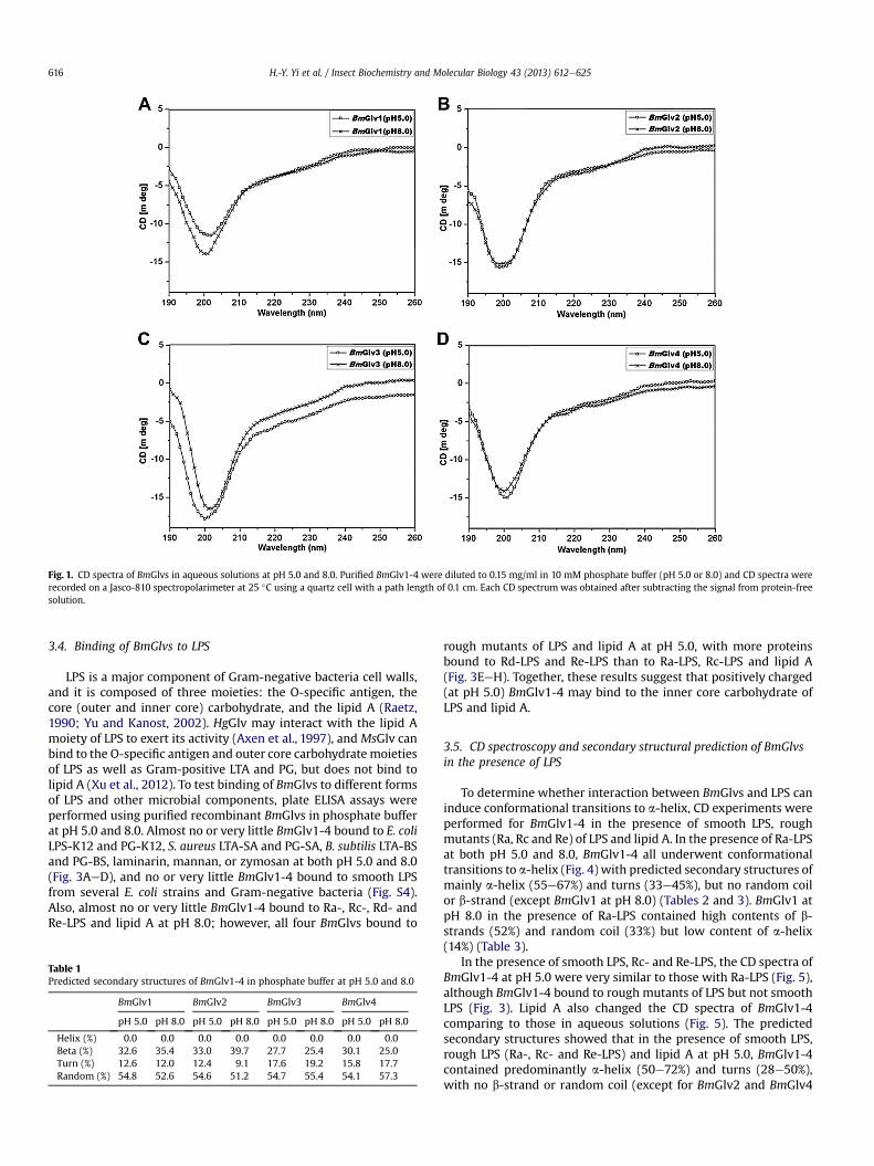
Fig. 1. CD spectra of BmGlvs in aqueous solutions at pH 5.0 and 8.0. Purified BmGlv1-4 were diluted to 0.15 mg/ml in 10 mM phosphate buffer (pH 5.0 or 8.0) and CD spectra wererecorded on a Jasco-810 spectropolarimeter at 25 �C using a quartz cell with a path length of 0.1 cm. Each CD spectrum was obtained after subtracting the signal from protein-freesolution.
H.-Y. Yi et al. / Insect Biochemistry and Molecular Biology 43 (2013) 612e625616
3.4. Binding of BmGlvs to LPS
LPS is a major component of Gram-negative bacteria cell walls,and it is composed of three moieties: the O-specific antigen, thecore (outer and inner core) carbohydrate, and the lipid A (Raetz,1990; Yu and Kanost, 2002). HgGlv may interact with the lipid Amoiety of LPS to exert its activity (Axen et al., 1997), and MsGlv canbind to the O-specific antigen and outer core carbohydratemoietiesof LPS as well as Gram-positive LTA and PG, but does not bind tolipid A (Xu et al., 2012). To test binding of BmGlvs to different formsof LPS and other microbial components, plate ELISA assays wereperformed using purified recombinant BmGlvs in phosphate bufferat pH 5.0 and 8.0. Almost no or very little BmGlv1-4 bound to E. coliLPS-K12 and PG-K12, S. aureus LTA-SA and PG-SA, B. subtilis LTA-BSand PG-BS, laminarin, mannan, or zymosan at both pH 5.0 and 8.0(Fig. 3AeD), and no or very little BmGlv1-4 bound to smooth LPSfrom several E. coli strains and Gram-negative bacteria (Fig. S4).Also, almost no or very little BmGlv1-4 bound to Ra-, Rc-, Rd- andRe-LPS and lipid A at pH 8.0; however, all four BmGlvs bound to
Table 1Predicted secondary structures of BmGlv1-4 in phosphate buffer at pH 5.0 and 8.0
BmGlv1 BmGlv2 BmGlv3 BmGlv4
pH 5.0 pH 8.0 pH 5.0 pH 8.0 pH 5.0 pH 8.0 pH 5.0 pH 8.0
Helix (%) 0.0 0.0 0.0 0.0 0.0 0.0 0.0 0.0Beta (%) 32.6 35.4 33.0 39.7 27.7 25.4 30.1 25.0Turn (%) 12.6 12.0 12.4 9.1 17.6 19.2 15.8 17.7Random (%) 54.8 52.6 54.6 51.2 54.7 55.4 54.1 57.3
rough mutants of LPS and lipid A at pH 5.0, with more proteinsbound to Rd-LPS and Re-LPS than to Ra-LPS, Rc-LPS and lipid A(Fig. 3EeH). Together, these results suggest that positively charged(at pH 5.0) BmGlv1-4 may bind to the inner core carbohydrate ofLPS and lipid A.
3.5. CD spectroscopy and secondary structural prediction of BmGlvsin the presence of LPS
To determine whether interaction between BmGlvs and LPS caninduce conformational transitions to a-helix, CD experiments wereperformed for BmGlv1-4 in the presence of smooth LPS, roughmutants (Ra, Rc and Re) of LPS and lipid A. In the presence of Ra-LPSat both pH 5.0 and 8.0, BmGlv1-4 all underwent conformationaltransitions to a-helix (Fig. 4) with predicted secondary structures ofmainly a-helix (55e67%) and turns (33e45%), but no random coilor b-strand (except BmGlv1 at pH 8.0) (Tables 2 and 3). BmGlv1 atpH 8.0 in the presence of Ra-LPS contained high contents of b-strands (52%) and random coil (33%) but low content of a-helix(14%) (Table 3).
In the presence of smooth LPS, Rc- and Re-LPS, the CD spectra ofBmGlv1-4 at pH 5.0 were very similar to those with Ra-LPS (Fig. 5),although BmGlv1-4 bound to rough mutants of LPS but not smoothLPS (Fig. 3). Lipid A also changed the CD spectra of BmGlv1-4comparing to those in aqueous solutions (Fig. 5). The predictedsecondary structures showed that in the presence of smooth LPS,rough LPS (Ra-, Rc- and Re-LPS) and lipid A at pH 5.0, BmGlv1-4contained predominantly a-helix (50e72%) and turns (28e50%),with no b-strand or random coil (except for BmGlv2 and BmGlv4
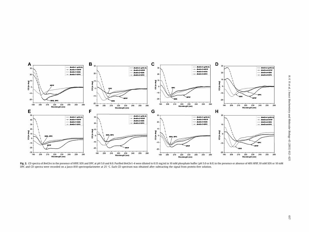
Fig. 2. CD spectra of BmGlvs in the presence of HFIP, SDS and DPC at pH 5.0 and 8.0. Purified BmGlv1-4 were diluted to 0.15 mg/ml in 10 mM phosphate buffer (pH 5.0 or 8.0) in the presence or absence of 40% HFIP, 10 mM SDS or 10 mMDPC and CD spectra were recorded on a Jasco-810 spectropolarimeter at 25 �C. Each CD spectrum was obtained after subtracting the signal from protein-free solution.
H.-Y.Yi
etal./
InsectBiochem
istryand
Molecular
Biology43
(2013)612
e625
617
Table 2Predicted secondary structures of BmGlv1-4 in the presence of HFIP and Ra-LPS at pH 5.0
BmGlv1 BmGlv2 BmGlv3 BmGlv4
pH 5.0 þHFIP þRa-LPS pH 5.0 þHFIP þRa-LPS pH 5.0 þHFIP þRa-LPS pH 5.0 þHFIP þRa-LPS
Helix (%) 0.0 57.6 54.8 0.0 57.6 58.4 0.0 32.7 67.0 0.0 59.5 62.1Beta (%) 32.6 0.0 0.0 33.0 0.0 0.0 27.7 25.9 0.0 30.1 0.0 0.0Turn (%) 12.6 0.0 45.2 12.4 11.0 41.6 17.6 0.0 33.0 15.8 24.5 37.9Random (%) 54.8 42.4 0.0 54.6 31.4 0.0 54.7 41.4 0.0 54.1 16.0 0.0
H.-Y. Yi et al. / Insect Biochemistry and Molecular Biology 43 (2013) 612e625618
with smooth LPS) (Table 4), suggesting that LPS and lipid A caninduce conformational transitions from random coil to a-helix inBmGlv1-4. The concentrations of LPS and lipid A used in the CDexperimentwere 0.15mg/ml (w15 mM for smooth LPS), whichwerehigher than the critical micelle concentration (CMC) of LPS (CMCfor smooth LPS from E. coli 0111:B4 is 1.3e1.6 mM) (Yu et al., 2006).Together, these results suggest that hydrophobic property of LPSand lipid A is required for conformational transitions from randomcoil to a-helix in BmGlv1-4. We also tried the CD experiments withLTA and PG, but could not obtain CD spectra due to poor solubilityof LTA and PG and precipitation problem after mixing LTA and PGwith BmGlvs proteins.
3.6. Activity of BmGlvs against E. coli with rough LPS
Gloverins from different lepidopteran species show activitiesagainst E. coli, Gram-positive bacteria, fungi or a virus (Axen et al.,1997; Mackintosh et al., 1998; Lundstrom et al., 2002; Hwang andKim, 2011; Xu et al., 2012; Moreno-Habel et al., 2012). It is notclear whether the activity of gloverin against different microbes isrelated to pH (net charge), conformational transition to a-helix,and/or binding to microbial surface. We showed above that pHalone cannot cause conformational transitions to a-helix in BmGlvs(Fig. 1 and S2) but pH is important for binding of BmGlvs to roughLPS (Fig. 3), and hydrophobic environment (HFIP, smooth and roughLPS, or lipid A) is required for conformational transitions to a-helixin BmGlvs (Figs. 2, 4 and 5, Tables 2e4, S1 and S2). To determinecorrelations between the activity of BmGlvs and structural confor-mation/binding ability, antimicrobial activity of BmGlv1-4 wastested against several Gram-negative and Gram-positive bacteria aswell as fungi. BmGlv1-4 were inactive against Gram-negative E. coliDH5a and S. marcescens with smooth LPS at pH 5.0 and 8.0, or thefungi S. cerevisiae (at pH 5.0 and 8.0) and C. neoformans (at pH 8.0),but were active against C. neoformans at pH 5.0 (Fig. S5). BmGlv1-4were also inactive against Gram-positive S. aureus, B. subtilis,B. cereus, and B. thuringiensis at both pH 5.0 and 8.0 (Fig. S6). Theseresults were consistent with the binding properties of BmGlv1-4 asthey did not bind to smooth LPS, LTA, PG, laminarin, mannan andzymosan (Fig. 3AeD and S4).
We then tested the activity of BmGlvs against E. coli mutantstrains containing rough LPS since BmGlv1-4 bound to rough LPS atpH 5.0 (Fig. 3EeH). BmGlv1-4 (w10 mM each) at pH 5.0 were activeagainst E. coli D21 (with Ra-LPS), E. coli D21e7 (with Rc-LPS), E. coliD21f1 (with Rd-LPS) and E. coli D21f2 (with Re-LPS) mutant strains,
Table 3Predicted secondary structures of BmGlv1-4 in the presence of HFIP and Ra-LPS at pH 8.
BmGlv1 BmGlv2
pH 8.0 þHFIP þRa-LPS pH 8.0 þHFIP þRa-
Helix (%) 0.0 44.9 14.0 0.0 25.6 63.0Beta (%) 35.4 2.1 52.1 39.7 34.7 0.0Turn (%) 12.0 10.9 0.7 9.1 0.9 37.0Random (%) 52.6 42.0 33.2 51.2 38.8 0.0
but were inactive against these E. coli mutant strains at pH 8.0(Fig. 6). The concentration of BmGlvs used in the bacteria clearanceassay was 10 mM for each protein, which was within the physio-logical concentration of total BmGlvs in E. coli-induced hemolymph(7e9 mM) (Fig. S1C). Using lower concentrations of recombinantBmGlvs at pH 5.0, the activity of BmGlvs against E. coli D21 andE. coli D21f2 was observed at 2 mM for BmGlv1, 2 and 4 and even at0.4 mM for BmGlv1 and 2 (Fig. 7), indicating that the activity ofBmGlvs against E. coli mutant strains is dose-dependent. We alsoconfirmed the activity of BmGlvs using bacterial viability assay.Significantly lower numbers of viable E. coli D21 and E. coli D21f2cells were observed when the bacterial cells were treated withBmGlvs (w10 mM) at pH 5.0 for 2 h (Fig. 8). The number of viableE. coli D21f2 cells was decreased but not significantly after treatingwith BmGlv3 compared to the control. In addition, the activity ofBmGlv3 was significantly lower than that of BmGlv1, 2 or 4 (Fig. 8).Since BmGlv1-4 bound to rough mutants of LPS at pH 5.0 (Fig. 3EeH), together these results suggest that binding of BmGlvs to E. colisurface (via rough LPS) is required for the activity of BmGlvs againstE. coli.
4. Discussion
Gloverin belongs to the glycine-rich AMP family and has beenidentified only in lepidopteran insects so far. Gloverins from mostlepidopteran species are basic (pI w8.3) or highly basic (pI > 9.0)(Xu et al., 2012), but H. virescens gloverin, A. mylitta gloverin 2, andfour B. mori gloverins are slightly acidic to neutral (pI w 5.5e7.2).Even basic gloverins from different lepidopteran species show ac-tivities against different microbes, including E. coli, Gram-positivebacteria, fungi, and a virus (Axen et al., 1997; Mackintosh et al.,1998; Lundstrom et al., 2002; Hwang and Kim, 2011; Xu et al.,2012; Moreno-Habel et al., 2012). B. mori gloverins are also activeagainst E. coli (Kawaoka et al., 2008; Mrinal and Nagaraju, 2008).Thus, it is not clear whether the activity of gloverin is related to itspI or not. HgGlv (pI w8.3) is active against E. coli, and it adoptsrandom coil conformation in solution but has a-helical conforma-tion in the presence of HFIP (Axen et al., 1997).MsGlv (pIw9.3) canbind to LPS but is inactive against E. coli (Xu et al., 2012). Therefore,it is also not clear whether the activity of gloverin is related to itsbinding ability to microbial surface and/or conformational transi-tion to a-helix.
Most linear AMPs are unfolded in aqueous solutions and theycan change to defined structures only after interacting with
0
BmGlv3 BmGlv4
LPS pH 8.0 þHFIP þRa-LPS pH 8.0 þHFIP þRa-LPS
0.0 26.5 65.4 0.0 46.6 63.425.4 30.7 0.0 25.0 0.0 0.019.2 0.0 34.6 17.7 19.5 36.655.4 42.8 0.0 57.3 34.0 0.0
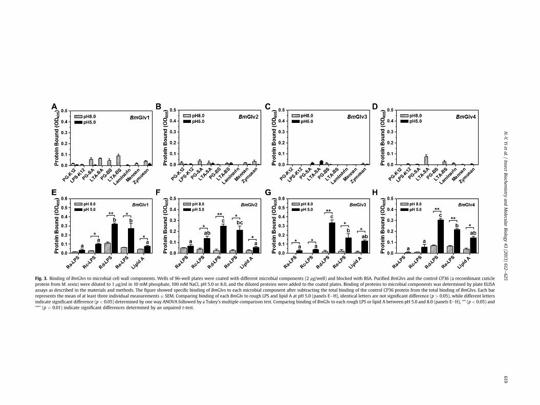
Fig. 3. Binding of BmGlvs to microbial cell wall components. Wells of 96-well plates were coated with different microbial components (2 mg/well) and blocked with BSA. Purified BmGlvs and the control CP36 (a recombinant cuticleprotein from M. sexta) were diluted to 1 mg/ml in 10 mM phosphate, 100 mM NaCl, pH 5.0 or 8.0, and the diluted proteins were added to the coated plates. Binding of proteins to microbial components was determined by plate ELISAassays as described in the materials and methods. The figure showed specific binding of BmGlvs to each microbial component after subtracting the total binding of the control CP36 protein from the total binding of BmGlvs. Each barrepresents the mean of at least three individual measurements � SEM. Comparing binding of each BmGlv to rough LPS and lipid A at pH 5.0 (panels EeH), identical letters are not significant difference (p > 0.05), while different lettersindicate significant difference (p < 0.05) determined by one way ANOVA followed by a Tukey’s multiple comparison test. Comparing binding of BmGlv to each rough LPS or lipid A between pH 5.0 and 8.0 (panels EeH), ‘*’ (p < 0.05) and‘**’ (p < 0.01) indicate significant differences determined by an unpaired t-test.
H.-Y.Yi
etal./
InsectBiochem
istryand
Molecular
Biology43
(2013)612
e625
619
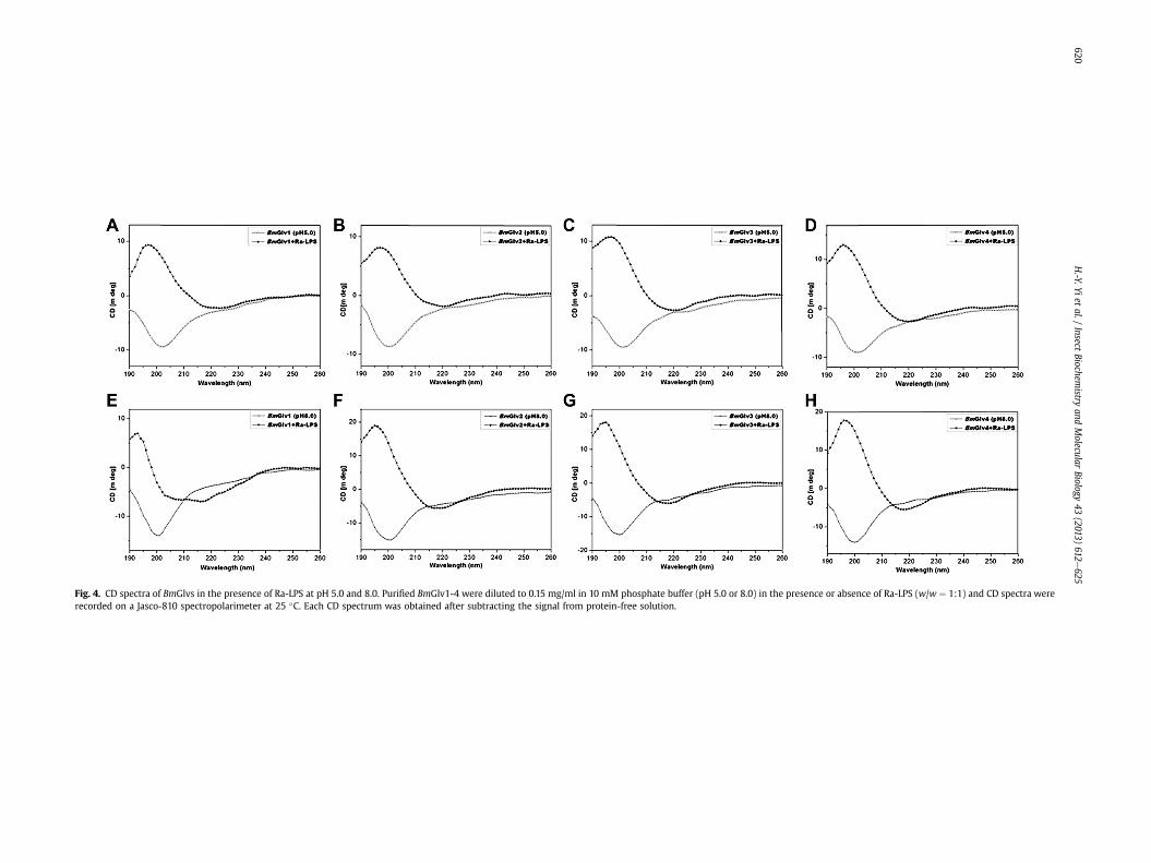
Fig. 4. CD spectra of BmGlvs in the presence of Ra-LPS at pH 5.0 and 8.0. Purified BmGlv1-4 were diluted to 0.15 mg/ml in 10 mM phosphate buffer (pH 5.0 or 8.0) in the presence or absence of Ra-LPS (w/w ¼ 1:1) and CD spectra wererecorded on a Jasco-810 spectropolarimeter at 25 �C. Each CD spectrum was obtained after subtracting the signal from protein-free solution.
H.-Y.Yi
etal./
InsectBiochem
istryand
Molecular
Biology43
(2013)612
e625
620
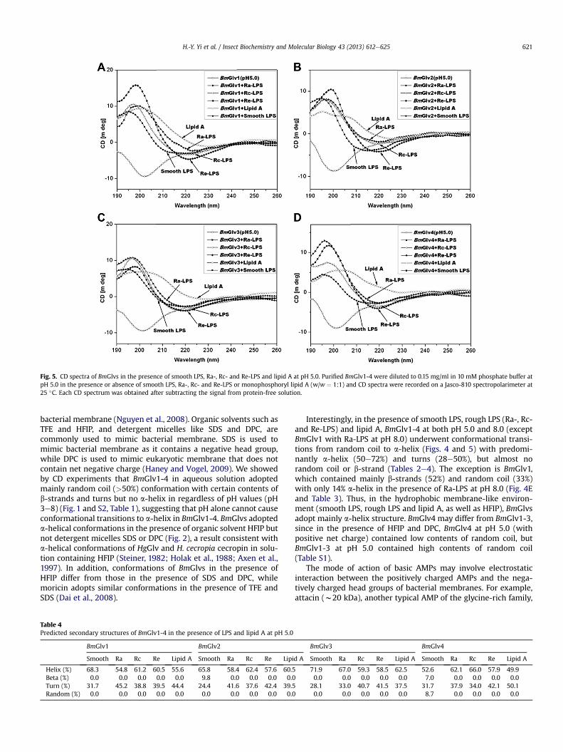
Fig. 5. CD spectra of BmGlvs in the presence of smooth LPS, Ra-, Rc- and Re-LPS and lipid A at pH 5.0. Purified BmGlv1-4 were diluted to 0.15 mg/ml in 10 mM phosphate buffer atpH 5.0 in the presence or absence of smooth LPS, Ra-, Rc- and Re-LPS or monophosphoryl lipid A (w/w ¼ 1:1) and CD spectra were recorded on a Jasco-810 spectropolarimeter at25 �C. Each CD spectrum was obtained after subtracting the signal from protein-free solution.
H.-Y. Yi et al. / Insect Biochemistry and Molecular Biology 43 (2013) 612e625 621
bacterial membrane (Nguyen et al., 2008). Organic solvents such asTFE and HFIP, and detergent micelles like SDS and DPC, arecommonly used to mimic bacterial membrane. SDS is used tomimic bacterial membrane as it contains a negative head group,while DPC is used to mimic eukaryotic membrane that does notcontain net negative charge (Haney and Vogel, 2009). We showedby CD experiments that BmGlv1-4 in aqueous solution adoptedmainly random coil (>50%) conformation with certain contents ofb-strands and turns but no a-helix in regardless of pH values (pH3e8) (Fig. 1 and S2, Table 1), suggesting that pH alone cannot causeconformational transitions to a-helix in BmGlv1-4. BmGlvs adopteda-helical conformations in the presence of organic solvent HFIP butnot detergent micelles SDS or DPC (Fig. 2), a result consistent witha-helical conformations of HgGlv and H. cecropia cecropin in solu-tion containing HFIP (Steiner, 1982; Holak et al., 1988; Axen et al.,1997). In addition, conformations of BmGlvs in the presence ofHFIP differ from those in the presence of SDS and DPC, whilemoricin adopts similar conformations in the presence of TFE andSDS (Dai et al., 2008).
Table 4Predicted secondary structures of BmGlv1-4 in the presence of LPS and lipid A at pH 5.0
BmGlv1 BmGlv2
Smooth Ra Rc Re Lipid A Smooth Ra Rc Re Lipi
Helix (%) 68.3 54.8 61.2 60.5 55.6 65.8 58.4 62.4 57.6 60.5Beta (%) 0.0 0.0 0.0 0.0 0.0 9.8 0.0 0.0 0.0 0.0Turn (%) 31.7 45.2 38.8 39.5 44.4 24.4 41.6 37.6 42.4 39.5Random (%) 0.0 0.0 0.0 0.0 0.0 0.0 0.0 0.0 0.0 0.0
Interestingly, in the presence of smooth LPS, rough LPS (Ra-, Rc-and Re-LPS) and lipid A, BmGlv1-4 at both pH 5.0 and 8.0 (exceptBmGlv1 with Ra-LPS at pH 8.0) underwent conformational transi-tions from random coil to a-helix (Figs. 4 and 5) with predomi-nantly a-helix (50e72%) and turns (28e50%), but almost norandom coil or b-strand (Tables 2e4). The exception is BmGlv1,which contained mainly b-strands (52%) and random coil (33%)with only 14% a-helix in the presence of Ra-LPS at pH 8.0 (Fig. 4Eand Table 3). Thus, in the hydrophobic membrane-like environ-ment (smooth LPS, rough LPS and lipid A, as well as HFIP), BmGlvsadopt mainly a-helix structure. BmGlv4 may differ from BmGlv1-3,since in the presence of HFIP and DPC, BmGlv4 at pH 5.0 (withpositive net charge) contained low contents of random coil, butBmGlv1-3 at pH 5.0 contained high contents of random coil(Table S1).
The mode of action of basic AMPs may involve electrostaticinteraction between the positively charged AMPs and the nega-tively charged head groups of bacterial membranes. For example,attacin (w20 kDa), another typical AMP of the glycine-rich family,
BmGlv3 BmGlv4
d A Smooth Ra Rc Re Lipid A Smooth Ra Rc Re Lipid A
71.9 67.0 59.3 58.5 62.5 52.6 62.1 66.0 57.9 49.90.0 0.0 0.0 0.0 0.0 7.0 0.0 0.0 0.0 0.0
28.1 33.0 40.7 41.5 37.5 31.7 37.9 34.0 42.1 50.10.0 0.0 0.0 0.0 0.0 8.7 0.0 0.0 0.0 0.0
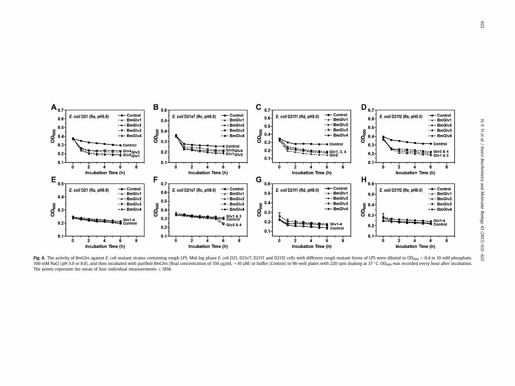
Fig. 6. The activity of BmGlvs against E. coli mutant strains containing rough LPS. Mid-log phase E. coli D21, D21e7, D21f1 and D21f2 cells with different rough mutant forms of LPS were diluted to OD600 ¼ 0.4 in 10 mM phosphate,100 mM NaCl (pH 5.0 or 8.0), and then incubated with purified BmGlvs (final concentration of 150 mg/ml, w10 mM) or buffer (Control) in 96-well plates with 220 rpm shaking at 37 �C. OD600 was recorded every hour after incubation.The points represent the mean of four individual measurements � SEM.
H.-Y.Yi
etal./
InsectBiochem
istryand
Molecular
Biology43
(2013)612
e625
622
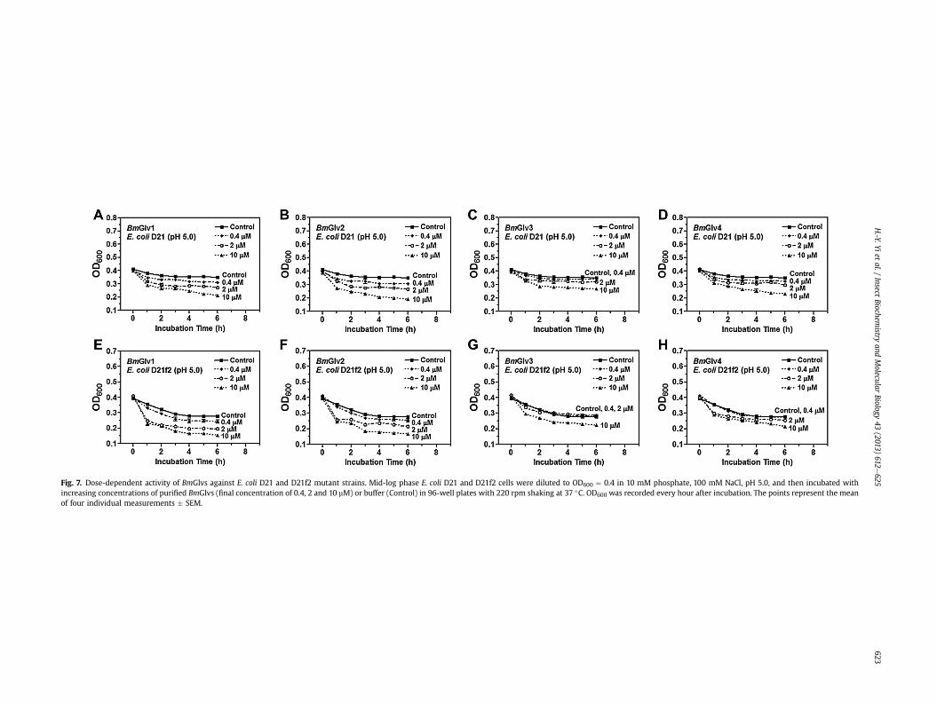
Fig. 7. Dose-dependent activity of BmGlvs against E. coli D21 and D21f2 mutant strains. Mid-log phase E. coli D21 and D21f2 cells were diluted to OD600 ¼ 0.4 in 10 mM phosphate, 100 mM NaCl, pH 5.0, and then incubated withincreasing concentrations of purified BmGlvs (final concentration of 0.4, 2 and 10 mM) or buffer (Control) in 96-well plates with 220 rpm shaking at 37 �C. OD600 was recorded every hour after incubation. The points represent the meanof four individual measurements � SEM.
H.-Y.Yi
etal./
InsectBiochem
istryand
Molecular
Biology43
(2013)612
e625
623

Fig. 8. Viability of E. coli D21 and D21f2 cells after treatment with BmGlvs. Mid-log phase E. coli D21 and D21f2 cells were diluted to OD600 ¼ 0.4 in 10 mM phosphate, 100 mM NaCl,pH 5.0, and then incubated with purified recombinant BmGlvs (final concentration of w10 mM) or buffer (Control) in 96-well plates with 220 rpm shaking at 37 �C for 2 h. Thebacterial cells were serially diluted with 10 mM phosphate, 100 mM NaCl, pH 5.0, and aliquots of diluted bacterial cells were plated on LB-agar plates containing streptomycinsulfate (50 mg/ml) and ampicilin (10 mg/ml). The plates were incubated at 37 �C overnight and the numbers of viable bacterial cells were recorded. The bars represent the mean ofthree individual measurements � SEM. Comparing the control group and BmGlvs treated groups, identical letters are not significant difference (p > 0.05), while different lettersindicate significant difference (p < 0.05) determined by one way ANOVA followed by a Tukey’s multiple comparison test.
H.-Y. Yi et al. / Insect Biochemistry and Molecular Biology 43 (2013) 612e625624
acts on the outer membrane of E. coli by electrostatic binding tolipid A and interaction with the acyl chain of lipid A and phos-pholipids in the outer membrane (Carlsson et al., 1998). Our ELISAassays showed that BmGlvs did not bind to smooth LPS, LTA, PG,laminarin, mannan and zymosan at both pH 5.0 and 8.0 (Fig. 3AeDand S4), but bound to roughmutants of LPS and lipid A at pH 5.0 butnot pH 8.0, with more proteins bound to Rd-LPS and Re-LPS than toRa-LPS, Rc-LPS and lipid A (Fig. 3EeH). These results suggest thatpositively net charge in BmGlvs may contribute to binding ofBmGlvs to rough LPS and lipid A, and BmGlvs may bind to the innercore carbohydrate of LPS. CD experiments showed that in thepresence of smooth LPS, Ra-, Rc- and Re-LPS and lipid A, randomcoils in BmGlvs were converted to a-helix (Figs. 4 and 5), whileBmGlvs did not bind to smooth LPS (Fig. 3AeD and S4). These re-sults suggest that binding to LPS/lipid A is not required butmembrane-like environment is necessary for conformationaltransitions of BmGlvs from random coil to a-helix.
Activity assays showed that BmGlvs were inactive against E. coliDH5a and S. marcescens with smooth LPS, and inactive against twofungal strains and several Gram-positive bacteria, but were activeagainst C. neoformans at pH 5.0 (Figs. S5 and S6). These results areconsistent with the binding properties of BmGlvs as they did notbind to smooth LPS, LTA, PG, laminarin, mannan or zymosan(Fig. 3AeD and S4). However, BmGlvs at pH 5.0 were active againstE. coli mutant strains with rough LPS (Ra-, Rc-, Rd- and Re-LPS)(Fig. 6AeD), a result consistent with HgGlv (Axen et al., 1997), butinactive at pH 8.0 against these E. coli mutant strains (Fig. 6EeH).The activity of recombinant BmGlvs against E. coli mutant strainswas dose-dependent (Fig. 7) and within physiological concentra-tions in hemolymph, as the high concentration of recombinantBmGlvs used in the assay was w10 mM, which was closed to theestimated concentration (7e9 mM) of total native BmGlvs in theE. coli-induced hemolymph of B. mori larvae (Fig. S1C). Comparedthe four BmGlvs, BmGlv1, 2 and 4 had similar high activity againstE. coli, while BmGlv3 had lower activity than BmGlv1, 2 and 3(Fig. 8).
BmGlvs bound to rough mutants of LPS at pH 5.0 but not at pH8.0 (Fig. 3EeH), but rough LPS caused conformational transition ofBmGlvs to a-helix at both pH 5.0 and 8.0 (Figs. 4 and 5). These re-sults suggest that binding of BmGlvs to rough LPS on E. coli surfaceis required for the activity of BmGlvs. BmGlvs at acidic pH wereactive against E. coli, suggesting that they may have higher activityagainst bacteria in the midgut. We believe that conformational
transition from random coil to a-helix in BmGlvs is the key for poreformation on bacterial membrane, but binding of BmGlvs to bac-terial surface via rough LPS is the prerequisite for the activity ofBmGlvs. Thus, whether a gloverin is active against Gram-negativebacteria, Gram-positive bacteria, fungi or viruses may depend onits binding to microbial surface and conformational transition to a-helix. We also tried CD experiments for BmGlvs in the presence ofLTA, PG, laminarin, mannan or zymosan to test interactions be-tween BmGlvs and these microbial components and determinewhether conformational transitions to a-helix occur, but failed toobtain CD spectra due to poor solubility of these microbial com-ponents. In order to better understand the mode of action of glo-verins and the mechanisms of conformational transitions to a-helix, it is necessary to determine three-dimensional structure ofgloverin, which will be one of our future goals.
Acknowledgments
This work was supported, in whole or in part, by National In-stitutes of Health Grant GM066356 (to X.Q.Y.). Support for theworkwas also derived from the “973”National Basic Research Program ofChina (No. 2012CB114600) (to Y.C., X.J.D. andW.Y.Y.) and the Projectof Science and Technology New Star in Zhu Jiang Guangzhou City(2012J2200083) (to W.Y.Y.).
Appendix A. Supplementary data
Supplementary data related to this article can be found at http://dx.doi.org/10.1016/j.ibmb.2013.03.013.
References
Abdel-Latief, M., Hilker, M., 2008. Innate immunity: eggs of Manduca sexta are ableto respond to parasitism by Trichogramma evanescens. Insect Biochem. Mol.Biol. 38, 136e145.
Ando, K., Okada, M., Natori, S., 1987. Purification of sarcotoxin II, antibacterialproteins of Sarcophaga peregrina (flesh fly) larvae. Biochemistry 26, 226e230.
Axen, A., Carlsson, A., Engstrǒm, A., Bennich, H., 1997. Gloverin, an antibacterialprotein from the immune hemolymph of Hyalophora pupae. Eur. J. Biochem.247, 614e619.
Brown, S.E., Howard, A., Kasprzak, A.B., Gordon, K.H., East, P.D., 2009. A peptidomicsstudy reveals the impressive antimicrobial peptide arsenal of the wax mothGalleria mellonella. Insect Biochem. Mol. Biol. 39, 792e800.
Bulet, P., Stocklin, R., 2005. Insect antimicrobial peptides: structures, properties andgene regulation. Protein Pept. Lett. 12, 3e11.
H.-Y. Yi et al. / Insect Biochemistry and Molecular Biology 43 (2013) 612e625 625
Bulet, P., Cociancich, S., Dimarcq, J.L., Lambert, J., Reichhart, J.M., Hoffmann, D.,Hetru, C., Hoffmann, J.A., 1991. Isolation from a coleopteran insect of a novelinducible antibacterial peptide and of new members of the insect defensinfamily. J. Biol. Chem. 266, 24520e24525.
Bulet, P., Hetru, C., Dimarcq, J.L., Hoffmann, D., 1999. Antimicrobial peptides in in-sects: structure and function. Dev. Comp. Immunol. 23, 329e344.
Carlsson, A., Nystrom, T., Cock, H., Bennich, H., 1998. Attacin, an insect immuneprotein-binds LPS and triggers the specific inhibition of bacterial outer mem-brane protein synthesis. Microbiology 144, 2179e2188.
Casteels, P., Ampe, C., Jacobs, F., Tempst, P., 1993. Functional and chemical charac-terization of hymenoptaecin, an antibacterial polypeptide that is infection-inducible in the honeybee (Apis mellifera). J. Biol. Chem. 268, 7044e7054.
Chae, J.-H., Kurokawa, K., So, Y.-I., Hwang, H.O., Kim, M.-S., Park, J.-W., Jo, Y.-H.,Lee, Y.S., Lee, B.L., 2012. Purification and characterization of tenecin 4, a newanti-gram-negative bacterial peptide from the beetle Tenebrio nolitor. Dev.Comp. Immunol. 36, 540e546.
Cheng, T., Zhao, P., Liu, C., Xu, P., Gao, Z., Xia, Q., Xiang, Z., 2006. Structures, regu-latory regions, and inductive expression patterns of antimicrobial peptide genesin the silkworm Bombyx mori. Genomics 87, 356e365.
Cociancich, S., Dupont, A., Hegy, G., Lanot, R., Holder, F., Hetru, C., Hoffmann, J.A.,Bulet, P., 1994. Novel inducible antibacterial peptides from a hemipteran insect,the sap-sucking bug Pyrrhocoris apterus. Biochem. J. 300, 567e575.
Cornet, B., Bonmatin, J.M., Hetru, C., Hoffmann, J.A., Ptak, M., Vovelle, F., 1995.Refined three-dimensional solution structure of insect defensin A. Structure 3,435e448.
Dai, H., Rayaprolu, S., Gong, Y., Huang, R., Prakash, O., Jiang, H., 2008. Solutionstructure, antibacterial activity, and expression profile of Manduca sexta mor-icin. J. Pept. Sci. 10, 1002e1016.
Etebari, K., Palfreyman, R.W., Schlipalius, D., Nielsen, L.K., Glatz, R.V., Asgari, S., 2011.Deep sequencing-based transcriptome analysis of Plutella xylostella larvaeparasitized by Diadegma semiclausum. BMC Genomics 12, 446.
Eum, J.H., Seo, Y.R., Yoe, S.M., Kang, S.W., Han, S.S., 2007. Analysis of the immune-inducible genes of Plutella xylostella using expressed sequence tags and cDNAmicroarray. Dev. Comp. Immunol. 31, 1107e1120.
Gandhe-Archana, S., Arunkumar, K.P., John-Serene, H., Nagaraju, J., 2006. Analysis ofbacteria-challenged wild silkmoth, Antheraea mylitta (Lepidoptera) tran-scriptome reveals potential immune genes. BMC Genomics 184, 1471e2164.
Haney, E.F., Vogel, H.J., 2009. NMR of antimicrobial peptides. ISSN: 0066-4103 65.http://dx.doi.org/10.1016/S0066-4103(08)00201-9.
Hoffmann, J.A., 1995. Innate immunity of insects. Immunology 7, 4e10.Holak, T.A., Engstrom, A., Kraulis, P.J., Lindeberg, G., Bennich, H., Jones, A.,
Gronenborn, A.M., Clore, G.M., 1988. The solution conformation of the anti-bacterial peptide cecropin a: a nuclear magnetic resonance and dynamicalsimulated annealing study. Biochemistry 27, 7620e7629.
Holzwarth, G., Doty, P., 1965. The ultraviolet circular dichroism of polypeptides.J. Am. Chem. Soc. 87, 218e228.
Hultmark, D., Engstrom, A., Andersson, K., Steiner, H., Bennich, H., Boman, H.G.,1983. Insect immunity. attacins, a family of antibacterial proteins from Hyalo-phora cecropia. EMBO J. 2, 571e576.
Hwang, J., Kim, Y., 2011. RNA interference of an antimicrobial peptide, gloverin, ofthe beet armyworm, Spodoptera exigua, enhances susceptibility to Bacillusthuringiensis. J. Invertebr Pathol. 108, 194e200.
Kaneko, Y., Furukawa, S., Tanaka, H., Yamakawa, M., 2007. Expression of antimi-crobial peptide genes encoding Enbocin and Gloverin isoforms in the silkworm,Bombyx mori. Biosci. Biotechnol. Biochem. 71, 2233e2241.
Kawaoka, S., Katsuma, S., Daimon, T., Isono, R., Omuro, N., Mita, K., Shimada, T.,2008. Functional analysis of four Gloverin-like genes in the silkworm, Bombyxmori. Arch. Insect Biochem. Physiol. 67, 87e96.
Lamberty, M., Caille, A., Landon, C., Tassin-Moindrot, S., Hetru, C., Bulet, P.,Vovelle, F., 2001. Solution structures of the antifungal heliomicin and a selectedvariant with both antibacterial and antifungal activities. Biochemistry 40,11995e12003.
Landon, C., Sodano, P., Hetru, C., Hoffmann, J., Ptak, M., 1997. Solution structure of droso-mycin, the first inducible antifungal protein from insects. Protein Sci. 6, 1878e1884.
Laszlo-Otvos, J.R., 2000. Antibacterial peptides isolated from insects. J. Pept. Sci. 6,497e511.
Lavine, M.D., Strand, M.R., 2002. Insect hemocytes and their role in immunity. InsectBiochem. Mol. Biol. 32, 1295e1309.
Lemaitre, B., Hoffmann, J., 2007. The host defense of Drosophila melanogaster. Annu.Rev. Immunol. 25, 697e743.
Lundstrom, A., Liu, G., Kang, D., Berzins, K., Steiner, H., 2002. Trichoplusia ni gloverin,an inducible immune gene encoding an antibacterial insect protein. InsectBiochem. Mol. Biol. 32, 795e801.
Mackintosh, J.A., Gooley, A.A., Karuso, P.H., Beattie, A.J., Jardine, D.R., Veal, D.A., 1998.A gloverin-like antibacterial protein is synthesized in Helicoverpa armigerafollowing bacterial challenge. Dev. Comp. Immunol. 22, 387e399.
Moreno-Habel, D.A., Biglang-Awa, I.M., Dulce, A., Luu, D.D., Garcia, P., Weers, P.M.,Haas-Stapleton, E.J., 2012. Inactivation of the budded virus of Autographa cal-ifornica M nucleopolyhedrovirus by gloverin. J. Invertebr Pathol. 110, 92e101.
Mrinal, N., Nagaraju, J., 2008. Intron loss is associated with gain of function in theevolution of the gloverin family of antibacterial genes in Bombyx mori. J. Biol.Chem. 283, 23376e23387.
Nguyen, L.T., Prenner, E.J., Vogel, H.J., 2008. Structural characterization of antimi-crobial peptides by NMR spectroscopy. In: Webb, Graham A. (Ed.), ModernMagnetic Resonance, pp. 1315e1323.
Raetz, C.R., 1990. Biochemistry of endotoxins. Annu. Rev. Biochem. 59, 129e170.Seitz, V., Clermont, A., Wedde, M., Hummel, M., Vilcinskas, A., Schlatterer, K.,
Podsiadlowski, L., 2003. Identification of immunorelevant genes from greaterwax moth (Galleria mellonella) by a subtractive hybridization approach. Dev.Comp. Immunol. 27, 207e215.
Sideri, M., Tsakas, S., Markoutsa, E., Lampropoulou, M., Marmaras, V.J., 2007. Innateimmunity in insects: surface-associated dopa decarboxylase-dependent path-ways regulate phagocytosis, nodulation and melanization in medfly haemo-cytes. Immunology 123, 528e537.
Silva, P.D., Jouvensal, L., Lamberty, M., Bulet, P., Caille, A., Vovelle, F., 2003. Solutionstructure of termicin, an antimicrobial peptide from the termite Pseudacan-thotermes spiniger. Protein Sci. 12, 438e446.
Silva, J.L., Barbosa, J.F., Bravo, J.P., Souza, E.M., Huergo, L.F., Pedrosa, F.O., Esteves, E.,Daffre, S., Fernandez, M.A., 2010. Induction of a gloverin-like antimicrobialpolypeptide in the sugarcane borer Diatraea saccharalis challenged by septicinjury. Braz. J. Med. Biol. Res. 43, 431e436.
Steiner, H., 1982. Secondary structure of the cecropins: antibacterial peptides fromthe moth Hyalophora cecropia. FEBS Lett. 137, 283e287.
Tiffany, M.L., Krimm, S., 1972. Effect of temprature on the circular dichroism spectraof polypeptides in the extended state. Biopolymers 11, 2309e2316.
Xu, X.X., Zhong, X., Yi, H.Y., Yu, X.Q., 2012.Manduca sexta gloverin binds microbial com-ponents and is active against bacteria and fungi. Dev. Comp. Immunol. 38, 275e284.
Yang, J.T., Wu, C.S., Martinez, H.M., 1986. Calculation of protein conformation fromcircular dichroism. Methods Enzymol. 130, 208e269.
Yu, X.Q., Kanost, M.R., 2000. Immulectin-2, a lipopolysaccharide-specific lectin froman insect, Manduca sexta, is induced in response to gram-negative bacteria.J. Biol. Chem. 275, 37373e37381.
Yu, X.Q., Kanost, M.R., 2002. Binding of hemolin to bacterial lipopolysaccharide andlipoteichoic acid. An immunoglobulin superfamily member from insects as apattern-recognition receptor. Eur. J. Biochem. 269, 1827e1834.
Yu, X.Q., Tracy, M.E., Ling, E., Scholz, F.R., Trenczek, T., 2005. A novel C-typeimmulectin- 3 from Manduca sexta is translocated from hemolymph into thecytoplasm of hemocytes. Insect Biochem. Mol. Biol. 35, 285e295.
Yu, L., Tan, M., Ho, B., Ding, J.L., Wohland, T., 2006. Determination of critical micelleconcentrations and aggregation numbers by fluorescence correlation spec-troscopy: aggregation of a lipopolysaccharide. Anal. Chim. Acta 556, 216e225.
Zhu, Y., Johnson, T.J., Myers, A.A., Kanost, M.R., 2003. Identification by subtractivesuppression hybridization of bacteria-induced genes expressed in Manducasexta fat body. Insect Biochem. Mol. Biol. 33, 541e559.