Embed Size (px)
Citation preview
Innate Immunity: InflammationChapter 6
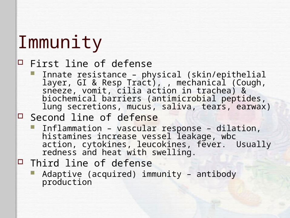
Immunity First line of defense
Innate resistance – physical (skin/epithelial layer, GI & Resp Tract), , mechanical (Cough, sneeze, vomit, cilia action in trachea) & biochemical barriers (antimicrobial peptides, lung secretions, mucus, saliva, tears, earwax)
Second line of defense Inflammation – vascular response – dilation, histamines
increase vessel leakage, wbc action, cytokines, leucokines, fever. Usually redness and heat with swelling.
Third line of defense Adaptive (acquired) immunity – antibody production
First Line of Defense Physical and mechanical barriers
Skin Linings of the gastrointestinal, genitourinary, and
respiratory tracts Sloughing off of cells Coughing and sneezing Flushing Vomiting Mucus and cilia
First Line of Defense Biochemical barriers
Synthesized and secreted saliva, tears, earwax, sweat, and sebum
Antimicrobial peptides Cathelicidins, defensins, and collectins
Normal bacterial flora
Second Line of Defense Inflammatory response
Caused by a variety of materials Infection, mechanical damage, ischemia, nutrient
deprivation, temperature extremes, radiation, etc.
Local manifestations Vascular response
Blood vessel dilation, increased vascular permeability and leakage, white blood cell adherence to the inner walls of the vessels and migration through the vessels
Inflammation Goals
Limit and control the inflammatory process Prevent and limit infection and further damage Interact with components of the adaptive immune
system Prepare the area of injury for healing
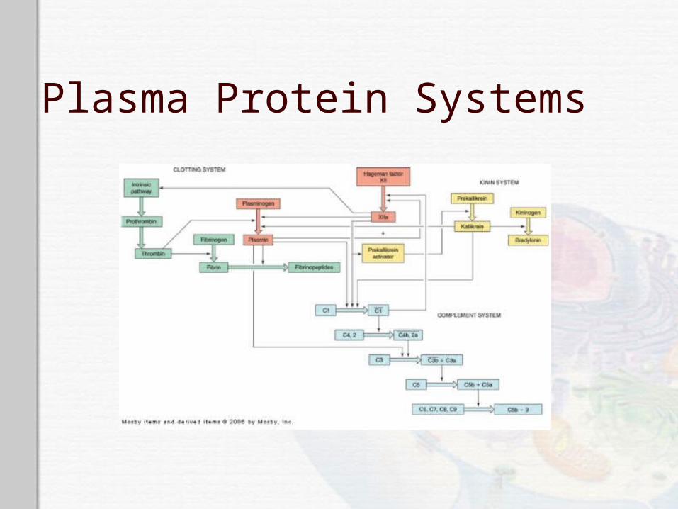
Plasma Protein Systems Protein systems
Complement system Coagulation system Kinin system
All contain inactive enzymes (proenzymes) Sequentially activated
First proenzyme is converted to an active enzyme Substrate of the activated enzyme becomes the next
component in the series
Plasma Protein Systems Complement system
Can destroy pathogens directly Activates or collaborates with every other
component of the inflammatory response Pathways
Classical Lectin Alternative
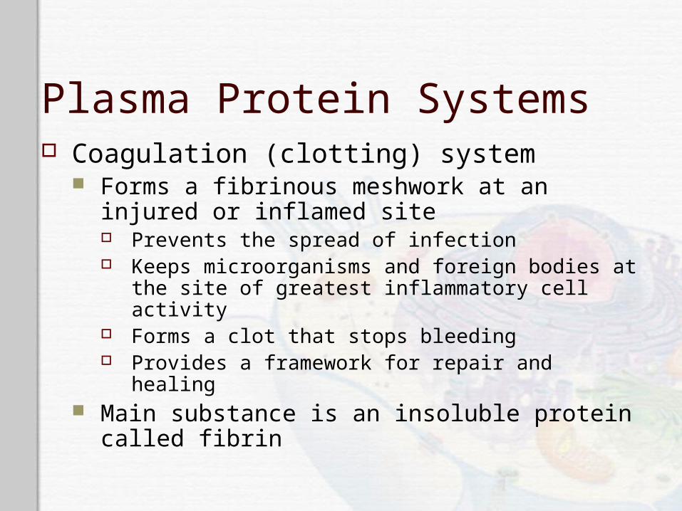
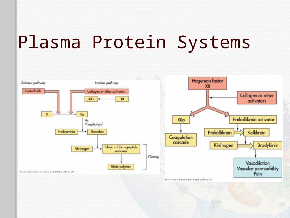
Plasma Protein Systems Coagulation (clotting) system
Forms a fibrinous meshwork at an injured or inflamed site Prevents the spread of infection Keeps microorganisms and foreign bodies at the site
of greatest inflammatory cell activity Forms a clot that stops bleeding Provides a framework for repair and healing
Main substance is an insoluble protein called fibrin
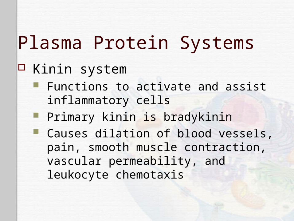
Plasma Protein Systems Kinin system
Functions to activate and assist inflammatory cells
Primary kinin is bradykinin Causes dilation of blood vessels, pain, smooth
muscle contraction, vascular permeability, and leukocyte chemotaxis
Plasma Protein Systems
Plasma Protein Systems
Cellular Mediators of Inflammation Cellular components
Granulocytes, platelets, monocytes, and lymphocytes
Cell surface receptors Pattern recognition receptors (PRRs) Pathogen-associated molecular patterns (PAMPs) Toll-like receptors Complement receptors Scavenger receptors

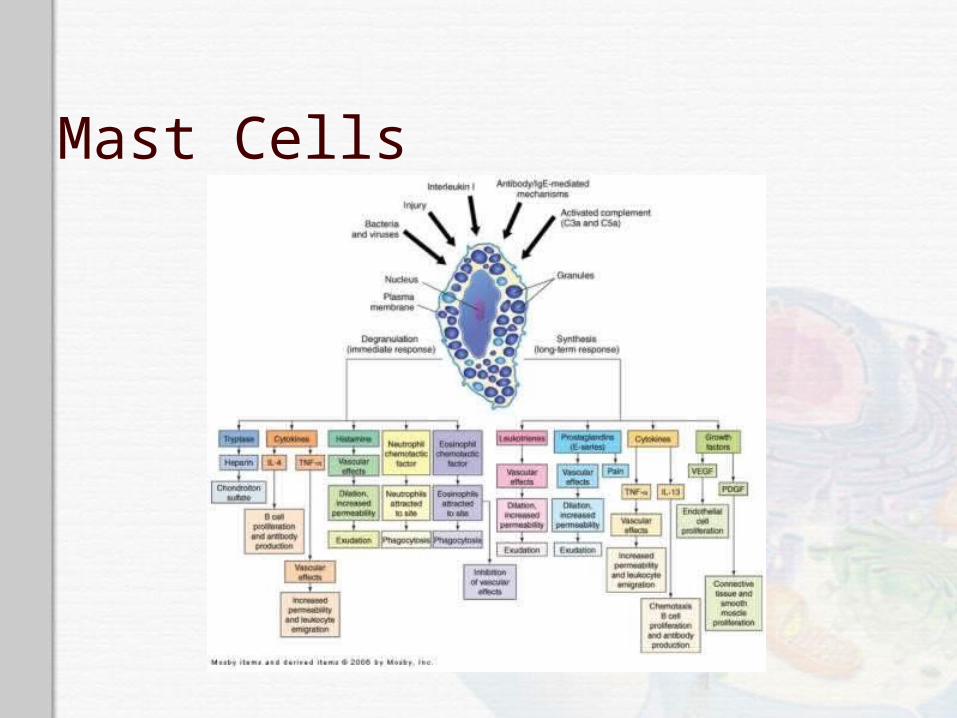
Mast Cells Cellular bags of granules located in the loose
connective tissues close to blood vessels Skin, digestive lining, and respiratory tract
Activation Physical injury, chemical agents, immunologic
processes, and toll-like receptors Chemical release in two ways
Degranulation and synthesis of lipid-derived chemical mediators
Mast Cell Degranulation Histamine
Vasoactive amine that causes temporary, rapid constriction of the large blood vessels and the dilation of the postcapillary venules
Retraction of endothelial cells lining the capillaries
Receptors H1 receptor (proinflammatory)
H2 receptor (anti-inflammatory)
Histamine Receptors
H1 receptor Proinflammatory Present in smooth muscle cells of the bronchi
H2 receptor Anti-inflammatory Present on parietal cells of the stomach mucosa
Induces the secretion of gastric acid
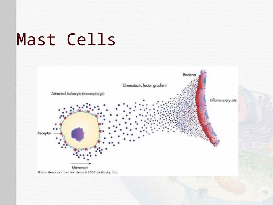
Mast Cell Degranulation Chemotactic factors
Neutrophil chemotactic factor Attracts neutrophils
Eosinophil chemotactic factor of anaphylaxis (ECF-A) Attracts eosinophils
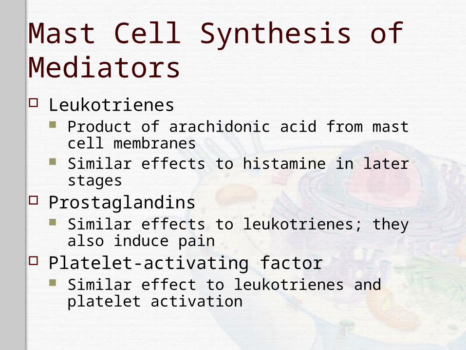
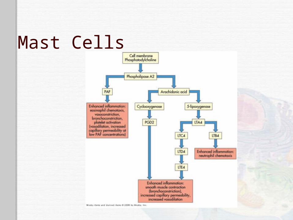
Mast Cell Synthesis of Mediators Leukotrienes
Product of arachidonic acid from mast cell membranes
Similar effects to histamine in later stages Prostaglandins
Similar effects to leukotrienes; they also induce pain
Platelet-activating factor Similar effect to leukotrienes and platelet activation
Mast Cells
Mast Cells
Mast Cells
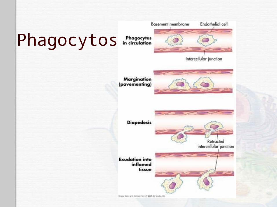
Phagocytosis Process by which a cell ingests and disposes
of foreign material Production of adhesion molecules Margination (pavementing)
Adherence of leukocytes to endothelial cells Diapedesis
Emigration of cells through the endothelial junctions
Phagocytosis
Phagocytosis Steps
Opsonization, recognition, and adherence Engulfment Phagosome formation Fusion with lysosomal granules Destruction of the target
Phagocytes Neutrophils
Also referred to as polymorphonuclear neutrophils (PMNs)
Predominate in early inflammatory responses Ingest bacteria, dead cells, and cellular debris Cells are short lived and become a component of
the purulent exudate
Phagocytes Monocytes and macrophages
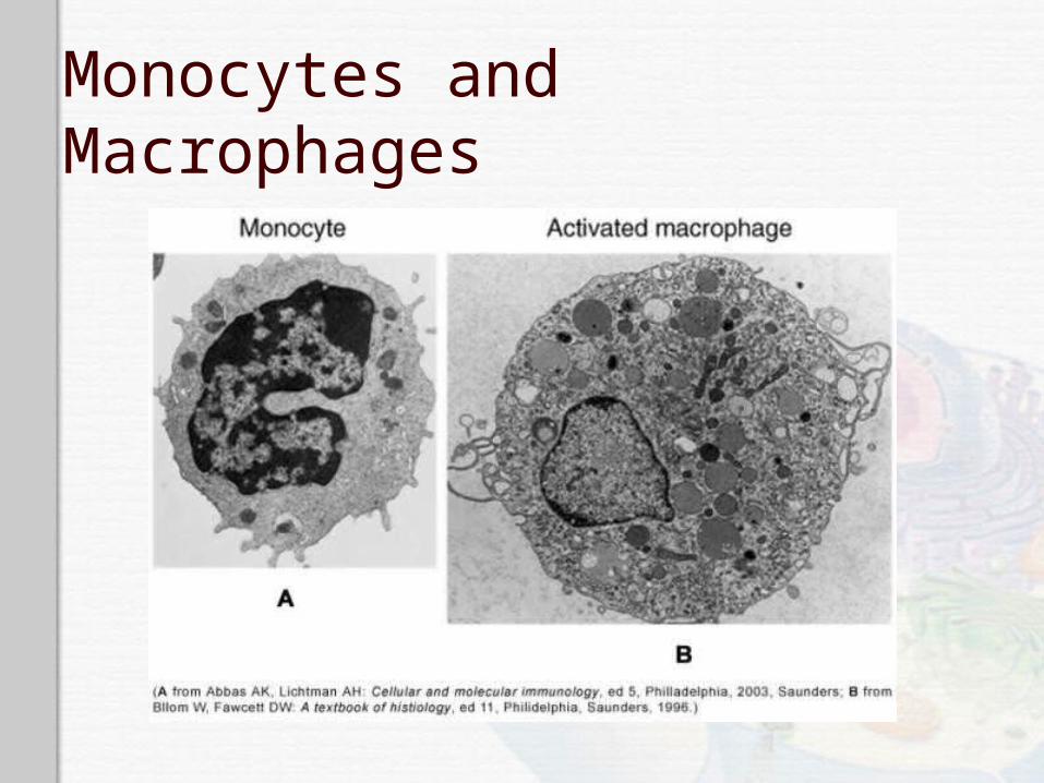
Monocytes are produced in the bone marrow, enter the circulation, and migrate to the inflammatory site, where they develop into macrophages
Macrophages typically arrive at the inflammatory site 3 to 7 days after neutrophils
Macrophage activation results in increased size, plasma membrane area, glucose metabolism, number of lysosomes, and secretory products
Monocytes and Macrophages
Phagocytes Eosinophils
Mildly phagocytic Duties
Defense against parasites and regulation of vascular mediators
Phagocytes Natural killer (NK) cells
Function is to recognize and eliminate cells infected with viruses and some function in eliminating cancer cells
Platelets Activation results in degranulation and interaction
with components of the coagulation system
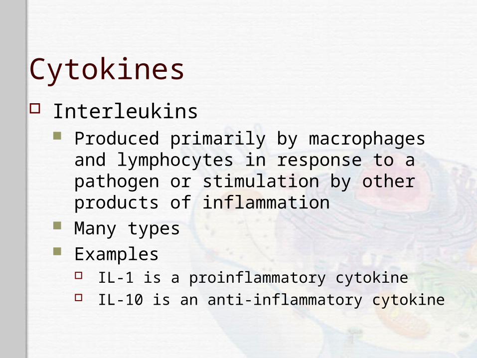
Cytokines Interleukins
Produced primarily by macrophages and lymphocytes in response to a pathogen or stimulation by other products of inflammation
Many types Examples
IL-1 is a proinflammatory cytokine IL-10 is an anti-inflammatory cytokine
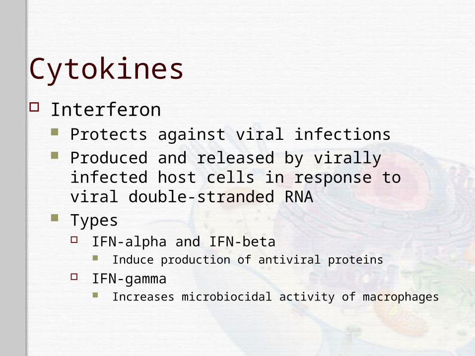
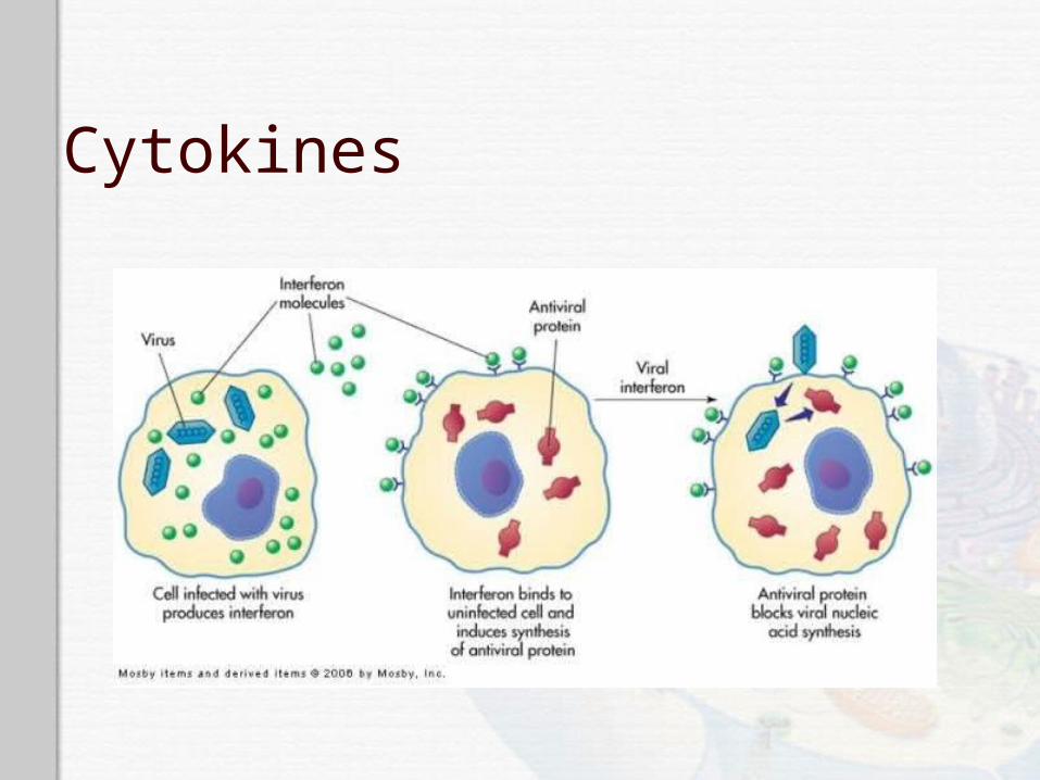
Cytokines Interferon
Protects against viral infections Produced and released by virally infected host
cells in response to viral double-stranded RNA Types
IFN-alpha and IFN-beta Induce production of antiviral proteins
IFN-gamma Increases microbiocidal activity of macrophages
Cytokines
Cytokines Tumor necrosis factor–alpha
Secreted by macrophages in response to PAMP and toll-like receptor recognition Induces fever by acting as an endogenous pyrogen Increases synthesis of inflammatory serum proteins Causes muscle wasting (cachexia) and intravascular
thrombosis

Cytokines
Local Manifestations of Inflammation Results from vascular changes and
corresponding leakage of circulating components into the tissue Heat Redness Swelling Pain
Exudative Fluids Serous exudate
Watery exudate: indicates early inflammation Fibrinous exudate
Thick, clotted exudate: indicates more advanced inflammation
Purulent exudate Pus: indicates a bacterial infection
Hemorrhagic exudate Exudate contains blood: indicates bleeding

Systemic Manifestations of Inflammation Fever
Caused by exogenous and endogenous pyrogens Act directly on the hypothalamus
Leukocytosis Increased numbers of circulating leukocytes
Increased plasma protein synthesis Acute-phase reactants
C-reactive protein, fibrinogen, haptoglobin, amyloid, ceruloplasmin, etc.
Chronic Inflammation Inflammation lasting 2 weeks or longer Often related to an unsuccessful acute
inflammatory response Other causes of chronic inflammation:
High lipid and wax content of a microorganism Ability to survive inside the macrophage Toxins Chemicals, particulate matter, or physical irritants

Chronic Inflammation
Chronic Inflammation Characteristics
Dense infiltration of lymphocytes and macrophages
Granuloma formation Epithelioid cell formation Giant cell formation
Resolution and Repair Regeneration Resolution
Returning injured tissue to the original structure and function
Repair Replacement of destroyed tissue with scar tissue Scar tissue
Composed primarily of collagen to restore the tensile strength of the tissue
Resolution and Repair Débridement
Cleaning up the dissolved clots, microorganisms, erythrocytes, and dead tissue cells
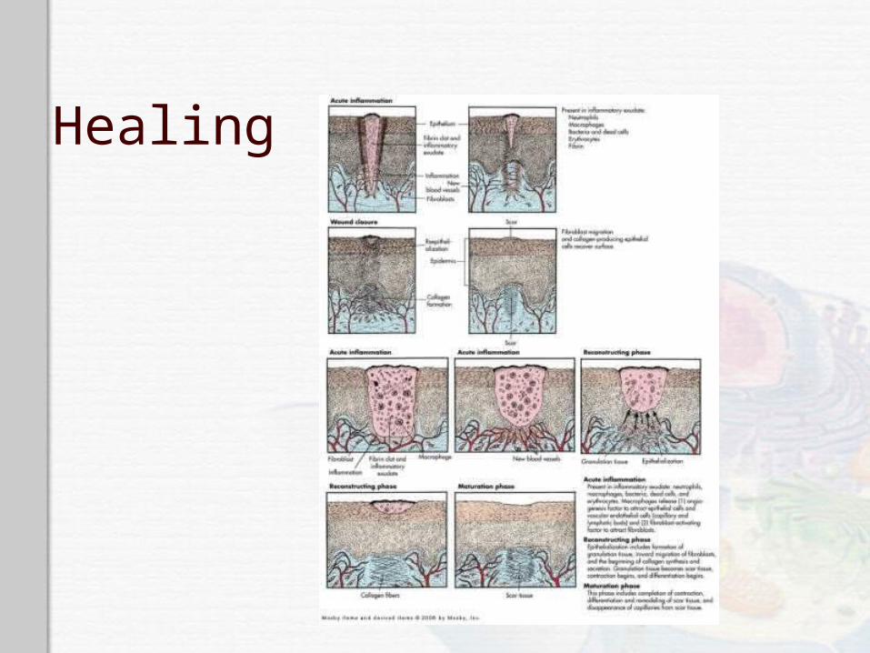
Healing Filling in the wound Sealing the wound (epithelialization) Shrinking the wound (contraction)
Healing Primary intention
Wounds that heal under conditions of minimal tissue loss
Secondary intention Wounds that require a great deal more tissue
replacement Open wound
Healing Reconstructive phase
Fibroblast proliferation Collagen synthesis Epithelialization Contraction
Myofibroblasts
Cellular differentiation

Healing Maturation phase
Continuation of cellular differentiation Scar tissue formation Scar remodeling
Healing

Dysfunctional Wound Healing Dysfunction during inflammatory response
Hemorrhage Fibrous adhesion Infection Excess scar formation Wound sepsis Hypovolemia Hypoproteinemia Anti-inflammatory steroids

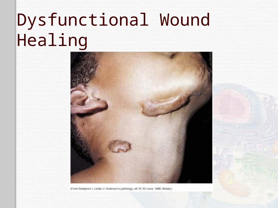
Dysfunctional Wound Healing Dysfunctional during reconstructive phase
Impaired collagen matrix assembly Keloid scar Hypertrophic scar
Impaired epithelialization Anti-inflammatory steroids, hypoxemia, and
nutritional deficiencies
Impaired contraction Contracture
Dysfunctional Wound Healing
Dysfunctional Wound Healing Wound disruption
Dehiscence Wound pulls apart at the suture line
Excessive strain and obesity are causes
Increases risk of wound sepsis
Pediatrics Neonates have transiently depressed
inflammatory and immune function Neutrophils are not capable of efficient
chemotaxis Neonates express complement deficiency Deficient in collectins and collectin-like
proteins
Elderly Impaired inflammation is likely a result of
chronic illness Diabetes, cardiovascular disease, etc.
Chronic medication intake decreases the inflammatory response
Healing response is diminished due to loss of the regenerative ability of the skin
Infections are more common in the elderly