Embed Size (px)
Citation preview
Seediscussions,stats,andauthorprofilesforthispublicationat:https://www.researchgate.net/publication/313265127
Injectableplateletrichfibrin(i-PRF):opportunitiesinregenerativedentistry?
ArticleinClinicalOralInvestigations·February2017
DOI:10.1007/s00784-017-2063-9
CITATIONS
0
READS
625
7authors,including:
Someoftheauthorsofthispublicationarealsoworkingontheserelatedprojects:
InvestigatetheeffectoflowercentrifugationspeedandtimeonPlateletRichFibrin(PRF)
formulationsleadingtotheiradvancedprotocols:A-PRF+andi-PRFViewproject
Anti-bacterialnanostructuresandbiomimeticmaterialsViewproject
RichardJMiron
NovaSoutheasternUniversity
105PUBLICATIONS1,026CITATIONS
SEEPROFILE
YufengZhang
WuhanUniversity
108PUBLICATIONS1,703CITATIONS
SEEPROFILE
ShahramGhanaati
GoetheUniversityofFrankfurt/Main;Universi…
99PUBLICATIONS1,108CITATIONS
SEEPROFILE
JosephChoukroun
Goethe-UniversitätFrankfurtamMain
39PUBLICATIONS1,728CITATIONS
SEEPROFILE
AllcontentfollowingthispagewasuploadedbyRichardJMironon07February2017.
Theuserhasrequestedenhancementofthedownloadedfile.
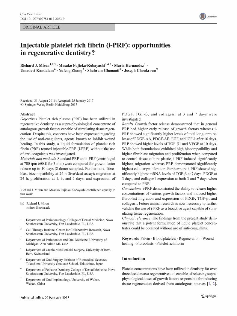
ORIGINAL ARTICLE
Injectable platelet rich fibrin (i-PRF): opportunitiesin regenerative dentistry?
Richard J. Miron1,2,3& Masako Fujioka-Kobayashi1,4,5 & Maria Hernandez1 &
Umadevi Kandalam6& Yufeng Zhang7 & Shahram Ghanaati8 & Joseph Choukroun9
Received: 31 August 2016 /Accepted: 25 January 2017# Springer-Verlag Berlin Heidelberg 2017
AbstractObjectives Platelet rich plasma (PRP) has been utilized inregenerative dentistry as a supra-physiological concentrate ofautologous growth factors capable of stimulating tissue regen-eration. Despite this, concerns have been expressed regardingthe use of anti-coagulants, agents known to inhibit woundhealing. In this study, a liquid formulation of platelet richfibrin (PRF) termed injectable-PRF (i-PRF) without the useof anti-coagulants was investigated.Materials and methods Standard PRP and i-PRF (centrifugedat 700 rpm (60G) for 3 min) were compared for growth factorrelease up to 10 days (8 donor samples). Furthermore, fibro-blast biocompatibility at 24 h (live/dead assay); migration at24 h; proliferation at 1, 3, and 5 days, and expression of
PDGF, TGF-β, and collagen1 at 3 and 7 days wereinvestigated.Results Growth factor release demonstrated that in generalPRP had higher early release of growth factors whereas i-PRF showed significantly higher levels of total long-term re-lease of PDGF-AA, PDGF-AB, EGF, and IGF-1 after 10 days.PRP showed higher levels of TGF-β1 and VEGF at 10 days.While both formulations exhibited high biocompatibility andhigher fibroblast migration and proliferation when comparedto control tissue-culture plastic, i-PRF induced significantlyhighest migration whereas PRP demonstrated significantlyhighest cellular proliferation. Furthermore, i-PRF showed sig-nificantly highest mRNA levels of TGF-β at 7 days, PDGF at3 days, and collagen1 expression at both 3 and 7 days whencompared to PRP.Conclusions i-PRF demonstrated the ability to release higherconcentrations of various growth factors and induced higherfibroblast migration and expression of PDGF, TGF-β, andcollagen1. Future animal research is now necessary to furthervalidate the use of i-PRF as a bioactive agent capable of stim-ulating tissue regeneration.Clinical relevance The findings from the present study dem-onstrate that a potent formulation of liquid platelet concen-trates could be obtained without use of anti-coagulants.
Keywords Fibrin . Blood platelets . Regeneration .Woundhealing . Fibroblasts . Platelet rich fibrin
Introduction
Platelet concentrations have been utilized in dentistry for overthree decades as a regenerative tool capable of releasing supra-physiological doses of growth factors responsible for inducingtissue regeneration derived from autologous sources [1, 2].
Richard J. Miron and Masako Fujioka-Kobayashi contributed equally tothis work.
* Richard J. [email protected]
1 Department of Periodontology, College of Dental Medicine, NovaSoutheastern University, Fort Lauderdale, FL, USA
2 Cell Therapy Institute, Center for Collaborative Research, NovaSoutheastern University, Fort Lauderdale, FL, USA
3 Department of Periodontics and Oral Medicine, University ofMichigan, Ann Arbor, MI, USA
4 Department of Cranio-Maxillofacial Surgery, University of Bern,Bern, Switzerland
5 Department of Oral Surgery, Institute of Biomedical Sciences,Tokushima University Graduate School, Tokushima, Japan
6 Department of Pediatric Dentistry, College of DentalMedicine, NovaSoutheastern University, Fort Lauderdale, FL, USA
7 Department of Oral Implantology, University of Wuhan,Wuhan, China
Clin Oral InvestDOI 10.1007/s00784-017-2063-9
Since then, platelet rich plasma (PRP) was developed havingwidespread use not only in regenerative dentistry but also inmaxillofacial surgery, orthopedic surgery, and esthetic medi-cine [3–7]. Despite this, various concerns have been raisedincluding the use of bovine thrombin and various other anti-coagulants, known suppressors of tissue regeneration [3, 8, 9].For these reasons, platelet rich fibrin (PRF) was developed asa first source of autogenous blood-derived growth factors har-vested without the use of anti-coagulants [10]. PRF thereforeforms a three-dimensional fibrin matrix that may further serveas a scaffold for tissue regeneration by bearing the feature ofacting as a barrier membrane in guided bone and tissue regen-eration (GBR, GTR) procedures while simultaneously hold-ing a number of growth factors responsible for wound healing[11–13].
Over the past decade [10], PRF has gained tremendousmomentum having been utilized for a variety of dental andmedical procedures. In the dental field, PRF has been utilizedfor the treatment of extraction sockets [14–17], gingival reces-sions [18–20], palatal wound closure [21–23], regeneration ofperiodontal defects [24], and hyperplastic gingival tissues[25]. In other medical fields, PRF has been utilized for thesuccessful management of hard-to-heal leg ulcers includingdiabetic foot ulcers, venous leg ulcers, and chronic leg ulcers[26]. Furthermore, hand ulcers, facial soft tissue defects, lap-aroscopic cholecystectomy, deep nasolabial folds, facial de-fects, superficial rhytids, acne scars, lipostructure surgical pro-cedures, chronic rotator cuff tears, and acute traumatic eardrum perforations have also all been treated with PRF [26].Reported advantages include faster wound healing, faster an-giogenesis, low costs, and complete immune-biocompatibility[27–30]. Therefore, while initial and early experiments re-vealed that up to six to eight times higher than normal bloodconcentrations could be reached with PRP, including platelet-derived growth factor (PDGF), vascular endothelial growthfactor (VEGF), and transforming growth factor-beta1(TGF-β1) [31], its use includes anti-coagulants which havealso been reported to inhibit wound healing and research hassince aimed to develop new protocols without the use of anti-coagulants. Very recently, Anitua et al. proposed removinganti-coagulant agents altogether from platelet rich in growthfactor (PRGF) formulations following approximately 20 yearsof research utilizing such therapies [32].
In this study, the development of an injectable formulationof PRF (termed i-PRF) has been pursued with the aim ofdelivering to clinicians an easy to use platelet concentrate inliquid formulation which can be either utilized alone or com-bined easily with various biomaterials. Taking advantage ofslower and shorter centrifugation speeds, a higher presence ofregenerative cells with higher concentrations of growth factorscan be observed when compared to other formulations of PRFutilizing higher centrifugation speeds as highlighted by ourgroup’s previous research [33, 34]. The aim of the present
study was to compare i-PRF to the clinically utilized PRPfor total growth factor release as well as on human gingivalfibroblast cell biocompatibility and activity. Cells were cul-tured with PRP or i-PRF and investigated for cell migration,proliferation, and messenger RNA (mRNA) levels of growthfactors (PDGF and TGF-β1) as well as collagen1 mRNAlevels in vitro.
Materials and methods
Preparation of PRP and i-PRF
Blood samples were collected with the informed consent of 6volunteer donors (12 total samples), and blood was then proc-essed for PRP and i-PRF preparation. All blood samples wereobtained from members of our laboratory between the ages of30 and 60. PRP platelet concentration was prepared via aprotocol as previously described [35]. Briefly, 10 ml of wholeblood with adding 1.0 ml of acid citrate dextrose (ACD) so-lution (Sigma, St. Louis, MO, USA) was centrifuged at1000 rpm (123×g) for 7 min at room temperature. The upperlayer with buffy coat was transferred to a new, sterile centri-fuge tube. The second spin was performed at 3000 rpm(1107×g) for 10 min at room temperature, and finally, thePRP was collected (Duo Centrifuge, Process for PRF, Nice,France). For i-PRF preparation, two tubes of 10 ml of wholeblood without anticoagulant were centrifuged at 700 rpm for3 min (60×g) at room temperature by a Duo Centrifuge(Process for PRF, Nice, France). The upper liquid layer wascollected as i-PRF. Collected PRP and i-PRF samples weretransferred to six-well plastic culture dishes with 5 ml of cul-ture media (DMEM; Gibco, Life technologies, Carlsbad, CA,USA) and processed as further described.
Protein quantification with ELISA
In order to determine the amount of released growth factors(GFs) from PRP and i-PRF at 15 min, 60 min, 8 h, 1 day,3 days, and 10 days, samples were placed into a shakingincubator (60 rotations per minute) at 37 °C to allow for GFrelease into the culture media. At each time point, 5 ml ofculture media was collected, frozen at −20 °C, and replacedwith 5 ml of additional culture media. Protein quantificationwas carried out using ELISA. At designated time points,PDGF-AA (DY221, range = 15.60–1000 pg/ml), PDGF-AB(DY222, range = 15.60–1000 pg/ml), PDGF-BB (DY220,31.20–2000 pg/ml), TGF-β1 (DY240, range = 31.20–2000 pg/ml), VEGF (DY293B, range = 31.20–2000 pg/ml),EGF (DY236, range = 3.91–250 pg/ml), and IGF-1 (DY291,range = 31.20–2000 pg/ml) were quantified using an ELISAkit (DuoSet, R&D Systems, Minneapolis, MN, USA) accord-ing to the manufacturer’s protocol as previously described
Clin Oral Invest
[35]. Absorbance was measured at 450 and 570 nm using aDTX880 microplate reader (Beckman Coulter, Brea, CA,USA) and subtracted at 570 nm from the readings at450 nm. All samples were measured in duplicate, and threeindependent experiments were performed for each plateletconcentrate.
Cell culture
Platelet concentrates including PRP and i-PRF were incubatedfor 3 days in a spinning chamber at 37 °C, and thereafter,conditioned media were collected and utilized in future exper-iments as 20% of the total volume. Human gingival fibroblasts(HGF-1) were purchased from ATCC (Manassas, VA, USA).All cells were detached from tissue culture plastic using0.25% EDTA-trypsin (Gibco) prior to reaching confluency.Cells used for experimental seeding were from passages 4–6as previously described. Cells were cultured in a humidifiedatmosphere at 37 °C in a growth medium consisting ofDMEM (Gibco), 10% fetal bovine serum (FBS; Gibco), and1% antibiotics (Gibco). Media was changed two times perweek. Cells were seeded with 20% conditioned media fromPRP and i-PRF contained within a growth medium at a den-sity of 10,000 cells for cell viability and proliferation experi-ments and 50,000 cells for real-time PCR per well in 24-wellplates. Standard tissue culture plastic (TCP) was utilized as acontrol.
Cell viability assay
At 24 h post cell seeding, cells were evaluated using a live-dead staining assay according to the manufacturer’s protocol(Enzo Life Sciences AG; Lausen, Switzerland). Fluorescentimages were quantified with an inverted fluorescent micro-scope (IX51, OLYMPUS, Tokyo, Japan). Thereafter, cellswere expressed as percentages of live versus dead cells fol-lowing cell culture growth with TCP, PRP, and i-PRF. Theexperiments were performed in triplicate with three indepen-dent experiments performed.
Cell migration assay
The migration assay was performed using 24-well plates andpolyethylene terephthalate cell culture inserts with a pore sizeof 8 μm (Falcon, Corning Inc., Corning, NY, USA). The 20%platelet conditioned media in DMEM containing 10% FBSwere filled into the lower compartment of the wells. Afterbeing starved in DMEM containing 0.5% FBS for 12 h,10,000 resuspended cells were seeded in the upper compart-ment. After 24 h, cells were fixed with 4% formaldehyde for2 min. Thereafter, cells were permeabilized by acetone(Sigma) for 15 min and stained with hematoxylin solution(Sigma) for 20 min. The upper side of the filter membrane
were rinsed and gently wiped by a cotton swab to removethe cell debris. The numbers of cells on the lower side of thefilter were counted under a microscope. The experiments wereperformed in triplicate with three independent experimentsperformed.
Proliferation assay
Cells were quantified using an MTS colorimetric assay(Promega, Madison, WI, USA) at 1, 3, and 5 days for cellproliferation as previously described [36]. At desired timepoints, cells were washed with phosphate-buffered saline(PBS) and quantified using a DTX880 microplate reader.The experiments were performed in triplicate with three inde-pendent experiments performed.
Real-time PCR analysis
Total RNAwas harvested at 3 and 7 days post stimulating forHGF-1 cells to investigate mRNA levels of TGF-β, PDGF,and collagen1a2 (COL1a2). Primer and probe sequences forgenes were fabricated with primer sequences according toTable 1. RNA isolation was performed using High PureRNA Isolation Kit (Roche, Basel, Switzerland). Real-timeRT-PCR was performed using Roche Master Mix and quanti-fied on the StepOne™ plus Real time PCR system fromApplied Biosystems (Foster City, CA, USA). The ΔΔCt meth-od was used to calculate gene expression levels normalized tothe expression of GAPDH. The experiments were performedin triplicate with three independent experiments performed.
Statistical analysis
All experiments were performed in triplicate with three inde-pendent experiments for each condition. Means and standarderrors (SE) were calculated, and data were analyzed for statis-tical significance using one-way analysis for cell viability andmigration assay, two-way analysis of variance for ELISA,proliferation assay, and real-time PCR analysis with Tukeytest (*p values <0.05 were considered significant) byGraphPad Prism 6.0 software (GraphPad Software, Inc., LaJolla, CA, USA).
Results
Growth factor release from PRP and i-PRF
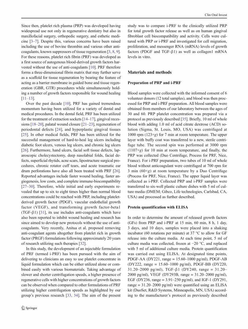
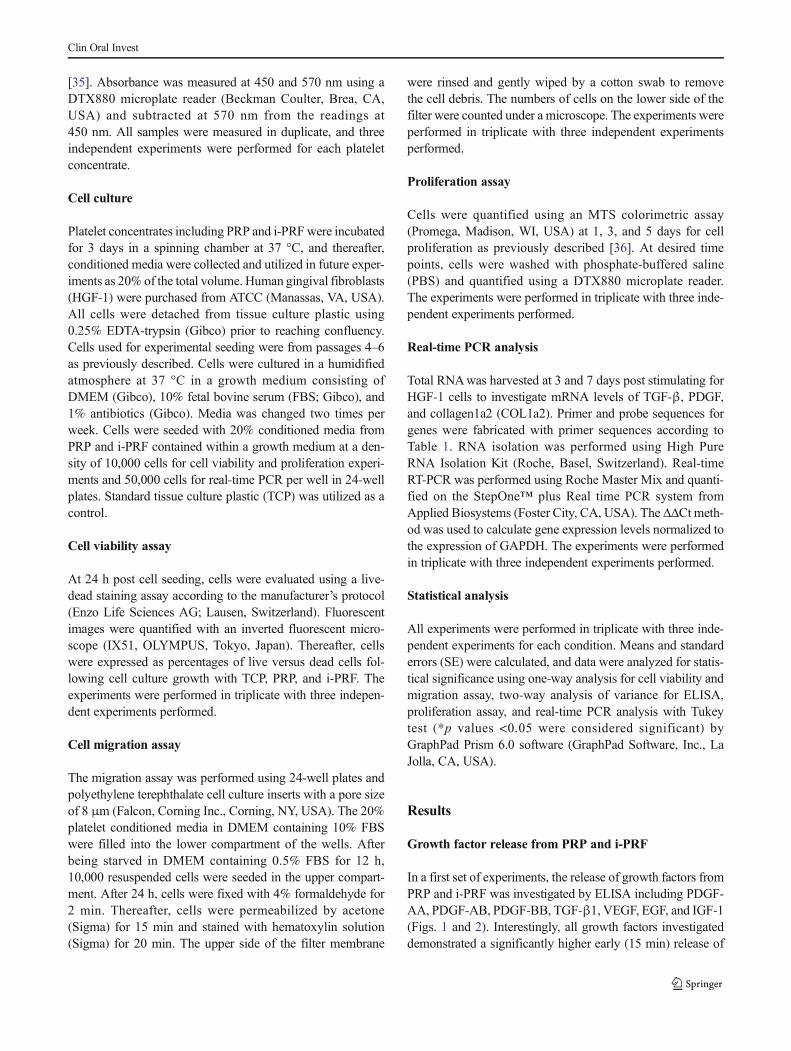
In a first set of experiments, the release of growth factors fromPRP and i-PRF was investigated by ELISA including PDGF-AA, PDGF-AB, PDGF-BB, TGF-β1, VEGF, EGF, and IGF-1(Figs. 1 and 2). Interestingly, all growth factors investigateddemonstrated a significantly higher early (15 min) release of
Clin Oral Invest
growth factor from PRP when compared to i-PRF (with theexception of IGF, Figs. 1 and 2). Thereafter, the total release ofgrowth factors was quantified up to a 10-day period (Figs. 1and 2). It was found that PDGF-AA, PDGF-AB, EGF, andIGF-1 all demonstrated higher total growth factors releasedfrom i-PRF when compared to PRP. Interestingly, however,
total growth factor release of PDGF-BB, VEGF, and TGF-β1were significantly higher in PRP when compared to i-PRF(Figs. 1f and 2b, d). These results point to the fact that variousspin protocols/cell types found in PRP/i-PRF are likely re-sponsible for the variations as discussed later.
Biocompatibility of PRP and i-PRF on human gingivalfibroblasts
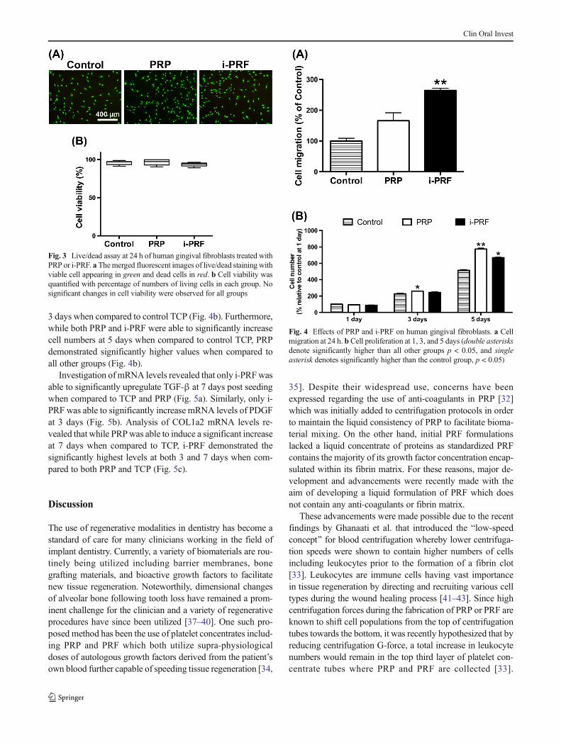
In the first cell culture experiment, the effects of PRP and i-PRF were investigated on cell viability of human gingivalfibroblasts. It was found that both PRP and i-PRF demonstrat-ed excellent cell cytocompatibility by demonstrating most no-tably high living cells (green cells, Fig. 3) with very few ob-servable dead cells (red cells). It was therefore concluded thatboth PRP and i-PRF were fully biocompatible under the pres-ent in vitro cell culture model (Fig. 3).
Fig. 1 ELISA protein quantification at each time point of a PDGF-AA, cPDGF-AB, and e PDGF-BB over a 10-day period. Total accumulatedgrowth factor released over a 10-day period for b PDGF-AA, d PDGF-
AB, and f PDGF-BB (*p < 0.05 signifies significant difference betweengroups, and *p < 0.05 signifies significantly higher than the other group)
Table 1 List of primersequences for real-timePCR
Gene Primer sequence
hTGF-β F actactacgccaaggaggtcac
hTGF-β R tgcttgaacttgtcatagatttcg
hPDGF F cacacctcctcgctgtagtattta
hPDGF R gttatcggtgtaaatgtcatccaa
hCOL1a2 F cccagccaagaactggtatagg
hCOL1a2 R ggctgccagcattgatagtttc
hGAPDH F agccacatcgctcagacac
hGAPDH R gcccaatacgaccaaatcc
Clin Oral Invest
Influence of PRP and i-PRF on human gingival fibroblastactivity
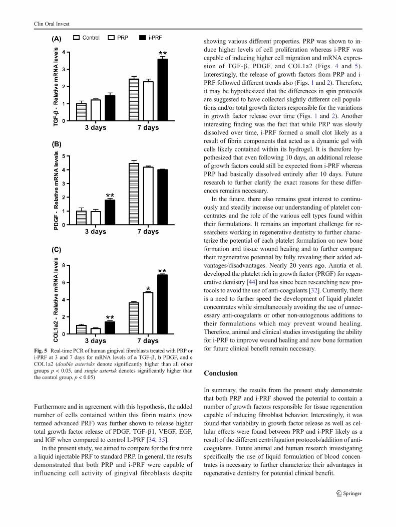
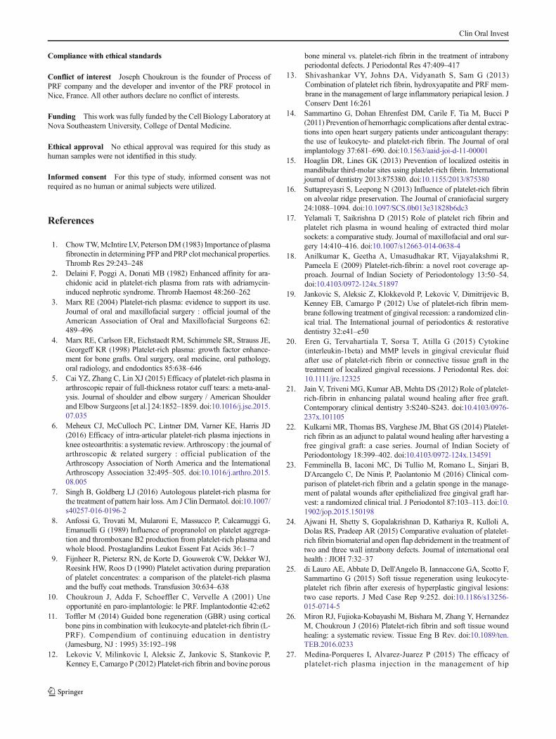
Following confirmation of high cell survival in the biocom-patibility assays, PRP and i-PRF were then investigated ongingival fibroblasts migration, proliferation, and mRNA ex-pression of TGF-β, PDGF, and COL1a2 (Figs. 4 and 5). It
was first found that while PRP induced a 180% increase in cellmigration when compared to control TCP, a significantlyhigher increase was observed when cells were cultured withi-PRF (Fig. 4a). It was found that a 270% increase was ob-served when fibroblasts were cultured with i-PRF, significant-ly higher than all other groups (Fig. 4a). Thereafter, it wasfound that PRP induced significantly higher cell numbers at
Fig. 2 ELISA protein quantification at each time point of a TGF-β1, cVEGF, e EGF, and g IGF-1 over a 10-day period. Total accumulatedgrowth factor released over a 10-day period for b TGF-β1, d VEGF, f
EGF, and h IGF-1 (*p < 0.05 signifies significantly higher than the othergroup)
Clin Oral Invest
3 days when compared to control TCP (Fig. 4b). Furthermore,while both PRP and i-PRF were able to significantly increasecell numbers at 5 days when compared to control TCP, PRPdemonstrated significantly higher values when compared toall other groups (Fig. 4b).
Investigation ofmRNA levels revealed that only i-PRFwasable to significantly upregulate TGF-β at 7 days post seedingwhen compared to TCP and PRP (Fig. 5a). Similarly, only i-PRF was able to significantly increase mRNA levels of PDGFat 3 days (Fig. 5b). Analysis of COL1a2 mRNA levels re-vealed that while PRPwas able to induce a significant increaseat 7 days when compared to TCP, i-PRF demonstrated thesignificantly highest levels at both 3 and 7 days when com-pared to both PRP and TCP (Fig. 5c).
Discussion
The use of regenerative modalities in dentistry has become astandard of care for many clinicians working in the field ofimplant dentistry. Currently, a variety of biomaterials are rou-tinely being utilized including barrier membranes, bonegrafting materials, and bioactive growth factors to facilitatenew tissue regeneration. Noteworthily, dimensional changesof alveolar bone following tooth loss have remained a prom-inent challenge for the clinician and a variety of regenerativeprocedures have since been utilized [37–40]. One such pro-posed method has been the use of platelet concentrates includ-ing PRP and PRF which both utilize supra-physiologicaldoses of autologous growth factors derived from the patient’sown blood further capable of speeding tissue regeneration [34,
35]. Despite their widespread use, concerns have beenexpressed regarding the use of anti-coagulants in PRP [32]which was initially added to centrifugation protocols in orderto maintain the liquid consistency of PRP to facilitate bioma-terial mixing. On the other hand, initial PRF formulationslacked a liquid concentrate of proteins as standardized PRFcontains the majority of its growth factor concentration encap-sulated within its fibrin matrix. For these reasons, major de-velopment and advancements were recently made with theaim of developing a liquid formulation of PRF which doesnot contain any anti-coagulants or fibrin matrix.
These advancements were made possible due to the recentfindings by Ghanaati et al. that introduced the Blow-speedconcept^ for blood centrifugation whereby lower centrifuga-tion speeds were shown to contain higher numbers of cellsincluding leukocytes prior to the formation of a fibrin clot[33]. Leukocytes are immune cells having vast importancein tissue regeneration by directing and recruiting various celltypes during the wound healing process [41–43]. Since highcentrifugation forces during the fabrication of PRP or PRF areknown to shift cell populations from the top of centrifugationtubes towards the bottom, it was recently hypothesized that byreducing centrifugation G-force, a total increase in leukocytenumbers would remain in the top third layer of platelet con-centrate tubes where PRP and PRF are collected [33].
Fig. 4 Effects of PRP and i-PRF on human gingival fibroblasts. a Cellmigration at 24 h. b Cell proliferation at 1, 3, and 5 days (double asterisksdenote significantly higher than all other groups p < 0.05, and singleasterisk denotes significantly higher than the control group, p < 0.05)
Fig. 3 Live/dead assay at 24 h of human gingival fibroblasts treated withPRP or i-PRF. a Themerged fluorescent images of live/dead staining withviable cell appearing in green and dead cells in red. b Cell viability wasquantified with percentage of numbers of living cells in each group. Nosignificant changes in cell viability were observed for all groups
Clin Oral Invest
Furthermore and in agreement with this hypothesis, the addednumber of cells contained within this fibrin matrix (nowtermed advanced PRF) was further shown to release highertotal growth factor release of PDGF, TGF-β1, VEGF, EGF,and IGF when compared to control L-PRF [34, 35].
In the present study, we aimed to compare for the first timea liquid injectable PRF to standard PRP. In general, the resultsdemonstrated that both PRP and i-PRF were capable ofinfluencing cell activity of gingival fibroblasts despite
showing various different properties. PRP was shown to in-duce higher levels of cell proliferation whereas i-PRF wascapable of inducing higher cell migration and mRNA expres-sion of TGF-β, PDGF, and COL1a2 (Figs. 4 and 5).Interestingly, the release of growth factors from PRP and i-PRF followed different trends also (Figs. 1 and 2). Therefore,it may be hypothesized that the differences in spin protocolsare suggested to have collected slightly different cell popula-tions and/or total growth factors responsible for the variationsin growth factor release over time (Figs. 1 and 2). Anotherinteresting finding was the fact that while PRP was slowlydissolved over time, i-PRF formed a small clot likely as aresult of fibrin components that acted as a dynamic gel withcells likely contained within its hydrogel. It is therefore hy-pothesized that even following 10 days, an additional releaseof growth factors could still be expected from i-PRF whereasPRP had basically dissolved entirely after 10 days. Futureresearch to further clarify the exact reasons for these differ-ences remains necessary.
In the future, there also remains great interest to continu-ously and steadily increase our understanding of platelet con-centrates and the role of the various cell types found withintheir formulations. It remains an important challenge for re-searchers working in regenerative dentistry to further charac-terize the potential of each platelet formulation on new boneformation and tissue wound healing and to further comparetheir regenerative potential by fully revealing their added ad-vantages/disadvantages. Nearly 20 years ago, Anutia et al.developed the platelet rich in growth factor (PRGF) for regen-erative dentistry [44] and has since been researching new pro-tocols to avoid the use of anti-coagulants [32]. Currently, thereis a need to further speed the development of liquid plateletconcentrates while simultaneously avoiding the use of unnec-essary anti-coagulants or other non-autogenous additions totheir formulations which may prevent wound healing.Therefore, animal and clinical studies investigating the abilityfor i-PRF to improve wound healing and new bone formationfor future clinical benefit remain necessary.
Conclusion
In summary, the results from the present study demonstratethat both PRP and i-PRF showed the potential to contain anumber of growth factors responsible for tissue regenerationcapable of inducing fibroblast behavior. Interestingly, it wasfound that variability in growth factor release as well as cel-lular effects were found between PRP and i-PRF likely as aresult of the different centrifugation protocols/addition of anti-coagulants. Future animal and human research investigatingspecifically the use of liquid formulation of blood concen-trates is necessary to further characterize their advantages inregenerative dentistry for potential clinical benefit.
Fig. 5 Real-time PCR of human gingival fibroblasts treated with PRP ori-PRF at 3 and 7 days for mRNA levels of a TGF-β, b PDGF, and cCOL1a2 (double asterisks denote significantly higher than all othergroups p < 0.05, and single asterisk denotes significantly higher thanthe control group, p < 0.05)
Clin Oral Invest
Compliance with ethical standards
Conflict of interest Joseph Choukroun is the founder of Process ofPRF company and the developer and inventor of the PRF protocol inNice, France. All other authors declare no conflict of interests.
Funding This work was fully funded by the Cell Biology Laboratory atNova Southeastern University, College of Dental Medicine.
Ethical approval No ethical approval was required for this study ashuman samples were not identified in this study.
Informed consent For this type of study, informed consent was notrequired as no human or animal subjects were utilized.
References
1. Chow TW,McIntire LV, PetersonDM (1983) Importance of plasmafibronectin in determining PFP and PRP clot mechanical properties.Thromb Res 29:243–248
2. Delaini F, Poggi A, Donati MB (1982) Enhanced affinity for ara-chidonic acid in platelet-rich plasma from rats with adriamycin-induced nephrotic syndrome. Thromb Haemost 48:260–262
3. Marx RE (2004) Platelet-rich plasma: evidence to support its use.Journal of oral and maxillofacial surgery : official journal of theAmerican Association of Oral and Maxillofacial Surgeons 62:489–496
4. Marx RE, Carlson ER, Eichstaedt RM, Schimmele SR, Strauss JE,Georgeff KR (1998) Platelet-rich plasma: growth factor enhance-ment for bone grafts. Oral surgery, oral medicine, oral pathology,oral radiology, and endodontics 85:638–646
5. Cai YZ, Zhang C, Lin XJ (2015) Efficacy of platelet-rich plasma inarthroscopic repair of full-thickness rotator cuff tears: a meta-anal-ysis. Journal of shoulder and elbow surgery / American Shoulderand Elbow Surgeons [et al.] 24:1852–1859. doi:10.1016/j.jse.2015.07.035
6. Meheux CJ, McCulloch PC, Lintner DM, Varner KE, Harris JD(2016) Efficacy of intra-articular platelet-rich plasma injections inknee osteoarthritis: a systematic review. Arthroscopy : the journal ofarthroscopic & related surgery : official publication of theArthroscopy Association of North America and the InternationalArthroscopy Association 32:495–505. doi:10.1016/j.arthro.2015.08.005
7. Singh B, Goldberg LJ (2016) Autologous platelet-rich plasma forthe treatment of pattern hair loss. Am J Clin Dermatol. doi:10.1007/s40257-016-0196-2
8. Anfossi G, Trovati M, Mularoni E, Massucco P, Calcamuggi G,Emanuelli G (1989) Influence of propranolol on platelet aggrega-tion and thromboxane B2 production from platelet-rich plasma andwhole blood. Prostaglandins Leukot Essent Fat Acids 36:1–7
9. Fijnheer R, Pietersz RN, de Korte D, Gouwerok CW, Dekker WJ,Reesink HW, Roos D (1990) Platelet activation during preparationof platelet concentrates: a comparison of the platelet-rich plasmaand the buffy coat methods. Transfusion 30:634–638
10. Choukroun J, Adda F, Schoeffler C, Vervelle A (2001) Uneopportunité en paro-implantologie: le PRF. Implantodontie 42:e62
11. Toffler M (2014) Guided bone regeneration (GBR) using corticalbone pins in combination with leukocyte-and platelet-rich fibrin (L-PRF). Compendium of continuing education in dentistry(Jamesburg, NJ : 1995) 35:192–198
12. Lekovic V, Milinkovic I, Aleksic Z, Jankovic S, Stankovic P,Kenney E, Camargo P (2012) Platelet-rich fibrin and bovine porous
bone mineral vs. platelet-rich fibrin in the treatment of intrabonyperiodontal defects. J Periodontal Res 47:409–417
13. Shivashankar VY, Johns DA, Vidyanath S, Sam G (2013)Combination of platelet rich fibrin, hydroxyapatite and PRF mem-brane in the management of large inflammatory periapical lesion. JConserv Dent 16:261
14. Sammartino G, Dohan Ehrenfest DM, Carile F, Tia M, Bucci P(2011) Prevention of hemorrhagic complications after dental extrac-tions into open heart surgery patients under anticoagulant therapy:the use of leukocyte- and platelet-rich fibrin. The Journal of oralimplantology 37:681–690. doi:10.1563/aaid-joi-d-11-00001
15. Hoaglin DR, Lines GK (2013) Prevention of localized osteitis inmandibular third-molar sites using platelet-rich fibrin. Internationaljournal of dentistry 2013:875380. doi:10.1155/2013/875380
16. Suttapreyasri S, Leepong N (2013) Influence of platelet-rich fibrinon alveolar ridge preservation. The Journal of craniofacial surgery24:1088–1094. doi:10.1097/SCS.0b013e31828b6dc3
17. Yelamali T, Saikrishna D (2015) Role of platelet rich fibrin andplatelet rich plasma in wound healing of extracted third molarsockets: a comparative study. Journal of maxillofacial and oral sur-gery 14:410–416. doi:10.1007/s12663-014-0638-4
18. Anilkumar K, Geetha A, Umasudhakar RT, Vijayalakshmi R,Pameela E (2009) Platelet-rich-fibrin: a novel root coverage ap-proach. Journal of Indian Society of Periodontology 13:50–54.doi:10.4103/0972-124x.51897
19. Jankovic S, Aleksic Z, Klokkevold P, Lekovic V, Dimitrijevic B,Kenney EB, Camargo P (2012) Use of platelet-rich fibrin mem-brane following treatment of gingival recession: a randomized clin-ical trial. The International journal of periodontics & restorativedentistry 32:e41–e50
20. Eren G, Tervahartiala T, Sorsa T, Atilla G (2015) Cytokine(interleukin-1beta) and MMP levels in gingival crevicular fluidafter use of platelet-rich fibrin or connective tissue graft in thetreatment of localized gingival recessions. J Periodontal Res. doi:10.1111/jre.12325
21. Jain V, Triveni MG, Kumar AB, Mehta DS (2012) Role of platelet-rich-fibrin in enhancing palatal wound healing after free graft.Contemporary clinical dentistry 3:S240–S243. doi:10.4103/0976-237x.101105
22. Kulkarni MR, Thomas BS, Varghese JM, Bhat GS (2014) Platelet-rich fibrin as an adjunct to palatal wound healing after harvesting afree gingival graft: a case series. Journal of Indian Society ofPeriodontology 18:399–402. doi:10.4103/0972-124x.134591
23. Femminella B, Iaconi MC, Di Tullio M, Romano L, Sinjari B,D'Arcangelo C, De Ninis P, Paolantonio M (2016) Clinical com-parison of platelet-rich fibrin and a gelatin sponge in the manage-ment of palatal wounds after epithelialized free gingival graft har-vest: a randomized clinical trial. J Periodontol 87:103–113. doi:10.1902/jop.2015.150198
24. Ajwani H, Shetty S, Gopalakrishnan D, Kathariya R, Kulloli A,Dolas RS, Pradeep AR (2015) Comparative evaluation of platelet-rich fibrin biomaterial and open flap debridement in the treatment oftwo and three wall intrabony defects. Journal of international oralhealth : JIOH 7:32–37
25. di Lauro AE, Abbate D, Dell'Angelo B, Iannaccone GA, Scotto F,Sammartino G (2015) Soft tissue regeneration using leukocyte-platelet rich fibrin after exeresis of hyperplastic gingival lesions:two case reports. J Med Case Rep 9:252. doi:10.1186/s13256-015-0714-5
26. Miron RJ, Fujioka-Kobayashi M, Bishara M, Zhang Y, HernandezM, Choukroun J (2016) Platelet-rich fibrin and soft tissue woundhealing: a systematic review. Tissue Eng B Rev. doi:10.1089/ten.TEB.2016.0233
27. Medina-Porqueres I, Alvarez-Juarez P (2015) The efficacy ofplatelet-rich plasma injection in the management of hip
Clin Oral Invest
osteoarthritis: a systematic review protocol. Musculoskeletal care.doi:10.1002/msc.1115
28. Salamanna F, Veronesi F, MaglioM, Della Bella E (2015) New andemerging strategies in platelet-rich plasma application in musculo-skeletal regenerative procedures. general overview on still openquestions and outlook 2015:846045. doi:10.1155/2015/846045
29. Albanese A, Licata ME, Polizzi B, Campisi G (2013) Platelet-richplasma (PRP) in dental and oral surgery: from the wound healing tobone regeneration. Immunity & ageing : I & A 10:23. doi:10.1186/1742-4933-10-23
30. Panda S, Doraiswamy J,Malaiappan S, Varghese SS, Del FabbroM(2014) Additive effect of autologous platelet concentrates in treat-ment of intrabony defects: a systematic review and meta-analysis. JInvestig Clin Dent. doi:10.1111/jicd.12117
31. Peerbooms JC, van Laar W, Faber F, Schuller HM, van der HoevenH, Gosens T (2010) Use of platelet rich plasma to treat plantarfasciitis: design of a multi centre randomized controlled trial.BMC Musculoskelet Disord 11:69–69. doi:10.1186/1471-2474-11-69
32. Anitua E, Prado R, TroyaM, ZalduendoM, de la FuenteM, PinoA,Muruzabal F, Orive G (2016) Implementation of a more physiolog-ical plasma rich in growth factor (PRGF) protocol: anticoagulantremoval and reduction in activator concentration. Platelets 27:459–466. doi:10.3109/09537104.2016.1143921
33. Ghanaati S, Booms P, Orlowska A, Kubesch A, Lorenz J,Rutkowski J, Landes C, Sader R, Kirkpatrick C, Choukroun J(2014) Advanced platelet-rich fibrin: a new concept for cell-basedtissue engineering by means of inflammatory cells. The Journal oforal implantology 40:679–689. doi:10.1563/aaid-joi-D-14-00138
34. Fujioka-Kobayashi M, Miron RJ, Hernandez M, Kandalam U,Zhang Y, Choukroun J (2016) Optimized platelet rich fibrin withthe low speed concept: growth factor release, biocompatibility andcellular response. J Periodontol:1–17. doi:10.1902/jop.2016.160443
35. Kobayashi E, Fluckiger L, Fujioka-Kobayashi M, Sawada K,Sculean A, Schaller B, Miron RJ (2016) Comparative release of
growth factors from PRP, PRF, and advanced-PRF. Clinical oralinvestigations. doi:10.1007/s00784-016-1719-1
36. Miron RJ, Bosshardt DD, Hedbom E, ZhangY, Haenni B, Buser D,Sculean A (2012) Adsorption of enamel matrix proteins to abovine-derived bone grafting material and its regulation of celladhesion, proliferation, and differentiation. J Periodontol 83:936–947. doi:10.1902/jop.2011.110480
37. Horowitz R, Holtzclaw D, Rosen PS (2012) A review on alveolarridge preservation following tooth extraction. The journal ofevidence-based dental practice 12:149–160. doi:10.1016/s1532-3382(12)70029-5
38. Lee CT, Chiu TS, Chuang SK, Tarnow D, Stoupel J (2014)Alterations of the bone dimension following immediate implantplacement into extraction socket: systematic review and meta-anal-ysis. J Clin Periodontol 41:914–926. doi:10.1111/jcpe.12276
39. Morjaria KR, Wilson R, Palmer RM (2014) Bone healing aftertooth extraction with or without an intervention: a systematic re-view of randomized controlled trials. Clin Implant Dent Relat Res16:1–20. doi:10.1111/j.1708-8208.2012.00450.x
40. Tan WL, Wong TL, Wong MC, Lang NP (2012) A systematicreview of post-extractional alveolar hard and soft tissue dimension-al changes in humans. Clin Oral Implants Res 23(Suppl 5):1–21.doi:10.1111/j.1600-0501.2011.02375.x
41. Bielecki T, Dohan Ehrenfest DM, Everts PA,Wiczkowski A (2012)The role of leukocytes from L-PRP/L-PRF in wound healing andimmune defense: new perspectives. Curr Pharm Biotechnol 13:1153–1162
42. Martin P (1997) Wound healing–aiming for perfect skin regenera-tion. Science 276:75–81
43. Barrick B, Campbell EJ, Owen CA (1999) Leukocyte proteinasesin wound healing: roles in physiologic and pathologic processes.Wound Repair Regen 7:410–422
44. Anitua E (1999) Plasma rich in growth factors: preliminary resultsof use in the preparation of future sites for implants. Int J OralMaxillofac Implants 14:529–535
Clin Oral Invest
View publication statsView publication stats