Embed Size (px)
Citation preview

NE40CH24-Vogels ARI 1 June 2017 12:42
RE V I E W
S
IN
AD V A
NC
E
Inhibitory Plasticity: Balance,Control, and CodependenceGuillaume Hennequin,1,* Everton J. Agnes,2,*and Tim P. Vogels2
1Computational and Biological Learning Lab, Department of Engineering, University ofCambridge, Cambridge CB2 3EJ, United Kingdom2Centre for Neural Circuits and Behaviour, Department of Physiology, Anatomy and Genetics,University of Oxford, Oxford OX1 3SR, United Kingdom
Annu. Rev. Neurosci. 2017. 40:557–79
The Annual Review of Neuroscience is online atneuro.annualreviews.org
https://doi.org/10.1146/annurev-neuro-072116-031005
Copyright c© 2017 by Annual Reviews.All rights reserved
∗These authors contributed equally to this review.
Keywords
inhibition, GABA, synaptic plasticity, network dynamics, feedback control,modeling
Abstract
Inhibitory neurons, although relatively few in number, exert powerful con-trol over brain circuits. They stabilize network activity in the face of strongfeedback excitation and actively engage in computations. Recent studies re-veal the importance of a precise balance of excitation and inhibition in neuralcircuits, which often requires exquisite fine-tuning of inhibitory connections.We review inhibitory synaptic plasticity and its roles in shaping both feedfor-ward and feedback control. We discuss the necessity of complex, codepen-dent plasticity mechanisms to build nontrivial, functioning networks, and weend by summarizing experimental evidence of such interactions.
557
NE40CH24-Vogels ARI 1 June 2017 12:42
GABA:γ -aminobutyric acid,an inhibitoryneurotransmitter
Codependence:an umbrella term thatdescribes helpingrelationships betweensynapse types in whichactivity in one typesupports or enablesplasticity in the other
Contents
1. INTRODUCTION . . . . . . . . . . . . . . . . . . . . . . . . . . . . . . . . . . . . . . . . . . . . . . . . . . . . . . . . . . . . 5582. INHIBITORY SYNAPTIC PLASTICITY AND ITS SPIKE-TIMING
DEPENDENCE. . . . . . . . . . . . . . . . . . . . . . . . . . . . . . . . . . . . . . . . . . . . . . . . . . . . . . . . . . . . . . . 5593. GLOBAL NETWORK EFFECTS OF INHIBITORY SYNAPTIC
PLASTICITY ON THE BALANCE OF EXCITATION AND INHIBITION . . 5604. INHIBITORY SYNAPTIC PLASTICITY: A KEY INGREDIENT IN ROBUST
COMPUTATIONS. . . . . . . . . . . . . . . . . . . . . . . . . . . . . . . . . . . . . . . . . . . . . . . . . . . . . . . . . . . . 5635. INHIBITORY MEMORIES . . . . . . . . . . . . . . . . . . . . . . . . . . . . . . . . . . . . . . . . . . . . . . . . . . . . 564
5.1. Memory Engrams . . . . . . . . . . . . . . . . . . . . . . . . . . . . . . . . . . . . . . . . . . . . . . . . . . . . . . . . . . 5655.2. Memories That Involve Inhibition. . . . . . . . . . . . . . . . . . . . . . . . . . . . . . . . . . . . . . . . . . . 5665.3. Beyond the Engram . . . . . . . . . . . . . . . . . . . . . . . . . . . . . . . . . . . . . . . . . . . . . . . . . . . . . . . . 568
6. NEGATIVE FEEDBACK CONTROL . . . . . . . . . . . . . . . . . . . . . . . . . . . . . . . . . . . . . . . . . 5686.1. Amplifying Networks . . . . . . . . . . . . . . . . . . . . . . . . . . . . . . . . . . . . . . . . . . . . . . . . . . . . . . . 5686.2. Short-Term Memory Networks . . . . . . . . . . . . . . . . . . . . . . . . . . . . . . . . . . . . . . . . . . . . . 5706.3. Stabilized Supralinear Networks. . . . . . . . . . . . . . . . . . . . . . . . . . . . . . . . . . . . . . . . . . . . . 5716.4. Inhibitory Plasticity and Optimal Feedback Control . . . . . . . . . . . . . . . . . . . . . . . . . . 571
7. CODEPENDENT PLASTICITY . . . . . . . . . . . . . . . . . . . . . . . . . . . . . . . . . . . . . . . . . . . . . . 5718. DISCUSSION. . . . . . . . . . . . . . . . . . . . . . . . . . . . . . . . . . . . . . . . . . . . . . . . . . . . . . . . . . . . . . . . . . 573
1. INTRODUCTION
Learning relies on stereotypical plasticity rules to adjust distinct synaptic connections. Until re-cently, both memories and signaling pathways were thought to be established predominantlythrough the modification of excitatory synapses, creating directionally or recurrently wired ex-citatory cell assemblies that carry out most of the processing work. Such excitatory recurrencehas a natural tendency toward catastrophic runaway activity, which calls for additional layers offlexible control via negative feedback. At the circuit level, negative feedback is mediated mainlyby GABAergic interneurons and their plastic synapses, and it has emerged as a major mechanismto stabilize and shape the functionality of neuronal dynamics.
In this article, we discuss how such negative feedback control can be learned by inhibitorysynaptic plasticity (ISP). We begin with a review of experimental studies of GABAergic spike-timing-dependent rules, which—due to the technical difficulty of probing the activity of the diverseminority of inhibitory neurons—remain less well characterized than their excitatory counterparts.The links between the learning rules, the resulting shape of inhibitory architectures, and theirfunction remain opaque.
In the next sections, we describe how modelers, confronted with such heterogeneous andsometimes perplexing experimental observations, have used tentative, ad hoc, and at times ex-perimentally unsubstantiated learning rules, often to impose a certain function on the networkdynamics by, for instance, controlling the balance of excitation and inhibition (E/I balance). In-deed, E/I balance has emerged as a staple of neuronal processing, and ISP is thus an ideal controlmechanism to stabilize intricate microprocessing pathways and memories.
We end with the argument that much insight into the mechanics and function of inhibitoryplasticity can be gleaned from the rich literature of control theory. We review network models inwhich inhibitory feedback is critical and relies on known control algorithms. The complexity of
558 Hennequin · Agnes · Vogels

NE40CH24-Vogels ARI 1 June 2017 12:42
these algorithms makes their biological plausibility questionable, at least if their implementationmust remain limited to modifications based strictly on simple forms of pre- and postsynapticevents. Interestingly, recent experimental studies suggest that we might have unduly confinedmodels of learning to local environments of single types of synapses: Diverse synaptic plasticitymechanisms often act in concert with one another, and their efficacy is tightly controlled by theactivity of other synapses in their proximity. Probing the nature of such codependent interactionsnecessitates unprecedented experimental intricacy paired with reductionist theory and data-drivenmodeling, highlighting the reciprocal reliance of theory and experiment.
2. INHIBITORY SYNAPTIC PLASTICITY AND ITS SPIKE-TIMINGDEPENDENCE
The plasticity of excitatory synapses has been intensively studied and displays a bewildering collec-tion of vastly different spike-timing-dependent learning rules (Abbott & Nelson 2000, Markramet al. 2011). Similar studies of inhibitory synapses have been relatively scarce (Komatsu & Iwakiri1993, Kano 1995, Aizenman et al. 1998, Holmgren & Zilberter 2001, Kilman et al. 2002, Woodinet al. 2003, Haas et al. 2006, Maffei et al. 2006, Kurotani et al. 2008, Hartmann et al. 2008, D’amour& Froemke 2015) but even more perplexing. To our best knowledge, only four studies to date havesuccessfully shown spike-timing-dependent plasticity in GABAergic synapses (iSTDP) (Holmgren& Zilberter 2001, Woodin et al. 2003, Haas et al. 2006, D’amour & Froemke 2015; but see Gaiarsaet al. 2002, Maffei et al. 2006, Vogels et al. 2013 for other, non-spike-timing-dependent mecha-nisms). In each of these four studies, the inhibitory cell type was not known and the target cell wasexcitatory. The identified learning rules were all different, and their functional consequences oftenwere not intuitive. For example, in somatosensory cortical slices of rat (Figure 1a) (Holmgren
a
Δw
I (%
)
0
50
100
150
200
250
–30 –20 –10 0 10 20 30
50
0
100
150
200
250
–100 –50 0 50 1000
50
100
150
200
250
–900 –600 –300 00
50
100
150
200
250
–100 –50 0 50 100
b c d× (25 to 40)
tpost – tpre (ms) tpost – tpre (ms) tpost – tpre (ms) tpost – tpre (ms)
× (320 to 600) × 150 × 60
Figure 1Spike-timing-dependent inhibitory synaptic plasticity in experiments. (a) Plasticity of inhibitory synapses from layer 2/3 fast spikingnonaccommodating neurons onto pyramidal neurons in rat cortical slices. Relative changes (percentage) in inhibitory postsynapticpotential (IPSP) size depend on the timing difference, tpost − tpre, between the first spike in a 200-ms postsynaptic burst (dashed lines)and a presynaptic action potential [see inset, showing an inhibitory neuron (blue) and its excitatory postsynaptic target (red), with theirrespective spike trains during the induction protocol]. (b) Spike-timing-dependent plasticity (STDP) of inhibitory synapses onto stellatecells in rat entorhinal cortex. Repeated pairing of pre- and postsynaptic spikes, separated by tpost − tpre (inset), changes the relative IPSPslopes (dots). (c) STDP of inhibitory synapses in rat hippocampus. Repeated pairing (inset) similar to that in panel b affects both synapticconductances and inhibitory reversal potentials. Each dot summarizes the two effects by showing the relative change in the magnitude ofinhibitory postsynaptic currents (IPSCs). Gray dots represent the original data from Woodin et al. (2003) for near-coincident pre–postspikes; black dots represent recalculated IPSPs. (d) STDP of inhibitory synapses onto layer 5 pyramidal cells in mouse auditory cortex.Dots represent relative changes in peak IPSC, under a similar induction protocol as in panels b and c. Panel a modified from Holmgren& Zilberter (2001), panel b from Haas et al. (2006), panel c from Woodin et al. (2003), and panel d from D’amour & Froemke (2015).
www.annualreviews.org • Inhibitory Plasticity 559

NE40CH24-Vogels ARI 1 June 2017 12:42
Spike-timing-dependent plasticity(STDP): modificationof synaptic efficaciesthat depends on therelative timing of pre-and postsynapticaction potentials
Local learning rule:mathematicaldescription of synapticchanges involvingvariables that canconceivably be directlyaccessed at the synapse(e.g., pre- andpostsynaptic actionpotentials)
& Zilberter 2001), postsynaptic bursts were paired with sole presynaptic spikes to reveal that, aslong as there is a single GABAergic spike within the temporal vicinity of a 200-ms-long burst, theGABAergic synapse depresses. Only when inhibitory spiking occurs more than 100 ms after theend of the postsynaptic burst does strengthening of the inhibitory connection occur, with unclearconsequences for network dynamics.
An asymmetric plasticity window on timescales more in line with classical excitatory STDP wasobserved in slices of rat entorhinal cortex (Haas et al. 2006), where pre- before postsynaptic spikesled to strengthening and the reverse ordering led to weakening (Figure 1b). Surprising in thiscase were the temporally shifted peaks of maximal efficacy changes, away from strictly coincidentpre- and postsynaptic spikes.
Similarly mysterious is a learning rule observed in rat hippocampal cultures and slices (Woodinet al. 2003) in which sole presynaptic events decreased the peak amplitude of the synaptic con-ductance. Furthermore, temporally proximal spike pairs effected changes in the local chloridereversal potential, and temporally distant spike pairs tended to trigger a mixture of both effects(although not significantly). Notably, the changes in synaptic currents were recorded at −90 mV,so that depolarizations in the reversal potential increased synaptic currents in the original data;thus, it appeared as though the synapses were strengthened. However, the same reversal potentialchanges would actually lead to an effective weakening of the synapses in the working range of−65to−50 mV (Figure 1c). Such exclusively weakening responses could conceivably lead to large E/Iimbalances and catastrophic runaway activity, and can thus be ruled out as the only determinantof inhibitory efficacy. Indeed, two follow-up studies have shown that the above mechanism maybe accompanied by strengthening of inhibitory synapses from other neurons (Ormond & Woodin2009, 2011). Additionally, it has been observed that the amplitude and direction of inhibitorysynaptic changes can be affected by the activity state of the network (Turrigiano 2012), and themembrane potential at the time of induction also plays a role in shaping synaptic change (Maffeiet al. 2006). Thus, each of the above experimental observations may be a momentary snapshot ofa complicated set of synaptic interactions (see Section 7).
Another symmetric learning rule was discovered in slices of mouse auditory cortex (layer 5pyramidal neurons) (Figure 1d ) (D’amour & Froemke 2015). Unlike in the study by Woodin et al.(2003), the protocol triggered changes in inhibitory synaptic conductance rather than in reversalpotential for temporally proximal pre- and postsynaptic pairs regardless of their order. For distalpairs, results were inconsistent, but in contrast to the previous iSTDP windows, the synapses weremore often strengthened than weakened, a scenario potentially leading to a silent, inhibition-dominated network. Inhibitory synaptic modifications were often observed concurrently withexcitatory changes and required N-methyl-D-aspartate (NMDA) receptor activation (Komatsu1994), again suggesting concerted changes in more than one synapse type (Section 7).
3. GLOBAL NETWORK EFFECTS OF INHIBITORY SYNAPTICPLASTICITY ON THE BALANCE OF EXCITATION AND INHIBITION
None of these iSTDP windows allow easy intuition regarding how they could affect corticalfunction, but theorists have begun to explore the role of ISP by trying to distill the essenceof the plasticity mechanisms discussed above into simple learning rules amenable to theoreticalanalysis (Hendin et al. 1997, Haas et al. 2006, Vogels et al. 2011, Luz & Shamir 2012, Wilmeset al. 2016, Kleberg et al. 2014, Weber & Sprekeler 2017, Barrett et al. 2016). A natural placeto begin is a minimal model of neuronal network dynamics (Figure 3a), the so-called balancednetwork (Tsodyks & Sejnowski 1995, van Vreeswijk & Sompolinsky 1998, Brunel 2000), of whichthe most common form consists of randomly and sparsely connected excitatory and inhibitory
560 Hennequin · Agnes · Vogels
NE40CH24-Vogels ARI 1 June 2017 12:42
Recurrent
Feedforward
van Vreeswijk & Sompolinsky (1998)Brunel (2000)
Kumar et al. (2008)Renart et al. (2010)
Vogels et al.(2011; recurrent network)
Ahmadian et al. (2013)
Kremkow et al. (2010)Kleberg et al. (2014)Vogels et al.(2011; feedforward network)
Vogels & Abbott (2009)
Boerlin et al. (2013)Barrett et al. (2016)
Litwin-Kumar & Doiron (2014)Zenke et al. (2015)
Murphy & Miller (2009)Hennequin et al. (2014)
Boerlin et al. (2013)Barrett et al. (2016)
Litwin-Kumar & Doiron (2014)Zenke et al. (2015)
Murphy & Miller (2009)Hennequin et al. (2014)
Shadlen &Newsome (1998)
Shadlen &Newsome (1998)
Rubin et al.(2015)
Rubin et al.(2015)
Global Detailed
Loose
Tigh
t
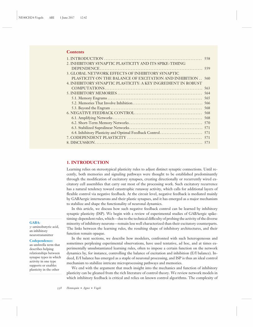
Loose versus tight: The balance is deemed tight if E and I inputs to single neurons balance each other on fasttimescales, and loose otherwise. Dynamically, tight balance in a recurrent network manifests itselfby the presence of very large negative eigenvalues in the (effective) connectivity matrix, expressingstrong inhibitory dominance in the dynamics of the corresponding eigenmodes. In classicalbalanced spiking networks (van Vreeswijk & Sompolinsky 1998, Renart et al. 2010), very large Eand I inputs must cancel tightly to leave a small remainder; in the stabilized supralinear network(Ahmadian et al. 2013, Rubin et al. 2015), moderately large E and I inputs need only cancel loosely.
Global versus detailed: When the spatial patterns of E and I inputs to a given neuron [denoted by vectors he(t) and hi(t)]balance each other among many dimensions [i.e., one can find many input directions v for whichthe projections vThe(t) and vThi(t) correlate temporally], the balance is said to be detailed, or highdimensional. In recurrent networks, this situation arises when feedback inhibition stabilizes a largenumber of instabilities in the E → E connectivity (e.g., Hennequin et al. 2014). When detailed balanceis also tight (cf. above), it is often referred to as precise balance. In random, unstructured networks,balance usually occurs only globally, i.e., in the summed E and summed I inputs in each neuron[corresponding to a single v ≈ (1,1,...,1)], reflecting overall balance of E and I population activities.
Recurrent versus feedforward: Balance can arise either from dynamic feedback inhibition (recurrent) or from feedforwardinhibition (even in a recurrent network).
Figure 2How balanced? Theoretical research has studied many facets of the balance of excitation and inhibition (E/I balance) in neuronalnetworks. We collected terminology from the literature, with definitions based on both phenomenology and considerations ofdynamics.
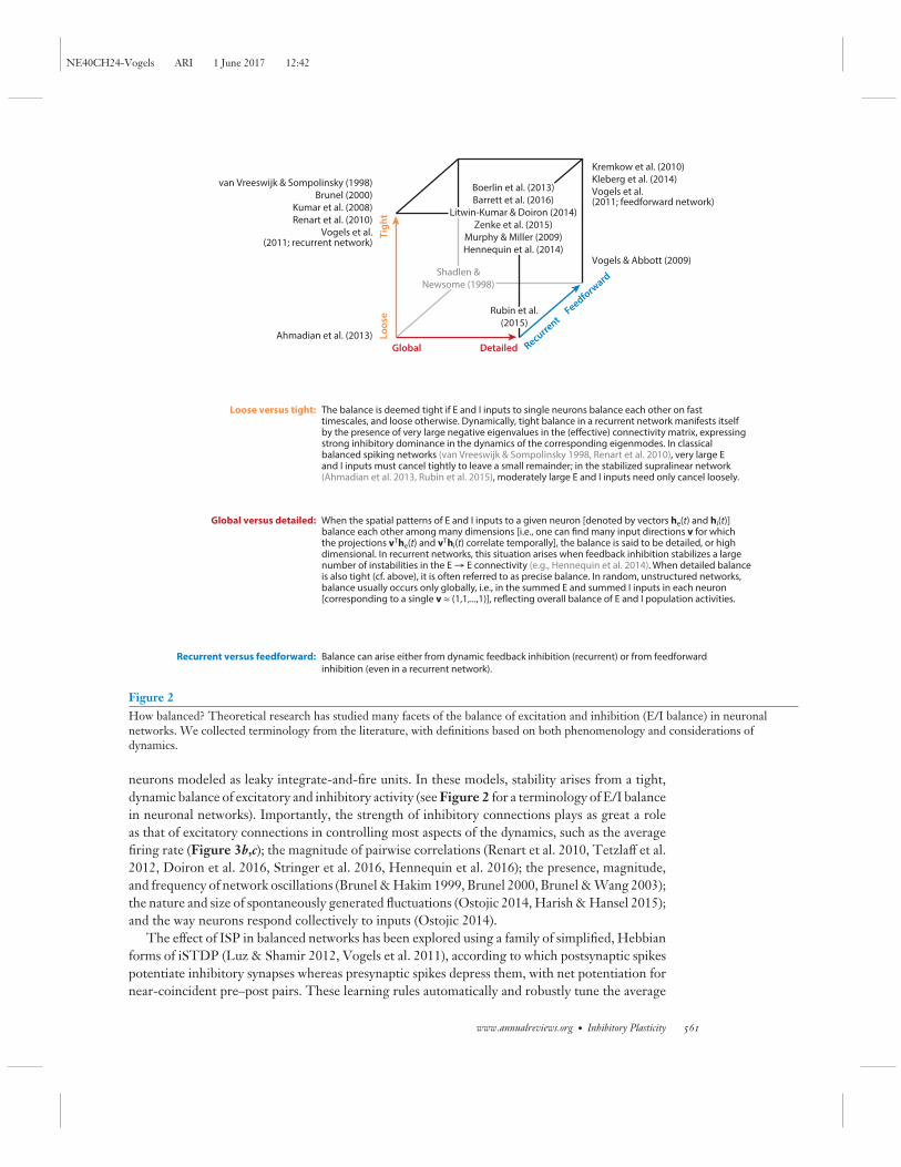
neurons modeled as leaky integrate-and-fire units. In these models, stability arises from a tight,dynamic balance of excitatory and inhibitory activity (see Figure 2 for a terminology of E/I balancein neuronal networks). Importantly, the strength of inhibitory connections plays as great a roleas that of excitatory connections in controlling most aspects of the dynamics, such as the averagefiring rate (Figure 3b,c); the magnitude of pairwise correlations (Renart et al. 2010, Tetzlaff et al.2012, Doiron et al. 2016, Stringer et al. 2016, Hennequin et al. 2016); the presence, magnitude,and frequency of network oscillations (Brunel & Hakim 1999, Brunel 2000, Brunel & Wang 2003);the nature and size of spontaneously generated fluctuations (Ostojic 2014, Harish & Hansel 2015);and the way neurons respond collectively to inputs (Ostojic 2014).
The effect of ISP in balanced networks has been explored using a family of simplified, Hebbianforms of iSTDP (Luz & Shamir 2012, Vogels et al. 2011), according to which postsynaptic spikespotentiate inhibitory synapses whereas presynaptic spikes depress them, with net potentiation fornear-coincident pre–post pairs. These learning rules automatically and robustly tune the average
www.annualreviews.org • Inhibitory Plasticity 561
NE40CH24-Vogels ARI 1 June 2017 12:42
40 Hz
20 Hz
100 ms
0.2
0.4
0.6
0.8
1.0
2
Rate (Hz)
1,000 0
Neu
rons
Neu
rons
Exci
tato
ry P
SPsi
ze (m
V)
Inhibitory PSPsize (mV)
0 >100
5 Hz10 Hz
p (in
-deg
ree)
p (in
-deg
ree)
p (in
-deg
ree)
In-degree
p (r
ate)
p (r
ate)
p (r
ate)
Firing rate (Hz)
a
b
c d
Input Input
Excitatory Inhibitory
Increasing heterogeneity
Compensation via ISPUnstableUnstable
1 2ISP
1
2
2,000 >1002010
8040
864
Figure 3Role of inhibition and inhibitory synaptic plasticity (ISP) in balanced networks. (a) Schematic of an excitatory/inhibitory networkarchitecture; two large populations of excitatory (red) and inhibitory (blue) neurons are randomly and sparsely coupled and receiveexternal input. (b) Phase diagram of the classical Brunel (2000) model of excitatory/inhibitory dynamics. The network is unstable iffeedback inhibition is too weak (white area left of the gray line). In the stable region, the overall network firing rate (color coded) varies withthe inhibitory and excitatory postsynaptic potential (PSP) sizes (x and y axes). Regions of oscillatory activity are not shown.(c) Representative spike rasters and corresponding fluctuations of the population firing rate in the different regimes labeled� and� inpanel b. (d) Randomly connecting any pair of neurons with a fixed probability results in a narrow distribution of in-degrees (top left) anda fairly homogeneous distribution of firing rates (top right). Greater heterogeneity in in-degrees (middle left) causes most cells to becomesilent and a number to fire at saturation (middle right). Homeostatic inhibitory plasticity (based on postsynaptic activity only)rehomogenizes firing rates (bottom). Panel d modified from Landau et al. (2016).
inhibitory input in single cells, effectively balancing them with the excitatory inputs and stabiliz-ing postsynaptic firing rates near a target value (Figure 3b,c). The resulting firing rates dependmainly on the ratio of the potentiation and depression parts of the iSTDP window (Vogels et al.2011, Luz & Shamir 2012). Any positive (respectively, negative) deviation in postsynaptic firingrate from the target is soon suppressed by strengthening (respectively, weakening) of inhibitoryinput synapses. Thus, whereas feedback inhibition stabilizes random, recurrent excitation at themillisecond timescale, its plasticity stabilizes firing rates on a slower timescale at which the globalE/I balance might be disrupted, for instance, due to slow ongoing modifications of excitatorysynapses. ISP thus maintains a tight and global balance.
One can more fully appreciate the benefits of a self-organized E/I balance when contemplatingthe delicate sensitivity of balanced networks to various deviations from standard uniform wiringstatistics (Barrett 2012, Rosenbaum & Doiron 2014). For example, introducing weak clusteringamong excitatory neurons produces large and slow fluctuations in the activity of the neurongroups (Litwin-Kumar & Doiron 2012). In this regime, the E/I balance is very sensitive to smallasymmetries in the wiring diagram, which typically result in winner-take-all behavior. ISP isan effective compensatory mechanism: Its strong tendency toward firing-rate homeostasis doesnot allow a single cluster of neurons to monopolize the dynamics (Litwin-Kumar & Doiron2014), resulting in a group-specific, detailed E/I balance. Similarly, implementing a more realisticdegree of heterogeneity in the wiring matrix by broadening the distributions of in-degrees causesa dramatic sparsification of the firing rates in the network, with most neurons becoming entirely
562 Hennequin · Agnes · Vogels
NE40CH24-Vogels ARI 1 June 2017 12:42
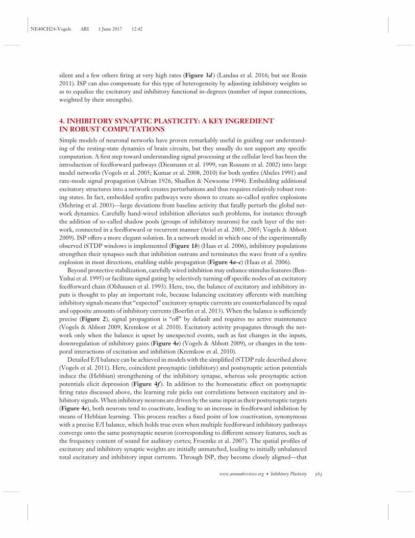
silent and a few others firing at very high rates (Figure 3d ) (Landau et al. 2016; but see Roxin2011). ISP can also compensate for this type of heterogeneity by adjusting inhibitory weights soas to equalize the excitatory and inhibitory functional in-degrees (number of input connections,weighted by their strengths).
4. INHIBITORY SYNAPTIC PLASTICITY: A KEY INGREDIENTIN ROBUST COMPUTATIONS
Simple models of neuronal networks have proven remarkably useful in guiding our understand-ing of the resting-state dynamics of brain circuits, but they usually do not support any specificcomputation. A first step toward understanding signal processing at the cellular level has been theintroduction of feedforward pathways (Diesmann et al. 1999, van Rossum et al. 2002) into largemodel networks (Vogels et al. 2005; Kumar et al. 2008, 2010) for both synfire (Abeles 1991) andrate-mode signal propagation (Adrian 1926, Shadlen & Newsome 1994). Embedding additionalexcitatory structures into a network creates perturbations and thus requires relatively robust rest-ing states. In fact, embedded synfire pathways were shown to create so-called synfire explosions(Mehring et al. 2003)—large deviations from baseline activity that fatally perturb the global net-work dynamics. Carefully hand-wired inhibition alleviates such problems, for instance throughthe addition of so-called shadow pools (groups of inhibitory neurons) for each layer of the net-work, connected in a feedforward or recurrent manner (Aviel et al. 2003, 2005; Vogels & Abbott2009). ISP offers a more elegant solution. In a network model in which one of the experimentallyobserved iSTDP windows is implemented (Figure 1b) (Haas et al. 2006), inhibitory populationsstrengthen their synapses such that inhibition outruns and terminates the wave front of a synfireexplosion in most directions, enabling stable propagation (Figure 4a–c) (Haas et al. 2006).
Beyond protective stabilization, carefully wired inhibition may enhance stimulus features (Ben-Yishai et al. 1995) or facilitate signal gating by selectively turning off specific nodes of an excitatoryfeedforward chain (Olshausen et al. 1993). Here, too, the balance of excitatory and inhibitory in-puts is thought to play an important role, because balancing excitatory afferents with matchinginhibitory signals means that “expected” excitatory synaptic currents are counterbalanced by equaland opposite amounts of inhibitory currents (Boerlin et al. 2013). When the balance is sufficientlyprecise (Figure 2), signal propagation is “off” by default and requires no active maintenance(Vogels & Abbott 2009, Kremkow et al. 2010). Excitatory activity propagates through the net-work only when the balance is upset by unexpected events, such as fast changes in the inputs,downregulation of inhibitory gains (Figure 4e) (Vogels & Abbott 2009), or changes in the tem-poral interactions of excitation and inhibition (Kremkow et al. 2010).
Detailed E/I balance can be achieved in models with the simplified iSTDP rule described above(Vogels et al. 2011). Here, coincident presynaptic (inhibitory) and postsynaptic action potentialsinduce the (Hebbian) strengthening of the inhibitory synapse, whereas sole presynaptic actionpotentials elicit depression (Figure 4f ). In addition to the homeostatic effect on postsynapticfiring rates discussed above, the learning rule picks out correlations between excitatory and in-hibitory signals. When inhibitory neurons are driven by the same input as their postsynaptic targets(Figure 4e), both neurons tend to coactivate, leading to an increase in feedforward inhibition bymeans of Hebbian learning. This process reaches a fixed point of low coactivation, synonymouswith a precise E/I balance, which holds true even when multiple feedforward inhibitory pathwaysconverge onto the same postsynaptic neuron (corresponding to different sensory features, such asthe frequency content of sound for auditory cortex; Froemke et al. 2007). The spatial profiles ofexcitatory and inhibitory synaptic weights are initially unmatched, leading to initially unbalancedtotal excitatory and inhibitory input currents. Through ISP, they become closely aligned—that
www.annualreviews.org • Inhibitory Plasticity 563

NE40CH24-Vogels ARI 1 June 2017 12:42
a
e f g
c
Before
Gate
...Excitatory
Inhibitory
d
Mem
bran
epo
tent
ial (
mV
) 200 ms
Depolarized
Hyperpolarized–80
–60
–40
–20
20
60
100
Ave
rage
curr
ent (
pA)
Signal number1 2 3 4 5 6 7 8
InhibitoryExcitatory
Before After
20
60
100
Signal number1 2 3 4 5 6 7 8
ΔwI
ΔwI
Long-termpotentiation
Long-term depression
b
tpost – tpre
tpost – tpre
After
Figure 4Functional consequences of inhibitory synaptic plasticity (ISP) for signal propagation. (a) Feedforward chain of excitatory neurons withfeedback inhibition. (b) Schematic of the inhibitory spike-timing-dependent plasticity (iSTDP) rule from Haas et al. (2006). (c) (Left)Synfire explosion (runaway activity propagates radially) in a neuronal network with two-dimensional topology. (Right) Followingactivity-dependent modifications of inhibitory synapses according to panel b, feedback inhibition preemptively suppresses activitypropagation in all but one direction. (d) Experimental evidence of detailed balance in vivo. Neighboring excitatory neurons weredepolarized (blue) and hyperpolarized (red) to extract inhibitory and excitatory currents, respectively, revealing their strong temporalcorrelation. (e) Schematic of a feedforward, gated inhibition motif. ( f ) ISP rule proposed by Vogels et al. (2011). ( g) Split excitatoryand inhibitory currents evoked by inputs going through each of eight feedforward inhibition motifs similar to panel e. Initially, strongand pathway-tuned excitation dominates over weak, unspecific inhibition (left). After learning with iSTDP as in panel f, excitatory andinhibitory tuning curves become precisely matched (right). Panel d modified from Okun & Lampl (2008) and panel g from Vogels et al.(2011).
is, exhibit detailed balance (Figure 4g) (Vogels et al. 2011)—even when perturbed by excitatoryplasticity (Clopath et al. 2016), consistent with the maintenance of a stimulus-specific, detailedE/I balance in auditory cortex (Froemke et al. 2007).
In feedforward networks, detailed balance is usually also tight, with excitatory and inhibitoryinputs tracking each other with the short delay of disynaptic feedforward inhibition. Such temporaltracking has also been observed in experiments (Figure 4d ) (Okun & Lampl 2008, Cafaro & Rieke2010, Moore & Nelson 1998, Wehr & Zador 2003, Shu et al. 2003), but one should take carein interpreting such close temporal correlations as a signature of detailed balance in a recurrentnetwork. For example, the balanced network model (Figure 3) exhibits tight, but not detailed,balance. One possible signature of detailed balance is that the temporal correlation of excitatoryand inhibitory inputs recorded in the same cell should be—on average—larger than those ofexcitatory and inhibitory inputs recorded in different cells (Hennequin et al. 2014). Estimatingthe former is difficult in practice, as it would require holding a cell’s membrane potential at thereversal potentials of excitation and inhibition simultaneously (or in very quick succession; Cafaro& Rieke 2010). How (local) synaptic plasticity rules can establish tight and detailed balance inrecurrent networks is still unknown.
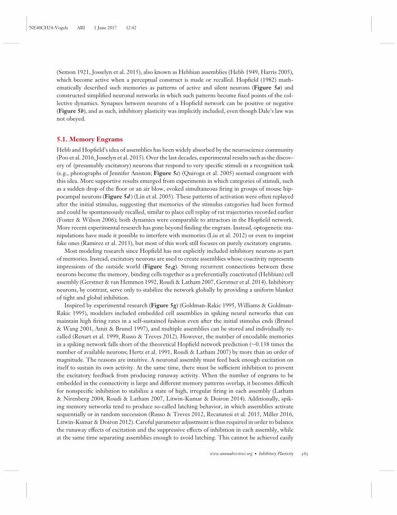
5. INHIBITORY MEMORIES
Memories of rich detail, recalled by simple commands or contextual cues, arguably involve elab-orate plasticity mechanisms, and they have inspired a long line of scientific inquiry. A com-mon hypothesis is that memories consist of groups of neurons that combine into engrams
564 Hennequin · Agnes · Vogels
NE40CH24-Vogels ARI 1 June 2017 12:42
(Semon 1921, Josselyn et al. 2015), also known as Hebbian assemblies (Hebb 1949, Harris 2005),which become active when a perceptual construct is made or recalled. Hopfield (1982) math-ematically described such memories as patterns of active and silent neurons (Figure 5a) andconstructed simplified neuronal networks in which such patterns become fixed points of the col-lective dynamics. Synapses between neurons of a Hopfield network can be positive or negative(Figure 5b), and as such, inhibitory plasticity was implicitly included, even though Dale’s law wasnot obeyed.
5.1. Memory Engrams
Hebb and Hopfield’s idea of assemblies has been widely absorbed by the neuroscience community(Poo et al. 2016, Josselyn et al. 2015). Over the last decades, experimental results such as the discov-ery of (presumably excitatory) neurons that respond to very specific stimuli in a recognition task(e.g., photographs of Jennifer Aniston; Figure 5c) (Quiroga et al. 2005) seemed congruent withthis idea. More supportive results emerged from experiments in which categories of stimuli, suchas a sudden drop of the floor or an air blow, evoked simultaneous firing in groups of mouse hip-pocampal neurons (Figure 5d ) (Lin et al. 2005). These patterns of activation were often replayedafter the initial stimulus, suggesting that memories of the stimulus categories had been formedand could be spontaneously recalled, similar to place cell replay of rat trajectories recorded earlier(Foster & Wilson 2006); both dynamics were comparable to attractors in the Hopfield network.More recent experimental research has gone beyond finding the engram. Instead, optogenetic ma-nipulations have made it possible to interfere with memories (Liu et al. 2012) or even to imprintfake ones (Ramirez et al. 2013), but most of this work still focuses on purely excitatory engrams.
Most modeling research since Hopfield has not explicitly included inhibitory neurons as partof memories. Instead, excitatory neurons are used to create assemblies whose coactivity representsimpressions of the outside world (Figure 5e,g). Strong recurrent connections between theseneurons become the memory, binding cells together as a preferentially coactivated (Hebbian) cellassembly (Gerstner & van Hemmen 1992, Roudi & Latham 2007, Gerstner et al. 2014). Inhibitoryneurons, by contrast, serve only to stabilize the network globally by providing a uniform blanketof tight and global inhibition.
Inspired by experimental research (Figure 5g) (Goldman-Rakic 1995, Williams & Goldman-Rakic 1995), modelers included embedded cell assemblies in spiking neural networks that canmaintain high firing rates in a self-sustained fashion even after the initial stimulus ends (Brunel& Wang 2001, Amit & Brunel 1997), and multiple assemblies can be stored and individually re-called (Renart et al. 1999, Russo & Treves 2012). However, the number of encodable memoriesin a spiking network falls short of the theoretical Hopfield network prediction (∼0.138 times thenumber of available neurons; Hertz et al. 1991, Roudi & Latham 2007) by more than an order ofmagnitude. The reasons are intuitive. A neuronal assembly must feed back enough excitation onitself to sustain its own activity. At the same time, there must be sufficient inhibition to preventthe excitatory feedback from producing runaway activity. When the number of engrams to beembedded in the connectivity is large and different memory patterns overlap, it becomes difficultfor nonspecific inhibition to stabilize a state of high, irregular firing in each assembly (Latham& Nirenberg 2004, Roudi & Latham 2007, Litwin-Kumar & Doiron 2014). Additionally, spik-ing memory networks tend to produce so-called latching behavior, in which assemblies activatesequentially or in random succession (Russo & Treves 2012, Recanatesi et al. 2015, Miller 2016,Litwin-Kumar & Doiron 2012). Careful parameter adjustment is thus required in order to balancethe runaway effects of excitation and the suppressive effects of inhibition in each assembly, whileat the same time separating assemblies enough to avoid latching. This cannot be achieved easily
www.annualreviews.org • Inhibitory Plasticity 565

NE40CH24-Vogels ARI 1 June 2017 12:42
through global inhibition, which often fails to control the specific E/I balance required in eachpattern.
5.2. Memories That Involve Inhibition
Precise E/I balance (Figure 4d ) (Froemke et al. 2007, Okun & Lampl 2008, Cafaro & Rieke2010, Xue et al. 2014) can be obtained through a concurrent adjustment of the excitatory andinhibitory weights, but specifically tuned, unique “Mexican hat” or shadow pool connectivity forevery neuron is exceedingly difficult to construct by hand. ISP offers an easy way to achievesuch cotuning. In a network model with embedded excitatory Hebbian assemblies, a simpleform of iSTDP inspired by experiments (Figure 4f ) (Vogels et al. 2011, Froemke et al. 2007,Sudhakaran et al. 2012) progressively carves out assembly-specific negative feedback loops through
e Inhibitory
Excitatory
Engr
am
Inhibitory
Excitatory
Inhibitory
Excitatory
Engr
am
a
h
k
Engr
am
f
b
0.40 0.8–0.4 1.2
Effec
t (Se
ssio
ns 3
–4)
20
10
30
0
3
6
–3
–6
CSS
(a.u
.)
0
Δ GABA concentration (μM/g)
i j
l m
c
1 s
Tria
l
d
MDA 2
MDA 1
MD
A 3
1 2
Session number
**
*
3 4
1 sN
euro
n
5
0
10
200 ms 20 Hz
Cue
1 s
Cueg
Tria
lRa
te
50 H
z
Target Move onset
566 Hennequin · Agnes · Vogels

NE40CH24-Vogels ARI 1 June 2017 12:42
activity-dependent strengthening of specific inhibitory feedback connections that may stem frommany, even all, inhibitory neurons in the network (Vogels et al. 2011). If a specific group ofinterneurons receives strong excitation from the assembly, it will be those neurons that will,by the nature of their correlated activity, form the strongest inhibitory synapses onto the as-sembly (Figure 5h,i ) (Duarte & Morrison 2014, Litwin-Kumar & Doiron 2014, Barron et al.2016a). Importantly, it is now this pair of amalgamated excitatory and inhibitory neuron groups,as opposed to excitatory neurons only, that defines the memory engram. As expected from therate-homogenizing effect of the learning rule, the firing rates of the neurons within the engrambecome indistinguishable from those of the rest of the network. However, the memory can stillbe reactivated through (even partial, local or network-wide) disruption of the E/I balance.
The inclusion of interneurons in the engram theory of memory gives rise to testable predictions.In humans, the strength of association between two stimuli can be estimated via a functionalmagnetic resonance imaging measurement termed cross-stimulus suppression (Figure 5j) (Barronet al. 2016b), in which the association of two unrelated stimuli leads to an increase in adaptationwhen these stimuli are sequentially presented (Barron et al. 2016a). This adaptation effect isthought to be a consequence of newly strengthened excitatory synapses between the corticalrepresentations of the associated stimuli. Over 24 h the effect fades, presumably due to rebalancingof the perturbed representations through inhibitory plasticity. The associative effect resurfaceswhen the global E/I balance within the brain region of interest is disrupted by means of transcranialdirect current stimulation (tDCs). A strong correlation between the measured reduction of GABA(Stagg et al. 2009) and the strength of the resurfacing effect was observed (Figure 5j), furthersupporting the idea that memory assemblies at least partially comprise inhibitory neurons (Barronet al. 2016a). This notion also underlies recent research in songbirds showing that inhibitoryneurons participate in, and protect, memories of newly acquired songs (Vallentin et al. 2016).
In a more bare-bones model of inhibitory memories introduced by Hendin et al. (1997), coac-tivation of presynaptic inhibition and postsynaptic excitation leads to weakening of the inhibitorysynapse, and prominent presynaptic activity leads to its strengthening [i.e., a near inverse of therule used by Vogels et al. (2011) and very similar in nature to that of Ormond & Woodin (2009,2011)]. It produces memory engrams in which the activity of excitatory neurons represents the
←−−−−−−−−−−−−−−−−−−−−−−−−−−−−−−−−−−−−−−−−−−−−−−−−−−−−−−−−−−−−−−−−−−−−−−−−−−−−−−−−−−−−−−−−−−Figure 5Inhibitory plasticity and memory formation in recurrent networks. (a) Example binary memory pattern as traditionally embedded in the(b) (schematized) connectivity matrix of a Hopfield network (red, excitatory weights; blue, inhibitory weights). (c) A single neuron inhuman hippocampus responds selectively to pictures of a well-known actress (top) presented for 1 s (dashed lines) in repeated trials; spikeraster (middle) and trial-averaged histogram (bottom). (d) (Left) Raster plot of multiple neurons in mouse hippocampus respondingselectively to an aversive stimulus (floor drop; Stim 1). (Right) The population dynamics describe low-dimensional trajectories during(black) and after (red and green loops) the experience. (e) Schematic and ( f) corresponding connectivity matrix of a network embedding apurely excitatory engram. ( g) Response of two neurons in monkey prefrontal cortex before, during, and after the presentation of avisual cue. The stimulus triggers persistent, elevated activity in the left neuron but not in the right neuron. (h) Schematic and(i) corresponding connectivity matrix of a network with an embedded engram comprising both excitatory and inhibitory neurons.( j) Experimental evidence for inhibitory memories in humans. The activity footprint of learning simple associations could be localizedin functional magnetic resonance imaging (fMRI) (left) through cross-stimulus suppression (CSS; middle). CSS was measured in theregion of interest (ROI; orange) over four sessions. The initially strong effect (Session 1) faded away over 24 h (Sessions 2 and 3). Aftertranscranial direct current stimulation (tDCs) over the ROI, CSS resurfaced (Session 4), and the strength of resurgence was correlatedwith the measured reduction of GABA concentration during tDCs stimulation (right). (k) Schematic and (l) corresponding connectivitymatrix of a network with globally distributed memories with recurrently strengthened excitatory and inhibitory connections. (m) Trial-averaged activity of a single neuron in monkey primary motor cortex before and during execution of various reaching movements (colorcoded). Panel c modified from Quiroga et al. (2005), panel d from Lin et al. (2005), panel g from Williams & Goldman-Rakic (1995),panel j from Barron et al. (2016a), and panel m from Churchland et al. (2012).
www.annualreviews.org • Inhibitory Plasticity 567

NE40CH24-Vogels ARI 1 June 2017 12:42
memory, but the connections that store it are purely inhibitory, because there is no recurrentexcitation. Increased activity in excitatory neurons stems merely from feedforward inputs and thefact that inhibitory neurons suppress the activity of all excitatory neurons outside the memorypattern.
5.3. Beyond the Engram
All of the above variations of the engram theory of memory entail a few active neurons thatrepresent a memory in a sea of silence. Hopfield’s original formulation was subtly different: Halfof all neurons were active for any given memory, many more than in any engram mentionedabove. Recent research on dynamical systems has revisited this idea, proposing that neuronalnetworks represent memories through the dynamic paths traced out by the activity of the wholepopulation (Maass et al. 2002, Sussillo & Abbott 2009, Laje & Buonomano 2013, Hennequinet al. 2014, Song et al. 2016). As such, nearly the entire network is involved (Figure 5k), andconsequently one could expect strong, seemingly indiscriminate connections between neurons,balanced tightly by appropriate inhibition (Figure 5l ). Indeed, network models built with the helpof nonlocal ISP rules to obey such constraints display spatiotemporal activity patterns in responseto stimuli (Figure 5m) that can be used to drive stereotypical activation of readout neurons withhigh reliability (Hennequin et al. 2014).
Finally, a hybrid of purely local engrams and globally distributed memories has emerged fromrecent research on conceptual memory organization. Constantinescu et al. (2016) showed thatthe particular hexagonal activity pattern of grid cells, thought to be a unique signature of spatialmemory (Rowland et al. 2016), may in fact be a broader principle for the representation of abstractknowledge. The distributed nature of the activity elicited by specific concepts would be in linewith globally embedded memories, whereas its spatial periodicity hints at the local organization ofindividual inhibitory populations. The specificity of the inhibitory feedback that sustains grid cellformation (Couey et al. 2013, Buetfering et al. 2014) may implicate ISP as a necessary componentof the formation of grid cell–like populations (Widloski & Fiete 2014, Weber & Sprekeler 2017).
6. NEGATIVE FEEDBACK CONTROL
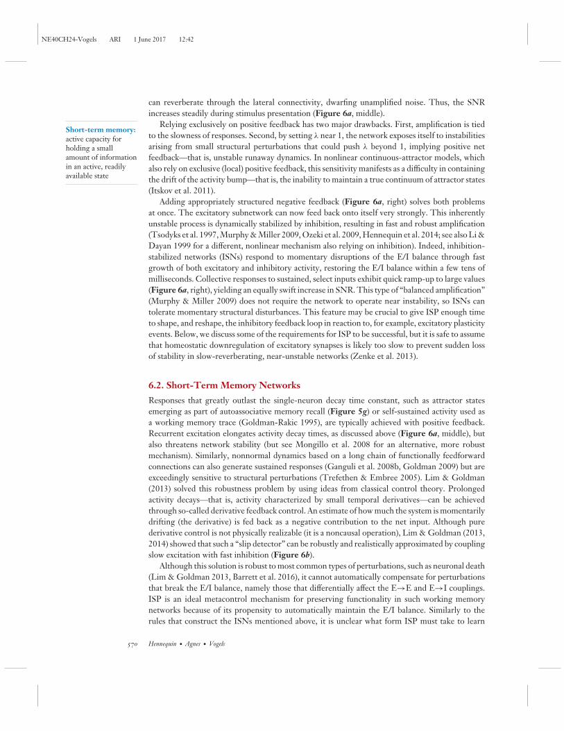
Sophisticated negative feedback is beginning to emerge as a fundamental component of neuralcircuit organization. Neuroscientists have begun to incorporate long-matured ideas from controltheory (Astrom & Murray 2010) to understand how well-balanced combinations of positive andnegative feedback enable fast and robust responses with high sensitivity to input signals and lowsensitivity to noise.
6.1. Amplifying Networks
High input sensitivity often manifests as amplification of certain input facets and removal of allother “noise.” Multiple lines of evidence suggest that sensory cortices perform such recurrent,selective amplification of their inputs (Kenet et al. 2003, Fiser et al. 2004, Luczak et al. 2009,Berkes et al. 2011, Bathellier et al. 2012), and several dynamical mechanisms have been proposed.They are easiest to illustrate in networks with exclusively local connectivity (Figure 6a,c). Weaklateral connectivity in such networks will simply relay inputs and any added noise, integratingboth on a characteristic single-neuron timescale, τ ∼ 5–50 ms (Figure 6a, left). A better signal-to-noise ratio (SNR) can be obtained when the network selectively amplifies the input signal, asis achieved by strengthening local excitatory connections (Figure 6a, middle) (Ben-Yishai et al.1995, Goldberg et al. 2004, Ganguli et al. 2008a, Ponce-Alvarez et al. 2013, Wimmer et al. 2014).
568 Hennequin · Agnes · Vogels

NE40CH24-Vogels ARI 1 June 2017 12:42
5 Hz
20 ms
Stimulus on
0
20
40
0 2 4
Responses to individual stimuliSummed responsesResponse to combined stimuli
5 Hz0
30
60
–10
Single neuroninput/output
Stimulus 50 ms
10 Hz
Excitatoryneurons
Excitatory Excitatory Inhibitory
Rate
(Hz)
Time (s)
Excitatory neurons
Weak input regime
Excitatory neurons
Strong input regime
Rate
(Hz)
Input (mV)
a b
c
d
Input Derivative feedback(slip detector)
Optimalinhibitoryplasticity
Unstable Stable dynamics
100
Figure 6Role of inhibition in functional models of recurrent neuronal networks. (a) Response to a step input ofa network with weak, negligible connectivity (left); distance-dependent excitatory connectivity (middle); anddistance-dependent, strong excitatory/inhibitory (E/I) connectivity (right). The input comprises a smoothbump (signal) and ongoing noise. (b) Derivative controller, or slip detector, implemented as an E–I–E feedbackloop (top), and corresponding success in producing sustained responses for working memory (bottom). (c) (Left)A stabilized supralinear network (SSN) with distance-dependent connectivity and a threshold-quadraticstatic nonlinearity in each neuron. (Middle) Response of the E cells (red line) to the superpositionof two weak input bumps centered on the two gray arrows. The response is larger than the sum (black) of theindividual responses ( gray, barely visible under black) to the two stimuli. (Right) Same, for the superpositionof two strong inputs—the response is now approximately normalized (i.e., less than the sum of theparts). (d) Inhibitory stabilization of strong random excitatory connectivity (left) through optimal inhibitorywiring yields networks (middle) that selectively amplify some inputs (generating rich transient responses;top right) while ignoring others (bottom right). Panel a modified from Murphy & Miller (2009), panel bfrom Lim & Goldman (2013), panel c from Rubin et al. (2015), and panel d from Hennequin et al. (2014).
This causes the network to feed back onto itself a fraction λ of its own activity, establishing aneffective integration time constant, ∼τ/(1 − λ), that can be very long if λ ≈ 1. In response toa localized stimulus, the network activity forms a response “bump” whose amplitude ramps upon this new collective timescale, eventually reaching a value ∝ 1/(1 − λ) � 1, indicating strongamplification (Figure 6a, middle). Crucially, only signal components with low spatial frequency
www.annualreviews.org • Inhibitory Plasticity 569
NE40CH24-Vogels ARI 1 June 2017 12:42
Short-term memory:active capacity forholding a smallamount of informationin an active, readilyavailable state
can reverberate through the lateral connectivity, dwarfing unamplified noise. Thus, the SNRincreases steadily during stimulus presentation (Figure 6a, middle).
Relying exclusively on positive feedback has two major drawbacks. First, amplification is tiedto the slowness of responses. Second, by setting λ near 1, the network exposes itself to instabilitiesarising from small structural perturbations that could push λ beyond 1, implying positive netfeedback—that is, unstable runaway dynamics. In nonlinear continuous-attractor models, whichalso rely on exclusive (local) positive feedback, this sensitivity manifests as a difficulty in containingthe drift of the activity bump—that is, the inability to maintain a true continuum of attractor states(Itskov et al. 2011).
Adding appropriately structured negative feedback (Figure 6a, right) solves both problemsat once. The excitatory subnetwork can now feed back onto itself very strongly. This inherentlyunstable process is dynamically stabilized by inhibition, resulting in fast and robust amplification(Tsodyks et al. 1997, Murphy & Miller 2009, Ozeki et al. 2009, Hennequin et al. 2014; see also Li &Dayan 1999 for a different, nonlinear mechanism also relying on inhibition). Indeed, inhibition-stabilized networks (ISNs) respond to momentary disruptions of the E/I balance through fastgrowth of both excitatory and inhibitory activity, restoring the E/I balance within a few tens ofmilliseconds. Collective responses to sustained, select inputs exhibit quick ramp-up to large values(Figure 6a, right), yielding an equally swift increase in SNR. This type of “balanced amplification”(Murphy & Miller 2009) does not require the network to operate near instability, so ISNs cantolerate momentary structural disturbances. This feature may be crucial to give ISP enough timeto shape, and reshape, the inhibitory feedback loop in reaction to, for example, excitatory plasticityevents. Below, we discuss some of the requirements for ISP to be successful, but it is safe to assumethat homeostatic downregulation of excitatory synapses is likely too slow to prevent sudden lossof stability in slow-reverberating, near-unstable networks (Zenke et al. 2013).
6.2. Short-Term Memory Networks
Responses that greatly outlast the single-neuron decay time constant, such as attractor statesemerging as part of autoassociative memory recall (Figure 5g) or self-sustained activity used asa working memory trace (Goldman-Rakic 1995), are typically achieved with positive feedback.Recurrent excitation elongates activity decay times, as discussed above (Figure 6a, middle), butalso threatens network stability (but see Mongillo et al. 2008 for an alternative, more robustmechanism). Similarly, nonnormal dynamics based on a long chain of functionally feedforwardconnections can also generate sustained responses (Ganguli et al. 2008b, Goldman 2009) but areexceedingly sensitive to structural perturbations (Trefethen & Embree 2005). Lim & Goldman(2013) solved this robustness problem by using ideas from classical control theory. Prolongedactivity decays—that is, activity characterized by small temporal derivatives—can be achievedthrough so-called derivative feedback control. An estimate of how much the system is momentarilydrifting (the derivative) is fed back as a negative contribution to the net input. Although purederivative control is not physically realizable (it is a noncausal operation), Lim & Goldman (2013,2014) showed that such a “slip detector” can be robustly and realistically approximated by couplingslow excitation with fast inhibition (Figure 6b).
Although this solution is robust to most common types of perturbations, such as neuronal death(Lim & Goldman 2013, Barrett et al. 2016), it cannot automatically compensate for perturbationsthat break the E/I balance, namely those that differentially affect the E→E and E→I couplings.ISP is an ideal metacontrol mechanism for preserving functionality in such working memorynetworks because of its propensity to automatically maintain the E/I balance. Similarly to therules that construct the ISNs mentioned above, it is unclear what form ISP must take to learn
570 Hennequin · Agnes · Vogels
NE40CH24-Vogels ARI 1 June 2017 12:42
slip detectors, but the success of the model indicates that future experiments on ISP might benefitfrom the riches of control theory.
6.3. Stabilized Supralinear Networks
Although the balanced network model explains important aspects of cortical firing, such as highvariability and low synchronicity (Figure 3), it is unable to account for basic circuit-level nonlin-earities, such as the normalization of cortical responses to multiple stimuli (Carandini & Heeger2012). This is because the tight E/I balance forces the network to respond linearly to changesin external input (van Vreeswijk & Sompolinsky 1996, Rosenbaum & Doiron 2014). Miller andcolleagues (Ahmadian et al. 2013, Rubin et al. 2015; see also Persi et al. 2011) showed that looseE/I balance in the so-called stabilized supralinear network (SSN) enables nonlinear collectivebehavior. Positive feedback arises from the combination of recurrent excitation and supralinearsingle-neuron nonlinearities (Figure 6c, left) (Priebe & Ferster 2008): The greater the excitatoryactivity is, the steeper the excitatory neurons’ input/output curves at their operating points, and sothe stronger the functional excitatory recurrent connectivity. This process would normally resultin inconsistent, explosive scaling of responses, but appropriately shaped feedback inhibition pre-vents it. Consequently, the SSN responds superlinearly to the superposition of two weak inputs(Figure 6c, center) but sublinearly to the superposition of two stronger inputs (Figure 6c, right)(Ahmadian et al. 2013), consistent with contrast-dependent normalization in V1. Several predic-tions of this simple model, including periodicity of tuning to stimulus size and contrast-dependentresonance, were successfully tested in recent experiments (Rubin et al. 2015).
6.4. Inhibitory Plasticity and Optimal Feedback Control
In all the models discussed above, functionality emerges from dynamic stabilization, requiringfeedback inhibition to reflect the spatial structure of the instabilities contained in the recurrentE→E connectivity. When this structure is simple (e.g., when instabilities arise only in a cou-ple of low-spatial-frequency modes), optimal inhibitory feedback is often just as simple, may beconstructed by hand in models, and could even be grown from genetic blueprints during devel-opment. Yet, cortical circuits evolve constantly at the micro scale (Chklovskii et al. 2004). Is italways possible for inhibition to tame high-dimensional recurrent instabilities? What form shouldISP take to succeed? Using methods from control theory and optimization, Hennequin et al.(2014) constructed complex ISNs by progressively adjusting each inhibitory synaptic weight ina strongly connected random network. This method could robustly stabilize a large number ofinitially unstable activity modes (Figure 6d, left), but the optimal stabilization algorithm couldnot easily be mapped onto a realistic form of ISP. Much like the simpler ISNs described above,the resulting stability-optimized circuits (SOCs) (Figure 6d, middle) responded vigorously toprivileged, balance-breaking input patterns (Figure 6d, top right) while almost completely ignor-ing others (Figure 6d, bottom right). However, owing to the complexity of recurrent excitatorymotifs, amplification occurs by way of rich internal dynamics that elicit multiphasic responsessimilar to those of neurons in the motor cortex of monkeys executing movements (Figure 5m)(Churchland et al. 2012). Such diverse responses might form useful basis functions to assemblemotor primitives, and they suggest that ISP lies at the heart of sequence learning.
7. CODEPENDENT PLASTICITY
The complexity of the control problems in which inhibition participates is perplexing to anyonewishing to understand how feasible learning rules may solve them. Indeed, optimal solutions
www.annualreviews.org • Inhibitory Plasticity 571

NE40CH24-Vogels ARI 1 June 2017 12:42
derived from control theory (e.g., for the construction of SOCs, described above) often prescribesynaptic modifications based on information not readily available at single synapses. This is difficultto reconcile with the de facto definition of a feasible rule as one that uses only local pre- andpostsynaptic spike trains (Morrison et al. 2008), possibly with a global reward signal (Fremauxet al. 2010, Fremaux & Gerstner 2016). One cannot strictly rule out the existence of simple—yetsufficiently effective—approximations of control-theoretic optimal constructs, which the braincould implement using elementary learning rules. Alternatively, complexity might emerge fromwell-orchestrated combinations of plasticity mechanisms, similar to those hand-tuned to enableformation of stable memories in cortical network models (Litwin-Kumar & Doiron 2014, Duarte& Morrison 2014, Zenke et al. 2015). Intrinsic, self-governed orchestration may result fromsophisticated cross talk between different types of synapses, and feasible synaptic plasticity may bemore complex than originally anticipated.
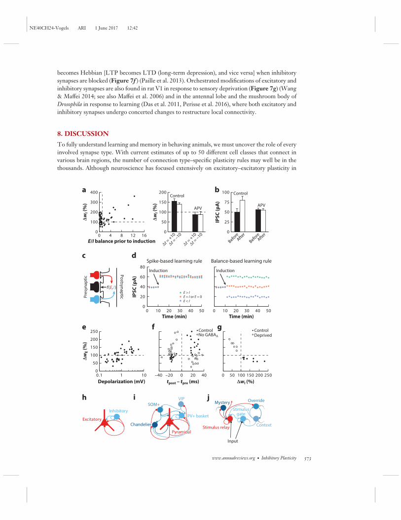
Recent experimental evidence from studies that monitored more than one synapse simulta-neously demonstrates such coordinated effects. In mouse auditory cortex, inhibitory synapseschange according to the E/I balance prior to induction (Figure 7a, left) (D’amour & Froemke2015), namely the combined state of excitation and inhibition. Moreover, plasticity is completelyabolished when NMDA receptors are blocked (Figure 7a, right) (D’amour & Froemke 2015), asalso reported in rat cerebellum (Figure 7b) (Mapelli et al. 2016). The fact that synaptic changesoccur only when excitatory inputs are coactive and the E/I balance is perturbed suggests thatISP integrates neighboring excitatory and inhibitory inputs (Figure 7c) to maintain a local E/Ibalance rather than, for instance, to impose an activity set point (as in Vogels et al. 2011). Theconsequences of such codependent learning rules for network function and architecture have notbeen explored in network models, but they may be vastly different from those of purely pre–postspike-based ISP (Figure 7d ).
Similarly, excitatory synapses depend on activity beyond pre–post spike pairs. For example,long-term potentiation (LTP) requires coactivation of additional excitatory afferents (Debanneet al. 1996, Sjostrom & Hausser 2006, Sjostrom et al. 2001), reflected in the dependence of LTPon the postsynaptic membrane potential (Figure 7e) (Sjostrom & Hausser 2006, Clopath et al.2010). Additionally, in rat corticostriatal excitatory synapses, an anti-Hebbian STDP window
−−−−−−−−−−−−−−−−−−−−−−−−−−−−−−−−−−−−−−−−−−−−−−−−−−−−−−−−−−−−−−−−−−−−−−−−−−−−−−−−−−−−−−−−−−→Figure 7Codependent synaptic plasticity. (a) (Left) Changes in inhibitory synaptic efficacy as a function of the ratio of excitatory (E) toinhibitory (I) peak currents just before the induction protocol (same data set as in Figure 1d ). (Right) N-Methyl-D-aspartate (NMDA)receptor blockade (APV) prevents plasticity otherwise induced by near-coincident pre- and postsynaptic spikes separated by �t.(b) Inhibitory synaptic plasticity (ISP) in rat cerebellum in a tetanus protocol is also affected by NMDA receptor blockade (APV).(c) Illustration of synaptic plasticity codependence. A synapse (black, middle) is modified according to a function f (E, I ) of biochemicalsignals shared by neighboring E (red) and I (blue) synapses. (d) Illustration of the difference between ISP induced by a purely spike-based (left) and a balance-based (right) learning rule. When the order of the spikes is the same (for example, pre before post), thespike-based learning rule (Vogels et al. 2011) induces the same change independently of the balance prior to induction. For acodependent learning rule, the amount of excitation and inhibition received by a neuron influences the amount and sign of the synapticweight change. (e) Excitatory synaptic plasticity depends on membrane potential depolarization. ( f) GABAA receptor blockade flips thesign of the spike-timing-dependent plasticity (STDP) window in rat corticostriatal synapses, under a protocol similar to that shown inFigure 1d. ( g) Long-term modifications of E and I synapses are negatively correlated in rat V1, but the magnitude of the effect ismodulated by sensory experience (compare monocular deprivation with control). (h–j) Most computational studies have focused on asimplistic microcircuit motif with a single E and I cell type (h), largely ignoring the wide diversity of interneuron types and connectionsfound in cortex (i), potentially serving an equally diverse array of functions ( j). Abbreviations: IPSC, inhibitory postsynaptic current;PV+, parvalbumin positive; SOM+, somatostatin positive; VIP, vasoactive intestinal polypeptide expressing. Panel a modified fromD’amour & Froemke (2015), panel b from Mapelli et al. (2016), panel e from Sjostrom & Hausser (2006), panel f from Paille et al.(2013), and panel g from Wang & Maffei (2014).
572 Hennequin · Agnes · Vogels
NE40CH24-Vogels ARI 1 June 2017 12:42
becomes Hebbian [LTP becomes LTD (long-term depression), and vice versa] when inhibitorysynapses are blocked (Figure 7f ) (Paille et al. 2013). Orchestrated modifications of excitatory andinhibitory synapses are also found in rat V1 in response to sensory deprivation (Figure 7g) (Wang& Maffei 2014; see also Maffei et al. 2006) and in the antennal lobe and the mushroom body ofDrosophila in response to learning (Das et al. 2011, Perisse et al. 2016), where both excitatory andinhibitory synapses undergo concerted changes to restructure local connectivity.
8. DISCUSSION
To fully understand learning and memory in behaving animals, we must uncover the role of everyinvolved synapse type. With current estimates of up to 50 different cell classes that connect invarious brain regions, the number of connection type–specific plasticity rules may well be in thethousands. Although neuroscience has focused extensively on excitatory–excitatory plasticity in
Pres
ynap
tic
Postsynaptic
a
c
h
Time (min) Time (min)
IPSC
(pA
)
Spike-based learning rule
IPSC
(pA
)
E > IE = I or E = 0E < I
Depolarization (mV)
BeforeAfte
r
BeforeAfte
r
Induction
ControlNo GABAA
b
g
d
e f
i j
ControlDeprived
Induction
Excitatory
PyramidalPyramidal
PV+ basket
ChandelierChandelier
VIPSOM+
Inhibitory
ΔwI (
%)
ΔwI (%)
ΔwE (
%)
ΔwI (
%)
E/I balance prior to inductionΔt
= +10
Δt = –10
Δt = +10
Δt = –10
Control
APV
0
50
100
150
200
250
0.1 1 10 –40 –20 0 20 40 0 50 100 150 200 250
0
100
200
300
400
0 4 8 12 16
Context
Stimulus gateStimulus gate
Mystery
Stimulus relay
Override
Input
0
50
100
150
200
0
25
50
75
100 Control
APV
0 10 20 30 40 500
20
40
60
80
0 10 20 30 40 50
Balance-based learning rule
f(E, I)
tpost – tpre (ms)
www.annualreviews.org • Inhibitory Plasticity 573

NE40CH24-Vogels ARI 1 June 2017 12:42
experiment and theory, other connection types are moving into the focus of scientific inquiry.Inhibitory plasticity is becoming a major player in the organization of neural circuits. Moreover,the emergence of function seems to be borne out of interactions between different forms ofplasticity, which recent experiments have barely begun to explore. Successful identification ofthese mechanisms and their function will require embracing the complexities of architectures withmore than just two cell types and learning rules (Figure 7h–j). Bringing together sophisticatedexperiments with integrative, reductionist theoretical research will let us distill the essential motifsthat organize myriads of synapses, allowing us to eventually articulate a canonical model of synapticlearning.
DISCLOSURE STATEMENT
The authors are not aware of any affiliations, memberships, funding, or financial holdings thatmight be perceived as affecting the objectivity of this review.
ACKNOWLEDGMENTS
The authors thank L.F. Abbott, A. Maffei, G. Miesenbock, W. Podlaski, J. Stroud, and M. Woodinfor feedback on figures and manuscript. The writing of this review was supported by a Well-come Trust Seed Award to G.H. (202111/Z/16/Z), a Sir Henry Dale Wellcome Trust and RoyalSociety Grant to T.P.V. (WT100000), and a postdoctoral grant from Brazilian CNPq to E.J.A.(235144/2014-2).
LITERATURE CITED
Abbott L, Nelson S. 2000. Synaptic plasticity: taming the beast. Nat. Neurosci. 3:1178–83Abeles M. 1991. Corticonics: Neural Circuits of the Cerebral Cortex. Cambridge, UK: Cambridge Univ. PressAdrian ED. 1926. The impulses produced by sensory nerve endings. J. Physiol. 61:49–72Ahmadian Y, Rubin DB, Miller KD. 2013. Analysis of the stabilized supralinear network. Neural Comput.
25:1994–2037Aizenman CD, Manis PB, Linden DJ. 1998. Polarity of long-term synaptic gain change is related to postsyn-
aptic spike firing at a cerebellar inhibitory synapse. Neuron 21:827–35Amit DJ, Brunel N. 1997. Model of global spontaneous activity and local structured activity during delay
periods in the cerebral cortex. Cereb. Cortex 7:237–52Astrom KJ, Murray RM. 2010. Feedback Systems: An Introduction for Scientists and Engineers. Princeton, NJ:
Princeton University PressAviel Y, Horn D, Abeles M. 2005. Memory capacity of balanced networks. Neural Comput. 17:691–713Aviel Y, Mehring C, Abeles M, Horn D. 2003. On embedding synfire chains in a balanced network. Neural
Comput. 15:1321–40Barrett D. 2012. Computation in balanced networks. PhD thesis, Univ. Coll. LondonBarrett DT, Deneve S, Machens CK. 2016. Optimal compensation for neuron loss. eLife 5:e12454Barron H, Vogels T, Emir U, Makin T, O’Shea J, et al. 2016a. Unmasking latent inhibitory connections in
human cortex to reveal dormant cortical memories. Neuron 90:191–203Barron HC, Garvert MM, Behrens TEJ. 2016b. Repetition suppression: a means to index neural representa-
tions using BOLD? Philos. Trans. R. Soc. B 371:20150355Bathellier B, Ushakova L, Rumpel S. 2012. Discrete neocortical dynamics predict behavioral categorization
of sounds. Neuron 76:435–49Ben-Yishai R, Bar-Or RL, Sompolinsky H. 1995. Theory of orientation tuning in visual cortex. PNAS 92:3844Berkes P, Orban G, Lengyel M, Fiser J. 2011. Spontaneous cortical activity reveals hallmarks of an optimal
internal model of the environment. Science 331:83–87
574 Hennequin · Agnes · Vogels

NE40CH24-Vogels ARI 1 June 2017 12:42
Boerlin M, Machens CK, Deneve S. 2013. Predictive coding of dynamical variables in balanced spikingnetworks. PLOS Comput. Biol. 9:e1003258
Brunel N. 2000. Dynamics of sparsely connected networks of excitatory and inhibitory spiking neurons.J. Comput. Neurosci. 8:183–208
Brunel N, Hakim V. 1999. Fast global oscillations in networks of integrate-and-fire neurons with low firingrates. Neural Comput. 11:1621–71
Brunel N, Wang XJ. 2001. Effects of neuromodulation in a cortical network model of object working memorydominated by recurrent inhibition. J. Comput. Neurosci. 11:63–85
Brunel N, Wang XJ. 2003. What determines the frequency of fast network oscillations with irregular neuraldischarges? I. Synaptic dynamics and excitation–inhibition balance. J. Neurophysiol. 90:415–30
Buetfering C, Allen K, Monyer H. 2014. Parvalbumin interneurons provide grid cell–driven recurrent inhi-bition in the medial entorhinal cortex. Nat. Neurosci. 17:710–18
Cafaro J, Rieke F. 2010. Noise correlations improve response fidelity and stimulus encoding. Nature 468:964–67
Carandini M, Heeger DJ. 2012. Normalization as a canonical neural computation. Nat. Rev. Neurosci. 13:51–62Chklovskii DB, Mel BW, Svoboda K. 2004. Cortical rewiring and information storage. Nature 431:782–88Churchland MM, Cunningham JP, Kaufman MT, Foster JD, Nuyujukian P, et al. 2012. Neural population
dynamics during reaching. Nature 487:51–56Clopath C, Busing L, Vasilaki E, Gerstner W. 2010. Connectivity reflects coding: a model of voltage-based
STDP with homeostasis. Nat. Neurosci. 13:344–52Clopath C, Vogels TP, Froemke RC, Sprekeler H. 2016. Receptive field formation by interacting excitatory
and inhibitory synaptic plasticity. bioRxiv 066589. https://doi.org/10.1101/066589Constantinescu AO, O’Reilly JX, Behrens TEJ. 2016. Organizing conceptual knowledge in humans with a
gridlike code. Science 352:1464–68Couey JJ, Witoelar A, Zhang SJ, Zheng K, Ye J, et al. 2013. Recurrent inhibitory circuitry as a mechanism
for grid formation. Nat. Neurosci. 16:318–24D’amour J, Froemke R. 2015. Inhibitory and excitatory spike-timing-dependent plasticity in the auditory
cortex. Neuron 86:514–28Das S, Sadanandappa MK, Dervan A, Larkin A, Lee JA, et al. 2011. Plasticity of local GABAergic interneurons
drives olfactory habituation. PNAS 108:E646–54Debanne D, Gahwiler BH, Thompson SM. 1996. Cooperative interactions in the induction of long-term
potentiation and depression of synaptic excitation between hippocampal CA3-CA1 cell pairs in vitro.PNAS 93:11225–30
Diesmann M, Gewaltig MO, Aertsen A. 1999. Stable propagation of synchronous spiking in cortical neuralnetworks. Nature 402:529–33
Doiron B, Litwin-Kumar A, Rosenbaum R, Ocker GK, Josi K. 2016. The mechanics of state-dependentneural correlations. Nat. Neurosci. 19:383–93
Duarte RCF, Morrison A. 2014. Dynamic stability of sequential stimulus representations in adapting neuronalnetworks. Front. Comput. Neurosci. 8:124
Fiser J, Chiu C, Weliky M. 2004. Small modulation of ongoing cortical dynamics by sensory input duringnatural vision. Nature 431:573–78
Foster DJ, Wilson MA. 2006. Reverse replay of behavioural sequences in hippocampal place cells during theawake state. Nature 440:680–83
Fremaux N, Gerstner W. 2016. Neuromodulated spike-timing-dependent plasticity, and theory of three-factorlearning rules. Front. Neural Circuits 9:85
Fremaux N, Sprekeler H, Gerstner W. 2010. Functional requirements for reward-modulated spike-timing-dependent plasticity. J. Neurosci. 30:13326–37
Froemke RC, Merzenich MM, Schreiner CE. 2007. A synaptic memory trace for cortical receptive fieldplasticity. Nature 450:425–29
Gaiarsa JL, Caillard O, Ben-Ari Y. 2002. Long-term plasticity at GABAergic and glycinergic synapses: mech-anisms and functional significance. Trends Neurosci. 25:564–70
Ganguli S, Bisley JW, Roitman JD, Shadlen MN, Goldberg ME, Miller KD. 2008a. One-dimensional dy-namics of attention and decision making in LIP. Neuron 58:15–25
www.annualreviews.org • Inhibitory Plasticity 575

NE40CH24-Vogels ARI 1 June 2017 12:42
Ganguli S, Huh D, Sompolinsky H. 2008b. Memory traces in dynamical systems. PNAS 105:18970–75Gerstner W, Kistler WM, Naud R, Paninski L. 2014. Neuronal Dynamics: From Single Neurons to Networks and
Models of Cognition. Cambridge, UK: Cambridge Univ. PressGerstner W, van Hemmen JL. 1992. Associative memory in a network of ‘spiking’ neurons. Netw. Comput.
Neural Syst. 3:139–64Goldberg JA, Rokni U, Sompolinsky H. 2004. Patterns of ongoing activity and the functional architecture of
the primary visual cortex. Neuron 42:489–500Goldman MS. 2009. Memory without feedback in a neural network. Neuron 61:621–34Goldman-Rakic PS. 1995. Cellular basis of working memory. Neuron 14:477–85Haas JS, Nowotny T, Abarbanel HDI. 2006. Spike-timing-dependent plasticity of inhibitory synapses in the
entorhinal cortex. J. Neurophysiol. 96:3305–13Harish O, Hansel D. 2015. Asynchronous rate chaos in spiking neuronal circuits. PLOS Comput. Biol.
11:e1004266Harris KD. 2005. Neural signatures of cell assembly organization. Nat. Rev. Neurosci. 6:399–407Hartmann K, Bruehl C, Golovko T, Draguhn A. 2008. Fast homeostatic plasticity of inhibition via activity-
dependent vesicular filling. PLOS ONE 3:e2979Hebb DO. 1949. The Organization of Behavior: A Neuropsychological Approach. New York: WileyHendin O, Horn D, Tsodyks MV. 1997. The role of inhibition in an associative memory model of the olfactory
bulb. J. Comput. Neurosci. 4:173–82Hennequin G, Ahmadian Y, Rubin DB, Lengyel M, Miller KD. 2016. Stabilized supralinear network
dynamics account for stimulus-induced changes of noise variability in the cortex. bioRxiv 094334.https://doi.org/10.1101/094334
Hennequin G, Vogels TP, Gerstner W. 2014. Optimal control of transient dynamics in balanced networkssupports generation of complex movements. Neuron 82:1394–406
Hertz J, Krogh A, Palmer R. 1991. Introduction to the Theory of Neural Computation. Redwood City, CA:Addison-Wesley
Holmgren CD, Zilberter Y. 2001. Coincident spiking activity induces long-term changes in inhibition ofneocortical pyramidal cells. J. Neurosci. 21:8270–77
Hopfield JJ. 1982. Neural networks and physical systems with emergent collective computational abilities.PNAS 79:2554–58
Itskov V, Hansel D, Tsodyks M. 2011. Short-term facilitation may stabilize parametric working memory trace.Front. Comput. Neurosci. 5:40
Josselyn SA, Kohler S, Frankland PW. 2015. Finding the engram. Nat. Rev. Neurosci. 16:521–34Kano M. 1995. Plasticity of inhibitory synapses in the brain: a possible memory mechanism that has been
overlooked. Neurosci. Res. 21:177–82Kenet T, Bibitchkov D, Tsodyks M, Grinvald A, Arieli A. 2003. Spontaneously emerging cortical represen-
tations of visual attributes. Nature 425:954–56Kilman V, van Rossum MCW, Turrigiano GG. 2002. Activity deprivation reduces miniature IPSC amplitude
by decreasing the number of postsynaptic GABAA receptors clustered at neocortical synapses. J. Neurosci.22:1328–37
Kleberg FI, Fukai T, Gilson M. 2014. Excitatory and inhibitory STDP jointly tune feedforward neural circuitsto selectively propagate correlated spiking activity. Front. Comput. Neurosci. 8:53
Komatsu Y. 1994. Age-dependent long-term potentiation of inhibitory synaptic transmission in rat visualcortex. J. Neurosci. 14:6488–99
Komatsu Y, Iwakiri M. 1993. Long-term modification of inhibitory synaptic transmission in developing visualcortex. NeuroReport 4:907–10
Kremkow J, Aertsen A, Kumar A. 2010. Gating of signal propagation in spiking neural networks by balancedand correlated excitation and inhibition. J. Neurosci. 30:15760–68
Kumar A, Rotter S, Aertsen A. 2010. Spiking activity propagation in neuronal networks: reconciling differentperspectives on neural coding. Nat. Rev. Neurosci. 11:615–27
Kumar A, Schrader S, Aertsen A, Rotter S. 2008. The high-conductance state of cortical networks. NeuralComput. 20:1–43
576 Hennequin · Agnes · Vogels

NE40CH24-Vogels ARI 1 June 2017 12:42
Kurotani T, Yamada K, Yoshimura Y, Crair MC, Komatsu Y. 2008. State-dependent bidirectional modificationof somatic inhibition in neocortical pyramidal cells. Neuron 57:905–16
Laje R, Buonomano DV. 2013. Robust timing and motor patterns by taming chaos in recurrent neuralnetworks. Nat. Neurosci. 16:925–33
Landau I, Egger R, Dercksen V, Oberlaender M, Sompolinsky H. 2016. The impact of structural heterogeneityon excitation–inhibition balance in cortical networks. Neuron 92:1106–21
Latham PE, Nirenberg S. 2004. Computing and stability in cortical networks. Neural Comput. 16:1385–412Li Z, Dayan P. 1999. Computational differences between asymmetrical and symmetrical networks. Netw.
Comput. Neural Syst. 10:59–77Lim S, Goldman MS. 2013. Balanced cortical microcircuitry for maintaining information in working memory.
Nat. Neurosci. 16:1306–14Lim S, Goldman MS. 2014. Balanced cortical microcircuitry for spatial working memory based on corrective
feedback control. J. Neurosci. 34:6790–806Lin L, Osan R, Shoham S, Jin W, Zuo W, Tsien JZ. 2005. Identification of network-level coding units for
real-time representation of episodic experiences in the hippocampus. PNAS 102:6125–30Litwin-Kumar A, Doiron B. 2012. Slow dynamics and high variability in balanced cortical networks with
clustered connections. Nat. Neurosci. 15:1498–505Litwin-Kumar A, Doiron B. 2014. Formation and maintenance of neuronal assemblies through synaptic
plasticity. Nat. Commun. 5:5319Liu X, Ramirez S, Pang PT, Puryear CB, Govindarajan A, et al. 2012. Optogenetic stimulation of a hippocam-
pal engram activates fear memory recall. Nature 484:381–85Luczak A, Bartho P, Harris KD. 2009. Spontaneous events outline the realm of possible sensory responses in
neocortical populations. Neuron 62:413–25Luz Y, Shamir M. 2012. Balancing feed-forward excitation and inhibition via Hebbian inhibitory synaptic
plasticity. PLOS Comput. Biol. 8:e1002334Maass W, Natschlager T, Markram H. 2002. Real-time computing without stable states: a new framework
for neural computation based on perturbations. Neural Comput. 14:2531–60Maffei A, Nataraj K, Nelson SB, Turrigiano GG. 2006. Potentiation of cortical inhibition by visual deprivation.
Nature 443:81–84Mapelli J, Gandolfi D, Vilella A, Zoli M, Bigiani A. 2016. Heterosynaptic GABAergic plasticity bidirectionally
driven by the activity of pre- and postsynaptic NMDA receptors. PNAS 113:9898–903Markram H, Gerstner W, Sjostrom PJ. 2011. A history of spike-timing-dependent plasticity. Front. Synaptic
Neurosci. 3:1–24Mehring C, Hehl U, Kubo M, Diesmann M, Aertsen A. 2003. Activity dynamics and propagation of syn-
chronous spiking in locally connected random networks. Biol. Cybern. 88:395–408Miller P. 2016. Itinerancy between attractor states in neural systems. Curr. Opin. Neurobiol. 40:14–22Mongillo G, Barak O, Tsodyks M. 2008. Synaptic theory of working memory. Science 319:1543–46Moore CI, Nelson SB. 1998. Spatio-temporal subthreshold receptive fields in the vibrissa representation of
rat primary somatosensory cortex. J. Neurophysiol. 80:2882–92Morrison A, Diesmann M, Gerstner W. 2008. Phenomenological models of synaptic plasticity based on spike
timing. Biol. Cybern. 98:459–78Murphy BK, Miller KD. 2009. Balanced amplification: a new mechanism of selective amplification of neural
activity patterns. Neuron 61:635–48Okun M, Lampl I. 2008. Instantaneous correlation of excitation and inhibition during ongoing and sensory-
evoked activities. Nat. Neurosci. 11:535–37Olshausen BA, Anderson CH, Essen DV. 1993. A neurobiological model of visual attention and invariant
pattern recognition based on dynamic routing of information. J. Neurosci. 13:4700–19Ormond J, Woodin MA. 2009. Disinhibition mediates a form of hippocampal long-term potentiation in area
CA1. PLOS ONE 4:e7224Ormond J, Woodin MA. 2011. Disinhibition-mediated LTP in the hippocampus is synapse specific. Front.
Cell. Neurosci. 5:17Ostojic S. 2014. Two types of asynchronous activity in networks of excitatory and inhibitory spiking neurons.
Nat. Neurosci. 17:594–600
www.annualreviews.org • Inhibitory Plasticity 577
NE40CH24-Vogels ARI 1 June 2017 12:42
Ozeki H, Finn IM, Schaffer ES, Miller KD, Ferster D. 2009. Inhibitory stabilization of the cortical networkunderlies visual surround suppression. Neuron 62:578–92
Paille V, Fino E, Du K, Morera-Herreras T, Perez S, et al. 2013. GABAergic circuits control spike-timing-dependent plasticity. J. Neurosci. 33:9353–63
Perisse E, Owald D, Barnstedt O, Talbot CB, Huetteroth W, Waddell S. 2016. Aversive learning and appetitivemotivation toggle feed-forward inhibition in the Drosophila mushroom body. Neuron 90:1086–99
Persi E, Hansel D, Nowak L, Barone P, van Vreeswijk C. 2011. Power-law input-output transfer functionsexplain the contrast-response and tuning properties of neurons in visual cortex. PLOS Comput. Biol.7:e1001078
Ponce-Alvarez A, Thiele A, Albright TD, Stoner GR, Deco G. 2013. Stimulus-dependent variability and noisecorrelations in cortical MT neurons. PNAS 110:13162–67
Poo MM, Pignatelli M, Ryan TJ, Tonegawa S, Bonhoeffer T, et al. 2016. What is memory? The present stateof the engram. BMC Biol. 14:40
Priebe NJ, Ferster D. 2008. Inhibition, spike threshold, and stimulus selectivity in primary visual cortex.Neuron 57:482–97
Quiroga RQ, Reddy L, Kreiman G, Koch C, Fried I. 2005. Invariant visual representation by single neuronsin the human brain. Nature 435:1102–7
Ramirez S, Liu X, Lin PA, Suh J, Pignatelli M, et al. 2013. Creating a false memory in the hippocampus.Science 341:387–91
Recanatesi S, Katkov M, Romani S, Tsodyks M. 2015. Neural network model of memory retrieval. Front.Comput. Neurosci. 9:149
Renart A, de la Rocha J, Bartho P, Hollender L, Parga N, et al. 2010. The asynchronous state in corticalcircuits. Science 327:587
Renart A, Parga N, Rolls ET. 1999. Associative memory properties of multiple cortical modules. Netw. Comput.Neural Syst. 10:237–55
Rosenbaum R, Doiron B. 2014. Balanced networks of spiking neurons with spatially dependent recurrentconnections. Phys. Rev. X 4:021039
Roudi Y, Latham PE. 2007. A balanced memory network. PLOS Comput. Biol. 3:e141Rowland DC, Roudi Y, Moser MB, Moser EI. 2016. Ten years of grid cells. Annu. Rev. Neurosci. 39:19–40Roxin A. 2011. The role of degree distribution in shaping the dynamics in networks of sparsely connected
spiking neurons. Front. Comput. Neurosci. 5:8Rubin D, Van Hooser S, Miller K. 2015. The stabilized supralinear network: a unifying circuit motif underlying
multi-input integration in sensory cortex. Neuron 85:402–17Russo E, Treves A. 2012. Cortical free-association dynamics: distinct phases of a latching network. Phys. Rev.
E 85:051920Semon RW. 1921. The Mneme. London: Allen & UnwinShadlen MN, Newsome WT. 1994. Noise, neural codes and cortical organization. Curr. Opin. Neurobiol.
4:569–79Shu Y, Hasenstaub A, McCormick DA. 2003. Turning on and off recurrent balanced cortical activity. Nature
423:288–93Sjostrom PJ, Hausser M. 2006. A cooperative switch determines the sign of synaptic plasticity in distal dendrites
of neocortical pyramidal neurons. Neuron 51:227–38Sjostrom PJ, Turrigiano GG, Nelson SB. 2001. Rate, timing, and cooperativity jointly determine cortical
synaptic plasticity. Neuron 32:1149–64Song HF, Yang GR, Wang XJ. 2016. Training excitatory-inhibitory recurrent neural networks for cognitive
tasks: a simple and flexible framework. PLOS Comput. Biol. 12:e1004792Stagg CJ, Best JG, Stephenson MC, O’Shea J, Wylezinska M, et al. 2009. Polarity-sensitive modulation of
cortical neurotransmitters by transcranial stimulation. J. Neurosci. 29:5202–6Stringer C, Pachitariu M, Steinmetz NA, Okun M, Bartho P, et al. 2016. Inhibitory control of correlated
intrinsic variability in cortical networks. eLife 5:e19695Sudhakaran IP, Holohan EE, Osman S, Rodrigues V, Vijayraghavan K, Ramaswami M. 2012. Plasticity of
recurrent inhibition in the Drosophila antennal lobe. J. Neurosci. 32:7225–31
578 Hennequin · Agnes · Vogels

NE40CH24-Vogels ARI 1 June 2017 12:42
Sussillo D, Abbott L. 2009. Generating coherent patterns of activity from chaotic neural networks. Neuron63:544–57
Tetzlaff T, Helias M, Einevoll GT, Diesmann M. 2012. Decorrelation of neural-network activity by inhibitoryfeedback. PLOS Comput. Biol. 8:e1002596
Trefethen LN, Embree M. 2005. Spectra and Pseudospectra: The Behavior of Nonnormal Matrices and Operators.Princeton, NJ: Princeton Univ. Press
Tsodyks MV, Sejnowski T. 1995. Rapid state switching in balanced cortical network models. Netw. Comput.Neural Syst. 6:111–24
Tsodyks MV, Skaggs WE, Sejnowski TJ, McNaughton BL. 1997. Paradoxical effects of external modulationof inhibitory interneurons. J. Neurosci. 17:4382–88
Turrigiano G. 2012. Homeostatic synaptic plasticity: local and global mechanisms for stabilizing neuronalfunction. Cold Spring Harb. Perspect. Biol. 4:a005736
Vallentin D, Kosche G, Lipkind D, Long MA. 2016. Inhibition protects acquired song segments during vocallearning in zebra finches. Science 351:267–71
van Rossum MCW, Turrigiano GG, Nelson SB. 2002. Fast propagation of firing rates through layerednetworks of noisy neurons. J. Neurosci. 22:1956–66
van Vreeswijk C, Sompolinsky H. 1996. Chaos in neuronal networks with balanced excitatory and inhibitoryactivity. Science 274:1724
van Vreeswijk C, Sompolinsky H. 1998. Chaotic balanced state in a model of cortical circuits. Neural Comput.10:1321–71
Vogels TP, Abbott LF. 2009. Gating multiple signals through detailed balance of excitation and inhibition inspiking networks. Nat. Neurosci. 12:483–91
Vogels TP, Froemke RC, Doyon N, Gilson M, Haas JS, et al. 2013. Inhibitory synaptic plasticity: spiketiming-dependence and putative network function. Front. Neural Circuits 7:119
Vogels TP, Rajan K, Abbott LF. 2005. Neural network dynamics. Annu. Rev. Neurosci. 28:357–76Vogels TP, Sprekeler H, Zenke F, Clopath C, Gerstner W. 2011. Inhibitory plasticity balances excitation and
inhibition in sensory pathways and memory networks. Science 334:1569–73Wang L, Maffei A. 2014. Inhibitory plasticity dictates the sign of plasticity at excitatory synapses. J. Neurosci.
34:1083–93Weber SN, Sprekeler H. 2017. Learning place cells, grid cells and invariances: a unifying model. bioRxiv
102525. https://doi.org/10.1101/102525Wehr M, Zador A. 2003. Balanced inhibition underlies tuning and sharpens spike timing in auditory cortex.
Nature 426:442–46Widloski J, Fiete I. 2014. A model of grid cell development through spatial exploration and spike time–
dependent plasticity. Neuron 83:481–95Williams GV, Goldman-Rakic PS. 1995. Modulation of memory fields by dopamine D1 receptors in prefrontal
cortex. Nature 376:572–75Wilmes KA, Sprekeler H, Schreiber S. 2016. Inhibition as a binary switch for excitatory plasticity in pyramidal
neurons. PLOS Comput. Biol. 12:e1004768Wimmer K, Nykamp DQ, Constantinidis C, Compte A. 2014. Bump attractor dynamics in prefrontal cortex
explains behavioral precision in spatial working memory. Nat. Neurosci. 17:431–39Woodin MA, Ganguly K, Poo M. 2003. Coincident pre-and postsynaptic activity modifies GABAergic synapses
by postsynaptic changes in Cl-transporter activity. Neuron 39:807–20Xue M, Atallah BV, Scanziani M. 2014. Equalizing excitation–inhibition ratios across visual cortical neurons.
Nature 511:596–600Zenke F, Agnes EJ, Gerstner W. 2015. Diverse synaptic plasticity mechanisms orchestrated to form and
retrieve memories in spiking neural networks. Nat. Commun. 6:6922Zenke F, Hennequin G, Gerstner W. 2013. Synaptic plasticity in neural networks needs homeostasis with a
fast rate detector. PLOS Comput. Biol. 9:e1003330
www.annualreviews.org • Inhibitory Plasticity 579