Embed Size (px)
Citation preview
1302 VOLUME 44 | NUMBER 12 | DECEMBER 2012 Nature GeNetics
ALS, also known as Lou Gehrig’s disease, is a late-onset neurode-generative disease characterized by loss of motor neurons, progres-sive weakness and eventual paralysis and death within 3–5 years of diganosis1. Most ALS cases are sporadic (SALS), but 10% are famil-ial (FALS), of which ~20% result from mutations in the SOD1 gene (encoding Cu/Zn superoxide dismutase 1)2. SOD1 mutations are thought to cause disease by a toxic gain of function3, and, thus, strat-egies to lower SOD1 levels are being pursued4. However, SOD1 muta-tions account for only a small percentage of ALS cases, and additional therapeutic strategies are needed.
RNA-binding proteins and RNA-processing pathways have recently been implicated in ALS5,6. The RNA-binding protein TDP-43 has been found in cytoplasmic inclusions in the spinal cord neurons of most ALS cases without SOD1 mutation7,8, and muta-tions in TARDBP, which encodes TDP-43, have been identified in FALS and SALS cases9–13. Mutations in FUS, which encodes another RNA-binding protein (FUS, also called TLS), have also been found in some ALS cases14–18. Therefore, therapies targeting TDP-43 and/or FUS could be effective in cases not caused by SOD1 mutation. Notably, TDP-43 inclusions occur in many frontotemporal demen-tia cases, and targeting TDP-43 might be an effective therapeutic strategy for these patients. Efforts are under way to define the
mechanisms by which TDP-43 and FUS and defects in RNA processing pathways contribute to ALS.
We have used yeast models to illuminate mechanisms underpinning TDP-43 and FUS aggregation and cellular toxicity19–21. TDP-43 forms aggregates in the cytoplasm of yeast cells and is toxic, reca-pitulating two key features of TDP-43 relevant to human disease19. We used a genome-wide plasmid overexpression screen to identify modifiers of aggregation and toxicity. One modifier of toxicity was Pbp1, whose human homolog, ataxin 2, harbors a polyglutamine tract that is expanded (>34 glutamines) in spinocerebellar ataxia type 2. We found that intermediate-length polyglutamine expan-sions (~27–33 glutamines) in ataxin 2 are a genetic risk factor for ALS22,23. Additional genetic modifiers of TDP-43 toxicity in yeast might provide further insight into pathogenic mechanisms and could represent novel therapeutic targets.
Here, we report results from a genome-wide loss-of-function screen to identify yeast genes that modify TDP-43 toxicity. In our previ-ous screen (ref. 22 and A.D.G., unpublished data), we used a library of yeast overexpression plasmids. Here, we interrogated collections of nonessential yeast knockouts and knockdowns of essential genes. Loss-of-function screens may be useful to identify therapeutic targets for which inhibitors could be developed. Among the most potent
Inhibition of RNA lariat debranching enzyme suppresses TDP-43 toxicity in ALS disease modelsMaria Armakola1,2,12, Matthew J Higgins3,12, Matthew D Figley1, Sami J Barmada4,5, Emily A Scarborough6, Zamia Diaz6, Xiaodong Fang1, James Shorter6, Nevan J Krogan3,7,8, Steven Finkbeiner4,5,9, Robert V Farese Jr3,10,11,13 & Aaron D Gitler1,13
Amyotrophiclateralsclerosis(ALS)isadevastatingneurodegenerativediseaseprimarilyaffectingmotorneurons.MutationsinthegeneencodingTDP-43causesomeformsofthedisease,andcytoplasmicTDP-43aggregatesaccumulateindegeneratingneuronsofmostindividualswithALS.Thus,strategiesaimedattargetingthetoxicityofcytoplasmicTDP-43aggregatesmaybeeffective.Here,wereportresultsfromtwogenome-wideloss-of-functionTDP-43toxicitysuppressorscreensinyeast.ThestrongestsuppressorofTDP-43toxicitywasdeletionofDBR1,whichencodesanRNAlariatdebranchingenzyme.Weshowthat,intheabsenceofDbr1enzymaticactivity,introniclariatsaccumulateinthecytoplasmandlikelyactasdecoystosequesterTDP-43,preventingitfrominterferingwithessentialcellularRNAsandRNA-bindingproteins.KnockdownofDbr1inahumanneuronalcelllineorinprimaryratneuronsisalsosufficienttorescueTDP-43toxicity.OurfindingsprovideinsightintoTDP-43–mediatedcytotoxicityandsuggestthatdecreasingDbr1activitycouldbeapotentialtherapeuticapproachforALS.
1Department of Genetics, Stanford University School of Medicine, Stanford, California, USA. 2Neuroscience Graduate Group, Perelman School of Medicine at the University of Pennsylvania, Philadelphia, Pennsylvania, USA. 3Gladstone Institute of Cardiovascular Disease, J David Gladstone Institutes, San Francisco, California, USA. 4Gladstone Institute of Neurological Disease, Taube-Koret Center, Hellman Family Foundation Program, J David Gladstone Institutes, San Francisco, California, USA. 5Department of Neurology, University of California, San Francisco, San Francisco, California, USA. 6Department of Biochemistry and Biophysics, Perelman School of Medicine at the University of Pennsylvania, Philadelphia, Pennsylvania, USA. 7Department of Cellular and Molecular Pharmacology, University of California, San Francisco, San Francisco, California, USA. 8California Institute for Quantitative Biosciences, San Francisco, California, USA. 9Department of Physiology, University of California, San Francisco, San Francisco, California, USA. 10Department of Medicine, University of California, San Francisco, San Francisco, California, USA. 11Department of Biochemistry & Biophysics, University of California, San Francisco, San Francisco, California, USA. 12These authors contributed equally to this work. 13These authors jointly directed this work. Correspondence should be addressed to A.D.G. ([email protected]) or R.V.F. ([email protected]).
Received 10 July; accepted 10 September; published online 28 October 2012; doi:10.1038/ng.2434
A rt i c l e snp
g©
201
2 N
atur
e A
mer
ica,
Inc.
All
right
s re
serv
ed.
Nature GeNetics VOLUME 44 | NUMBER 12 | DECEMBER 2012 1303
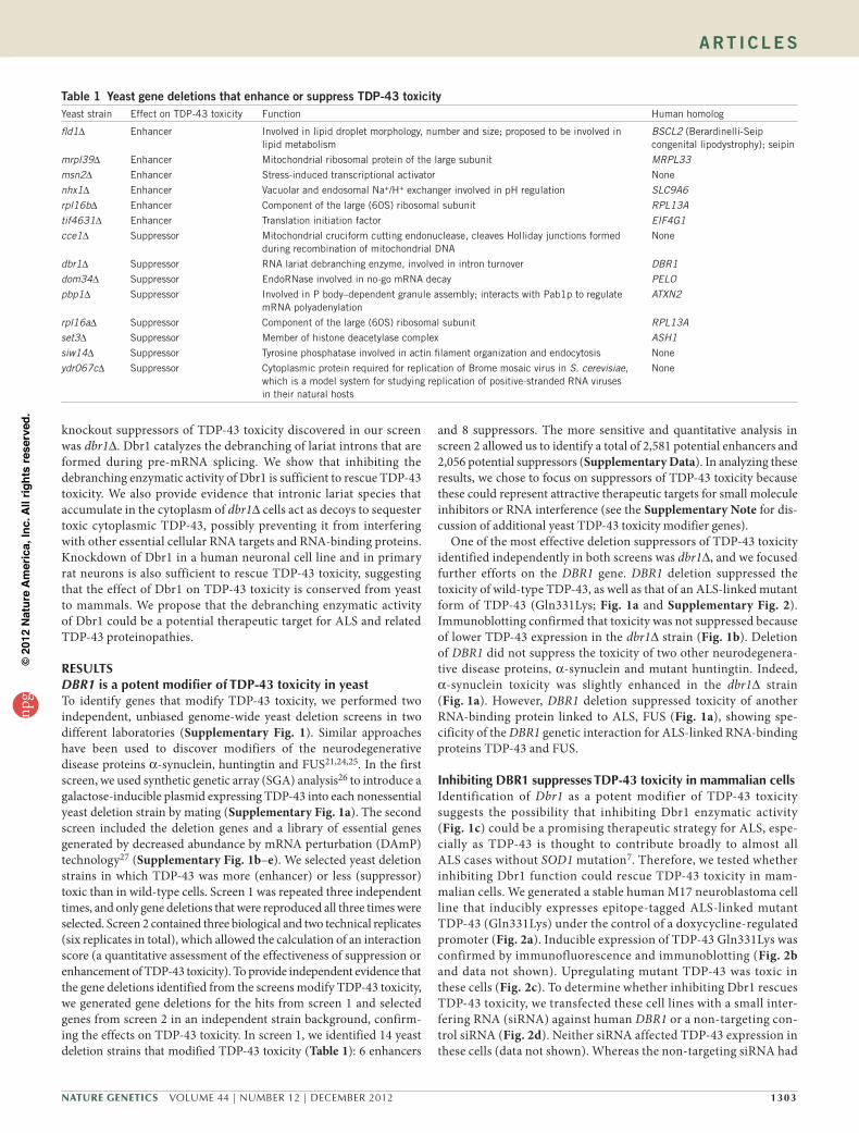
knockout suppressors of TDP-43 toxicity discovered in our screen was dbr1∆. Dbr1 catalyzes the debranching of lariat introns that are formed during pre-mRNA splicing. We show that inhibiting the debranching enzymatic activity of Dbr1 is sufficient to rescue TDP-43 toxicity. We also provide evidence that intronic lariat species that accumulate in the cytoplasm of dbr1∆ cells act as decoys to sequester toxic cytoplasmic TDP-43, possibly preventing it from interfering with other essential cellular RNA targets and RNA-binding proteins. Knockdown of Dbr1 in a human neuronal cell line and in primary rat neurons is also sufficient to rescue TDP-43 toxicity, suggesting that the effect of Dbr1 on TDP-43 toxicity is conserved from yeast to mammals. We propose that the debranching enzymatic activity of Dbr1 could be a potential therapeutic target for ALS and related TDP-43 proteinopathies.
RESULTSDBR1isapotentmodifierofTDP-43toxicityinyeastTo identify genes that modify TDP-43 toxicity, we performed two independent, unbiased genome-wide yeast deletion screens in two different laboratories (Supplementary Fig. 1). Similar approaches have been used to discover modifiers of the neurodegenerative disease proteins α-synuclein, huntingtin and FUS21,24,25. In the first screen, we used synthetic genetic array (SGA) analysis26 to introduce a galactose-inducible plasmid expressing TDP-43 into each nonessential yeast deletion strain by mating (Supplementary Fig. 1a). The second screen included the deletion genes and a library of essential genes generated by decreased abundance by mRNA perturbation (DAmP) technology27 (Supplementary Fig. 1b–e). We selected yeast deletion strains in which TDP-43 was more (enhancer) or less (suppressor) toxic than in wild-type cells. Screen 1 was repeated three independent times, and only gene deletions that were reproduced all three times were selected. Screen 2 contained three biological and two technical replicates (six replicates in total), which allowed the calculation of an interaction score (a quantitative assessment of the effectiveness of suppression or enhancement of TDP-43 toxicity). To provide independent evidence that the gene deletions identified from the screens modify TDP-43 toxicity, we generated gene deletions for the hits from screen 1 and selected genes from screen 2 in an independent strain background, confirm-ing the effects on TDP-43 toxicity. In screen 1, we identified 14 yeast deletion strains that modified TDP-43 toxicity (Table 1): 6 enhancers
and 8 suppressors. The more sensitive and quantitative analysis in screen 2 allowed us to identify a total of 2,581 potential enhancers and 2,056 potential suppressors (Supplementary Data). In analyzing these results, we chose to focus on suppressors of TDP-43 toxicity because these could represent attractive therapeutic targets for small molecule inhibitors or RNA interference (see the Supplementary Note for dis-cussion of additional yeast TDP-43 toxicity modifier genes).
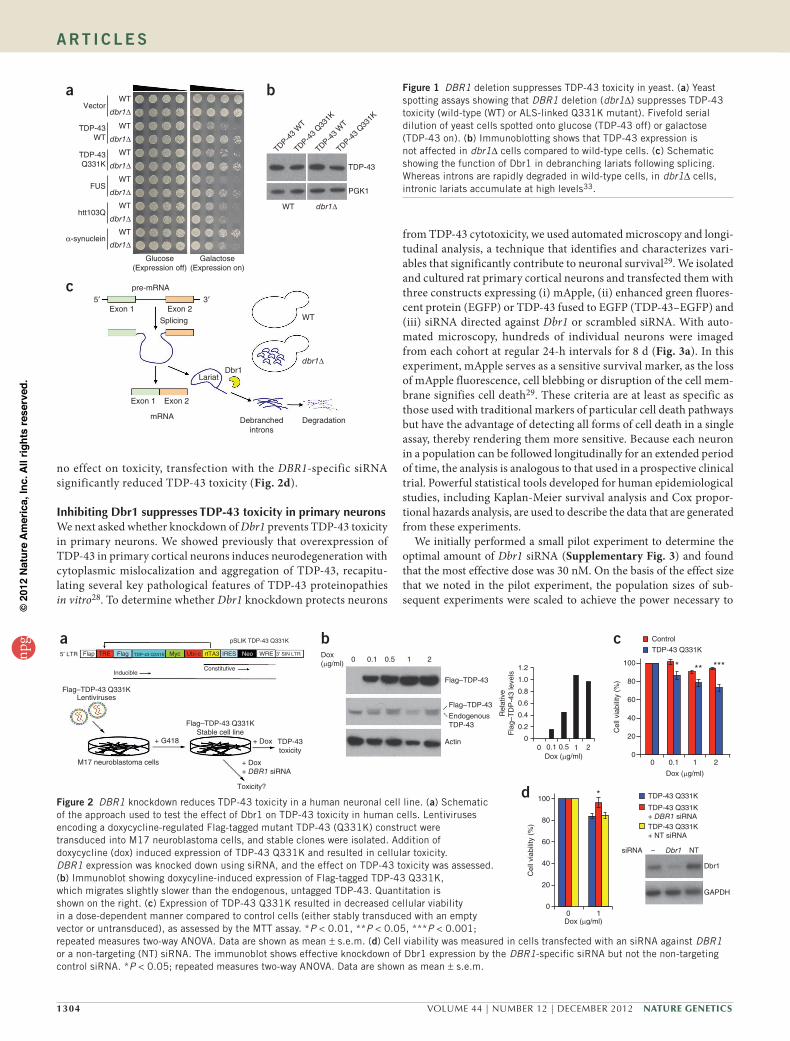
One of the most effective deletion suppressors of TDP-43 toxicity identified independently in both screens was dbr1∆, and we focused further efforts on the DBR1 gene. DBR1 deletion suppressed the toxicity of wild-type TDP-43, as well as that of an ALS-linked mutant form of TDP-43 (Gln331Lys; Fig. 1a and Supplementary Fig. 2). Immunoblotting confirmed that toxicity was not suppressed because of lower TDP-43 expression in the dbr1∆ strain (Fig. 1b). Deletion of DBR1 did not suppress the toxicity of two other neurodegenera-tive disease proteins, α-synuclein and mutant huntingtin. Indeed, α-synuclein toxicity was slightly enhanced in the dbr1∆ strain (Fig. 1a). However, DBR1 deletion suppressed toxicity of another RNA-binding protein linked to ALS, FUS (Fig. 1a), showing spe-cificity of the DBR1 genetic interaction for ALS-linked RNA-binding proteins TDP-43 and FUS.
InhibitingDBR1suppressesTDP-43toxicityinmammaliancellsIdentification of Dbr1 as a potent modifier of TDP-43 toxicity suggests the possibility that inhibiting Dbr1 enzymatic activity (Fig. 1c) could be a promising therapeutic strategy for ALS, espe-cially as TDP-43 is thought to contribute broadly to almost all ALS cases without SOD1 mutation7. Therefore, we tested whether inhibiting Dbr1 function could rescue TDP-43 toxicity in mam-malian cells. We generated a stable human M17 neuroblastoma cell line that inducibly expresses epitope-tagged ALS-linked mutant TDP-43 (Gln331Lys) under the control of a doxycycline-regulated promoter (Fig. 2a). Inducible expression of TDP-43 Gln331Lys was confirmed by immunofluorescence and immunoblotting (Fig. 2b and data not shown). Upregulating mutant TDP-43 was toxic in these cells (Fig. 2c). To determine whether inhibiting Dbr1 rescues TDP-43 toxicity, we transfected these cell lines with a small inter-fering RNA (siRNA) against human DBR1 or a non-targeting con-trol siRNA (Fig. 2d). Neither siRNA affected TDP-43 expression in these cells (data not shown). Whereas the non-targeting siRNA had
table 1 Yeast gene deletions that enhance or suppress tDP-43 toxicityYeast strain Effect on TDP-43 toxicity Function Human homolog
fld1∆ Enhancer Involved in lipid droplet morphology, number and size; proposed to be involved in lipid metabolism
BSCL2 (Berardinelli-Seip congenital lipodystrophy); seipin
mrpl39∆ Enhancer Mitochondrial ribosomal protein of the large subunit MRPL33msn2∆ Enhancer Stress-induced transcriptional activator None
nhx1∆ Enhancer Vacuolar and endosomal Na+/H+ exchanger involved in pH regulation SLC9A6rpl16b∆ Enhancer Component of the large (60S) ribosomal subunit RPL13Atif4631∆ Enhancer Translation initiation factor EIF4G1cce1∆ Suppressor Mitochondrial cruciform cutting endonuclease, cleaves Holliday junctions formed
during recombination of mitochondrial DNANone
dbr1∆ Suppressor RNA lariat debranching enzyme, involved in intron turnover DBR1dom34∆ Suppressor EndoRNase involved in no-go mRNA decay PELOpbp1∆ Suppressor Involved in P body–dependent granule assembly; interacts with Pab1p to regulate
mRNA polyadenylationATXN2
rpl16a∆ Suppressor Component of the large (60S) ribosomal subunit RPL13Aset3∆ Suppressor Member of histone deacetylase complex ASH1siw14∆ Suppressor Tyrosine phosphatase involved in actin filament organization and endocytosis None
ydr067c∆ Suppressor Cytoplasmic protein required for replication of Brome mosaic virus in S. cerevisiae, which is a model system for studying replication of positive-stranded RNA viruses in their natural hosts
None
A rt i c l e snp
g©
201
2 N
atur
e A
mer
ica,
Inc.
All
right
s re
serv
ed.
1304 VOLUME 44 | NUMBER 12 | DECEMBER 2012 Nature GeNetics
A rt i c l e s
no effect on toxicity, transfection with the DBR1-specific siRNA significantly reduced TDP-43 toxicity (Fig. 2d).
InhibitingDbr1suppressesTDP-43toxicityinprimaryneuronsWe next asked whether knockdown of Dbr1 prevents TDP-43 toxicity in primary neurons. We showed previously that overexpression of TDP-43 in primary cortical neurons induces neurodegeneration with cytoplasmic mislocalization and aggregation of TDP-43, recapitu-lating several key pathological features of TDP-43 proteinopathies in vitro28. To determine whether Dbr1 knockdown protects neurons
from TDP-43 cytotoxicity, we used automated microscopy and longi-tudinal analysis, a technique that identifies and characterizes vari-ables that significantly contribute to neuronal survival29. We isolated and cultured rat primary cortical neurons and transfected them with three constructs expressing (i) mApple, (ii) enhanced green fluores-cent protein (EGFP) or TDP-43 fused to EGFP (TDP-43–EGFP) and (iii) siRNA directed against Dbr1 or scrambled siRNA. With auto-mated microscopy, hundreds of individual neurons were imaged from each cohort at regular 24-h intervals for 8 d (Fig. 3a). In this experiment, mApple serves as a sensitive survival marker, as the loss of mApple fluorescence, cell blebbing or disruption of the cell mem-brane signifies cell death29. These criteria are at least as specific as those used with traditional markers of particular cell death pathways but have the advantage of detecting all forms of cell death in a single assay, thereby rendering them more sensitive. Because each neuron in a population can be followed longitudinally for an extended period of time, the analysis is analogous to that used in a prospective clinical trial. Powerful statistical tools developed for human epidemiological studies, including Kaplan-Meier survival analysis and Cox propor-tional hazards analysis, are used to describe the data that are generated from these experiments.
We initially performed a small pilot experiment to determine the optimal amount of Dbr1 siRNA (Supplementary Fig. 3) and found that the most effective dose was 30 nM. On the basis of the effect size that we noted in the pilot experiment, the population sizes of sub-sequent experiments were scaled to achieve the power necessary to
c
WTExon 2
Exon 2
mRNA
Splicing
pre-mRNA
Exon 1
Exon 1
3′5′
dbr1∆Dbr1
Lariat
Debranchedintrons
Degradation
b
TDP-43
WT
TDP-43
PGK1
WT dbr1∆
TDP-43
WT
TDP-43
Q331K
TDP-43
Q331K
aVector
TDP-43WT
TDP-43Q331K
FUS
htt103Q
α-synuclein
Glucose(Expression off)
Galactose(Expression on)
WT
dbr1∆
WT
dbr1∆
WT
dbr1∆
WT
dbr1∆
WT
dbr1∆
WT
dbr1∆
Figure 1 DBR1 deletion suppresses TDP-43 toxicity in yeast. (a) Yeast spotting assays showing that DBR1 deletion (dbr1∆) suppresses TDP-43 toxicity (wild-type (WT) or ALS-linked Q331K mutant). Fivefold serial dilution of yeast cells spotted onto glucose (TDP-43 off) or galactose (TDP-43 on). (b) Immunoblotting shows that TDP-43 expression is not affected in dbr1∆ cells compared to wild-type cells. (c) Schematic showing the function of Dbr1 in debranching lariats following splicing. Whereas introns are rapidly degraded in wild-type cells, in dbr1∆ cells, intronic lariats accumulate at high levels33.
a pSLIK TDP-43 Q331K
5′ LTR Flap TRE Flag TDP-43 Q331K
InducibleConstitutive
Myc Ubi-c rtTA3 IRES Neo WRE 3′ SIN LTR
Flag–TDP-43 Q331KLentiviruses
Flag–TDP-43 Q331KStable cell line
M17 neuroblastoma cells
+ G418 + Dox
+ Dox+ DBR1 siRNA
Toxicity?
TDP-43toxicity
b0 0.1 0.5 1 2
Flag–TDP-43
Flag–TDP-43
EndogenousTDP-43
Actin
Dox(µg/ml) 1.2
1.0
0.8
0.6
0.4
0.2
0.1 0.5 1 20
0
Rel
ativ
eF
lag–
TD
P-4
3 le
vels
Dox (µg/ml)
c100
Cel
l via
bilit
y (%
) 80
Control
TDP-43 Q331K
* ** ***
60
40
20
00 0.1
Dox (µg/ml)
1 2
d
Cel
l via
bilit
y (%
)
TDP-43 Q331K
TDP-43 Q331K+ DBR1 siRNATDP-43 Q331K+ NT siRNA
siRNA Dbr1
Dbr1
GAPDH
NT–
*100
80
60
40
20
00 1Dox (µg/ml)
Figure 2 DBR1 knockdown reduces TDP-43 toxicity in a human neuronal cell line. (a) Schematic of the approach used to test the effect of Dbr1 on TDP-43 toxicity in human cells. Lentiviruses encoding a doxycycline-regulated Flag-tagged mutant TDP-43 (Q331K) construct were transduced into M17 neuroblastoma cells, and stable clones were isolated. Addition of doxycycline (dox) induced expression of TDP-43 Q331K and resulted in cellular toxicity. DBR1 expression was knocked down using siRNA, and the effect on TDP-43 toxicity was assessed. (b) Immunoblot showing doxycyline-induced expression of Flag-tagged TDP-43 Q331K, which migrates slightly slower than the endogenous, untagged TDP-43. Quantitation is shown on the right. (c) Expression of TDP-43 Q331K resulted in decreased cellular viability in a dose-dependent manner compared to control cells (either stably transduced with an empty vector or untransduced), as assessed by the MTT assay. *P < 0.01, **P < 0.05, ***P < 0.001; repeated measures two-way ANOVA. Data are shown as mean ± s.e.m. (d) Cell viability was measured in cells transfected with an siRNA against DBR1 or a non-targeting (NT) siRNA. The immunoblot shows effective knockdown of Dbr1 expression by the DBR1-specific siRNA but not the non-targeting control siRNA. *P < 0.05; repeated measures two-way ANOVA. Data are shown as mean ± s.e.m.
npg
© 2
012
Nat
ure
Am
eric
a, In
c. A
ll rig
hts
rese
rved
.
Nature GeNetics VOLUME 44 | NUMBER 12 | DECEMBER 2012 1305
A rt i c l e s
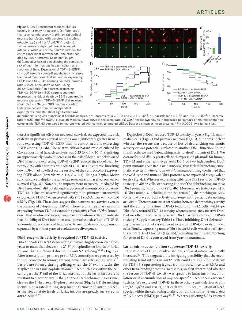
detect a significant effect on neuronal survival. As expected, the risk of death in primary cortical neurons was significantly greater in neu-rons expressing TDP-43–EGFP than in control neurons expressing EGFP alone (Fig. 3b). The relative risk or hazard ratio calculated by Cox proportional hazards analysis was 2.23 (P < 1 × 10⁻⁹), signifying an approximately twofold increase in the risk of death. Knockdown of Dbr1 in neurons expressing TDP-43–EGFP reduced the risk of death by nearly 20%, with a hazards ratio of 0.81 (P = 0.04). In contrast, knocking down Dbr1 had no effect on the survival of the control cohort express-ing EGFP alone (hazards ratio 1.2, P = 0.3). Using a Kaplan-Meier survival curve to display the same data revealed a similar effect on neuron survival (Fig. 3c). Notably, the improvement in survival mediated by Dbr1 knockdown did not depend on decreased amounts of cytoplasmic TDP-43. In fact, we observed significantly more primary neurons with cytoplasmic TDP-43 when treated with Dbr1 siRNA than with control siRNA (Fig. 3d). These data suggest that neurons can survive even in the presence of cytoplasmic TDP-43. These results in primary neurons expressing human TDP-43 extend the protective effect of Dbr1 knock-down that we observed in yeast and in neuroblastoma cells and indicate that the ability of Dbr1 inhibition to suppress the toxic effects of TDP-43 accumulation is conserved from yeast to mammalian cells, organisms separated by a billion years of evolutionary divergence.
Dbr1enzymaticactivityisrequiredforTDP-43toxicityDBR1 encodes an RNA debranching enzyme, highly conserved from yeast to man, that cleaves the 2′–5′ phosphodiester bonds of lariat introns that are formed during pre-mRNA splicing30,31 (Fig. 1c). After transcription, primary pre-mRNA transcripts are processed by the spliceosome to remove introns, which are released as lariats32. Lariats are formed during splicing when the 5′ exon attacks the 3′ splice site in a nucleophilic manner. RNA nucleases within the cell can digest the 3′ tail of the lariat introns, but the lariat structure is resistant to digestion until Dbr1, a specialized debranching enzyme, cleaves the 2′-hydroxyl-5′-phosphate bond (Fig. 1c). Debranching seems to be a rate-limiting step for the turnover of intronic RNA, as the steady-state levels of lariat introns are greatly increased in dbr1∆ cells31,33.
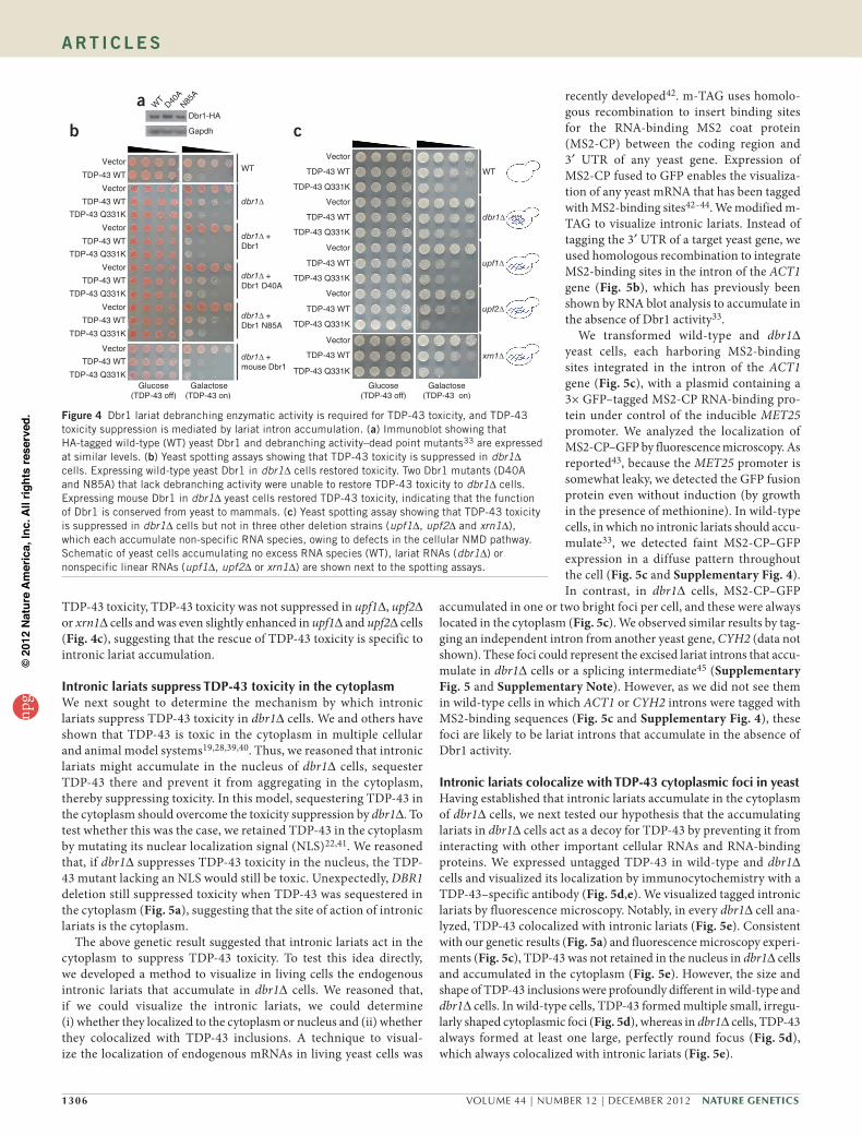
Depletion of Dbr1 reduced TDP-43 toxicity in yeast (Fig. 1), mam-malian cells (Fig. 2) and primary neurons (Fig. 3), but it was unclear whether the rescue was because of loss of debranching enzymatic activity or was potentially related to another Dbr1 function. To test this directly, we used ‘debranching activity–dead’ mutants of Dbr1. We cotransformed dbr1∆ yeast cells with expression plasmids for human TDP-43 and either wild-type yeast Dbr1 or two independent Dbr1 point mutants (Asp40Ala or Asn85Ala) that lack debranching enzy-matic activity in vitro and in vivo33. Immunoblotting confirmed that the wild-type and mutant Dbr1 proteins were expressed at equivalent levels (Fig. 4a). Whereas expressing wild-type Dbr1 restored TDP-43 toxicity to dbr1∆ cells, expressing either of the debranching-inactive Dbr1 point mutants did not (Fig. 4b). Moreover, we tested a panel of 16 Dbr1 mutants, including some that retain full debranching activity, some that have lost all activity and some with partial debranching activity33. There was an exact correlation between debranching activity and the ability to restore TDP-43 toxicity to dbr1∆ cells: wild-type Dbr1 fully restored TDP-43 toxicity, whereas completely inactive Dbr1 had no effect, and partially active Dbr1 partially restored TDP-43 toxicity (Supplementary Table 1). Thus, inhibiting Dbr1 debranch-ing enzymatic activity is sufficient to rescue TDP-43 toxicity in yeast cells. Finally, expressing mouse Dbr1 in dbr1∆ cells was also sufficient to restore TDP-43 toxicity (Fig. 4b), indicating that the debranching function of Dbr1 is conserved from yeast to mammals.
LariatintronaccumulationsuppressesTDP-43toxicityIn the absence of Dbr1, steady-state levels of lariat introns are greatly increased33. This suggested the intriguing possibility that the accu-mulating lariat introns in dbr1∆ cells could act as a kind of decoy for TDP-43, sequestering it away from important cellular RNAs and other RNA-binding proteins. To test this, we first determined whether the rescue of TDP-43 toxicity was specific to lariat intron accumu-lation or if accumulation of any nonspecific RNA species rescued toxicity. We expressed TDP-43 in three other yeast deletion strains (upf1∆, upf2∆ and xrn1∆) that each result in accumulation of RNA species within the cell, owing to alterations in the nonsense-mediated mRNA decay (NMD) pathway34–38. Whereas deleting DBR1 rescued
Figure 3 Dbr1 knockdown reduces TDP-43 toxicity in primary rat neurons. (a) Automated fluorescence microscopy of primary rat cortical neurons transfected with constructs encoding mApple (top) and TDP-43–EGFP (bottom). Two neurons are depicted here at repeated intervals. While one of the neurons lives for the entire experiment (arrowheads), the other has died by 144 h (arrows). Scale bar, 10 µm. (b) Cumulative hazard plot showing the cumulative risk of death for neurons in each cohort as a function of time. Expression of TDP-43–EGFP (n = 380 neurons counted) significantly increases the risk of death over that of neurons expressing EGFP alone (n = 245 neurons counted; hazards ratio = 2.2). Knockdown of Dbr1 using 30 nM Dbr1 siRNA in neurons expressing TDP-43–EGFP (n = 300 neurons counted) decreases the risk of death by 19% compared to neurons expressing TDP-43–EGFP that received scrambled siRNA (n = 380 neurons counted). Data were pooled from two independent experiments, and statistical significance was determined using Cox proportional hazards analysis. ***, hazards ratio = 2.23 and P < 1 × 10⁻⁹; **, hazards ratio = 1.83 and P < 1 × 10⁻⁴; *, hazards ratio = 0.81 and P = 0.04. (c) Kaplan-Meier survival curve of the same data. (d) Dbr1 knockdown results in increased percentage of neurons containing cytoplasmic TDP-43 compared to neurons treated with control, scrambled siRNA. Data are shown as mean ± s.e.m. *P < 0.0005, two-tailed t test.
c 100
80
*
60
40
20
0
0 50 100Time (h)
150 200
EGFP + scrambled siRNAEGFP + Dbr1 siRNATDP-43 + scrambled siRNATDP-43 + Dbr1 siRNA
Per
cent
sur
viva
l
a 24 h 48 h 72 h 97 h 121 h 144 h 168 h 193 h
TD
P-4
3–E
GF
Pm
App
le
d40
Neu
rons
with
cyto
plas
mic
TD
P-4
3 (%
)
30
20
10
Scram
bled
siRNA Db
r1
siRNA
0
*
b0.8
0.6
0.4
0.2
0
0 50 100Time (h)
150 200
TDP-43 + scrambled siRNATDP-43 + Dbr1 siRNA
EGFP + scrambled siRNAEGFP + Dbr1 siRNA
Cum
ulat
ive
risk
of d
eath *
*****
npg
© 2
012
Nat
ure
Am
eric
a, In
c. A
ll rig
hts
rese
rved
.
1306 VOLUME 44 | NUMBER 12 | DECEMBER 2012 Nature GeNetics
A rt i c l e s
TDP-43 toxicity, TDP-43 toxicity was not suppressed in upf1∆, upf2∆ or xrn1∆ cells and was even slightly enhanced in upf1∆ and upf2∆ cells (Fig. 4c), suggesting that the rescue of TDP-43 toxicity is specific to intronic lariat accumulation.
IntroniclariatssuppressTDP-43toxicityinthecytoplasmWe next sought to determine the mechanism by which intronic lariats suppress TDP-43 toxicity in dbr1∆ cells. We and others have shown that TDP-43 is toxic in the cytoplasm in multiple cellular and animal model systems19,28,39,40. Thus, we reasoned that intronic lariats might accumulate in the nucleus of dbr1∆ cells, sequester TDP-43 there and prevent it from aggregating in the cytoplasm, thereby suppressing toxicity. In this model, sequestering TDP-43 in the cytoplasm should overcome the toxicity suppression by dbr1∆. To test whether this was the case, we retained TDP-43 in the cytoplasm by mutating its nuclear localization signal (NLS)22,41. We reasoned that, if dbr1∆ suppresses TDP-43 toxicity in the nucleus, the TDP-43 mutant lacking an NLS would still be toxic. Unexpectedly, DBR1 deletion still suppressed toxicity when TDP-43 was sequestered in the cytoplasm (Fig. 5a), suggesting that the site of action of intronic lariats is the cytoplasm.
The above genetic result suggested that intronic lariats act in the cytoplasm to suppress TDP-43 toxicity. To test this idea directly, we developed a method to visualize in living cells the endogenous intronic lariats that accumulate in dbr1∆ cells. We reasoned that, if we could visualize the intronic lariats, we could determine (i) whether they localized to the cytoplasm or nucleus and (ii) whether they colocalized with TDP-43 inclusions. A technique to visual-ize the localization of endogenous mRNAs in living yeast cells was
recently developed42. m-TAG uses homolo-gous recombination to insert binding sites for the RNA-binding MS2 coat protein (MS2-CP) between the coding region and 3′ UTR of any yeast gene. Expression of MS2-CP fused to GFP enables the visualiza-tion of any yeast mRNA that has been tagged with MS2-binding sites42–44. We modified m-TAG to visualize intronic lariats. Instead of tagging the 3′ UTR of a target yeast gene, we used homologous recombination to integrate MS2-binding sites in the intron of the ACT1 gene (Fig. 5b), which has previously been shown by RNA blot analysis to accumulate in the absence of Dbr1 activity33.
We transformed wild-type and dbr1∆ yeast cells, each harboring MS2-binding sites integrated in the intron of the ACT1 gene (Fig. 5c), with a plasmid containing a 3× GFP–tagged MS2-CP RNA-binding pro-tein under control of the inducible MET25 promoter. We analyzed the localization of MS2-CP–GFP by fluorescence microscopy. As reported43, because the MET25 promoter is somewhat leaky, we detected the GFP fusion protein even without induction (by growth in the presence of methionine). In wild-type cells, in which no intronic lariats should accu-mulate33, we detected faint MS2-CP–GFP expression in a diffuse pattern throughout the cell (Fig. 5c and Supplementary Fig. 4). In contrast, in dbr1∆ cells, MS2-CP–GFP
accumulated in one or two bright foci per cell, and these were always located in the cytoplasm (Fig. 5c). We observed similar results by tag-ging an independent intron from another yeast gene, CYH2 (data not shown). These foci could represent the excised lariat introns that accu-mulate in dbr1∆ cells or a splicing intermediate45 (Supplementary Fig. 5 and Supplementary Note). However, as we did not see them in wild-type cells in which ACT1 or CYH2 introns were tagged with MS2-binding sequences (Fig. 5c and Supplementary Fig. 4), these foci are likely to be lariat introns that accumulate in the absence of Dbr1 activity.
IntroniclariatscolocalizewithTDP-43cytoplasmicfociinyeastHaving established that intronic lariats accumulate in the cytoplasm of dbr1∆ cells, we next tested our hypothesis that the accumulating lariats in dbr1∆ cells act as a decoy for TDP-43 by preventing it from interacting with other important cellular RNAs and RNA-binding proteins. We expressed untagged TDP-43 in wild-type and dbr1∆ cells and visualized its localization by immunocytochemistry with a TDP-43–specific antibody (Fig. 5d,e). We visualized tagged intronic lariats by fluorescence microscopy. Notably, in every dbr1∆ cell ana-lyzed, TDP-43 colocalized with intronic lariats (Fig. 5e). Consistent with our genetic results (Fig. 5a) and fluorescence microscopy experi-ments (Fig. 5c), TDP-43 was not retained in the nucleus in dbr1∆ cells and accumulated in the cytoplasm (Fig. 5e). However, the size and shape of TDP-43 inclusions were profoundly different in wild-type and dbr1∆ cells. In wild-type cells, TDP-43 formed multiple small, irregu-larly shaped cytoplasmic foci (Fig. 5d), whereas in dbr1∆ cells, TDP-43 always formed at least one large, perfectly round focus (Fig. 5d), which always colocalized with intronic lariats (Fig. 5e).
WT
dbr1∆
upf1∆
upf2∆
xrn1∆
Glucose(TDP-43 off)
Galactose(TDP-43 on)
Vector
TDP-43 WT
TDP-43 Q331K
Vector
TDP-43 WT
TDP-43 Q331K
Vector
TDP-43 WT
TDP-43 Q331K
Vector
TDP-43 WT
TDP-43 Q331K
Vector
TDP-43 WT
TDP-43 Q331K
c
aDbr1-HA
Gapdh
WT
D40A
N85A
WT
dbr1∆
dbr1∆ +Dbr1
dbr1∆ +mouse Dbr1
dbr1∆ +Dbr1 D40A
dbr1∆ +Dbr1 N85A
Vector
TDP-43 WT
Glucose(TDP-43 off)
Galactose(TDP-43 on)
Vector
TDP-43 WT
TDP-43 Q331K
Vector
TDP-43 WT
TDP-43 Q331K
Vector
TDP-43 WT
TDP-43 Q331K
Vector
TDP-43 WT
TDP-43 Q331K
Vector
TDP-43 WT
TDP-43 Q331K
b
Figure 4 Dbr1 lariat debranching enzymatic activity is required for TDP-43 toxicity, and TDP-43 toxicity suppression is mediated by lariat intron accumulation. (a) Immunoblot showing that HA-tagged wild-type (WT) yeast Dbr1 and debranching activity–dead point mutants33 are expressed at similar levels. (b) Yeast spotting assays showing that TDP-43 toxicity is suppressed in dbr1∆ cells. Expressing wild-type yeast Dbr1 in dbr1∆ cells restored toxicity. Two Dbr1 mutants (D40A and N85A) that lack debranching activity were unable to restore TDP-43 toxicity to dbr1∆ cells. Expressing mouse Dbr1 in dbr1∆ yeast cells restored TDP-43 toxicity, indicating that the function of Dbr1 is conserved from yeast to mammals. (c) Yeast spotting assay showing that TDP-43 toxicity is suppressed in dbr1∆ cells but not in three other deletion strains (upf1∆, upf2∆ and xrn1∆), which each accumulate non-specific RNA species, owing to defects in the cellular NMD pathway. Schematic of yeast cells accumulating no excess RNA species (WT), lariat RNAs (dbr1∆) or nonspecific linear RNAs (upf1∆, upf2∆ or xrn1∆) are shown next to the spotting assays.
npg
© 2
012
Nat
ure
Am
eric
a, In
c. A
ll rig
hts
rese
rved
.

Nature GeNetics VOLUME 44 | NUMBER 12 | DECEMBER 2012 1307
A rt i c l e s
IntroniclariatscompetewithRNAsforbindingtoTDP-43We also assessed whether the TDP-43 ribonucleoprotein complex contains lariats. We expressed Flag-tagged TDP-43 in wild-type and dbr1∆ cells and immunoprecipitated TDP-43 from cell lysates with an antibody to Flag. The associated RNA was separated by two- dimensional electrophoresis. Linear RNA molecules migrate in a diagonal on two-dimensional gels, but lariat and lariat breakdown products migrate in a separate arc, owing to the reduced mobility conferred by their altered structures (Supplementary Fig. 6a). As expected, in wild-type and dbr1∆ cells, TDP-43 associated with RNA. However, in dbr1∆ cells, the TDP-43 ribonucleoprotein complex contained additional RNA species, consistent with intronic lariats.
Finally, to directly test our hypothesis that the intronic lariats that form in dbr1∆ cells act as decoys for TDP-43 by competing with it for binding to other cellular RNAs, we performed in vitro electrophoretic mobility shift assays. Incubation of a radioactively labeled RNA probe known to bind TDP-43 (ref. 46) with recombinant TDP-43 results in a gel shift (Supplementary Fig. 6b,c). We isolated total RNA from wild-type and dbr1∆ cells and tested the ability of these RNAs to compete for binding to TDP-43. As expected, RNAs from wild-type and dbr1∆ cells competed for TDP-43 binding (Supplementary Fig. 6b), consistent with TDP-43 binding a large number of RNA targets47. To specifically test the ability of intronic lariats to compete for binding to TDP-43 (to act as decoys), we treated RNAs isolated from wild-type and dbr1∆ cells
with RNase R, which degrades all RNAs except for branched lariats48. When these RNase R–treated samples were incubated with the TDP-43 binding reaction, only the RNase R–treated RNAs isolated from dbr1∆ cells competed TDP-43 away from its binding site (Supplementary Fig. 6c). Thus, dbr1∆ cells contain an RNase R–resistant species, likely intronic lariats, that compete for binding to TDP-43.
DISCUSSIONUsing two unbiased genetic screens in yeast, we discovered Dbr1 as a potent modifier of TDP-43 toxicity. We provide evidence that, in the absence of Dbr1, intronic lariats accumulate in the cytoplasm, and we propose that these act as decoys to sequester TDP-43, pre-venting it from interfering with essential RNAs and RNA-binding proteins. Thus, we suggest that the debranching enzymatic activity of Dbr1 could be a novel therapeutic target to combat toxic effects from cytoplasmic TDP-43 aggregation in ALS. The additional genetic modifiers of TDP-43 toxicity uncovered from these two yeast screens (Table 1 and Supplementary Data) will hopefully provide even more insight into disease mechanisms and open new avenues for therapeutic intervention.
Recent studies indicate that RNA binding is an important compo-nent of TDP-43 toxicity in cellular and animal models19,22,49,50. One potential mechanism of neurotoxicity is that TDP-43 aggregates in the cytoplasm, sequestering essential cellular RNAs and RNA-binding
f
dbr1∆WT
TDP-43cytoplasmicaggregates
Essential RNAsand RNA-binding
proteins
Introniclariats
c
WT
dbr1∆
MS2-CP–GFP (×3) DAPI Merge
eWT +
TDP-43
dbr1∆ +vector
dbr1∆ +TDP-43
dbr1∆ +TDP-43
MS2-CP–GFP (×3) DAPI MergeTDP-43d
WT
dbr1∆
dbr1∆
DAPI MergeTDP-43
b MS2L (×2)
Exon ExonHomologous recombination
Cre recombination
CPGFP
GFPGFP
MS2-CP–GFP(×3)expression
Intron
Sphis5+
Sphis5+
a
VectorWT
WT
WT
dbr1∆
dbr1∆
dbr1∆
TDP-43 WT
Glucose(TDP-43 off)
Galactose(TDP-43 on)
TDP-43 ∆NLS
Figure 5 Intronic lariats colocalize with TDP-43 cytoplasmic foci in yeast. (a) The toxicity of a TDP-43 mutant that is retained in the cytoplasm (∆NLS) is also suppressed by dbr1∆. (b) Strategy to visualize lariat introns in living cells. Homologous recombination was used to insert an MS2 RNA-binding sequence in the intron of the ACT1 gene. A GFP-tagged MS2-CP protein was used to visualize accumulated intronic lariats. (c) In wild-type cells, lariat introns did not accumulate, and the MS2-CP–GFP signal was faint and diffusely localized throughout the cell. In contrast, in dbr1∆ cells, MS2-CP–GFP accumulated in one or two bright foci per cell, and these were always located in the cytoplasm. Scale bar, 5 µm. (d) The size and shape of TDP-43 cytoplasmic inclusions were different in dbr1∆ cells compared to wild-type cells. Whereas in wild-type cells, TDP-43 formed multiple small, irregularly shaped foci (arrowheads), in dbr1∆ cells, there was always at least one large, perfectly round focus per cell (arrows). Scale bar, 5 µm. (e) Untagged TDP-43 was expressed in wild-type and dbr1∆ cells and visualized by immunocytochemistry with a TDP-43–specific antibody. TDP-43 cytoplasmic foci colocalized with tagged lariat introns (arrows). Scale bar, 5 µm. (f) A model of how lariat intron accumulation suppresses TDP-43 cytoplasmic toxicity. In wild-type cells, TDP-43 aggregates in the cytoplasm and could interfere, via RNA binding, with essential RNAs and RNA-binding proteins. When Dbr1 activity is inhibited (for example, in dbr1∆ cells), lariat introns accumulate in the cytoplasm and might act as decoys, sequestering TDP-43, thereby preventing it from interfering with important cellular RNAs.
npg
© 2
012
Nat
ure
Am
eric
a, In
c. A
ll rig
hts
rese
rved
.
1308 VOLUME 44 | NUMBER 12 | DECEMBER 2012 Nature GeNetics
A rt i c l e s
proteins away from their normal functions. This could lead to cata-strophic changes in RNA metabolism, owing to defects in the sta-bility, splicing, transport and/or translation of essential RNAs. This effect might be more deleterious to motor neurons if motor neuron– specific transcripts were preferentially sequestered by TDP-43 or even if motor neurons were simply more sensitive to subtle alterations in any of these RNA metabolic pathways. TDP-43 loss of function likely also contributes to disease. TDP-43 is depleted from the nuclei of affected neuronal populations in individuals with ALS8, and downregulation of TDP-43 in mouse leads to widespread dysregu-lation of splicing47. Finally, TDP-43 missense variants might act in a dominant-negative manner, interfering with the function of the wild-type protein51,52. These three pathogenic mechanisms (gain of toxic properties in the cytoplasm, loss of function in the nucleus and dominant-negative effects) are not mutually exclusive and likely all contribute to ALS pathogenesis.
Inhibiting Dbr1 function might overcome or prevent cytoplasmic TDP-43 (and FUS) aggregates from disrupting critical RNAs and RNA-binding proteins. We propose that the accumulated intronic lariat species in dbr1∆ cells suppress TDP-43 toxicity by acting as decoys, causing TDP-43 to bind to them rather than to important cellular RNA targets (Fig. 5f). The catalog of genome-wide RNA targets for TDP-43 is emerging47,53–55. Notably, TDP-43 seems to pref-erentially bind to long intronic pre-mRNA sites rather than coding exonic regions or UTRs47. This might explain why the intronic RNA sequences (lariats) that accumulate in dbr1∆ cells sequester TDP-43 away from other RNA species in the cell and suppress TDP-43 toxicity. TDP-43 might also preferentially recognize the lariat branch-point structure, or binding to the lariat might promote nucleation or other alterations to TDP-43 conformation.
For Dbr1 to be a therapeutic target, several questions must be addressed. First, could inhibition of Dbr1 debranching activity be toxic itself? Indeed, the highest concentration of Dbr1 siRNA (50 nM) in primary rat neurons increased the risk of death (Supplementary Fig. 3), suggesting that too much Dbr1 inhibition could be deleteri-ous. However, in budding yeast, DBR1 deletion is tolerated (Fig. 1a), and siRNA knockdown in M17 cells does not result in growth inhibi-tion (ref. 56 and M.A. and A.D.G., unpublished data). Nevertheless, it will be important to determine what level of Dbr1 inhibition is toler-ated and whether an appropriate therapeutic index can be achieved. An additional consideration for this approach in mammalian cells is the non-coding RNAs in intronic regions that depend on Dbr1 activity. Expression of these non-coding RNAs might be dysregulated by Dbr1 inhibition, and the consequences of this must be assessed. Furthermore, as abnormal accumulation of RNA is hypothesized to trigger autoimmunity and there would likely be a large pool of lariats in human cells, the effects of Dbr1 inhibition on autoimmunity should be analyzed. Although, to our knowledge, Dbr1 inhibitors are not available, our new method to visualize intronic lariats in living cells (Fig. 4) will facilitate screening of chemical compound libraries for small molecule Dbr1 inhibitors.
There are currently no TDP-43–directed therapies for ALS or related TDP-43 proteinopathies. Antisense oligonucleotides and RNA interference approaches are emerging as attractive therapeutic strategies in neurodegenerative diseases in which decreasing levels of a toxic mutant protein might be efficacious57–60. Indeed, treating a mouse model of inherited ALS (caused by a mutation in SOD1) with antisense oligonucleotides to SOD1 significantly slowed disease progression4. This approach offers tremendous promise for treat-ing patients with ALS with SOD1 mutations, but, as mutations in SOD1 account for only ~2–5% of all ALS cases, additional therapeutic
strategies are needed. Because TDP-43 appears to contribute to ALS pathogenesis broadly7, targeting TDP-43 with antisense approaches could be effective. However, it is unknown whether TDP-43 contrib-utes to disease via loss- or gain-of-function mechanisms61, mandating caution in pursuing TDP-43–lowering approaches. We present an alternative strategy: by targeting Dbr1 using small molecule inhibitors or antisense oligonucleotides, it might be possible to antagonize the toxic effects of TDP-43 in the cytoplasm without interfering with potentially critical functions in the nucleus.
URLs. Study protocols, http://www.gladstone.ucsf.edu/gladstone/site/finkbeiner/.
METHODSMethods and any associated references are available in the online version of the paper.
Note: Supplementary information is available in the online version of the paper.
AcKNowlEDGMENtSWe thank K. Lynch and S. Smith for helpful suggestions and discussions about RNA and splicing; T. Nakaya for advice and assistance with lentivirus transduction experiments and analysis; C. Kurischko for advice and assistance with visualizing P bodies and stress granules; Q. Mitrovich and A. Plocik for advice with running the two-dimensional nucleic acid gels; S. Collins and D. Cameron for useful advice and assistance in performing the data analysis for screen 2; B. Hodges, D. Hosangadi, P. Patel, P. Nathanson and C. Mrejen for assistance with yeast experiments; J. Epstein and A. Raphael for critical comments on the manuscript and helpful suggestions; and G. Howard for editorial assistance. A. Elden helped with initial stages of this project. We are grateful to J. Gerst (Weizmann Institute) for providing the yeast m-TAG plasmids, R. Parker (University of Arizona) for sharing the P-body and stress granule marker plasmids and J. Weibezahn (University of California, San Francisco) for providing temperature-sensitive CDC48 (cdc48-3, SM 4783) and wild-type CDC48 isogenic (SM 5124) yeast strains. We thank B. Schwer (Weill Cornell Medical College) for providing the yeast mutant Dbr1 expression plasmids. We thank C. Boone (University of Toronto) for the MATα strain Y7092. This work was supported by US National Institutes of Health (NIH) Director’s New Innovator Awards 1DP2OD004417 (to A.D.G.) and 1DP2OD002177 (to J.S.), NIH grants NS065317 and NS065317 (to A.D.G.), NS067354 (to J.S.), GM084448, GM084279, GM081879 and GM098101 (to N.J.K.), NS39074 and NS045491 (to S.F.) and NS072233 (to S.J.B.), a New Scholar in Aging Award from the Ellison Medical Foundation (to J.S.), a grant from the Packard Center for ALS Research at Johns Hopkins (A.D.G. and J.S.), a grant from the Consortium for Frontotemporal Research (to R.V.F.), NIH grant 2P01AG02074 (to S.F.), a grant from the ALS Association (to S.F.) and the Taube-Koret Center and Hellman Family Foundation (to S.F.). A.D.G. is a Pew Scholar in the Biomedical Sciences, supported by The Pew Charitable Trusts, and a Rita Allen Foundation Scholar. N.J.K. is a Searle Scholar and a Keck Young Investigator. R.V.F. is an Investigator of the Gladstone Institutes. The J. David Gladstone Institutes received support from National Center for Research Resources Grant RR18928.
AUtHoR coNtRIBUtIoNSM.A., M.J.H., M.D.F., S.J.B., J.S., N.J.K., S.F., R.V.F. and A.D.G. designed the experiments. M.A., M.J.H., M.D.F., S.J.B., J.S., E.A.S., Z.D., X.F. and A.D.G. performed the research. M.A., M.J.H., M.D.F., S.J.B., J.S., N.J.K., S.F., R.V.F. and A.D.G. analyzed and interpreted data. M.A., M.J.H., M.D.F., S.J.B., S.F., R.V.F. and A.D.G. wrote the manuscript with contributions from all authors.
coMPEtING FINANcIAl INtEREStSThe authors declare competing financial interests: details are available in the online version of the paper.
Published online at http://www.nature.com/doifinder/10.1038/ng.2434. Reprints and permissions information is available online at http://www.nature.com/reprints/index.html.
1. Boillée, S., Vande Velde, C. & Cleveland, D.W. ALS: a disease of motor neurons and their nonneuronal neighbors. Neuron 52, 39–59 (2006).
2. Rosen, D.R. et al. Mutations in Cu/Zn superoxide dismutase gene are associated with familial amyotrophic lateral sclerosis. Nature 362, 59–62 (1993).
npg
© 2
012
Nat
ure
Am
eric
a, In
c. A
ll rig
hts
rese
rved
.
Nature GeNetics VOLUME 44 | NUMBER 12 | DECEMBER 2012 1309
A rt i c l e s
3. Bruijn, L.I. et al. Aggregation and motor neuron toxicity of an ALS-linked SOD1 mutant independent from wild-type SOD1. Science 281, 1851–1854 (1998).
4. Smith, R.A. et al. Antisense oligonucleotide therapy for neurodegenerative disease. J. Clin. Invest. 116, 2290–2296 (2006).
5. Lagier-Tourenne, C., Polymenidou, M. & Cleveland, D.W. TDP-43 and FUS/TLS: emerging roles in RNA processing and neurodegeneration. Hum. Mol. Genet. 19, R46–R64 (2010).
6. Gitler, A.D. & Shorter, J. RNA-binding proteins with prion-like domains in ALS and FTLD-U. Prion 5, 179–187 (2011).
7. Mackenzie, I.R. et al. Pathological TDP-43 distinguishes sporadic amyotrophic lateral sclerosis from amyotrophic lateral sclerosis with SOD1 mutations. Ann. Neurol. 61, 427–434 (2007).
8. Neumann, M. et al. Ubiquitinated TDP-43 in frontotemporal lobar degeneration and amyotrophic lateral sclerosis. Science 314, 130–133 (2006).
9. Pesiridis, G.S., Lee, V.M. & Trojanowski, J.Q. Mutations in TDP-43 link glycine-rich domain functions to amyotrophic lateral sclerosis. Hum. Mol. Genet. 18, R156–R162 (2009).
10. Rutherford, N.J. et al. Novel mutations in TARDBP (TDP-43) in patients with familial amyotrophic lateral sclerosis. PLoS Genet. 4, e1000193 (2008).
11. Sreedharan, J. et al. TDP-43 mutations in familial and sporadic amyotrophic lateral sclerosis. Science 319, 1668–1672 (2008).
12. Van Deerlin, V.M. et al. TARDBP mutations in amyotrophic lateral sclerosis with TDP-43 neuropathology: a genetic and histopathological analysis. Lancet Neurol. 7, 409–416 (2008).
13. Yokoseki, A. et al. TDP-43 mutation in familial amyotrophic lateral sclerosis. Ann. Neurol. 63, 538–542 (2008).
14. Belzil, V.V. et al. Mutations in FUS cause FALS and SALS in French and French Canadian populations. Neurology 73, 1176–1179 (2009).
15. Corrado, L. et al. Mutations of FUS gene in sporadic amyotrophic lateral sclerosis. J. Med. Genet. 47, 190–194 (2010).
16. Hewitt, C. et al. Novel FUS/TLS mutations and pathology in familial and sporadic amyotrophic lateral sclerosis. Arch. Neurol. 67, 455–461 (2010).
17. Kwiatkowski, T.J. Jr. et al. Mutations in the FUS/TLS gene on chromosome 16 cause familial amyotrophic lateral sclerosis. Science 323, 1205–1208 (2009).
18. Vance, C. et al. Mutations in FUS, an RNA processing protein, cause familial amyotrophic lateral sclerosis type 6. Science 323, 1208–1211 (2009).
19. Johnson, B.S., McCaffery, J.M., Lindquist, S. & Gitler, A.D. A yeast TDP-43 proteinopathy model: exploring the molecular determinants of TDP-43 aggregation and cellular toxicity. Proc. Natl. Acad. Sci. USA 105, 6439–6444 (2008).
20. Johnson, B.S. et al. TDP-43 is intrinsically aggregation-prone, and amyotrophic lateral sclerosis–linked mutations accelerate aggregation and increase toxicity. J. Biol. Chem. 284, 20329–20339 (2009).
21. Sun, Z. et al. Molecular determinants and genetic modifiers of aggregation and toxicity for the ALS disease protein FUS/TLS. PLoS Biol. 9, e1000614 (2011).
22. Elden, A.C. et al. Ataxin-2 intermediate-length polyglutamine expansions are associated with increased risk for ALS. Nature 466, 1069–1075 (2010).
23. Lagier-Tourenne, C. & Cleveland, D.W. Neurodegeneration: an expansion in ALS genetics. Nature 466, 1052–1053 (2010).
24. Willingham, S., Outeiro, T.F., DeVit, M.J., Lindquist, S.L. & Muchowski, P.J. Yeast genes that enhance the toxicity of a mutant huntingtin fragment or α-synuclein. Science 302, 1769–1772 (2003).
25. Giorgini, F., Guidetti, P., Nguyen, Q., Bennett, S.C. & Muchowski, P.J. A genomic screen in yeast implicates kynurenine 3-monooxygenase as a therapeutic target for Huntington disease. Nat. Genet. 37, 526–531 (2005).
26. Tong, A.H. et al. Systematic genetic analysis with ordered arrays of yeast deletion mutants. Science 294, 2364–2368 (2001).
27. Schuldiner, M. et al. Exploration of the function and organization of the yeast early secretory pathway through an epistatic miniarray profile. Cell 123, 507–519 (2005).
28. Barmada, S.J. et al. Cytoplasmic mislocalization of TDP-43 is toxic to neurons and enhanced by a mutation associated with familial amyotrophic lateral sclerosis. J. Neurosci. 30, 639–649 (2010).
29. Arrasate, M., Mitra, S., Schweitzer, E.S., Segal, M.R. & Finkbeiner, S. Inclusion body formation reduces levels of mutant huntingtin and the risk of neuronal death. Nature 431, 805–810 (2004).
30. Arenas, J. & Hurwitz, J. Purification of a RNA debranching activity from HeLa cells. J. Biol. Chem. 262, 4274–4279 (1987).
31. Chapman, K.B. & Boeke, J.D. Isolation and characterization of the gene encoding yeast debranching enzyme. Cell 65, 483–492 (1991).
32. Domdey, H. et al. Lariat structures are in vivo intermediates in yeast pre-mRNA splicing. Cell 39, 611–621 (1984).
33. Khalid, M.F., Damha, M.J., Shuman, S. & Schwer, B. Structure-function analysis of yeast RNA debranching enzyme (Dbr1), a manganese-dependent phosphodiesterase. Nucleic Acids Res. 33, 6349–6360 (2005).
34. Franks, T.M., Singh, G. & Lykke-Andersen, J. Upf1 ATPase-dependent mRNP disassembly is required for completion of nonsense-mediated mRNA decay. Cell 143, 938–950 (2010).
35. Gatfield, D. & Izaurralde, E. Nonsense-mediated messenger RNA decay is initiated by endonucleolytic cleavage in Drosophila. Nature 429, 575–578 (2004).
36. Lejeune, F., Li, X. & Maquat, L.E. Nonsense-mediated mRNA decay in mammalian cells involves decapping, deadenylating, and exonucleolytic activities. Mol. Cell 12, 675–687 (2003).
37. Larimer, F.W., Hsu, C.L., Maupin, M.K. & Stevens, A. Characterization of the XRN1 gene encoding a 5′→3′ exoribonuclease: sequence data and analysis of disparate protein and mRNA levels of gene-disrupted yeast cells. Gene 120, 51–57 (1992).
38. van Dijk, E.L. et al. XUTs are a class of Xrn1-sensitive antisense regulatory non-coding RNA in yeast. Nature 475, 114–117 (2011).
39. Igaz, L.M. et al. Dysregulation of the ALS-associated gene TDP-43 leads to neuronal death and degeneration in mice. J. Clin. Invest. 121, 726–738 (2011).
40. Zhang, Y.J. et al. Aberrant cleavage of TDP-43 enhances aggregation and cellular toxicity. Proc. Natl. Acad. Sci. USA 106, 7607–7612 (2009).
41. Winton, M.J. et al. Disturbance of nuclear and cytoplasmic TAR DNA-binding protein (TDP-43) induces disease-like redistribution, sequestration, and aggregate formation. J. Biol. Chem. 283, 13302–13309 (2008).
42. Haim, L., Zipor, G., Aronov, S. & Gerst, J.E. A genomic integration method to visualize localization of endogenous mRNAs in living yeast. Nat. Methods 4, 409–412 (2007).
43. Haim-Vilmovsky, L. & Gerst, J.E. m-TAG: a PCR-based genomic integration method to visualize the localization of specific endogenous mRNAs in vivo in yeast. Nat. Protoc. 4, 1274–1284 (2009).
44. Haim-Vilmovsky, L. & Gerst, J.E. Visualizing endogenous mRNAs in living yeast using m-TAG, a PCR-based RNA aptamer integration method, and fluorescence microscopy. Methods Mol. Biol. 714, 237–247 (2011).
45. Mayas, R.M., Maita, H., Semlow, D.R. & Staley, J.P. Spliceosome discards intermediates via the DEAH box ATPase Prp43p. Proc. Natl. Acad. Sci. USA 107, 10020–10025 (2010).
46. Buratti, E. & Baralle, F.E. Characterization and functional implications of the RNA binding properties of nuclear factor TDP-43, a novel splicing regulator of CFTR exon 9. J. Biol. Chem. 276, 36337–36343 (2001).
47. Polymenidou, M. et al. Long pre-mRNA depletion and RNA missplicing contribute to neuronal vulnerability from loss of TDP-43. Nat. Neurosci. 14, 459–468 (2011).
48. Suzuki, H. et al. Characterization of RNase R–digested cellular RNA source that consists of lariat and circular RNAs from pre-mRNA splicing. Nucleic Acids Res. 34, e63 (2006).
49. Voigt, A. et al. TDP-43–mediated neuron loss in vivo requires RNA-binding activity. PLoS ONE 5, e12247 (2010).
50. Li, Y. et al. A Drosophila model for TDP-43 proteinopathy. Proc. Natl. Acad. Sci. USA 107, 3169–3174 (2010).
51. Yang, C. et al. The C-terminal TDP-43 fragments have a high aggregation propensity and harm neurons by a dominant-negative mechanism. PLoS ONE 5, e15878 (2010).
52. Estes, P.S. et al. Wild-type and A315T mutant TDP-43 exert differential neurotoxicity in a Drosophila model of ALS. Hum. Mol. Genet. 20, 2308–2321 (2011).
53. Sephton, C.F. et al. Identification of neuronal RNA targets of TDP-43–containing ribonucleoprotein complexes. J. Biol. Chem. 286, 1204–1215 (2011).
54. Tollervey, J.R. et al. Characterizing the RNA targets and position-dependent splicing regulation by TDP-43. Nat. Neurosci. 14, 452–458 (2011).
55. Xiao, S. et al. RNA targets of TDP-43 identified by UV-CLIP are deregulated in ALS. Mol. Cell Neurosci. 47, 167–180 (2011).
56. Ye, Y., De Leon, J., Yokoyama, N., Naidu, Y. & Camerini, D. DBR1 siRNA inhibition of HIV-1 replication. Retrovirology 2, 63 (2005).
57. Xia, H., Mao, Q., Paulson, H.L. & Davidson, B.L. siRNA-mediated gene silencing in vitro and in vivo. Nat. Biotechnol. 20, 1006–1010 (2002).
58. Xia, H. et al. RNAi suppresses polyglutamine-induced neurodegeneration in a model of spinocerebellar ataxia. Nat. Med. 10, 816–820 (2004).
59. Singer, O. et al. Targeting BACE1 with siRNAs ameliorates Alzheimer disease neuropathology in a transgenic model. Nat. Neurosci. 8, 1343–1349 (2005).
60. Kordasiewicz, H.B. et al. Sustained therapeutic reversal of Huntington′s disease by transient repression of Huntingtin synthesis. Neuron 74, 1031–1044 (2012).
61. Da Cruz, S. & Cleveland, D.W. Understanding the role of TDP-43 and FUS/TLS in ALS and beyond. Curr. Opin. Neurobiol. 21, 904–919 (2011).
npg
© 2
012
Nat
ure
Am
eric
a, In
c. A
ll rig
hts
rese
rved
.
Nature GeNetics doi:10.1038/ng.2434
ONLINEMETHODSYeast strains, media and plasmids. The dbr1∆ strain was obtained by replac-ing the DBR1 coding region with a KanMX4 cassette in the BY4741 or W303 strain and verified by colony PCR. CEN and 2-micron galactose-inducible TDP-43 (ref. 28), 2-micron ∆NLS-TDP-43-YFP22, FUS21, α-synuclein62 and Htt103Q expression plasmids were as described63. The yeast Dbr1 mutant expression plasmids33 and m-TAG plasmids pLOXHIS5MS2L, pSH47 and pMS2-CP-GFP (×3) were as described44.
Yeast TDP-43 toxicity modifier screens. We used the SGA technique to screen the collection of nonessential-only yeast knockout strains (screen 1, performed in the laboratory of A.D.G.) or a collection of nonessential yeast knockout strains combined with essential genes reduced in expression by DAmP27 (screen 2, performed in the laboratories of R.V.F. and N.K.) for modifiers of TDP-43 toxicity. These screens were performed as described26,64,65, with some modifications66, using a Singer RoToR HDA (Singer Instruments). For screen 1, the galactose-inducible TDP-43 expression construct (pAG416Gal–TDP-43) was introduced into MATα strain Y7092 to generate the query strain. This query strain was mated to the yeast haploid deletion collection of nonessen-tial genes (MATα, each gene deleted by KanMX cassette; Fig. 1a). Haploid mutants harboring the TDP-43 expression plasmid were grown in the pres-ence of glucose (TDP-43 expression off) or galactose (TDP-43 expression on). After growth at 30 °C for 2 d, plates were photographed, and colony sizes were measured by ImageJ image analysis software as described67. The entire screen was repeated three times, and only hits that were reproduced all three times were selected for further validation by random spore analysis on DNA sequencing of deletion strain barcodes.
For screen 2, a control query strain containing the empty vector was gen-erated by transforming the yeast strain BY5563 (Matα) with p415-Gal-GFP, and the experimental query strain was generated by transforming BY5563 (Matα) with p415-Gal–TDP-43–GFP. Both strains were mated with the com-bined yeast knockout DAmP collection (Matα). Haploid yeast containing the deletion/DAmP yeast gene with the control or experiment plasmid were selected as described65. Three biological replicates were isolated by selecting for haploid yeast after sporulation on three plates. Each biological replicate was duplicated to generate six replicates (three biological and two technical replicates per biological replicate). Plates were imaged at several time points after final inoculation on glucose or galactose. Colony size was assessed as described67.
Isolation and culturing of primary cortical neurons. Rat cortical neurons were isolated from embryonic day 20–21 rat (Sprague Dawley) pups, cultured in serum-free medium in 96-well plates for 4 d in vitro and then transfected using Lipofectamine 2000 (Invitrogen; see URLs for full protocols). Neurons were transfected with plasmids (pGW1 backbone) encoding mApple and either TDP-43–EGFP or EGFP and with siRNA directed against Dbr1 (SMARTpool, Dharmacon) or scrambled siRNA (Dharmacon).
Live-cell imaging for longitudinal analysis. For neuronal survival analysis, we used a robotic imaging system as described68,69. Briefly, approximately 24–48 h after transfection, images were acquired with an inverted Nikon micro-scope (TE2000) equipped with PerfectFocus, an extra-long working distance (ELWD) 20× objective lens and an Andor Clara 16-bit, ultra-cooled camera. Illumination was provided by an adjustable, high-intensity and long-lasting xenon lamp, and a liquid light guide was used to maximize the signal-to-noise ratio. The stage and shutter movements, fluorescence excitation and emission filters, focusing and image acquisition were fully automated and were con-trolled through a 64-bit host computer running a combination of proprietary software (ImagePro, Media Cybernetics) and custom-designed algorithms.
Survival analysis. Digitized images from the red fluorescent protein (RFP; mApple) and green fluorescent protein (GFP; TDP-43–EGFP) channels were assembled into montages with ImagePro, and cells were prospectively counted in the RFP channel by original code developed in Matlab. Cell death was marked by loss of fluorescence, membrane rupture or neurite retraction. The time of death for each neuron was defined as the last time the neuron was seen. Kaplan-Meier and cumulative risk of death curves were generated using the
survival package in R. Statistical significance of survival differences between cohorts of neurons was determined by the log-rank test, and Cox proportional hazards analysis was used to measure the relative change in the risk of death, the hazard ratio.
Two-dimensional nucleic acid gels. Nucleic acid pellets from immunopre-cipitation were resuspended in 5 µl of nuclease-free water and 5 µl of 2× nucleic acid loading dye (0.025 g xylene cyanol and 0.025 g bromophenol blue in 100% formamide) and were heated at 85 °C for 5 min. Nucleic acids were separated in the first dimension by loading samples into a prerun (300 V for 10 min) 5% polyacrylamide gel (19:1 acrylamide:bis-acrylamide) containing 7 M urea and 1× TBE (90 mM Tris base, 90 mM boric acid, 2 mM EDTA, pH 8.0) and electrophoresing them at 300 V. Nucleic acids were separated in the second dimension by placing the entire lane from the first-dimension gel above the second-dimension gel. The second-dimension gel was exactly the same as the first-dimension gel, except that it contained 10% polyacrylamide. Samples were electrophoresed at 300 V until xylene cyanol dye reached the bottom of the second-dimension gel. Two-dimensional nucleic acid gels were washed briefly in water and then placed in 1× SYBR Gold (Invitrogen) solu-tion prepared in water. Gels were protected from light and gently agitated at 23–25 °C in 1× SYBR Gold solution for 45 min. Images of the gels were captured on an ultraviolet light box at an excitation wavelength of 302 nm with a 4-s exposure time.
Genomic tagging strategy to visualize intronic lariats. To visualize intronic lariats in dbr1∆ cells, we adapted the m-TAG protocol42 for use with introns. We used PCR-based chromosomal gene tagging, using homologous recom-bination to integrate the loxPøSphis5+øloxPøMS2L cassette into the intron of either the ACT1 gene or the CYH2 gene in wild-type or dbr1∆ cells. For ACT1, we inserted the MS2L cassette at nucleotide 159 of the ACT1 genomic DNA sequence (ACT1/YFL039C on chromosome VI from coordinates 54,696 to 53,260). For CYH2, we inserted the MS2L cassette at nucleotide 303 of the CYH2 genomic DNA sequence (RPL28/YGL103W on chromosome VII from coordinates 310,967 to 311,927). To excise the loxP-flanked Sphis5 cassette, we transformed cells with plasmid pSH47, which expresses Cre recombinase under control of a galactose-regulated promoter. Proper integration of MS2L sequences was verified by colony PCR and DNA sequencing.
Visualizing tagged intronic lariats by fluorescence microscopy. We visual-ized MS2L-tagged intronic lariats in wild-type and dbr1∆ cells as described42. We transformed wild-type and dbr1∆ cells harboring an MS2L-tagged ACT1 intron with pMS2–CP-GFP(×3), which expresses MS2-CP fused to three cop-ies of GFP under the control of the MET25 promoter. Yeast cells were fixed with 70% ethanol and stained with DAPI in Vectashield mounting medium. Colocalization of an MS2L-tagged intronic lariat with TDP-43 was determined using fluorescence microscopy to detect lariats (GFP) and immunocytochem-istry with a TDP-43–specific antibody to detect untagged TDP-43.
TDP-43 lentiviruses. To generate a lentiviral vector for the condi-tional expression of TDP-43 Gln331Lys, a Gateway entry clone encoding N-terminally Flag-tagged and C-terminally Myc-tagged TDP-43 Gln331Lys was used in a Gateway LR reaction (Invitrogen) to transfer it to the pSLIK-Neo lentiviral destination vector. Recombinant lentiviruses were produced as described70. For lentiviral transduction, human M17 neuroblastoma cells (ATCC) were incubated with lentiviral supernatants for 6 h at 37 °C. Stable cell lines were selected by culturing transduced cells for 5 d in growth medium (minimum essential medium (MEM) alpha/F-12 (Invitrogen) sup-plemented with 10% heat-inactivated FBS, 100 U/ml penicillin and 100 µg/ml streptomycin (complete medium) with 100 µg/ml Geneticin). Individual clones were selected and analyzed for inducible expression by immunoblot-ting and immunofluorescence analyses.
TDP-43 toxicity assays in mammalian cells. We assessed the effect of TDP-43 on cell proliferation in the MTT assay. M17 cells stably transduced with either an empty vector or with the vector encoding TDP-43 Gln331Lys were seeded in 96-well plates. To induce TDP-43 Gln331Lys expression, doxycycline (0.1, 1 or 2 µg/ml) was added to the growth medium, and cells were incubated
npg
© 2
012
Nat
ure
Am
eric
a, In
c. A
ll rig
hts
rese
rved
.
Nature GeNeticsdoi:10.1038/ng.2434
for 3 d. To measure proliferation, MTT (3-(4,5-dimethylthiazol-2-yl)-2,5-diphenyltetrazolium bromide) was added to each well, and cells were incub-ated for 4 h at 37 °C. Acidic isopropanol (40 mM HCl) was then added to each well to solubilize the blue formazan crystals. Absorbance at 570 nm was read with a Tecan Safire II plate reader. Absorbance measurements were normalized to the absorbance of cells not treated with doxycycline and used to calculate the percent viability under each condition. Two-way ANOVA was used to evaluate differences in means between two groups, and P values of <0.05 were considered statistically significant. Each condition was replicated at least four times, and each experiment was independently repeated at least three times.
siRNA knockdown of DBR1 in M17 cells. siRNA targeting human DBR1 and non-targeting control siRNA were purchased from Thermo-Scientific (DBR1, J-008290-08-0005; non-targeting, D-001810-01-05). siRNA knockdown was validated by immunoblotting with a Dbr1-specific antibody (Proteintech, rab-bit polyclonal antibody, 16019-1-AP). The M17 cell line with inducible TDP-43 Gln331Lys expression was transfected with 50 nM human DBR1 siRNA or non-targeting control siRNA using RNAi Lipofectamine (Invitrogen). After 2 d, cells were transferred to medium containing doxycycline (0 and 1 µg/ml) to induce expression of TDP-43, and toxicity was then assessed by MTT assay after 2 d (on day 4 after transfection). TDP-43 toxicity in M17 cells transfected with DBR1 siRNA was compared to toxicity in cells transfected with non- targeting control siRNA or empty vector.
TDP-43 in vitro RNA-binding assay. Glutathione S-transferase (GST)–TDP-43 was purified from Escherichia coli as described20. RNA binding assays were performed as described71. Briefly, competitor RNA was purified from wild-type or dbr1∆ W303 yeast using the RNeasy Mini kit (Qiagen) and was either left untreated or was treated with RNase R to degrade any linear RNA structures while leaving lariat RNA intact. After RNase R treatment, RNA was purified
using the RNeasy MinElute Cleanup kit (Qiagen). A radioactively labeled RNA probe, UG6, which is known to bind TDP-43 (ref. 46), was generated by T7 polymerase–catalyzed transcription of a DNA template in the presence of [32P]-labeled UTP. Standard binding reactions were carried out in a 10-µl volume containing 4 mM MgCl2, 25 mM phosphocreatine, 1.25 mM ATP, 1.3% polyvinyl alcohol, 25 ng of yeast tRNA, 0.8 mg of BSA, 1 mM DTT, 0.1 µl Rnasin (Promega, 40 U/ml), 75 mM KCl, 10 mM Tris, pH 7.5, 0.1 mM EDTA and 10% glycerol. Binding reactions containing GST–TDP-43 (0.5 µM) and [32P]-labeled UG6 (1 µl) were incubated for 15 min at 30 °C in the presence of various dilutions of competitor RNA (0–12.9 µg). Binding reactions were then transferred to ice, and heparin was added to a final concentration of 0.5 µg/µl. Reactions were analyzed on a 4.5% native gel (acrylamide:bis-acrylamide (29:1), Bio-Rad).
62. Cooper, A.A. et al. Alpha-synuclein blocks ER-Golgi traffic and Rab1 rescues neuron loss in Parkinson′s models. Science 313, 324–328 (2006).
63. Duennwald, M.L. & Lindquist, S. Impaired ERAD and ER stress are early and specific events in polyglutamine toxicity. Genes Dev. 22, 3308–3319 (2008).
64. Tong, A.H. et al. Global mapping of the yeast genetic interaction network. Science 303, 808–813 (2004).
65. Tong, A.H. & Boone, C. Synthetic genetic array analysis in Saccharomyces cerevisiae. Methods Mol. Biol. 313, 171–192 (2006).
66. Armakola, M., Hart, M.P. & Gitler, A.D. TDP-43 toxicity in yeast. Methods 53, 238–245 (2011).
67. Collins, S.R., Schuldiner, M., Krogan, N.J. & Weissman, J.S. A strategy for extracting and analyzing large-scale quantitative epistatic interaction data. Genome Biol. 7, R63 (2006).
68. Sharma, P., Ando, D.M., Daub, A., Kaye, J.A. & Finkbeiner, S. High-throughput screening in primary neurons. Methods Enzymol. 506, 331–360 (2012).
69. Arrasate, M. & Finkbeiner, S. Automated microscope system for determining factors that predict neuronal fate. Proc. Natl. Acad. Sci. USA 102, 3840–3845 (2005).
70. Shinbo, Y. et al. Proper SUMO-1 conjugation is essential to DJ-1 to exert its full activities. Cell Death Differ. 13, 96–108 (2006).
71. Rothrock, C.R., House, A.E. & Lynch, K.W. HnRNP L represses exon splicing via a regulated exonic splicing silencer. EMBO J. 24, 2792–2802 (2005).
npg
© 2
012
Nat
ure
Am
eric
a, In
c. A
ll rig
hts
rese
rved
.