Embed Size (px)
Citation preview

Full Terms & Conditions of access and use can be found athttps://www.tandfonline.com/action/journalInformation?journalCode=ibmg20
Critical Reviews in Biochemistry and Molecular Biology
ISSN: 1040-9238 (Print) 1549-7798 (Online) Journal homepage: https://www.tandfonline.com/loi/ibmg20
Inhibition of farnesyl pyrophosphate (FPP)and/or geranylgeranyl pyrophosphate (GGPP)biosynthesis and its implication in the treatmentof cancers
Daniel D. Waller, Jaeok Park & Youla S. Tsantrizos
To cite this article: Daniel D. Waller, Jaeok Park & Youla S. Tsantrizos (2019): Inhibition offarnesyl pyrophosphate (FPP) and/or geranylgeranyl pyrophosphate (GGPP) biosynthesis and itsimplication in the treatment of cancers, Critical Reviews in Biochemistry and Molecular Biology
To link to this article: https://doi.org/10.1080/10409238.2019.1568964
Published online: 18 Feb 2019.
Submit your article to this journal
View Crossmark data

REVIEW ARTICLE
Inhibition of farnesyl pyrophosphate (FPP) and/or geranylgeranylpyrophosphate (GGPP) biosynthesis and its implication in thetreatment of cancers
Daniel D. Wallera, Jaeok Parkb,c and Youla S. Tsantrizosb,c
aDepartment of Medicine, McGill University, Montreal, Canada; bDepartment of Chemistry, McGill University, Montreal, Canada;cDepartment of Biochemistry, McGill University, Montreal, Canada
ABSTRACTDysregulation of isoprenoid biosynthesis is implicated in numerous biochemical disorders thatplay a role in the onset and/or progression of age-related diseases, such as hypercholesterolemia,osteoporosis, various cancers, and neurodegeneration. The mevalonate metabolic pathway isresponsible for the biosynthesis of the two key isoprenoid metabolites, farnesyl pyrophosphate(FPP) and geranylgeranyl pyrophosphate (GGPP). Post-translational prenylation of various pro-teins, including the small GTP-binding proteins (GTPases), with either FPP or GGPP is vital forproper localization and activation of these proteins. Prenylated GTPases play a critical role in cellsignaling, proliferation, cellular plasticity, oncogenesis, and cancer metastasis. Pre-clinical andclinical studies strongly suggest that inhibition of protein prenylation can be an effective treat-ment for non-skeletal cancers. In this review, we summarize the most recent drug discoveryefforts focusing on blocking protein farnesylation and/or geranylgeranylation and the biochem-ical and structural data available in guiding the current on-going studies in drug discovery.Furthermore, we provide a summary on the biochemical association between disruption of pro-tein prenylation, endoplasmic reticulum (ER) stress, unfolded protein response (UPR) signaling,and cancer.
ARTICLE HISTORYReceived 23 November 2018Revised 9 January 2019Accepted 9 January 2019
KEYWORDSIsoprenoids; cancer;mevalonate; GTPases;metabolism; age-related diseases
Introduction
The mevalonate pathway is responsible for the biosyn-thesis of all mammalian isoprenoids (Figure 1). Thesemetabolites serve as the starting material for numerousother essential biomolecules, including steroids, bileacids, lipoproteins, vitamin D, heme A, ubiquinone, doli-chol, and isopentenyladenine (Goldstein and Brown1990). Consequently, this pathway is crucial in a pleth-ora of biological processes that maintain the integrityof cell membranes (e.g. cholesterol), the balance ofreproductive hormones (e.g. estradiol, progesterone,testosterone), electron transport mechanisms (e.g. ubi-quinone), glycoprotein biosynthesis (dolichol), andmodifications of tRNAs (e.g. biosynthesis of isopenteny-ladenine). Additionally, isoprenoids are essential for thepost-translational prenylation and activation of manyproteins that are important to human health.
The rate-limiting step of the mevalonate pathway iscatalyzed by hydroxymethylglutaryl coenzyme A (HMG-CoA) reductase, leading to the formation of mevalonic
acid. This metabolite is the immediate precursor of the5-carbon isoprenoid units, isopentenyl pyrophosphate(IPP) and dimethylallyl pyrophosphate (DMAPP;Figure 1). In humans, the first branching point of thepathway is occupied by the enzyme farnesyl pyrophos-phate synthase (hFPPS), which is responsible for thecatalytic elongation of DMAPP first to the C-10 metab-olite geranyl pyrophosphate (GPP) and then to the C-15isoprenoid farnesyl pyrophosphate (FPP). The immedi-ate downstream enzyme, the human geranylgeranylpyrophosphate synthase (hGGPPS) catalyzes the exten-sion of the FPP substrate to the C-20 isoprenoid gera-nylgeranyl pyrophosphate (GGPP).
Post-translational modification of proteins witheither FPP or GGPP is estimated to account for approxi-mately 2% of all mammalian proteins (Nguyen et al.2009). Known farnesylated proteins include many of thesmall GTP-binding proteins (GTPases), such as the Rassuperfamily (e.g. H/K/N-Ras) (Kho et al. 2004), but alsoothers proteins, such as the DnaJ chaperone proteins
CONTACT Youla S. Tsantrizos [email protected] Department of Chemistry and Department of Biochemistry, McGill University, 801Sherbrooke Street West, Montreal, Quebec H3A 1A1, Canada� 2019 Informa UK Limited, trading as Taylor & Francis Group
CRITICAL REVIEWS IN BIOCHEMISTRY AND MOLECULAR BIOLOGYhttps://doi.org/10.1080/10409238.2019.1568964

(Kampinga and Craig 2010; Stark et al. 2014) and theprecursor peptide of the nuclear lamin A (Young et al.2005; Chang et al. 2012). Geranylgeranylated GTPasesinclude the Rho family of proteins (e.g. RhoA/B/C), theRas-related proteins Rap1A, Rac-1 and Rab GTPases,and Cdc42. Post-translational prenylation of these pro-teins provides them with the ability to associate specif-ically with cellular membranes and participate in aplethora of biochemical mechanisms that are essentialto cell survival, cell signaling, and proliferation (Takaiet al. 2001), biological events that play a critical role inoncogenesis and cancer metastasis. It is noteworthythat there is also a strong association between prenyla-tion and the synaptic plasticity of neurons (Hottmanand Li 2014), as well as neurodegeneration andAlzheimer’s disease (Eckert et al. 2009; Hooff et al. 2010;Hooff et al. 2012; De Schutter et al. 2014; Pelleieuxet al. 2018).
In the past, drug discovery efforts targeting differentsteps of the mevalonate pathway focused mainly oninhibition of HMG-CoA reductase and hFPPS for thepurpose of treating hypercholesterolemia and lyticbone disease, respectively. The statins are the bestexample of highly successful drugs targeting HMG-CoAreductase and widely used to reduce the risk of
cardiovascular diseases. Statins represent an excellentexample of a clinically validated class of prophylacticdrugs that targets an essential metabolic pathway withminimal or negligible adverse effects.
Bisphosphonate drugs (BPs) are effective antiresorp-tive agents for the treatment of osteoporosis that wereinitially reported in the 1960s (Russell 2011). Althoughall BP drugs bind to bone and block osteoclastic activ-ity, the molecular mechanism of action of the earlieranalogs (i.e. the non-nitrogen containing analogs), suchas clodronate (1a) and etidronate (1b), is different fromthat of the more potent (second generation analogs)nitrogen-containing BPs (N-BPs) which inhibit hFPPSwith significant selectivity. The N-BP family of com-pounds includes hydrocarbon-based analogs, such aspamidronate (2a), alendronate (2b) and ibandronatoe(3), as well as heteroaromatic compounds, such as zole-dronic acid (4a), risedronic acid (5a) and minodronicacid; some structures shown in Figure 2. Several excel-lent review articles have been written specifically onthe therapeutic value of N-BPs and their osteoclast-mediated inhibition of bone resorption (Dunford et al.2001; Dunford 2010; Fournier et al. 2010; Ebetino et al.2011; Russell, 2011). Due to the high charge density ofthe bisphosphonate moiety, which exists as the trianion
Figure 1. Schematic representation of the mevalonate pathway, indicating the major biochemical steps in isoprenoid biosyn-thesis. Important classes of clinically validated inhibitors of key enzymes in the pathway are indicated (e.g. statins and bisphosph-onates) (see colour version of this figure at www.tandfonline.com/ibmg).
2 D. D. WALLER ET AL.

under physiological conditions, BPs and N-BPs sufferfrom poor drug-like properties (in the classical sense).Nonetheless, N-BPs are important human therapeuticsthat improve the quality of life for patients with lyticbone diseases. For example, approximately 50% of allpost-menopausal women suffer from osteoporosis andusually treated with N-BPs (Eastell et al. 2011) andapproximately 70–80% of breast and prostate cancersultimately metastasize to bone. In multiple myeloma(MM) patients, osteolytic lesions are one of several hall-mark clinical features and more than 90% of thesepatients will develop bone lesions at some stage oftheir disease (Bianch and Munshi 2015). Skeletal mani-festations of the aforementioned cancers are a majorcause of morbidity that can be characterized by severepain, impaired mobility, bone fractures, spinal cordcompression, and hypercalcemia.
In addition to the hFPPS-mediated antiresorptiveproperties of N-BPs, numerous biochemical studieshave suggested a strong association between the inhib-ition of protein prenylation and cancer cell survival(Clendening et al. 2010; Sorrentino et al. 2014; Mullenet al. 2016) or metastasis (Dudakovic et al. 2011).Although, inhibition of the mevalonate pathway at theHMG-CoA reductase step with statins has also beenimplicated in better survival of patients with variouscancers (Nielsen et al. 2012; Kubatka et al. 2014), includ-ing breast cancer (Garwood et al. 2010) and MM(Sanfilippo et al. 2016), drug discovery in oncology hasfocused mainly on the downstream enzymes, hFPPS,hGGPPS, and their corresponding prenyl transferaseenzymes, farnesyl transferase (FTase), as well as geranyl-geranyl transferases (GGTase) I and II (Figure 1). Thisreview will focus mainly on recent efforts aiming atdownregulating the intracellular levels of FPP and/orGGPP biosynthesis, and consequently, the prenylationof proteins implicated in cancer.
The structure and function of the humanfarnesyl pyrophosphate synthase (hFPPS)
The mechanism of action of the early BP compounds(e.g. Figure 2; 1a) does not involve inhibition of anyparticular enzyme of the isoprenoid pathway. Instead,metabolic incorporation of these compounds into sta-ble ATP derivatives is believed to interfere with ATP-dependent cellular pathways (Rogers et al. 1992). Incontrast, the main biological target of the N-BP drugs isthe hFPPS enzyme, blocking the biosynthesis of FPPand modulating a large biochemical cascade thatimpacts both upstream and downstream events in themevalonate pathway (Figure 1). The details of the
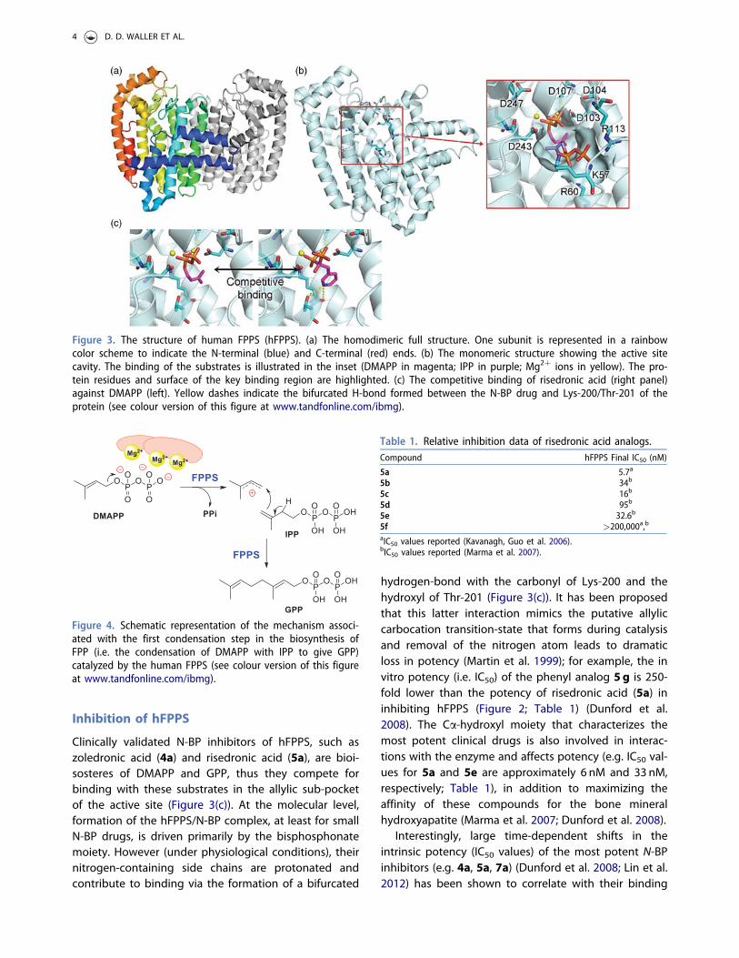
interactions between N-BP drugs and hFPPS were firstreported in 2006, independently by scientists fromNovartis (Rondeau et al. 2006) and the StructuralGenomic Consortium at the University of Oxford(Kavanagh, Guo et al. 2006). Their crystallographic stud-ies revealed that hFPPS consists of a large cavity, havingtwo charged surfaces on opposite walls of the cavityand only a small, partly lipophilic region (Figure 3). Oneof these surfaces is composed of two conserved aspar-tate-rich motifs (117DDIMD121, 257DDYLD261) that bindthe pyrophosphate moiety of the substrates DMAPP/GPP via metal-mediated interactions with three magne-sium cations (Mg2þ; Figure 3(b)). Adjacent to this sur-face is the small lipophilic region, which is lined withthe side chains of Phe98 and Phe99 (commonly referredto as the “capping” phenyls), which binds the lipophilictail of the enzyme’s catalytic product (i.e. the lipophilictail of GPP and FPP). The capping phenyls define thesize of this lipophilic pocket and control the extent ofisoprenoid polymerization to a maximum chain lengthof C-15 (Tarshis et al. 1996). Mutation of these phenylshas been shown to result in errors in the final length ofthe product’s hydrophobic side chain.
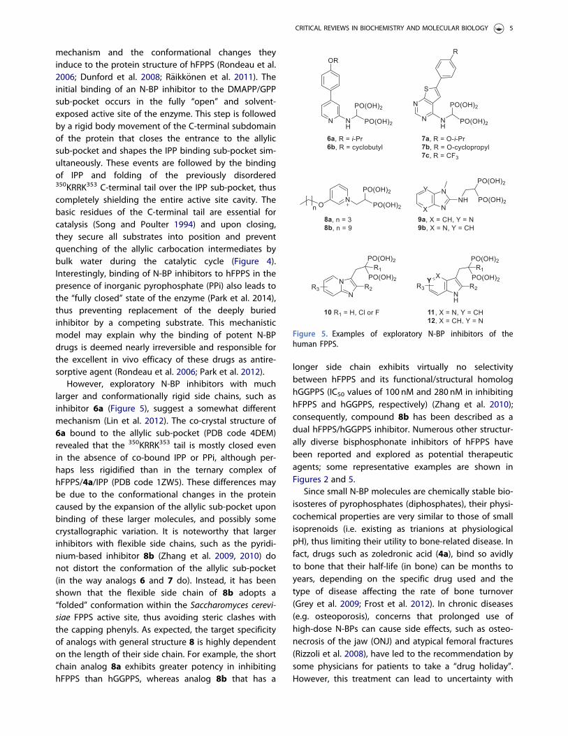
The IPP binding sub-pocket is at the opposite wall ofthe DMAP/GPP binding cavity and lined with the posi-tively-charged side chains of Arg and Lys residues,which interact directly with the IPP’s pyrophosphatemoiety (Figure 3(b)). The lipophilic side chains of thesubstrates lie against each other within van der Waalsdistance. Each condensation step in the polymerizationreaction is driven by the dissociation of the pyrophos-phate moiety of DMAP (or GPP in the second step) togive an allylic carbocation intermediate (this sub-pocketis also known as the allylic sub-pocket), which is subse-quently captured by the double bond of an IPP unitwith concerted deprotonation of IPP (Figure 4) (Poulteret al. 1978).
Figure 2. Examples of clinically validated N-BP inhibitors ofthe human FPPS and some of their derivatives.
CRITICAL REVIEWS IN BIOCHEMISTRY AND MOLECULAR BIOLOGY 3
Inhibition of hFPPS
Clinically validated N-BP inhibitors of hFPPS, such aszoledronic acid (4a) and risedronic acid (5a), are bioi-sosteres of DMAPP and GPP, thus they compete forbinding with these substrates in the allylic sub-pocketof the active site (Figure 3(c)). At the molecular level,formation of the hFPPS/N-BP complex, at least for smallN-BP drugs, is driven primarily by the bisphosphonatemoiety. However (under physiological conditions), theirnitrogen-containing side chains are protonated andcontribute to binding via the formation of a bifurcated
hydrogen-bond with the carbonyl of Lys-200 and thehydroxyl of Thr-201 (Figure 3(c)). It has been proposedthat this latter interaction mimics the putative allyliccarbocation transition-state that forms during catalysisand removal of the nitrogen atom leads to dramaticloss in potency (Martin et al. 1999); for example, the invitro potency (i.e. IC50) of the phenyl analog 5g is 250-fold lower than the potency of risedronic acid (5a) ininhibiting hFPPS (Figure 2; Table 1) (Dunford et al.2008). The Ca-hydroxyl moiety that characterizes themost potent clinical drugs is also involved in interac-tions with the enzyme and affects potency (e.g. IC50 val-ues for 5a and 5e are approximately 6 nM and 33 nM,respectively; Table 1), in addition to maximizing theaffinity of these compounds for the bone mineralhydroxyapatite (Marma et al. 2007; Dunford et al. 2008).
Interestingly, large time-dependent shifts in theintrinsic potency (IC50 values) of the most potent N-BPinhibitors (e.g. 4a, 5a, 7a) (Dunford et al. 2008; Lin et al.2012) has been shown to correlate with their binding
Figure 4. Schematic representation of the mechanism associ-ated with the first condensation step in the biosynthesis ofFPP (i.e. the condensation of DMAPP with IPP to give GPP)catalyzed by the human FPPS (see colour version of this figureat www.tandfonline.com/ibmg).
Table 1. Relative inhibition data of risedronic acid analogs.Compound hFPPS Final IC50 (nM)
5a 5.7a
5b 34b
5c 16b
5d 95b
5e 32.6b
5f >200,000a,b
aIC50 values reported (Kavanagh, Guo et al. 2006).bIC50 values reported (Marma et al. 2007).
Figure 3. The structure of human FPPS (hFPPS). (a) The homodimeric full structure. One subunit is represented in a rainbowcolor scheme to indicate the N-terminal (blue) and C-terminal (red) ends. (b) The monomeric structure showing the active sitecavity. The binding of the substrates is illustrated in the inset (DMAPP in magenta; IPP in purple; Mg2þ ions in yellow). The pro-tein residues and surface of the key binding region are highlighted. (c) The competitive binding of risedronic acid (right panel)against DMAPP (left). Yellow dashes indicate the bifurcated H-bond formed between the N-BP drug and Lys-200/Thr-201 of theprotein (see colour version of this figure at www.tandfonline.com/ibmg).
4 D. D. WALLER ET AL.
mechanism and the conformational changes theyinduce to the protein structure of hFPPS (Rondeau et al.2006; Dunford et al. 2008; R€aikk€onen et al. 2011). Theinitial binding of an N-BP inhibitor to the DMAPP/GPPsub-pocket occurs in the fully “open” and solvent-exposed active site of the enzyme. This step is followedby a rigid body movement of the C-terminal subdomainof the protein that closes the entrance to the allylicsub-pocket and shapes the IPP binding sub-pocket sim-ultaneously. These events are followed by the bindingof IPP and folding of the previously disordered350KRRK353 C-terminal tail over the IPP sub-pocket, thuscompletely shielding the entire active site cavity. Thebasic residues of the C-terminal tail are essential forcatalysis (Song and Poulter 1994) and upon closing,they secure all substrates into position and preventquenching of the allylic carbocation intermediates bybulk water during the catalytic cycle (Figure 4).Interestingly, binding of N-BP inhibitors to hFPPS in thepresence of inorganic pyrophosphate (PPi) also leads tothe “fully closed” state of the enzyme (Park et al. 2014),thus preventing replacement of the deeply buriedinhibitor by a competing substrate. This mechanisticmodel may explain why the binding of potent N-BPdrugs is deemed nearly irreversible and responsible forthe excellent in vivo efficacy of these drugs as antire-sorptive agent (Rondeau et al. 2006; Park et al. 2012).
However, exploratory N-BP inhibitors with muchlarger and conformationally rigid side chains, such asinhibitor 6a (Figure 5), suggest a somewhat differentmechanism (Lin et al. 2012). The co-crystal structure of6a bound to the allylic sub-pocket (PDB code 4DEM)revealed that the 350KRRK353 tail is mostly closed evenin the absence of co-bound IPP or PPi, although per-haps less rigidified than in the ternary complex ofhFPPS/4a/IPP (PDB code 1ZW5). These differences maybe due to the conformational changes in the proteincaused by the expansion of the allylic sub-pocket uponbinding of these larger molecules, and possibly somecrystallographic variation. It is noteworthy that largerinhibitors with flexible side chains, such as the pyridi-nium-based inhibitor 8b (Zhang et al. 2009, 2010) donot distort the conformation of the allylic sub-pocket(in the way analogs 6 and 7 do). Instead, it has beenshown that the flexible side chain of 8b adopts a“folded” conformation within the Saccharomyces cerevi-siae FPPS active site, thus avoiding steric clashes withthe capping phenyls. As expected, the target specificityof analogs with general structure 8 is highly dependenton the length of their side chain. For example, the shortchain analog 8a exhibits greater potency in inhibitinghFPPS than hGGPPS, whereas analog 8b that has a
longer side chain exhibits virtually no selectivitybetween hFPPS and its functional/structural homologhGGPPS (IC50 values of 100 nM and 280 nM in inhibitinghFPPS and hGGPPS, respectively) (Zhang et al. 2010);consequently, compound 8b has been described as adual hFPPS/hGGPPS inhibitor. Numerous other structur-ally diverse bisphosphonate inhibitors of hFPPS havebeen reported and explored as potential therapeuticagents; some representative examples are shown inFigures 2 and 5.
Since small N-BP molecules are chemically stable bio-isosteres of pyrophosphates (diphosphates), their physi-cochemical properties are very similar to those of smallisoprenoids (i.e. existing as trianions at physiologicalpH), thus limiting their utility to bone-related disease. Infact, drugs such as zoledronic acid (4a), bind so avidlyto bone that their half-life (in bone) can be months toyears, depending on the specific drug used and thetype of disease affecting the rate of bone turnover(Grey et al. 2009; Frost et al. 2012). In chronic diseases(e.g. osteoporosis), concerns that prolonged use ofhigh-dose N-BPs can cause side effects, such as osteo-necrosis of the jaw (ONJ) and atypical femoral fractures(Rizzoli et al. 2008), have led to the recommendation bysome physicians for patients to take a “drug holiday”.However, this treatment can lead to uncertainty with
Figure 5. Examples of exploratory N-BP inhibitors of thehuman FPPS.
CRITICAL REVIEWS IN BIOCHEMISTRY AND MOLECULAR BIOLOGY 5

respect to the type of drug and dose used, as well asthe duration of treatment for different patients (Eastellet al. 2011). Although ONJ is fairly uncommon inpatients treated for osteoporosis, it is a long-lasting dis-order that can occur in patients treated for bone cancermetastasis with high doses of intravenous bisphospho-nates (Hoff et al. 2008; Dimopoulos et al. 2009;Ripamonti et al. 2009). The systemic half-life of currentN-BP drugs is extremely low; for example, after i.v.administration of zoledronic acid (4a), 50% of the dosegets bound to bone and the rest is rapidly cleared bythe kidneys. Consequently, the dose-limiting toxicity ofzoledronic acid is based on nephrotoxicity (Skerjanecet al. 2003; Weiss et al. 2008).
In order to increase the systemic exposure of N-BPdrugs to non-skeletal tissues, POM esters (Zhang et al.2006) and peptide pro-drugs (Ezra et al. 2000), as wellas formulation with liposomes (Shmeeda et al. 2010),have been investigated, unfortunately with limited suc-cess. Past efforts also focused on replacing the Ca-hydroxyl moiety, in order to reduce the pKa of thebisphosphonate and decrease the affinity of thesedrugs for bone (Marma et al. 2007; Jahnke and Henry2010). For example, the Ca-deoxy analog of risedronicacid, analog 5b, the Ca-halogenated derivatives 5c–5e,as well as the less polar phosphonocarboxylate 5f havebeen investigated (Marma et al. 2007). A clear drop inintrinsic potency was observed that correlates withdecreased charge density on the bisphosphonatepharmacophore and potentially a steric clash with theprotein surface in the case of the larger halide atoms(Table 1). Replacement of one phosphonate moietywith the less charged carboxylic acid (e.g. analog 5f),leads to essentially an inactive compound againsthFPPS (an IC50 value greater than 200 mM was reported)(Marma et al. 2007). It is noteworthy that the phospho-nocarboxylate analog 5f was found to be a weak inhibi-tor of the prenyl transferase enzyme GGTase II (alsoknown as Rab geranylgeranyl transferase; RGGT) withIC50 vales in the double digit micromolar range (IC50 of�24 lM). The ability of this compound to reduce theviability of J774 cells was also explored and an EC50value of 2.6mM was reported. However, at such highconcentrations of a compound (i.e. mM concentrations),there is significant concern that reduction in cell viabil-ity may be (at least in part) due to non-select-ive toxicity.
Numerous other structurally diverse N-BP inhibitorsof hFPPS with much larger lipophilic side chains havealso been explored as selective inhibitors of hFPPS(Figure 5) (examples include: Dunford et al. 2001;Simoni et al. 2008; De Schutter et al. 2010; Lolli et al.
2010; Ebetino et al. 2010a; Ebetino et al. 2010b; Ebetinoet al. 2010c; De Schutter et al. 2012; Leung, Langilleet al. 2013; Leung, Park et al. 2013; De Schutter et al.2014; Gritzalis et al. 2015). Unfortunately, none of thesecompounds exhibit the ability to block cancer cell pro-liferation at a therapeutically relevant, low nanomolarpotency range. In contrast, the dual hFPPS/hGGPPSinhibitor pyridinium 8b was shown to block the viabilityof MCF-7 breast cancer cells with an EC50 of�100–200 nM (Zhang et al. 2009). Some insight intowhether a compound is selectively binding to itsintended biological target in cells can be gained by co-treating the cells with a toxic concentration of aninhibitor and farnesol (FOH) or geranylgeraniol (GGOH),to circumvent the effects of the inhibitor. The prenylalcohols (FOH and GGOH) are metabolically convertedto their corresponding pyrophosphate isoprenoids incells (Crick et al. 1995; Fliesler and Keller 1995) andtherefore, can rescue cells from growth inhibition orapoptosis, assuming these are caused by intracellularFPP or GGPP depletion. For example, rescue of cellgrowth inhibition (e.g. MCF-7 breast and PC-3 prostatecancer cells) and protein prenylation impairment wheninduced by zoledronic acid (4a) has been observedwith both FOH and GGOH (Jagdev et al. 2001), althoughthe effect was more pronounced with GGOH (Goffinetet al. 2006). However, neither FOH nor GGOH was ableto revert the growth inhibition of MCF-7 cells caused byinhibitor 8b (Zhang et al. 2009), perhaps suggestingmore complex intracellular effects associated with themechanism of action of this compound.
In spite of the numerous biochemical studies indicat-ing some (weak) activity in blocking the proliferation ofvarious types of cancer cells, including prostate (Iguchiet al. 2010; Mani et al. 2012), breast (R€aikk€onen et al.2010; Dedes et al. 2012), and colorectal (Notarnicolaet al. 2004) cancers, human glioblastoma (Cimini et al.2011), and MM, clinical validation of an hFPPS inhibitoras a bona fide anti-tumor agent is still elusive. Higherexpression of hFPPS has been observed in human pros-tate cancer tissues (as compared to controls), suggest-ing an association between abnormally high levels ofprenylation and disease progression (Todenh€ofer et al.2013). Similarly, whole genome sequencing of MMtumors from 38MM patients demonstrated that 50% ofthese patients harbored either K-Ras or N-Ras codingmutations, underscoring the importance of prenylation/farnesylation of these oncogenes in MM (Chapmanet al. 2011). More importantly, a randomized clinicaltrial (>1700 patients) has shown that when standardchemotherapy is supplemented with zoledronic acid(4a) it leads to a statistically significant increase in the
6 D. D. WALLER ET AL.

disease progression-free survival and overall survival ofMM patients, as compared to patients treated withchemotherapy plus clodronic acid (1a); the latter com-pound is not an inhibitor of hFPPS (Morgan et al. 2010;Morgan et al. 2012). Analogous observations were alsomade in a randomized clinical trial (1803 patients)involving premenopausal breast cancer patients treatedwith standard adjuvant chemotherapy plus zoledronicacid (Gnant et al. 2009); however, these findingsseemed to be inconsistent for different age groups ofbreast cancer patients (Coleman et al. 2011).
Although most of the above studies attributed theanti-tumor effects observed (albeit minimal) to thedecrease in prenylation of various oncogenic GTPases,as a consequence of inhibiting hFPPS, the upstream lev-els of isoprenoids in the mevalonate pathway are alsosimultaneously affected. For example, intracellular accu-mulation of IPP has been shown to cause an increase inthe concentration of an ATP-derivative adduct, knownas ApppI (14; Figure 6). ApppI inhibits the mitochon-drial adenine nucleotide translocase (ANT) enzyme,inducing cell apoptosis (M€onkk€onen et al. 2006;Mitrofan et al. 2009). IPP is also a natural antigen thatdirectly stimulates cd T cells expressing Vc2Vd2 T cellreceptors and is strongly implicated in the humaninnate immune response against tumors (Morita et al.2007). As previously mentioned, ATP derivatives 15 and16 of the non-nitrogen-containing BP, clodronate (1a)and etidronate (1b), respectively (Figure 6), have beenidentified and proposed to be the molecular mediatorsof the osteoclast apoptosis observed with these com-pounds (Rogers et al. 2011).
Additionally, short hairpin RNA-mediated knockdownof hFPPS in hematopoietic and non-hematopoietictumor cell lines has been shown to activate Vc2Vd2 Tcells and induce IFN-c secretion (Li et al. 2009; Wang
et al. 2011). Immunostimulation and increased Vc9Vd2T cell-mediated cytotoxicity has been observed in ani-mal models of human breast cancer after treatmentwith N-BPs, suggesting an adjuvant immunosurveillancerole induced by N-BPs in cancer chemotherapy(Benzaïd et al. 2012). Evidence for the stimulation ofVc2Vd2-bearing T cells by N-BPs has also beenobserved in MM patients treated with pamidronic acid(2a) (Kunzmann et al. 1999) and prostate cancerpatients treated with zoledronic acid (4a) (Naoe et al.2010). In the case of prostate cancer, the observed Tcell effects coincided with reduction in serum prostate-specific antigen (PSA), providing further support of thehypothesis that N-BPs can contribute to an anti-tumorimmune response in vivo (Naoe et al. 2010). Activationof cd T cells in vitro correlates specifically with inhib-ition of hFPPS and has not been observed with structur-ally related N-BPs that target other downstream prenylsynthase enzymes, such as hGGPPS, hSQS, or decap-renyl pyrophosphate synthase (hDPPS) (Zhanget al. 2010).
Allosteric inhibition of the hFPPS
To date, all inhibitors of hFPPS reported that bind tothe active site of the enzyme are characterized by abisphosphonate pharmacophore. For many years, it hasbeen assumed that the chemical nature of such mole-cules limits their cell membrane permeability and distri-bution to non-skeletal tissues, thus compromising theirclinical validation as true anti-neoplastic agents. Thisreasonable hypothesis, in addition to the inability todiscover selective active site inhibitors of hFPPS thatexhibit low nanomolar potency in cell-based anti-tumorassays, has fueled efforts towards the identification ofallosteric inhibitors for this target. Initially, use of frag-ment-based screening by NMR and X-ray crystallog-raphy allowed the identification of such compoundsand showed that they bind to an allosteric pocket, nearthe IPP binding site (Jahnke et al. 2010). More recently,the biological role of this allosteric pocket was shownto bind the FPP catalytic product of the enzyme, lock-ing its conformation in an inactive form and conse-quently, providing a feed-back mechanism forcontrolling the intracellular levels of isoprenoid biosyn-thesis in vivo (Park, Zielinski et al. 2017). To date, manystructurally diverse non-bisphosphonate inhibitors havebeen reported that bind to this allosteric pocket withhigh affinity, including analogs 17–22 (Figure 7)(Cotesta et al. 2010; Jahnke et al. 2010; Marzinzik et al.2015; Park, Leung et al. 2017). In vitro potencies in thelow nanomolar range have been observed with some
Figure 6. Structures of the ATP (19) metabolites of IPP (20),clodronate (21), and etidronate (22).
CRITICAL REVIEWS IN BIOCHEMISTRY AND MOLECULAR BIOLOGY 7
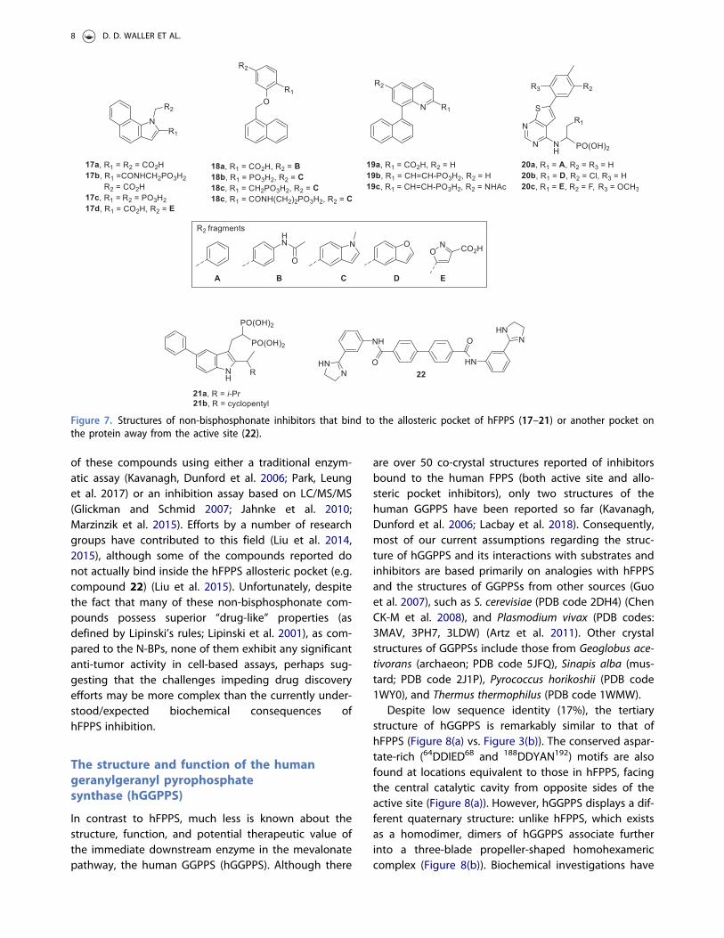
of these compounds using either a traditional enzym-atic assay (Kavanagh, Dunford et al. 2006; Park, Leunget al. 2017) or an inhibition assay based on LC/MS/MS(Glickman and Schmid 2007; Jahnke et al. 2010;Marzinzik et al. 2015). Efforts by a number of researchgroups have contributed to this field (Liu et al. 2014,2015), although some of the compounds reported donot actually bind inside the hFPPS allosteric pocket (e.g.compound 22) (Liu et al. 2015). Unfortunately, despitethe fact that many of these non-bisphosphonate com-pounds possess superior “drug-like” properties (asdefined by Lipinski’s rules; Lipinski et al. 2001), as com-pared to the N-BPs, none of them exhibit any significantanti-tumor activity in cell-based assays, perhaps sug-gesting that the challenges impeding drug discoveryefforts may be more complex than the currently under-stood/expected biochemical consequences ofhFPPS inhibition.
The structure and function of the humangeranylgeranyl pyrophosphatesynthase (hGGPPS)
In contrast to hFPPS, much less is known about thestructure, function, and potential therapeutic value ofthe immediate downstream enzyme in the mevalonatepathway, the human GGPPS (hGGPPS). Although there
are over 50 co-crystal structures reported of inhibitorsbound to the human FPPS (both active site and allo-steric pocket inhibitors), only two structures of thehuman GGPPS have been reported so far (Kavanagh,Dunford et al. 2006; Lacbay et al. 2018). Consequently,most of our current assumptions regarding the struc-ture of hGGPPS and its interactions with substrates andinhibitors are based primarily on analogies with hFPPSand the structures of GGPPSs from other sources (Guoet al. 2007), such as S. cerevisiae (PDB code 2DH4) (ChenCK-M et al. 2008), and Plasmodium vivax (PDB codes:3MAV, 3PH7, 3LDW) (Artz et al. 2011). Other crystalstructures of GGPPSs include those from Geoglobus ace-tivorans (archaeon; PDB code 5JFQ), Sinapis alba (mus-tard; PDB code 2J1P), Pyrococcus horikoshii (PDB code1WY0), and Thermus thermophilus (PDB code 1WMW).
Despite low sequence identity (17%), the tertiarystructure of hGGPPS is remarkably similar to that ofhFPPS (Figure 8(a) vs. Figure 3(b)). The conserved aspar-tate-rich (64DDIED68 and 188DDYAN192) motifs are alsofound at locations equivalent to those in hFPPS, facingthe central catalytic cavity from opposite sides of theactive site (Figure 8(a)). However, hGGPPS displays a dif-ferent quaternary structure: unlike hFPPS, which existsas a homodimer, dimers of hGGPPS associate furtherinto a three-blade propeller-shaped homohexamericcomplex (Figure 8(b)). Biochemical investigations have
Figure 7. Structures of non-bisphosphonate inhibitors that bind to the allosteric pocket of hFPPS (17–21) or another pocket onthe protein away from the active site (22).
8 D. D. WALLER ET AL.

even reported the formation of octameric complexes insolution (Miyagi et al. 2007). The core of the dimer-dimer interface is composed of the hydrophobic surfaceformed by the N-terminal residues Tyr18, Phe76, Pro77,Ile82, and Tyr83 of one subunit and the C-terminal resi-dues Ile233, Ile243, and Tyr246 of the contacting sub-unit. This contact is further stabilized by the H-bondinteractions between Glu14 and Thr228, and Gln21 andTyr246 (Figure 8(b), inset). Interestingly, the hexamericquaternary structure is unique only to mammalian andinsect GGPPS. A sequence alignment analysis indicatedthat the residues forming the inter-dimer region inhGGPPS are not conserved in plant, fungal, archaeal, orbacterial orthologs (Kavanagh, Dunford et al. 2006); thedimeric nature of these orthologs have been confirmedcrystallographically over the years.
The catalytic mechanism of hGGPPS is also pre-sumed to be virtually identical to that of hFPPS (Figure4). The pyrophosphate moiety of the allylic substrate(i.e. FPP) binds against a highly conserved patch ofnegatively charged side chains via three Mg2þ-medi-ated interactions. However, the capping phenyls(Phe98/99) observed in the hFPPS structure that bindthe allylic substrates’ isoprenyl tail are replaced by thesmaller side chains of Ala59 and Ser60 in the hGGPPS,thus allowing space for the C-20 isoprenoid product ofthis enzyme. The IPP binding sub-pocket of hGGPPS isalso lined with basic residues (as in hFPPS), which
interact directly with the pyrophosphate of IPP (i.e.Arg28, His57, Arg73, and Arg74). During the catalyticreaction, the pyrophosphate of FPP dissociates to pro-duce the allylic carbocation, which is then captured bythe IPP double bond (Figure 4). Consistent with thishypothesis, the three amino acid residues presumed tostabilize the allylic carbocation intermediate formed inthe hFPPS active site (i.e. from DMAPP or GPP) are allconserved in hGGPPS active site (i.e. Lys151, Thr152,and Gln185).
Inhibitor design of hGGPPS
To date, very few selective inhibitors of hGGPPS havebeen reported (Figure 9) and although none of theseinhibitors have yet advanced to clinical development acouple have been evaluated in vivo. It is noteworthythat the value of developing selective inhibitors ofhGGPPS as potential anti-tumor agents has beendebated in the literacture (Zhang et al. 2009). A reason-able assumption is that inhibitors of hFPPS can directlyblock the farnesylation of mutated, oncogenic Ras pro-teins, and indirectly downregulate the intracellular lev-els of GGPP (via depletion of the FPP substrate ofhGGPPS), thus will also block the prenylation of GGPP-dependent GTPases (Figure 1). Based on this assump-tion, compounds that inhibit exclusively hGGPPS werepresumed to be less effective as anti-tumor agents than
Figure 8. The structure of hGGPS. (a) The tertiary structure showing the conserved DDXXD/N motifs. (b) The homohexamericcomplex of wild-type human GGPPS. Monomeric subunits are indicated by different colors. The inset shows residues forminginter-dimer H-bonds (yellow dashes). (c) Compound 29c bound to the active site of a dimeric mutant form of hGGPPS (Y246D;PDB code 6C57) (see colour version of this figure at www.tandfonline.com/ibmg).
CRITICAL REVIEWS IN BIOCHEMISTRY AND MOLECULAR BIOLOGY 9

those inhibiting hFPPS, or dual hFPPS/hGGPPS inhibi-tors, such as the pyridinium bisphosphonate inhibitor8b (Figure 9). As mentioned earlier, compound 8b hasbeen described as a dual hFPPS/hGGPPS inhibitors andis �100-fold more potent than zoledronic acid (4a;Figure 3) in blocking tumor cell growth; the N-BP drugzoledronic acid is the most potent and selective hFPPSinhibitor. Both compounds were tested in MCF-7 breastcancer cells and EC50 values of �100–200 nM and�15 mM were reported for 8b and 4a, respectively(Zhang et al. 2009). Interestingly, the anti-tumor effectsof 4a in MM has been attributed (at least in part) to itsability to indirectly block geranylgeranylation ofGTPases (e.g. Rap1A), by inhibiting hFPPS and causingintracellular depletion of FPP (Guenther et al. 2010).
Examples of hGGPPS inhibitors reported, include thepolyaromatic bisphosphonates 23 and 24 (Guo et al.2007; Zhang et al. 2009), the isoprenoid derivatives25–28 (Shull et al. 2006; Wiemer et al. 2007; Barneyet al. 2010), and the C-2 substituted thienopyrimidinebisphosphonates (C2ThP-BPs) 29 (Lacbay et al. 2018).Although the natural product gerfelin (31) has alsobeen reported to inhibit hGGPPS (IC50 of 12 mM)(Zenitani et al. 2003; Kanoh et al. 2013), its catechol-based structure is a known pan-assay interference struc-tural motif that binds metals and interferes with redox
cellular functions (Baell and Walters 2014).Bisphosphonate analogs 23–29 are all more selective ininhibiting hGGPPS than hFPPS.
Some insight on the enzyme-ligand interactions hasbeen provided by the co-crystal structure of bacterialGGPP bound to the human GGPPS (Kavanagh, Dunfordet al. 2006). This structure revealed that the pyrophos-phate of the GGPP ligand was bound to the DDXX(D/N)motifs in the allylic site, and its hydrocarbon tailextended into a deep mostly lipophilic channel, whichis lined with aliphatic and aromatic side chains andlocated below the active site. The GGPP ligand in thisstructure was an unintentional purification artifact,derived from the heterologous bacterial expression ofthe human recombinant GGPPS enzyme. This hGGPPS-GGPP complex is thought to represent a feedbackinhibitory state and is consistent with the observationthat both GGPP and 3-azaGGPP can act as competitiveinhibitor of this enzyme with respect to its FPP sub-strate (Kavanagh, Guo et al. 2006; Kavanagh, Dunfordet al. 2006). Given that hGGPPS and other GGPPSs havetwo large hydrophobic sub-pockets, one within andanother near the active site cavity (the latter is referredto as the GGPP inhibitor channel), it is not surprisingthat larger lipophilic bisphosphonates can bind to theseenzymes in multiple binding modes (Guo et al. 2007).
Figure 9. Structures of selective hGGPPS inhibitors or dual hFPPS/hGGPPS inhibitors.
10 D. D. WALLER ET AL.
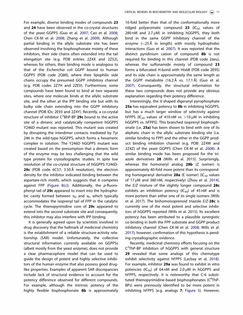
For example, diverse binding modes of compounds 23and 24 have been observed in the co-crystal structuresof the yeast GGPPS (Guo et al. 2007; Cao et al. 2008;Chen CK-M et al. 2008; Zhang et al. 2009). Althoughpartial binding in the allylic substrate site has beenobserved involving the bisphosphonate moiety of theseinhibitors, their side chains often extended into the tailelongation site (e.g. PDB entries 2Z4X and 2Z52),whereas for others, their binding mode is analogous tothat of the Escherichia coli GGPP bound to humanGGPPS (PDB code 2Q80), where their lipophilic sidechains occupy the presumed GGPP inhibitory channel(e.g. PDB codes 2Z7H and 2ZEV). Furthermore, somecompounds have been found to bind at two separatesites, where one molecule binds at the allylic substratesite, and the other at the IPP binding site but with itsbulky side chain extending into the GGPP inhibitorychannel (PDB IDs: 2E93 and 2Z4Y). Recently, a co-crystalstructure of inhibitor C2BhP-BP 29c bound to the activesite of a dimeric and catalytically competent hGGPPSY246D mutant was reported. This mutant was createdby disrupting the interdimer contacts mediated by Tyr246 in the wild-type hGGPPS, which forms a hexamericcomplex in solution. The Y246D hGGPPS mutant wascreated based on the presumption that a dimeric formof the enzyme may be less challenging that the wildtype protein for crystallographic studies. In spite lowresolution of the co-crystal structure of hGGPPS Y246D-29c (PDB code 6C57; 3.50 Å resolution), the electrondensity for the inhibitor indicated binding between theaspartate-rich motifs, which suggests that it competesagainst FPP (Figure 8(c)). Additionally, the p-fluoro-phenyl tail of 29c appeared to insert into the hydropho-bic cavity formed between aD and aF, which typicallyaccommodates the isoprenyl tail of FPP in the catalyticcycle. The thienopyrimidine core of 29c appeared toextend into the second substrate site and consequently,this inhibitor may also interfere with IPP binding.
It is generally agreed upon by scientists involved indrug discovery that the hallmark of medicinal chemistryis the establishment of a reliable structure-activity rela-tionship (SAR) model. Unfortunately, the collectivestructural information currently available on GGPPSs(albeit mostly from the yeast enzyme), does not providea clear pharmacophore model that can be used toguide the design of potent and highly selective inhibi-tors of the human enzyme that also possess good drug-like properties. Examples of apparent SAR discrepanciesinclude lack of structural evidence to account for thepotency difference observed for different compounds.For example, although the intrinsic potency of thehighly flexible bisphosphonate 8b is approximately
10-fold better than that of the conformationally moreridged polyaromatic compound 23 (IC50 values of280 nM and 2.7 mM) in inhibiting hGGPPS, they bothbind in the same GGPP inhibitory channel of theenzyme (�25Å in length) with mostly hydrophobicinteractions (Guo et al. 2007). It was reported that thedistinct pyridinium cation of compound 8b is notrequired for binding in this channel (PDB code 2zeu),whereas the sulfonamide moiety of compound 23forms a bifurcated H-bond with His68 (PDB code 2E95)and its side chain is approximately the same length asthe GGPP metabolite (16.2 Å vs. 17.1 Å) (Guo et al.2007). Consequently, the structural information forthese two compounds does not provide any obviousexplanation regarding their potency difference.
Interestingly, the V-shaped digeranyl pyrophosphate25a has equivalent potency to 8b in inhibiting hGGPPS,but has a much larger window of selectivity againsthFPPS (IC50 values of 410 nM vs >10 mM in inhibitinghGGPPS vs. hFPPS). This branched isoprenyl bisphosph-onate (i.e. 25a) has been shown to bind with one of itsaliphatic chain in the allylic substrate binding site (i.e.similar binding to FPP) and the other in the GGPP prod-uct binding inhibition channel (e.g. PDB: 2Z4W and2Z4Z) of the yeast GGPPS (Chen CK-M et al. 2008). Asimilar binding mode has been proposed for the tri-azole derivatives 28 (Wills et al. 2015). Surprisingly,whereas the homoneryl analog 28b (Z isomer) isapproximately 40-fold more potent than its correspond-ing homogeranyl derivative 28a (E isomer) (IC50 valuesof 17mM and 380 nM, respectively) (Zhou et al. 2014),the E/Z mixture of the slightly longer compound 28cexhibits an inhibition potency (IC50) of 45 nM and ismore portent than either one of its single isomers (Willset al. 2017). The bishomoisoprenoid triazole E/Z-28c iscurrently one of the most potent and selective inhibi-tors of hGGPPS reported (Wills et al. 2015). Its excellentpotency has been attributed to a plausible synergisticco-binding in both the FPP substrate and GGPP productinhibitory channel (Chen CK-M et al. 2008; Wills et al.2017); however, confirmation of this hypothesis is pend-ing crystallographic evidence.
Recently, medicinal chemistry efforts focusing on theC2ThP-BP inhibitors of hGGPPS with general structure29 revealed that some analogs of this chemotypeexhibit selectivity against hFPPS (Lacbay et al. 2018).For example, inhibitor 29a was found to exhibit in vitropotencies (IC50) of 64 nM and 2.0 mM in hGGPPS andhFPPS, respectively. It is noteworthy that C-6 substi-tuted thienopyrimidine-based bisphosphonates (C6ThP-BPs) were previously identified to be more potent ininhibiting hFPPS (e.g. analogs 7; Figure 5). However,
CRITICAL REVIEWS IN BIOCHEMISTRY AND MOLECULAR BIOLOGY 11

synthesis of the C6ThP-BP derivative 30, having theexact same sidechain as the C2ThP-BP analog 29arevealed that 30 was totally inactive in both enzymaticassays at concentrations up to 10mM. These resultsstrongly suggest that very subtle differences in themolecular recognition elements involved in protein–li-gand interactions are critical for both potency and tar-get selectivity.
The exquisite precision with which hFPPS andhGGPPS differentiate between their respective sub-strates, in spite the fact that these substrates have verylittle structural diversity and significant conformationalflexibility (i.e. C-5, C-10, or C-15 olefinic side chain), sur-passes our current understanding of the molecular rec-ognition elements dictating ligand selectivity forhGGPPS versus hFPPS. Therefore, the notion that SARstudies focusing on hFPPS and hGGPPS can simply beguided by the presence of a bisphosphonate pharma-cophore and the size and length of the side chainare inaccurate.
Preclinical in vivo evaluation ofhGGPPS inhibitors
Collectively, and in spite of all the challenges, pastefforts have generated a number of molecular tools (i.e.several compounds shown in Figure 9) that can providesome insight into the plausible clinical relevance ofhGGPPS inhibitors. For example, in vivo treatment withthe digeranyl bisphosphonate 25a (IC50 value of200 nM in inhibiting hGGPPS) in a mouse model pro-duced a decrease in the progression of pulmonaryfibrosis after lung injury (Osborn-Heaford et al. 2015).Similarly, the hydroxylated derivative 25b was reportedto decelerate prostate cancer metastasis in miceinjected with luciferase-expressing PC-3 prostate cancercells (Reilly et al. 2015, 2017). However, the potency of25b in inhibiting hGGPPS in vitro (IC50) is only 0.8 mM,and this compound is not toxic to luciferase-expressingPC-3 prostate cells in a MTT assay (an EC50 value ofapproximately 100mM was estimated), thus castingsome uncertainty as to its intracellular selectivity in tar-get engagement and exact biochemical mechanism. Incontrast, inhibitor E/Z-28c blocks the proliferation ofMM cells with significant potency (EC50 of 190 ± 58 nMin RPMI-8226 cells) (Wills et al. 2015). Therefore, its invivo properties are of interest and under investigation;preliminary metabolic stability, toxicology, and pharma-cokinetics studies were recently reported (Haney et al.2018). Inhibitor E/Z-28c was reported to be metabolic-ally stable in human and mouse liver microsomes and
to disrupt geranylgeranylation of Rap1A in CD-1 miceliver, kidney, and spleen, thus confirming systemic dis-tribution and target engagement in vivo. However, sin-gle i.v. dosing in CD-1 mice revealed a maximumtolerated dose of 0.5mg/kg, while doses higher than1mg/kg resulted in liver toxicity.
Similarly, the C2ThP-BP inhibitor 29d (EC50 value of100–150 nM) was shown to be approximately equipo-tent to E/Z-28c and to doxorubicin in blocking the pro-liferation of RPMI-8226 cells. However, the hGGPPSinhibitor 29d is significantly less toxic to normal bron-chial cells (NHBE) than doxorubicin (Lacbay et al. 2018).Pre-clinical evaluation of this compound revealed thatit is metabolically stable in male CD-1 mouse (MLM),Sprague-Dawley rat (RLM) and human (HLM) livermicrosomes, with a half-life clearance greater than 2 hin all species. The anti-myeloma properties of 29d wereevaluated in aged Vk�MYC transgenic mice (average50weeks old), which faithfully recapitulates the charac-teristics of the human MM disease and mimic the thera-peutic responses of MM patients to clinically validateddrugs (Chesi et al. 2012; Gomez-Palou et al. 2013). Micewith disease burden measurable by serum protein elec-trophoresis (i.e. M-protein levels higher than 15% oftotal serum proteins, a biomarker of MM disease bur-den) were treated with 12 doses of compound 29d at3mg/kg/d or vehicle (phosphate buffered saline; PBS)by intraperitoneal injection over a period of 14 d. At theend of treatment, Western blot analysis of peripheralblood mononuclear cell (PBMC) lysates clearly showedinhibition of Rap 1A geranylgeranylation, confirmingsystemic exposure and target engagement in vivo.Additionally, proof of anti-myeloma efficacy was alsoconfirmed, as evidenced by a significant decrease inserum M-protein seen in the mice treated with 29d ascompared to an increase in animals treated withvehicle. It is noteworthy that the half-life of mouseimmunoglobulins (i.e. M-protein) is approximately 7 d(Vieira and Rajewsky 1988). Therefore, the observeddecrease in M-protein after less than 2 weeks of treat-ment is an exciting result that clearly proves the in vivoanti-myeloma efficacy of inhibitor 29d. Althoughassessment of the blood chemistry of plasma samplesfrom the treated mice revealed some liver toxicity, theobservations were highly variable between animals.Furthermore, there was no correlation between theincrease of alanine transaminase (ALT) and aspartatetransaminase (AST) levels and decrease of M-proteinlevels that could potentially imply a mechanism-basedtoxicity. However, more in-depth investigations arerequired to confirm the safety of this compound.
12 D. D. WALLER ET AL.

Biochemical association between disruption ofprotein prenylation, endoplasmic reticulumstress, unfolded protein response (UPR)signaling, and cancer
Continued proliferation and immune evasion within harshtumor microenvironments (e.g. hypoxic, nutrient-limited,etc.) requires malignant cells to co-opt cellular homeo-static mechanisms. One important adaptive mechanismutilized by cancer cells is the unfolded protein response(UPR), which comprises a set of signaling pathways thatare initiated to overcome accumulation of unfolded pro-tein within the endoplasmic reticulum, a situation referredto as ER stress (Walter and Ron 2011). Evidence for ERstress and corresponding activation of UPR signaling hasbeen broadly reported across many human cancer types(Wang and Kaufman 2014). The mammalian UPR is initi-ated by three ER transmembrane sensor proteins: inositolrequiring enzyme 1 alpha (IRE1a), PKR-like ER kinase(PERK), and activating transcription factor 6 (ATF6). Theconcerted downstream effects of these three ER stresstransducers is to expand the size and protein folding cap-acity of the ER, to remove misfolded protein via increasedER-associated degradation (ERAD), and to transientlydecrease ER protein folding demand by two means: Ire1-regulated mRNA decay (RIDD) and global repression ofprotein translation by PERK through phosphorylation ofeIF2a. If these various UPR adaptive mechanisms fail toreduce the burden of unfolded protein in the ER in atimely fashion, UPR signaling transitions from adaptive topro-apoptotic responses (Tabas and Ron 2011).
Evidence implicating the UPR in tumorigenesis,inflammation, tissue invasion, angiogenesis, immuneevasion, and resistance to cancer therapy have beenthe subject of numerous excellent reviews (Urra et al.2016; Avril et al. 2017; Cubillos-Ruiz et al. 2017), sothese important topics will not be discussed in detailhere. Instead this section focuses on how inhibitors ofisoprenoid biosynthesis impact upon ER proteostasisand the UPR homeostatic mechanisms that areemployed by cancer cells to help tolerate elevatedsecretory protein demands and to sustain proliferationwithin harsh tumor microenvironments.
Over the last decade, it has been appreciated that invitro statin exposure can result in apoptosis of cancercell lines from various tumor types, including: glioma(Jones et al. 1994), promyelocytic leukemia (Perez-Salaand Mollinedo 1994), mesothelioma (Rubins et al.1998), acute myeloid leukemia (AML) (Xia et al. 2001),and MM (van de Donk et al. 2002). The principal causeof statin-induced apoptosis in AML, MM, and lymphomacells was shown to result primarily from indirect impair-ment of protein geranylgeranylation (Xia et al. 2001;
van de Donk, Kamphuis, et al. 2003; van de Donk,Schotte, et al. 2003), occurring via depletion of cellularreserves of the substrates required for both isoprenoidpyrophosphate synthase enzymes, hFPPS and hGGPPS.Similarly, hFPPS-targeting bisphosphonates have beenshown to induce apoptosis in cancer cell lines, albeittypically at relatively high concentrations (20–100 mM)(Aparicio et al. 1998; Fromigue et al. 2000; Lee et al.2001). As with the apoptosis caused by statin-mediatedHMG-CoA reductase inhibition, it has been reportedthat apoptotic responses to N-BPs that are selectiveinhibitors of hFPPS appear to be predominantly due toimpaired geranylgeranylation (Goffinet et al. 2006;Okamoto et al. 2014). Despite several decades ofresearch on the topic, a complete and detailed mechan-ism for the observed (direct) anti-tumor activities of sta-tins and N-BPs are still lacking. There is pressing needfor further studies to delineate the complex cell biologythat is elicited during inhibition of isoprenoid biosyn-thesis and so this remains an area of great interest.
Regarding the mechanism of apoptosis induced byimpaired isoprenoid biosynthesis, it has been shown thattreatment of human cells with either statins or hFPPS-tar-geting N-BPs, results in elevated ER stress and corre-sponding activation of UPR signaling (Chen JC et al. 2008;Ghavami et al. 2012; Lan et al. 2013). For example, it wasshown that statin treatment can activate all threebranches of the mammalian UPR in cultured human air-way cells (Ghavami et al. 2012). Autophagy was alsoshown to be induced following statin exposure and,more importantly, both chemical (bafilomycin A1) or gen-etic (ATG3 or ATG5 knockout cells, or ATG7 knockdown)impairment of this autophagic response was shown toaugment the apoptosis seen in response to statin treat-ment (Ghavami et al. 2012, 2014). It is well establishedthat UPR activation leads to upregulation of autophagy(Bernales et al. 2006), thus providing yet another layer ofcomplexity to the sophisticated cellular adaptiveresponses that are instigated when ER proteostasis is dis-rupted by inhibition of isoprenoid biosynthesis.
Given that inhibition of isoprenoid biosynthesisresults in increased ER stress, autophagy, and ultimatelyapoptosis, it is evident that cancers with high secretoryprotein burdens, such as immunoglobulin-secretingMM cells, should be ideally suited for evaluating thetherapeutic potential of novel hGGPPS inhibitors.Indeed, it has been shown that depletion of cellularGGPP by lovastatin, zoledronic acid (4a), or direct inhib-ition of hGGPPS by the digeranyl inhibitor 25a leads todisrupted intracellular light chain trafficking and a con-comitant activation of UPR signaling in MM cells(Holstein and Hohl 2011). The disruption of ER-to-Golgi
CRITICAL REVIEWS IN BIOCHEMISTRY AND MOLECULAR BIOLOGY 13

trafficking of MM light chains is thought to result fromimpaired geranylgeranylation of a variety of RabGTPases that are known to be important for intracellu-lar vesicular trafficking (Hutagalung and Novick 2011).
Inhibition of prenyl transferase enzymes
Previously, in addition to direct inhibition of the humanenzymes involved in the biosynthesis of the isoprenoidsFPPS and GGPPS, the prenyl transferase enzymes,FTase, GGTase I and II (Figure 1), also received consider-able attention as potential therapeutic targets in oncol-ogy. Efforts towards the discovery of anti-tumor agentsthat specifically target the prenyl transferase enzymeFTase led to a number of disappointing clinical trials,before it was realized that a biochemical redundancymechanism allows K-Ras activation by geranylgeranyla-tion (catalyzed by GGTase I), which takes over the taskof Ras prenylation, when FTase is inhibited (Yokoyamaet al. 1997; Rowinsky 2006). This redundancy mechan-ism has been blamed for the failure of FTase inhibitors(e.g. tipifarnib) to demonstrate significant clinicalefficacy in the intended treatments of pancreatic (90%K-Ras mutations), lung and colon carcinomas (�30% K-Ras mutations) (Sparano et al. 2009). Nonetheless,clinical development of some FTase inhibitors is still on-going and a phase III registration-directed trial of tipi-farnib in H-RAS mutant head and neck squamous cellcarcinomas (HNSCC) was recently initiated.
Investigations in support of GGTase I as a valid thera-peutic target have also been reported. Genetic studieshave shown that conditional deletion of the geneencoding the b-subunit of GGTase I in myeloid andlung cancer, almost completely eliminates proliferationand tumor formation in mice, leading to markedlyimproved survival (Sjogren et al. 2007). This study andothers have suggested that inhibition of geranylgerany-lation may be a useful strategy for treating K-Ras-induced malignancies, in addition to other human dis-eases that are driven by GGPP specific prenylation ofproteins. For greater anti-tumor efficacy, dual inhibitorsof the prenyl transferase enzymes FTase and GGTase Ihave also been evaluated (Lerner et al. 1997); examplesinclude the Merck compound L-778,123, a dual inhibitorof FTase and GGTase I with in vitro inhibition potencies(IC50 values) of 2 nM and 98 nM, respectively (Lobellet al. 2002). This compound was purposely designed asa dual FTase/GGTase I inhibitor in order to overcomethe biochemical redundancy mechanism describedabove and consequently, completely block K-Ras preny-lation (Whyte et al. 1997). L-778,123 was advanced tophase I clinical trials in patients with pancreatic cancer,
but it was eventually withdrawn from clinical develop-ment due to its toxicity (Martin et al. 2004).
In summary, although some interest remains inpotentially treating cancer by downregulating proteinprenylation at the prenyl transferase enzymes (i.e.FTase, GGTase I and II), due to the apparent lack ofstringent substrate selectivity of these enzymes and theabove mentioned redundancy mechanism, interest inthis approach has markedly declined in recent years.
Conclusions and future directions
The cumulative knowledge gained from the abovebody of literature is that blocking protein prenylationby inhibiting hFPPS and/or hGGPPS, thus decreasingthe intracellular levels of FPP and GGPP isoprenoidsand consequently, protein prenylation results in ERstress, UPR signaling, autophagy, and (ultimately) apop-tosis across many different cancer types; these effectsappear to be caused predominantly by impaired pro-tein geranylgeranylation. One plausible reason for thehigher efficacy of hGGPPS inhibitors could be related tothe lower levels of expression of this enzyme in cancercells as compared to hFPPS (Lacbay et al. 2018). Highintracellular levels of a biological target can be a majorchallenge for achieving clinical efficacy, since a highdosing regimen would likely be required that couldcompromise the therapeutic window.
Therefore, given the central importance of GGPPdepletion in inducing cancer cell apoptosis, a numberof research groups have directed their more recentefforts towards the identification of selective inhibitorsof hGGPPS. This direct targeting of hGGPPS has alreadyyielded compounds with markedly improved cell-basedanti-tumor potency (e.g. 28c, 29d) when compared toestablished hFPPS targeting N-BP drugs (Wiemer et al.2007; Zhou et al. 2014; Wills et al. 2015; Wills et al.2017; Lacbay et al. 2018). In addition, direct inhibitionof the downstream hGGPPS enzyme may avoid some ofthe unintended consequences that occur when moreupstream enzymes in the mevalonate pathway, specific-ally hFPPS or HMG-CoA reductase, are inhibited. Todate, a very limited number of groups have reportedhGGPPS inhibitors that are sufficiently potent and havereasonable biopharmaceutical properties to allow pre-clinical assessment in animal models (Reilly et al. 2017;Haney et al. 2018; Lacbay et al. 2018).
In spite of all the challenges and past disappointments,recent studies clearly suggest that there is a strong bio-chemical connection between prenylation and cancerthus, strongly suggesting that this field of research is wor-thy of further investigation. The relationship between
14 D. D. WALLER ET AL.

isoprenoids and cancer is likely more complex than initiallypresumed and skepticism about the design of drugs tar-geting human metabolic pathways (that are essential toboth heathy and malignant cells) should not be ignored.Currently, an estimated 15% of the world’s population isover the age of 60 and this group is expected to increaseto approximately 25% by 2050. For many individuals, alonger life will also mean more years of suffering due toage-related diseases, particularly cancer, which accountsfor approximately 23% of the leading causes of death inthe US (De Magalh~aes et al. 2017). Oncology is clearly anarea of major unmet therapeutic needs and changes inmetabolic pathways are implicated in both cancer progres-sion and malignancies of the ageing population, such asMM, breast, and prostate cancer. Furthermore, cellular sen-escence (a state of cell cycle arrest) is induced by cellularstress and believed to play a central role in ageing andage-related diseases, such as cancer and Alzheimer’s dis-ease (Childs et al. 2017). In addition to the strong associ-ation between intracellular levels of prenylated GTPasesand cancer, other prenylated proteins, such as the farnesy-lated precursors of nuclear lamins (prelamin A) play a crit-ical role in cellular senescence. The latter knowledge maysuggest that hFPPS inhibitors may also find new applica-tions in medicine, assuming compounds with good drug-like properties can be identified. Induction of cellular sen-escence has been proposed as a potent tumor-suppressivemechanism that arrests cell proliferation (Freund et al.2012). Inhibition of processing of the farnesylated prelaminA to the mature nuclear lamina has been shown to inducesenescence and block cancer cell migration, a required bio-logical process for cancer metastasis (Matralis et al. 2018).Therefore, multidisciplinary investigations that can lead tobetter understanding of the role of protein prenylation incancer could provide new therapeutic agents and uncoverinsights in biomedical sciences that can address the pre-vention of many cancers in the aging population.
Disclosure statement
No potential conflict of interest was reported by the authors.
ORCID
Youla S. Tsantrizos http://orcid.org/0000-0002-6231-7498
References
Aparicio A, Gardner A, Tu Y, Savage A, Berenson J,Lichtenstein A. 1998. In vitro cytoreductive effects on mul-tiple myeloma cells induced by bisphosphonates.Leukemia. 12:220–229.
Artz JD, Wernimont AK, Dunford JE, Schapira M, Dong A,Zhao Y, Lew J, Russell RGG, Ebetino FH, Oppermann U,et al. 2011. Molecular characterization of a novel geranyl-geranyl pyrophosphate synthase from Plasmodium para-sites. J Biol Chem. 286:3315–3322.
Avril T, Vauleon E, Chevet E. 2017. Endoplasmic reticulumstress signaling and chemotherapy resistance in solid can-cers. Oncogenesis. 6:e373.
Baell J, Walters MA. 2014. Chemistry: chemical con artists foildrug discovery. Nature. 513:481–483.
Barney RJ, Wasko BM, Dudakovic A, Hohl RJ, Wiemer DF.2010. Synthesis and biological evaluation of a series ofaromatic bisphosphonates. Bioorg Med Chem. 18:7212–7220.
Benzaïd I, M€onkk€onen H, Bonnelye E, M€onkk€onen J,Cl�ezardin P. 2012. In vivo phosphoantigen levels inbisphosphonate-treated human breast tumors triggerVc9Vd2 T-cell antitumor cytotoxicity through ICAM-1engagement. Clin Cancer Res. 18:6249–6259.
Bernales S, McDonald KL, Walter P. 2006. Autophagy counter-balances endoplasmic reticulum expansion during theunfolded protein response. PLoS Biol. 4:e423.
Bianch G, Munshi N. 2015. Pathogenesis beyond the cancerclone(s) in multiple myeloma. Blood. 125:3049–3058.
Cao R, Chen CK, Guo RT, Wang AH, Oldfield E. 2008.Structures of a potent phenylalkyl bisphosphonate inhibi-tor bound to farnesyl and geranylgeranyl diphosphatesynthases. Proteins. 73:431–439.
Chang SY, Hudon-Miller SE, Yang SH, Jung H-J, Lee JM,Farber E, Subramanian T, Andres DA, Spielmann HP,Hrycyna CA, et al. 2012. Inhibitors of protein geranylgera-nyltransferase-I lead to prelamin A accumulation in cellsby inhibiting ZMPSTE24. J Lipid Res. 52:1176–1182.
Chapman MA, Lawrence MS, Keats JJ, Cibulskis K, Sougnez C,Schinzel AC, Harview CL, Brunet JP, Ahmann GJ, Adli M,et al. 2011. Initial genome sequencing and analysis of mul-tiple myeloma. Nature. 471:467–472.
Chen CK-M, Hudock MP, Zhang Y, Guo R-T, Cao R, No JH,Liang P-H, Ko T-P, Chang T-H, Chang S-C, et al. 2008.Inhibition of geranylgeranyl diphosphate synthase bybisphosphonates: a crystallographic and computationalinvestigation. J Med Chem. 51:5594–5607.
Chen JC, Wu ML, Huang KC, Lin WW. 2008. HMG-CoA reduc-tase inhibitors activate the unfolded protein response andinduce cytoprotective GRP78 expression. Cardiovasc Res.80:138–150.
Chesi M, Matthews GM, Garbitt VM, Palmer SE, Shortt J,Lefebure M, Stewart AK, Johnstone RW, Bergsage PL. 2012.Drug response in a genetically engineered mouse modelof multiple myeloma is predictive of clinical efficacy.Blood. 120:376–385.
Childs BG, Gluscevic M, Baker DJ, Laberge R-M, Marquess D,Dananberg J, van Deursen JM. 2017. Senescent cells: anemerging target for diseases of ageing. Nat Rev DrugDiscov. 16:718–735.
Cimini E, Piacentini P, Sacchi A, Gioia C, Leone S, Lauro GM,Martini F, Agrati C. 2011. Zoledronic acid enhances Vd2 T-lymphocyte antitumor response to human glioma celllines. Int J Immunopathol Pharmacol. 24:139–148.
Clendening JW, Pandyra A, Boutros PC, Ghamrasni SE,Khosravi F, Trentin GA, Martirosyan A, Hakem A, Hakem R,Jurisica I, et al. 2010. Dysregulation of the mevalonate
CRITICAL REVIEWS IN BIOCHEMISTRY AND MOLECULAR BIOLOGY 15

pathway promotes transformation. Proc Natl Acad SciUSA. 107:15051–15056.
Coleman RE, Marshall H, Cameron D, Dodwell D, BurkinshawR, Keane M, Gil M, Houston SJ, Grieve RJ, Barrett-Lee PJ,et al. 2011. Breast-cancer adjuvant therapy with zoledronicacid. N Engl J Med. 365:1396–1405.
Cotesta S, Glickman JF, Jahnke W, Marzinzik A, Ofner S,Rondeau J-M, Zoller T. 2010. Salicylic acid derivativesbeing farnesyl pyrophosphate synthase activity inhibitors.WO 2010/043584 A1.
Crick DC, Andres DA, Waechter CJ. 1995. Farnesol is utilizedfor protein isoprenylation and the biosynthesis of choles-terol in mammalian cells. Biochem Biophys Res Commun.211:590–599.
Cubillos-Ruiz JR, Bettigole SE, Glimcher LH. 2017.Tumorigenic and immunosuppressive effects of endoplas-mic reticulum stress in cancer. Cell. 168:692–706.
Dedes PG, Gialeli C, Tsonis AI, Kanakis I, Theocharis AD,Kletsas D, Tzanakakis GN, Karamanos NK. 2012. Expressionof matrix macromolecules and functional properties ofbreast cancer cells are modulated by the bisphosphonatezoledronic acid. Biochem Biophys Acta. 1820:1926–1939.
De Magalh~aes JP, Stevens M, Thornton D. 2017. The businessof anti-aging science. Trends Biotechnol Biotech. 35:1062–1073.
De Schutter JW, Park J, Leung CY, Gormley P, Lin Y-S, Hu Z,Berghuis AM, Poirier J, Tsantrizos YS. 2014. Multistagescreening reveals chameleon ligands of the human farne-syl pyrophosphate synthase: implications to drug discov-ery for neurodegenerative diseases. J Med Chem. 57:5764–5776.
De Schutter JW, Shaw J, Lin Y-S, Tsantrizos YS. 2012. Designof potent bisphosphonate inhibitors of the human farnesylpyrophosphate synthase via targeted interactions with theactive site “‘capping’ phenyls”. Bioorg Med Chem. 20:5583–5591.
De Schutter JW, Zaretsky S, Welbourn S, Pause A, TsantrizosYS. 2010. Novel bisphosphonate inhibitors of the humanfarnesyl pyrophosphate synthase. Bioorg Med Chem Lett.20:5781–5786.
Dimopoulos MA, Kastritis E, Bamia C, Melakopoulos I, Gika D,Roussou M, Migkou M, Eleftherakis-Papaiakovou E,Christoulas D, Terpos E, et al. 2009. Reduction of osteo-necrosis of the jaw (ONJ) after implementation of prevent-ive measures in patients with multiple myeloma treatedwith zoledronic acid. Ann Oncol. 20:117–120.
Dudakovic A, Tong H, Hohl RJ. 2011. Geranylgeranyl diphos-phate depletion inhibits breast cancer cell migration.Invest New Drugs. 29:912–920.
Dunford JE. 2010. Molecular targets of the nitrogen contain-ing bisphosphonates: the molecular pharmacology ofprenyl synthase inhibition. Curr Pharm Des. 16:2961–2969.
Dunford JE, Kwaasi AA, Rogers MJ, Barnett BL, Ebetino FH,Russell RGG, Oppermann U, Kavanagh KL. 2008. Structure-activity relationship among the nitrogen containingbisphosphonates in clinical use and other analogues: time-dependent inhibition of human farnesyl pyrophosphatesynthase. J Med Chem. 51:2187–2195.
Dunford JE, Thompson K, Coxon FP, Luckma SP, Hahn FM,Poulter CD, Ebetino FH, Rogers MJ. 2001. Structure-activityrelationships for inhibition of farnesyl diphosphate syn-thase in vitro and inhibition of bone resorption in vivo by
nitrogen-containing bisphosphonates. J Pharmacol ExpTher. 296:235–242.
Eastell R, Walsh JS, Watts NB, Siris E. 2011. Bisphosphonatesfor postmenopausal osteoporosis. Bone. 49:82–88.
Ebetino FH, Hogan A-M, Sun S, Tsoumpra MK, Duan X, TriffittJT, Kwaasi AA, Dunford JE, Barnett BL, Oppermann U,et al. 2011. The relationship between the chemistry andbiological activity of the bisphosphonates. Bone. 49:20–33.
Ebetino FH, Mazur A, Lundy MW, Russell RGG. 2010a. 4-Azaindole bisphosphonates. WO 2010/033980 A2.
Ebetino FH, Mazur A, Lundy MW, Russell RGG. 2010b. 5-Azaindole bisphosphonates WO 2010/033981 A2
Ebetino FH, Mazur A, Lundy MW, Russell RGG. 2010c.Imidazo[1,2-a]pyridinyl Bisphosphonates. WO 2010/033978A2
Eckert GP, Hooff GP, Strandjord DM, Igbavboa U, Volmer DA,M€uller WE, Wood WG. 2009. Regulation of the brain iso-prenoids farnesyl- and geranylgeranylpyrophosphate isaltered in male Alzheimer patients. Neurobiol Dis. 35:251–257.
Ezra A, Hoffma A, Breuer E, Alferiev IS, M€onkk€onen J,Hanany-Rozen NE, Weiss G, Stepensky D, Gati I, Cohen H,et al. 2000. Peptide prodrug approach for improvingbisphosphonate oral absorption. J Med Chem. 43:3641–3652.
Fliesler SJ, Keller RK. 1995. Metabolism of [3H]farnesol tocholesterol and cholesterogenic intermediates in the livingrat eye. Biochem Biophys Res Commun. 210:695–702.
Fournier PG, Stresing V, Ebetino FH, Cl�ezardin P. 2010. Howdo bisphosphonates inhibit bone metastasis in vivo.NeoPlasia. 12:571–578.
Freund A, Laberge R-M, Demaria M, Campisi J. 2012. LaminB1 loss is a senescence-associated biomarker. Mol BiolCell. 23:2066–2075.
Fromigue O, Lagneaux L, Body JJ. 2000. Bisphosphonatesinduce breast cancer cell death in vitro. J Bone Miner Res.15:2211–2221.
Frost ML, Siddique M, Blake GM, Moore AE, Marsden PK,Schleyer PJ, Eastell R, Fogelman I. 2012. Regional bonemetabolism at the lumbar spine and hip following discon-tinuation of alendronate and risedronate treatment inpostmenopausal women. Osteoporos Int. 23:2107–2116.
Garwood ER, Kumar AS, Baehner FL, Moore DH, Au A, HyltonN, Flowers CI, Garber J, Lesnikoski B-A, Hwang ES, et al.2010. Fluvastatin reduces proliferation and increases apop-tosis in women with high grade breast cancer. BreastCancer Res Treat. 119:137–144.
Ghavami S, Sharma P, Yeganeh B, Ojo OO, Jha A, MutaweMM, Kashani HH, Los MJ, Klonisch T, Unruh H. 2014.Airway mesenchymal cell death by mevalonate cascadeinhibition: integration of autophagy, unfolded proteinresponse and apoptosis focusing on Bcl2 family proteins.Biochem Biophys Acta. 1843:1259–1271.
Ghavami S, Yeganeh B, Stelmack GL, Kashani HH, Sharma P,Cunnington R, Rattan S, Bathe K, Klonisch T, Dixon IM.2012. Apoptosis, autophagy and ER stress in mevalonatecascade inhibition-induced cell death of human atrialfibroblasts. Cell Death Dis. 3:e330.
Glickman JF, Schmid A. 2007. Farnesyl pyrophosphate syn-thase: real-time kinetics and inhibition by nitrogen-con-taining bisphosphonates in a scintillation assay. AssayDrug Dev Technol. 5:205–214.
16 D. D. WALLER ET AL.

Gnant M, Mlineritsch B, Schippinger W, Luschin-EbengreuthG, P€ostlberger S, Menzel C, Jakesz R, Seifert M, Hubalek M,Bjelic-Radisic V, et al. 2009. Endocrine therapy plus zole-dronic acid in premenopausal breast cancer. N Engl JMed. 360:679–691.
Goffinet M, Thoulouzan M, Pradines A, Lajoie-Mazenc I,Weinbaum C, Faye JC, Seronie-Vivien S. 2006. Zoledronicacid treatment impairs protein geranyl-geranylation forbiological effects in prostatic cells. BMC Cancer. 6:60.
Goldstein JL, Brown MS. 1990. Regulation of the mevalonatepathway. Nature. 343:425–430.
Gomez-Palou M, Fang H, Kremer R, Sebag M. 2013. A trans-genic model of multiple myeloma bone disease showsprofound mesenchymal stem cell impairment. Blood. 112:130.
Grey A, Bolland MJ, Wattie D, Horne A, Gamble G, Reid IR.2009. The antiresorptive effects of a single dose of zoledr-onate persists for two years: arandomized placebo-controltrial in osteopenic postmenopausal women. J ClinEndocrinol Metab. 94:538–544.
Gritzalis D, Park J, Chiu W, Cho H, Lin Y-S, De Schutter JW,Lacbay CM, Zielinski M, Berghuis AM, Tsantrizos YS. 2015.Probing the molecular and structural elements of ligandsbinding to the active site versus an allosteric pocket ofthe human farnesyl pyrophosphate synthase. Bioorg MedChem Lett. 25:1117–1123.
Guenther A, Gordon S, Tiemann M, Burger R, Bakker F, GreenJR, Baum W, Roelofs AJ, Rogers MJ, Gramatzki M. 2010.The bisphosphonate zoledronic acid has antimyelomaactivity in vivo by inhibition of protein prenylation. Int JCancer. 9:239–246.
Guo RT, Cao R, Liang PH, Ko TP, Chang TH, Hudock MP, JengWY, Chen CK, Zhang Y, Song Y, et al. 2007.Bisphosphonates target multiple sites in both cis- andtrans-prenyltransferases. Proc Natl Acad Sci USA. 104:10022–10027.
Haney SL, Chhonker YS, Varney ML, Talmon G, Murry DJ,Holstein SA. 2018. Preclinical investigation of a potent ger-anylgeranyl diphosphate synthase inhibitor. Invest NewDrugs. 36:810–818.
Hoff AO, Toth BB, Altundag K, Johnson MM, Warneke CL, HuM, Nooka A, Sayegh G, Guarneri V, Desrouleaux K, et al.2008. Frequency and risk factors associated with osteo-necrosis of the jaw in cancer patients treated with intra-venous bisphosphonates. J Bone Miner Res. 23:826–836.
Holstein SA, Hohl RJ. 2011. Isoprenoid biosynthetic pathwayinhibition disrupts monoclonal protein secretion and indu-ces the unfolded protein response pathway in multiplemyeloma cells. Leuk Res. 35:551–559.
Hooff GP, Wood WG, Kim J-H, Igbavboa U, Ong W-Y, M€ullerWE, Eckert GP. 2012. Brain isoprenoids farnesyl pyrophos-phate and geranylgeranyl pyrophosphate are increased inaged mice. Mol Neurobiol. 46:179–185.
Hooff GP, Wood WG, M€uller WE, Eckert GP. 2010.Isoprenoids, small GTPases and Alzheimer’s disease.Biochim Biophys Acta. 1801:896–905.
Hottman DA, Li L. 2014. Protein prenylation and synapticplasticity: implications for Alzheimer’s diseases. MolNeurobiol. 50:177–185.
Hutagalung AH, Novick PJ. 2011. Role of Rab GTPases inmembrane traffic and cell physiology. Physiol Rev. 91:119–149.
Iguchi K, Tatsuda Y, Usui S, Hirano K. 2010. Pamidronateinhibits antiapoptotic bcl-2 expression though inhibitionof the mevalonate pathway in prostate cancer PC-3 cells.Eur J Pharmacol. 641:35–40.
Jagdev SP, Coleman RE, Shipman CM, Rostami HA, CroucherPI. 2001. The bisphosphonate, zoledronic acid, inducesapoptosis of breast cancer cells: evidence for synergy withpaclitaxel. Br J Cancer. 84:1126–1134.
Jahnke W, Henry C. 2010. An in vitro assay to measure tar-geted drug delivery to bone mineral. ChemMedChem. 5:770–776.
Jahnke W, Rondeau J-M, Cotesta S, Marzinzik A, Pell�e X,Geiser M, Strauss A, G€otte M, Bitsch F, Hemmig R, et al.2010. Allosteric non-bisphosphonate FPPS inhibitors iden-tified by fragment-based discovery. Nat Chem Biol. 6:660–666.
Jones KD, Couldwell WT, Hinton DR, Su Y, He S, Anker L, LawRE. 1994. Lovastatin induces growth inhibition and apop-tosis in human malignant glioma cells. Biochem BiophysRes Commun. 205:1681–1687.
Kampinga HH, Craig EA. 2010. The HSP70 chaperone machin-ery: J proteins as drivers of functional specificity. Nat RevMol Cell Biol. 11:579–592.
Kanoh N, Suzuki T, Kawatani M, Katou Y, Osada H, IwabuchiY. 2013. Dual structure-activity relationship of osteoclasto-genesis inhibitor methyl gerfelin based on TEG scanning.Bioconjugate Chem. 24:44–52.
Kavanagh KL, Dunford JE, Bunkoczi G, Russell RG,Oppermann U. 2006. The crystal structure of human gera-nylgeranyl pyrophosphate synthase reveals a novel hex-americ arrangement and inhibitory product binding. J BiolChem. 281:22004–22012.
Kavanagh KL, Guo K, Dunford JE, Wu X, Knapp S, Ebetino FH,Rogers MJ, Russell RG, Oppermann U. 2006. The molecularmechanism of nitrogen-containing bisphosphonates asantiosteoporosis drugs. Proc Natl Acad Sci USA. 103:7829–7834.
Kho Y, Kim SC, Jiang C, Barma D, Kwon SW, Cheng J,Jaunbergs J, Weinbaum C, Tamanoi F, Falck J, Zhao Y.2004. A tagging-via-substrate technology for detectionand proteomics of farnesylated proteins. Proc Natl AcadSci USA. 101:12479–12484.
Kubatka P, Kruzliak P, Rotrekl V, Jelinkova S, MladosievicovaB. 2014. Statins in oncological research: from experimentalstudies to clinical practice. Crit Rev Oncol Hematol. 92:296–311.
Kunzmann V, Bauer E, Wilhelm M. 1999. cd Gamma/delta T-cell stimulation by pamidronate. N Engl J Med. 340:737–738.
Lacbay CM, Waller DD, Park J, G�omez Palou M, Vincent F,Huang X-F, Ta V, Berghuis AM, Sebag M, Tsantrizos YS.2018. Unraveling the prenylation-cancer paradox in mul-tiple myeloma with novel geranylgeranyl pyrophosphatesynthase (GGPPS) inhibitors. J Med Chem. 61:6904–6917.
Lan YC, Chang CL, Sung MT, Yin PH, Hsu CC, Wang KC, LeeHC, Tseng LM, Chi CW. 2013. Zoledronic acid-inducedcytotoxicity through endoplasmic reticulum stress trig-gered REDD1-mTOR pathway in breast cancer cells.Anticancer Res. 33:3807–3814.
Lee MV, Fong EM, Singer FR, Guenette RS. 2001.Bisphosphonate treatment inhibits the growth of prostatecancer cells. Cancer Res. 61:2602–2608.
CRITICAL REVIEWS IN BIOCHEMISTRY AND MOLECULAR BIOLOGY 17

Lerner EC, Zhang T-T, Knowles DB, Qian Y, Hamilton AD,Sebti SM. 1997. Inhibition of the prenylation of K-Ras, butnot H- or N-Ras, is highly resistant to CAAX peptidomimet-ics and requires both a farnesyltransferase and a geranyl-geranyltransferase I inhibitor in human tumor cell lines.Oncogene. 15:1283–1288.
Leung CY, Langille AM, Mancuso J, Tsantrizos YS. 2013.Discovery of thienopyrimidine-based inhibitors of thehuman farnesyl pyrophosphate synthase – parallel synthe-sis of analogs via a thrimethylsilyl ylidene intermediate.Bioorg Med Chem Lett. 21:2229–2240.
Leung CY, Park J, De Schutter JW, Sebag M, Berghuis AM,Tsantrizos YS. 2013. Thienopyrimidine bisphosphonate(ThPBP) inhibitors of the human farnesyl pyrophosphatesynthase: optimization and characterization of the modeof inhibition. J Med Chem. 56:7939–7950.
Li J, Herold MJ, Kimmel B, M€uller I, Rincon-Orozco B,Kunzmann V, Herrmann T. 2009. Reduced expression ofthe mevalonate pathway enzyme farnesyl pyrophosphatesynthase unveils recognition of tumor cells byVgamma9Vdelta2 T cells. J Immunol. 182:8118–8124.
Lin Y-S, Park J, De Schutter JW, Huang XF, Berghuis AM,Sebag M, Tsantrizos YS. 2012. Design and synthesis ofactive site inhibitors of the human farnesyl pyrophosphatesynthase – apoptosis and inhibition of ERK phosphoryl-ation in multiple myeloma cells. J Med Chem. 55:3201–3215.
Lipinski CA, Lombardo F, Dominy BW, Feeney PJ. 2001.Experimental and computational approaches to estimatesolubility and permeability in drug discovery and develop-ment settings. Adv Drug Deliv Rev. 46:3–26.
Liu J, Liu W, Ge H, Gao J, He Q, Su L, Xu J, Gu L-Q, Huang Z-S, Li D. 2014. Syntheses and characterization of non-bisphosphonate quinoline derivatives as new FPPS inhibi-tors. Biochimica et Biophysica Acta. 1840:1051–1062.
Liu Y-L, Cao R, Wang Y, Oldfield E. 2015. Farnesyl diphos-phate synthase inhibitors with unique ligand-binding geo-metries. ACS Med Chem Lett. 6:349–354.
Lobell RB, Liu D, Buser CA, Davide JP, DePuy E, Hamilton K,Koblan KS, Lee Y, Mosser S, Motzel SL, et al. 2002.Preclinical and clinical pharmacodynamic assessment of L-778, 123, a dual inhibitor of farnesyl protein transferaseand geranylgeranyl protein transferase type-I. Mol CancerTher. 1:747–758.
Lolli ML, Rolando B, Tosco P, Chaurasia S, Di Stilo A,Lazzarato L, Gorassini E, Ferracini R, Oliaro-Bosso S,Fruttero R, et al. 2010. Synthesis and preliminary pharma-cological characterization of a new class of nitrogen-con-taining bisphosphonates (N-BPs). Bioorg Med Chem. 18:2428–2438.
Mani J, Vallo S, Barth K, Makarevi�c J, Juengel E, Bartsch G,Wiesner C, Haferkamp A, Blaheta RA. 2012. Zoledronic acidinfluences growth, migration and invasive activity of pros-tate cancer cells in vitro. Prostate Cancer Prostatic Dis. 15:250–255.
Marma MS, Xia Z, Stewart C, Coxon F, Dunford JE, Baron R,Kashemirov BA, Ebetino FH, Triffitt JT, Russell GG, et al.2007. Synthesis and biological evaluation of a-halogenatedbisphosphonate and phosphonocarboxylate analogues ofrisedronate. J Med Chem. 50:5967–5975.
Martin MB, Arnold W, Heath HT, III, Urbina JA, Oldfield E.1999. Nitrogen-containing bisphosphonates as carbocation
transition state analogs for isoprenoid biosynthesis.Biochem Biophys Res Commun. 263:754–758.
Martin NE, Brunner TB, Kiel KD, DeLaney TF, Regine WF,Mohiuddin M, Rosato EF, Haller DG, Stevenson JP, SmithD, et al. 2004. A phase I trial of the dual farnesyltransfer-ase and geranylgeranyltransferase inhibitor L-778,123 andradiotherapy for locally advanced pancreatic cancer.Clinical Cancer Res. 10:5447–5454.
Marzinzik AL, Amstutz R, Bold G, Bourgier E, Cotesta S,Glickman JF, G€otte M, Henry C, Lehmann S, Hartwieg JCD,et al. 2015. Discovery of novel allosteric non-bisphospho-nate inhibitors of farnesyl pyrophosphate synthase byintegrated lead finding. ChemMedChem. 10:1884–1891.
Matralis AN, Xanthopoulos D, Huot G, Lopes-Paciencia S,Cole C, Vries H, Ferbeyre G, Tsantrizos YS. 2018. Moleculartools that block maturation of the nuclear laminA anddecelerate cancer cell migration. Bioorg Med Chem. 26:5547–5554.
Mitrofan LM, Pelkonen J, M€onkk€onen J. 2009. The level ofATP analog and isopentenyl pyrophosphate correlateswith zoledronic acid-induced apoptosis in cancer cells invitro. Bone. 45:1153–1160.
Miyagi Y, Matsumura Y, Sagami H. 2007. Human geranylger-anyl diphosphate synthase is an octamer in solution. JBiochem. 142:377–381.
M€onkk€onen H, Auriola S, Lehenkari P, Kellinsalmi M, HassinenIE, Veps€al€ainen J, M€onkk€onen J. 2006. A new endogenousATP analog (ApppI) inhibits the mitochondrial adeninenucleotide translocase (ANT) and is responsible for theapoptosis induced by nitrogen-containing bisphospho-nates. Br J Pharmacol. 147:437–445.
Morgan GJ, Davies FE, Gregory WM, Cocks K, Bell SE, SzubertAJ, Navarro-Coy N, Drayson MT, Owen RG, Feyler S, et al.2010. First-line treatment with zoledronic acid as com-pared with clodronic acid in multiple myeloma (MRCMyeloma IX): a randomised controlled trial. Lancet. 376:1989–1999.
Morgan GJ, Davies FE, Gregory WM, Szubert AJ, Bell SE,Drayson MT, Owen RG, Ashcroft AJ, Jackson GH, Child JA.2012. Effects of induction and maintenance plus long-termbisphosphonates on bone disease in patients with mul-tiple myeloma: the Medical Research Council Myeloma IXTrial. Blood. 119:5374–5383.
Morita CT, Jin C, Sarikonda G, Wang H. 2007. Nonpeptideantigens, presentation mechanisms, and immunologicalmemory of human Vc2Vd2 T cells: discriminating friendsfrom foe through the recognition of prenyl pyrophosphateantigens. Immunol Rev. 215:59–76.
Mullen PJ, Yu R, Longo J, Archer MC, Penn LZ. 2016. Theinterplay between cell signalling and the mevalonatepathway in cancer. Nat Rev Cancer. 16:718–731.
Naoe M, Ogawa Y, Takeshita K, Morita J, Shichijo T, Fuji K,Fukagai T, Iwamoto S, Terao S. 2010. Zoledronate stimu-lates cd T cells in prostate cancer patients. Oncol Res. 18:493–501.
Nguyen UTT, Guo Z, Delon C, Wu Y, Deraeve C, Fr€anzel B,Bon RS, Blankenfeldt W, Goody RS, Waldmann H, et al.2009. Analysis of the eukaryotic prenylome by isoprenoidaffinity tagging. Nat Chem Biol. 5:227–235.
Nielsen SF, Nordestgaard BG, Bojesen SE. 2012. Statin useand reduced cancer-related mortality. N Engl J Med. 367:1792–1802.
18 D. D. WALLER ET AL.
Notarnicola M, Messa C, Cavallini A, Bifulco M, Tecce MF,Eletto D, Di Leo A, Montemurro S, Laezza C, Caruso MG.2004. Higher farnesyl diphosphate synthase activity inhuman colorectal cancer inhibition of cellular apoptosis.Oncology. 67:351–358.
Okamoto S, Jiang Y, Kawamura K, Shingyoji M, Tada Y,Sekine I, Takiguchi Y, Tatsumi K, Kobayashi H, Shimada H,et al. 2014. Zoledronic acid induces apoptosis and S-phasearrest in mesothelioma through inhibiting Rab family pro-teins and topoisomerase II actions. Cell Death Dis. 5:e1517
Osborn-Heaford HL, Murthy S, Gu L, Larson-Casey JL, RyanAJ, Shi L, Glogauer M, Neighbors JD, Hohl R, Carter AB.2015. Targeting the isoprenoid pathway to abrogate pro-gression of pulmonary fibrosis. Free Radic Biol Med. 86:47–56.
Park J, Leung CY, Matralis AN, Lacbay CM, Tsakos M,Fernandez De Troconiz G, Berghuis AM, Tsantrizos YS.2017. Pharmacophore mapping of thienopyrimidine-basedmonophosphonate (ThP-MP) inhibitors of the human far-nesyl pyrophosphate synthase. J Med Chem. 60:2119–2134.
Park J, Lin YS, De Schutter JW, Tsantrizos YS, Berghuis AM.2012. Ternary complex structures of human farnesyl pyro-phosphate synthase bound with a novel inhibitor and sec-ondary ligands provide insights into the molecular detailsof the enzyme’s active site closure. BMC Struct Biol. 12:32.
Park J, Lin YS, Tsantrizos YS, Berghuis AM. 2014. Structure ofhuman farnesyl pyrophosphate synthase in complex withan aminopyridine bisphosphonate and two molecules ofinorganic phosphate. Acta Crystallogr F Struct BiolCommun. 70:299–304.
Park J, Zielinski M, Magder A, Tsantrizos YS, Berghuis AM.2017. Human farnesyl pyrophosphate synthase is allosteri-cally inhibited by its own preoducts. Nat Commun. 8:14132. /
Pelleieux S, Picard C, Lamarre-Th�eroux L, Dea D, Leduc V,Tsantrizos YS, Poirier J. 2018. Isoprenoids and tau path-ology in sporadic Alzheimer’s disease. Neurobiol Aging.65:132–139.
Perez-Sala D, Mollinedo F. 1994. Inhibition of isoprenoid bio-synthesis induces apoptosis in human promyelocytic HL-60 cells. Biochem Biophys Res Commun. 1993:1209–1215.
Poulter CD, Argyle JC, Mash EA. 1978. Farnesyl pyrophos-phate synthetase. Mechanistic studies of the 1’-4 couplingreaction with 2-fluorogeranyl pyrophosphate. J Biol Chem.253:7227–7233.
R€aikk€onen J, M€onkk€onen H, Auriola S, M€onkk€onen J. 2010.Mevalonate pathway intermediates downregulate zole-dronic acid-induced isopentenyl pyrophosphate and ATPanalog formation in human breast cancer cells. BiochemPharmacol. 79:777–783.
R€aikk€onen J, Taskinen M, Dunford JE, M€onkk€onen H, AuriolaS, M€onkk€onen J. 2011. Correlation between time-depend-ent inhibition of human farnesyl pyrophosphate synthaseand blockade of mevalonate pathway by nitrogen-contain-ing bisphosphonates in cultured cells. Biochem BiophysRes Commun. 407:663–667.
Reilly JE, Neighbors JD, Hohl RJ. 2017. Targeting protein ger-anylgeranylation slows tumor development in a murinemodel of prostate cancer metastasis. Cancer Biol Ther. 18:872–882.
Reilly JE, Neighbors JD, Tong H, Henry MD, Hohl RJ. 2015.Targeting geranylgeranylation reduces adrenal glandtumor burden in a murine model of prostate cancermetastasis. Clin Exp Metastasis. 32:555–566.
Ripamonti CI, Maniezzo M, Campa T, Fagnoni E, Brunelli C,Saibene G, Bareggi C, Ascani L, Cislaghi E. 2009. Decreasedoccurrence of osteonecrosis of the jaw after implementa-tion of dental preventive measures in solid tumourpatients with bone metastases treated with bisphospho-nates. The experience of the National Cancer Institute ofMilan. Ann Oncol. 20:137–145.
Rizzoli R, Burlet N, Cahall D, Delmas PD, Eriksen EF,Felsenberg D, Grbic J, Jontell M, Landesberg R, Laslop A,et al. 2008. Osteonecrosis of the jaw and bisphosphonatetreatment for osteoporosis. Bone. 42:841–847.
Rogers MJ, Crockett JC, Coxon FP, M€onkk€onen J. 2011.Biochemical and molecular mechanisms of action ofbisphosphonates. Bone. 49:34–41.
Rogers MJ, Russell RGG, Blackburn GM, Williamson MP, WattsDJ. 1992. Metabolism of halogenated bisphosphonates bythe cellular slime mould Dictyostelium discoideum.Biochem Biophys Res Commun. 189:414–423.
Rondeau JM, Bitsch F, Bourgier E, Geiser M, Hemmig R,Kroemer M, Lehmann S, Ramage P, Rieffel S, Strauss A,et al. 2006. Structural basis for the exceptional in vivo effi-cacy of bisphosphonate drugs. ChemMedChem. 1:267–273.
Rowinsky EK. 2006. Lately, it occurs to me what a long,strange trip it’s been for the farnesyltransferase inhibitors.J Clin Oncol. 24:2981.
Rubins JB, Greatens T, Kratzke RA, Tan AT, Polunovsky VA,Bitterman P. 1998. Lovastatin induces apoptosis in malig-nant mesothelioma cells. Am J Respir Crit Care Med. 157:1616–1622.
Russell RGG. 2011. Bisphosphonates: the first 40 years. Bone.49:2–19.
Sanfilippo KM, Keller J, Gage BF, Luo S, Wang T-F, MoskowitzG, Gumbel J, Blue B, O’Brian K, Carson KR. 2016. Statinsare associated with reduced mortality in multiple mye-loma. J Clin Oncol. 34:4008–4014.
Shmeeda H, Amitay Y, Gorin J, Tzemach D, Mak L, Ogorka J,Kumar S, Zhang JA, Gabizon A. 2010. Delivery of zole-dronic acid encapsulated in folate-targeted liposomeresults in potent in vitro cytotoxic activity on tumor cells.J Control Rel. 146:76–83.
Shull LW, Wiemer AJ, Hohl RJ, Wiemer DF. 2006. Synthesisand biological activity of isoprenoid bisphosphonates.Bioorg Med Chem. 14:4130–4136.
Simoni D, Gebbia N, Invidiata FP, Eleopra M, Marchetti P,Rondanin R, Baruchello R, Provera S, Marchioro C,Tolomeo M, et al. 2008. Design, synthesis, and biologicalevaluation of novel aminobisphosphonates possessing anin vivo antitumor activity through a cd-T lymphocytes-mediated activation mechanism. J Med Chem. 51:6800–6807.
Sjogren A-KM, Andersson KME, Liu M, Cutts BA, Karlsson C,Wahlstrom AM, Dalin M, Weinbaum C, Casey PJ, TarkowskiA, et al. 2007. GGTase-I deficiency reduces tumor forma-tion and improves survival in mice with K-RAS–inducedlung cancer. J Clin Invest. 117:1294–1304.
Skerjanec A, Berenson J, Hsu C, Major P, Miller WH, Jr RaveraC, Schran H, Seaman J, Waldmeier F. 2003. The
CRITICAL REVIEWS IN BIOCHEMISTRY AND MOLECULAR BIOLOGY 19

pharmacokinetics and pharmacodynamics of zoledronicacid in cancer patients with varying degrees of renal func-tion. J Clin Pharmacol. 43:154–162.
Sparano JA, Moulder S, Kazi A, Coppola D, Negassa A,Vahdat L, Li T, Pellegrino C, Fineberg S, Munster P, et al.2009. Phase II trial of tipifarnib plus neoadjuvant doxorubi-cin-cyclophosphamide in patients with clinical stage IIB-IICbreast cancer. Clin Cancer Res. 15:2942–2948.
Song L, Poulter CD. 1994. Yeast farnesyl-diphosphate syn-thase: site-directed mutagenesis of residues in highly con-served prenyltransferase domains I and II. Proc Natl AcadSci USA. 91:3044–3048.
Sorrentino G, Ruggeri N, Specchia V, Cordenonsi M, Mano M,Dupont S, Manfrin A, Ingallina E, Sommaggio R, Piazza S,et al. 2014. Metabolic control of YAP and TAZ by themevalonate pathway. Nat Cell Biol. 16:357–366.
Stark JL, Mehla K, Chaika N, Acton TB, Xiao R, Singh PK,Montelione GT, Powers R. 2014. Structure and function ofhuman DnaJ homologue subfamily a member 1 (DNAJA1)and its relationship to pancreatic cancer. Biochemistry. 53:1360–1372.
Tabas I, Ron D. 2011. Integrating the mechanisms of apop-tosis induced by endoplasmic reticulum stress. Nat CellBiol. 13:184–190.
Takai Y, Sasaki T, Matozaki T. 2001. Small GTP-binding pro-teins. Physiol Rev. 81:153–208.
Tarshis LC, Proteau PJ, Kellogg BA, Sacchettini JC, Poulter CD.1996. Regulation of product chain length by isoprenyldiphosphate synthases. Proc Natl Acad Sci USA. 93:15018–15023.
Todenh€ofer T, Hennenlotter J, K€uhs U, Gerber V, Gakis G,Vogel U, Aufderklamm S, Merseburger A, Knapp J, StenzlA, et al. 2013. Altered expression of farnesyl pyrophos-phate synthase in prostate cancer: evidence for a role ofthe mevalonate pathway in disease progression? World JUrol. 31:345–350.
Urra H, Dufey E, Avril T, Chevet E, Hetz C. 2016. Endoplasmicreticulum stress and the hallmarks of cancer. TrendsCancer. 2:252–262.
van de Donk NW, Kamphuis MM, Lokhorst HM, Bloem AC.2002. The cholesterol lowering drug lovastatin induces celldeath in myeloma plasma cells. Leukemia. 16:1362–1371.
van de Donk NW, Kamphuis MM, van Kessel B, Lokhorst HM,Bloem AC. 2003. Inhibition of protein geranylgeranylationinduces apoptosis in myeloma plasma cells by reducingMcl-1 protein levels. Blood. 102:3354–3362.
van de Donk NW, Schotte D, Kamphuis MM, van Marion AM,van Kessel B, Bloem AC, Lokhorst HM. 2003. Protein gera-nylgeranylation is critical for the regulation of survival andproliferation of lymphoma tumor cells. Clin Cancer Res. 9:5735–5748.
Vieira P, Rajewsky K. 1988. The half-lives of serum immuno-globulins in adult mice. Eur J Immunol. 18:313–316.
Walter P, Ron D. 2011. The unfolded protein response: fromstress pathway to homeostatic regulation. Science. 334:1081–1086.
Wang H, Sarikonda G, Puan K-J, Tanaka Y, Feng J, Giner J-L,Cao R, M€onkk€onen J, Oldfield E, Morita CT. 2011. Indirect
stimulation of human Vc2Vd2 T cells through alterationsin isoprenoid metabolism. J Immunol. 187:5099–5113.
Wang M, Kaufman RJ. 2014. The impact of the endoplasmicreticulum protein-folding environment on cancer develop-ment. Nat Rev Cancer. 14:581–597.
Weiss HM, Pfaar U, Schweitzer A, Wiegand H, Skerjanec A,Schran H. 2008. Biodistribution and plasma protein bind-ing of zoledronic acid. Drug Metab Dispos. 36:2043–2049.
Whyte DB, Kirschmeier P, Hockenberry TN, Nunez-Oliva I,James L, Catino JJ, Bishop WR, Pai J-K. 1997. K- and N-Rasare geranylgeranylated in cells treated with farnesyl pro-tein transferase inhibitors. J Biol Chem. 272:14459–14464.
Wiemer AJ, Tong H, Swanson KM, Hohl RJ. 2007. Digeranylbisphosphonate inhibits geranylgeranyl pyrophosphatesynthase. Biochem Biophys Res Commun. 353:921–925.
Wills VS, Allen C, Holstein SA, Wiemer DF. 2015. Potent tri-azole bisphosphonate inhibitor of geranylgeranyl diphos-phate synthase. ACS Med Chem Lett. 6:1195–1198.
Wills VS, Metzger JI, Allen C, Varney ML, Wiemer DF, HolsteinSA. 2017. Bishomoisoprenoid triazole bisphosphonates asinhibitors of geranylgeranyl diphosphate synthase. BioorgMed Chem. 25:2437–2444.
Xia Z, Tan MM, Wong WW, Dimitroulakos J, Minden MD,Penn LZ. 2001. Blocking protein geranylgeranylation isessential for lovastatin-induced apoptosis of human acutemyeloid leukemia cells. Leukemia. 15:1398–1407.
Yokoyama K, Zimmerman K, Scholten J, Gelb MH. 1997.Differential prenyl pyrophosphate binding to mammalianprotein geranylgeranyltransferase-I and protein farnesyl-transferase and its consequence on the specificity of pro-tein prenylation. J Biol Chem. 272:3944–3952.
Young SG, Fong LG, Michaelis S. 2005. Prelamin A, Zmpste24,misshapen cell nuclei, and progeria-new evidence suggest-ing that protein farnesylation could be important for dis-ease pathogenesis. J Lipid Res. 46:2531–2558.
Zenitani S, Tashiro S, Shindo K, Nagai K, Suzuki K, Imoto M.2003. Gerfelin, a novel inhibitor of geranylgeranyl diphos-phate synthase from Beauveria feline QN22047. J Antibiot.56:617–621.
Zhang Y, Leon A, Song Y, Studer D, Haase C, Koscielski LA,Oldfield E. 2006. Activity of nitrogen-containing and non-nitrogen-containing bisphosphonates on tumor cell lines. JMed Chem. 49:5804–5814.
Zhang Y, Cao R, Yin F, Hudock MP, Guo R-T, Krysiak K,Mukherjee S, Gao Y-G, Robinson H, Song Y, et al. 2009.Lipophilic bisphosphonates as dual farnesyl/geranylgeranyldiphosphate synthase inhibitors: an X-ray and NMR inves-tigation. J Am Chem Soc. 131:5153–5162.
Zhang Y, Cao R, Yin F, Lin F-Y, Wang H, Krysiak K, No J-H,Mukkamala D, Houlihan K, Li J, et al. 2010. Lipophilic pyri-dinium bisphosphonates: potent gammadelta T cell stimu-lators. Angew Chem Int Ed Engl. 49:1136–1138.
Zhou X, Ferree SD, Wills VS, Born EJ, Tong H, Wiemer DF,Holstein SA. 2014. Geranyl and neryl triazole bisphospho-nates as inhibitors of geranylgeranyl diphosphate syn-thase. Bioorg Med Chem. 22:2791–2798.
20 D. D. WALLER ET AL.