Embed Size (px)
Citation preview
Pergamoln
Biochemical Pharmacology, Vol. 50. No. 6, pp. 861-867. 1995. Copyright 8 1995 Elseviex Science Inc.
Printed in Great Britain. All rights reserved
0006-2952(95)00255-3 WO&2952/95 $9.50 + 0.00
INHIBITION OF DIHYDROOROTATE DEHYDROGENASE BY THE
IMMUNOSUPPRESSIVE AGENT LEFLUNOMIDE
SHARON GREENE, KIYOTAKA WATANABE, JULIE BRAATZ-TRULSON AND LILLIAN LOU*
Institute of Biochemistry and Cell Biology, Syntex Discovery Research, Palo Alto, CA 94303, U.S.A.
(Received 20 December 1994, accepted 26 April 1995)
Abstract-Leflunomide [HWA 486 or RS-34821, 5-methyl-N-(4trifluoromethylphenyl)~4-isoxazole carboxi- mide] is an immunosuppressive agent effective in the treatment of rheumatoid arthritis. In spite of its clinical potential, its mechanism of action has not been elucidated. Recent studies suggest that leflunomide may interfere with the metabolism of pyrimidine nucleotides. In our studies, the active metabolite of leflunomide, RS-61980 (A77 1726, 2..hydroxyethylidene-cyanoacetic acid-&rifluoromethyl anilide), was cytostatic towards a human T-lymphoblastoma cell line (A3.01). The inhibition of growth could be overcome completely by uridine. The other nucleosides, cytidine, adenosine and guanosine, did not overcome the effect of the compound. Since uridine is a precursor for the salvage synthesis of UMP, we propose that RS-61980 may be inhibiting the de now pathway of UMP synthesis. Using human cells, the six enzymes catalyzing de nova UMP biosynthesis were tested for their sensitivity toward8 RS-61980. Only one of the enzymes, dihydroortate dehydrogenase (DHODH, EC 1.3.3.1) was inhibited by RS-61980 with a K, value of 2.7 + 0.7 pM. The other five enzymes were not affected. The inhibition exhibited mixed-type kinetics towards both substrates, dihydroorotic acid and coenzyme Q. These results suggest that the molecular target of leflunomide action is DHODH. The immunomodulating activity may tie related to the inhibition of UMP synthesis in proliferating lymphocytes.
Key words: leflunomide; pyrimidine nucleotide synthesis; de ~OVO pathway; Brequinar; dihydroortate dehydro- genase; uridine
The enzymes in the de nova synthesis of purine and pyrimidine nucleotides are proven targets for immuno- suppressive agents. Mycophenolate mofetil [RS-61443, 6-(1,3-dihydro-4-hytlroxy-6-methoxy-7-methyl-3-oxo- 5-isobenzofuranyl)-4-methyl-2-(4-morpholinyl)ethyl es- ter)], a prodrug of mycophenolic acid, is efficacious in the treatment of transplant rejection and rheumatoid ar- thritis [ 1, 21. Mycophenolic acid is a potent inhibitor of IMPDHt (EC 1.1.1.2.05), a key enzyme of de nova syn- thesis of purine nucleotides [for a review, see Ref. 31. Brequinar SodiumTM [NSC 368 390, 6-fluoro-2-(2’-flu- oro-l,l’-biphenyl-4-y])-3-methyl-4-quinoline carboxylic acid sodium salt, Brequinar], an inhibitor of DHODH in the de nuvo synthesis of pyrimidine nucleotides, is ef- fective in preventing graft rejection [for a review, see Ref. 41.
Leflunomide is a novel immunosuppressive agent that is effective towards several autoimmune diseases and rejection of transplants in animals (Fig. 1) [for reviews, see Refs. 5 and 61. It effectively reduces inflammation in animal models of rheumatoid arthritis, such as adjuvant arthritis and proteoglycan-induced progressive polyar- thritis [7, 81. It is currently being investigated in clinical trials for the treatment of rheumatoid arthritis. Despite its
* Corresponding author: Dr. Lillian Lou, Galileo Laborato- ries, 1320 Chesapeake Terrace, Sunnyvale, CA 94089. Tel. (408) 745-6857; FAX (408) 541-9071.
t Abbreviations: IMF’DH, inosine 5’-monophosphate dehy- drogenase; OMP, orotidine 5’-monophosphate; PRPP, 5-phos- phoribosyl- 1 -pyrophosp hate; UMP, uridine 5’-monophosphate: DHODH, dihydroorotate dehydrogenase; CPS, carbamyl phos- phate synthetase II; ATC, aspartate transcarbamylase; DHOase, dihydroorotase; OPRT, orotate phosphoribosyl transferase; OMPDC, orotidine 5’-monophosphate decarboxylase; and PBL, peripheral blood lymphocytes.
promising potential as an immunomodulatory drug, its molecular target has not been elucidated clearly. It has been proposed that the mechanism of action is related to the inhibition of both activation and proliferation of T lymphocytes [S]. Several groups have reported that the levels of interleukin 2 (IL-2) production, IL-2 receptor expression, and tyrosine kinase activity are reduced by the drug [9-121. However, the inhibition of these events was very weak, and the data are highly controversial, since they have not been entirely reproducible among different groups of investigators. More importantly, the doses required for the inhibition of these events were substantially higher than the doses needed for inhibition of growth. These results do not explain the in vitro and in vivo potency of leflunomide. It is unlikely that inhi- bition of these early events of lymphocyte activation, such as cytokine production, receptor up-regulation and tyrosine kinase stimulation, represents the main feature of the immunosuppressive action of leflunomide [12].
Leflunomide is a general cytostatic agent for a wide variety of animal cells. In viva, it is metabolized rapidly and the primary active metabolite is RS-61980 (see Fig. 1 for structure) [ 131. The drug action seems to be late- acting in the cell cycle, inhibiting the progression from the S to G,/M phase. It inhibits DNA and RNA synthe- ses, and its potency is unchanged even when added to lymphocytes that are past the early stages of activation. Unlike mycophenolic acid, the action of leflunomide is not overcome by purine nucleotides. Similar to Brequi- nar, however, its effects can be overcome by either uri- dine or cytidine in human PBL.$
$ Cherwinski H, Cohn RG, Cheung P, Webster DJ, Caufield JP, Young JM and Ransom IT, Manuscript submitted for pub- lication. Cited with permission.
861
862 S. GREENE et al.
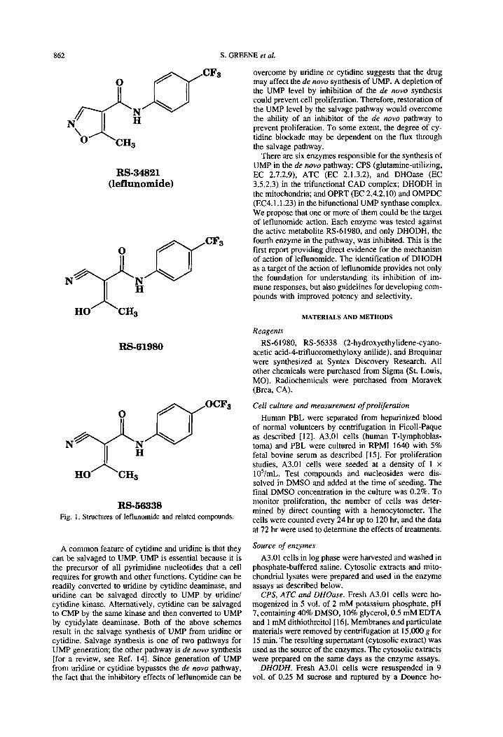
RS-34821 (leflunomide)
0
RS-61980
OCFs Cell culture and measurement of proliferation
Human PBL were separated from heparinized blood of normal volunteers by centrifugation in Ficoll-Paque as described [12]. A3.01 cells (human T-lymphoblas- toma) and PBL were cultured in RPM1 1640 with 5% fetal bovine serum as described [15]. For proliferation studies, A3.01 cells were seeded at a density of 1 x
lO’/mL. Test compounds and nucleosides were dis- solved in DMSO and added at the time of seeding. The final DMSO concentration in the culture was 0.2%. To monitor proliferation, the number of cells was deter- mined by direct counting with a hemocytometer. The cells were counted every 24 hr up to 120 hr, and the data at 72 hr were used to determine the effects of treatments.
HOA,, 3
RS-56338 Fig. 1. Structures of leflunomide and related compounds.
A common feature of cytidine and uridine is that they can be salvaged to UMP. UMP is essential because it is the precursor of all pyrimidine nucleotides that a cell requires for growth and other functions. Cytidine can be readily converted to uridine by cytidine deaminase, and uridine can be salvaged directly to UMP by uridine/ cytidine kinase. Alternatively, cytidine can be salvaged to CMP by the same kinase and then converted to UMP by cytidylate deaminase. Both of the above schemes result in the salvage synthesis of UMP from uridine or cytidine. Salvage synthesis is one of two pathways for UMP generation; the other pathway is de now synthesis [for a review, see Ref. 141. Since generation of UMP from uridine or cytidine bypasses the de nova pathway, the fact that the inhibitory effects of leflunomide can be
overcome by uridine or cytidine suggests that the drug may affect the de nova synthesis of UMP. A depletion of the UMP level by inhibition of the de nova synthesis could prevent cell proliferation. Therefore, restoration of the UMP level by the salvage pathway would overcome the ability of an inhibitor of the de nova pathway to prevent proliferation. To some extent, the degree of cy- tidine blockade may be dependent on the flux through the salvage pathway.
There are six enzymes responsible for the synthesis of UMP in the de nova pathway: CPS (glutamine-utilizing, EC 2.7.2.9), ATC (EC 2.1.3.2), and DHOase (EC 3.5.2.3) in the trifunctional CAD complex; DHODH in the mitochondria; and OPRT (EC 2.4.2.10) and OMPDC (EC4.1 .1.23) in the bifunctional UMP synthase complex. We propose that one or more of them could be the target of lefhrnomide action. Each enzyme was tested against the active metabolite RS-61980, and only DHODH, the fourth enzyme in the pathway, was inhibited. This is the first report providing direct evidence for the mechanism of action of leflunomide. The identification of DHODH as a target of the action of leflunomide provides not only the foundation for understanding its inhibition of im- mune responses, but also guidelines for developing com- pounds with improved potency and selectivity.
MATERIALS AND METHODS
Reagents
RS-61980, RS-56338 (2-hydroxyethylidene-cyano- acetic acid-4-trifluoromethyloxy anilide), and Brequinar were synthesized at Syntex Discovery Research. All other chemicals were purchased from Sigma (St. Louis, MO). Radiochemicals were purchased from Moravek (Brea, CA).
Source of enzymes
A3.01 cells in log phase were harvested and washed in phosphate-buffered saline. Cytosolic extracts and mito- chondrial lysates were prepared and used in the enzyme assays as described below.
CPS, ATC and DHOase. Fresh A3.01 cells were ho- mogenized in 5 vol. of 2 mM potassium phosphate, pH 7, containing 40% DMSO, 10% glycerol, 0.5 mM EDTA and 1 mM dithiothreitol[16]. Membranes and particulate materials were removed by centrifugation at 15,000 g for 15 min. The resulting supematant (cytosolic extract) was used as the source of the enzymes. The cytosolic extracts were prepared on the same days as the enzyme assays.
DHODH. Fresh A3.01 cells were resuspended in 9 vol. of 0.25 M sucrose and ruptured by a Dounce ho-
Dihydroorotate dehydrogenase, a target of leflunomide 863
mogenizer. Intact mitochondria were prepared as de- scribed [17]. Mitochondria were solubilized in 0.1 M Tris-Cl, 10% glycerol and 0.03% Lubrol and used as the source of the enzyme [ 181.
OPRT and OMPDC. The cytosolic extracts of A3.01 cells were prepared as described above for CPS, ATC and DHOase except for one modification, which was omitting DMSO from the lysis buffer.
Cytidine deaminase (EC 3.5.4.5). Cytidine deaminase activity was measured from both intracellular and extra- cellular sources. For intracellular levels of enzyme, the cytosolic extracts of A3.01 cells and human PBL were prepared exactly as described above for OPRT and OMPDC. To measure extracellular cytidine deaminase, culture medium (72 hr after seeding) was used as a source of enzyme.
product of the reaction, was treated with an equal vol- ume of a color reagent (antipyrine/sulfuric acid and di- acetyl monoxime/acetic acid) for the detection of ureido compounds. The procedures of Prescott and Jones [19] were followed exactly. Briefly, the color development was carried out in the dark for 17 hr at room temperature and then in the light for 70 min at 45”. The absorbance at 466 nm was measured immediately. Under these con- ditions, carbamyl phosphate did not give a signal. For the measurement of the background level of ureido com- pounds in the assay, reaction samples were prepared where either carbamyl phosphate or aspartic acid was omitted. The background of this assay procedure was insignificantly low (less than 1%).
Determination of enzyme activity
CPS. The activity of CPS was determined by measur- ing the formation of [‘4C]carbamyl aspartate in a cou- pled reaction. Reaction mixtures consisted of 50 mM potassium-HEPES, pH 7.4, 10% glycerol, 2 mM dithio- threitol, 10 mM magnesium chloride, 1 mM ATP, 1 mM glutamine, 5 mM sodium bicarbonate and 90 pM L-
[U-i4C]aspartic acid (30 mCi/mmol). Cytosolic extracts (25 pg protein), which contained CPS, ATC and other enzymes, were added to start the reaction (final reaction volume of 50 pL). This reaction mixture allowed the formation of carbamyl phosphate catalyzed by CPS and the subsequent format Ion of [‘4C]carbamyl aspartate cat- alyzed by ATC. Since exogenous carbamyl phosphate was not added to the reaction mixtures, [14C]carbamyl aspartate could only be formed as a result of the CPS reaction. For a measurement of the background level of [i4C]carbamyl aspartate formation, substrates (magne- sium ion, ATP, glutamine and sodium bicarbonate) re- quired for CPS were omitted from the reaction mixture. This level was confirmed to be low (less than 2%).
DHOase. DHOase was assayed by the formation of carbamyl aspartate in the reverse reaction. Reaction mix- tures consisted of 100 mM Tris-Cl, pH 8.5, and 20 pM dihydroorotic acid. The assay conditions and procedures of detecting carbamyl aspartate were exactly as de- scribed above for ATC.
DHODH. The activity of DHODH was measured by the formation of [‘4C]orotic acid from [‘4C]dihydro- erotic acid. The reaction mixtures consisted of 75 mM Tris-Cl, pH 7.8, 5 mM potassium cyanide, 0.6 mM co- enzyme Q (Q30), and 10 pM t,-[2-‘4C]dihydroorotic acid (50 mCi/mmol), as described [ 181. The reaction was initiated by the addition of mitochondrial lysate (1.6 pg protein). After 10 min at 40”, a carrier solution was added, and the reaction was stopped by heating at 100’ for 3 min. The carrier solution contained 50 nmol dihy- droorotic acid and 12.5 nmol erotic acid. The radioactive product and substrate were separated by ion-pairing HPLC and quantitated by liquid scintillation counting as described above for CPS. A significant level of [i4C]orotic acid formation was detected in the absence of coenzyme Q (see Results and Fig. 6).
After 30 min at 40’. a solution containing carriers was added, and the reaction was stopped by heating the sam- ples at 100” for 3 min. The carriers were 1 pmol aspartic acid, 2.5 pmol carbarnyl aspartate, and 0.04 pmol dihy- droorotic acid. Radioactive compounds were separated by ion-pairing HPLC using a Cl8 Microsorb column (Rainin, Emeryville, CA). The materials of interest were eluted at a flow rate c’f 0.5 mL/min with 40 mM potas- sium phosphate, pH 6,5 mM tetrabutylammonium phos- phate (IPC-A reagent, Alltech Associates, Deerfield, IL). Under these condition:;, the retention time values were 3, 5.2, and 7.4 min for aspartic acid, dihydroorotic acid, and carbamyl aspartate, respectively. The carriers marked the elution positions of these metabolites, and the elution profile was monitored by optical density at 230 nm. Eluents were collected, and the amounts of radioactivity co-migrating with each carrier were mea- sured by liquid scintillation counting.
OPRT and OMPDC. The two enzymes were assayed together in the same reaction. The assay mixture con- sisted of 40 mM Tris-Cl, pH 8,5 mM magnesium chlo- ride, 50 uM PRPP and 10 pM [5-3H]orotic acid (0.1 CYmmol). The reaction was initiated with cytosolic ex- tracts (100 pg protein) in a final volume of 100 pL. After 15 min at 40”, a carrier solution was added, and the reaction was stopped by heating at 100” for 3 min. The carrier solution consisted of 10 nmol each of erotic acid, OMP and UMP. The radioactive compounds were sep- arated by HPLC using a Partisil 10 SAX column (What- man) with a gradient of 20 mM (buffer A) to 50 mM (buffer B) potassium phosphate, pH 5.9, as follows: 0 to 15 min, 0% B; 15 to 30 mitt, linear gradient to 100% B. The flow rate was 1 mL/min. Under these conditions, the retention time values were 4.8, 10.3, and 50.7 min for erotic acid, UMP and OMP, respectively. The elution profiles of the carriers were monitored by optical density at 260 nm. The radioactive eluents were collected and then quantitated by scintillation counting.
ATC. ATC activity was assayed by the formation of Cytidine deaminase. Cytidine deaminase associated carbamyl aspartate using a calorimetric method. Reac- with A3.01 cells or human PBL was measured by the tion mixtures consisted of 100 mM Tris-Cl, pH 9, 2.5 formation of [3H]uridine from r3H]cytidine. The reaction mM L-aspartic acid and 0.5 mM carbamyl phosphate. mixture consisted of 20 mM Tris-Cl, pH 7.4, 1 mM Cytosolic extracts (25 pg protein) were added to start the EDTA, 1 mM dithiothreitol and 0.2 mM [5-3H]cytidine reaction (final volume of 100 p.L). After 20 min at 40”, (0.1 Ci/mmol). Cytosolic extract (13-37 pg protein) or the samples were stopped with 300 pL of 5% trichloro- culture medium (26-31 pg protein) was added to initiate acetic acid. Cytosolic proteins were allowed to precipi- the reaction, which proceeded at 40” for 30 min. A car- tate for 30 min at 0” and then were removed by centrif- rier solution that contained 5 nmol each of cytidine and ugation. The resulting supematant, which contained the uridine was added to the mixture, and the reaction was
S. GREENE et al.
0.8
0.6
0.4
0.2
0.0 0 1 10 100
[RS-619801 (PM)
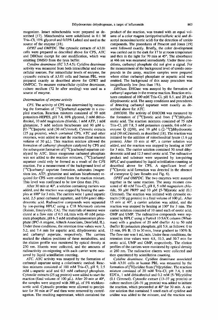
Fig. 2. Growth inhibition of A.301 cells by RS-61980. A3.01 cells were seeded at 1 x ld/mL, and proliferation was moni- tored by the number of cells. RS-61980 was added at the time of seeding. The cell number, after 72 hr. was determined by direct counting. The data were fitted to the computer program Systat for ~csa determination. Cell viability was monitored by trypan blue exclusion. Cell death was typically less than 3% at
the highest inhibitor concentration.
stopped by heating at 100” for 3 min. Radioactive cyti- dine and uridine were separated by HPLC using a Cl8 Microsorb column eluted with 5 mM potassium phos- phate, pH 5.9, at a flow rate of 1 ml/mitt. The retention time values for cytidine and uridine were 6 and 8.6 min, respectively. The radioactive product and substrate were quantitated as described above.
Determination of inhibition constants
Inhibition and kinetic constants were determined by fitting the initial velocity data to appropriate equations using nonlinear regression analysis by the computer pro- gram Systat. Determinations are reported as “value + asymptotic standard error.”
a.
= 1.0 2
.; 0.8
5 Z 0.6 :’
RESULTS
Effect of RS-61980 on cell proliferation
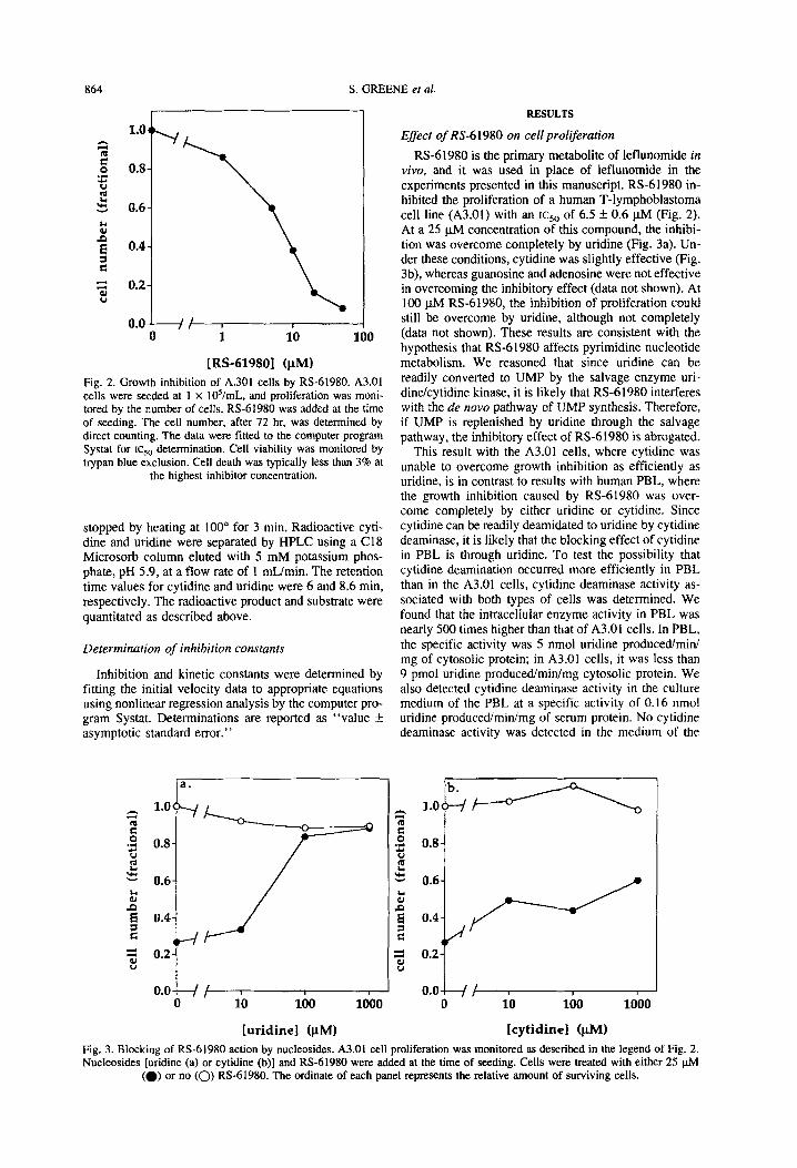
RS-61980 is the primary metabolite of leflunomide in vivo, and it was used in place of leflunomide in the experiments presented in this manuscript. RS-61980 in- hibited the proliferation of a human T-lymphoblastoma cell line (A3.01) with an ~cso of 6.5 f 0.6 pM (Fig. 2). At a 25 pM concentration of this compound, the inhibi- tion was overcome completely by uridine (Fig. 3a). Un- der these conditions, cytidine was slightly effective (Fig. 3b), whereas guanosine and adenosine were not effective in overcoming the inhibitory effect (data not shown). At 100 pM RS-61980, the inhibition of proliferation could still be overcome by uridine, although not completely (data not shown). These results are consistent with the hypothesis that RS-61980 affects pyrimidine nucleotide metabolism. We reasoned that since uridine can be readily converted to UMP by the salvage enzyme uri- dine/cytidine kinase, it is likely that RS-61980 interferes with the de now pathway of UMP synthesis. Therefore, if UMP is replenished by uridine through the salvage pathway, the inhibitory effect of RS-61980 is abrogated.
This result with the A3.01 cells, where cytidine was unable to overcome growth inhibition as efficiently as uridine, is in contrast to results with human PBL, where the growth inhibition caused by RS-61980 was over- come completely by either uridine or cytidine. Since cytidine can be readily deamidated to uridine by cytidine deaminase, it is likely that the blocking effect of cytidine in PBL is through uridine. To test the possibility that cytidine deamination occurred more efficiently in PBL than in the A3.01 cells, cytidine deaminase activity as- sociated with both types of cells was determined. We found that the intracellular enzyme activity in PBL was nearly 500 times higher than that of A3.01 cells. In PBL, the specific activity was 5 nmol uridine produced/mini mg of cytosolic protein; in A3.01 cells, it was less than 9 pmol uridine produced/min/mg cytosolic protein. We also detected cytidine deaminase activity in the culture medium of the PBL at a specific activity of 0.16 nmol uridine produced/min/mg of serum protein. No cytidine deaminase activity was detected in the medium of the
1.0 b. k---cm
pi
[uridinel (PM) [cytidine] (PM)
Fig. 3. Blocking of RS-61980 action by nucleosides. A3.01 cell proliferation was monitored as described in the legend of Fig. 2. Nucleosides [uridine (a) or cytidine (b)] and RS-61980 were added at the time of seeding. Cells were treated with either 25 phi
(a) or no (0) RS-61980. The ordinate of each panel represents the relative amount of surviving cells.
Dihydroorotate dehydrogenase, a target of leflunomide
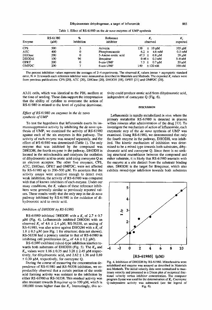
Table 1. Effect of RS-61980 on the de nova enzymes of UMP synthesis
865
Enzyme
CPS ATC DHOase DHODH OPRT OMPDC
RS-61’380 % Reference (VW Inhibition inhibitor
sot1 5 Acivicin 4ocI 0 Phosphonoacetic 2OC 0 5-Amino erotic acid 1OC 96 Brequinar 200 0 6-aza-UMP 2OC 7 6-aza-UMP
Ki KI observed expected
139 * lO@vl 105 plvl 6.2 * 4.6 mM 0.5 mM
47.3 k 0.8 ~&l 28 W 0.46 f 0.3 nM 5-8 nM 7.3 * 0.7 ph4 20 W
140 + 120nM 100 nM
The percent inhibition values represent the averages of 2-4 experiments. The observed Ki values (mean + asymptotic standard error, N Z= 3) towards each reference inhibitor were measured as described in Materials and Methods. The expected K, values were from previous publications: CPS [20], ATC [20], DHOase [20], DHODH [18], OPRT [21] and OMPDC [20].
A3.01 cells, which was identical to the PBL medium at the time of seeding. These data support the interpretation that the ability of cytidine to overcome the action of RS-61980 is related to the level of cytidine deaminase.
Effect of RS-61980 on enzymes in the de novo synthesis of LIMP
To test the hypothesis that leflunomide exerts its im- munosuppressive activity by inhibiting the de novo syn- thesis of UMP, we examined the activity of RS-61980 against each of the sir. enzymes in this pathway. The activity of each enzyme was assayed separately, and the effect of RS-61980 was determined (Table 1). The only enzyme that was inh.ibited by the compound was DHODH, the fourth enzyme in the pathway. DHODH is located in the mitochondria and catalyzes the oxidation of dihydroorotic acid to erotic acid using coenzyme Q as an electron acceptor. The other five enzymes, CPS, ATC, DHOase, OPRT and OMPDC, were not affected by RS-61980 up to 20&500 ~.LM. To ascertain that the activity assays were sensitive enough to detect even weak inhibition, the activity of RS-61980 was compared with that of known inhibitors of each enzyme. Under our assay conditions, the K, values of these reference inhib- itors were generally similar to previously reported val- ues. These results verify that the only step in the de novo pathway inhibited by RS-61980 is the oxidation of di- hydroorotic acid to erotic acid.
Inhibition of DHODH by RS-61980
RS-61980 inhibited DHODH with a Ki of 2.7 f 0.7 PM (Fig. 4). Leflunomide inhibited DHODH with an observed Ki of 4.6 + 2.4 PM; RS-56338, an analog of RS-61980, was also actl:ve against DHODH with a Ki of 1.8 f 0.5 PM (see Fig. 1 for structure; data not shown). RS-56338 had a potency similar to that of RS-61980 in inhibiting cell proliferation (IC,, of 4.6 f 0.2 PM).
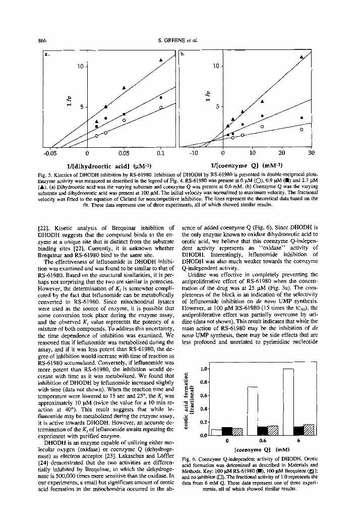
RS-61980 exhibited mixed-type inhibition kinetics to- wards both substrates of DHODH (Fig. 5). The Kii and K, values were 1.16 * 0.35 and 3.28 f 2.45 FM respec- tively, for dihydroorotic acid, and 2.62 It 1.36 and 0.69 f 0.39 PM, respectively, for coenzyme Q.
During the course of measuring the concentration de- pendence of RS-61980 ;and RS-56338 inhibition, we re- producibly observed that a certain portion of the erotic acid forming activity was resistant to the inhibition by either RS-61980 or RS-56338. This residual activity was also resistant towards B:requinar up to 100 FM, which is
tivity could produce erotic acid from dihydroorotic acid, independent of coenzyme Q (Fig. 6).
DISCUSSION
Leflunomide is rapidly metabolized in vivo, where the primary metabolite RS-61980 is detected in plasma within minutes after administration of the drug [ 131. To investigate the mechanism of action of leflunomide, each enzymatic step of the de novo synthesis of UMP was examined. Using RS-61980, we demonstrated that only the fourth enzyme in the pathway, DHODH, was inhib- ited. The kinetic mechanism of inhibition was deter- mined to be a mixed type towards both substrates, dihy- droorotic acid and coenzyme Q. Since there is no strik- ing structural resemblance between the compound and either substrate, it is likely that RS-61980 interacts with the enzyme at a site distinct from the substrate binding sites. DHODH is the target for Brequinar, which also exhibits mixed-type inhibition towards both substrates
i -i ti i i ii i io
IRS-619801 (FM)
Fig. 4. Inhibition of DHODH by RS-61980. Mitochondria were solubilized and enzyme was assayed as described in Materials and Methods. The initial velocity data were normalized to max- imum velocity and presented in a Dixon plot of reciprocal frac- tional velocity versus inhibitor concentration. The computer program Systat was used for the determination of K* &enzyme Q-independent activity was subtracted (see the legend of
100,000 times higher than the Kp Interestingly, this ac- -. .I rig. 0).
866 S. GREENE et al.
ia
l/[dihydroortic acid] @M-l) l/[coenzyme Q] (mM-1) Fig. 5. Kinetics of DHODH inhibition by RS-61980. Inhibition of DHODH by RS-61980 is presented in double-reciprocal plots. Enzyme activity was measured as described in the legend of Fig. 4. RS-61980 was present at 0 ph4 (0). 0.9 pM (m) and 2.7 ph4 (A). (a) Dihydroortic acid was the varying substrate and coenzyme Q was present at 0.6 mM. (b) Coenzyme Q was the varying substrate and dihydroorotic acid was present at 100 pM. The initial velocity was normalized to maximum velocity. The fractional velocity was fitted to the equation of Cleland for noncompetitive inhibition. The lines represent the theoretical data based on the
fit. These data represent one of three experiments, all of which showed similar results.
[22]. Kinetic analysis of Brequinar inhibition of DHODH suggests that the compound binds to the en- zyme at a unique site that is distinct from the substrate binding sites [22]. Currently, it is unknown whether Brequinar and RS-61980 bind to the same site.
The effectiveness of leflunomide in DHODH inhibi- tion was examined and was found to be similar to that of RS-61980. Based on the structural similarities, it is per- haps not surprising that the two are similar in potencies. However, the determination of Ki is somewhat compli- cated by the fact that leflunomide can be metabolically converted to RS-61980. Since mitochondrial lysates were used as the source of enzyme, it is possible that some conversion took place during the enzyme assay, and the observed Ki value represents the potency of a mixture of both compounds. To address this uncertainty, the time dependence of inhibition was examined. We reasoned that if leflunomide was metabolized during the assay, and if it was less potent than RS-61980, the de- gree of inhibition would increase with time of reaction as RS-61980 accumulated. Conversely, if leflunomide was more potent than RS-61980, the inhibition would de- crease with time as it was metabolized. We found that inhibition of DHODH by leflunomide increased slightly with time (data not shown). When the reaction time and temperature were lowered to 15 set and 25”, the Ki was approximately 10 @I (twice the value for a IO-min re- action at 40’). This result suggests that while le- flunomide may be metabolized during the enzyme assay, it is active towards DHODH. However, an accurate de- termination of the Ki of leflunomide awaits repeating the experiment with purified enzyme.
DHODH is an enzyme capable of utilizing either mo- lecular oxygen (oxidase) or coenzyme Q (dehydroge- nase) as electron acceptor [23]. Lakaschus and Laffler [24] demonstrated that the two activities are differen- tially inhibited by Brequinar, to which the dehydroge- nase is 500,000 times more sensitive than the oxidase. In our experiments, a small but significant amount of erotic acid formation in the mitochondria occurred in the ab-
sence of added coenzyme Q (Fig. 6). Since DHODH is the only enzyme known to oxidize dihydroorotic acid to erotic acid, we believe that this coenzyme Q-indepen- dent activity represents an “oxidase” activity of DHODH. Interestingly, leflunomide inhibition of DHODH was also much weaker towards the coenzyme Q-independent activity.
Uridine was effective in completely preventing the antiproliferative effect of RS-61980 when the concen- tration of the drug was at 25 pM (Fig. 3a). The com- pleteness of the block is an indication of the selectivity of leflunomide inhibition on de now UMP synthesis. However, at 100 pM RS-61980 (15 times the lc,,), the antiproliferative effect was partially overcome by uri- dine (data not shown). This result indicates that while the main action of RS-61980 may be the inhibition of de now UMP synthesis, there may be side effects that are less profound and unrelated to pyrimidine nucleotide
L 0.6
L 6
[coenzyme Q] (mM)
Fig. 6. Coenzyme Q-independent activity of DHODH. Orotic acid formation was determined as described in Materials and Methods. Key: 100 pM RS-61980 (m. 100 w Brequinar (a); and no inhibitor (0). The fractional activity of 1 .O represents the data from 6 mM Q. These data represent one of three experi-
ments, all of which showed similar results.
Dihydroorotate dehydrogenase, a target of leflunomide 867
metabolism. These “side effects” occur at higher con- 10. centrations of the drug. As a comparison, Brequinar at 500 nM (12.5 times the ICKY, which is 40 nM) was com- pletely overcome by uridine (data not shown). Using the ll. uridine effect as a criterion for selectivity, leflunomide appears to be less selective than Brequinar. RS-61980 is less potent than Brequinar in both cell growth and 12. DHODH inhibition by three orders of magnitude.
In summary, this study suggests a molecular target for leflunomide, a novel and clinically effective immuno- suppressive agent. This should enable further efforts to 13. develop compounds with improved potency and selec- tivity.
Acknowledgements-We thank Dr. Howard Schulman for pro- viding valuable suggestions for preparing the manuscript. 14.
1.
2.
3.
4.
5.
6.
7.
8.
9.
IREFERENCES
Allison AC and Eug;ui EM, Mycophenolate mofetil: A ra- tionally designed immunosuppressive drug. C/in Truns- p&f 7: 96-l 12, 19’33. Goldblum R, Therapy of rheumatoid arthritis with myco- phenolate mofetil. Clin Exp Rheumarol 8: Sl17-Sll9, 1993. Wu J, Mycophenolate mofetil: Molecular mechanisms of action. Perspect Drug Discovery Design 2: 185-204, 1994. Makowka L, Sher LS and Cramer DV, The development of Brequinar as an immunosuppressive drug for transplanta- tion. Immunol Rev 136: 51-70, 1993. Bartlett RR, Dimitrijevic M, Mattar T, Zielinski T, Ger- mann T, Rude E, Thoenes GH, Kuchle CC, Schorlemmer HU, Bremer E, Finnegan A and Schleyerbach R, Le- flunomide (HWA 486), a novel immunomodulating com- pound for the treatment of autoimmune disorders and re- actions leading to trzlnsplantation rejection. Agents Actions 32: 10-21, 1991. Thomson AW and Starzl TE, New immunosuppressive drugs: Mechanistic Insights and potential therapeutic ad- vances. Immunol Rell 136: 71-98, 1993. Hambleton P and McMahon S, Drug actions on delayed- type hypersensitivity in rats with developing and estab- lished adjuvant arthritis. Agents Acrions 29: 328-332, 1990. Glant TT. Mikecz I<, Bartlett RR, Deak F, Thonar EJ, Williams JM, Mattar T, Kuettner KE and Schleyerbach R, Immunomodulation of proteoglycan-induced progressive polyarthritis by leflunomide. lmmunopharmacology 23, 105-l 16, 1992. Chong AS, Finnefan A, Jiang X, Gebel H, Sankary HN, Foster P and Williams JW, Leflunomide, a novel immuno- suppressive agent. The mechanism of inhibition of T cell proliferation. TranspJanration 55: 1361-l 366, 1993.
15.
16.
17.
18.
19.
20.
21.
22.
23.
24.
Zielinski T, Muller HJ and Bartlett RR, Effects of le- flunomide (HWA 486) on expression of lymphocyte acti- vation markers. Agents Actions 38: C8O-C82, 1993. Mattar T, Kochhar K, Bartlett R, Bremer EG and Finnegan A, Inhibition of the epidennal growth factor receptor tyro- sine kinase activity by leflunomide. FEES Lert 334: 161- 164, 1993. Cherwinski HM, McMarley D, Schatzman R, Devens B and Ransom JT, The immunosuppressant leflunomide in- hibits lymphocyte progression through cell cycle by a novel mechanism. J Pharmacol Exp Ther 272: 460-468, 1994. Bartlett RR, Mattar T, Weithmann U, Anagnostopoulos H, Popovic S and Schleyerback R, Leflunomide (HWA486): A novel immunorestoring drug. In: Therapeutic Ap- proaches to Inflammatory Diseases (Eds. Lewis AJ, Doherty NS and Ackerman NR), pp. 215-228. Elsevier Science Publishing, New York, 1989. Jones ME, Pyrimidine nucleotide biosynthesis in animals: Genes, enzymes and regulation of UMP biosynthesis. Annu Rev Biochem 49: 253-279, 1980. Hirst M, Haliday E, Nakamura J and Lou L, Human GMP synthetase: Protein purification, cloning and functional ex- pression of cDNA. J Biol Chem 269: 23830-23837, 1994. Mori M and Tatibana M, A multienzyme complex of car- bamoyl-phosphate synthase (glutamine):aspartate carbam- oyltransferase:dihydroorotase (rat ascites hepatoma cells and rat liver). Methods Enzymol51: 11 l-121, 1978. Dileepan KN and Kennedy J, Rapid conversion of newly- synthesized orotate to uridine 5’-monophosphate by rat liver cytosolic enzymes. FEBS Lelt 153: 1-5, 1983. Chen S-F, Ruben RL and Dexter DL. Mechanism of action of the novel anticancer agent 6-fluoro-2-Q’-fluoro-l,l’-bi- phenyl-4-yl)-3-methyl-4-quinolinecarboxylic acid sodium salt (NCS 368390): Inhibition of de nova pyrimidine nu- cleotide biosynthesis. Cancer Res 46: 50145019, 1986. Prescott LM and Jones ME, Modified methods for the de- termination of carbamyl aspartate. Anal Biochem 32: 408- 419, 1969. Kensler TW and Cooney DA, Inhibitors of the de nova pyrimidine pathway. In:. Design of Enzyme Inhibitors as Drugs (Eds. Sandler A and Smith HJ), PP. 379-401. Oxford Uniie&ty Press, New York, 1989. . _ Jones ME, Kavipurapu PR and Traut TW, Orotate phos- phoribosyltransferase:orotidylate decarboxylase (Ehrlich ascites cell). Methods Enzymol51: 155-167, 1978. Chen S-F, Perrella FW, Behrens DL and Papp LM, Inhi- bition of dihydroorotate dehydrogenase activity by brequi- nar sodium. Cancer Res 52: 3521-3527, 1992. Forman HJ and Kennedy J, Purification of the primary dihydroortate dehydrogenase (oxidase) from rat liver mito- chondria. Prep Biochem 7: 345-355, 1977.
Lakaschus G and Lbffler M, Differential susceptibility of dihydroorotate dehydrogenase/oxidase to Brequinar so- dium (NCS 368 390) in vitro. Biochem Pharmacol 43: 1025-1030, 1992.
BP 50:6-F