Embed Size (px)
Citation preview
University of Kentucky University of Kentucky
UKnowledge UKnowledge
Theses and Dissertations--Chemical and Materials Engineering Chemical and Materials Engineering
2015
INHALABLE NANOCOMPOSITES AND ANTICANCER AGENTS FOR INHALABLE NANOCOMPOSITES AND ANTICANCER AGENTS FOR
CANCER THERAPY CANCER THERAPY
Nathanael A. Stocke University of Kentucky, [email protected]
Right click to open a feedback form in a new tab to let us know how this document benefits you. Right click to open a feedback form in a new tab to let us know how this document benefits you.
Recommended Citation Recommended Citation Stocke, Nathanael A., "INHALABLE NANOCOMPOSITES AND ANTICANCER AGENTS FOR CANCER THERAPY" (2015). Theses and Dissertations--Chemical and Materials Engineering. 55. https://uknowledge.uky.edu/cme_etds/55
This Doctoral Dissertation is brought to you for free and open access by the Chemical and Materials Engineering at UKnowledge. It has been accepted for inclusion in Theses and Dissertations--Chemical and Materials Engineering by an authorized administrator of UKnowledge. For more information, please contact [email protected].
STUDENT AGREEMENT: STUDENT AGREEMENT:
I represent that my thesis or dissertation and abstract are my original work. Proper attribution
has been given to all outside sources. I understand that I am solely responsible for obtaining
any needed copyright permissions. I have obtained needed written permission statement(s)
from the owner(s) of each third-party copyrighted matter to be included in my work, allowing
electronic distribution (if such use is not permitted by the fair use doctrine) which will be
submitted to UKnowledge as Additional File.
I hereby grant to The University of Kentucky and its agents the irrevocable, non-exclusive, and
royalty-free license to archive and make accessible my work in whole or in part in all forms of
media, now or hereafter known. I agree that the document mentioned above may be made
available immediately for worldwide access unless an embargo applies.
I retain all other ownership rights to the copyright of my work. I also retain the right to use in
future works (such as articles or books) all or part of my work. I understand that I am free to
register the copyright to my work.
REVIEW, APPROVAL AND ACCEPTANCE REVIEW, APPROVAL AND ACCEPTANCE
The document mentioned above has been reviewed and accepted by the student’s advisor, on
behalf of the advisory committee, and by the Director of Graduate Studies (DGS), on behalf of
the program; we verify that this is the final, approved version of the student’s thesis including all
changes required by the advisory committee. The undersigned agree to abide by the statements
above.
Nathanael A. Stocke, Student
Dr. J. Zach Hilt, Major Professor
Dr. Thomas Dziubla, Director of Graduate Studies
Title Page INHALABLE NANOCOMPOSITES AND ANTICANCER AGENTS FOR CANCER
THERAPY
Title page
DISSERTATION
A dissertation submitted in partial fulfillment of the
requirements for the degree of Doctor of Philosophy in the
College of Engineering
at the University of Kentucky
By
Nathanael Aaron Stocke
Lexington, Kentucky
Director: Dr. J. Zach Hilt, Associate Professor of Chemical & Materials Engineering
Lexington, Kentucky
2015
Copyright © Nathanael Aaron Stocke 2015
Abstract
ABSTRACT OF DISSERTATION
INHALABLE NANOCOMPOSITES AND ANTICANCER AGENTS FOR CANCER
THERAPY
Cancer is designated as the leading cause of mortality worldwide and lung cancer
is responsible for nearly 30% of all cancer related deaths. Over the last few decades
mortality rates have only marginally increased and rates of recurrence remain high.
These factors, among others, suggest the need for more innovative treatment modalities
in lung cancer therapy. Targeted pulmonary delivery is well established for treating
pulmonary diseases such as asthma and provides a promising platform for lung cancer
therapy. Increasing local deposition of anticancer agents (ACAs) and reducing systemic
exposure of these toxic moieties could lead to better therapeutic outcomes and higher
quality of life for lung cancer patients receiving such harsh chemotherapy regimens. In
this work, a novel lung cancer treatment modality is presented wherein ACAs are
incorporated into inhalable dry powder composites for targeted delivery to the pulmonary
tract. Additionally, nanoparticles were added to inhalable composites to increase the
therapeutic potential of these unique materials.
A variety of dry powder composites were formulated via spray drying and the
physicochemical properties of the resulting systems were characterized. Additionally, the
performance of the cargo incorporated into these composites was evaluated in order to
insure the activity of the components after release from the inhalable dry powders. The
aerodynamic performance of the dry powder systems was evaluated with the Next
Generation Impactor® to determine if these materials were suitable for inhalation
purposes.
Iron oxide (Fe3O4) magnetic nanoparticles were synthesized and incorporated into
dry powders to examine the feasibility of administering these materials to the lungs for
remotely actuated hyperthermia. Remote heating studies were performed on the
nanoparticles released from these composites using a custom Taylor Winfield®
alternating magnetic field source, and in vitro hyperthermia studies were performed using
advanced multicellular spheroid cell culture models. These studies elicited the
effectiveness of these systems on physiologically relevant models. In addition to the iron
oxide composites, dry powders were formulated with two common ACAs, cisplatin and
erlotinib, for inhalable chemotherapy. The activity of the drugs released from these
composites was evaluated on the human pulmonary lung cancer cell lines A549 and
H358 and compared with the free form of the drugs in order to evaluate the effectiveness
of these therapies. Finally, responsive hydrogel nanoparticles (HNPs) that contain the
ability to respond to environmental changes in pH were synthesized and evaluated as
responsive drug carriers. The response of these particles to pH was evaluated and their
stability was examined before and after inclusion into dry powder composites. Overall,
inhalable dry powder nanocomposites are promising materials for innovative lung cancer
treatment modalities and have the potential to provide a safer and more effective option
for addressing this devastating disease.
KEYWORDS: Lung cancer, pulmonary delivery, nanoparticles,
hyperthermia, anticancer agents
Nathanael Aaron Stocke o
Student’s Signature
April 30, 2015 o
Date
INHALABLE NANOCOMPOSITES FOR THE TREATEMENT OF LUNG
CANCER
By
Nathanael Aaron Stocke
Dr. J. Zach Hilt o
Director of Dissertation
Dr. Thomas Dziubla o
Director of Graduate Studies
April, 2015_________
o

Dedicated to my loving grandparents, John and Agnus Mitchell
iii
Acknowledgements
ACKNOWLEDGEMENTS
This dissertation would not have been possible without guidance and
encouragement from multiple individuals in my life and I am beyond thankful for those
who have been a part of this journey. To start, I would like to thank my advisor, Dr. J.
Zach Hilt for all of his continuous support. Throughout my time spent here at the
University of Kentucky, I have sought Dr. Hilt’s counsel countless times. While most of
these encounters have revolved around science, his influence has reached far beyond
simply molding my scientific intellect; his character and integrity have inspired me to
pursue professionalism at the highest level, while showing me that it is possible to be a
committed husband and loving father though it all. Truthfully, I would not be where I am
today if not for the invaluable teaching, mentoring, and advising I received from Dr. Hilt.
Secondly, I would like to express gratitude to my clinical advisor Dr. Susanne
Arnold, MD. During my first year as a graduate student, I reached out to Dr. Arnold to
obtain her advice regarding the direction of my research project. She generously gave
her time and counsel that proved to be invaluable in years to come. Thereafter, Dr.
Arnold accepted my invitation to become the clinical advisor for my project with the
National Cancer Institute. Her contributions shaped many fundamental aspects of my
projects, and I am grateful for her expert advice and unapologetic candidness during our
encounters.
To the other members of my committee, Dr. Dziubla, Dr. Anderson, and Dr.
Upreti, I am grateful for their input over the past few years. I have approached each of
iv
these individuals at varying times throughout my graduate work. Each of them
responded as if I am one of their own graduate students, with a willing and supportive
approach.
Additionally, I would like to express appreciation to my fellow graduate students
and lab mates, without whom this work would have been impossible. Specifically, I
would like to thank members of the Hilt lab group, both past and present: Ashley Lewis,
Samantha Meenach, Robert Wydra, Anastasia Hauser, Angela Gutierrez, Shuo Tang,
Rohit Bhandari, Trang Mai. Additionally, I would like to thank the following graduate
students from other lab groups: Daniel Schlipfgphd, Jenn Fisher, Andrew Vasilakes,
David Cochran, and Jacob Lilly. Each of these individuals have played an influential part
in facilitating my progress through graduate school, regardless of whether it be an tedious
scientific conversation or an enjoyable social activity.
I would also like to specifically thank Robert Wydra for the thousands of times I
asked for his advice and the hundreds of times he pleasantly responded. Truthfully, Rob
started his graduate work the year before me and has been an irreplaceable resource
through this experience. More than just a fellow graduate student, Rob has been an
ongoing friend and I will always cherish the time we spent together becoming young
scientists.
Lastly, I would be remiss if I failed to mention my friends and family who played
such an important role, albeit unnoticed by many, in my progression throughout the last
several years. They have been the rock that I stand on and their encouragement that has
helped me persevere. Specifically, my parents, Richard and Susan, my brothers,
Jonathan and Eric, my sisters Sarah and Madeline, and my closest friends have shown
v
love and support throughout my time spent here at the University of Kentucky and I owe
this entire dissertation to them.
When I started my graduate work, I had a strong desire to be part of a research
project involving lung cancer, as my grandfather’s passing from this debilitating disease
had a marked influence on my life. Beyond anything else, I wanted my family to be
proud of my research and their support throughout my graduate work has confirmed this
each and every day.
In closing, I would like to say that what you get by achieving your goals is not as
important as what you become by achieving your goals. Thank you all for individually
being a part of what this dissertation and I have become.
vi
Table of Contents
ACKNOWLEDGEMENTS ............................................................................................... iii
Table of Contents ............................................................................................................... vi
List of Tables…………………………………………………………………………….xi
List of Figures ................................................................................................................... xii
Chapter 1 Introduction ........................................................................................................ 1
1.1 Objectives ........................................................................................................... 3
Chapter 2 Pulmonary Delivery for Targeted Lung Cancer Therapy .................................. 5
2.1 Types of Lung Cancer ........................................................................................ 5
2.2 Current Non-Surgical Treatments of NSCLC .................................................... 7
2.2.1 Treatment options based on pathological stage .................................... 8
2.2.2 Personalized treatment of NSCLC patients ........................................ 10
2.3 Pulmonary Delivery ......................................................................................... 12
2.3.1 Pulmonary delivery for lung cancer treatment.................................... 14
2.3.2 Dry powders for inhalation ................................................................. 15
2.4 Hydrogels Nanoparticles .................................................................................. 17
2.5 Iron Oxide (Fe3O4) magnetic nanoparticles ..................................................... 19
2.6 Magnetic nanoparticles and pulmonary delivery ............................................. 21
2.7 Mucus Barriers and particle transport .............................................................. 28
Chapter 3 Formulation and Characterization of Inhalable Magnetic Nanocomposite
Microparticles (MnMs) for Targeted Pulmonary Delivery via Spray Drying .................. 34
3.0 Abstract ............................................................................................................ 34
3.1 Introduction ...................................................................................................... 35
3.2 Material and methods ....................................................................................... 39
3.2.1 Materials ............................................................................................. 39
3.2.2 Synthesis of iron oxide magnetic nanoparticles (MNPs) .................... 40
3.2.3 Formulation of magnetic nanocomposite microparticles (MnMs) with
spray drying ................................................................................................. 40
3.2.4 Electron microscopy ........................................................................... 41
vii
3.2.5 Thermal gravimetric analysis (TGA) .................................................. 42
3.2.6 Karl Fischer titration ........................................................................... 43
3.2.7 Differential scanning calorimetry (DSC) ............................................ 43
3.2.8 X-ray powder diffraction (XRPD) ...................................................... 44
3.2.9 Particle size analysis ........................................................................... 44
3.2.10 In vitro aerosol dispersion performance with the Next Generation
ImpactorTM
(NGITM
) .................................................................................... 45
3.2.11 Alternating magnetic field (AMF) Heating studies .......................... 46
3.2.12 Cytotoxicity tests .............................................................................. 47
3.2.13 Statistics ............................................................................................ 48
3.3 Results and Discussion ..................................................................................... 48
3.3.1 Physiochemical characterization ......................................................... 48
3.3.2 Aerosol performance of MnMs ........................................................... 54
3.3.3 AMF heating of MNPs released from MnMs ..................................... 57
3.3.4 In vitro cytotoxicity of MnMs exposed to human lung cell lines ....... 58
3.4 Conclusions ...................................................................................................... 60
Chapter 4 Remote controlled thermal therapy with magnetic nanocomposite
microparticles induces cell death in triple negative breast cancer micrometastasic tumor
tissue analogs 61
4.1 Introduction ...................................................................................................... 61
4.2 Material and methods ....................................................................................... 63
4.2.1 Materials ............................................................................................. 63
4.2.2 Formulation of inhalable magnetic nanocomposite microparticles .... 63
4.2.3 Formation of triple negative breast cancer micrometastasic tumor
tissue analogs (TM analogs) ........................................................................ 64
4.2.4 MNP-treatment of TM analogs ........................................................... 65
4.2.5 Remotely actuated hyperthermia using a custom alternating magnetic
field (AM) .................................................................................................... 65
4.2.6 Fluorescent imaging on TM analogs................................................... 66
4.2.7 Quantification of cell death using Sytox ............................................. 67
viii
4.2.8 Prussian blue staining for iron ............................................................ 67
4.2.9 Transmission electron microscopy (TEM) on TM analogs ................ 68
4.3 Results and Discussion ..................................................................................... 69
4.4 Conclusions ...................................................................................................... 80
Chapter 5 Formulation and Characterization of Inhalable Anticancer Agents for Targeted
Pulmonary Delivery via Spray Drying ............................................................................. 81
5.0 Abstract ............................................................................................................ 81
5.1 Introduction ...................................................................................................... 82
5.2 Materials and Methods ..................................................................................... 85
5.2.1 Materials ............................................................................................. 85
5.2.2 Inhalable ACA systems....................................................................... 86
5.2.3 Spray Drying ....................................................................................... 87
5.2.4 Scanning Electron Microscopy (SEM) on Inhalable Powders ........... 88
5.2.5 Determination of ACA loading in spray dried composites ................. 88
5.2.6 Differential scanning calorimetry (DSC) ............................................ 89
5.2.7 X-ray powder diffraction (XRPD) ...................................................... 90
5.2.8 Particle size analysis ........................................................................... 90
5.2.9 In vitro aerosol dispersion performance with the Next Generation
ImpactorTM (NGITM) ................................................................................ 90
5.2.10 Cytotoxicity tests .............................................................................. 92
5.2.11 Statistics ............................................................................................ 93
5.3 Results and Discussion ..................................................................................... 93
5.3.1 Physiochemical Characterization ........................................................ 93
5.3.2 Aerosol performance of inhalable powders ...................................... 101
5.3.3 In vitro activity of ACAs .................................................................. 104
5.4 Conclusions .................................................................................................... 106
Chapter 6 Responsive Hydrogel Nanoparticles for Pulmonary Delivery ....................... 107
6.0 Abstract .......................................................................................................... 107
6.1 Introduction .................................................................................................... 108
6.2 Material and methods ..................................................................................... 110
ix
6.2.1 Materials ........................................................................................... 110
6.2.2 Synthesis of responsive hydrogel nanoparticles ............................... 111
6.2.3 Electron microscopy and particle sizing ........................................... 112
6.2.4 Colloidal stability of HNPs ............................................................... 112
6.2.5 pH response of HNP diameter with dynamic light scattering........... 113
6.2.6 pH response of HNP diameter with dynamic light scattering........... 113
6.2.7 Inhalable HNP composites from spray drying .................................. 113
6.2.8 Aerosol performance with the Next Generation ImpactorTM
(NGITM
)
.................................................................................................................... 114
6.3 Results and Discussion ................................................................................... 116
6.4 Conclusions .................................................................................................... 131
Chapter 7 Conclusions .................................................................................................... 132
Appendix A Transport in PEG-Based Hydrogels: Role of Water Content and Crosslinker
Molecular Weight ........................................................................................................... 137
A.0 ABSTRACT .................................................................................................. 137
A.1 INTRODUCTION ......................................................................................... 137
A.2 Material and methods .................................................................................... 141
A.2.1 Materials........................................................................................... 141
A.2.2 Synthesis of hydrogels through UV polymerization ........................ 141
A.2.3 Conversion of hydrogels through Fourier Transform Infrared
Spectroscopy (FTIR) analysis .................................................................... 142
A.2.4 Swelling studies ............................................................................... 143
A.2.5 FCS setup ......................................................................................... 143
A.2.6 FCS Data Analysis ........................................................................... 144
A.3 Results and Discussion .................................................................................. 145
A.3.1. Hydrogel synthesis .......................................................................... 145
A.3.2 Swelling studies ............................................................................... 147
A.3.2 Probe diffusion in PEG hydrogels ................................................... 151
A.4 Conclusions ................................................................................................... 156
Appendix B Supplemental Figures ................................................................................. 158

x
References .................................................................................................................. 159
Vita .................................................................................................................. 177
xi
List of Tables
Table 3.1: Summary of experimental conditions for inhalable dry powders……………41
Table 3.2: MnM and MNP diameters, MNP loadings, and water content of MnMs……51
Table 3.3: Aerosol performance properties for inhalable MnMs………………………..57
Table 4.1: Summary of treatment groups for TM analogs for inhalable dry powders….66
Table 5.1: Summary of experimental conditions for inhalable dry powders……………87
Table 5.2: Diameter from SEM and drug loading………………………………………97
Table 5.3: Aerosol performance properties for inhalable MnMs (mean ± std. dev.)…..104
Table 6.1: Aerosol performance of HNP composites for inhalation (mean ± std. dev.).129
xii
List of Figures
Figure 2.1: From reference (Li, Kung et al. 2013). Evolution of non–small-cell lung
cancer (NSCLC) subtyping from histologic to molecular based. Data adapted (Pao and
Girard 2011). EGFR, epidermal growth factor receptor; HER2, human epidermal growth
factor receptor2; MAP2K1, mitogen-activated protein kinase kinase 1………………….7
Figure 2.2: Taken from reference (Shepherd, Pereira et al. 2005). Kaplan-Meier Curve
for Overall Survival among All Patients Randomly Assigned to Erlotinib or Placebo….12
Figure 2.3: A nanomagnetosol solution comprising PEI-pDNA gene vectors coding for
the luciferase gene and SPIONs was nebulized to Balb/c-mice (n03) either with (a) or
without (b) an external magnetic gradient field applied to the right chest. Twenty-four
hours after nebulization luciferase activity in the lungs was measured using in vivo
bioluminescence imaging in mice. The lungs were removed subsequently to conduct an
ex vivo luciferase assay, revealing a mean luciferase expression of 7.2 pg luciferase per
gram tissue in mice that had been exposed to a magnetic gradient and no luciferase in
mice without a magnetic gradient during the nebulization procedure (c). Taken from
reference (Hasenpusch, Geiger et al. 2012)……………………………………………...26
Figure 2.4: A nanomagnetosol solution comprising PEI-pDNA gene vectors coding for
the luciferase gene and SPIONs was nebulized to Balb/c-mice (n03) either with (a) or
without (b) an external magnetic gradient field applied to the right chest. Twenty-four
hours after nebulization luciferase activity in the lungs was measured using in vivo
xiii
bioluminescence imaging in mice. The lungs were removed subsequently to conduct an
ex vivo luciferase assay, revealing a mean luciferase expression of 7.2 pg luciferase per
gram tissue in mice that had been exposed to a magnetic gradient and no luciferase in
mice without a magnetic gradient during the nebulization procedure (c). Taken from
reference (Hasenpusch, Geiger et al. 2012)……………………………………………...28
Figure 2.5: From reference (Wang, Lai et al. 2008). Mucoinert (open symbols) vs.
Mucoadhesive (filled symbols) behaviour of nanoparticles figure………………………31
Figure 2.6: From reference (Sonaje, Lin et al. 2009). Behaviour of oral (♦) and
subcutaneous (■) insulin solution and insulin-loaded nanoparticles (▲) in diabetic
rats………………………………………………………………………………………..33
Figure 3.1: Schematic representation of the formulation and application of magnetic
nanoparticle microcomposites (MnMs). From left to right we see the initial formation of
these inhalable dry powders through spray-drying a feed consisting of iron oxide MNPs
dispersed in a D-mannitol solution of methanol. Upon collection these materials were
loaded into a capsule and placed into a dry powder inhaler for in vitro aerosol dispersion
performance studies in order to model their predictive deposition patterns in the lungs.
Finally, the heating properties of the MNPs released from these MnMs upon dissolution
were verified by exposing them to an alternating magnetic field………………………..39
xiv
Figure 3.2: SEM images of a) MF20, b) MF5, and c) MF0 dry powders. Magnification
5,000x…………………………………………………………………………………….49
Figure 3.3: TEM images of a) MF20, b) MF5, and c) MF0 dry powders………………49
Figure 3.4: XRPD diffractograms for MnM powders…………………………………..53
Figure 3.5: DSC thermograms for MnM powders………………………………………54
Figure 3.6: Aerosol dispersion performance plot for MnM powders using the NGI at 60
L/min……………………………………………………………………………………..56
Figure 3.7: Heating curves for MnMs under AMF exposure…………………………...58
Figure 3.8: In vitro cytotoxicity results for human A549 alveolar epithelial
adenocarcinoma cells after 24 hours of exposure to MnMs at 37°C…………………….59
Figure 4.1: Microscopy images (n=3) of TM analogs after MNP-exposure (-AMF). The
treatment groups are arranged in columns with increasing MNP dose from left to right.
Left column is control group (n=3); middle column is LD MNPs; right column is HD
MNPs. Each panel consists of the following 4 images: green fluorescent (MEF cells, top
left), red fluorescent (4T1, top right), bright-field (bottom left), and overlay (bottom
right). Images were captured t=5 days post MNP exposure…………………………….70
xv
Figure 4.2: Comparison of TM analogs with- (+AMF; bottom row) and without (-AMF;
top row) application of AMF for 1-hour. The treatment groups are arranged in columns
with increasing MNP dose from left to right. Left column is control group (n=3); middle
column is LD MNPs; right column is HD MNPs. Each panel consists of the following 4
images: green fluorescent (MEF cells, top left), red fluorescent (4T1, top right), bright-
field (bottom left), and overlay (bottom right). Images were captured t=5 days after AMF
exposure………………………………………………………………………………….71
Figure 4.3: Representative fluorescent images of (top row; a-c) and bright-field (bottom
row; d-f) images of spheroids stained with SYTOX® Blue dead cell stain and not
exposed to AMF. From left to right the columns represent treatments of control (a,d)
MNPs at 0.1 mg/mL (b,e) and MNPs at 1 mg/mL (c,f)………………………………….73
Figure 4.4: Quantification of dead cell count measured as the mean integrated density of
blue signal from fluorescent images……………………………………………………..74
Figure 4.5: Prussian blue staining of multicellular breast cancer micrometastasic
spheroids. From left to right the columns represent treatments of control (a,d) MNPs at
0.1 mg/mL (b,e) and MNPs at 1 mg/mL (c,f) with the top row (a-c) being treatments
without AMF exposure and the bottom row (d-f) exposed to AMF……………………..75
xvi
Figure 4.6: TEM images of the center of TM analogs. Control –AMF (left column; a, d),
LD –AMF (middle column; b, e), and HD +AMF (right column; c, f) at magnifications of
x2900 (top row; a-c) and x6800 (bottom row; d-f). Red arrows point at extracellular
material in MNP treatments……………………………………………………………...76
Figure 4.7: TEM images of cells displaying massive vacuolization of the cytoplasm at
magnifications of a) x6800, b) 11,000, and c) 23,000. Red box shows where adjacent
image is located in lower mag image…………………………………………………….77
Figure 4.8: TEM images of the edge of TM analogs. Control –AMF (left column; a, d),
LD –AMF (middle column; b, e), and HD +AMF (right column; c, f) at magnifications of
x2900 (top row; a-c) and x6800 (bottom row; d-f). Red arrows point at large MNP
agglomerates……………………………………………………………………………..78
Figure 4.9: TEM images of HD MNP -AMF TM analogs at x2900 (a, d), x4800 (b, e)
and x6800 (c, f). Top row (a-c) are images from center of TM analog and bottom row (d-
f) are images from edge………………………………………………………………….79
Figure 5.1: Representative SEM images of a) MC0 and b) MC10 at magnification of
5,000x with inset at 30,000x …………………………………………………………….95
Figure 5.2: Representative SEM images of a) ME0 and b) ME10 at magnification of
5,000x with inset at 30,000x……………………………………………………………..96
xvii
Figure 5.3: XRPD diffractograms for raw cisplatin (top), MC10 (middle) and MC0
(bottom)…………………………………………………………………………………..98
Figure 5.4: XRPD diffractograms for raw erlotinib (top), ME10 (middle) and ME0
(bottom)…………………………………………………………………………………..99
Figure 5.5: DSC thermograms for raw cisplatin, MC10, and MC10…………………..100
Figure 5.6: DSC thermograms for raw erlotinib, ME0, and ME10……………………101
Figure 5.7: Aerosol dispersion performance plot for MC0 and MC10 powders………103
Figure 5.8: Aerosol dispersion performance plot for ME0 and ME10 powders………103
Figure 5.9: In vitro comparison of raw cisplatin and MC10 in human lung cancer cell
lines A549 (left) and H358 (right)……………………………………………………...105
Figure 5.10: In vitro comparison of raw erlotinib and ME10 in human lung cancer cell
lines A549 (left) and H358 (right) ……………………………………………………..105
Figure 6.1: Representation of responsive hydrogel nanoparticles at varying pH
conditions from low (left) to high (right)……………………………………………….118
xviii
Figure 6.2: Hydrodynamic diameter of HNP120s a) over a large range of pH values and
b) zoomed in above transition pH………………………………………………………119
Figure 6.3: Hydrodynamic diameter of HNP270s a) over a large range of pH values and
b) zoomed in above transition pH……………………………………………………....119
Figure 6.4: Hydrodynamic diameter of HNP120 (diamonds) and HNP270 (squares)
showing difference in transition pH…………………………………………………….120
Figure 6.5: SEM images of HNP120s at a) x100k and b) x180k. ImageJ® size analysis
shows 82.6 nm ± 4.3 nm………………………………………………………………..122
Figure 6.6: SEM images of HNP270s a) x35k and b) x150k. ImageJ® size analysis
shows 133.4 nm ± 12.2 nm……………………………………………………………..122
Figure 6.7: Representative TEM images of representative HNP120s on copper TEM grid
illustrating spherical morphology and size uniformity. The diameters of two
representative particles are: a) 80.7 nm and b) 88.7 nm………………………………..124
Figure 6.8: High mag (x500k) TEM image of individual HNP120 with fast Fourier
Transform inset showing amorphous character of HNPs………………………………124
xix
Figure 6.9: Stability of HNP120s above and below transition pH…………………….126
Figure 6.10: SEM image of SD HNP120 composite dry powder with individual particle
shown in top right corner inset………………………………………………………….127
Figure 6.11: Aerosol dispersion performance plot for inhalable responsive HNPs…...128
Figure 6.12: Hydrodynamic diameter of HNP120s and SD HNP120s a) over a large
range of pH values and b) zoomed above transition pH………………………………..130
Figure 6.13: Stability of HNP120s and SD HNP120s above transition pH shows
population of SD HNP120s that fall out of suspension………………………………...131
1
Chapter 1 Introduction
Cancer is designated as the leading cause of mortality worldwide with an estimated 8.2
million deaths in 2012 (Ferlay, Soerjomataram et al. 2013). In the United States, one in
four deaths is caused by this devastating disease with nearly 30% of these resulting from
lung cancer (Siegel, Naishadham et al. 2012). Non-small cell lung cancer (NSCLC)
accounts for approximately 85% of all lung cancers and is the leading cause of cancer
mortality worldwide (Mellas, Elmesbahi et al. 2010, Bonomi, Pilotto et al. 2011, Tucker,
Laguna et al. 2012).
Although the initial treatment of NSCLC differs depending on stage, physical
health of the patient, and a variety of other factors, the preferred treatment is surgical
resection; however, most NSCLCs are diagnosed as inoperable stage III or stage IV
malignancies (Fathi and Brahmer 2008, Mazzone and Mekhail 2012, Tucker, Laguna et
al. 2012). Unfortunately, over 40% of patients develop recurrences after surgery even
with adjuvant treatments such as radiation or chemotherapy (Bonomi, Pilotto et al. 2011,
Tucker, Laguna et al. 2012). This, among other factors, contributes to 5- and 10-year
mortality rates of less than 15 and 7% respectfully (Crino, Weder et al. 2010). Hence,
there is a great need for more effective non-surgical treatments of NSCLC, and,
considering the large mortality rates, any improvement on current strategies would bring
about a great benefit.
Targeted pulmonary delivery is well established for treating pulmonary diseases
such as asthma and displays characteristics that are promising for its translation into a
novel lung cancer treatment strategy (Hasenpusch, Geiger et al. 2012). For NSCLC
2
therapy, this route of drug delivery could lead to increased concentrations of ACAs in
lung tissue and therefore reduced systemic side effects when compared to an equivalent
intravenous dose. Hence, it is possible that targeted pulmonary delivery could be used to
administer local doses that are higher than current clinical practices and thus result in a
more effective treatment modality for NSCLC patients.
Inhalable therapies, traditionally, consist of liquid droplets or solid particles with
a mass median aerosol diameter (MMAD) of <5 𝜇m in order to be respirable (Hickey,
Mansour et al. 2007, Xu, Mansour et al. 2010). Common inhalable therapies include
nebulizers, metered dose inhalers, and dry powder inhalers (DPIs). Dry powder
inhalation systems are preferable to traditional metered dose inhalers and nebulizers
because of superior stability and ease of use for associated devices (El-Gendy and
Berkland 2009). Conversely, spray drying provides the ability to reproducibly formulate
dry powders consisting of microparticles that are suitable for pulmonary delivery and
offers the ability to control certain physiochemical properties of these particles by
adjusting process parameters (Vanbever, Mintzes et al. 1999, Mobus, Siepmann et al.
2012, Odziomek, Sosnowski et al. 2012, Shen, Chen et al. 2012). Additionally, spray-
drying allows for easy production of aerodynamically adequate carriers of nanoparticles
and ACAs that could further enhance the effectiveness of locally delivering the therapy.
This dissertation includes an investigation and discussion of a range of inhalable
nanocomposites for the targeted treatment of lung cancer with the overall goal being the
development of a novel lung cancer treatment modality that could improve on current
non-surgical clinical practices. These nanocomposites consisted of iron oxide magnetic
nanoparticles (MNPs), anticancer agents (ACAs) and hydrogel nanoparticles (HNPs) in
3
varying combinations. Formulation of the inhalable powders was accomplished via spray
drying and Chapter 2 presents a background of the nanomaterials and formulation
techniques.
1.1 Objectives
The overall objective of this dissertation was to examine the feasibility and
potential of spray drying nanoparticles and anticancer agents into inhalable composite
powders as innovative treatment modalities for patients with primary and secondary lung
cancer. This was accomplished through the following four projects:
1. “Formulation and Characterization of Inhalable Magnetic Nanocomposite
Microparticles for Targeted Pulmonary Delivery”
2. “Remote controlled thermal therapy with magnetic nanocomposite microparticles
induces cell death in triple negative breast cancer micrometastasic tumor tissue
analogs”
3. “Inhalable Anticancer Agents for Targeted Pulmonary Delivery and Lung Cancer
Therapy”
4. “Responsive Hydrogel Nanoparticles for Pulmonary Delivery”
This dissertation begins with background on the relevant aspects of this research in
Chapter 2. Current non-surgical treatments of non-small cell lung cancer, magnetic
nanoparticles (MNPs), pulmonary delivery, and hydrogel nanoparticles (HNPs) are
reviewed in order to highlight their potential in cancer therapy applications. Chapter 3,
titled “Formulation and Characterization of Inhalable Magnetic Nanocomposite
4
Microparticles for Targeted Pulmonary Delivery,” involves the incorporation of magnetic
nanoparticles into dry powder composites via spray drying. The physicochemical
properties of these composites are then thoroughly evaluated and their potential for
targeted remote-controlled thermal therapy is examined by heating studies in an
alternating magnetic field (AMF) and in vitro aerosol performance studies. Chapter 4 is a
continuation of Chapter 3 wherein the magnetic nanoparticle microcomposites (MnMs)
formulated in Chapter 3 are evaluated for their application in metastatic triple-negative
breast cancer (TNBC). Specifically, the fourth chapter is titled “Remote controlled
thermal therapy with magnetic nanocomposite microparticles induces cell death in triple
negative breast cancer micrometastasic tumor tissue analogs.” Chapter 5, titled
“Inhalable Anticancer Agents for Targeted Pulmonary Delivery and Lung Cancer
Therapy” presents the results of incorporating two commonly administered anticancer
agents, cisplatin and erlotinib, into inhalable dry powder composites. The aerodynamic
performance of these dry powders is examined and the activity of the released ACAs is
studied on two human lung cell lines. Chapter 6, “Responsive Hydrogel Nanoparticles
for Pulmonary Delivery,” reports the synthesis and incorporation of responsive hydrogel
nanoparticles (HNPs) into inhalable composites through spray drying. These HNPs
respond to changes in pH by swelling and/or agglomerating and have potential for future
development into triggered drug release carriers that respond to the reduced pH in the
tumor environment. Finally, Chapter 7 reports the conclusions of this research.
Copyright © Nathanael Aaron Stocke 2015
5
Chapter 2 Pulmonary Delivery for Targeted Lung Cancer Therapy
Over the last several decades great efforts have been poured into lung cancer research.
Billions of dollars in funding ($254 million from NIH in 2014 alone) and countless hours
of research have been invested in attempts to, ultimately, find a cure for this pervasive
disease (Health 2015). However, the return on this investment has not been as fruitful as
anticipated, as survival rates have shown minimal improvements over the last 40 years.
Still, much effort is needed to improve these dismal survival rates and enhance the
quality of life for lung cancer patients.
2.1 Types of Lung Cancer
Traditionally, primary lung cancer can be split into two main histological types
called small cell lung cancer (SCLC) and non-small cell lung cancer (NSCLC), with the
latter accounting for approximately 80-85% of all cases (Molina, Yang et al. 2008,
Zarogoulidis, Zarogoulidis et al. 2013). SCLC, the less common histological type, is split
into two sub groups referred to as pure- and combined small-cell lung cancer (SCLC and
CSCLC, respectively) (Wallace, Arya et al. 2014). In CSCLC any additional component
of NSCLC subtype is present along with SCLC and it represents approximately 30% of
all SCLC cases (Wagner, Kitabayashi et al. 2009). There is a higher proportion of SCLC
patients who are smokers than in NSCLC patients and SCLC is has very low 5-year
survival rates of 1-2% for extensive-stage SCLC (Wallace, Arya et al. 2014).
Non-small cell lung cancer represents the majority population of lung cancer
patients and, traditionally, can be split into three major subtypes referred to as squamous
6
cell carcinoma, large cell carcinoma, and adenocarcinoma (Zarogoulidis, Zarogoulidis et
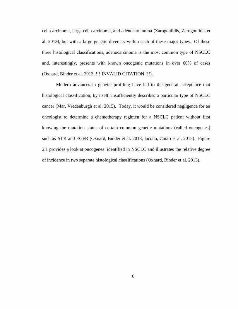
al. 2013), but with a large genetic diversity within each of these major types. Of these
three histological classifications, adenocarcinoma is the most common type of NSCLC
and, interestingly, presents with known oncogenic mutations in over 60% of cases
(Oxnard, Binder et al. 2013, !!! INVALID CITATION !!!).
Modern advances in genetic profiling have led to the general acceptance that
histological classification, by itself, insufficiently describes a particular type of NSCLC
cancer (Mar, Vredenburgh et al. 2015). Today, it would be considered negligence for an
oncologist to determine a chemotherapy regimen for a NSCLC patient without first
knowing the mutation status of certain common genetic mutations (called oncogenes)
such as ALK and EGFR (Oxnard, Binder et al. 2013, Iacono, Chiari et al. 2015). Figure
2.1 provides a look at oncogenes identified in NSCLC and illustrates the relative degree
of incidence in two separate histological classifications (Oxnard, Binder et al. 2013).
7
Figure 2.1: From reference (Li, Kung et al. 2013). Evolution of non–small-cell lung
cancer (NSCLC) subtyping from histologic to molecular based. Data adapted (Pao and
Girard 2011). EGFR, epidermal growth factor receptor; HER2, human epidermal growth
factor receptor2; MAP2K1, mitogen-activated protein kinase kinase 1.
2.2 Current Non-Surgical Treatments of NSCLC
Non-surgical approaches to treating locally advanced NSCLC are primarily
combinations of adjuvant- and neoadjuvant radiotherapy (RT) and chemotherapy (CT).
Late stage disease is treated with primarily with systemic therapies such as CT and
targeted agents. Different guidelines exist for treatment options of NSCLC patients but
8
these decisions are largely dictated by stage and surgery preference (when possible).
Staging of NSCLC is completed using the TNM system that depends on tumor size (T),
lymph node involvement (N) and degree of metastasis (Institute 2015). Here we provide
a quick glance of current treatment approaches for different stages of NSCLC patients in
order to provide a convenient overview of these options, yet we understand that cancer is
a highly heterogeneous disease and requires case-by-case treatment plans that do not
necessarily fall under the generalizations provided here. Additionally, we present
background information on the most commonly employed chemotherapeutic agent for
NSCLC patients, discuss trends in personalizing NSCLC therapy, and provide
background on erlotinib (Tarveva®), which was the first successful drug for genetically
targeted NSCLC treatment.
2.2.1 Treatment options based on pathological stage
As expected, the prognosis for NSCLC patients depends on pathological stage,
with lower stages having superior survival rates (Zarogoulidis, Zarogoulidis et al. 2013).
Stage I-IIIB patients who do not have surgical resection (by choice or circumstance) are
typically administered a form of RT or placed into a clinical trial (Institute 2015). For
patients not entering a clinical trial, the type of RT administered as well as the
combination of CT varies based on a number of factors (Institute 2015).
For those patients who undergo surgical resection, stage I cases are not generally
given adjuvant CT or RT unless admitted to a clinical trial involving such approach
(Zarogoulidis, Zarogoulidis et al. 2013). Large meta-analyses have confirmed the benefit
of cisplatin-based adjuvant CT for resected stage II-IIIA NSCLC patients, leading to the
9
designation of this treatment modality as the gold standard for such cases (Pisters, Evans
et al. 2007, Robinson, Ruckdeschel et al. 2007, Scott, Howington et al. 2007, Crino,
Weder et al. 2010, Zarogoulidis, Zarogoulidis et al. 2013); however, some guidelines
include neoadjuvant CT as a treatment option as well as RT alone for certain patients
such as stage IIIA cases (Institute 2015). Radiotherapy is also administered in
combination with platinum-based CT for certain resected stages II-IIIA patients and
lymph node involvement plays a large role in prescribing this treatment modality
(Zarogoulidis, Zarogoulidis et al. 2013).
Advanced stage IIIB-IV have a very poor prognosis and therefore a variety of CT
and RT treatment options are considered as well as maintenance therapy or best
supportive care. However, the standard first-line treatment for advanced NSCLC patients
is a platinum-based two drug combination regimen (Gatzemeier, Pluzanska et al. 2007).
Recently, it was suggested that stage IIIB-IV and inoperable patients receive 4 cycles of
cisplatin-based CT along with a third-generation cytotoxic or cytostatic drug
(Zarogoulidis, Zarogoulidis et al. 2013).
Cisplatin was first examined for anticancer activity in 1968 after showing
inhibition of cell proliferation in bacteria cultures (Rosenberg 1985). Since its approval
for medical use in 1978 by the United States Food and Drug Administration (FDA), this
anticancer agent (ACA) has been used in the treatment of a variety of cancers such as
ovarian, testicular, and bladder as well as head and neck cancer (Babincova, Altanerova
et al. 2008). Cisplatin is still considered the gold-standard for treatment of many types of
NSCLC and is classified as a cytotoxic ACA (Zarogoulidis, Zarogoulidis et al. 2013).
One of the main mechanisms of cisplatin-induced cytotoxicity lies in the ability of
10
cisplatin to form irreversible crosslinks in DNA causing G1 cell cycle arrest and
apoptosis [13]. While cisplatin is a highly toxic and nonspecific ACA, more recent
efforts to personalize cancer treatment have led to the development of drugs targeted to
patients with specific genetic alterations.
2.2.2 Personalized treatment of NSCLC patients
Personalized treatment of NSCLC patients with molecularly targeted therapies is
revolutionizing the way researchers and clinicians approach this devastating disease (Li,
Kung et al. 2013, Oxnard, Binder et al. 2013). These rational therapies are based on
commonly mutated genes in NSCLC patients (called oncogenes) and the resulting drugs
have provided an unprecedented step forward in treatment of a disease that has only seen
marginal improvement over the last 40 years (Oxnard, Binder et al. 2013). The success
of these treatments is also leading to paradigm shift in treatment selection from
empirical- to rational design and will undoubtedly play a role in future guidelines for
standard treatment options (Li, Kung et al. 2013). Our increased understanding of
oncogenes and recent groundbreaking clinical studies with targeted therapies are
transforming the way we treat NSCLC patients by using molecularly targeted anticancer
agents for personalized therapy based on genetic profiles. One early example of this is
erlotinib (Rosell, Carcereny et al. 2012, Oxnard, Binder et al. 2013, Mar, Vredenburgh et
al. 2015).
Erlotinib (Tarceva®), a small molecule tyrosine kinase inhibitor (TKI), was
approved for the treatment of NSCLC and is given to patients with tumor associated
epidermal growth factor receptor (EGFR) mutations as it showed a survival benefit for
11
such patients (Eberhard, Johnson et al. 2005, Shepherd, Pereira et al. 2005, Gatzemeier,
Pluzanska et al. 2007). Unlike cytotoxic drugs (e.g. cisplatin), cytostatic drugs such as
erlotinib prevent proliferation of cells as opposed to inducing cellular death (Gatzemeier,
Pluzanska et al. 2007). The mechanism by which erlotinib, and other EFGR-TKIs,
prevent cancer growth involves binding the intracellular domain of EGFR at the
adenosine triphosphate binding site (Smith 2005). Binding of erlotinib to EGRF inhibits
phosphorylation and thereby prevents activation of multiple downstream pathways
including the PI3K-AKT-mTOR and RAS-RAF-MEK-ERK pathways involved in cell
survival and proliferation, respectively (Hirte 2013).
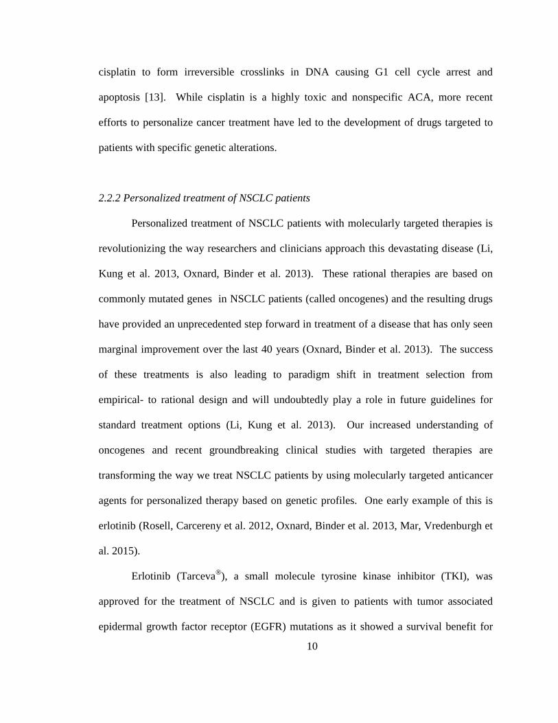
The groundbreaking Phase III study completed by Shepherd et al. showed a better
Kaplan-Meier curve for overall survival (OS) (Figure 2.2) in EGFR+ patients given
erlotinib versus those treated with standard chemotherapy, and illustrated the
effectiveness of targeted therapies in NSCLC therapy (Rosell, Carcereny et al. 2012).
Additionally, over 25% of all lung cancers, and up to 80% of NSCLCs, over express
EGFR; hence, NSCLC represents a large number of patients whom are candidates for
targeted therapy with targeted therapies such as erlotinib (Gatzemeier, Pluzanska et al.
2007, Kanthala, Pallerla et al. 2015). Figure 2.2 is from (Rosell, Carcereny et al. 2012).
12
Figure 2.2: Taken from reference (Shepherd, Pereira et al. 2005). Kaplan-Meier Curve
for Overall Survival among All Patients Randomly Assigned to Erlotinib or Placebo.
2.3 Pulmonary Delivery
Targeted pulmonary delivery is well established for treating diseases of the lung
such as asthma and displays characteristics that are promising for its translation into a
novel lung cancer treatment modality (Hasenpusch, Geiger et al. 2012). The performance
of a pharmaceutical aerosol is quantified with a variety of parameters, the most important
of which is the aerodynamic diameter, 𝐷𝐴. The aerodynamic diameter of a given particle
13
(or droplet) is largely dependent on size, shape, surface roughness, and density (Chow,
Tong et al. 2007, Vehring 2008). Qualitatively, 𝐷𝐴 represents the diameter of a
geometrically perfect sphere of unit density, and zero surface roughness, that would reach
the same terminal velocity in air as the actual, non-ideal, particle (Chow, Tong et al.
2007, Vehring 2008). Quantitatively, it is derived by equating the terminal velocities of
the ideal and non-ideal particle as shown in equation 2.1 below (Anthony J. and Mansour
2009):
𝑉𝑇 = 𝜌𝑝𝑔𝐷𝑝
2𝐶(𝐷𝑝)
𝜅𝑝18𝜂=
𝜌0𝑔𝐷𝐴2𝐶(𝐷𝐴)
𝜅018𝜂 (Equation 2.1)
Here, the subscript p indicates properties of the particle of interest and A indicates those
properties of the idealized aerodynamic particle (or droplet); g is the acceleration of
gravity; 𝜂 is the viscosity of air; 𝐷𝑖 , 𝜌𝑖, 𝜅𝑖, and 𝐶(𝐷𝑖), are the diameter, density, shape
factor, and slip correction factor of species i (which, for the idealized case, become unity
for 𝜌𝑖 and 𝜅𝑖). For particles with a diameter > 1 𝜇m this equation becomes:
𝐷𝐴 = 𝐷𝑝√𝜌
𝜅𝑝 (Equation 2.2)
Additionally, the majority of pharmaceutical aerosols display spherical morphology or
have rotational symmetry, therefore the shape factor, 𝜅𝑝, is equal to one (Anthony J. and
Mansour 2009).
Experimentally, the aerodynamic diameter is determined through inertial
impaction studies using cascade impactors and is referred to as the mass median aerosol
diameter (MMAD). A large advantage to such empirical determination of the
aerodynamic diameter is not needing to know the shape factor or density of the aerosol
formulation (Anthony J. and Mansour 2009). Usually, inhalable aerosol therapies consist
14
of liquid droplets or solid particles with a MMAD < 5 𝜇m in order to be respirable
(Hickey, Mansour et al. 2007, Xu, Mansour et al. 2010).
2.3.1 Pulmonary delivery for lung cancer treatment
Traditionally, anticancer agents (ACAs) are administered intravenously or, to a
lesser extent, orally for systemic delivery (Carvalho, Carvalho et al. 2011). However,
studies have shown that systemic delivery results in relatively low concentration of drug
in the desired site within the lungs, and this, among other factors, has lead researchers to
examine inhalation as means of administering ACAs to patients with lung malignancies
(Vaughn, McConville et al. 2006, Carvalho, Carvalho et al. 2011).
For NSCLC therapy, pulmonary delivery could lead to increased concentrations
of ACAs in lung tissue and therefore reduced systemic side effects when compared to an
equivalent systemic dose. Conversely, reports have indicated as much as a 10-fold
increase in drug concentration within the lungs when administered via pulmonary
delivery relative to systemic delivery via oral administration (Vaughn, McConville et al.
2006). Hence, it is possible that targeted pulmonary delivery could be used to administer
local doses that are higher than current clinical practices and thus result in an effective
treatment modality for NSCLC patients.
Phase I studies of inhaled cisplatin (via nebulization) reached no maximum
tolerated dose and observed no dose limiting toxicity at up to 60 mg/m2 compared with
clinically accepted dosages of 50-100 mg/m2 (Wittgen, Kunst et al. 2007). Additionally,
phase I/II studies of inhalable doxorubicin showed no systemic toxicity for all
administered doses; however, dose-limiting occurred due to direct effects on the upper
15
and lower respiratory tract (Otterson, Villalona-Calero et al. 2007, Otterson, Villalona-
Calero et al. 2010).
A study evaluating the effectiveness of inhaled carboplatin (nebulized liquid
solution) revealed a significant increase in survival for stage IV NSCLC patients who
received a combination of intravenous and pulmonary drug administration as opposed to
IV- or pulmonary only (Zarogoulidis, Eleftheriadou et al. 2012). This suggests that even
advanced-stage NSCLC patients could potentially benefit from a portion of their
chemotherapy being delivered via inhalation and illustrates the potential for inhaled
ACAs in both local and metastatic lung malignancies. This potential of inhaled
chemotherapy in secondary lung cancer patients was examined in a phase Ib/IIa study
with inhaled cisplatin (via nebulization) on metastatic osteosarcoma patients. Two out of
the 14 patients in this study had no pulmonary disease one year after inhalation of
therapy, thereby confirming the potential of pulmonary delivery for both primary and
secondary lung cancer patients (Chou, Bell et al. 2007). One limitation of traditional
inhalation techniques used in most clinical studies (i.e. nebulization) is low aqueous
solubility of hydrophobic anticancer agents such as taxanes, but this limitation could be
overcome through dry powder inhalation (Carvalho, Carvalho et al. 2011).
2.3.2 Dry powders for inhalation
Dry powder formulations are promising in biomedical applications due to
propellant-free, low-cost devices (Rahimpour, Kouhsoltani et al. 2014) as well as
improved stability of the formulation as a result of the dry state (Carpenter, Pikal et al.
1997). Devices for administering dry powders (i.e. dry powder inhalers or DPIs) are
16
breath-activated, which can reduce variability associated with inhaling liquid aerosols by
avoiding necessary coordination of patients (Timsina, Martin et al. 1994, Todo, Okamoto
et al. 2001). Additionally, dry powder formulations do not require active ingredients to
be water soluble and, considering the wide variety of anticancer agents, offer a more
promising approach than liquid aerosols for treating this disease. The primary particles in
dry powder formulations can also be tailored by particle engineering technologies such as
spray drying and therefore designed for regional targeting within the lungs, which is not
possible to the same extent with liquid aerosols (Daniher and Zhu 2008, Son and
McConville 2008).
Spray drying provides the ability to reproducibly formulate dry powders,
consisting of microparticles, that are suitable for pulmonary delivery and offers the
ability to control certain physiochemical properties of these particles by adjusting process
parameters (Vanbever, Mintzes et al. 1999, Mobus, Siepmann et al. 2012, Odziomek,
Sosnowski et al. 2012, Shen, Chen et al. 2012). Additionally, this technique enables easy
production of powders composed of multiple chemical species by simply dispersing them
in the feed solvent (Stocke, Meenach et al. 2014). Incorporating small molecules, such as
most ACAs, into spray dried powders simply requires selecting a solvent appropriate for
spray drying in which your excipients and active ingredients are soluble and optimizing
the conditions for the particular formulation.
Nanoparticles can also be incorporated into composite powders through spray
drying. Incorporating nanoparticles into larger composites is thought to be necessary, as
some have suggested they are too small for pulmonary delivery due to lack of deposition
and subsequent exhalation (Azarmi, Tao et al. 2006, Stocke, Meenach et al. 2014).
17
Hence, spray-drying can be used to incorporate ACAs and multifunctional nanoparticles
into inhalable dry powders thereby opening up an entire field of nano-based drug delivery
platforms that could be utilized to treat NSCLC patients. These factors, among others,
have led to the assertion that dry powders are superior to liquid based formulations (El-
Gendy and Berkland 2009, Rahimpour, Kouhsoltani et al. 2014).
2.4 Hydrogels Nanoparticles
Hydrogels represent an important class of biomaterials with a wide variety of
applications including, among others, tissue engineering, biosensors, drug delivery, and
medical implants (Peppas, Keys et al. 1999, Peppas, Bures et al. 2000, Kopecek 2009).
Traditionally, hydrogels are defined as three-dimensional polymeric networks formed by
crosslinking hydrophilic polymers, and many of their applications center around gels at
equilibrium (e.g. contact lenses) (Peppas, Bures et al. 2000, Peppas, Hilt et al. 2006).
However, the responsive behavior of hydrogel materials provides them with unique
capabilities in biomedical applications (Gupta, Vermani et al. 2002).
Responsive hydrogels have been examined thoroughly over the past several years
and researchers have used a variety of approaches to achieve desired functionality
(Gupta, Vermani et al. 2002). By incorporating various monomers, crosslinkers, and
synthesis conditions one can tune the hydrogel to respond in a variety of manners
including degradation of the hydrogel network or swelling/shrinking of the gel
(Ghandehari, Kopeckova et al. 1997, De, Aluru et al. 2002). Additionally, the response
of the hydrogel can be initiated by a variety of external stimuli such as heat, specific
analytes, and changes in pH (Soppimath, Aminabhavi et al. 2002, Yang, Peters et al.
18
2008, Anthony J. and Mansour 2009). Although hydrogels are generally fabricated at the
macroscale, hydrogel nanoparticles represent a unique class of responsive nanoparticles.
Hydrogel nanoparticles (or nanogels) are crosslinked structures that, as with the
bulk-scale counterparts, have a high water content that provides enhanced
biocompatibility (Oh, Drumright et al. 2008). When these particles are formulated to be
< 200 nm these particles are excellent candidates as drug carriers due to receptor
mediated cellular uptake and prolonged circulation times by eluding uptake by the
mononuclear phagocyte system (Oh, Drumright et al. 2008). Additionally, the structure
of these particles provides them with a unique interior that is in contact with its
surroundings and thereby yields drug carrier systems with a vast range of applications.
Previous reports of delivering hydrogel particles to the lungs via pulmonary
administration demonstrated the ability to engineer micron-sized hydrogel particles that
can be delivered to the lungs as stand-alone carriers (El-Sherbiny and Smyth 2010, Du,
El-Sherbiny et al. 2014, Secret, Kelly et al. 2014). Liquid aerosols of hydrogel micro-
and nanoparticle suspensions were also accomplished using metered dose inhalers and
nebulizers (Farhat, Holloway et al. 2009, Selvam, El-Sherbiny et al. 2011). Additionally,
the in vivo administration of 220 nm nanogels showed sustained release of a model
peptide after pulmonary administration, thereby illustrating the potential of HNPs
administered via inhalation (Lee, Lee et al. 2012). As such, hydrogel nanoparticles
represent a class of materials that has promise for incorporation into novel drug delivery
systems such as inhalable composites for lung cancer applications.
19
2.5 Iron Oxide (Fe3O4) magnetic nanoparticles
A large amount of cancer-related research has pointed to the benefits of utilizing
nanotechnology in the development of novel treatment strategies (Pankhurst, Connolly et
al. 2003, Ferrari 2005, Peer, Karp et al. 2007, Cho, Wang et al. 2008). Iron oxide (Fe3O4)
magnetic nanoparticles (MNPs) represent a unique class of nanomaterials and have
generated considerable interest in biomedicine due to their broad applicability in
biomedical fields. Tissue engineering, biosensing, drug delivery, and hyperthermia
represent a few such applications in which MNPs have great potential (Reddy, Arias et al.
2012). Additionally, MNPs have been approved by the FDA for several commercial
products including their use as an MRI contrast agent. The dual potential of these
materials in therapy and diagnostics – so called theranostics – makes MNPs especially
attractive for future development in cancer applications (Pankhurst, Connolly et al. 2003,
Frimpong and Hilt 2010, He, David et al. 2013). .
Over the years, a wide variety of MNP-based drug delivery systems have been
developed through techniques such as surface coatings and magnetic composites
(Satarkar and Hilt 2008, Satarkar and Hilt 2008, Hawkins, Bottom et al. 2012, Wydra,
Kruse et al. 2013, Kruse, Meenach et al. 2014, Stocke, Meenach et al. 2015). Many of
these applications take advantage of the unique capability of MNPs to heat in the
presence of an alternating magnetic field (AMF). Under such conditions, MNPs generate
thermal energy through frictional (Brownian) and magnetic (Neel) relaxation processes,
and this heat can be used to trigger other therapies, increase transport of particles, or
induce hyperthermia as a thermal treatment (Frimpong, Fraser et al. 2007, Satarkar and
20
Hilt 2008, McGill, Cuylear et al. 2009, Yakacki, Satarkar et al. 2009, Knecht, Ali et al.
2012).
The ability of a MNP system to heat in an alternating magnetic field can be
quantified by calculating a specific absorption rate (SAR), which is defined in equation
2.3 below:
𝑆𝐴𝑅 = ∑ 𝐶𝑖𝑚𝑖
𝑚
𝑑𝑇
𝑑𝑡 (Equation 2.3)
In this equation mi, Ci, are the mass and specific heat capacity of component i,
respectively, and m is the mass of iron oxide. SAR values are dependent on the magnetic
field strength and are therefore reported along with this value. Higher SAR values for
MNP systems are desirable in order to obtain maximum heating and minimum field
strength. One specific, and widely studied, application of heat generation from MNPs is
hyperthermia.
Hyperthermia is loosely defined as mildly heating the body tissue to an elevated
temperature and can be administered to the whole body, regionally, or locally (Vertrees,
Leeth et al. 2002, Wust, Hildebrandt et al. 2002). Despite mixed reports of what
temperature range constitutes hyperthermia, it is generally accepted that above 46°C the
treatment is referred to as thermoablation, and some have suggested 41-46°C as the range
of hyperthermia (Vernon, Hand et al. 1996, Jordan, Scholz et al. 1999, Wust, Hildebrandt
et al. 2002, Babincova, Altanerova et al. 2008, Colombo, Carregal-Romero et al. 2012,
Ohguri, Imada et al. 2012, Xu, Karmakar et al. 2012). In isolation, hyperthermia is a
viable treatment option for cancer patients and in combination with chemotherapeutic
agents such as cisplatin it has shown a synergistic effect as defined by Valeriote and Lin
(Valeriote and Lin 1975, Vernon, Hand et al. 1996, Jordan, Scholz et al. 1999, Wust,
21
Hildebrandt et al. 2002, Babincova, Altanerova et al. 2008, Ohguri, Imada et al. 2012,
Xu, Karmakar et al. 2012). Additionally, recent reports of aerosols containing MNPs
have elicited the ability to administer remotely actuated hyperthermia to the lungs in a
targeted manner (Sadhukha, Wiedmann et al. 2013).
2.6 Magnetic nanoparticles and pulmonary delivery
Over the past decade, a growing interest in delivering MNPs directly to the lungs
via pulmonary delivery is evident by the number of inhalation-based publications, which
include delivery of uncoated MNPs, cubic nanoaggregates, magnetic liposomes, core-
shell MNPs, high aspect ratio MNPs and dry powder magnetic composites (Ally, Martin
et al. 2005, Ally, Amirfazli et al. 2006, Dames, Gleich et al. 2007, Martin and Finlay
2008, Redman, Martin et al. 2011, Hasenpusch, Geiger et al. 2012, Ragab, Rohani et al.
2012, McBride, Price et al. 2013, Ragab and Rohani 2013, Sadhukha, Wiedmann et al.
2013, Verma, Crosbie-Staunton et al. 2013, Nahar, Absar et al. 2014, Tewes, Ehrhardt et
al. 2014, Stocke, Meenach et al. 2015). The attractiveness of such systems lies in the
ability to noninvasively administer and localize these unique nanomaterials through
pulmonary delivery, thereby presenting a drug delivery platform with obvious potential in
a wide variety of applications. However, as with most biomedical applications, the
therapeutic potential of inhalable MNPs must be balanced with safety considerations.
A number of reports regarding the safety of inhaled magnetic particles have
presented mixed conclusions regarding the local and systemic toxicity of this route of
administration; however, some have concluded that nanoparticles delivered to the
pulmonary tract show no evidence of histological changes and temporary pulmonary
22
toxicity (Dames, Gleich et al. 2007, Cho, Cho et al. 2009, Choi, Oh et al. 2009, Kwon,
Kim et al. 2009, Hasenpusch, Geiger et al. 2012, Srinivas, Rao et al. 2012, Szalay, Tatrai
et al. 2012, Oakes, Scadeng et al. 2013, Verma, Crosbie-Staunton et al. 2013). Although
the details of these reports are beyond the scope of this review, we acknowledge these
considerations and point the readers toward potential drawbacks of this approach before
continuing our discussion on inhalable applications of MNPs for inhalable therapies.
Recently, our group reported the incorporation of MNPs into inhalable dry
powder composites, via spray drying, for potential applications in localized hyperthermia
(Stocke, Meenach et al. 2015). We showed that these dry powders displayed good
aerodynamic performance and that high loadings of MNPs were achieved while retaining
adequate aerodynamic behavior. The embedded MNPs retained their heating capabilities
under AMF exposure, upon released from the composites, and showed only modest in
vitro toxicity at relatively high concentrations in A549 human lung cancer cells studies.
These in vitro results highlight the potential of formulating inhalable dry powder
composites for targeted hyperthermia applications.
Sadhuka et al. reported in vivo hyperthermia studies with aerosols containing
EGFR-targeted magnetic nanoparticles for hyperthermia applications (Sadhukha,
Wiedmann et al. 2013). In this work, the previously reported EGFR-targeting peptide
(YHWYGYTPQNVI) (Li, Zhao et al. 2005) was conjugated to magnetic nanoparticles
and then delivered to female Fox Chase SCID® Beige mice via inhalation of an aerosol
generated through ultrasonic atomization of liquid suspensions. Their results showed that
1-week post inhalation, EGFR-targeted MNPs were retained in the lungs at
concentrations higher than MNPs conjugated to a scrambled peptide. Additionally, a
23
murine orthotopic NSCLC model was used to examine the effectiveness of these
materials for localized hyperthermia. These experiments revealed no difference in lung
tumor bioluminescence among untreated-saline, non-targeted-, and targeted MNP
systems when no AMF exposure was administered. Conversely, AMF-induced
hyperthermia resulted in a statistically significant decrease in lung tumor
bioluminescence for the EGFR-targeted MNPs exposed to AMF. These studies show in
vivo proof-of-concept for noninvasive pulmonary delivery and targeting of MNPs for
localized hyperthermia. In addition to physically targeting MNPs through inhalation, a
second degree of targeting magnetic aerosols is achievable through magnetic fields.
For over 30 years, targeting magnetic material to specific regions within the body
has interested researchers, but most of these reports have focused on particles in
circulation (Mosbach and Schroder 1979, Widder and Senyei 1983, Driscoll, Morris et al.
1984, Pankhurst, Connolly et al. 2003). The rationale behind magnetic targeting is the
same as all targeted drug delivery systems wherein localizing the therapy increases the
potency within desired tissue while reducing side effects in other areas of the body. In
addition to passive targeting of magnetic nanoparticles (through the enhanced permeation
and retention (EPR) effect), active targeting can be achieved through a variety of
approaches including surface modification, site-specific injection, and the application of a
magnetic field (Silva, Silva et al. 2007). Targeting magnetic particles in circulation has
been previously reviewed (Dobson 2006, Silva, Silva et al. 2007) so here we will focus
our discussion on this technique as it pertains to MNP-containing aerosols.
Magnetic targeting of aerosols is a relatively new application of biomedical
targeting wherein inhaled therapies can be directed to preferentially accumulate in
24
desired regions of the lungs. This method is particularly attracting for patients with
diseased lungs, as it combines two degrees of targeting: 1) physically targeting the
affected tissue through pulmonary delivery alone (discussed above) and 2) further site-
specific enhancement of MNP-deposition through magnetic guidance. To our
knowledge, the first report of magnetically guided aerosols for therapeutic applications
was in 2005 by Ally et al. This work described theoretical concepts of magnetic aerosols
and validated these hypotheses through foundational in vitro studies showing magnetic
guidance of magnetic aerosols (Ally, Martin et al. 2005).
The in vivo validation of magnetic targeting as a therapeutically viable treatment
modality was reported by Dames et al. in 2007. Liquid droplets containing MNPs – so-
called magnetosols – were administered via nebulization and intratracheal intabation
(Dames, Gleich et al. 2007). Computer-aided simulations suggested that a magnetic flux
gradient of ∇B > 100 Tm-1
was necessary for targeting the magnetosols. The achieved
this by placing the tip of a customized electromagnet over the right lobe of female
BALB/c mice during inhalation. They showed enhanced deposition of MNPs in the
targeted regions of the lungs wherein the amount of MNPs deposited in the right
(targeted) lung was increased by a factor of 8 relative to the untargeted (left) lung.
Additionally, histological evaluation demonstrated no disadvantageous effect on lung
function from the magnetosols with- or without application of magnetic field.
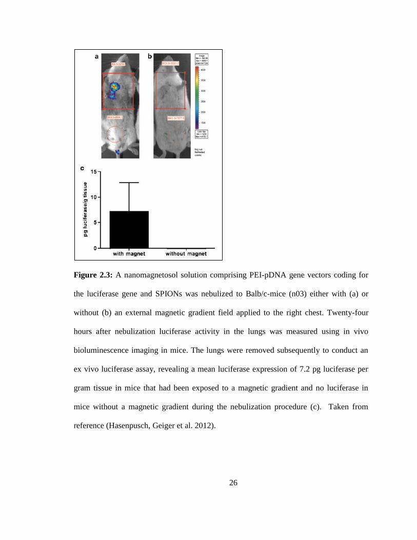
The Rudolph group also published a recent study where they delivered magnetic
aerosols containing model drugs to BALB/c mice via voluntary inhalation of nebulized
liquid droplets (Hasenpusch, Geiger et al. 2012). In this work, the authors used
mathematical simulations to optimize a portable magnet consisting of four identical
25
magnets arranged in a quadupole and then attached this magnet to the chest of the mice
during inhalation. This quadupole resulted in a magnetic field of 0.2 Telsa and a
magnetic gradient of 140 Tm-1
2 mm from the surface of the magnet, which is the
distance they estimated between the animals’ chest and the lung tissue. After inhalation,
the MNP-content in the lungs was determined using a previously reported method of
magnettrelaxometry (Dames, Gleich et al. 2007) as well as a colorimetric determination
of non-heme, and both methods revealed a 2.1-fold increase in the targeted right lung of
the animals relative to the untargeted left lung (no difference was observed when
inhalation was administered in the absence of a magnetic field). Additionally, a plasmid
DNA (coding for the reporter gene luciferase) was complexed with 25 kDa PEI and
added to the pre-nebulized aerosol suspension. The in vivo bioluminescence was
measured 24 hours after inhalation (using an IVIS-100 system) and the transgene
expression is illustrated in Figure 2.3.
26
Figure 2.3: A nanomagnetosol solution comprising PEI-pDNA gene vectors coding for
the luciferase gene and SPIONs was nebulized to Balb/c-mice (n03) either with (a) or
without (b) an external magnetic gradient field applied to the right chest. Twenty-four
hours after nebulization luciferase activity in the lungs was measured using in vivo
bioluminescence imaging in mice. The lungs were removed subsequently to conduct an
ex vivo luciferase assay, revealing a mean luciferase expression of 7.2 pg luciferase per
gram tissue in mice that had been exposed to a magnetic gradient and no luciferase in
mice without a magnetic gradient during the nebulization procedure (c). Taken from
reference (Hasenpusch, Geiger et al. 2012).
27
This image clearly shows luciferase expression in the targeted right lung of the mice
(Figure 2.3a) and no detectable expression in the untargeted left lung (Figure 2.3b).
Further, ex vivo quantification of luciferase (Figure 2.3c) quantitatively confirmed the
findings of the bioluminescence imaging. In addition to enhanced deposition,
histopathological analysis revealed no alterations in the lungs of mice after inhalation of
magnetosols. These studies show the potential of MNPs in pulmonary delivery by
confirming the ability to provide two levels of active targeting through inhaling MNPs
directly to the lungs and magnetically guiding them for site-specific accumulation.
In addition to enhanced deposition, reports have indicated the possibility of
improving the retention of MNPs by applying a magnetic field after deposition from
aerosols (Ally, Amirfazli et al. 2006). Ally et al. reported the theoretical factors affecting
the retention of the magnetic particles once they reach the lungs and compared their
findings with ex vivo experiments. The focus of this study was the upper airways where
the main mechanism of particle clearance is the mucociliary escalator, so their model
focused on the ability of magnetic fields to aid in overcoming this clearance mechanism.
Figure 2.4 illustrates the concept of this work schematically. Their results indicated that
the viscosity of the mucus was the most important factor affecting the retention of the
magnetic particles and illustrate the potential of magnetic fields to enhance retention of
particles in within the lungs which are very efficient at eliminating particulate matter.
Others have shown interest in enhancing mucus penetration of MNPs through magnetic
field and were able to accomplish this in mucus models; however, they were unable to
reproduce these results in actual mucus due to the highly rigid structures within the
mucus mesh.
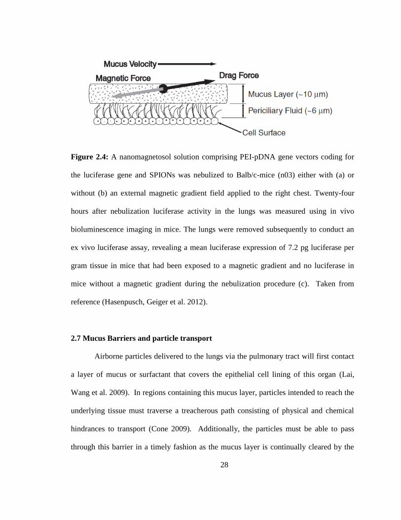
28
Figure 2.4: A nanomagnetosol solution comprising PEI-pDNA gene vectors coding for
the luciferase gene and SPIONs was nebulized to Balb/c-mice (n03) either with (a) or
without (b) an external magnetic gradient field applied to the right chest. Twenty-four
hours after nebulization luciferase activity in the lungs was measured using in vivo
bioluminescence imaging in mice. The lungs were removed subsequently to conduct an
ex vivo luciferase assay, revealing a mean luciferase expression of 7.2 pg luciferase per
gram tissue in mice that had been exposed to a magnetic gradient and no luciferase in
mice without a magnetic gradient during the nebulization procedure (c). Taken from
reference (Hasenpusch, Geiger et al. 2012).
2.7 Mucus Barriers and particle transport
Airborne particles delivered to the lungs via the pulmonary tract will first contact
a layer of mucus or surfactant that covers the epithelial cell lining of this organ (Lai,
Wang et al. 2009). In regions containing this mucus layer, particles intended to reach the
underlying tissue must traverse a treacherous path consisting of physical and chemical
hindrances to transport (Cone 2009). Additionally, the particles must be able to pass
through this barrier in a timely fashion as the mucus layer is continually cleared by the
29
mucociliary escalator (Lai, Wang et al. 2009). Hence, regardless of the novelty and
biocompatibility exhibited by a particulate drug delivery system, sufficient transport rates
are necessary to ensure any therapeutic benefit.
When selecting a coating or additive, it is crucial to consider the barrier through
which these particles must travel. Mucus is a common barrier to transport of colloidal
drug delivery systems and covers sites such as the eyes, lungs, nose, vagina, and upper GI
tract (Cone 2009). This viscoelastic fluid is very effective in trapping foreign particles
and exists as a complex, heterogeneous, aqueous mixture of proteins, lipids, DNA, ions,
cells, and cellular debris (Lai, Wang et al. 2009). The primary component of mucus is
mucin, which forms a crosslinked network able to hinder the diffusion of foreign particles
by physical and chemical means (Lai, O'Hanlon et al. 2007). Mucin fibers are connected
end-to-end via disulfide linkages and the resulting pores in this network are on the
nanometer scale (Sheehan, Kirkham et al. 2004, Lai, Wang et al. 2009). Additionally,
these amphiphilic proteins trap both hydrophobic and hydrophilic particles through low-
affinity interactions (Lai, Wang et al. 2009). Hence, selecting a system that minimizes
these interactions will result in more effective transport of the particles.
Recently, it was shown that the use of a polymer coating enabled particles as large
as 500 nm to traverse mucus at rates approaching unhindered diffusional transport as seen
in a phosphate buffer solution (Lai, O'Hanlon et al. 2007). Hanes’ group took a top-down
approach at designing their coating by attempting to recreate the ability of relatively large
viruses to efficiently transport through mucus (Olmsted, Padgett et al. 2001). Viruses
minimize interaction with mucin fibers by an adhesion-preventing mechanism wherein
their surfaces are densely coated with both positive and negative charges (Wada and
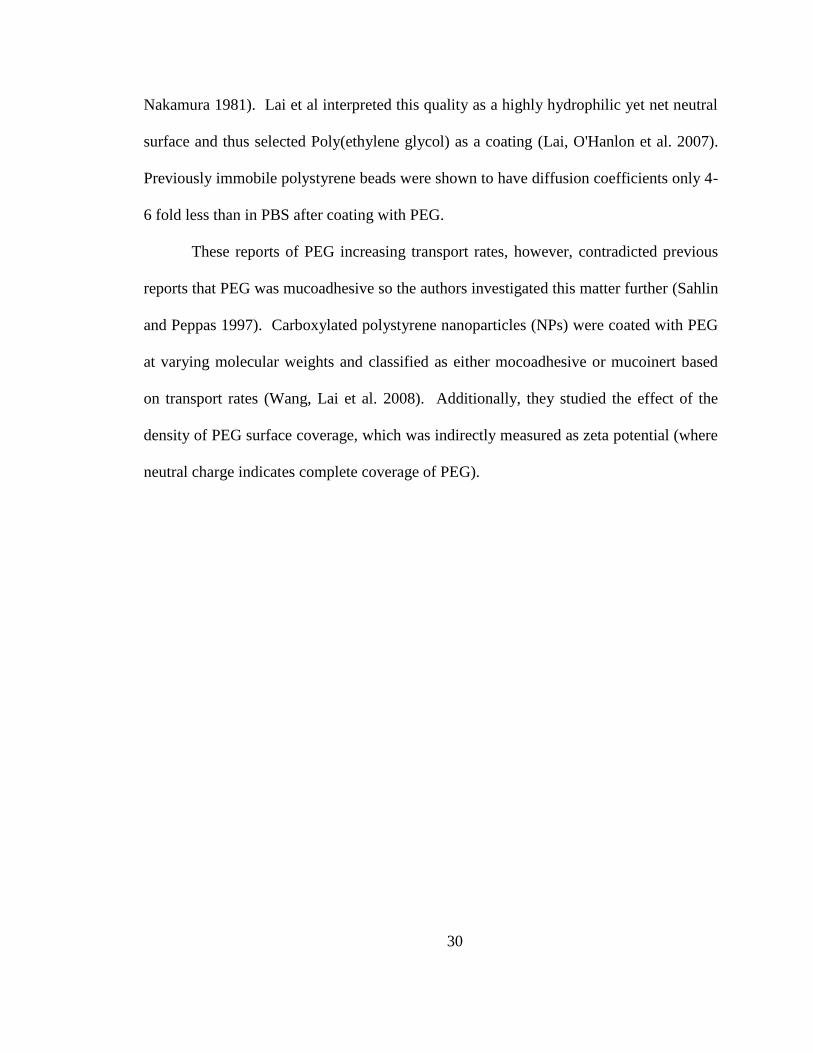
30
Nakamura 1981). Lai et al interpreted this quality as a highly hydrophilic yet net neutral
surface and thus selected Poly(ethylene glycol) as a coating (Lai, O'Hanlon et al. 2007).
Previously immobile polystyrene beads were shown to have diffusion coefficients only 4-
6 fold less than in PBS after coating with PEG.
These reports of PEG increasing transport rates, however, contradicted previous
reports that PEG was mucoadhesive so the authors investigated this matter further (Sahlin
and Peppas 1997). Carboxylated polystyrene nanoparticles (NPs) were coated with PEG
at varying molecular weights and classified as either mocoadhesive or mucoinert based
on transport rates (Wang, Lai et al. 2008). Additionally, they studied the effect of the
density of PEG surface coverage, which was indirectly measured as zeta potential (where
neutral charge indicates complete coverage of PEG).
31
Figure 2.5: From reference (Wang, Lai et al. 2008). Mucoinert (open symbols) vs.
Mucoadhesive (filled symbols) behaviour of nanoparticles figure.
Figure 2.5 shows a variety of particles reported to be mucoadhesive (filled symbols)
along with those said to be mucoinert (open symbols). Results indicate that there is a
specific range (shaded region) of PEG molecular weight and surface coverage for which
the coated nanoparticles are mucoinert. Their hypothesis is that by increasing the
molecular weight of PEG increases (and thus the hydrophilicity of the particles) the
32
particles avoid hydrophobic interactions with the mucin network, and the high surface
coverage of PEG (low zeta potential) minimizes electrostatic interactions. They reasoned
that PEG’s mucoadhesive properties, at higher MW PEG moieties, are due to physical
entanglement between PEG chains and mucin fibres. These reports illustrate the
complexity of engineering a system that allows particles to effectively transport through
the mucus barrier.
Another example of hindered transport is oral administration of proteins such as
insulin (Chen, Sonaje et al. 2011). The layer of endothelial cells lining the walls of the
upper GI tract is a very effective barrier to passive transport of molecules lager than 500-
700 Da (Chen, Sonaje et al. 2011, Muller 2011). Additionally, proteins often require
colloidal encapsulation as some reports indicate that 94-98% of orally administered
proteins are digested by GI proteases (Ganapathy, Gupta et al. 2006, Muller 2011).
Hence, an effective oral protein delivery system requires supramolecular particles to
navigate, or facilitate passage of proteins, to and through the intestinal epithelium in
order to reach the underlying blood vessels.
Sonaje et al. reported an insulin oral bioavailability of 15.1 ± 0.9% by
encapsulating insulin in pH-responsive poly-𝛾-glutamic acid (𝛾 -PGA) and chitosan (CS)
nanoparticles (Sonaje, Lin et al. 2010). Chitosan, a cationic polysaccharide, was chosen
because of its ability to increase paracellular permeability through the opening of tight
junctions between adjacent cells (Ranaldi, Marigliano et al. 2002, Sonaje, Lin et al.
2010). Positively charged 𝛾 -PGA was incorporated as an additive to facilitate ionic
gelation resulting in nanoparticles approximately 200 nm in diameter. As the particles
travel down the GI tract the increasing pH reduces the interaction between 𝛾-PGA and
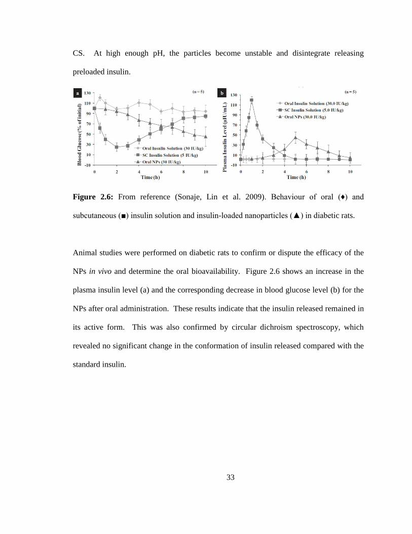
33
CS. At high enough pH, the particles become unstable and disintegrate releasing
preloaded insulin.
Figure 2.6: From reference (Sonaje, Lin et al. 2009). Behaviour of oral (♦) and
subcutaneous (■) insulin solution and insulin-loaded nanoparticles (▲) in diabetic rats.
Animal studies were performed on diabetic rats to confirm or dispute the efficacy of the
NPs in vivo and determine the oral bioavailability. Figure 2.6 shows an increase in the
plasma insulin level (a) and the corresponding decrease in blood glucose level (b) for the
NPs after oral administration. These results indicate that the insulin released remained in
its active form. This was also confirmed by circular dichroism spectroscopy, which
revealed no significant change in the conformation of insulin released compared with the
standard insulin.
34
Chapter 3 Formulation and Characterization of Inhalable Magnetic Nanocomposite
Microparticles (MnMs) for Targeted Pulmonary Delivery via Spray Drying
Inhalable composites for targeted pulmonary delivery of iron oxide (Fe3O4) magnetic
nanoparticles (MNPs) to the lungs were formulated through spray-drying. The resulting
aerodynamic performance of these composites as well as the activity of the MNPs
released upon dissolution was important considerations. The goal was to controllably
incorporate MNPs into powders with adequate aerodynamic performance and to be able
to release unhindered MNPs that retain their ability to remotely heat in the presence of an
alternating magnetic field. The chapter is taken directly or adapted from work published
in Stocke et al. (2014) Copyright 2014 Elsevier B.V. Used with permission from
Nathanael A Stocke, Samantha A. Meenach, Susanne M. Arnold, Heidi M. Mansour, and
J. Zach Hilt.
3.0 Abstract
Targeted pulmonary delivery facilitates the direct application of bioactive
materials to the lungs in a controlled manner and provides an exciting platform for
targeting magnetic nanoparticles (MNPs) to the lungs. Iron oxide MNPs remotely heat in
the presence of an alternating magnetic field (AMF) providing unique opportunities for
therapeutic applications such as hyperthermia. In this study, spray drying was used to
formulate magnetic nanocomposite microparticles (MnMs) consisting of iron oxide
MNPs and D-mannitol. The physicochemical properties of these MnMs were evaluated
and the in vitro aerosol dispersion performance of the dry powders was measured by the
35
Next Generation Impactor®. For all powders, the mass median aerosol diameter (MMAD)
was <5 m and deposition patterns revealed that MnMs could deposit throughout the
lungs. Heating studies with a custom AMF showed that MNPs retain excellent thermal
properties after spray drying into composite dry powders, with specific absorption ratios
(SAR) > 200 W/g, and in vitro studies on a human lung cell line indicated moderate
cytotoxicity of these materials. These inhalable composites present a class of materials
with many potential applications and pose a promising approach for thermal treatment of
the lungs through targeted pulmonary administration of MNPs.
3.1 Introduction
Targeted pulmonary inhalation aerosol delivery facilitates the direct application of
bioactive materials to the lungs in a controlled manner. Medical conditions such as
asthma, chronic obstructive pulmonary disease (COPD), and pulmonary infections
secondary to cystic fibrosis employ targeted pulmonary inhalation aerosol delivery as a
first-line treatment because of inherent advantages (Dolovich, Ahrens et al. 2005) such as
a fast onset of pharmaceutical action, higher local concentrations of drugs, and reduced
systemic side effects (Patton and Byron 2007). Further, the treatments of pulmonary
diseases with inhalation methods avoid first-pass metabolism of pharmaceutical agents
(Mansour, Rhee et al. 2009) resulting in significantly less drug required for aerosol
dosage forms to exert a therapeutic effect in the lungs.
Dry powder inhalation aerosol offers several advantages over liquid aerosol
formulations such as increased stability and sterility of formulations as well as
controllable particle size for targeting regions of the lung (Dolovich, Ahrens et al. 2005).
36
Additionally, dry powder formulations can be used to administer higher amounts of water
insoluble pharmaceutical actives as in contrast to liquid-based inhalation aerosol systems
such as nebulizers and pressurized meter dose inhalers (pMDIs).
Recently, there has been a growing interest in utilizing the advantages of
pulmonary delivery for treating lung diseases such as cystic fibrosis (CF) and lung cancer
thereby expanding the potential applications of successful aerosol formulations. Phase I
trials of inhaled cisplatin showed no dose-limiting toxicity or maximum tolerated
dose(Wittgen, Kunst et al. 2007) and a phase II study of inhaled doxorubicin showed a
favorable median survival compared to intravenous administration (Otterson, Villalona-
Calero et al. 2010). Traditionally, inhaled therapies such as these utilize aerosols
consisting of small molecule drugs and excipients; however, targeted pulmonary delivery
provides a platform for localizing novel multifunctional nanoparticles to the lungs
through direct, topical application (Azarmi, Roa et al. 2008, Mansour, Rhee et al. 2009).
The concept of delivering iron oxide MNPs1 to the lung for drug delivery
purposes has been explored recently due to their unique properties that provide novel
functionality to drug delivery systems as well as their FDA approval as marketed
pharmaceutical products for medical applications such as a contrast agent in magnetic
resonance imaging (MRI) (Pankhurst, Connolly et al. 2003, Dames, Gleich et al. 2007,
Frimpong and Hilt 2010, Xie, Zeng et al. 2010, Redman, Martin et al. 2011, McBride,
Price et al. 2013, Sadhukha, Wiedmann et al. 2013, Verma, Crosbie-Staunton et al.
1AMF: Alternating magnetic field; MMAD: Mass median aerosol diameter; MnM: Magnetic
nanocomposite microparticle; MNP: Magnetic nanoparticle; NGI: Next generation impactor
37
2013). One such functionality is the magnetic guidance of aerosols containing MNPs for
enhanced targeting to specific regions within the lungs. The Rudolph lab has
successfully directed magnetic aerosols, as nebulized MNP suspensions, to regions of the
lungs using magnetic fields (Dames, Gleich et al. 2007, Hasenpusch, Geiger et al. 2012).
These studies show the ability to deliver a higher dose of model drugs to specific regions
of the lungs and demonstrate one of the many potential advantages to incorporating
multifunctional MNPs to inhalation therapies.
Additional advantages of using MNP-containing aerosols include their ability to
remotely heat in the presence of an alternating magnetic field (AMF) providing enhanced
control over actuating the onset of therapy (Satarkar and Hilt 2008, Satarkar, Biswal et al.
2010). In suspension, MNPs generate heat through frictional (Brownian) and magnetic
(Néel) relaxation processes, and the heat generated from these particles can be used to
trigger other therapies, increase transport of particles, and induce hyperthermia as a
thermal treatment (Jordan, Scholz et al. 2006, McGill, Cuylear et al. 2009, Knecht, Ali et
al. 2012, Li, ten Hagen et al. 2013).
Hyperthermia is the heating of tissue to 41-45°C and can be applied locally,
regionally, or throughout the body. Additionally, it has shown promise in treating cancer
by enhancing chemotherapy and radiotherapy (Vernon, Hand et al. 1996, Horsman and
Overgaard 2007, Issels 2008). Locally targeted hyperthermia in the treatment of cancer
has the advantage of reducing undesired cellular death to healthy tissue and can be
accomplished with MNPs under AMF exposure (Jordan, Scholz et al. 1999, Frimpong
and Hilt 2010, Meenach, Hilt et al. 2010, Satarkar, Meenach et al. 2011). Previous
clinical trials have utilized magnetic nanoparticles under AMF exposure to demonstrate
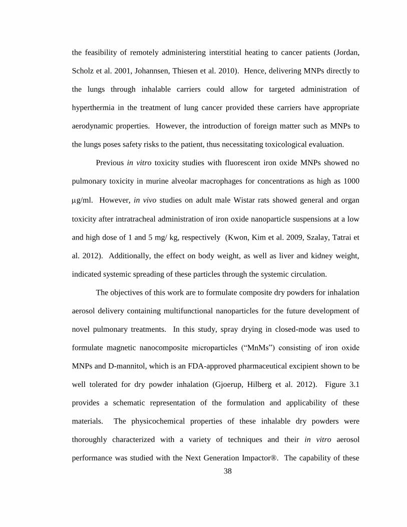
38
the feasibility of remotely administering interstitial heating to cancer patients (Jordan,
Scholz et al. 2001, Johannsen, Thiesen et al. 2010). Hence, delivering MNPs directly to
the lungs through inhalable carriers could allow for targeted administration of
hyperthermia in the treatment of lung cancer provided these carriers have appropriate
aerodynamic properties. However, the introduction of foreign matter such as MNPs to
the lungs poses safety risks to the patient, thus necessitating toxicological evaluation.
Previous in vitro toxicity studies with fluorescent iron oxide MNPs showed no
pulmonary toxicity in murine alveolar macrophages for concentrations as high as 1000
g/ml. However, in vivo studies on adult male Wistar rats showed general and organ
toxicity after intratracheal administration of iron oxide nanoparticle suspensions at a low
and high dose of 1 and 5 mg/ kg, respectively (Kwon, Kim et al. 2009, Szalay, Tatrai et
al. 2012). Additionally, the effect on body weight, as well as liver and kidney weight,
indicated systemic spreading of these particles through the systemic circulation.
The objectives of this work are to formulate composite dry powders for inhalation
aerosol delivery containing multifunctional nanoparticles for the future development of
novel pulmonary treatments. In this study, spray drying in closed-mode was used to
formulate magnetic nanocomposite microparticles (“MnMs”) consisting of iron oxide
MNPs and D-mannitol, which is an FDA-approved pharmaceutical excipient shown to be
well tolerated for dry powder inhalation (Gjoerup, Hilberg et al. 2012). Figure 3.1
provides a schematic representation of the formulation and applicability of these
materials. The physicochemical properties of these inhalable dry powders were
thoroughly characterized with a variety of techniques and their in vitro aerosol
performance was studied with the Next Generation Impactor®. The capability of these
39
materials to remotely heat in the presence of an AMF was examined and the in vitro
cytotoxicity of these materials was examined in a human lung cell line (A549).
Figure 3.1: Schematic representation of the formulation and application of magnetic
nanoparticle microcomposites (MnMs). From left to right we see the initial formation of
these inhalable dry powders through spray-drying a feed consisting of iron oxide MNPs
dispersed in a D-mannitol solution of methanol. Upon collection these materials were
loaded into a capsule and placed into a dry powder inhaler for in vitro aerosol dispersion
performance studies in order to model their predictive deposition patterns in the lungs.
Finally, the heating properties of the MNPs released from these MnMs upon dissolution
were verified by exposing them to an alternating magnetic field.
3.2 Material and methods
3.2.1 Materials
Iron(II) chloride tetrahydrate (FeCl2∙4H2O), iron(III) chloride hexahydrate
(FeCl3∙6H2O), and D-mannitol were obtained from Sigma Aldrich (St. Louis, MO).
Chloroform, anhydrous methanol, and high performance liquid chromatography-grade
methanol were purchased from Fisher Scientific (Pittsburgh, PA). Ultrahigh-purity
nitrogen gas was from Scott-Gross (Lexington, KY), and ammonium hydroxide
40
(NH4OH) was obtained from EMD chemicals (Gibbstown, NJ). Honeywell Riedel-de
Haen® Hydranal®-Coulomat AD, reagent for Karl Fischer (KF) coulometric titration,
was purchased from Sigma-Aldrich (St Louis, MO). Special anhydrous AQUA STAR
methanol for moisture determinations was purchased from EMD Chemicals, Inc.
(Darmstadt, Germany)
3.2.2 Synthesis of iron oxide magnetic nanoparticles (MNPs)
Aqueous co-precipitation was used to synthesize iron oxide nanoparticles.
Briefly, FeCl2∙4H2O (Fe2+
) and FeCl3∙6H2O (Fe3+
) salts were dissolved in 40 mL of
deionized (DI) water at a 2:1 Fe3+
:Fe2+
molar ratio. The solution was sonicated in a water
bath for approximately 20 min and transferred to a 3-neck 500 mL round bottom flask.
The round bottom flask was heated to 85°C under nitrogen flow with a mechanical stirrer
at 300 rpm. Once the vessel reached 85°C, 5 mL of NH4OH (28 % w/w) was added with
a syringe and the reaction was carried out for 1 hour. Recovered particles were washed
with ethanol via magnetic decantation and dried in a vacuum oven prior to spray drying.
3.2.3 Formulation of magnetic nanocomposite microparticles (MnMs) with spray drying
Spray drying was accomplished with a Büchi Mini Spray Dryer B290 coupled
with a B-295 Inert Loop and high-performance cyclone (all from Büchi Labortechnik,
Flawil, Switzerland) using parameters previously optimized by our lab (Li and Mansour
2011). Spray drying experiments were carried out in closed mode using UHP dry
nitrogen gas at a flow rate of 600 L/h, an inlet temperature of 150°C, and an aspirator rate
of 35 m3/h. Once the inlet temperature of 150°C was reached, pure methanol was fed to
41
the spray dryer until the outlet temperature was stable and this temperature was recorded,
as tabulated in Table 3.1. For feed mixtures, D-mannitol was dissolved in methanol and
iron oxide MNPs were weighed out and subsequently dispersed such that the combined
mass of these two components resulted in a dilute concentration of 0.1% wt/vol. The
MF20 and MF5 powders resulted in a MNP concentration of 0.20 mg/ml and 0.05 mg/ml,
respectively. Two liters of the feed mixture were fed to the spray dryer at a pump rate of
50% (15 mL/min) through a stainless steel nozzle with a diameter of 0.7 mm. For the
MF20 and MF5 experiments, which contained MNPs, the feed mixture was sonicated
throughout the experiment to insure adequate dispersion of MNPs. Powders were
collected and transferred to a glass desiccator over Drierite™ and stored in the freezer at -
23°C. Two different iron oxide loadings were incorporated into the powders and Table
3.1 summarizes these systems and their spray drying parameters.
Table 3.1: Summary of experimental conditions for inhalable dry powders.
System Initial MNPs (wt%) Inlet T (°C) Outlet T (°C)
MF20 20% 150 67
MF5 5% 150 58
MF0 0% 150 70
3.2.4 Electron microscopy
Scanning transmission electron microscopy (STEM) was carried out in order to
confirm the presence of iron oxide in the MnMs and to observe the spatial distribution of
42
the nanoparticles within the composites. Powders were dispersed in chloroform at 100
g/ml, added dropwise onto a support film grid (Electron Microscopy Sciences, Hatfield,
PA), and allowed to dry overnight in a desiccator over Drierite™. Chloroform was used
as a dispersant because D-Mannitol is practically insoluble in this organic solvent (Pena,
Daali et al. 2000). Images were captured with a JEOL 2100F Field Emission Electron
Microscope (JOEL, Tokyo, Japan) equipped with an Oxford EDS detector (Oxford
Instruments, Oxford, United Kingdom).
Scanning electron microscopy (SEM) was performed in order to observe the
particle size, surface roughness, shape and morphology. A Hitachi S4300 microscope
(Tokyo, Japan) was used to capture images. Double-sided adhesive carbon tabs were
adhered to aluminum studs (Ted Pella, Redding, CA) and blotted on weigh paper with
our dry powder. These samples were coated with a thin film of a gold/palladium alloy
using an Emscope SC400 sputter coating system at 20 mA for 2 minutes under argon gas.
For all electron microscopy experiments at least 3 independent samples were examined
and multiple images were examined for each sample.
3.2.5 Thermal gravimetric analysis (TGA)
Thermal gravimetric analysis was performed using a TA TGA Q50 (TA
Instruments New Castle, DE) in order to quantify the actual MNP loading in the powders.
Samples were placed in a Tzero™ alodined aluminum pan that was hermetically sealed
and then placed into the furnace along with an empty alodined aluminum hermetically
sealed reference pan. The temperature of the furnace was raised to 120°C at a rate of 5°C
/min and then held isothermally for 20 minutes to remove any trace solvent. The
43
temperature was then increased to 500°C and held isothermally for 30 minutes in order to
completely decompose any D-mannitol present and allow for calculating the actual mass
of iron oxide nanoparticles in the dry powders. Experiments were carried out in triplicate
3.2.6 Karl Fischer titration
Karl Fischer coulometric titration was carried out in order to determine the
residual water content in the powders. A 737 KF Coulometer coupled with 703 Ti Stand
(Metrohm Ltd., Antwerp, Belgium) was used. Approximately 5 mg of these powders
were dissolved into anhydrous methanol for moisture determinations and allowed to
sonicate for approximately 10 minutes to ensure complete dissolution of composites
thereby releasing the MNPs into suspension. The resulting dispersions were injected into
reaction cell filled with Hydranal® KF reagent with a 2 mL syringe and the amount of
water injected was measured. Pure solvent was also injected to determine the amount of
residual water in the solvent. The mass of water in the injected samples was used to
calculate the water content in the dry powders by subtracting out the water content of the
solvent.
3.2.7 Differential scanning calorimetry (DSC)
DSC was carried out in order to determine the thermal phase transitions of the dry
powders. An Auto Q20 Differential Scanning Calorimeter equipped with an RSC-90
refrigerated cooling system (TA Instruments-Waters LLC, New Castle, DE) and Tzero™
technology was used for measurements. Approximately 2 mg of sample was weighed
into a Tzero™ alodined aluminum pan that was hermetically sealed and then placed into
44
the furnace along with an empty alodined aluminum hermetically sealed reference pan.
The temperature was initialized at 0°C and subsequently raised to 250°C at a rate of
5.00°C /min.
3.2.8 X-ray powder diffraction (XRPD)
XRPD was used to examine the long range order of the MnMs. Powders were
analyzed by an X-ray diffractometer (Multiplex, Rigaku) with Cu K radiation (40 kV,
44 mA) in order to examine their crystallinity. Scans were obtained from 5 to 60° 2Θ
with step size of 0.2 and scan rate of 2 °/min.
3.2.9 Particle size analysis
Dynamic Light Scattering (DLS) was performed to determine the hydrodynamic
diameter of the MNPs before spray drying and after being released by dissolution of the
MnMs. Aqueous dispersions were made with a MNP concentration of 200 g/mL and
were read in a disposable cuvette using a Beckman Coulter® Delsa Nano Submicron
Particle Size and Zeta Potential Particle Analyzer (Beckman Coulter, Brea, California).
For these experiments, MNPs from the same synthesis batch were compared in order to
examine the effects of spray drying on particle diameter and avoid observing batch-to-
batch variability as seen in precipitation reactions.
SigmaScan® 5.0 software (Systat, SanJose, CA) was used to analyze SEM
images and digitally determine the mean size and standard deviation of MnMs.
Representative SEM images at a magnification of 5000x were analyzed by measuring the
diameter of at least 100 particles per image with the SigmaScan software.
45
3.2.10 In vitro aerosol dispersion performance with the Next Generation ImpactorTM
(NGITM
)
Impactor studies were carried out with a M170 Next Generation ImpactorTM
(MSP Corporation Shoreview, MN) in accordance with United States Pharmacopeia
(USP) Chapter <601> specifications on aerosols (2006). The conditions for these
experiments were previously reported (Meenach, Vogt et al. 2013, Wu, Zhang et al.
2013). The NGI was equipped with a stainless steel induction port (USP throat adaptor)
attachment and specialized stainless steel NGITM
gravimetric insert cups (NGI Model
170, MSP Corporation, Shoreview, Minnesota). Type A/E glass fiber filters with
diameter 55mm (Pall Corporation, NY) were placed on each stage of the impactor and
weighed before and after the experiment to determine the particle mass deposited.
Approximately 10 mg of powder was loaded into each clear hydroxypropyl
methylcellulose, size-3 capsule (Quali-V®) (QUALICAPS, Spain) and these capsules
were placed into a HandiHaler® DPI
TM device (Boehringer Ingelheim Pharmaceuticals,
Inc. Ridgefield, CT) attached to a customized rubber mouth piece connected to the
NGITM
. Three capsules were loaded and released for each sample and experiments were
performed in triplicate. The total dose for each sample was approximately 30 mg. A
Copley HCP5 vacuum pump (Copley Scientific, United Kingdom) and a Copley TPK
2000 critical flow controller (Copley Scientific, United Kingdom) were used to produce a
flow rate (Q) of 60 L/min in order to model the flow rate in a healthy adult lung. This
flow rate was adjusted before each experiment using a Copley DFM 2000 flow meter
(Copley Scientific, United Kingdom).
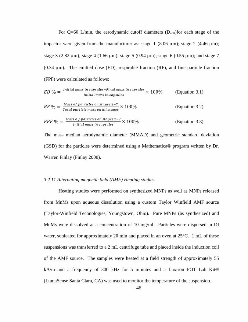
46
For Q=60 L/min, the aerodynamic cutoff diameters (Da50)for each stage of the
impactor were given from the manufacturer as: stage 1 (8.06 m); stage 2 (4.46 m);
stage 3 (2.82 m); stage 4 (1.66 m); stage 5 (0.94 m); stage 6 (0.55 m); and stage 7
(0.34 m). The emitted dose (ED), respirable fraction (RF), and fine particle fraction
(FPF) were calculated as follows:
𝐸𝐷 % = 𝐼𝑛𝑖𝑡𝑖𝑎𝑙 𝑚𝑎𝑠𝑠 𝑖𝑛 𝑐𝑎𝑝𝑠𝑢𝑙𝑒𝑠−𝐹𝑖𝑛𝑎𝑙 𝑚𝑎𝑠𝑠 𝑖𝑛 𝑐𝑎𝑝𝑠𝑢𝑙𝑒𝑠
𝐼𝑛𝑖𝑡𝑖𝑎𝑙 𝑚𝑎𝑠𝑠 𝑖𝑛 𝑐𝑎𝑝𝑠𝑢𝑙𝑒𝑠× 100% (Equation 3.1)
𝑅𝐹 % = 𝑀𝑎𝑠𝑠 𝑜𝑓 𝑝𝑎𝑟𝑡𝑖𝑐𝑙𝑒𝑠 𝑜𝑛 𝑠𝑡𝑎𝑔𝑒𝑠 2−7
𝑇𝑜𝑡𝑎𝑙 𝑝𝑎𝑟𝑡𝑖𝑐𝑙𝑒 𝑚𝑎𝑠𝑠 𝑜𝑛 𝑎𝑙𝑙 𝑠𝑡𝑎𝑔𝑒𝑠× 100% (Equation 3.2)
𝐹𝑃𝐹 % = 𝑀𝑎𝑠𝑠 𝑜 𝑓 𝑝𝑎𝑟𝑡𝑖𝑐𝑙𝑒𝑠 𝑜𝑛 𝑠𝑡𝑎𝑔𝑒𝑠 2−7
𝐼𝑛𝑖𝑡𝑖𝑎𝑙 𝑚𝑎𝑠𝑠 𝑖𝑛 𝑐𝑎𝑝𝑠𝑢𝑙𝑒𝑠× 100% (Equation 3.3)
The mass median aerodynamic diameter (MMAD) and geometric standard deviation
(GSD) for the particles were determined using a Mathematica® program written by Dr.
Warren Finlay (Finlay 2008).
3.2.11 Alternating magnetic field (AMF) Heating studies
Heating studies were performed on synthesized MNPs as well as MNPs released
from MnMs upon aqueous dissolution using a custom Taylor Winfield AMF source
(Taylor-Winfield Technologies, Youngstown, Ohio). Pure MNPs (as synthesized) and
MnMs were dissolved at a concentration of 10 mg/ml. Particles were dispersed in DI
water, sonicated for approximately 20 min and placed in an oven at 25°C. 1 mL of these
suspensions was transferred to a 2 mL centrifuge tube and placed inside the induction coil
of the AMF source. The samples were heated at a field strength of approximately 55
kA/m and a frequency of 300 kHz for 5 minutes and a Luxtron FOT Lab Kit®
(LumaSense Santa Clara, CA) was used to monitor the temperature of the suspension.
47
The specific absorption rate (SAR) is a calculation that normalizes the heating
curve to the concentration of MNPs and indicates the ability of the MNPs to heat. SAR
values were calculated for these data using equation 34:
𝑆𝐴𝑅 = ∑ 𝐶𝑖𝑚𝑖
𝑚
𝑑𝑇
𝑑𝑡 (Equation 3.4)
where mi, Ci, are the mass and specific heat capacity of component i, respectively. The
ratio dT/dt was determined as the slope of a linear trendline over a 30 second time
interval in the initial linear region of the heating curve. The mass of iron oxide, m, for
the MF20 and MF5 powders was calculated using results from TGA experiments.
3.2.12 Cytotoxicity tests
Human A549 alveolar epithelial adenocarcinoma cells were purchased from
American Type Culture Collection (Manassas, VA, USA). Cells were cultured in 75 cm2
flasks in Dulbecco’s modified eagle medium (DMEM) containing 10% FBS, 5%
PSTREP, and 1% Fungizone at 37C in a humidified incubator with 5% CO2. For
viability studies, A549 cells were seeded into 96-well plates at 10,000 cells/cm2, placed
in an incubator at 37°C and allowed to attach overnight. Cells were subsequently
exposed to varying concentrations of MnMs dispersed in DMEM media for 24 hours at
37°C. Following exposure, live cells were stained with calcein AM, which is hydrolyzed
to the fluorescent molecule calcein by intracellular esterases, and placed in a Biotek
Synergy Mx microplate reader (Biotek, Winooski, VT) to measure the fluorescent
intensity. Relative viability was calculated as the ratio of the mean fluorescence intensity
to that of the control, which contained media only. For each experiment 10 wells were
48
used for each concentration and experiments were performed in triplicate therefore and
the statistical analysis was performed with N=3.
3.2.13 Statistics
All experiments were performed in at least triplicate. The aerosolization studies
were experimentally designed by Design Expert™ 8.0.7.1 software (Stat-Ease Corp.,
MN, USA). SigmaPlot 12.3 was used for one-way ANOVA tests and when statisticaly
difference was observed a Holm-Sidak test was used to determine any significance in
observed data. A p-value of <0.05 was considered statistically significant.
3.3 Results and Discussion
3.3.1 Physiochemical characterization
Magnetic nanocomposite microparticles (MnMs) were successfully formulated
via spray drying and Figure 3.2 shows representative SEM images of the MnM powders.
All particle systems displayed smooth and spherical particle morphologies, which are
important for optimal aerodynamic performance. Additionally, these images show a
relatively uniform size distribution of the particles, which was confirmed by measuring
the geometric diameter of MnM particles with SigmaScan® (Table 3.2). Representative
STEM images of the MnMs show the successful loading of MNPs (Figure 3.3) via spray
drying thereby confirming our hypothesis that composite powders containing novel
magnetic nanoparticles can be formulated via spray drying in closed-mode. The presence
of MNPs within composite dry powders is evident as areas of higher contrast (brighter
regions) and energy dispersive X-ray spectroscopy confirmed these findings (data not
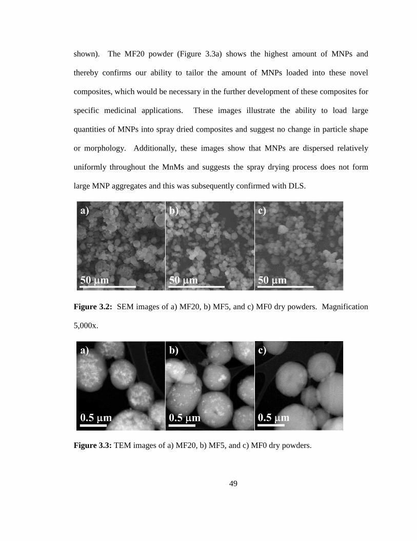
49
shown). The MF20 powder (Figure 3.3a) shows the highest amount of MNPs and
thereby confirms our ability to tailor the amount of MNPs loaded into these novel
composites, which would be necessary in the further development of these composites for
specific medicinal applications. These images illustrate the ability to load large
quantities of MNPs into spray dried composites and suggest no change in particle shape
or morphology. Additionally, these images show that MNPs are dispersed relatively
uniformly throughout the MnMs and suggests the spray drying process does not form
large MNP aggregates and this was subsequently confirmed with DLS.
Figure 3.2: SEM images of a) MF20, b) MF5, and c) MF0 dry powders. Magnification
5,000x.
Figure 3.3: TEM images of a) MF20, b) MF5, and c) MF0 dry powders.
50
DLS, Karl Fisher titration, and TGA were carried out to evaluate the diameters of MNPs
released from the composite particles, water content of the powders, and MNP loading in
MnMs, respectively. Table 3.2 summarizes the data from these experiments. DLS was
used to measure the hydrodynamic radii of the MNPs before incorporation into MnMs
and after release from composites via aqueous dissolution. The diameter of the iron
oxide MNPs before incorporation into MnM composites was 145.7 ± 2.9 nm. After spray
drying into MnMs, the diameters of the MNPs in the MF20 and MF5 powders were 159.0
± 3.8 nm and 144.2 ± 4.5 nm, respectively. These data confirmed minimal differences in
the particle diameters of the MNPs released from composites and confirms the absence of
agglomerate formation during the formation of MnMs with spray drying. Importantly,
these results show the ability to incorporate our MNPs into composite powders for
inhalation without losing the advantageous properties associated with their small size.
Karl Fisher titration demonstrated that the various MnM powders have low residual water
content which is optimal for aerodynamic performance and illustrate that these powders
can be stored in such a way to avoid water vapor uptake and the associated complications
such as agglomeration due to increased interparticulate interactions. It is interesting to
observe that MF20 powders showed a higher mean residual water content than MF5
although this difference was not statistically significantly. Additionally, the variability in
the water content was much higher for the MF20 powder and this could be due to residual
water present on the MNPs before spray drying. TGA allowed for the quantification of
MNP loading in the MnMs and revealed slightly higher loading values in the final spray
dried particles than in the initial feed conditions; however, the MNP loading was
relatively consistent from batch-to-batch. The increased loading of iron oxide in the final
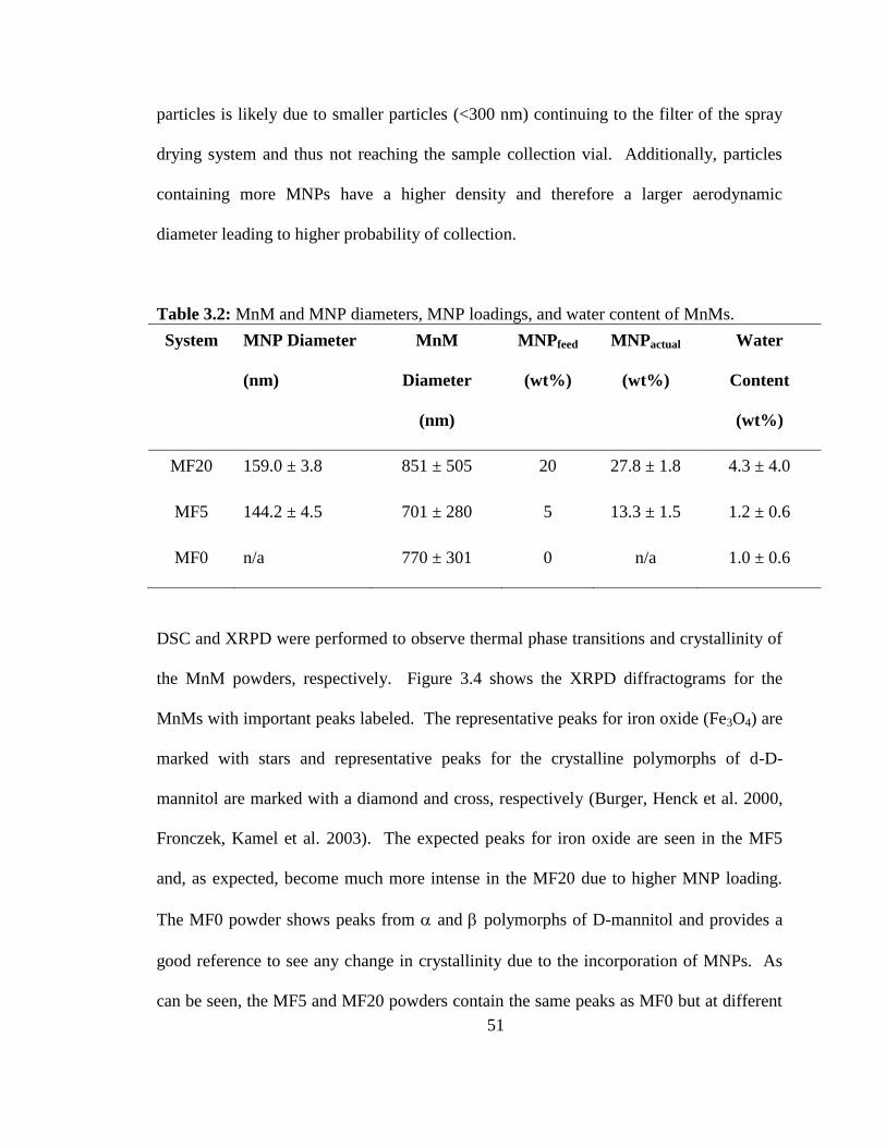
51
particles is likely due to smaller particles (<300 nm) continuing to the filter of the spray
drying system and thus not reaching the sample collection vial. Additionally, particles
containing more MNPs have a higher density and therefore a larger aerodynamic
diameter leading to higher probability of collection.
Table 3.2: MnM and MNP diameters, MNP loadings, and water content of MnMs.
System MNP Diameter
(nm)
MnM
Diameter
(nm)
MNPfeed
(wt%)
MNPactual
(wt%)
Water
Content
(wt%)
MF20 159.0 ± 3.8 851 ± 505 20 27.8 ± 1.8 4.3 ± 4.0
MF5 144.2 ± 4.5 701 ± 280 5 13.3 ± 1.5 1.2 ± 0.6
MF0 n/a 770 ± 301 0 n/a 1.0 ± 0.6
DSC and XRPD were performed to observe thermal phase transitions and crystallinity of
the MnM powders, respectively. Figure 3.4 shows the XRPD diffractograms for the
MnMs with important peaks labeled. The representative peaks for iron oxide (Fe3O4) are
marked with stars and representative peaks for the crystalline polymorphs of d-D-
mannitol are marked with a diamond and cross, respectively (Burger, Henck et al. 2000,
Fronczek, Kamel et al. 2003). The expected peaks for iron oxide are seen in the MF5
and, as expected, become much more intense in the MF20 due to higher MNP loading.
The MF0 powder shows peaks from and polymorphs of D-mannitol and provides a
good reference to see any change in crystallinity due to the incorporation of MNPs. As
can be seen, the MF5 and MF20 powders contain the same peaks as MF0 but at different
52
relative intensities. As an example, it is observed that there is a change in the ratio of
representative peaks for the and polymorphs at 2degree values of 13.6 and 14.5,
respectively. It can be seen that the incorporation of MNPs into the spray dried
composites results in a higher -polymorph character of mannitol in the solid state. This
increase in the amount of the -polymorph demonstrates that the incorporation of iron
oxide MNPs results in a polymorphic interconversion between the different crystalline
polymorphs of D-mannitol. This also suggests that such phenomena should be closely
monitored for future development of powders as conversion between crystalline
polymorphs of a given material can lead to changes in its physical properties such as
melting point and solubility among others, since polymorphs of the same material have
different Gibbs free energy states. DSC thermograms (Figure 3.5) reveal an endothermic
phase transition at approximately 165°C representing the melting point of D-mannitol
which is in excellent agreement with its reported melting temperature (Burger, Henck et
al. 2000). These findings indicate that these novel MnMs possess excellent thermal
stability and suggest, metaphorically, that these MnMs will “melt in your lungs, not in
your hands” (in actuality they will dissolve). These findings are important because they
show that the storage of these systems is insensitive to temperature for practical purposes.
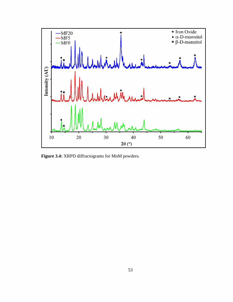
53
Figure 3.4: XRPD diffractograms for MnM powders.
54
Figure 3.5: DSC thermograms for MnM powders.
3.3.2 Aerosol performance of MnMs
The MnM powders showed excellent aerosol performance thereby confirming
that they are suitable for dry powder aerosol delivery. The aerosol performance of the
MnMs (Figure 3.6) indicates a large number of the particles being deposited on all stages
of the impactor for each powder system thereby illustrating the potential and the
versatility of these formulations to reach all regions of the lungs., This would be
advantageous for particular lung cancers such as bronchioloalveolar carcinomas, as well
as for carcinomas occurring in the upper lung region (e.g. bronchial carcinoma) which
can be targeted based on aerodynamic particle size based on the deposition on stage 1.
55
Specifically, particles below 10 microns but greater than 5 microns can effectively target
the upper lung region of the bronchi and bronchials (Suarez and Hickey 2000).
Additionally, these deposition patterns can be altered to shift the deposition pattern to
more specific regions by changing processing variables in spray drying as well as by
using other excipients (Suarez and Hickey 2000). Table 3.3 summarizes the aerosol
performance parameters for each system. The fine particle fraction (FPF) and respirable
fraction (RF) values for all of the powders were approximately 30% and 50%,
respectively, which are higher than the 10-20% values of currently marketed dry powder
inhalation (DPI) pharmaceutical products even in the absence of a lactose carrier (Suarez
and Hickey 2000). Additionally, these powders all displayed high emitted doses of
approximately 90% which demonstrates favorable dispersion efficiency. The mass
median aerosol diameter (MMAD) was calculated for the aerosols produced from the
MnM powders and all of these values were less than 5 m suggesting their ability to
adequately deposit throughout all regions of the lungs and effectively reach the lower
airways. Interestingly, we see similar deposition patterns between MF5 and MF0 while
the MF20 powder has slightly higher deposition on some of the earlier stages. This shift
in deposition pattern is most likely due to the significant increase in the density of the
composite particles as a result of such a high MNP-loading. Still, all three powders
displayed good aerosol performance and these results suggest that these MnMs are
suitable for DPI administration and suggest a new administration route for the delivery of
iron oxide nanoparticles. Additionally, these findings propose a novel method for
incorporating nanomaterials into composites that can subsequently be delivered to the
56
highly permeable tissue of the lungs assuming they can traverse the mucosal lining in
regions where it exists.
Figure 3.6: Aerosol dispersion performance plot for MnM powders using the NGI at 60
L/min.
57
Table 3.3: Aerosol performance properties for inhalable MnMs.
MF20 MF5 MF0
MMAD (μm) 4.5 ± 0.2 4.5 ± 1.0 4.4 ± 1.0
GSD (μm) 2.4 ± 0.1 2.8 ± 0.1 2.7 ± 0.3
FPF (%) 34.3 ± 3.8 31.2 ± 2.0 29.6 ± 6.8
RF (%) 56.5 ± 3.8 50.9 ± 5.6 50.7 ± 6.3
ED (%) 92.0 ± 2.9 92.5 ± 2.9 88.0 ± 6.7
3.3.3 AMF heating of MNPs released from MnMs
Representative heating curves for the MnM systems are shown in Figure 3.7 and
these data confirm that the MNPs retained excellent heating capabilities. Experiments
were performed at room temperature and therefore illustrate that the temperatures
reached for the MF20 powders are well above the hyperthermia range indicating
sufficient heat for the intended goals. Additionally, the range of heating between the
MF5 and MF20 powders suggests the ability to significantly change heating capability of
these powders by altering the MNP loading. The SAR of the iron oxide MNPs before
incorporation into MnM composites was 422 ± 18 W/g. After spray drying into MnMs,
the SAR values for the MF20 and MF5 powders were 317 ± 51 W/g and 252 ± 53 W/g,
respectively. These data illustrate that, although a drop in SAR was seen, the MNPs
show excellent heating capability and these SAR values compare favorably with previous
reports from our group (Wydra, Kruse et al. 2013). High SAR values suggest that
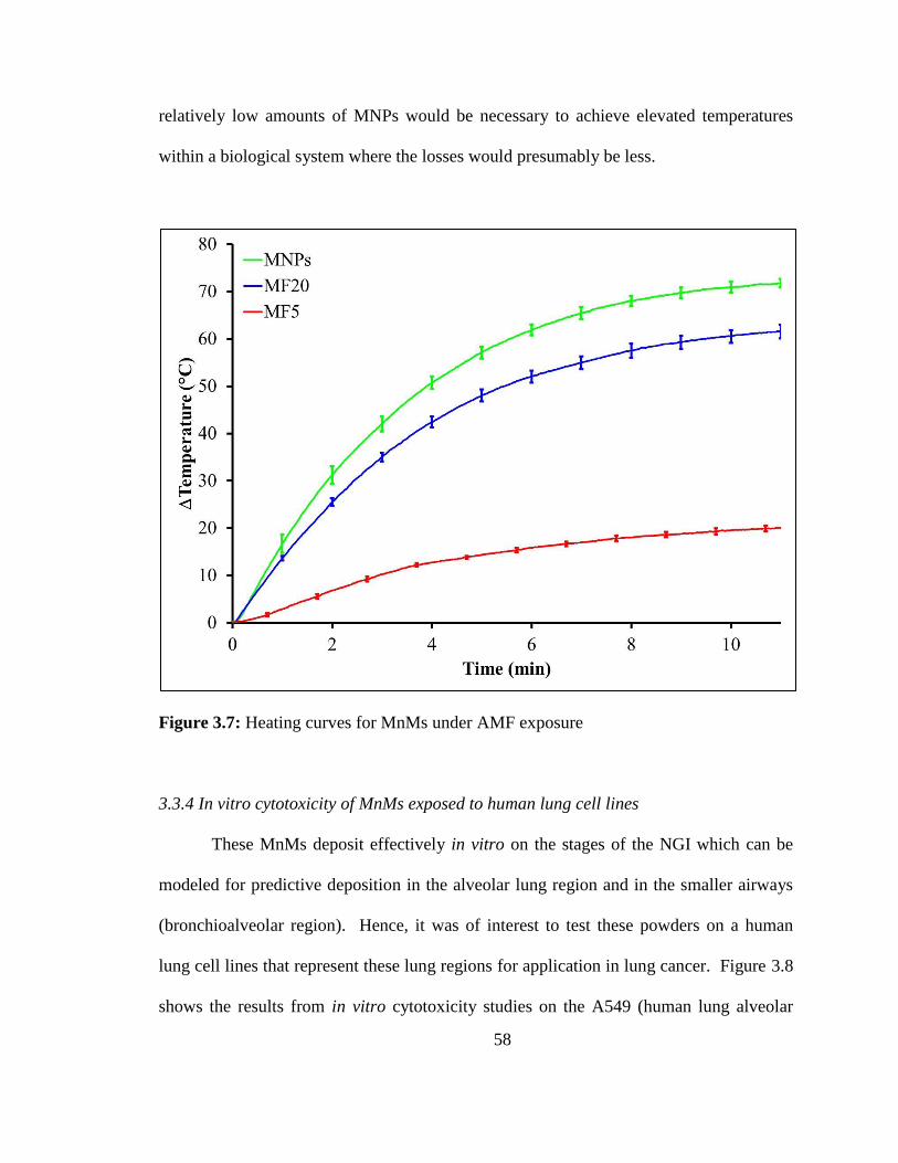
58
relatively low amounts of MNPs would be necessary to achieve elevated temperatures
within a biological system where the losses would presumably be less.
Figure 3.7: Heating curves for MnMs under AMF exposure
3.3.4 In vitro cytotoxicity of MnMs exposed to human lung cell lines
These MnMs deposit effectively in vitro on the stages of the NGI which can be
modeled for predictive deposition in the alveolar lung region and in the smaller airways
(bronchioalveolar region). Hence, it was of interest to test these powders on a human
lung cell lines that represent these lung regions for application in lung cancer. Figure 3.8
shows the results from in vitro cytotoxicity studies on the A549 (human lung alveolar
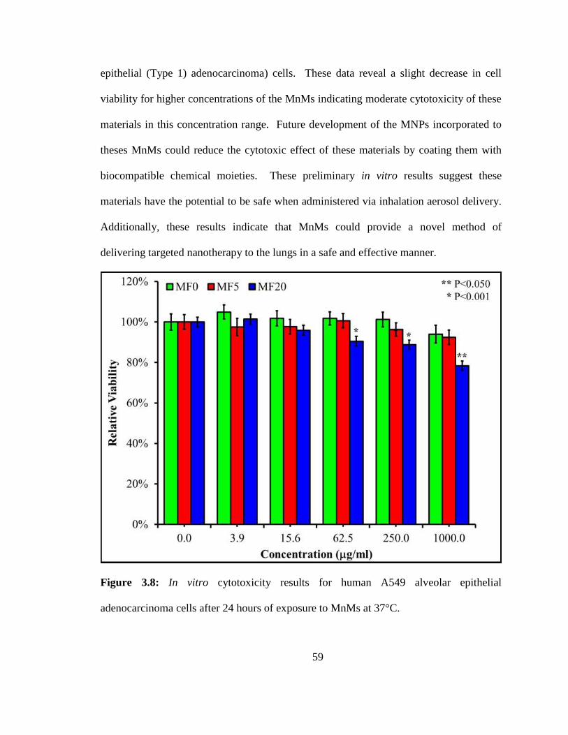
59
epithelial (Type 1) adenocarcinoma) cells. These data reveal a slight decrease in cell
viability for higher concentrations of the MnMs indicating moderate cytotoxicity of these
materials in this concentration range. Future development of the MNPs incorporated to
theses MnMs could reduce the cytotoxic effect of these materials by coating them with
biocompatible chemical moieties. These preliminary in vitro results suggest these
materials have the potential to be safe when administered via inhalation aerosol delivery.
Additionally, these results indicate that MnMs could provide a novel method of
delivering targeted nanotherapy to the lungs in a safe and effective manner.
Figure 3.8: In vitro cytotoxicity results for human A549 alveolar epithelial
adenocarcinoma cells after 24 hours of exposure to MnMs at 37°C.
60
3.4 Conclusions
Iron oxide MNPs were successfully incorporated into MnMs through spray drying
in closed-mode. Physicochemical characterization of these composites revealed the
potential to use these particles in the inhalation aerosol treatment of lung cancer via
targeted hyperthermia. Aerosol performance studies revealed favorable aerodynamic
properties for the MnM powders with FPF and RF values greater than 50% along with
ED values around 90%. The MMADs for all aerosols delivered by a currently marketed
DPI device were approximately 4.5 μm and the aerosol performance plots suggest the
potential for high deposition of these particles throughout the lungs. Heating studies
imply that these materials can be remotely heated after formulation and in vitro
cytotoxicity studies show moderate toxicity on human lung A549 cells. These data
provide initial evidence that these materials can potentially be targeted throughout the
lung including the upper lung region for application in bronchial lung cancer and also in
the mid-to-lower lung regions. Additionally, MnMs possess the ability to release MNPs
that remotely heat in the presence of an AMF. These novel composites present an
exciting class of materials with many potential applications and pose a promising
approach to a novel thermal treatment of the lungs through targeted pulmonary inhalation
aerosol delivery of MNPs.
61
Chapter 4 Remote controlled thermal therapy with magnetic nanocomposite
microparticles induces cell death in triple negative breast cancer micrometastasic
tumor tissue analogs
4.1 Introduction
Breast cancer is the second leading cause of death in women, behind lung cancer,
and 1-in-8 women are diagnosed with this dreadful disease (DeSantis, Ma et al. 2014).
Triple negative breast cancer (TNBC) is an aggressive subtype wherein the cells do not
express commonly targeted receptors for estrogen (ER), progesterone (PR), and human
epidermal growth factor 2 (HER2) (Hudis and Gianni 2011). TNBC accounts for
approximately 15% of all breast cancers and is very aggressive with a disproportionately
higher rate of metastasis (Andre and Zielinski 2012). Additionally, patients with distant
metastatic disease show a significantly shorter survival time with TNBC relative to all
other breast cancer types (Kassam, Enright et al. 2009). Currently there is an unmet need
for novel approaches to treating patients with metastatic TNBC and recent studies have
revealed that TNBC has a higher propensity of metastasizing to the lungs (Andre and
Zielinski 2012, Gerratana, Fanotto et al. 2015).
Pulmonary delivery presents a promising approach for delivering local therapy to
the lungs of both primary and secondary lung cancer patients due to higher local
concentrations and reduced systemic side effects (Otterson, Villalona-Calero et al. 2007,
Otterson, Villalona-Calero et al. 2010). Additionally, clinical trials with stage IV lung
cancer patients showed a statistically significant increase in median survival time when
carboplatin was administered via combined injection and inhalation as opposed to
injection only (Zarogoulidis, Eleftheriadou et al. 2012). These results suggest that
62
pulmonary delivery has the potential to complement traditional approaches for patients
with metastatic disease in the lungs. Additionally researchers have been interested in
delivering iron oxide magnetic nanoparticles to the lungs for thermal therapy applications
(Stocke, Meenach et al. 2014).
Iron oxide magnetic nanoparticles (MNPs) represent a unique class of materials
that have generated a considerable about of interest for biomedical applications (Hao,
Xing et al. 2010). The biocompatibility of MNPs has led to a variety of commercialized
biomedical applications (Mahmoudi, Sant et al. 2011) and their ability to be actuated
remotely via an alternating magnetic field (AMF) has spared interest in their therapeutic
potential (Jordan, Scholz et al. 1999, Frimpong and Hilt 2010). In the presence of an
AMF, iron oxide magnetic nanoparticles generate heat through Brownian and Neel
relaxation and this thermal energy can be used to actuate the onset of therapy or as a form
of therapy itself (Satarkar and Hilt 2008, Satarkar and Hilt 2008, Frimpong, Dou et al.
2010, Satarkar, Meenach et al. 2011, Wydra, Kruse et al. 2013). For cancer patients,
researchers have been interested in the potential advantages of thermal therapy both in
isolation and in combination with anticancer agents but clinical translation of such
therapies has been limited (Vertrees, Leeth et al. 2002, Babincova, Altanerova et al.
2008, Thiesen and Jordan 2008, Lubner, Brace et al. 2010, Kruse, Meenach et al. 2014).
Over the past several years, cellular spheroids have gained interest as more
representative cancer models due to large discrepancies between traditional two-
dimensional cell culture and physiological tumors (Bates, Edwards et al. 2000).
Additionally, multicellular spheroids attempt to more accurately represent the
heterogeneity present in the tumor environment by combining two or more cell types
63
(Kunz-Schughart, Kreutz et al. 1998). Conversely, our lab went a step further to develop
tumor tissue analogs composed of tumor cells as well as normal fibroblasts and
endothelial components (Upreti, Jamshidi-Parsian et al. 2011). The combination of
normal and neoplastic cells allows for a more accurate recapitulation of the microtumor
environment, which is often neglected with in vitro assays.
In this work, we developed TNBC micrometastatic tumor tissue analogs (TM
analogs) to study the potential of inhalable formulations for secondary lung cancer
patients with highly metastatic TNBC. The TM analogs were composed of the following
three murine cell lines: 4T1 breast cancer cells (ER-, PR-, and HER2- (Kau, Nagaraja et
al. 2012)), MEF fibroblast cells, and C166 endothelial cells. Iron oxide magnetic
nanoparticles were previously formulated into inhalable magnetic nanocomposites
(MnMs) via spray drying (Stocke, Meenach et al. 2014) and, here, were applied to the
TM analogs. Remote controlled thermal therapy on TM analogs was accomplished using
a custom alternating magnetic field (AMF) and the resulting tissues were analyzed both
quantitatively and qualitatively.
4.2 Material and methods
4.2.1 Materials
4.2.2 Formulation of inhalable magnetic nanocomposite microparticles
The formulation and characterization of the materials used in this work were
previously reported (Stocke, Meenach et al. 2014). Briefly, uncoated iron oxide magnetic
nanoparticles (MNPs) were synthesized via aqueous co-precipitation of ferric and ferrous
iron salts at a 2:1 ratio, respectively. The MNPs were washed with ethanol and water and
64
allowed to dry before incorporation into inhalable dry powder composites via spray
drying with a Büchi Mini Spray Dryer B290 coupled with a B-295 Inert Loop and high-
performance cyclone (all from Büchi Labortechnik, Flawil, Switzerland). D-mannitol
was dissolved in methanol and iron oxide MNPs were weighed out at 20 wt% based on
the combined mass of these two components, which resulted in a dilute concentration of
0.1% wt/vol in methanol. Two liters of the feed mixture were fed to the spray dryer at a
pump rate of 50% (15 mL/min) through a stainless steel nozzle with a diameter of 0.7
mm. Spray drying experiments were carried out in closed mode using UHP dry nitrogen
gas at a flow rate of 600 L/h, an inlet temperature of 150°C, an aspirator rate of 35 m3/h,
and under sonication via water bath sonicator. Powders were collected and transferred to
a glass desiccator over Drierite™ and stored in the freezer at -23°C.
4.2.3 Formation of triple negative breast cancer micrometastasic tumor tissue analogs
(TM analogs)
4T1-mcherry tumor cells, C166-GFP endothelial cells and murine embryonic
fibroblasts (MEF) were used to generated 3D TM analogs “hanging drops” of media
(Dulbecco modified Eagle medium with 10% fetal bovine serum and antibiotic mix).
Briefly, single a cell suspension of 4T1-mcherry cells, C166-GFP cells and MEF cells in
equal proportion (3000 cells/20μL) was dispensed on the inside of the lid of each well of
a 48-well cell culture plate (Greiner Cellstar, BioExpress, Kaysville, UT). The growth of
tumor tissue analogs was monitored over time until day 10 in a hanging drop of medium,
following which they were subject to MNP (Magnetic nanoparticle) treatment. The 3D
co-cultures/ tumor tissue analogs were subsequently transferred to optically clear Greiner
65
repellent plates for MNP and AMF treatment, imaging and analysis of the treatment
response.
4.2.4 MNP-treatment of TM analogs
MnM powders were dispersed in media at 1.0 mg/mL and 0.1 mg/mL and these
two treatment groups will be referred to as high dose (HD) and low dose (LD),
respectively. The media surrounding TM analogs was carefully removed and 50 𝜇l of the
MNP dispersions was transferred to each 96-well containing n=5 TM analogs. For
control groups fresh media was added to the 96-wells. Each treatment group consisted of
4 96-wells, totaling 20 TM analogs, and these groups are summarized in Table 4.1. The
TM analogs were allowed to incubate in the MNP suspensions for 24 hours at 37°C in a
humidified environment supplemented with 5% CO2. After 24 hours of MNP-exposure,
the TM analogs were rinsed with media 3 times and placed on a trans-well insert in a 35
mm petri dish. 2 ml of media was added to the dish in order to insure the TM analogs
had sufficient supply of nutrients during hyperthermia treatments.
4.2.5 Remotely actuated hyperthermia using a custom alternating magnetic field (AM)
For hyperthermia experiments each petri dish was placed directly above the coil
of a custom Taylor Winfield® Alternating Magnetic Field (AMF) Source. The AMF was
ran at a field strength of 55 kA/m at a frequency of 300 kHz and the AMF treatment
groups were exposed to the field for 1 hour. During the experiments a custom built
Styrofoam casing was used to enclose the coil and petri dish and a hot air supply was
used to hold the temperature of the surroundings to approximately 37°C. A Luxtron
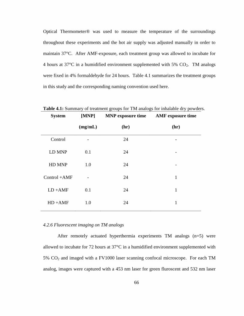
66
Optical Thermometer® was used to measure the temperature of the surroundings
throughout these experiments and the hot air supply was adjusted manually in order to
maintain 37°C. After AMF-exposure, each treatment group was allowed to incubate for
4 hours at 37°C in a humidified environment supplemented with 5% CO2. TM analogs
were fixed in 4% formaldehyde for 24 hours. Table 4.1 summarizes the treatment groups
in this study and the corresponding naming convention used here.
Table 4.1: Summary of treatment groups for TM analogs for inhalable dry powders.
System [MNP]
(mg/mL)
MNP exposure time
(hr)
AMF exposure time
(hr)
Control - 24 -
LD MNP 0.1 24 -
HD MNP 1.0 24 -
Control +AMF - 24 1
LD +AMF 0.1 24 1
HD +AMF 1.0 24 1
4.2.6 Fluorescent imaging on TM analogs
After remotely actuated hyperthermia experiments TM analogs (n=5) were
allowed to incubate for 72 hours at 37°C in a humidified environment supplemented with
5% CO2 and imaged with a FV1000 laser scanning confocal microscope. For each TM
analog, images were captured with a 453 nm laser for green fluroscent and 532 nm laser
67
for red fluroscent. Bright field images were captured to estimate the size of the TM
analogs. Images were processed using FV10-ASW 1.7 viewer software. Images were
analyzed using Olympus image analysis software and ImageJ 1.47v (National Institute of
Health, Bethesda, USA) as 8-bit. These images were reported along with a composite
wherein the 3 separate images were merged.
4.2.7 Quantification of cell death using Sytox
In order to quantify the amount of cell death TM analogs (n=5) were stained with
Sytox Blue Dead Cell Stain. SYTOX blue stain is a high-affinity nucleic acid stain that
easily penetrates cells with compromised plasma membranes. Spheroids were incubated
with SYTOX blue to observe the dead cells, which were excited with argon 406 lasers.
4.2.8 Prussian blue staining for iron
The amount of iron present in TM analogs was evaluated using a Prussian Blue
stain for iron content. Spheroids were harvested and processed through sucrose gradient
and then snap frozen in O.C.T, Tissue-Tek, Miles USA, Inc. (Elkhart, IN). Five
micrometer cryostat sections were obtained with the help of a Leica CM1950 cryotome
and transferred on to Superfrost™ Plus slides (Fischer Scientific; Boston, MA) for
detection of iron-oxide particles using Prussian blue staining. Sections were dipped in
freshly prepared 10% aqueous solution of potassium ferrocyanide and hydrochloric acid
for 20 minutes to stain iron accumulated within TM analogs. Sections were then washed
three times with distilled water and counterstained with nuclear red for 5 minutes. After
rinsing the slides, sections were dehydrated through grades of alcohol and then cleared
with xylene and permanent slides were made after coverslipping with permount media
68
(Sigma). Light microscopic images were obtained at 10x and 20x magnification with the
help of Olympus light microscope (Olympus America Inc., Melville, NY) for identifying
the distribution of iron-oxide nanoparticles within TM analogs analogs.
4.2.9 Transmission electron microscopy (TEM) on TM analogs
Transmission electron microscopy (TEM) was carried out on TM analogs (n=5)
in order to qualitatively observe MNP uptake and cellular characteristics after AMF
treatment. Immediately following 1 hour of AMF exposure the TM analogs were placed
in an incubator for 4 hours at 37°C in a humidified environment supplemented with 5%
CO2. Subsequently, TM analogs were transferred to a 20 mL glass vial and washed with
0.1 M Sorenson’s Phosphate Buffer (SPB) to remove media before fixing. The buffer
was removed and a 3.5% glutaraldehyde solution in 0.1 M SPB was added to the vial and
allowed to sit for 45 min at approximately 4°C. Subsequently, the fixate was removed
and the TM analogs were washed three times for 5 minutes in 0.1 M SPB supplemented
with 5% sucrose. After this, the SBS was removed and a 1% osmium tetroxide solution
in 0.1 M SPB was added to the vial containing the TM analogs and allowed to sit for 45
mins at 4°C. The osmium tetroxide solution was removed and TM analogs were washed
with 0.1 M SPB and dehydrated by subsequent ethanol washes at 50-, 70-, 80-, 90-, and
100% (twice) for 10 mins each. After the second wash with 100% ethanol the TM
analogs were washed with propylene oxide twice for 20 mins each. The propylene oxide
was then removed and the TM analogs were allowed to sit overnight under a mild heating
lamp in a 1:1 mixture of propylene oxide and an epoxy resin. The next day, the
propylene oxide and epoxy resin mixture was removed and the cells were washed with
69
just the epoxy resin twice for an hour each time. After the second wash was removed the
TM analogs were transferred to a centrifuge tube for sectioning, epoxy resin was added
and the samples were allowed to polymerize for 48 hours at 60°C. After polymerization,
the pellets were sliced into 70-90 µm sections with a Reichert-Jung Ultracut E microtome
and stained with a combination of uranyl acetate, (saturated solution in 70% ethanol) for
5 minutes, and lead citrate, (see attached recipe), for 2 minutes. The specimens were
observed using a Philips Tecnai Biotwin 12, manufactured by FEI.
4.3 Results and Discussion
The effect of MNPs, released from the inhalable dry composites, on the TM
analogs (n=5) was examined 5 days post MNP-exposure with fluorescent and brightfield
imaging (Figure 4.1) for three different MNP doses (control, LD, and HD). In addition to
illustrating the reproducibility of these spheroids, these data show no apparent adverse
effect on the mechanical stability of the TM analogs. Additionally, these data confirm
the presence of all three cell types wherein each panel shows evidence of the MEF
(green), 4T1 (red) and C166 (colorless) cells confirming their multicellular composition.
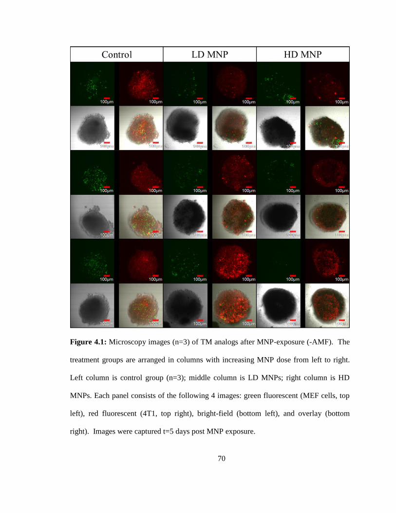
70
Figure 4.1: Microscopy images (n=3) of TM analogs after MNP-exposure (-AMF). The
treatment groups are arranged in columns with increasing MNP dose from left to right.
Left column is control group (n=3); middle column is LD MNPs; right column is HD
MNPs. Each panel consists of the following 4 images: green fluorescent (MEF cells, top
left), red fluorescent (4T1, top right), bright-field (bottom left), and overlay (bottom
right). Images were captured t=5 days post MNP exposure.
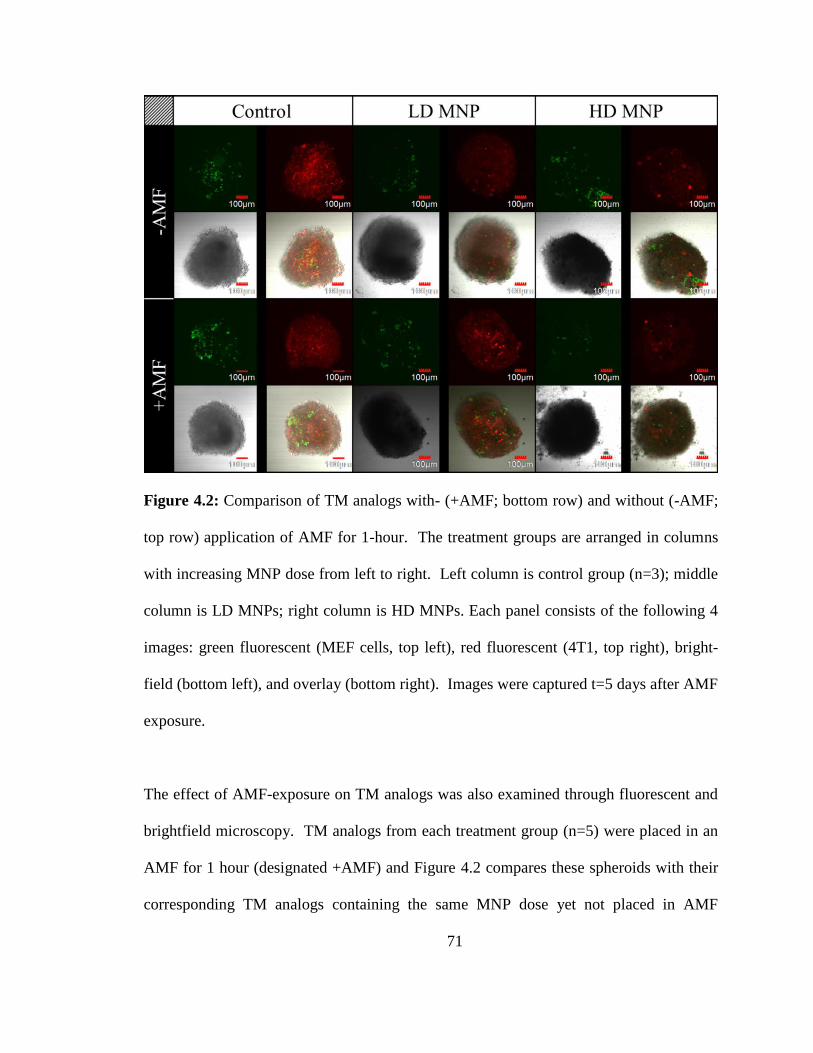
71
Figure 4.2: Comparison of TM analogs with- (+AMF; bottom row) and without (-AMF;
top row) application of AMF for 1-hour. The treatment groups are arranged in columns
with increasing MNP dose from left to right. Left column is control group (n=3); middle
column is LD MNPs; right column is HD MNPs. Each panel consists of the following 4
images: green fluorescent (MEF cells, top left), red fluorescent (4T1, top right), bright-
field (bottom left), and overlay (bottom right). Images were captured t=5 days after AMF
exposure.
The effect of AMF-exposure on TM analogs was also examined through fluorescent and
brightfield microscopy. TM analogs from each treatment group (n=5) were placed in an
AMF for 1 hour (designated +AMF) and Figure 4.2 compares these spheroids with their
corresponding TM analogs containing the same MNP dose yet not placed in AMF
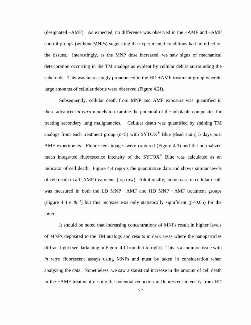
72
(designated –AMF). As expected, no difference was observed in the +AMF and –AMF
control groups (without MNPs) suggesting the experimental conditions had no effect on
the tissues. Interestingly, as the MNP dose increased, we saw signs of mechanical
deterioration occurring in the TM analogs as evident by cellular debris surrounding the
spheroids. This was increasingly pronounced in the HD +AMF treatment group wherein
large amounts of cellular debris were observed (Figure 4.2f).
Subsequently, cellular death from MNP and AMF exposure was quantified in
these advanced in vitro models to examine the potential of the inhalable composites for
treating secondary lung malignancies. Cellular death was quantified by staining TM
analogs from each treatment group (n=5) with SYTOX® Blue (dead stain) 5 days post
AMF experiments. Fluorescent images were captured (Figure 4.3) and the normalized
mean integrated fluorescence intensity of the SYTOX® Blue was calculated as an
indicator of cell death. Figure 4.4 reports the quantitative data and shows similar levels
of cell death in all -AMF treatments (top row). Additionally, an increase in cellular death
was measured in both the LD MNP +AMF and HD MNP +AMF treatment groups
(Figure 4.3 e & f) but this increase was only statistically significant (p<0.05) for the
latter.
It should be noted that increasing concentrations of MNPs result in higher levels
of MNPs deposited to the TM analogs and results in dark areas where the nanoparticles
diffract light (see darkening in Figure 4.1 from left to right). This is a common issue with
in vitro fluorescent assays using MNPs and must be taken in consideration when
analyzing the data. Nonetheless, we saw a statistical increase in the amount of cell death
in the +AMF treatment despite the potential reduction in fluorescent intensity from HD
73
MNPs. Conversely, non-specific toxic effects of MNPs could potentially be masked by
reducing the dead signal through diffraction.
Figure 4.3: Representative fluorescent images of (top row; a-c) and bright-field (bottom
row; d-f) images of spheroids stained with SYTOX® Blue dead cell stain and not
exposed to AMF. From left to right the columns represent treatments of control (a,d)
MNPs at 0.1 mg/mL (b,e) and MNPs at 1 mg/mL (c,f)
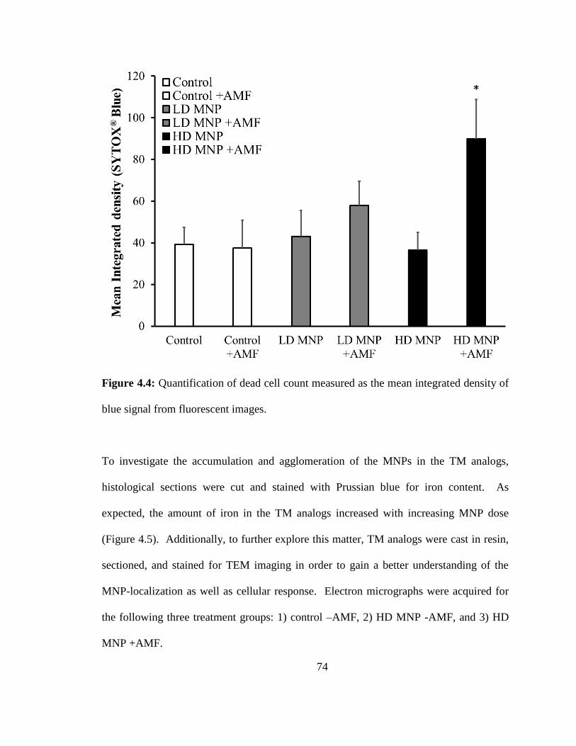
74
Figure 4.4: Quantification of dead cell count measured as the mean integrated density of
blue signal from fluorescent images.
To investigate the accumulation and agglomeration of the MNPs in the TM analogs,
histological sections were cut and stained with Prussian blue for iron content. As
expected, the amount of iron in the TM analogs increased with increasing MNP dose
(Figure 4.5). Additionally, to further explore this matter, TM analogs were cast in resin,
sectioned, and stained for TEM imaging in order to gain a better understanding of the
MNP-localization as well as cellular response. Electron micrographs were acquired for
the following three treatment groups: 1) control –AMF, 2) HD MNP -AMF, and 3) HD
MNP +AMF.
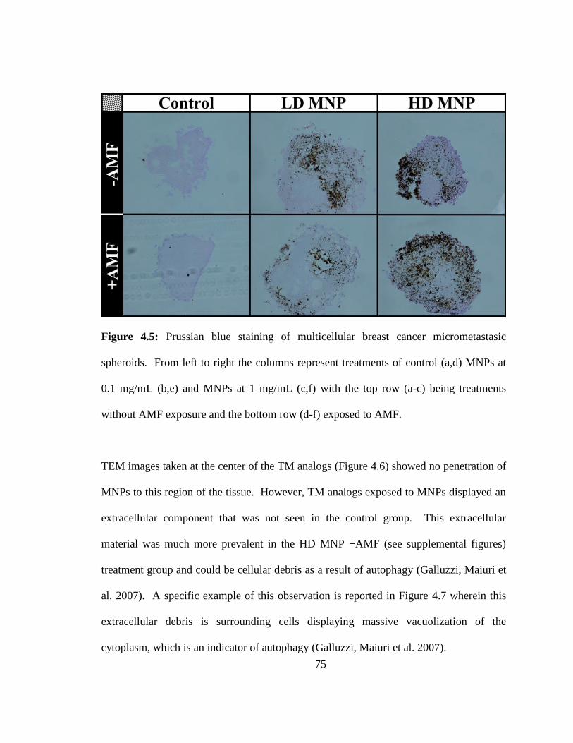
75
Figure 4.5: Prussian blue staining of multicellular breast cancer micrometastasic
spheroids. From left to right the columns represent treatments of control (a,d) MNPs at
0.1 mg/mL (b,e) and MNPs at 1 mg/mL (c,f) with the top row (a-c) being treatments
without AMF exposure and the bottom row (d-f) exposed to AMF.
TEM images taken at the center of the TM analogs (Figure 4.6) showed no penetration of
MNPs to this region of the tissue. However, TM analogs exposed to MNPs displayed an
extracellular component that was not seen in the control group. This extracellular
material was much more prevalent in the HD MNP +AMF (see supplemental figures)
treatment group and could be cellular debris as a result of autophagy (Galluzzi, Maiuri et
al. 2007). A specific example of this observation is reported in Figure 4.7 wherein this
extracellular debris is surrounding cells displaying massive vacuolization of the
cytoplasm, which is an indicator of autophagy (Galluzzi, Maiuri et al. 2007).
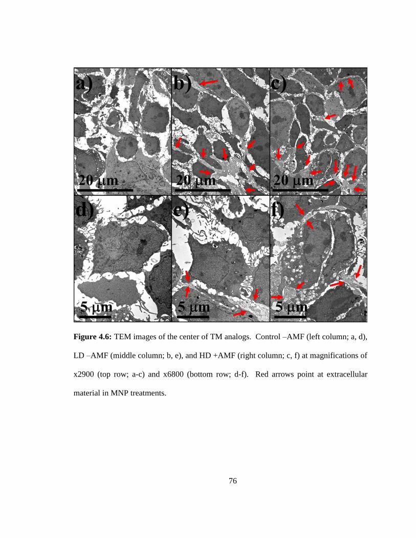
76
Figure 4.6: TEM images of the center of TM analogs. Control –AMF (left column; a, d),
LD –AMF (middle column; b, e), and HD +AMF (right column; c, f) at magnifications of
x2900 (top row; a-c) and x6800 (bottom row; d-f). Red arrows point at extracellular
material in MNP treatments.
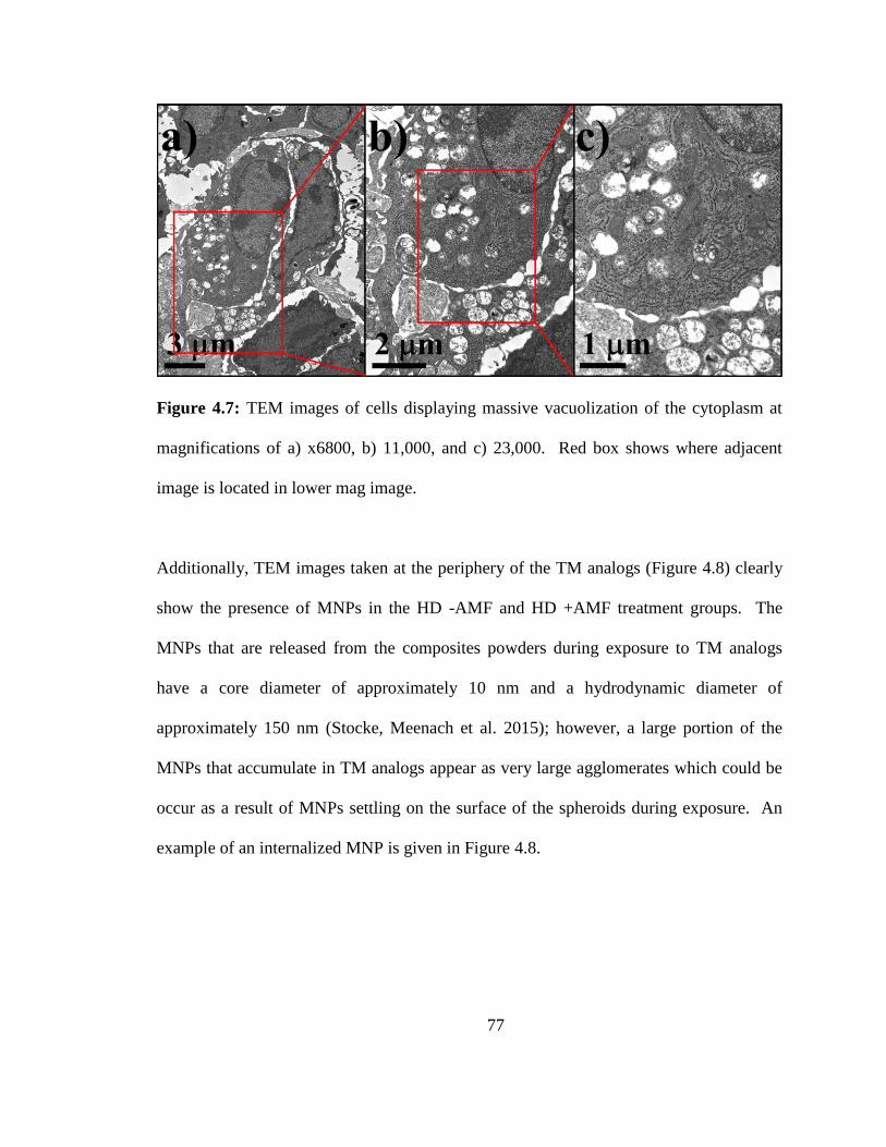
77
Figure 4.7: TEM images of cells displaying massive vacuolization of the cytoplasm at
magnifications of a) x6800, b) 11,000, and c) 23,000. Red box shows where adjacent
image is located in lower mag image.
Additionally, TEM images taken at the periphery of the TM analogs (Figure 4.8) clearly
show the presence of MNPs in the HD -AMF and HD +AMF treatment groups. The
MNPs that are released from the composites powders during exposure to TM analogs
have a core diameter of approximately 10 nm and a hydrodynamic diameter of
approximately 150 nm (Stocke, Meenach et al. 2015); however, a large portion of the
MNPs that accumulate in TM analogs appear as very large agglomerates which could be
occur as a result of MNPs settling on the surface of the spheroids during exposure. An
example of an internalized MNP is given in Figure 4.8.
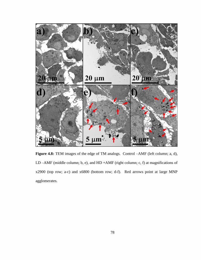
78
Figure 4.8: TEM images of the edge of TM analogs. Control –AMF (left column; a, d),
LD –AMF (middle column; b, e), and HD +AMF (right column; c, f) at magnifications of
x2900 (top row; a-c) and x6800 (bottom row; d-f). Red arrows point at large MNP
agglomerates.
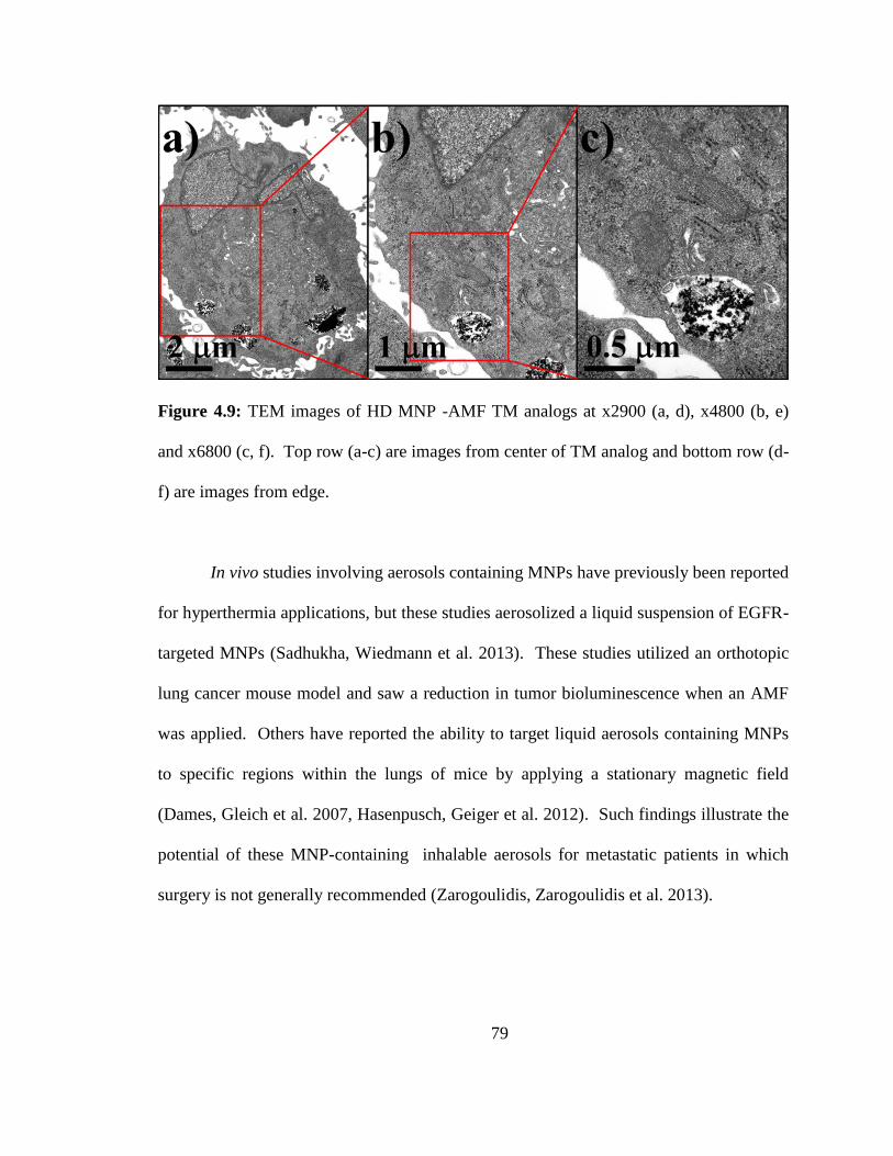
79
Figure 4.9: TEM images of HD MNP -AMF TM analogs at x2900 (a, d), x4800 (b, e)
and x6800 (c, f). Top row (a-c) are images from center of TM analog and bottom row (d-
f) are images from edge.
In vivo studies involving aerosols containing MNPs have previously been reported
for hyperthermia applications, but these studies aerosolized a liquid suspension of EGFR-
targeted MNPs (Sadhukha, Wiedmann et al. 2013). These studies utilized an orthotopic
lung cancer mouse model and saw a reduction in tumor bioluminescence when an AMF
was applied. Others have reported the ability to target liquid aerosols containing MNPs
to specific regions within the lungs of mice by applying a stationary magnetic field
(Dames, Gleich et al. 2007, Hasenpusch, Geiger et al. 2012). Such findings illustrate the
potential of these MNP-containing inhalable aerosols for metastatic patients in which
surgery is not generally recommended (Zarogoulidis, Zarogoulidis et al. 2013).
80
4.4 Conclusions
Triple negative breast cancer micrometastasic tumor tissue analogs (TM analogs)
were successfully formed and showed relatively consistent size and spherical
morphology, thereby highlighting their potential as advanced in vitro models of
secondary lung malignancies. Previously reported inhalable MnMs were applied to these
advanced models and showed no measureable adverse effects in the absence of AMF-
exposure. Conversely, remote controlled thermal therapy on was accomplished by
placing the TM analogs in an alternating magnetic field (AMF). TM analogs subjected to
a high enough concentration of MNPs showed a statistically significant increase in
cellular death after AMF-exposure and indicated the potential of these inhalable
composites for future development. These TM analogs provide an approved
physiologically relevant in vitro model of secondary lung malignancies from metastatic
TNBC and our findings suggest the potential of inhalable composites containing MNPs.
81
Chapter 5 Formulation and Characterization of Inhalable Anticancer Agents for
Targeted Pulmonary Delivery via Spray Drying
The ability to deliver two common anticancer agents was examined by spray drying these
drugs into inhalable composites. The aerodynamic performance of these composites was
examined and cell culture studies with two human lung cancer cell lines were carried out
to insure the drugs retained their activity. The goal was to formulate inhalable powders
containing ACAs with adequate aerodynamic performance and to be able to release
active ACAs. The chapter is taken directly or adapted from work that was recently
submitted to Pharmaceutical Research. Used with permission from Nathanael A Stocke,
Marjorie L. Guy, Shuo Tang, Heidi M. Mansour, Susanne M. Arnold, Heidi M. Mansour,
and J. Zach Hilt.
5.0 Abstract
Non-small cell lung cancer (NSCLC) is the leading cause of cancer mortality
worldwide. Patients diagnosed with this devastating disease have low survival rates and
high incidence of recurrence, thus highlighting the need for more effective lung cancer
treatment modalities. Pulmonary delivery provides a platform for noninvasive delivery
of novel materials to the lungs and has the potential for delivering equivalent local
anticancer agent (ACA) concentrations while reducing systemic side effects. Here, we
formulated inhalable powders consisting of the two FDA-approved ACAs cisplatin and
erlotinib (active drug in the non-inhalation drug product, Tarceva®
) and characterized
their physicochemical properties with a variety of techniques. Cascade impactor studies
82
were used to evaluate the aerodynamic performance of these powders and in vitro cell
studies determined the activity of the ACAs released from the powders in two human
lung cancer cell lines. Inhalable ACAs were examined with scanning electron
microscopy and showed spherical particle morphology and relatively uniform size
distribution yet unique surface characteristics (typo-suggest correcting and clarifying)
based on particle composition. The inhalable ACAs showed good aerodynamic
performance with MMAD values ranging from 3.0-5.6 m and all powders displaying
fine particle fractions >25% even in the absence of a carrier such as lactose. Finally, in
vitro cell culture studies revealed that cisplatin and erlotinib incorporated into inhalable
powders retained their activity upon release into media and confirmed the potential of
these systems for future development.
5.1 Introduction
Non-small cell lung cancer (NSCLC) accounts for 85% of all lung cancers and is
the leading cause of cancer mortality worldwide (Mellas, Elmesbahi et al. 2010, Bonomi,
Pilotto et al. 2011, Tucker, Laguna et al. 2012). While, surgical resection cures the
majority of early stage NSCLC, most patients are diagnosed with inoperable, incurable
stage III and IV lung cancer (Fathi and Brahmer 2008, Mazzone and Mekhail 2012,
Tucker, Laguna et al. 2012). Additionally, over 40% of NSCLC patients develop
recurrences even after surgery, and this contributes to 5- and 10-year mortality rates of
less than 15 and 7% respectfully (Crino, Weder et al. 2010, Bonomi, Pilotto et al. 2011,
Tucker, Laguna et al. 2012). Hence, there is a critical need for more effective lung
83
cancer treatment modalities consisting of noninvasive and nontoxic alternatives built
around novel materials with great therapeutic potential.
Chemotherapy, the mainstay of treatment in metastatic disease, causes significant
toxicity and this has driven researchers to develop targeted drug delivery systems.
Cisplatin is the most common anticancer agent (ACA) and was first examined for
anticancer activity in 1968 after showing inhibition of cell proliferation in bacteria
cultures (Rosenberg 1985). Since its approval for medical use in 1978 by the United
States Food and Drug Administration (FDA), cisplatin has been used in the treatment of a
variety of cancers such as ovarian, testicular, and bladder as well as head and neck cancer
(Babincova, Altanerova et al. 2008). While cisplatin is a highly toxic and nonspecific
ACA, more recent efforts to personalize cancer treatment have led to the development of
drugs targeted to patients with specific genetic alterations.
Erlotinib (Tarceva®) is a small molecule tyrosine kinase inhibitor (TKI), and was
approved for the treatment of NSCLC patients after showing a survival benefit for
patients with tumor associated epidermal growth factor receptor (EGFR) mutations
(Eberhard, Johnson et al. 2005). These findings have paved the way for a large increase
in efforts toward genetically personalized cancer treatment and represent an exciting field
of cancer-related drug delivery research.
In general, alternative drug delivery platforms focus on improving the safety
and/or efficacy of an active pharmaceutical ingredient. For chemotherapy, high toxicities
associated with systemic delivery of ACAs as well as low concentrations within tumors
advocate the exploration of alternative delivery options. Localizing the concentration of
84
ACAs to the lungs and lowering their corresponding systemic delivery could increase the
efficacy of for appropriate lung tumors.
Pulmonary delivery provides a platform for noninvasive delivery of novel
materials to the lungs and has the potential for delivering equivalent local ACA
concentrations while reducing systemic side effects. This administration route is well
established for treating other pulmonary diseases such as asthma (Hasenpusch, Geiger et
al. 2012) and displays characteristics that are promising for its translation into a novel
lung cancer treatment strategy (Hasenpusch, Geiger et al. 2012). A clinical study
evaluating the effectiveness of inhaled carboplatin (nebulized liquid solution) revealed a
significant increase in survival for stage IV NSCLC patients who received a combination
of intravenous and pulmonary drug administration as opposed to IV or pulmonary only.
This suggests that even advanced-stage NSCLC patients could potentially benefit from a
portion of their chemotherapy being delivered via inhalation. Hence, it is possible that
targeted pulmonary delivery could present a more effective or complimentary treatment
modality for certain NSCLC patients.
Inhalable therapies, traditionally, consist of liquid droplets or solid particles with
a mass median aerosol diameter (MMAD) of <5 m in order to be respirable (Hickey,
Mansour et al. 2007, Xu, Mansour et al. 2010). Common inhalable therapies include
nebulizers, metered dose inhalers, and dry powder inhalers (DPIs). Dry powder
inhalation systems are preferable to traditional metered dose inhalers and nebulizers
because of superior stability and ease of use for associated devices (El-Gendy and
Berkland 2009). Conversely, spray drying provides the ability to reproducibly formulate
dry powders consisting of microparticles that are suitable for pulmonary delivery and
85
offers the ability to control certain physiochemical properties of these particles by
adjusting process parameters (Vanbever, Mintzes et al. 1999, Mobus, Siepmann et al.
2012, Odziomek, Sosnowski et al. 2012, Shen, Chen et al. 2012). Additionally, spray-
drying allows for easy production of powders composed of multiple chemical species by
simply dispersing them in the feed solvent and therefore provides a platform for
formulating inhalable composites.
Here, we have formulated and characterized inhalable powders consisting of two
approved ACAs for lung cancer treatment cisplatin and erlotinib. The physicochemical
properties of these powders were examined by a variety of techniques and the ACA
loadings were determined. The aerodynamic performance of these powders was
determined with the Next Generation Impactor®. In vitro cell studies were used to
compare the inhalable powders to their respective free form of each drug in two human
lung cancer cell lines in order to evaluate the activity of the ACAs released from the
powders upon dissolution.
5.2 Materials and Methods
5.2.1 Materials
Cis-diamineplatinum(II) dichloride (cisplatin) was purchased from Sigma-Aldrich
(St Louis MO) and erlotinib hydrochloride salt was purchased from LC Laboratories
(Woburn, MA). Chloroform, anhydrous methanol, and high performance liquid
chromatography-grade methanol were purchased from Fisher Scientific (Pittsburgh, PA).
Ultrahigh-purity nitrogen gas was from Scott-Gross (Lexington, KY). Human alveolar
adenocarcinoma A549 cells and Human bronchioalveolar carcinoma H358 cells were
86
purchased from ATTC (Manassas, VA). Dulbecco’s modified eagle’s medium (DMEM),
penicillin-streptomycin (PSTREP), Gibco® fetal bovine serum, and calcein AM were
purchased from Life Technologies (Grand Island, NY). ULTRAgrade™ Solution
Platinum Standards for ICP were purchased from Ultra Scientific (New Kingstown, RI).
5.2.2 Inhalable ACA systems
Four different powders were produced by spray drying with a Buchi mini spray
dryer and these powders were designated MC0, ME0, MC10, and ME10. Due to the
insolubility of cisplatin in alcohol the powder named MC10 was spray dried with water
as the solvent and the mixture fed to the spray dryer was, by weight, 50% D-mannitol,
40% sodium chloride and 10% cisplatin in order to match the clinically administered
formulation of cisplatin (1984). The MC0 and ME0 powders were composed of D-
mannitol only and were spray dried with water and methanol as their solvent,
respectively. For ME10, methanol was used as the spray drying solvent and the
concentration of erlotinib, by weight, was 10% with the remaining solute being made up
of D-mannitol. Table 5.1 summarizes the inhalable ACA systems formulated.
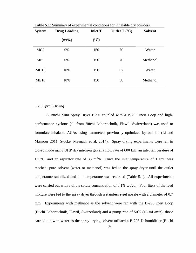
87
Table 5.1: Summary of experimental conditions for inhalable dry powders.
System Drug Loading
(wt%)
Inlet T
(°C)
Outlet T (°C) Solvent
MC0 0% 150 70 Water
ME0 0% 150 70 Methanol
MC10 10% 150 67 Water
ME10 10% 150 58 Methanol
5.2.3 Spray Drying
A Büchi Mini Spray Dryer B290 coupled with a B-295 Inert Loop and high-
performance cyclone (all from Büchi Labortechnik, Flawil, Switzerland) was used to
formulate inhalable ACAs using parameters previously optimized by our lab (Li and
Mansour 2011, Stocke, Meenach et al. 2014). Spray drying experiments were ran in
closed mode using UHP dry nitrogen gas at a flow rate of 600 L/h, an inlet temperature of
150°C, and an aspirator rate of 35 m3/h. Once the inlet temperature of 150°C was
reached, pure solvent (water or methanol) was fed to the spray dryer until the outlet
temperature stabilized and this temperature was recorded (Table 5.1). All experiments
were carried out with a dilute solute concentration of 0.1% wt/vol. Four liters of the feed
mixture were fed to the spray dryer through a stainless steel nozzle with a diameter of 0.7
mm. Experiments with methanol as the solvent were ran with the B-295 Inert Loop
(Büchi Labortechnik, Flawil, Switzerland) and a pump rate of 50% (15 mL/min); those
carried out with water as the spray-drying solvent utiliaed a B-296 Dehumidifier (Büchi
88
Labortechnik, Flawil, Switzerland) and were fed at a pump rate of 20% (5 mL/min).
Powders were collected and transferred to a glass desiccator over Drierite™ and stored in
the freezer at -23°C.
5.2.4 Scanning Electron Microscopy (SEM) on Inhalable Powders
A Hitachi S4300 Scanning Electron Microscope (Tokyo, Japan) was used to
capture images of inhalable ACA powders in order to observe the particle size, surface
roughness, shape and morphology. Double-sided adhesive carbon tabs were adhered to
aluminum studs (Ted Pella, Redding, CA) and blotted on weigh paper containing a small
amount of powder. These samples were coated with a thin film of a gold/palladium alloy
using an Emscope SC400 sputter coating system at 20 mA for 4 minutes under argon gas.
For all electron microscopy experiments at least 3 independent samples were examined
and multiple images were examined for each sample.
5.2.5 Determination of ACA loading in spray dried composites
UV/vis spectrometry was used to determine the loading of erlotinib in the ME10
powders. Erlotinib standards were read by the spectrometer at erlotinib’s absorption max
(max) of 346 nm to a standard curve. Subsequently, ME10 solutions at known
concentration were placed in the spectrometer and the absorbance was read at 346 nm in
order to calculate erlotinib loading. Additionally, a small amount (<20 mg) of erlotinib
was spray dried without mannitol and read on the spectrometer to assure no changes in
spectral properties. Samples containing equivalent amounts of D-mannitol were used in
order to normalize findings.
89
Inductively coupled plasma optical emission spectroscopy was used to quantify
cisplatin loading in the MC10 powders by measuring platinum levels in prepared samples
and comparing them with standard curves. A Varian Vista Pro ICP-OES was used with
argon gas for plasma generation at 1.2kW and 15L/min flow, and sample introduction at
0.9L/min nebulizer flow, 10 second replicate read time across 3 replicates, and 15 rpm
pump rate. A 1ppm yittrium solution from CPI International was used to adjust for any
physical interferences. ULTRAgrade™ Solution Platinum Standards were purchased
from Ultra Scientific (New Kingstown, RI) at 1000 mg/L and used for calibration curves.
A second standard stock was used as a laboratory control sample (LCS) at two different
concentrations and the wavelength showing the best agreement between the LCS samples
and their corresponding standard concentrations was selected for analysis. Prepared
solutions of MC10 at known concentrations were diluted into the range of the standard
curve. The resulting platinum concentrations were used to calculate cisplatin loading.
Additionally, solutions of cisplatin were injected to confirm the validity of the
concentration calculations.
5.2.6 Differential scanning calorimetry (DSC)
DSC was carried out in order to determine the thermal phase transitions of the dry
powders. An Auto Q20 Differential Scanning Calorimeter equipped with an RSC-90
refrigerated cooling system (TA Instruments-Waters LLC, New Castle, DE) and Tzero™
technology was used for measurements. Approximately 2 mg of sample was weighed
into a Tzero™ alodined aluminum pan that was hermetically sealed and then placed into
the furnace along with an empty alodined aluminum hermetically sealed reference pan.
90
The temperature was initialized at 0°C and subsequently raised to 250°C at a rate of
5.00°C /min.
5.2.7 X-ray powder diffraction (XRPD)
The long range order of the inhalable ACA powders was examined with XRPD.
Powders were analyzed by a D8 Discover X-ray diffractometer (Bruker, Billericia, MD)
with Cu K radiation (40 kV, 40 mA, 0.15418 nm) in order to examine their crystallinity.
Scans were obtained from 5 to 60° 2Θ with step size of 0.02 and scan rate of 1 °/min.
5.2.8 Particle size analysis
ImageJ® (NIH, Bethesda, MD) was used to analyze SEM images and digitally
determine the mean size and standard deviation of the geometric diameter of the
inhalable ACA powders. Representative SEM images at a magnification of 5000x were
analyzed by measuring the diameter of at least 100 particles per image with the ImageJ
software.
5.2.9 In vitro aerosol dispersion performance with the Next Generation ImpactorTM
(NGITM)
The M170 Next Generation ImpactorTM
(MSP Corporation Shoreview, MN) was
used for cascade impaction experiments in accordance with United States Pharmacopeia
(USP) Chapter <601> specifications on aerosols (2006) as previously reported (Meenach,
Vogt et al. 2013, Wu, Zhang et al. 2013, Stocke, Meenach et al. 2014). The NGI was
equipped with a stainless steel induction port (USP throat adaptor) attachment and
91
specialized stainless steel NGITM
gravimetric insert cups (NGI Model 170, MSP
Corporation, Shoreview, Minnesota). Type A/E glass fiber filters with diameter 55mm
(Pall Corporation, NY) were placed on each stage of the impactor and weighed before
and after the experiment to determine the particle mass deposited. Approximately 10 mg
of powder was loaded into each clear hydroxypropyl methylcellulose, size-3 capsule
(Quali-V®) (QUALICAPS, Spain) and these capsules were placed into a HandiHaler®
DPITM
device (Boehringer Ingelheim Pharmaceuticals, Inc. Ridgefield, CT) attached to a
customized rubber mouth piece connected to the NGITM
. Three capsules were loaded and
released for each sample and experiments were performed in triplicate. The total dose for
each sample was approximately 30 mg. A Copley HCP5 vacuum pump (Copley
Scientific, United Kingdom) and a Copley TPK 2000 critical flow controller (Copley
Scientific, United Kingdom) were used to produce a flow rate (Q) of 60 L/min in order to
model the flow rate in a healthy adult lung. This flow rate was adjusted before each
experiment using a Copley DFM 2000 flow meter (Copley Scientific, United Kingdom).
For Q=60 L/min, the aerodynamic cutoff diameters (Da50) for each stage of the
impactor were given from the manufacturer as: stage 1 (8.06 m); stage 2 (4.46 m);
stage 3 (2.82 m); stage 4 (1.66 m); stage 5 (0.94 m); stage 6 (0.55 m); and stage 7
(0.34 m). The emitted dose (ED), respirable fraction (RF), and fine particle fraction
(FPF) were calculated as follows:
𝐸𝐷 % = 𝐼𝑛𝑖𝑡𝑖𝑎𝑙 𝑚𝑎𝑠𝑠 𝑖𝑛 𝑐𝑎𝑝𝑠𝑢𝑙𝑒𝑠−𝐹𝑖𝑛𝑎𝑙 𝑚𝑎𝑠𝑠 𝑖𝑛 𝑐𝑎𝑝𝑠𝑢𝑙𝑒𝑠
𝐼𝑛𝑖𝑡𝑖𝑎𝑙 𝑚𝑎𝑠𝑠 𝑖𝑛 𝑐𝑎𝑝𝑠𝑢𝑙𝑒𝑠× 100% (Equation 5.1)
𝑅𝐹 % = 𝑀𝑎𝑠𝑠 𝑜𝑓 𝑝𝑎𝑟𝑡𝑖𝑐𝑙𝑒𝑠 𝑜𝑛 𝑠𝑡𝑎𝑔𝑒𝑠 2−7
𝑇𝑜𝑡𝑎𝑙 𝑝𝑎𝑟𝑡𝑖𝑐𝑙𝑒 𝑚𝑎𝑠𝑠 𝑜𝑛 𝑎𝑙𝑙 𝑠𝑡𝑎𝑔𝑒𝑠× 100% (Equation 5.2)
92
𝐹𝑃𝐹 % = 𝑀𝑎𝑠𝑠 𝑜 𝑓 𝑝𝑎𝑟𝑡𝑖𝑐𝑙𝑒𝑠 𝑜𝑛 𝑠𝑡𝑎𝑔𝑒𝑠 2−7
𝐼𝑛𝑖𝑡𝑖𝑎𝑙 𝑚𝑎𝑠𝑠 𝑖𝑛 𝑐𝑎𝑝𝑠𝑢𝑙𝑒𝑠× 100% (Equation 5.3)
The mass median aerodynamic diameter (MMAD) and geometric standard deviation
(GSD) for the particles were determined using a Mathematica® program written by Dr.
Warren Finlay (Finlay 2008).
5.2.10 Cytotoxicity tests
Cells were cultured in 75 cm2 flasks in DMEM containing 10% FBS, 5%
PSTREP, and 1% Fungizone at 37°C in a humidified incubator with 5% CO2. For
viability studies, cells were seeded into 96-well plates at 10,000 cells/cm2, placed in an
incubator at 37°C and allowed to attach overnight. Cells were dosed cisplatin or erlotinib
in raw form for 24 hours. Studies with MC10 and ME10 were carried out at equivalent
cisplatin and erlotinib concentrations, respectively, as calculated from drug loading
results. After exposure cells were allowed to grow in fresh media for 48 hours. At 72
hours media was removed, cells were washed twice with DPBS, and live cells were
stained with calcein AM, which is hydrolyzed to the fluorescent molecule calcein by
intracellular esterases. Quantification of live cells was accomplished with a Biotek
Synergy Mx microplate reader (Biotek, Winooski, VT) by measuring the fluorescent
intensity of calcein (ex/em 495 nm/517 nm). Relative viability was calculated as the ratio
of the mean fluorescence intensity to that of the control, which contained media only.
93
5.2.11 Statistics
All experiments were performed in at least triplicate. The aerosolization studies
were experimentally designed by Design Expert™ 8.0.7.1 software (Stat-Ease Corp.,
MN, USA). SigmaPlot 12.3 was used for one-way ANOVA tests and when statistically
difference was observed a Holm-Sidak test was used to determine any significance in
observed data. A p-value of <0.05 was considered statistically significant.
5.3 Results and Discussion
5.3.1 Physiochemical Characterization
Inhalable ACA powders obtained from spray drying experiments were examined
via scanning electron microscopy (SEM) and showed spherical particle morphology and
relatively uniform geometric size distribution (Figures 5.1 & 5.2). High mag images
showed varying surface characteristics for the powders spray dried in water with the
MC10 powder appearing to have small uniform crystals agglomerated together into the
composite as opposed to MC0, which displayed fibrous looking crystals. The fibrous
crystals in the MC0 powder appear tightly packed together thereby yielding a smoother
surface than the MC10 particles. Additionally, the particles from the MC10 powder,
although spherical, displayed more deviation in their morphology than the MC0 particles,
which were extremely uniform in spherical morphology.
The ME10 and ME0 powders (spray-dried in methanol) showed more
continuous/homogeneous appearing surfaces wherein the boarders of individual crystals
are not evident. Additionally, the spherical morphology of the particles was less
consistent than particles spray dried in water and some or the particles in the ME0
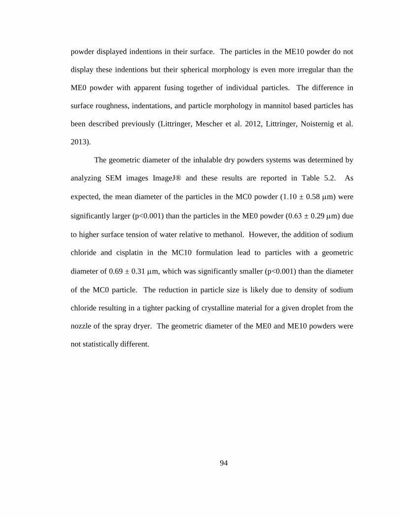
94
powder displayed indentions in their surface. The particles in the ME10 powder do not
display these indentions but their spherical morphology is even more irregular than the
ME0 powder with apparent fusing together of individual particles. The difference in
surface roughness, indentations, and particle morphology in mannitol based particles has
been described previously (Littringer, Mescher et al. 2012, Littringer, Noisternig et al.
2013).
The geometric diameter of the inhalable dry powders systems was determined by
analyzing SEM images ImageJ® and these results are reported in Table 5.2. As
expected, the mean diameter of the particles in the MC0 powder (1.10 ± 0.58 m) were
significantly larger (p<0.001) than the particles in the ME0 powder (0.63 ± 0.29 m) due
to higher surface tension of water relative to methanol. However, the addition of sodium
chloride and cisplatin in the MC10 formulation lead to particles with a geometric
diameter of 0.69 ± 0.31 m, which was significantly smaller (p<0.001) than the diameter
of the MC0 particle. The reduction in particle size is likely due to density of sodium
chloride resulting in a tighter packing of crystalline material for a given droplet from the
nozzle of the spray dryer. The geometric diameter of the ME0 and ME10 powders were
not statistically different.
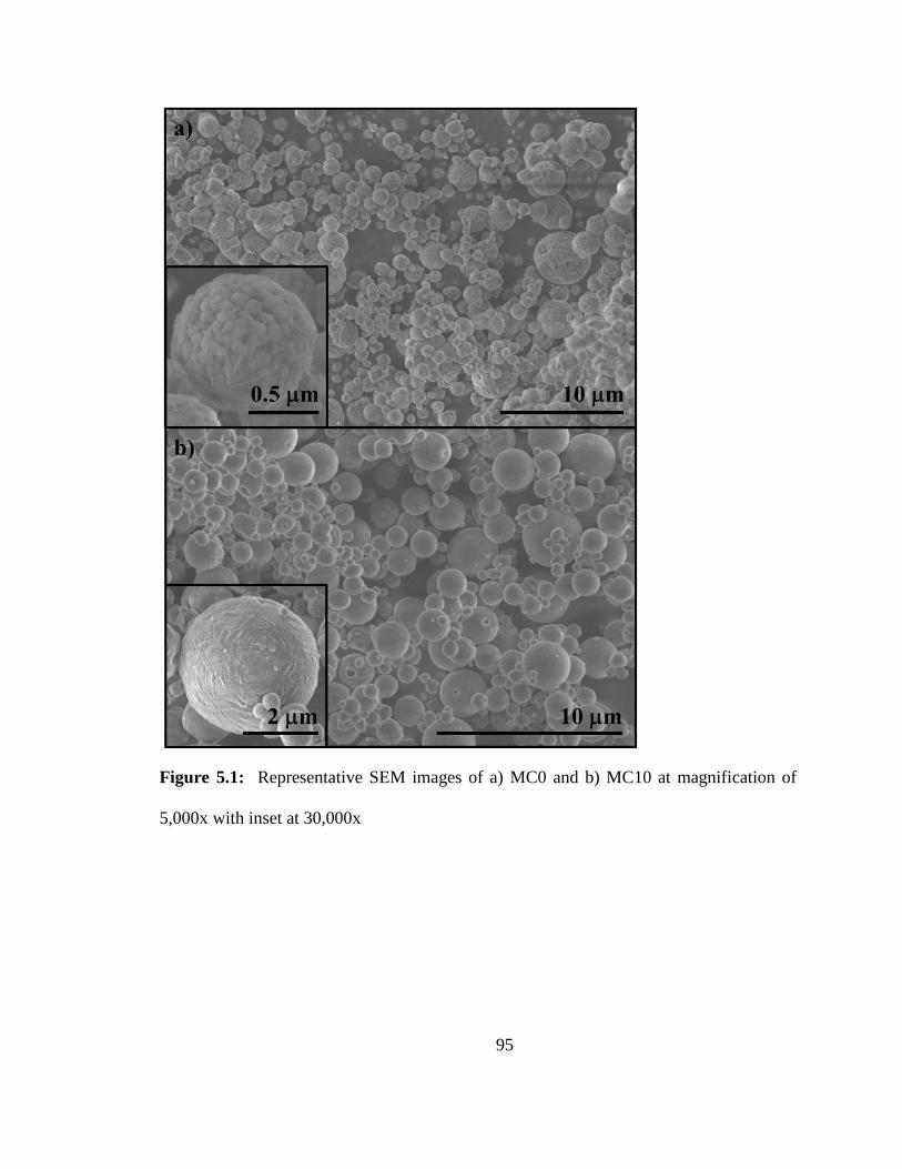
95
Figure 5.1: Representative SEM images of a) MC0 and b) MC10 at magnification of
5,000x with inset at 30,000x
96
Figure 5.2: Representative SEM images of a) ME0 and b) ME10 at magnification of
5,000x with inset at 30,000x
The loading of cisplatin in the MC10 powder was determined through ICP-OES
quantification of platinum in solutions of MC10. The platinum concentration, in
combination with the molecular weight of cisplatin, was used to back calculate a loading
of 10.0% ± 0.0% in the MC10 powder indicating a very controllable capability for these
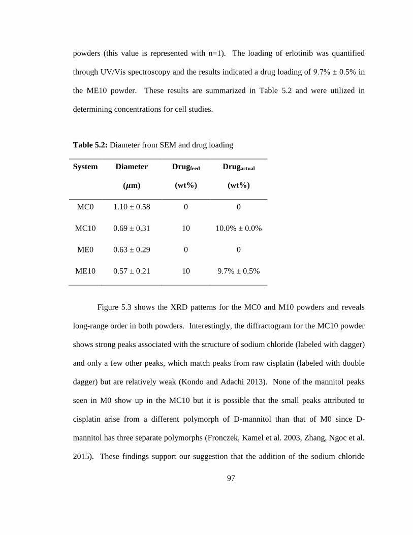
97
powders (this value is represented with n=1). The loading of erlotinib was quantified
through UV/Vis spectroscopy and the results indicated a drug loading of 9.7% ± 0.5% in
the ME10 powder. These results are summarized in Table 5.2 and were utilized in
determining concentrations for cell studies.
Table 5.2: Diameter from SEM and drug loading
System Diameter
(𝝁m)
Drugfeed
(wt%)
Drugactual
(wt%)
MC0 1.10 ± 0.58 0 0
MC10 0.69 ± 0.31 10 10.0% ± 0.0%
ME0 0.63 ± 0.29 0 0
ME10 0.57 ± 0.21 10 9.7% ± 0.5%
Figure 5.3 shows the XRD patterns for the MC0 and M10 powders and reveals
long-range order in both powders. Interestingly, the diffractogram for the MC10 powder
shows strong peaks associated with the structure of sodium chloride (labeled with dagger)
and only a few other peaks, which match peaks from raw cisplatin (labeled with double
dagger) but are relatively weak (Kondo and Adachi 2013). None of the mannitol peaks
seen in M0 show up in the MC10 but it is possible that the small peaks attributed to
cisplatin arise from a different polymorph of D-mannitol than that of M0 since D-
mannitol has three separate polymorphs (Fronczek, Kamel et al. 2003, Zhang, Ngoc et al.
2015). These findings support our suggestion that the addition of the sodium chloride
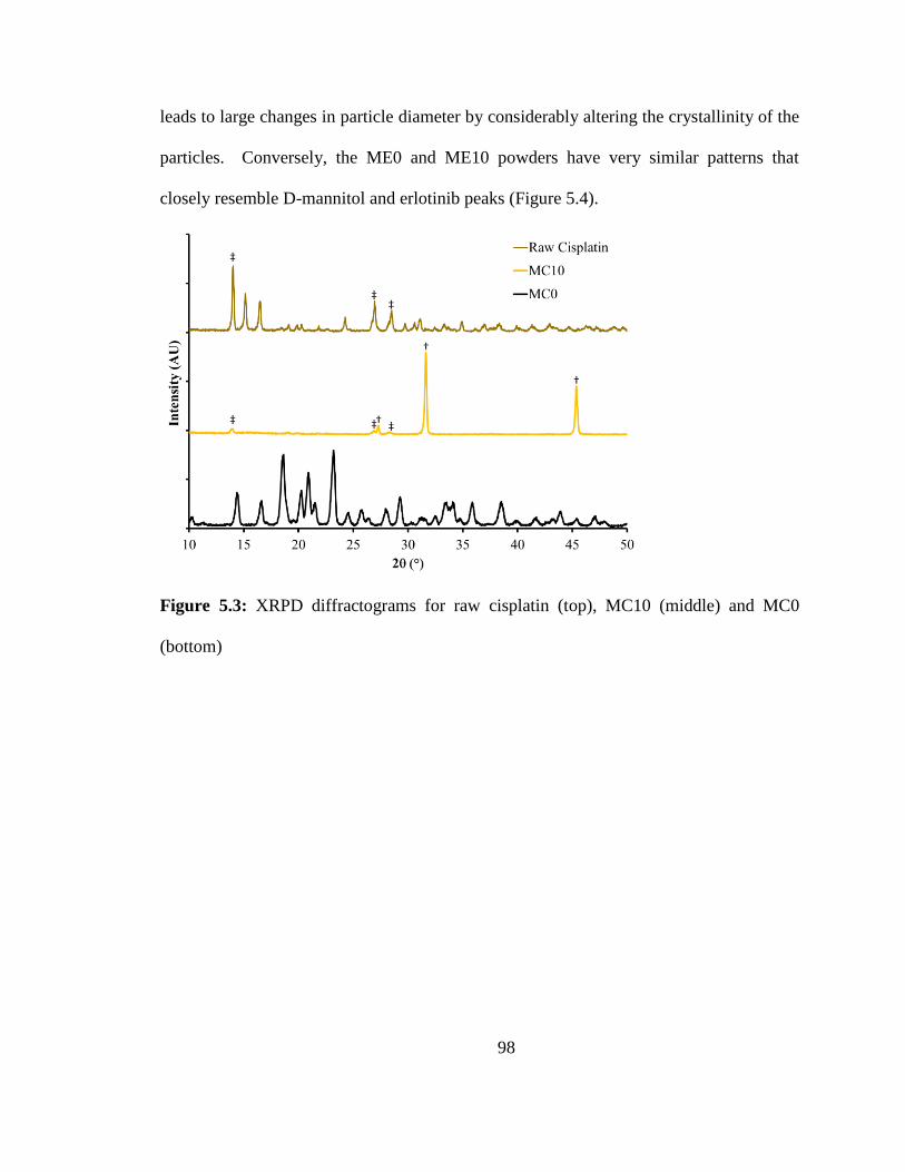
98
leads to large changes in particle diameter by considerably altering the crystallinity of the
particles. Conversely, the ME0 and ME10 powders have very similar patterns that
closely resemble D-mannitol and erlotinib peaks (Figure 5.4).
Figure 5.3: XRPD diffractograms for raw cisplatin (top), MC10 (middle) and MC0
(bottom)
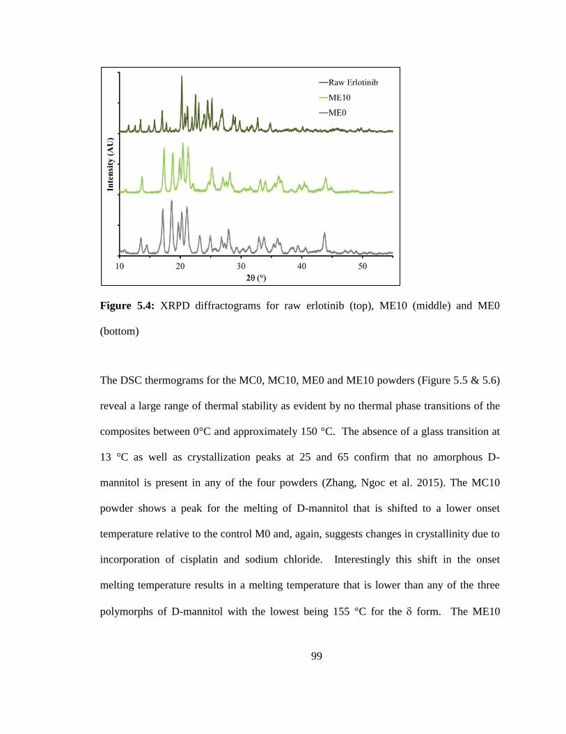
99
Figure 5.4: XRPD diffractograms for raw erlotinib (top), ME10 (middle) and ME0
(bottom)
The DSC thermograms for the MC0, MC10, ME0 and ME10 powders (Figure 5.5 & 5.6)
reveal a large range of thermal stability as evident by no thermal phase transitions of the
composites between 0°C and approximately 150 °C. The absence of a glass transition at
13 °C as well as crystallization peaks at 25 and 65 confirm that no amorphous D-
mannitol is present in any of the four powders (Zhang, Ngoc et al. 2015). The MC10
powder shows a peak for the melting of D-mannitol that is shifted to a lower onset
temperature relative to the control M0 and, again, suggests changes in crystallinity due to
incorporation of cisplatin and sodium chloride. Interestingly this shift in the onset
melting temperature results in a melting temperature that is lower than any of the three
polymorphs of D-mannitol with the lowest being 155 °C for the form. The ME10
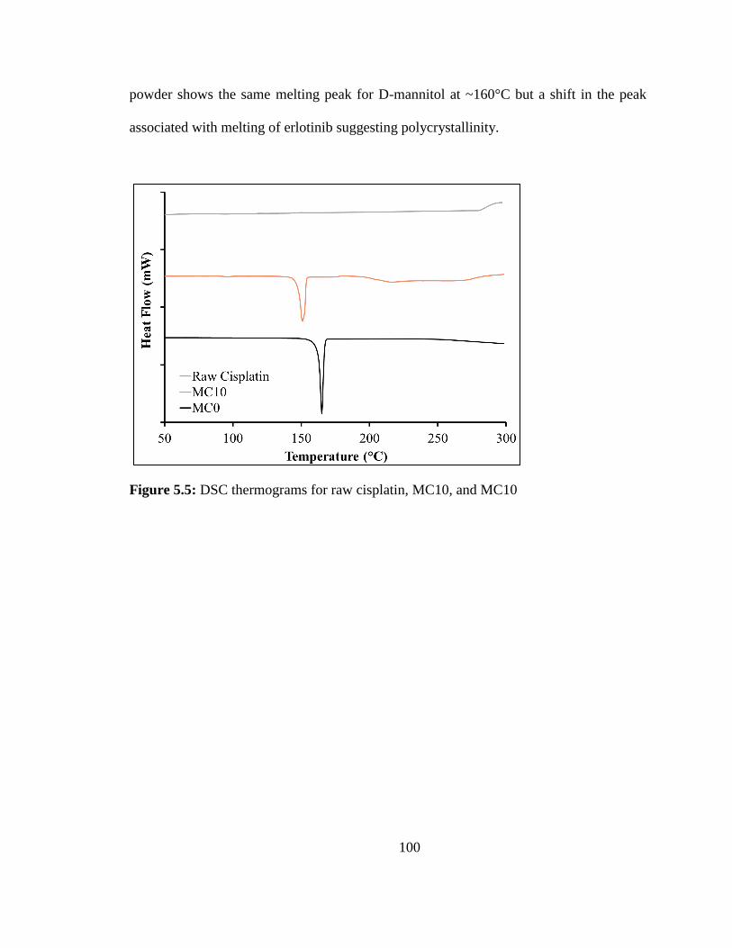
100
powder shows the same melting peak for D-mannitol at ~160°C but a shift in the peak
associated with melting of erlotinib suggesting polycrystallinity.
Figure 5.5: DSC thermograms for raw cisplatin, MC10, and MC10
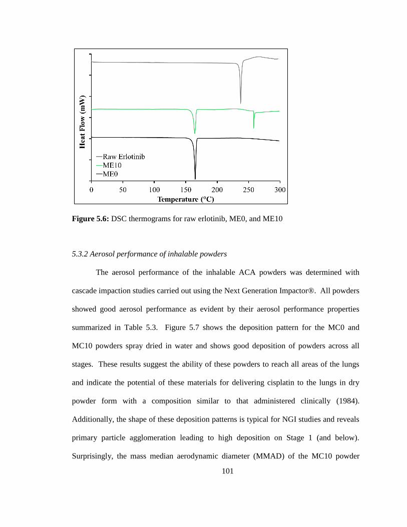
101
Figure 5.6: DSC thermograms for raw erlotinib, ME0, and ME10
5.3.2 Aerosol performance of inhalable powders
The aerosol performance of the inhalable ACA powders was determined with
cascade impaction studies carried out using the Next Generation Impactor®. All powders
showed good aerosol performance as evident by their aerosol performance properties
summarized in Table 5.3. Figure 5.7 shows the deposition pattern for the MC0 and
MC10 powders spray dried in water and shows good deposition of powders across all
stages. These results suggest the ability of these powders to reach all areas of the lungs
and indicate the potential of these materials for delivering cisplatin to the lungs in dry
powder form with a composition similar to that administered clinically (1984).
Additionally, the shape of these deposition patterns is typical for NGI studies and reveals
primary particle agglomeration leading to high deposition on Stage 1 (and below).
Surprisingly, the mass median aerodynamic diameter (MMAD) of the MC10 powder
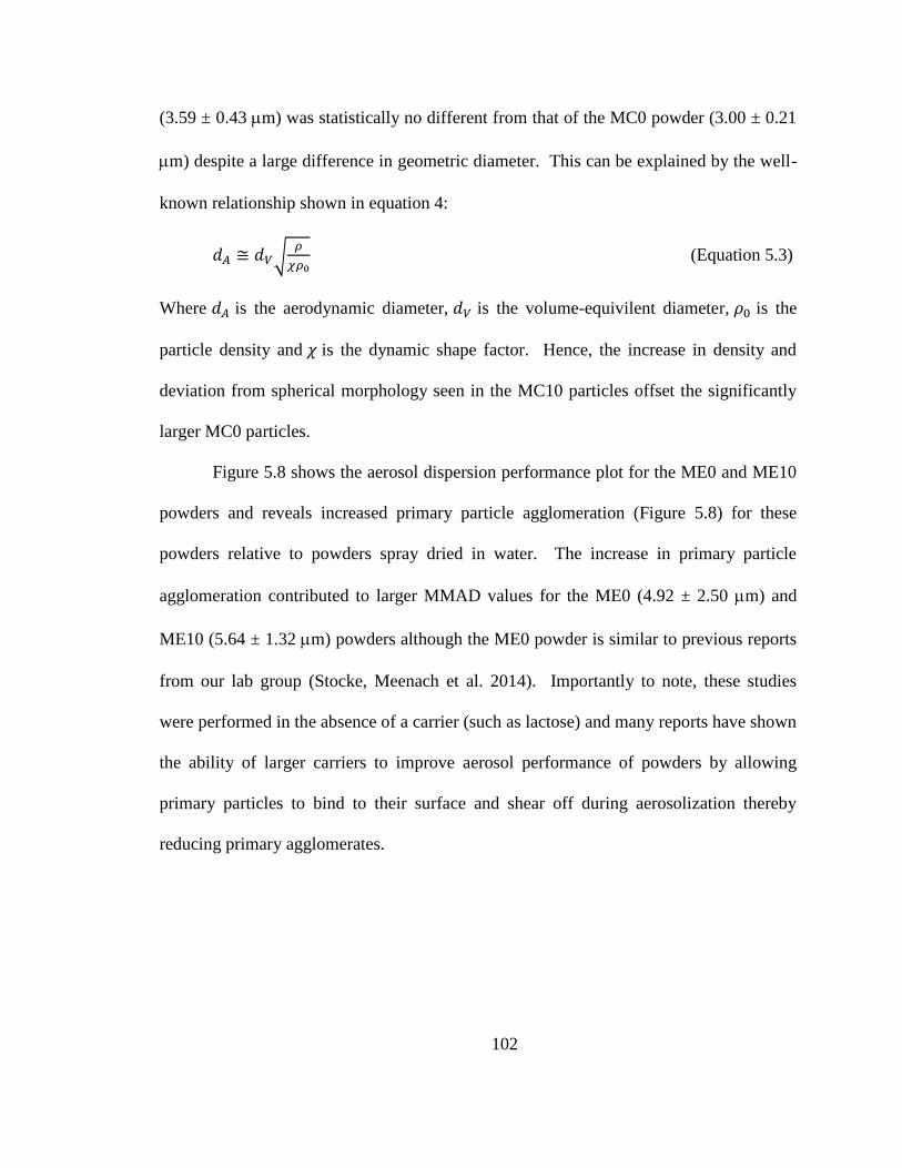
102
(3.59 ± 0.43 m) was statistically no different from that of the MC0 powder (3.00 ± 0.21
m) despite a large difference in geometric diameter. This can be explained by the well-
known relationship shown in equation 4:
𝑑𝐴 ≅ 𝑑𝑉√𝜌
𝜒𝜌0 (Equation 5.3)
Where 𝑑𝐴 is the aerodynamic diameter, 𝑑𝑉 is the volume-equivilent diameter, 𝜌0 is the
particle density and 𝜒 is the dynamic shape factor. Hence, the increase in density and
deviation from spherical morphology seen in the MC10 particles offset the significantly
larger MC0 particles.
Figure 5.8 shows the aerosol dispersion performance plot for the ME0 and ME10
powders and reveals increased primary particle agglomeration (Figure 5.8) for these
powders relative to powders spray dried in water. The increase in primary particle
agglomeration contributed to larger MMAD values for the ME0 (4.92 ± 2.50 m) and
ME10 (5.64 ± 1.32 m) powders although the ME0 powder is similar to previous reports
from our lab group (Stocke, Meenach et al. 2014). Importantly to note, these studies
were performed in the absence of a carrier (such as lactose) and many reports have shown
the ability of larger carriers to improve aerosol performance of powders by allowing
primary particles to bind to their surface and shear off during aerosolization thereby
reducing primary agglomerates.
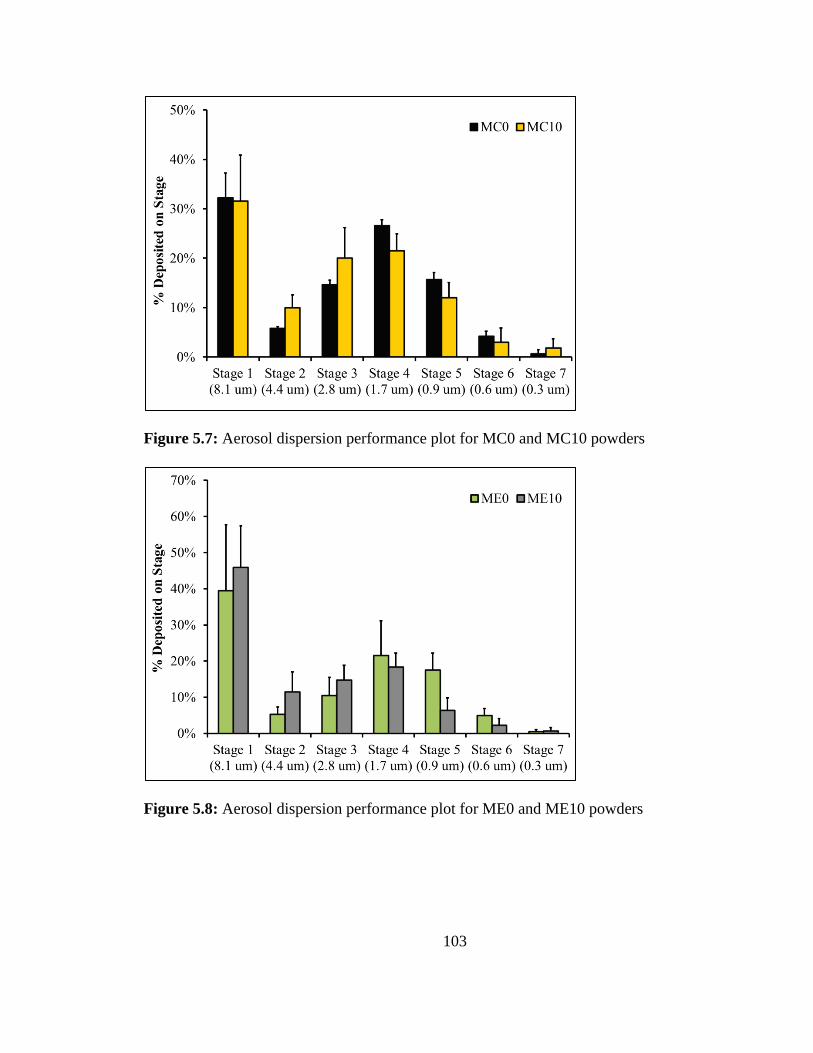
103
Figure 5.7: Aerosol dispersion performance plot for MC0 and MC10 powders
Figure 5.8: Aerosol dispersion performance plot for ME0 and ME10 powders
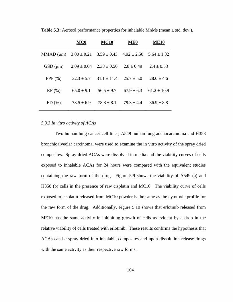
104
Table 5.3: Aerosol performance properties for inhalable MnMs (mean ± std. dev.).
MC0 MC10 ME0 ME10
MMAD (μm) 3.00 ± 0.21 3.59 ± 0.43 4.92 ± 2.50 5.64 ± 1.32
GSD (μm) 2.09 ± 0.04 2.38 ± 0.50 2.8 ± 0.49 2.4 ± 0.53
FPF (%) 32.3 ± 5.7 31.1 ± 11.4 25.7 ± 5.0 28.0 ± 4.6
RF (%) 65.0 ± 9.1 56.5 ± 9.7 67.9 ± 6.3 61.2 ± 10.9
ED (%) 73.5 ± 6.9 78.8 ± 8.1 79.3 ± 4.4 86.9 ± 8.8
5.3.3 In vitro activity of ACAs
Two human lung cancer cell lines, A549 human lung adenocarcinoma and H358
bronchioalveolar carcinoma, were used to examine the in vitro activity of the spray dried
composites. Spray-dried ACAs were dissolved in media and the viability curves of cells
exposed to inhalable ACAs for 24 hours were compared with the equivalent studies
containing the raw form of the drug. Figure 5.9 shows the viability of A549 (a) and
H358 (b) cells in the presence of raw cisplatin and MC10. The viability curve of cells
exposed to cisplatin released from MC10 powder is the same as the cytotoxic profile for
the raw form of the drug. Additionally, Figure 5.10 shows that erlotinib released from
ME10 has the same activity in inhibiting growth of cells as evident by a drop in the
relative viability of cells treated with erlotinib. These results confirms the hypothesis that
ACAs can be spray dried into inhalable composites and upon dissolution release drugs
with the same activity as their respective raw forms.
105
Figure 5.9: In vitro comparison of raw cisplatin and MC10 in human lung cancer cell lines A549
(left) and H358 (right)
Figure 5.10: In vitro comparison of raw erlotinib and ME10 in human lung cancer cell
lines A549 (left) and H358 (right)
Additionally, these findings suggest the potential for pulmonary delivery of
agents traditionally administered systemically (cisplatin intravenously and erlotinib
orally). The potential of these inhalable systems to target the delivery of ACAs to the
lungs could represent a potential improvement of current treatment modalities for certain
patients. Bronchioloalveolar carcinoma (BAC) is a form of adenocarcinoma that has a
prevalence of about 5% among NSCLC cases and this particular neoplastic classification
106
has a higher likelihood of response to epidermal growth factor receptor (EGFR) tyrosine
kinase inhibitors (TKIs) such as erlotinib (Jackman, Chirieac et al. 2005, Arenberg 2011).
A lung tumor classified as BAC must be non-invasive with a pure lepidic spreading
pattern making it a logical target for topical administration of anticancer agents ACAs
through pulmonary delivery. The localization of inhalable ACAs to the affected organ
through inhalation could show benefit for a large variety of lung cancers, and we
recognize that BAC represents a class of tumors that potentially shows significant
advantages from a treatment such as this.
5.4 Conclusions
Inhalable powders consisting of two common ACAs, cisplatin and erlotinib, were
successfully formulated by spray drying. The surface roughness, morphology, and size
of the particles was examined with SEM imaging and showed a significant difference in
the size of mannitol spray dried in water (MC0) verses methanol (ME0). Additionally,
the MC10 powder, containing 40% sodium chloride, displayed particles with rougher
surfaces, smaller geometric diameter, and more irregular spherical particle morphology.
The aerodynamic performance of the powders revealed good aerosol performance for the
powders in absence of a lactose carrier although the ME10 powder displayed the poorest
aerodynamic behavior. Cell studies with human lung cancer cell lines confirmed that the
ACAs incorporated into inhalable powders retained their activity upon release into media
and confirmed the potential of these systems for future development.
107
Chapter 6 Responsive Hydrogel Nanoparticles for Pulmonary Delivery
The chapter is taken directly or adapted from work published in Stocke et al. (2015)
Copyright 2015 Elsevier B.V. Used with permission from Nathanael A Stocke, Susanne
M. Arnold, and J. Zach Hilt.
6.0 Abstract
Nanoparticles represent one of the most widely studied classes of advanced drug
delivery platforms in recent years due to a wide range of unique properties and
capabilities that can be utilized to improve upon traditional drug administration.
Conversely, hydrogel nanoparticles (HNPs) – also called nanogels – represent a unique
class of materials that combine the intrinsic advantages of nanotechnology with the
inherent capabilities of hydrogels. Responsive hydrogels pose a particularly interesting
class of materials that can sense and respond to external stimuli and previous reports of
inhalable hydrogel particles have highlighted their potential in pulmonary delivery. Here,
we synthesized two different pH-responsive HNPs, designated HNP120 and HNP270, by
incorporating functional monomers with a common crosslinker and characterized their
physicochemical properties. One of the HNP systems was selected for incorporation into
a composite dry powder by spray drying, and the aerodynamic performance of the
resulting powder was evaluated. The HNP120s displayed a hydrodynamic diameter of
approximately 120 nm in their fully swollen state and a minimal diameter of around 80
nm while the HNP270s were approximately 270 nm and 115 nm, respectively. Electron
microscopy confirmed particle size- and morphological uniformity of the HNPs. The
108
HNP120s were spray dried into composite dry powders for inhalation and cascade
impaction studies showed good aerosol performance with a mass median aerosol
diameter (MMAD) of 4.82 ± 0.37 m and a fine particle fraction > 30%. The HNPs
released from the spray dried composites retained their responsive behavior thereby
illustrating the potential for these materials as intelligent drug delivery systems that
combine the advantages of nanotechnology, lung targeting through pulmonary delivery,
and stimuli-responsive hydrogels.
6.1 Introduction
Over the last few decades, exhaustive efforts have focused on developing
advanced drug delivery systems for improved therapeutic efficacy, enhanced patient
compliance, and optimal drug safety profiles (Peer, Karp et al. 2007). Nanoparticles
represent one of the most widely studied classes of advanced drug delivery platforms
with more than 25,000 publications over the last 10 years (Anselmo and Mitragotri
2014). Local delivery, environmental sensing, and responsive behavior are a few
properties of nanoparticulate drug delivery systems that can be utilized to improve upon
traditional drug administration (Courrier, Butz et al. 2002, Brannon-Peppas and
Blanchette 2004, Liu and Tang 2010, Coto-Garcia, Sotelo-Gonzalez et al. 2011, Baeza,
Colilla et al. 2015). Conversely, hydrogel nanoparticles (HNPs) – also called nanogels –
represent a unique class of materials that combine the intrinsic advantages of
nanotechnology with the inherent capabilities of hydrogels (Hamidi, Azadi et al. 2008,
Oh, Drumright et al. 2008).
109
Generally, hydrogels are defined as three-dimensional polymeric networks
composed of hydrophilic moieties and possess the ability to imbibe large quantities of
water (Peppas, Bures et al. 2000). Though traditionally made at the macroscopic scale,
hydrogels possess unique characteristics that are promising for nanoformulations. The
high water content of hydrogels results in materials that resemble living tissues and
contributes to the success of many hydrogel applications in research and medicine
(Rosiak and Yoshii 1999, Soppimath, Aminabhavi et al. 2002, Peppas, Hilt et al. 2006).
While many of these applications center around gels at equilibrium (e.g. contact lenses),
researchers have elicited a variety of mechanisms by which to control hydrogel behavior
resulting in intelligent materials with great potential in drug delivery.
Responsive hydrogels have been examined thoroughly over the past several years
and researchers have used a variety of approaches to achieve desired functionality
(Gupta, Vermani et al. 2002). By incorporating various monomers, crosslinkers, and
synthesis conditions one can tune the hydrogel to respond in a variety of manners
including degradation of the hydrogel network or swelling/shrinking of the gel
(Ghandehari, Kopeckova et al. 1997, De, Aluru et al. 2002). Additionally, the response
of the hydrogel can be initiated by a variety of external stimuli such as heat, specific
analytes, and changes in pH and these particles have potential in pulmonary
administration, which has shown promise for nanoparticle and drug delivery to the lungs
(Soppimath, Aminabhavi et al. 2002, Yang, Peters et al. 2008, Anthony J. and Mansour
2009).
Previous reports of delivering hydrogel particles to the lungs via pulmonary
administration demonstrated the ability to engineer micron-sized hydrogel particles that
110
can be delivered to the lungs as stand-alone carriers (El-Sherbiny and Smyth 2010, Du,
El-Sherbiny et al. 2014, Secret, Kelly et al. 2014). Liquid aerosols of hydrogel micro-
and nanoparticle suspensions were also accomplished using metered dose inhalers and
nebulizers (Farhat, Holloway et al. 2009, Selvam, El-Sherbiny et al. 2011). Additionally,
the in vivo administration of 220 nm nanogels showed sustained release of a model
peptide after pulmonary administration, thereby illustrating the potential of HNPs
administered via inhalation (Lee, Lee et al. 2012).
Here, we synthesized pH-responsive HNPs and characterized some of their
physicochemical properties. The responsive behavior of these HNPs was achieved by
incorporating functional monomers with a common crosslinker. Specifically, pH
responsive HNPs were formed by polymerizing methacrylic acid (MAA) with
poly(ethylene glycol) (n=400) diacrylate (PEG400DA). Dynamic light scattering was
used to examine the change in hydrodynamic diameter of two different HNP systems as a
function of pH and electron microscopy confirmed particle size- and morphological
uniformity. Additionally, one of the HNP systems was selected for incorporation into a
composite dry powder by spray drying and the aerodynamic performance of the resulting
powder was evaluated. The characteristics of the HNPs released from this inhalable
powder were examined and compared to the HNPs as synthesized.
6.2 Material and methods
6.2.1 Materials
Acrylic acid (AA), poly(ethylene glycol) 400 (PEG400DA) diacrylate, 9-
anthracenylmethyl methacrylate (9AM), sodium dodecyl sulfate (SDS). Hydrochloric
111
acid (HCL), Sodium Hydroxide (NaOH). Phosphate buffer saline (PBS) and acetic acid
buffer (AAB) stock solutions were made at a concentration of 100 mM and an ionic
strength of 1540 mM with sodium phosphate glacial acetic, respectfully. Ionic strength
of buffers were adjusted with sodium chloride (fisher). For pH adjustments hydrochloric
acid (Fisher) and sodium hydroxide were mixed with DI water to a final concentration of
0.1 M. Sigma-Aldrich (St Louis MO), Fisher Scientific (Pittsburgh, PA). Scott-Gross
(Lexington, KY). ATTC (Manassas, VA). Life Technologies (Grand Island, NY).
6.2.2 Synthesis of responsive hydrogel nanoparticles
Responsive hydrogel nanoparticles were prepared with acrylic acid (AA),
poly(ethylene glycol) 400 (PEG400DA) diacrylate, and 9-anthracenylmethyl
methacrylate (9AM) in an aqueous solution of sodium dodecyl sulfate (SDS) . The molar
ratio of AA:PEG400DA was 80:20 and 1mol% 9AM was incorporated based on total
monomer weight. Briefly, Acrylic acid was weighed into a 15 mL centrifuge tube and
9AM was dissolved into this liquid monomer. Subsequently, PEG400DA was weighed
into a separate 15 mL centrifuge tube. SDS was dissolved in 100 mL of DI water and
added to a 500 mL round bottom flask. A mechanical stir bar and a MODEL 260 J KEM
Digitial Temperature Controller were used during the experiment, which was carried out
for 45 mins at 100 rpm and 45°C. The reaction mixture was via dialysis for 72 hours.
Two different HNP systems were synthesized by changing the SDS concentration and
were labeled HNP120s and HNP270s corresponding to SDS concentrations of 3.0 mM
and 1.5 mM, respectively.
112
6.2.3 Electron microscopy and particle sizing
Scanning electron microscopy (SEM) was performed with a Hitachi S4300
microscope (Tokyo, Japan). Three milliliters of HNP solutions were transferred to a
weigh boat and allowed to evaporate under mild heating on a hot plate. Double-sided
adhesive carbon tabs were adhered to aluminum studs (Ted Pella, Redding, CA) and
blotted on dried HNPs. Samples were coated with a thin film of a gold/palladium alloy
using an Emscope SC400 sputter coating system at 20 mA for 1 minute under argon gas.
For all electron microscopy experiments at least 3 independent samples were examined
and multiple images were examined for each sample. ImageJ® software (NIH, Bethesda,
MD) was used to measure particle diameter in multiple (n>3) representative images.
6.2.4 Colloidal stability of HNPs
The colloidal stability of the HNPs was determined via UV/vis spectroscopy.
HNPs were dispersed in 10 mM PBS at 0.5 mg/mL and adjusted to desired pH with 0.1
M HCl or NaOH solutions. After adjusting the pH, the suspensions were transferred to a
quartz cuvette and scanned from 200 nm to 800 nm in order to insure no maxima or
minima occurred near desired wavelength of 600 nm, hence absorption is due to light
scattering from nanoparticles. A separate quartz cuvette was filled with 10 mM PBS and
used as a blank for the measurements, which were taken every 5 minutes for 12 hours at
600 nm. The stability was calculated by normalizing the absorption at time t to the initial
absorption and the data were plotted in excel.
113
6.2.5 pH response of HNP diameter with dynamic light scattering
Dynamic light scattering was used to measure the hydrodynamic diameter of
HNPs at varying pH conditions. HNPs were first dispersed in 10 mM PBS at a
concentration of 0.5 mg/mL and the pH was titrated to a range of values with 0.1 M HCl
and 0.1 M NaOH. At each pH value a sample of the mixture was placed in a disposable
cuvette and placed into a [model] Malvern ZetaSizer®. The z-averaged diameter of these
particles was recorded
6.2.6 pH response of HNP diameter with dynamic light scattering
Dynamic Light Scattering (DLS) was performed to determine the hydrodynamic
diameter of the HNPs at varying pH values. Readings were performed in disposable
cuvettes using a Malvern Instruments ZetaSizer® (Malvern Instruments, Malvern, United
Kingdom). Aqueous dispersions were made with HNP and PBS concentrations of 200
measurement the pH was adjusted using 0.1 M HCl and NaOH, recorded, and then placed
in ZetaSizer®.
6.2.7 Inhalable HNP composites from spray drying
Spray drying was accomplished with a Büchi Mini Spray Dryer B290 coupled
with a B-295 Inert Loop and high-performance cyclone (all from Büchi Labortechnik,
Flawil, Switzerland) using parameters previously optimized by our lab (Li and Mansour
2011). Spray drying experiments were carried out in closed mode using UHP dry
nitrogen gas at a flow rate of 600 L/h, an inlet temperature of 150°C, and an aspirator rate
114
of 35 m3/h. Once the inlet temperature of 150°C was reached, pure methanol was fed to
the spray dryer until the outlet temperature was stable. For feed mixtures, D-mannitol
was dissolved in methanol and iron HNPs were subsequently dispersed such that the
combined mass of these two components resulted in a dilute concentration of 0.1%
wt/vol. The HNP concentration in the feed was 10wt% based on the combined mass of
D-mannitol and HNPs. Five hundred milliliters of this mixture was fed to the spray dryer
at a pump rate of 50% (15 mL/min) through a stainless steel nozzle with a diameter of 0.7
mm. Powders were collected and transferred to a glass desiccator over Drierite™ and
stored in the freezer at -23°C.
6.2.8 Aerosol performance with the Next Generation ImpactorTM
(NGITM
)
Impactor studies were carried out with a M170 Next Generation ImpactorTM
(MSP Corporation Shoreview, MN) in accordance with United States Pharmacopeia
(USP) Chapter <601> specifications on aerosols (2006). The conditions for these
experiments were previously reported (Meenach, Vogt et al. 2013, Wu, Zhang et al.
2013). The NGI was equipped with a stainless steel induction port (USP throat adaptor)
attachment and specialized stainless steel NGITM
gravimetric insert cups (NGI Model
170, MSP Corporation, Shoreview, Minnesota). Type A/E glass fiber filters with
diameter 55mm (Pall Corporation, NY) were placed on each stage of the impactor and
weighed before and after the experiment to determine the particle mass deposited.
Approximately 10 mg of powder was loaded into each clear hydroxypropyl
methylcellulose, size-3 capsule (Quali-V®) (QUALICAPS, Spain) and these capsules
were placed into a HandiHaler® DPI
TM device (Boehringer Ingelheim Pharmaceuticals,
115
Inc. Ridgefield, CT) attached to a customized rubber mouth piece connected to the
NGITM
. Three capsules were loaded and released for each sample and experiments were
performed in triplicate. The total dose for each sample was approximately 30 mg. A
Copley HCP5 vacuum pump (Copley Scientific, United Kingdom) and a Copley TPK
2000 critical flow controller (Copley Scientific, United Kingdom) were used to produce a
flow rate (Q) of 60 L/min in order to model the flow rate in a healthy adult lung. This
flow rate was adjusted before each experiment using a Copley DFM 2000 flow meter
(Copley Scientific, United Kingdom).
For Q=60 L/min, the aerodynamic cutoff diameters (Da50)for each stage of the
impact
(FPF) were calculated as follows:
𝐸𝐷 % = 𝐼𝑛𝑖𝑡𝑖𝑎𝑙 𝑚𝑎𝑠𝑠 𝑖𝑛 𝑐𝑎𝑝𝑠𝑢𝑙𝑒𝑠−𝐹𝑖𝑛𝑎𝑙 𝑚𝑎𝑠𝑠 𝑖𝑛 𝑐𝑎𝑝𝑠𝑢𝑙𝑒𝑠
𝐼𝑛𝑖𝑡𝑖𝑎𝑙 𝑚𝑎𝑠𝑠 𝑖𝑛 𝑐𝑎𝑝𝑠𝑢𝑙𝑒𝑠× 100% (Equation 6.1)
𝑅𝐹 % = 𝑀𝑎𝑠𝑠 𝑜𝑓 𝑝𝑎𝑟𝑡𝑖𝑐𝑙𝑒𝑠 𝑜𝑛 𝑠𝑡𝑎𝑔𝑒𝑠 2−7
𝑇𝑜𝑡𝑎𝑙 𝑝𝑎𝑟𝑡𝑖𝑐𝑙𝑒 𝑚𝑎𝑠𝑠 𝑜𝑛 𝑎𝑙𝑙 𝑠𝑡𝑎𝑔𝑒𝑠× 100% (Equation 6.2)
𝐹𝑃𝐹 % = 𝑀𝑎𝑠𝑠 𝑜 𝑓 𝑝𝑎𝑟𝑡𝑖𝑐𝑙𝑒𝑠 𝑜𝑛 𝑠𝑡𝑎𝑔𝑒𝑠 2−7
𝐼𝑛𝑖𝑡𝑖𝑎𝑙 𝑚𝑎𝑠𝑠 𝑖𝑛 𝑐𝑎𝑝𝑠𝑢𝑙𝑒𝑠× 100% (Equation 6.3)
Additionally, the mass median aerodynamic diameter (MMAD) and geometric standard
deviation (GSD) for the particles were determined using a Mathematica® program
written by Dr. Warren Finlay (Finlay 2008).
116
6.3 Results and Discussion
Figure 6.1 includes a representation of the structure and responsive of the HNPs at
varying pH conditions, and the responsive behavior of the synthesized HNPs was
examined by measuring the hydrodynamic diameter of the particles as a function of pH
(Figures 6.2 & 6.3). At higher pH values, the carboxylic groups in MAA along the
hydrogel backbone are deprotonated providing a negative charge that stabilizes the
particles and thus prevents agglomeration as illustrated in Figure 6.1. As pH is lowered
the protonation of these functional groups results in less electrostatic repulsion between
chains and intrastrand hydrogen bonding. Both of these mechanisms lead to the collapse,
or deswelling, of the three dimensional network in the HNPs and therefore a reduction in
the hydrodynamic diameter of the HNPs. Eventually, increasing the proton concentration
results in in a transition of particle behavior wherein interparticulate hydrogen bonds
result in large agglomerates of particles leading to a massive increase in the
hydrodynamic diameter of the particles. Interestingly, this behavior is reversible wherein
raising the pH above the transition value thus giving these HNPs the unique ability to
reversibly sense and respond to changes in their environment.
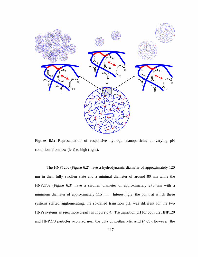
117
Figure 6.1: Representation of responsive hydrogel nanoparticles at varying pH
conditions from low (left) to high (right).
The HNP120s (Figure 6.2) have a hydrodynamic diameter of approximately 120
nm in their fully swollen state and a minimal diameter of around 80 nm while the
HNP270s (Figure 6.3) have a swollen diameter of approximately 270 nm with a
minimum diameter of approximately 115 nm. Interestingly, the point at which these
systems started agglomerating, the so-called transition pH, was different for the two
HNPs systems as seen more clearly in Figure 6.4. Tre transition pH for both the HNP120
and HNP270 particles occurred near the pKa of methacrylic acid (4.65); however, the
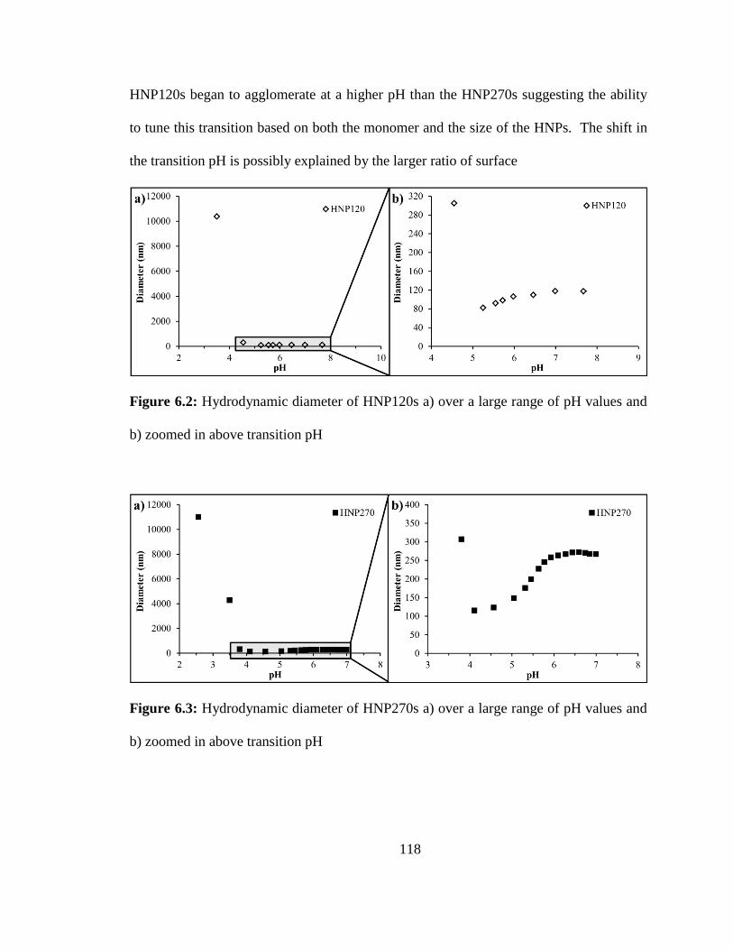
118
HNP120s began to agglomerate at a higher pH than the HNP270s suggesting the ability
to tune this transition based on both the monomer and the size of the HNPs. The shift in
the transition pH is possibly explained by the larger ratio of surface
Figure 6.2: Hydrodynamic diameter of HNP120s a) over a large range of pH values and
b) zoomed in above transition pH
Figure 6.3: Hydrodynamic diameter of HNP270s a) over a large range of pH values and
b) zoomed in above transition pH
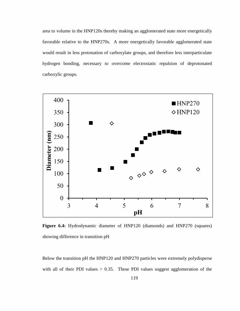
119
area to volume in the HNP120s thereby making an agglomerated state more energetically
favorable relative to the HNP270s. A more energetically favorable agglomerated state
would result in less protonation of carboxylate groups, and therefore less interparticulate
hydrogen bonding, necessary to overcome electrostatic repulsion of deprotonated
carboxylic groups.
Figure 6.4: Hydrodynamic diameter of HNP120 (diamonds) and HNP270 (squares)
showing difference in transition pH
Below the transition pH the HNP120 and HNP270 particles were extremely polydisperse
with all of their PDI values > 0.35. These PDI values suggest agglomeration of the
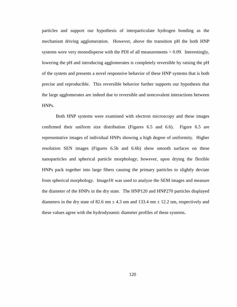
120
particles and support our hypothesis of interparticulate hydrogen bonding as the
mechanism driving agglomeration. However, above the transition pH the both HNP
systems were very monodisperse with the PDI of all measurements > 0.09. Interestingly,
lowering the pH and introducing agglomerates is completely reversible by raising the pH
of the system and presents a novel responsive behavior of these HNP systems that is both
precise and reproducible. This reversible behavior further supports our hypothesis that
the large agglomerates are indeed due to reversible and noncovalent interactions between
HNPs.
Both HNP systems were examined with electron microscopy and these images
confirmed their uniform size distribution (Figures 6.5 and 6.6). Figure 6.5 are
representative images of individual HNPs showing a high degree of uniformity. Higher
resolution SEN images (Figures 6.5b and 6.6b) show smooth surfaces on these
nanoparticles and spherical particle morphology; however, upon drying the flexible
HNPs pack together into large fibers causing the primary particles to slightly deviate
from spherical morphology. ImageJ® was used to analyze the SEM images and measure
the diameter of the HNPs in the dry state. The HNP120 and HNP270 particles displayed
diameters in the dry state of 82.6 nm ± 4.3 nm and 133.4 nm ± 12.2 nm, respectively and
these values agree with the hydrodynamic diameter profiles of these systems.
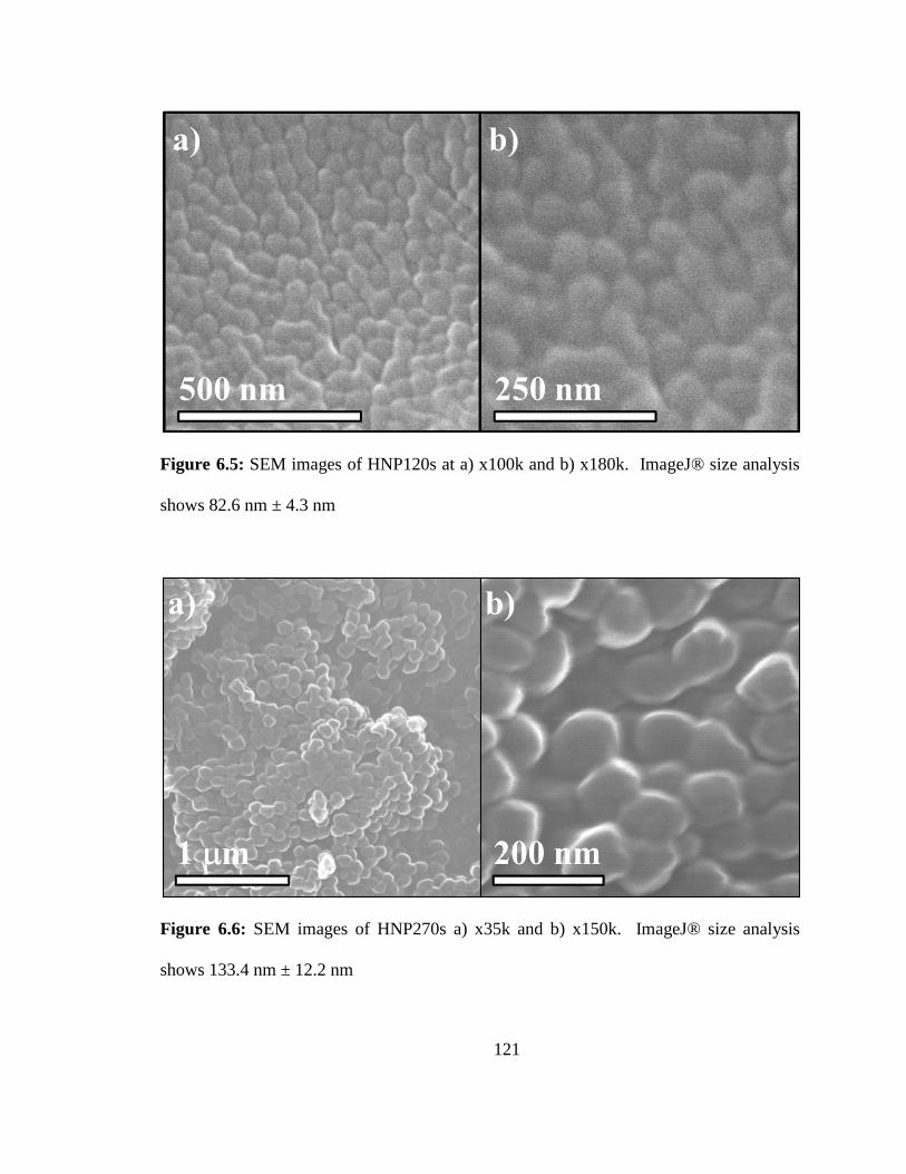
121
Figure 6.5: SEM images of HNP120s at a) x100k and b) x180k. ImageJ® size analysis
shows 82.6 nm ± 4.3 nm
Figure 6.6: SEM images of HNP270s a) x35k and b) x150k. ImageJ® size analysis
shows 133.4 nm ± 12.2 nm
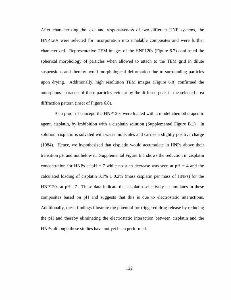
122
After characterizing the size and responsiveness of two different HNP systems, the
HNP120s were selected for incorporation into inhalable composites and were further
characterized. Representative TEM images of the HNP120s (Figure 6.7) confirmed the
spherical morphology of particles when allowed to attach to the TEM grid in dilute
suspensions and thereby avoid morphological deformation due to surrounding particles
upon drying. Additionally, high resolution TEM images (Figure 6.8) confirmed the
amorphous character of these particles evident by the diffused peak in the selected area
diffraction pattern (inset of Figure 6.8).
As a proof of concept, the HNP120s were loaded with a model chemotherapeutic
agent, cisplatin, by imbibition with a cisplatin solution (Supplemental Figure B.1). In
solution, cisplatin is solvated with water molecules and carries a slightly positive charge
(1984). Hence, we hypothesized that cisplatin would accumulate in HNPs above their
transition pH and not below it. Supplemental Figure B.1 shows the reduction in cisplatin
concentration for HNPs at pH = 7 while no such decrease was seen at pH = 4 and the
calculated loading of cisplatin 3.1% ± 0.2% (mass cisplatin per mass of HNPs) for the
HNP120s at pH =7. These data indicate that cisplatin selectively accumulates in these
composites based on pH and suggests that this is due to electrostatic interactions.
Additionally, these findings illustrate the potential for triggered drug release by reducing
the pH and thereby eliminating the electrostatic interaction between cisplatin and the
HNPs although these studies have not yet been performed.
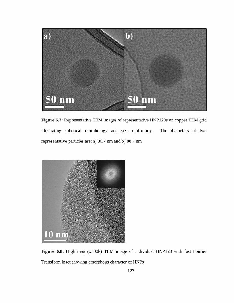
123
Figure 6.7: Representative TEM images of representative HNP120s on copper TEM grid
illustrating spherical morphology and size uniformity. The diameters of two
representative particles are: a) 80.7 nm and b) 88.7 nm
Figure 6.8: High mag (x500k) TEM image of individual HNP120 with fast Fourier
Transform inset showing amorphous character of HNPs
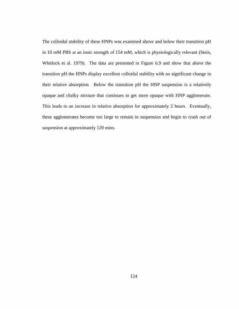
124
The colloidal stability of these HNPs was examined above and below their transition pH
in 10 mM PBS at an ionic strength of 154 mM, which is physiologically relevant (Stein,
Whitlock et al. 1979). The data are presented in Figure 6.9 and show that above the
transition pH the HNPs display excellent colloidal stability with no significant change in
their relative absorption. Below the transition pH the HNP suspension is a relatively
opaque and chalky mixture that continues to get more opaque with HNP agglomerate.
This leads to an increase in relative absorption for approximately 2 hours. Eventually,
these agglomerates become too large to remain in suspension and begin to crash out of
suspension at approximately 120 mins.
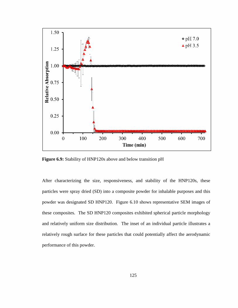
125
Figure 6.9: Stability of HNP120s above and below transition pH
After characterizing the size, responsiveness, and stability of the HNP120s, these
particles were spray dried (SD) into a composite powder for inhalable purposes and this
powder was designated SD HNP120. Figure 6.10 shows representative SEM images of
these composites. The SD HNP120 composites exhibited spherical particle morphology
and relatively uniform size distribution. The inset of an individual particle illustrates a
relatively rough surface for these particles that could potentially affect the aerodynamic
performance of this powder.
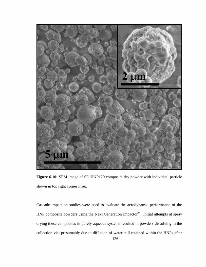
126
Figure 6.10: SEM image of SD HNP120 composite dry powder with individual particle
shown in top right corner inset.
Cascade impaction studies were used to evaluate the aerodynamic performance of the
HNP composite powders using the Next Generation Impactor®. Initial attempts at spray
drying these composites in purely aqueous systems resulted in powders dissolving in the
collection vial presumably due to diffusion of water still retained within the HNPs after
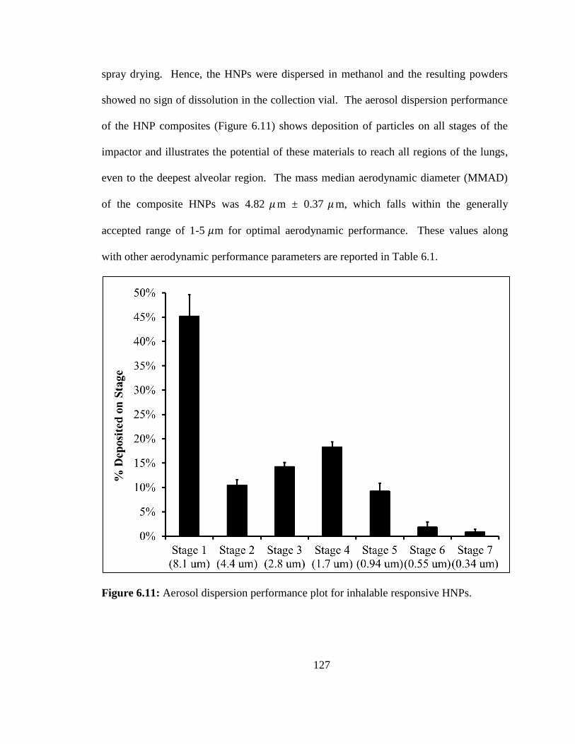
127
spray drying. Hence, the HNPs were dispersed in methanol and the resulting powders
showed no sign of dissolution in the collection vial. The aerosol dispersion performance
of the HNP composites (Figure 6.11) shows deposition of particles on all stages of the
impactor and illustrates the potential of these materials to reach all regions of the lungs,
even to the deepest alveolar region. The mass median aerodynamic diameter (MMAD)
of the composite HNPs was 4.82 𝜇 m ± 0.37 𝜇 m, which falls within the generally
accepted range of 1-5 𝜇m for optimal aerodynamic performance. These values along
with other aerodynamic performance parameters are reported in Table 6.1.
Figure 6.11: Aerosol dispersion performance plot for inhalable responsive HNPs.
128
Table 6.1: Aerosol performance of HNP composites for inhalation (mean ± std. dev.).
MMAD
(μm)
GSD (μm) FPF (%) RF (%) ED (%)
HNPs 4.82 ± 0.37 2.51 ± 0.18 31.7 ± 6.8 54.8 ± 4.4 78.5 ± 5.3
The responsiveness of the HNPs released from the composite HNP powders was
examined by again measuring their hydrodynamic diameter as a function of pH. Figure
6.12 shows a slight increase in the z-averaged diameter of the HNPs released from the
spray dried composites. This increase in z-averaged diameter is accompanied by an
increase of the PDI of these particles. For all measurements above the transition pH the
PDI varied between 0.1-0.2 indicating a slightly more polydisperse system relative to the
HNPs before spray drying. Importantly, the HNPs released from inhalable composites
still respond to environmental changes in pH and these responses continue to be
reversible. The increase in z-averaged diameter and PDI suggested a population of
particles that are in a permanently agglomerated state so we examined their colloidal
stability.
129
Figure 6.12: Hydrodynamic diameter of HNP120s and SD HNP120s a) over a large
range of pH values and b) zoomed above transition pH
Figure 6.13 compares the stability of the HNP120s to those released from the SD
HNP120 composites. The HNPs released from the spray dried composites have a
subpopulation of particles that fall out of suspension with time. This fraction of particles
likely represents permanent agglomerates that were introduced during the spray drying
process but only represents approximately 12.6% +/- 0.3% of the HNPs (assuming Beer’s
law).
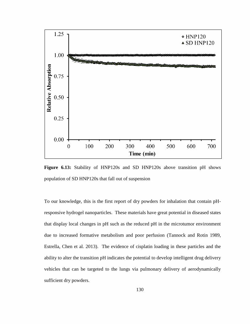
130
Figure 6.13: Stability of HNP120s and SD HNP120s above transition pH shows
population of SD HNP120s that fall out of suspension
To our knowledge, this is the first report of dry powders for inhalation that contain pH-
responsive hydrogel nanoparticles. These materials have great potential in diseased states
that display local changes in pH such as the reduced pH in the microtumor environment
due to increased formative metabolism and poor perfusion (Tannock and Rotin 1989,
Estrella, Chen et al. 2013). The evidence of cisplatin loading in these particles and the
ability to alter the transition pH indicates the potential to develop intelligent drug delivery
vehicles that can be targeted to the lungs via pulmonary delivery of aerodynamically
sufficient dry powders.
131
6.4 Conclusions
Responsive HNPs were successfully synthesized by incorporating MAA as a
functional monomer and PEG400DA as a crosslinker. DLS showed a reduction in
hydrodynamic diameter of the HNPS due to protonation of the carboxylic acids groups in
MAA with decreasing pH. The transition pH of these materials can be altered by changes
in the particle synthesis conditions suggesting the ability to tune this value even for a
given monomeric system. HNPs were successfully incorporated into inhalable
composites and retained their nanometer size as well as their responsive behavior. These
findings highlight the great potential for these materials as intelligent drug delivery
systems that combine the advantages of nanotechnology, lung targeting through
pulmonary delivery, and stimuli-responsive hydrogels.
Acknowledgements
This work would not have been possible without the efforts of Marjorie L. Guy
and financial support from the National Institute of Health’s National Cancer Institute on
grant number R25CA153954.
132
Chapter 7 Conclusions
Over the last several decades, survival rates of lung cancer patients have shown only
minimal improvements. This disease represents an area in oncology where much
progress is needed and alternative treatment modalities must be considered. Encouraging
progress in personalized therapy is opening doors of opportunity in oncology by
discovering genetically targeted moieties for individualized care. Additionally, the
modern progress of nanotechnology suggests impending success in cancer applications
and the combination with personalized anticancer agents could provide unique
therapeutic options. However, new therapies will require innovative drug-delivery
platforms in order to maximize their potential therapeutic advantages; pulmonary
delivery represents a promising approach for targeting novel cancer therapies and treating
primary and secondary lung malignancies.
In this dissertation, pulmonary delivery was investigated as a viable treatment
modality for delivering unique nanoparticles and anticancer agents directly to the lungs.
We began by examining the possibility of noninvasively administering magnetic
nanoparticles to the lungs, via inhalation, for their application as remote-controlled
thermal therapy agents. To accomplish this, we successfully formulated dry powder
composites consisting of iron oxide MNPs – so-called magnetic nanocomposite
microparticles – through advanced spray drying techniques. The physicochemical
characterization of these composites revealed their potential and aerosol performance
studies demonstrated favorable aerodynamic properties. Heating studies illustrated that
133
these materials retain excellent remote-heating capability after formulation and in vitro
cytotoxicity studies showed only moderate toxicity on human lung A549 cells.
To further examine the therapeutic potential of these materials, we utilized an
advanced in vitro model of metastatic triple negative breast cancer TNBC. This model
consisted of spheroids – so called TM analogs – composed of 4T1 breast cancer cells,
(ER-, PR-, and HER2-), MEF fibroblast cells, and C166 endothelial cells and represented
secondary lung malignancies that could be targeted with inhalable composites. TM
analogs were exposed to our MnMs and no measureable adverse effects were observed in
the absence of AMF-exposure. Conversely, remote-controlled thermal therapy on TM
analogs was accomplished with a custom alternating magnetic field (AMF) and showed a
statistically significant increase in cellular death for the higher MNP dose. These
findings highlighted the potential of these inhalable MnM composites for future
development as a targeted thermal treatment of lung cancer.
The next part of this dissertation examined feasibility of inhalable chemotherapy
wherein two anticancer agents (ACAs) commonly administered in the clinic were
formulated into dry powder composites for pulmonary delivery. Inhalable powders
consisting of cisplatin and erlotinib were successfully formulated by spray drying and
their physicochemical properties were evaluated. The aerodynamic diameter of these
powders revealed good aerosol performance even in absence of a lactose carrier although
the ME10 powder displayed the poorest aerodynamic behavior. Finally, cell studies with
human lung cancer cell lines confirmed that the ACAs incorporated into inhalable
powders retained their activity upon release into media and confirmed the potential of
these systems for future development.
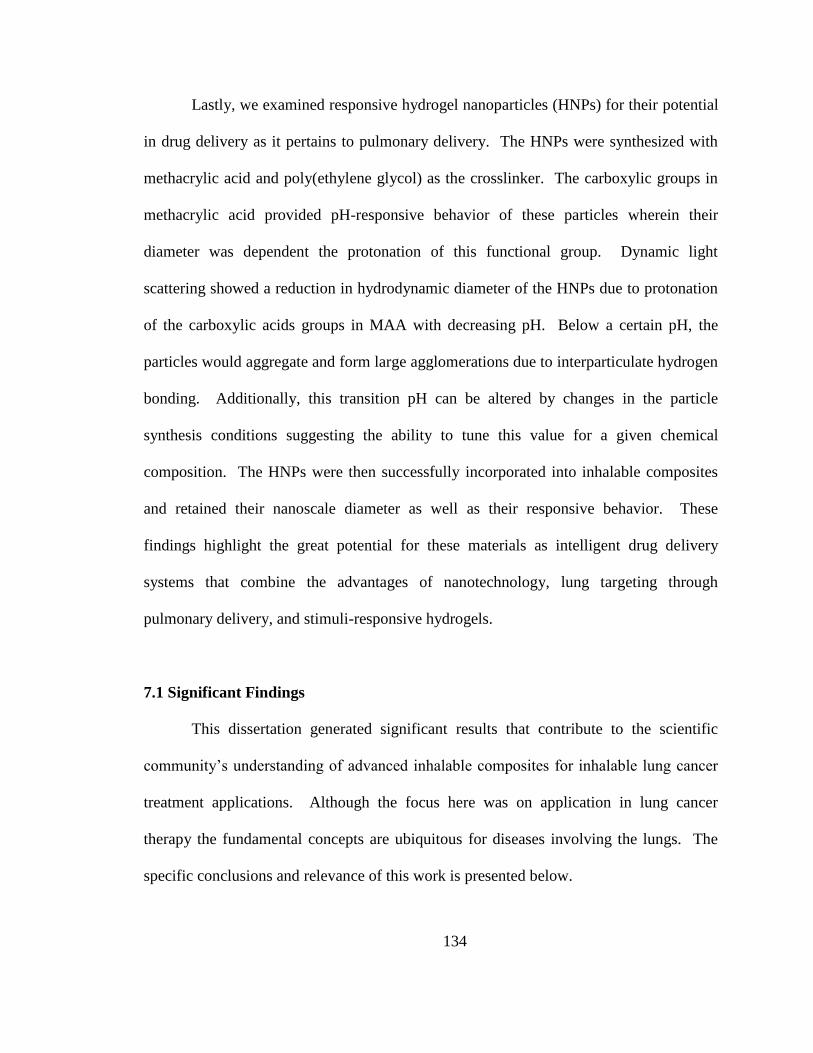
134
Lastly, we examined responsive hydrogel nanoparticles (HNPs) for their potential
in drug delivery as it pertains to pulmonary delivery. The HNPs were synthesized with
methacrylic acid and poly(ethylene glycol) as the crosslinker. The carboxylic groups in
methacrylic acid provided pH-responsive behavior of these particles wherein their
diameter was dependent the protonation of this functional group. Dynamic light
scattering showed a reduction in hydrodynamic diameter of the HNPs due to protonation
of the carboxylic acids groups in MAA with decreasing pH. Below a certain pH, the
particles would aggregate and form large agglomerations due to interparticulate hydrogen
bonding. Additionally, this transition pH can be altered by changes in the particle
synthesis conditions suggesting the ability to tune this value for a given chemical
composition. The HNPs were then successfully incorporated into inhalable composites
and retained their nanoscale diameter as well as their responsive behavior. These
findings highlight the great potential for these materials as intelligent drug delivery
systems that combine the advantages of nanotechnology, lung targeting through
pulmonary delivery, and stimuli-responsive hydrogels.
7.1 Significant Findings
This dissertation generated significant results that contribute to the scientific
community’s understanding of advanced inhalable composites for inhalable lung cancer
treatment applications. Although the focus here was on application in lung cancer
therapy the fundamental concepts are ubiquitous for diseases involving the lungs. The
specific conclusions and relevance of this work is presented below.
135
MNPs can be embedded into dry powder composites via spray drying and
the resulting powders can be formulated such that good aerodynamic
performance is achievable
The loading of MNPs can be varied in dry powder composites without
adversely affecting the aerosol behavior of the materials
MNPs incorporated to dry-powder composites release upon dissolution
and show no signs of agglomeration as evident by consistent
hydrodynamic diameter
Upon release from inhalable composites, MNPs retain their ability to
remotely heat in the presence of an alternating magnetic field although this
performance is slightly reduced as evident by a small reduction in the SAR
value
Metastatic triple negative breast cancer spheroids (TM analogs) show no
signs of mechanical detrition as a result of MNP exposure for 24 hours at
MNP concentrations as high as 1.0 mg/mL
MNPs are presumably non-toxic in TM analogs for concentrations up to
1.0 mg/mL as evidence by no reduction in dead cell count measured
through SYTOX® Blue staining and fluorescence quantification
Remote-controlled thermal treatment of TM analogs exposed to MNPs
results in mechanical deterioration of the spheroids and results in a
statistically significant increase in dead cell count as measured through
SYTOX® Blue staining and fluorescence quantification
136
Cisplatin and erlotinib can be formulated into inhalable dry powder
composites with acceptable aerodynamic performance
These composites show identical cytotoxic profiles in the human lung
cancer cell lines H358 and A549 thereby suggesting the released drugs
retain their physiological activity
Hydrogel nanoparticles (HNPs) respond to pH with changes in their
hydrodynamic diameter and exhibit a transition pH below which they form
large agglomerates, which are presumable due to interparticulate hydrogen
bonding
The incorporation of HNPs into inhalable drug delivery platforms is
possible and the release HNPs retain their responsive behavior despite a
small increase in their hydrodynamic diameter, which is likely due to an
agglomerated subpopulation as suggested by stability studies
Copyright © Nathanael Aaron Stocke 2015
137
Appendix A Transport in PEG-Based Hydrogels: Role of Water Content and
Crosslinker Molecular Weight
A.0 ABSTRACT
Polyethylene glycol (PEG) hydrogels are hydrophilic, high water content,
polymeric networks that represent excellent candidates as engineering biomaterials for
applications ranging from tissue engineering to drug and gene delivery. A key challenge
for many biomedical applications is the control of transport properties of small particles
within the resulting 3D crosslinked gels. In this work, we investigated the swelling
characteristics of a series of PEG-hydrogels as a function of both the water content at
synthesis and the molecular weight of the crosslinker used. Using Rhodamine 110 (R110)
as a model probe molecule, we examined the transport properties within the resulting
PEG-hydrogels by fluorescence correlation spectroscopy (FCS) measurements. We
determined a linear relationship between the R110 translational diffusion coefficients, D
and the volumetric swelling ratio of the formed hydrogels. Both increasing the water
content at synthesis and increasing crosslinker molecular weight are tunable parameters
resulting in more open hydrogel microstructures and increased transport of probe
molecules through the gel networks.
A.1 INTRODUCTION
Poly(ethylene glycol) (PEG) is one of the most commonly used polymers in
biomedicine because of its biocompatibility and has been approved for a wide range of
biomedical applications.[1]
Despite a few reports claiming that PEG can result in an
138
immune response,[2]
most studies have referred to PEG as a stealth polymer due to its
ability to avoid eliciting an immune response inside the body.[3]
A wide range of
materials utilizing PEG have been extensively studied including PEG coatings on particle
systems or devices,[4, 5]
PEG-linked proteins for enhanced performance,[6, 7]
micelle and
liposome carriers,[8, 9]
and intelligent hydrogels for drug delivery.[1, 10, 11]
Hydrogels represent an important and versatile class of biomedical materials
proving to be excellent candidates for a wide range of applications ranging from contact
lenses to tissue engineering.[12, 13]
Hydrogels consist of three dimensional networks
formed by crosslinking hydrophilic polymers and are capable of immobilizing a large
amount of water in their interior through capillary forces and surface tension.[14, 15]
Crosslinks in hydrogels can be either physical or chemical in nature, and both have
implications on the performance of the material.[16-18]
The amount of water a hydrogel
imbibes, among other factors, impacts the microenvironment within the gel. Control of
the gel microenvironment directly impacts the suitability of the resulting materials for its
intended application.
The microenvironment of a hydrogel is characterized by a variety of parameters.
One of the most important parameters is the volume fraction in the swollen state (v2,s) or
its reciprocal which is the volumetric swelling ratio (Q). These parameters are determined
by examining the swelling properties of a given hydrogel system and provide insight into
the microenvironment within the hydrogel. A greater swelling capacity implies a less
crowded microenvironment. The “crowdedness” of the microenvironment is quantified
distance between polymer chains with a network system. A variety of factors are known
139
to affect the mesh size of a hydrogel including: composition, charge, molecular weight of
the crosslinker, and crosslinking density.[11]
Hydrogels with higher crosslinking density
have less capacity to swell due to more tether points that restrict the movement within the
gel. These tie points can be covalent bonds, physical entanglements, ionic interactions, or
specific affinities between complementary moieties such as ligand-receptor
interactions.[16, 19]
Ultimately, the crosslinking of a hydrogel is what determines its three-
dimensional structure, and the number and nature of these crosslinks dramatically affects
the performance of the hydrogel by altering the mechanical properties, swelling
characteristics, and mesh sizes of the network. For applications that require transport of
small molecules through the hydrogels, such as drug delivery, dynamics within the
hydrogel are highly dependent on the mesh size which controls the mean free path of
diffusion.[20]
Fluorescence correlation spectroscopy (FCS) has been previously shown effective
to measure transport properties of small molecules in various polymeric systems.[21-24]
This includes the use of FCS to examine the diffusion of probe molecules in hydrogels.[17,
25-29] It has been shown that higher molar ratios of chemical crosslinkers lead to slower
diffusion of probe molecules.[30]
Less studied are the effects of water content during
hydrogel synthesis and the molecular weight of the crosslinker on the transport properties
of probe molecules.
In this work, we examined the role of final water content and crosslinker
molecular weight on both the swelling characteristics and the resulting diffusivity of
probe molecules within the PEG-hydrogels. An array of PEG-based hydrogels was
synthesized with varied solvent content during synthesis as well as varying the molecular
140
weight of the crosslinker. The two crosslinkers used in this study, poly(ethylene glycol)
400 diacrylate (PEG400DA) and poly(ethylene glycol) 4000 diacrylate (PEG4000DA),
are identical in chemical composition but differ by an order of magnitude in the average
molecular weight of the ethylene glycol chain. To examine nanoparticle transport
properties within the hydrogels, gels were washed, lyophilized, and subsequently imbibed
with a model fluorescent probe, rhodamine 110 (R110), at nanomolar concentrations. We
used FCS to measure the translational diffusion coefficient of R110 in the resulting PEG-
hydrogels. Additionally, the swelling characteristics of these gels were measured, and the
correlation between swelling and diffusion was examined (Figure A.1). We show that
increasing the solvent content during hydrogel synthesis leads to a microenvironment
consisting of fewer physical crosslinks and, therefore, less crowded hydrogels.
Additionally, we demonstrate that changing the molecular weight of the crosslinker has a
dramatic effect on the transport of probe molecules within the hydrogel due to increased
mesh sizes. Lastly, we determined an approximately linear relationship between the
translational diffusion coefficient, D, of a probe molecule and the volumetric swelling
ratio of the formed hydrogels.
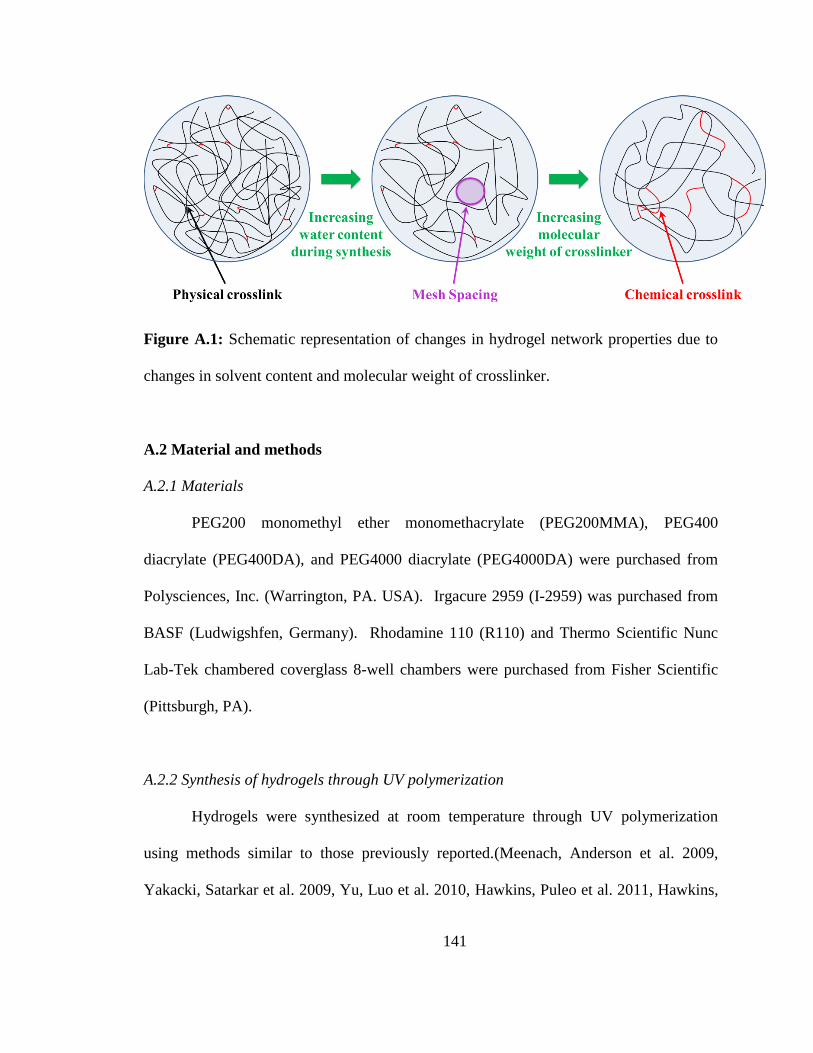
141
Figure A.1: Schematic representation of changes in hydrogel network properties due to
changes in solvent content and molecular weight of crosslinker.
A.2 Material and methods
A.2.1 Materials
PEG200 monomethyl ether monomethacrylate (PEG200MMA), PEG400
diacrylate (PEG400DA), and PEG4000 diacrylate (PEG4000DA) were purchased from
Polysciences, Inc. (Warrington, PA. USA). Irgacure 2959 (I-2959) was purchased from
BASF (Ludwigshfen, Germany). Rhodamine 110 (R110) and Thermo Scientific Nunc
Lab-Tek chambered coverglass 8-well chambers were purchased from Fisher Scientific
(Pittsburgh, PA).
A.2.2 Synthesis of hydrogels through UV polymerization
Hydrogels were synthesized at room temperature through UV polymerization
using methods similar to those previously reported.(Meenach, Anderson et al. 2009,
Yakacki, Satarkar et al. 2009, Yu, Luo et al. 2010, Hawkins, Puleo et al. 2011, Hawkins,
142
Tolbert et al. 2013) Briefly, acrylated PEG moieties were weighed out and mixed with
the water-soluble photoinitiator I-2959 (1 wt%). The water content during synthesis was
varied between 0-85 wt% based on the total mass of the system. This mixture was
pipetted between two glass plates containing a 1mm Teflon® spacer and held together
with binder clips. The plate was then exposed to 365 nm UV light for five minutes using
a Lesco Cure Max UV flood source (American ultraviolet, Torranc, CA) operating at
~10-11 mW/cm2. After polymerization, gels were punched into 4mm discs and washed
in DI water for 1 week. For measurement of probe diffusion by fluorescence correlation
spectroscopy (FCS), hydrogels were washed in DI water for 1 week then imbibed over
night with R110 at a concentration of 10 nM. Gels were subsequently removed and
placed into NUNC LabTek 8-well microscopy chambers (Nalgene Nunc International)
and measured directly by FCS.
A.2.3 Conversion of hydrogels through Fourier Transform Infrared Spectroscopy (FTIR)
analysis
In order to determine the conversion of the hydrogel systems, an attenuated total
refractive FTIR (ATR-FTIR) was used to ratio the intensity of the carbon-carbon double
bond at ~1647 cm-1
relative to the carbon-oxygen double bond in the carbonyl group
(~1725 cm-1
), which should remain constant. A small amount of the prepolymerized
mixture was placed on the liquid sample holder and a kinetic measurement was made
while the gel was polymerized on the FTIR stage using an EXFO OmniCure™ Series
2000 Spot Curing System (Lumen Dynamics Group, Mississauga, ON, Canada).
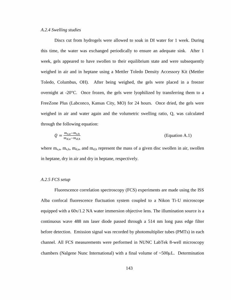
143
A.2.4 Swelling studies
Discs cut from hydrogels were allowed to soak in DI water for 1 week. During
this time, the water was exchanged periodically to ensure an adequate sink. After 1
week, gels appeared to have swollen to their equilibrium state and were subsequently
weighed in air and in heptane using a Mettler Toledo Density Accessory Kit (Mettler
Toledo, Columbus, OH). After being weighed, the gels were placed in a freezer
overnight at -20°C. Once frozen, the gels were lyophilized by transferring them to a
FreeZone Plus (Labconco, Kansas City, MO) for 24 hours. Once dried, the gels were
weighed in air and water again and the volumetric swelling ratio, Q, was calculated
through the following equation:
𝑄 =𝑚𝑠,𝑎−𝑚𝑠,ℎ
𝑚𝑑,𝑎−𝑚𝑑,ℎ (Equation A.1)
where ms,a, ms,h, md,a, and md,h represent the mass of a given disc swollen in air, swollen
in heptane, dry in air and dry in heptane, respectively.
A.2.5 FCS setup
Fluorescence correlation spectroscopy (FCS) experiments are made using the ISS
Alba confocal fluorescence fluctuation system coupled to a Nikon Ti-U microscope
equipped with a 60x/1.2 NA water immersion objective lens. The illumination source is a
continuous wave 488 nm laser diode passed through a 514 nm long pass edge filter
before detection. Emission signal was recorded by photomultiplier tubes (PMTs) in each
channel. All FCS measurements were performed in NUNC LabTek 8-well microscopy
chambers (Nalgene Nunc International) with a final volume of ~500L. Determination
144
of the focal volume was established via calibration against an aqueous solution of
Rhodamine 110 with known diffusion coefficient (D = 440 μm2 s
-1).(Gendron, Avaltroni
et al. 2008) Diffusion coefficients reported are calculated from an average of at least 10
FCS measurements, taken at various positions within the hydrogel, performed at room
temperature on a sample. Typical sampling times of ~30 seconds were used. All results
are expressed as mean values +/- the standard deviation of the recorded diffusion
coefficient. All FCS measurements were analyzed using VistaVision 4.0 Software.
A.2.6 FCS Data Analysis
As the basic principles of FCS have been described elsewhere,(Elson and Magde
1974, Hess, Huang et al. 2002) here we will give only a brief overview. FCS measures
the fluorescence fluctuations emitted from labelled molecules moving in and out of a
small confocal volume (~1 fL). The size of the effective illumination volume is fixed by
the confocal detection optics and the excitation profile of the focused laser beam and
characterized by measurements against a standard of a known diffusion constant. As
labeled particles diffuse through the detection volume, a signal I(t) is generated with
fluctuations around a mean value. The average intensity fluctuation is < 𝐼(𝑡) >= 𝐼(𝑡) −
𝛿𝐼(𝑡) where δI(t) donates the spontaneous deviation of the measured intensity around
the average intensity. For monodisperse particles diffusing by ideal Brownian motion,
dynamic information can be determined from the intensity fluctuations by means of a
time autocorrelation given by
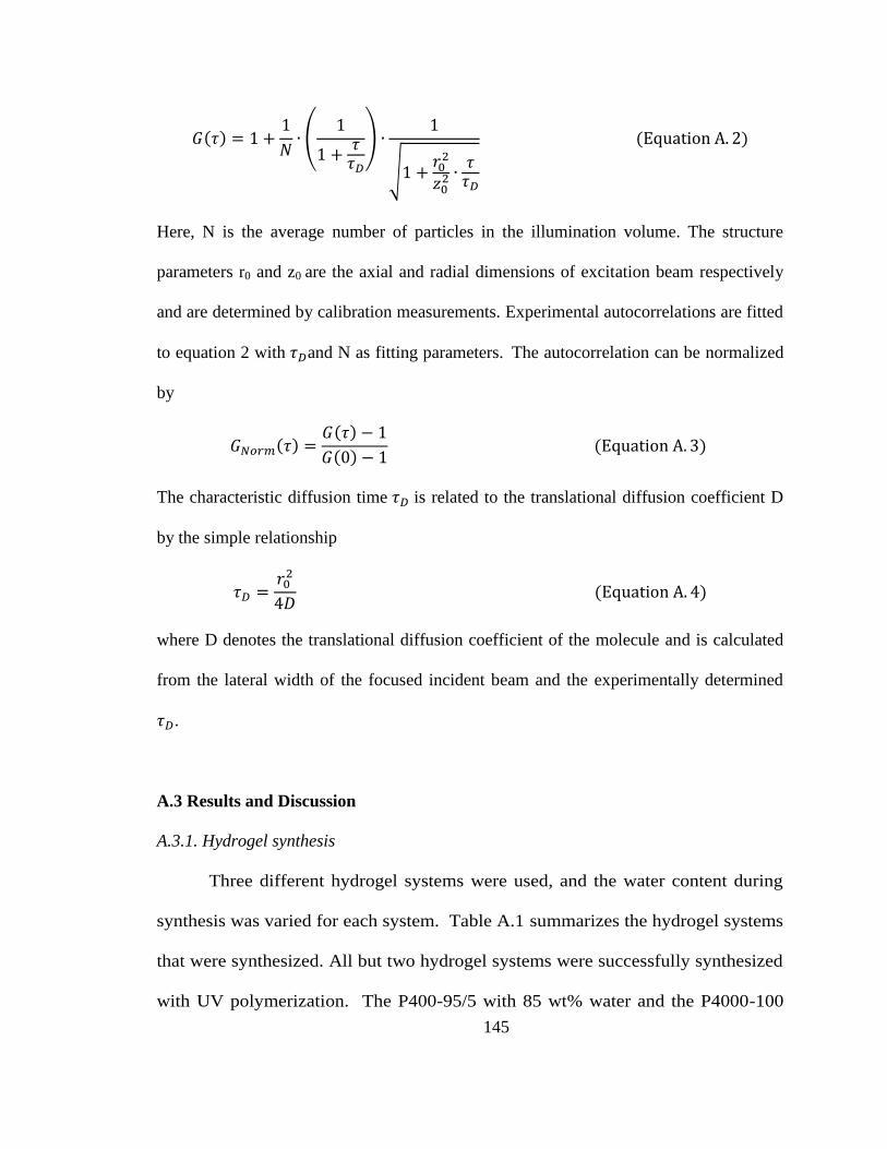
145
𝐺(𝜏) = 1 +1
𝑁∙ (
1
1 +𝜏
𝜏𝐷
) ∙1
√1 +𝑟0
2
𝑧02 ∙
𝜏𝜏𝐷
(Equation A. 2)
Here, N is the average number of particles in the illumination volume. The structure
parameters r0 and z0 are the axial and radial dimensions of excitation beam respectively
and are determined by calibration measurements. Experimental autocorrelations are fitted
to equation 2 with 𝜏𝐷and N as fitting parameters. The autocorrelation can be normalized
by
𝐺𝑁𝑜𝑟𝑚(𝜏) =𝐺(𝜏) − 1
𝐺(0) − 1 (Equation A. 3)
The characteristic diffusion time 𝜏𝐷 is related to the translational diffusion coefficient D
by the simple relationship
𝜏𝐷 =𝑟0
2
4𝐷 (Equation A. 4)
where D denotes the translational diffusion coefficient of the molecule and is calculated
from the lateral width of the focused incident beam and the experimentally determined
𝜏𝐷.
A.3 Results and Discussion
A.3.1. Hydrogel synthesis
Three different hydrogel systems were used, and the water content during
synthesis was varied for each system. Table A.1 summarizes the hydrogel systems
that were synthesized. All but two hydrogel systems were successfully synthesized
with UV polymerization. The P400-95/5 with 85 wt% water and the P4000-100
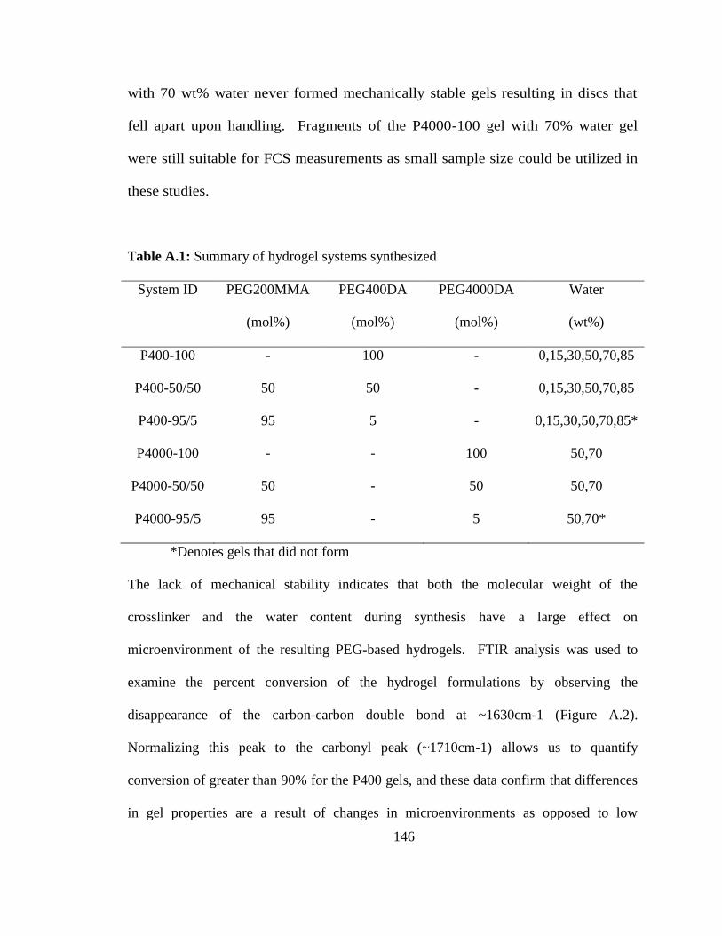
146
with 70 wt% water never formed mechanically stable gels resulting in discs that
fell apart upon handling. Fragments of the P4000-100 gel with 70% water gel
were still suitable for FCS measurements as small sample size could be utilized in
these studies.
Table A.1: Summary of hydrogel systems synthesized
System ID PEG200MMA
(mol%)
PEG400DA
(mol%)
PEG4000DA
(mol%)
Water
(wt%)
P400-100 - 100 - 0,15,30,50,70,85
P400-50/50 50 50 - 0,15,30,50,70,85
P400-95/5 95 5 - 0,15,30,50,70,85*
P4000-100 - - 100 50,70
P4000-50/50 50 - 50 50,70
P4000-95/5 95 - 5 50,70*
*Denotes gels that did not form
The lack of mechanical stability indicates that both the molecular weight of the
crosslinker and the water content during synthesis have a large effect on
microenvironment of the resulting PEG-based hydrogels. FTIR analysis was used to
examine the percent conversion of the hydrogel formulations by observing the
disappearance of the carbon-carbon double bond at ~1630cm-1 (Figure A.2).
Normalizing this peak to the carbonyl peak (~1710cm-1) allows us to quantify
conversion of greater than 90% for the P400 gels, and these data confirm that differences
in gel properties are a result of changes in microenvironments as opposed to low
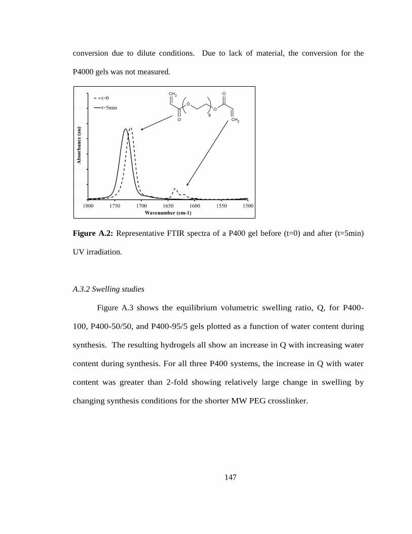
147
conversion due to dilute conditions. Due to lack of material, the conversion for the
P4000 gels was not measured.
Figure A.2: Representative FTIR spectra of a P400 gel before (t=0) and after (t=5min)
UV irradiation.
A.3.2 Swelling studies
Figure A.3 shows the equilibrium volumetric swelling ratio, Q, for P400-
100, P400-50/50, and P400-95/5 gels plotted as a function of water content during
synthesis. The resulting hydrogels all show an increase in Q with increasing water
content during synthesis. For all three P400 systems, the increase in Q with water
content was greater than 2-fold showing relatively large change in swelling by
changing synthesis conditions for the shorter MW PEG crosslinker.
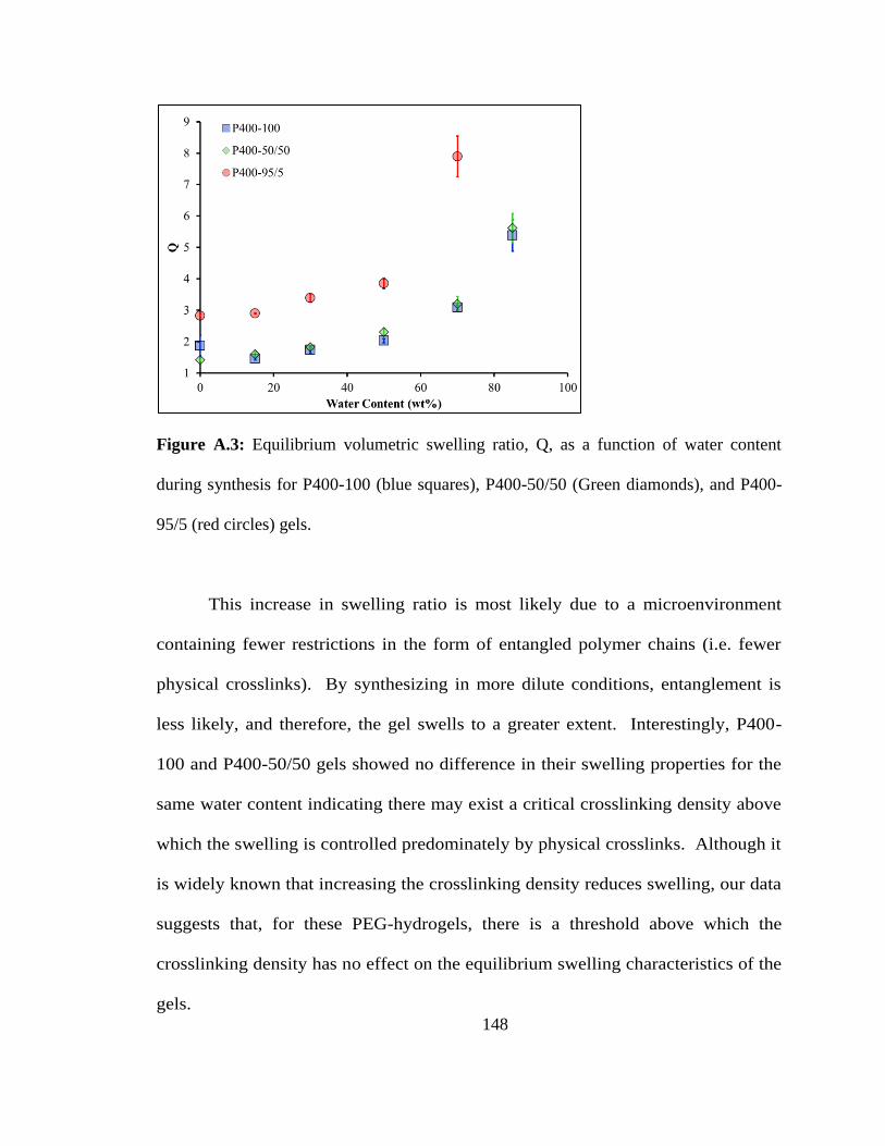
148
Figure A.3: Equilibrium volumetric swelling ratio, Q, as a function of water content
during synthesis for P400-100 (blue squares), P400-50/50 (Green diamonds), and P400-
95/5 (red circles) gels.
This increase in swelling ratio is most likely due to a microenvironment
containing fewer restrictions in the form of entangled polymer chains (i.e. fewer
physical crosslinks). By synthesizing in more dilute conditions, entanglement is
less likely, and therefore, the gel swells to a greater extent. Interestingly, P400-
100 and P400-50/50 gels showed no difference in their swelling properties for the
same water content indicating there may exist a critical crosslinking density above
which the swelling is controlled predominately by physical crosslinks. Although it
is widely known that increasing the crosslinking density reduces swelling, our data
suggests that, for these PEG-hydrogels, there is a threshold above which the
crosslinking density has no effect on the equilibrium swelling characteristics of the
gels.
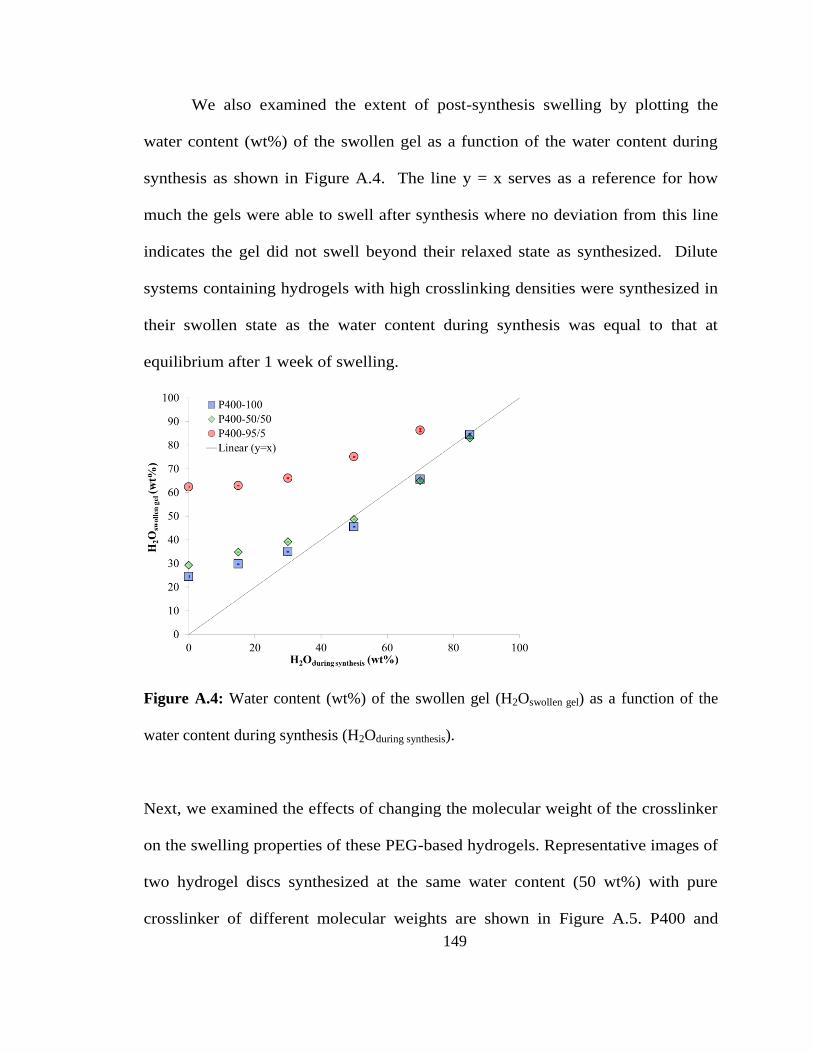
149
We also examined the extent of post-synthesis swelling by plotting the
water content (wt%) of the swollen gel as a function of the water content during
synthesis as shown in Figure A.4. The line y = x serves as a reference for how
much the gels were able to swell after synthesis where no deviation from this line
indicates the gel did not swell beyond their relaxed state as synthesized. Dilute
systems containing hydrogels with high crosslinking densities were synthesized in
their swollen state as the water content during synthesis was equal to that at
equilibrium after 1 week of swelling.
Figure A.4: Water content (wt%) of the swollen gel (H2Oswollen gel) as a function of the
water content during synthesis (H2Oduring synthesis).
Next, we examined the effects of changing the molecular weight of the crosslinker
on the swelling properties of these PEG-based hydrogels. Representative images of
two hydrogel discs synthesized at the same water content (50 wt%) with pure
crosslinker of different molecular weights are shown in Figure A.5. P400 and
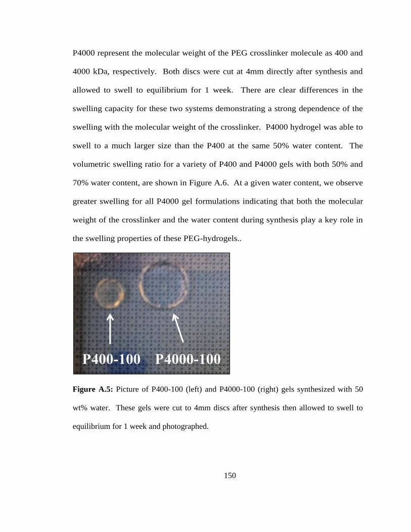
150
P4000 represent the molecular weight of the PEG crosslinker molecule as 400 and
4000 kDa, respectively. Both discs were cut at 4mm directly after synthesis and
allowed to swell to equilibrium for 1 week. There are clear differences in the
swelling capacity for these two systems demonstrating a strong dependence of the
swelling with the molecular weight of the crosslinker. P4000 hydrogel was able to
swell to a much larger size than the P400 at the same 50% water content. The
volumetric swelling ratio for a variety of P400 and P4000 gels with both 50% and
70% water content, are shown in Figure A.6. At a given water content, we observe
greater swelling for all P4000 gel formulations indicating that both the molecular
weight of the crosslinker and the water content during synthesis play a key role in
the swelling properties of these PEG-hydrogels..
Figure A.5: Picture of P400-100 (left) and P4000-100 (right) gels synthesized with 50
wt% water. These gels were cut to 4mm discs after synthesis then allowed to swell to
equilibrium for 1 week and photographed.
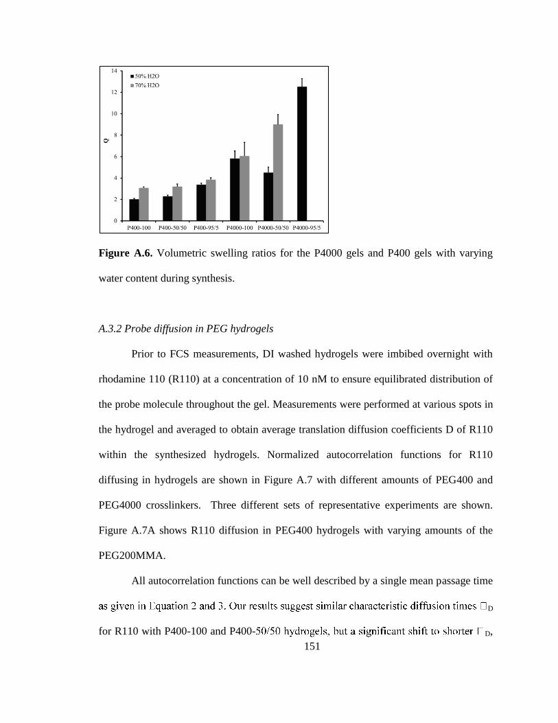
151
Figure A.6. Volumetric swelling ratios for the P4000 gels and P400 gels with varying
water content during synthesis.
A.3.2 Probe diffusion in PEG hydrogels
Prior to FCS measurements, DI washed hydrogels were imbibed overnight with
rhodamine 110 (R110) at a concentration of 10 nM to ensure equilibrated distribution of
the probe molecule throughout the gel. Measurements were performed at various spots in
the hydrogel and averaged to obtain average translation diffusion coefficients D of R110
within the synthesized hydrogels. Normalized autocorrelation functions for R110
diffusing in hydrogels are shown in Figure A.7 with different amounts of PEG400 and
PEG4000 crosslinkers. Three different sets of representative experiments are shown.
Figure A.7A shows R110 diffusion in PEG400 hydrogels with varying amounts of the
PEG200MMA.
All autocorrelation functions can be well described by a single mean passage time
D
for R110 with P400-100 and P400- D,
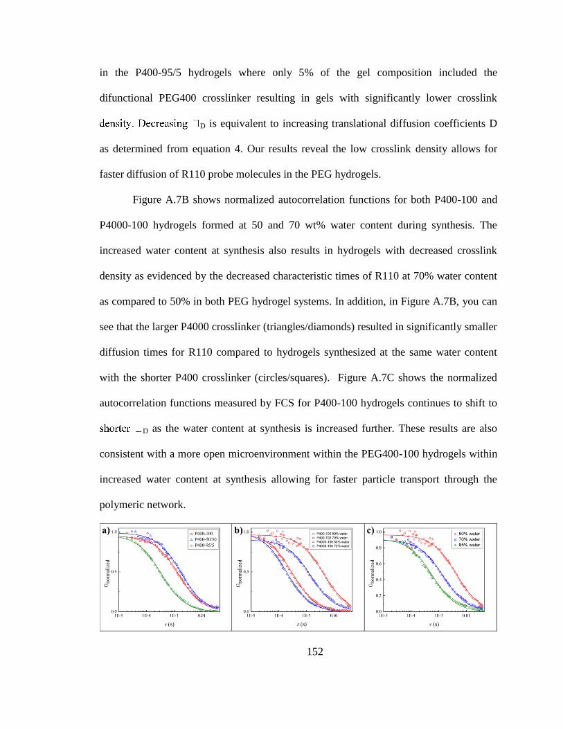
152
in the P400-95/5 hydrogels where only 5% of the gel composition included the
difunctional PEG400 crosslinker resulting in gels with significantly lower crosslink
D is equivalent to increasing translational diffusion coefficients D
as determined from equation 4. Our results reveal the low crosslink density allows for
faster diffusion of R110 probe molecules in the PEG hydrogels.
Figure A.7B shows normalized autocorrelation functions for both P400-100 and
P4000-100 hydrogels formed at 50 and 70 wt% water content during synthesis. The
increased water content at synthesis also results in hydrogels with decreased crosslink
density as evidenced by the decreased characteristic times of R110 at 70% water content
as compared to 50% in both PEG hydrogel systems. In addition, in Figure A.7B, you can
see that the larger P4000 crosslinker (triangles/diamonds) resulted in significantly smaller
diffusion times for R110 compared to hydrogels synthesized at the same water content
with the shorter P400 crosslinker (circles/squares). Figure A.7C shows the normalized
autocorrelation functions measured by FCS for P400-100 hydrogels continues to shift to
D as the water content at synthesis is increased further. These results are also
consistent with a more open microenvironment within the PEG400-100 hydrogels within
increased water content at synthesis allowing for faster particle transport through the
polymeric network.
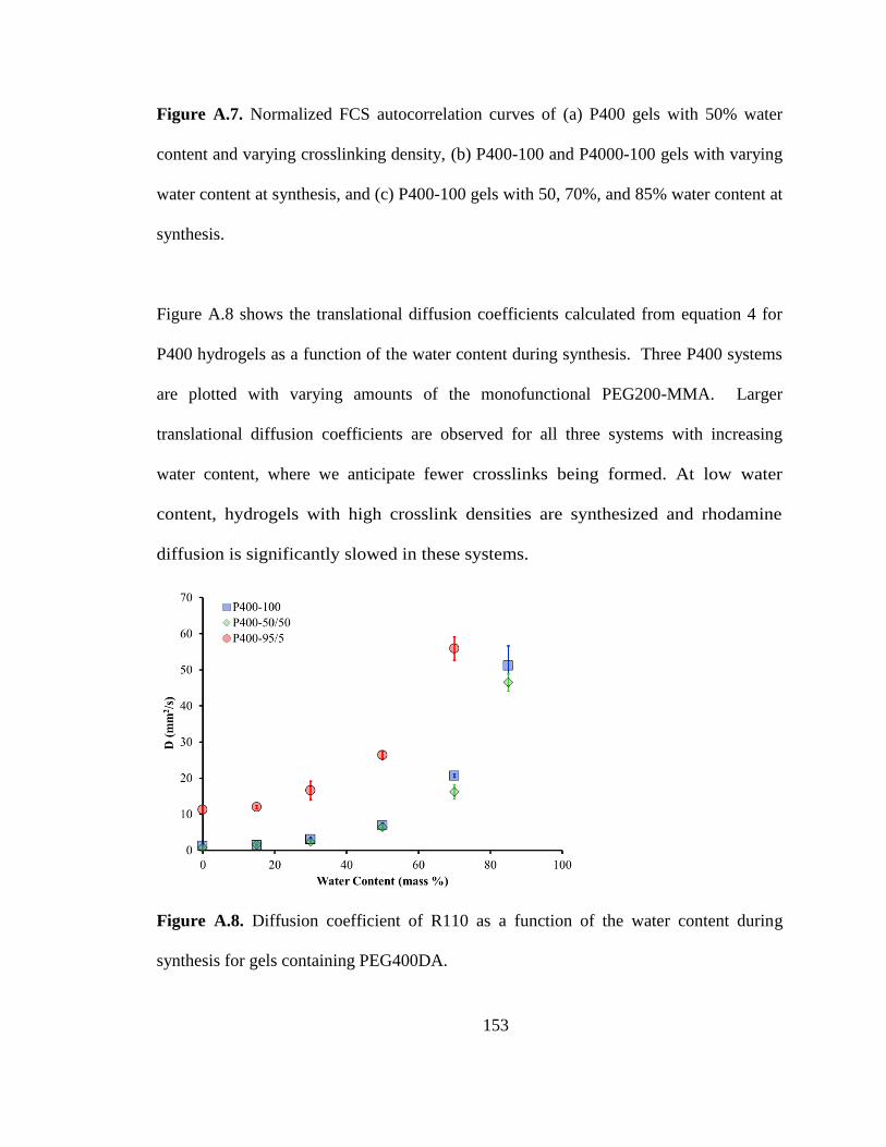
153
Figure A.7. Normalized FCS autocorrelation curves of (a) P400 gels with 50% water
content and varying crosslinking density, (b) P400-100 and P4000-100 gels with varying
water content at synthesis, and (c) P400-100 gels with 50, 70%, and 85% water content at
synthesis.
Figure A.8 shows the translational diffusion coefficients calculated from equation 4 for
P400 hydrogels as a function of the water content during synthesis. Three P400 systems
are plotted with varying amounts of the monofunctional PEG200-MMA. Larger
translational diffusion coefficients are observed for all three systems with increasing
water content, where we anticipate fewer crosslinks being formed. At low water
content, hydrogels with high crosslink densities are synthesized and rhodamine
diffusion is significantly slowed in these systems.
Figure A.8. Diffusion coefficient of R110 as a function of the water content during
synthesis for gels containing PEG400DA.
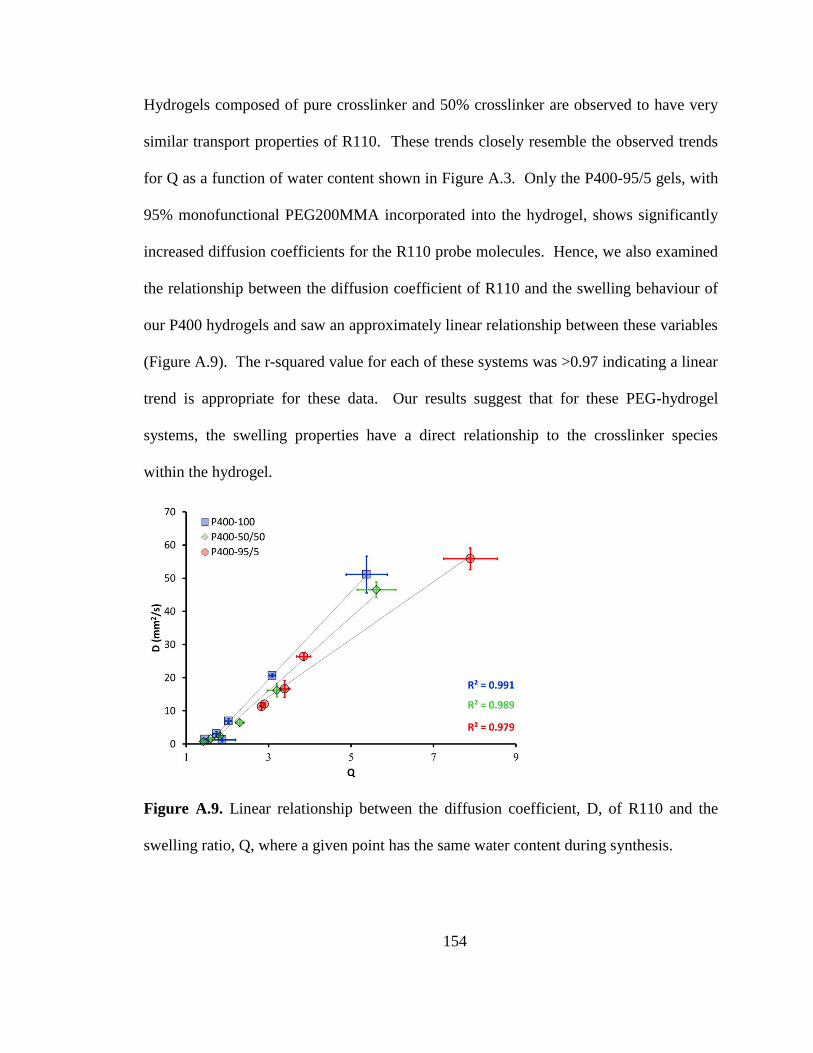
154
Hydrogels composed of pure crosslinker and 50% crosslinker are observed to have very
similar transport properties of R110. These trends closely resemble the observed trends
for Q as a function of water content shown in Figure A.3. Only the P400-95/5 gels, with
95% monofunctional PEG200MMA incorporated into the hydrogel, shows significantly
increased diffusion coefficients for the R110 probe molecules. Hence, we also examined
the relationship between the diffusion coefficient of R110 and the swelling behaviour of
our P400 hydrogels and saw an approximately linear relationship between these variables
(Figure A.9). The r-squared value for each of these systems was >0.97 indicating a linear
trend is appropriate for these data. Our results suggest that for these PEG-hydrogel
systems, the swelling properties have a direct relationship to the crosslinker species
within the hydrogel.
Figure A.9. Linear relationship between the diffusion coefficient, D, of R110 and the
swelling ratio, Q, where a given point has the same water content during synthesis.
155
Lastly, the translational diffusion coefficients of R110 in various P400 and P4000
gels at 50% and 70% water content at synthesis were determined and are plotted in
Figure A.10. These data illustrate that probe molecule transport in P4000 has a
dramatically larger diffusion coefficient when compared to the corresponding P400
gel due presumably to larger gel correlation lengths formed when using the longer
PEG crosslinker. As with P400 gels, R110 displayed almost identical translational
diffusion coefficients in the P4000-100 and P4000-50/50 gels suggesting that there
may exist a critical crosslinking density above which physical crosslinks dominate
the effects of the microenvironment on the transport of small molecules.
Interestingly, comparing the P400 and P4000 gels with high crosslinking density
(50 or 100%) we see that the ratio of molecular weight between the crosslinkers
(MWPEG400DA:MWPEG4000DA), 10.0, was almost the same as the ratio of
diffusion coefficients (DPEG400DA: DPEG4000DA), which was 11.2±2.4 and
9.7±0.7 for the systems with 50 and 100% crosslinking density, respectively. To
our knowledge, this is the first report of FCS being used to directly correlate the
diffusion coefficient of a molecular probe with the molecular weight of a
crosslinker. Control of the properties of synthesized hydrogels is critical to the
engineering of these biocompatible materials for various applications. Taken
together, our results suggest controlling the swelling ratio through molecular
weight of the crosslinks or varying water content at synthesis are viable means to
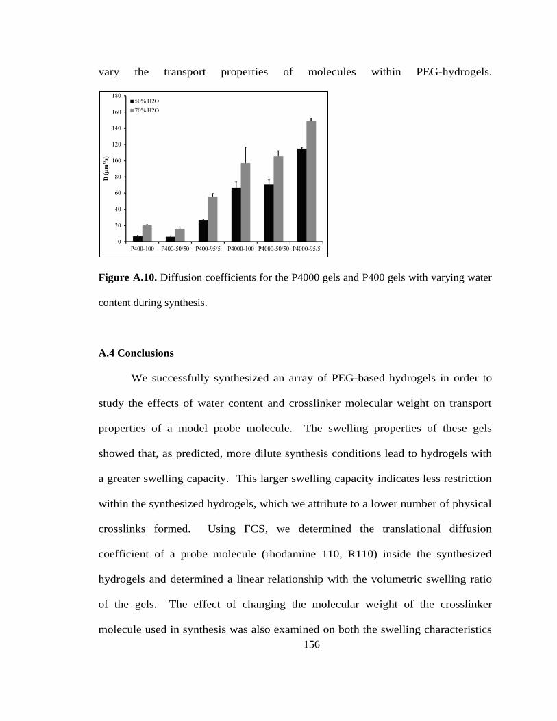
156
vary the transport properties of molecules within PEG-hydrogels.
Figure A.10. Diffusion coefficients for the P4000 gels and P400 gels with varying water
content during synthesis.
A.4 Conclusions
We successfully synthesized an array of PEG-based hydrogels in order to
study the effects of water content and crosslinker molecular weight on transport
properties of a model probe molecule. The swelling properties of these gels
showed that, as predicted, more dilute synthesis conditions lead to hydrogels with
a greater swelling capacity. This larger swelling capacity indicates less restriction
within the synthesized hydrogels, which we attribute to a lower number of physical
crosslinks formed. Using FCS, we determined the translational diffusion
coefficient of a probe molecule (rhodamine 110, R110) inside the synthesized
hydrogels and determined a linear relationship with the volumetric swelling ratio
of the gels. The effect of changing the molecular weight of the crosslinker
molecule used in synthesis was also examined on both the swelling characteristics
157
of the gel and transport of R110 inside the formed hydrogels. For all cases, P4000
hydrogels displayed greater swelling capacity and increased transport properties of
R110 as compared to the corresponding P400 gels. These results illustrate that the
water content during synthesis and the molecular weight of the crosslinker have a
dramatic impact on the microenvironment of these hydrogels and have significant
implications for engineering tunable hydrogels for specific biomedical
applications.
Acknowledgements:
NAS acknowledges the financial support from a NCI-CNTC pre-doctoral
traineeship and XZ acknowledge financial support from the University of Kentucky
Research Challenge Trust Fund (RCTF) Fellowship for Biochemistry. The project
described was supported by Grant Number 5R25CA153954 from the National Cancer
Institute and start-up funds from the University of Kentucky.
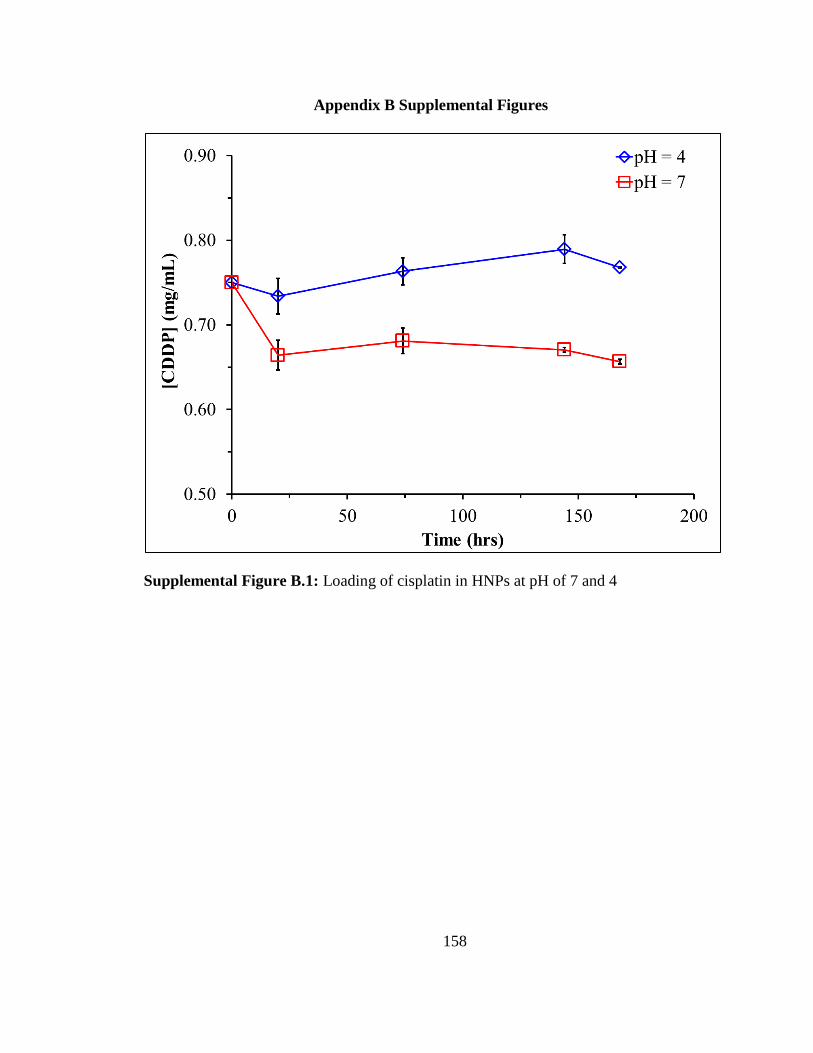
158
Appendix B Supplemental Figures
Supplemental Figure B.1: Loading of cisplatin in HNPs at pH of 7 and 4
159
References
(1984). "ANALYTICAL PROFILES OF DRUG SUBSTANCES - FOLEY,K." Journal
of the American Chemical Society 106(4): 1173-1173.
(2006). <601> Aerosols, Nasal Sprays, Metered-Dose Inhalers, and Dry Powder Inhalers
Monograph. USP 29-NF 24 The United States Pharmacopoeia and The National
Formulary: The Official Compendia of Standards. Rockville, MD, The United States
Pharmacopeial Convention, Inc. 29/24: 2617-2636.
(!!! INVALID CITATION !!!).
Ally, J., A. Amirfazli and W. Roa (2006). "Factors affecting magnetic retention of
particles in the upper airways: An in vitro and ex vivo study." Journal of Aerosol
Medicine-Deposition Clearance and Effects in the Lung 19(4): 491-509.
Ally, J., B. Martin, M. B. Khamesee, W. Roa and A. Amirfazli (2005). "Magnetic
targeting of aerosol particles for cancer therapy." Journal of Magnetism and Magnetic
Materials 293(1): 442-449.
Andre, F. and C. C. Zielinski (2012). "Optimal strategies for the treatment of metastatic
triple-negative breast cancer with currently approved agents." Annals of Oncology 23:
46-51.
Anselmo, A. C. and S. Mitragotri (2014). "An overview of clinical and commercial
impact of drug delivery systems." Journal of Controlled Release 190: 15-28.
Anthony J., H. and H. M. Mansour (2009). Delivery of Drugs by the Pulmonary Route.
Modern Pharmaceutics Volume 2: Applications and Advances. A. Florence and J.
Siepmann. New York, Taylor & Francis. 2: 191-219.
Arenberg, D. (2011). "Bronchioloalveolar Carcinoma." Seminars in Respiratory and
Critical Care Medicine 32(1): 52-61.
Azarmi, S., W. H. Roa and R. Loebenberg (2008). "Targeted delivery of nanoparticles for
the treatment of lung diseases." Advanced Drug Delivery Reviews 60(8): 863-875.
Azarmi, S., X. Tao, H. Chen, Z. L. Wang, W. H. Finlay, R. Lobenberg and W. H. Roa
(2006). "Formulation and cytotoxicity of doxorubicin nanoparticles carried by dry
powder aerosol particles." International Journal of Pharmaceutics 319(1-2): 155-161.
Babincova, M., V. Altanerova, C. Altaner, C. Bergemann and P. Babinec (2008). "In
vitro analysis of cisplatin functionalized magnetic nanoparticles in combined cancer
chemotherapy and electromagnetic hyperthermia." Ieee Transactions on Nanobioscience
7(1): 15-19.
160
Baeza, A., M. Colilla and M. Vallet-Regi (2015). "Advances in mesoporous silica
nanoparticles for targeted stimuli-responsive drug delivery." Expert Opinion on Drug
Delivery 12(2): 319-337.
Bates, R. C., N. S. Edwards and J. D. Yates (2000). "Spheroids and cell survival."
Critical Reviews in Oncology Hematology 36(2-3): 61-74.
Bonomi, M., S. Pilotto, M. Milella, F. Massari, S. Cingarlini, M. Brunelli, M. Chilosi, G.
Tortora and E. Bria (2011). "Adjuvant chemotherapy for resected non-small-cell lung
cancer: future perspectives for clinical research." Journal of Experimental & Clinical
Cancer Research 30.
Brannon-Peppas, L. and J. O. Blanchette (2004). "Nanoparticle and targeted systems for
cancer therapy." Advanced Drug Delivery Reviews 56(11): 1649-1659.
Burger, A., J. O. Henck, S. Hetz, J. M. Rollinger, A. A. Weissnicht and H. Stottner
(2000). "Energy/temperature diagram and compression behavior of the polymorphs of D-
mannitol." Journal of Pharmaceutical Sciences 89(4): 457-468.
Carpenter, J. F., M. J. Pikal, B. S. Chang and T. W. Randolph (1997). "Rational design of
stable lyophilized protein formulations: Some practical advice." Pharmaceutical Research
14(8): 969-975.
Carvalho, T. C., S. R. Carvalho and J. T. McConville (2011). "Formulations for
Pulmonary Administration of Anticancer Agents to Treat Lung Malignancies." Journal of
Aerosol Medicine and Pulmonary Drug Delivery 24(2): 61-80.
Chen, M. C., K. Sonaje, K. J. Chen and H. W. Sung (2011). "A review of the prospects
for polymeric nanoparticle platforms in oral insulin delivery." Biomaterials 32(36): 9826-
9838.
Cho, K. J., X. Wang, S. M. Nie, Z. Chen and D. M. Shin (2008). "Therapeutic
nanoparticles for drug delivery in cancer." Clinical Cancer Research 14(5): 1310-1316.
Cho, W. S., M. J. Cho, S. R. Kim, M. Choi, J. Y. Lee, B. S. Han, S. N. Park, M. K. Yu, S.
Jon and J. Jeong (2009). "Pulmonary toxicity and kinetic study of Cy5.5-conjugated
superparamagnetic iron oxide nanoparticles by optical imaging." Toxicology and Applied
Pharmacology 239(1): 106-115.
Choi, S. J., J. M. Oh and J. H. Choy (2009). "Toxicological effects of inorganic
nanoparticles on human lung cancer A549 cells." Journal of Inorganic Biochemistry
103(3): 463-471.
Chou, A. J., M. D. Bell, C. Mackinson, R. Gupta, P. A. Meyers and R. Gorlick (2007).
Phase Ib/IIa study of sustained release lipid inhalation targeting cisplatin by inhalation in
the treatment of patients with relapsed/progressive osteosarcoma metastatic to the lung.
Journal of Clinical Oncology, American Society of Clinical Oncology. 25: 9525.
161
Chow, A. H. L., H. H. Y. Tong, P. Chattopadhyay and B. Y. Shekunov (2007). "Particle
engineering for pulmonary drug delivery." Pharmaceutical Research 24(3): 411-437.
Colombo, M., S. Carregal-Romero, M. F. Casula, L. Gutierrez, M. P. Morales, I. B.
Bohm, J. T. Heverhagen, D. Prosperi and W. J. Parak (2012). "Biological applications of
magnetic nanoparticles." Chemical Society Reviews 41(11): 4306-4334.
Cone, R. A. (2009). "Barrier properties of mucus." Advanced Drug Delivery Reviews
61(2): 75-85.
Coto-Garcia, A. M., E. Sotelo-Gonzalez, M. Fernandez-Arguelles, R. Pereiro, J. M.
Costa-Fernandez and A. Sanz-Medel (2011). "Nanoparticles as fluorescent labels for
optical imaging and sensing in genomics and proteomics." Analytical and Bioanalytical
Chemistry 399(1): 29-42.
Courrier, H. M., N. Butz and T. F. Vandamme (2002). "Pulmonary drug delivery
systems: Recent developments and prospects." Critical Reviews in Therapeutic Drug
Carrier Systems 19(4-5): 425-498.
Crino, L., W. Weder, J. van Meerbeeck, E. Felip and E. G. W. Grp (2010). "Early stage
and locally advanced (non-metastatic) non-small-cell lung cancer: ESMO Clinical
Practice Guidelines for diagnosis, treatment and follow-up." Annals of Oncology 21:
v103-v115.
Dames, P., B. Gleich, A. Flemmer, K. Hajek, N. Seidl, F. Wiekhorst, D. Eberbeck, I.
Bittmann, C. Bergemann, T. Weyh, L. Trahms, J. Rosenecker and C. Rudolph (2007).
"Targeted delivery of magnetic aerosol droplets to the lung." Nature Nanotechnology
2(8): 495-499.
Daniher, D. I. and J. Zhu (2008). "Dry powder platform for pulmonary drug delivery."
Particuology 6(4): 225-238.
De, S. K., N. R. Aluru, B. Johnson, W. C. Crone, D. J. Beebe and J. Moore (2002).
"Equilibrium swelling and kinetics of pH-responsive hydrogels: Models, experiments,
and simulations." Journal of Microelectromechanical Systems 11(5): 544-555.
DeSantis, C., J. M. Ma, L. Bryan and A. Jemal (2014). "Breast Cancer Statistics, 2013."
Ca-a Cancer Journal for Clinicians 64(1): 52-62.
Dobson, J. (2006). "Magnetic micro- and nano-particle-based targeting for drug and gene
delivery." Nanomedicine 1(1): 31-37.
Dolovich, M. B., R. C. Ahrens, D. R. Hess, P. Anderson, R. Dhand, J. L. Rau, G. C.
Smaldone and G. Guyatt (2005). "Device selection and outcomes of aerosol therapy:
Evidence-based guidelines." Chest 127(1): 335-371.
162
Driscoll, C. F., R. M. Morris, A. E. Senyei, K. J. Widder and G. S. Heller (1984).
"MAGNETIC TARGETING OF MICROSPHERES IN BLOOD-FLOW." Microvascular
Research 27(3): 353-369.
Du, J., I. M. El-Sherbiny and H. D. Smyth (2014). "Swellable Ciprofloxacin-Loaded
Nano-in-Micro Hydrogel Particles for Local Lung Drug Delivery." Aaps Pharmscitech
15(6): 1535-1544.
Eberhard, D. A., B. E. Johnson, L. C. Amler, A. D. Goddard, S. L. Heldens, R. S. Herbst,
W. L. Ince, P. A. Janne, T. Januario, D. H. Johnson, P. Klein, V. A. Miller, M. A.
Ostland, D. A. Ramies, D. Sebisanovic, J. A. Stinson, Y. R. Zhang, S. Seshagiri and K. J.
Hillan (2005). "Mutations in the epidermal growth factor receptor and in KRAS are
predictive and prognostic indicators in patients with non-small-cell lung cancer treated
with chemotherapy alone and in combination with erlotinib." Journal of Clinical
Oncology 23(25): 5900-5909.
El-Gendy, N. and C. Berkland (2009). "Combination Chemotherapeutic Dry Powder
Aerosols via Controlled Nanoparticle Agglomeration." Pharmaceutical Research 26(7):
1752-1763.
El-Sherbiny, I. M. and H. D. C. Smyth (2010). "Novel cryomilled physically cross-linked
biodegradable hydrogel microparticles as carriers for inhalation therapy." Journal of
Microencapsulation 27(8): 657-668.
Elson, E. L. and D. Magde (1974). "Fluorescence Correlation Spectroscopy. 1.
Conceptual Basis and Theory." Biopolymers 13(1): 1-27.
Estrella, V., T. Chen, M. Lloyd, J. Wojtkowiak, H. H. Cornnell, A. Ibrahim-Hashim, K.
Bailey, Y. Balagurunathan, J. M. Rothberg, B. F. Sloane, J. Johnson, R. A. Gatenby and
R. J. Gillies (2013). "Acidity Generated by the Tumor Microenvironment Drives Local
Invasion." Cancer Research 73(5): 1524-1535.
Farhat, A., Y. Holloway, T. Jones, S. Taylor, S. Britland and D. Eagland (2009).
"Towards improved pulmonary delivery of budesonide using a nebulisable
nanoparticulate hydrogel." Journal of Pharmacy and Pharmacology 61: A43-A44.
Fathi, A. and J. Brahmer (2008). "Chemotherapy for Advanced Stage Non-Small Cell
Lung Cancer." Seminars in thoracic and cardiovascular surgery 20: 210-216.
Ferlay, J., I. Soerjomataram, M. Ervik, R. Dikshit, S. Eser, C. Mathers, M. Rebelo, D.
Parkin, D. Forman and F. Bray. (2013). "GLOBOCAN 2012 v1.0, Cancer Incidence and
Mortality Worldwide: IARC CancerBase No. 11 [Internet]." Retrieved 6/30/14, 2014,
from http://globocan.iarc.fr.
Ferrari, M. (2005). "Cancer nanotechnology: Opportunities and challenges." Nature
Reviews Cancer 5(3): 161-171.
163
Finlay, W. (2008). "The ARLA Respiratory Deposition Calculator." Retrieved 2008,
from http://www.mece.ualberta.ca/arla/impactor_mmad_calculator.html.
Frimpong, R. A., J. Dou, M. Pechan and J. Z. Hilt (2010). "Enhancing remote controlled
heating characteristics in hydrophilic magnetite nanoparticles via facile co-precipitation."
Journal of Magnetism and Magnetic Materials 322(3): 326-331.
Frimpong, R. A., S. Fraser and J. Z. Hilt (2007). "Synthesis and temperature response
analysis of magnetic-hydrogel nanocomposites." Journal of Biomedical Materials
Research Part A 80A(1): 1-6.
Frimpong, R. A. and J. Z. Hilt (2010). "Magnetic nanoparticles in biomedicine: synthesis,
functionalization and applications." Nanomedicine 5(9): 1401-1414.
Fronczek, F. R., H. N. Kamel and M. Slattery (2003). "Three polymorphs (alpha, beta
and delta) of D-mannitol at 100 K." Acta Crystallographica Section C-Crystal Structure
Communications 59: O567-O570.
Galluzzi, L., M. C. Maiuri, I. Vitale, H. Zischka, M. Castedo, L. Zitvogel and G.
Kroemer (2007). "Cell death modalities: classification and pathophysiological
implications." Cell Death and Differentiation 14(7): 1237-1243.
Ganapathy, V., N. Gupta and R. Martindale (2006). Protein digestion and absorption.
Physiology of the gastrointestinal tract, Burlington: Academic Press. II: 1667-1692.
Gatzemeier, U., A. Pluzanska, A. Szczesna, E. Kaukel, J. Roubec, F. De Rosa, J.
Milanowski, H. Karnicka-Mlodkowski, M. Pesek, P. Serwatowski, R. Ramlau, T.
Janaskova, J. Vansteenkiste, J. Strausz, G. M. Manikhas and J. Von Pawel (2007). "Phase
III study of erlotinib in combination with cisplatin and gemcitabine in advanced non-
small-cell lung cancer: The Tarceva Lung Cancer Investigation Trial." Journal of Clinical
Oncology 25(12): 1545-1552.
Gendron, P. O., F. Avaltroni and K. J. Wilkinson (2008). "Diffusion Coefficients of
Several Rhodamine Derivatives as Determined by Pulsed Field Gradient-Nuclear
Magnetic Resonance and Fluorescence Correlation Spectroscopy." Journal of
Fluorescence 18(6): 1093-1101.
Gerratana, L., V. Fanotto, M. Bonotto, S. Bolzonello, A. M. Minisini, G. Fasola and F.
Puglisi (2015). "Pattern of metastasis and outcome in patients with breast cancer."
Clinical & Experimental Metastasis 32(2): 125-133.
Ghandehari, H., P. Kopeckova and J. Kopecek (1997). "In vitro degradation of pH-
sensitive hydrogels containing aromatic azo bonds." Biomaterials 18(12): 861-872.
Gjoerup, J., O. Hilberg and E. Bendstrup (2012). "Inhaled mannitol in the treatment of
non-cystic fibrosis bronchiectasis in adults." Respirology 17(6): 927-932.
164
Gupta, P., K. Vermani and S. Garg (2002). "Hydrogels: from controlled release to pH-
responsive drug delivery." Drug Discovery Today 7(10): 569-579.
Hamidi, M., A. Azadi and P. Rafiei (2008). "Hydrogel nanoparticles in drug delivery."
Advanced Drug Delivery Reviews 60(15): 1638-1649.
Hao, R., R. J. Xing, Z. C. Xu, Y. L. Hou, S. Gao and S. H. Sun (2010). "Synthesis,
Functionalization, and Biomedical Applications of Multifunctional Magnetic
Nanoparticles." Advanced Materials 22(25): 2729-2742.
Hasenpusch, G., J. Geiger, K. Wagner, O. Mykhaylyk, F. Wiekhorst, L. Trahms, A.
Heidsieck, B. Gleich, C. Bergemann, M. K. Aneja and C. Rudolph (2012). "Magnetized
Aerosols Comprising Superparamagnetic Iron Oxide Nanoparticles Improve Targeted
Drug and Gene Delivery to the Lung." Pharmaceutical Research 29(5): 1308-1318.
Hawkins, A. M., C. E. Bottom, Z. Liang, D. A. Puleo and J. Z. Hilt (2012). "Magnetic
Nanocomposite Sol-Gel Systems for Remote Controlled Drug Release." Advanced
Healthcare Materials 1(1): 96-100.
Hawkins, A. M., D. A. Puleo and J. Z. Hilt (2011). "Effect of Macromer Synthesis Time
on the Properties of the Resulting Poly(beta-amino ester) Degradable Hydrogel." Journal
of Applied Polymer Science 122(2): 1420-1426.
.
Hawkins, A. M., M. E. Tolbert, B. Newton, T. A. Milbrandt, D. A. Puleo and J. Z. Hilt
(2013). "Tuning biodegradable hydrogel properties via synthesis procedure." Polymer
54(17): 4422-4426.
He, H. N., A. David, B. Chertok, A. Cole, K. Lee, J. Zhang, J. X. Wang, Y. Z. Huang and
V. C. Yang (2013). "Magnetic Nanoparticles for Tumor Imaging and Therapy: A So-
Called Theranostic System." Pharmaceutical Research 30(10): 2445-2458.
Health, N. I. o. (2015). "Estimates of Funding for Various Research, Condition, and
Disease Categories (RCDC)." Retrieved 4/22/15, 2015, from
http://report.nih.gov/categorical_spending.aspx.
Hess, S. T., S. Huang, A. A. Heikal and W. W. Webb (2002). "Biological and chemical
applications of fluorescence correlation spectroscopy: a review." Biochemistry 41(3):
697-705.
Hickey, A. J., H. M. Mansour, M. J. Telko, Z. Xu, H. D. C. Smyth, T. Mulder, R.
McLean, J. Langridge and D. Papadopoulos (2007). "Physical characterization of
component particles included in dry powder inhalers. I. Strategy review and static
characteristics." Journal of Pharmaceutical Sciences 96(5): 1282-1301.
165
Hirte, H. W. (2013). "Profile of erlotinib and its potential in the treatment of advanced
ovarian carcinoma." Oncotargets and Therapy 6.
Horsman, M. R. and J. Overgaard (2007). "Hyperthermia: a potent enhancer of
radiotherapy." Clinical Oncology 19(6): 418-426.
Hudis, C. A. and L. Gianni (2011). "Triple-Negative Breast Cancer: An Unmet Medical
Need." Oncologist 16: 1-11.
Iacono, D., R. Chiari, G. Metro, C. Bennati, G. Bellezza, M. Cenci, B. Ricciuti, A.
Sidoni, S. Baglivo, V. Minotti and L. Crino (2015). "Future options for ALK-positive
non-small cell lung cancer." Lung Cancer 87(3): 211-219.
Institute, N. C. (2015). "Non-Small Cell Lung Cancer Treatment (PDQ®)." Retrieved 4-
21-15, 2015, from http://www.cancer.gov/cancertopics/pdq/treatment/non-small-cell-
lung/Patient.
Issels, R. D. (2008). "Hyperthermia adds to chemotherapy." European Journal of Cancer
44(17): 2546-2554.
Jackman, D. M., L. R. Chirieac and P. A. Janne (2005). "Bronchioloalveolar carcinoma:
A review of the epidemiology, pathology, and treatment." Seminars in Respiratory and
Critical Care Medicine 26(3): 342-352.
Johannsen, M., B. Thiesen, P. Wust and A. Jordan (2010). "Magnetic nanoparticle
hyperthermia for prostate cancer." International Journal of Hyperthermia 26(8): 790-795.
Jordan, A., R. Scholz, K. Maier-Hauff, M. Johannsen, P. Wust, J. Nadobny, H. Schirra,
H. Schmidt, S. Deger, S. Loening, W. Lanksch and R. Felix (2001). "Presentation of a
new magnetic field therapy system for the treatment of human solid tumors with
magnetic fluid hyperthermia." Journal of Magnetism and Magnetic Materials 225(1-2):
118-126.
Jordan, A., R. Scholz, K. Maier-Hauff, F. K. H. van Landeghem, N. Waldoefner, U.
Teichgraeber, J. Pinkernelle, H. Bruhn, F. Neumann, B. Thiesen, A. von Deimling and R.
Felix (2006). "The effect of thermotherapy using magnetic nanoparticles on rat malignant
glioma." Journal of Neuro-Oncology 78(1): 7-14.
Jordan, A., R. Scholz, P. Wust, H. Fahling and R. Felix (1999). "Magnetic fluid
hyperthermia (MFH): Cancer treatment with AC magnetic field induced excitation of
biocompatible superparamagnetic nanoparticles." Journal of Magnetism and Magnetic
Materials 201: 413-419.
Kanthala, S., S. Pallerla and S. Jois (2015). "Current and future targeted therapies for
non-small-cell lung cancers with aberrant EGF receptors." Future Oncology 11(5): 865-
878.
166
Kassam, F., K. Enright, R. Dent, G. Dranitsaris, J. Myers, C. Flynn, M. Fralick, R.
Kumar and M. Clemons (2009). "Survival Outcomes for Patients with Metastatic Triple-
Negative Breast Cancer: Implications for Clinical Practice and Trial Design." Clinical
Breast Cancer 9(1): 29-33.
Kau, P., G. M. Nagaraja, H. Zheng, D. Gizachew, M. Galukande, S. Krishnan and A.
Asea (2012). "A mouse model for triple-negative breast cancer tumor-initiating cells
(TNBC-TICs) exhibits similar aggressive phenotype to the human disease." Bmc Cancer
12.
Knecht, L. D., N. Ali, Y. N. Wei, J. Z. Hilt and S. Daunert (2012). "Nanoparticle-
Mediated Remote Control of Enzymatic Activity." Acs Nano 6(10): 9079-9086.
Kondo, M. and S. Adachi (2013). "Optical Properties of NaCl:Sn2+ Phosphor
Synthesized from Aqueous NaCl/SnCl2/HCl Solution." Ecs Journal of Solid State
Science and Technology 2(1): R9-R15.
Kopecek, J. (2009). "Hydrogels: From Soft Contact Lenses and Implants to Self-
Assembled Nanomaterials." Journal of Polymer Science Part a-Polymer Chemistry
47(22): 5929-5946.
Kruse, A. M., S. A. Meenach, K. W. Anderson and J. Z. Hilt (2014). "Synthesis and
characterization of CREKA-conjugated iron oxide nanoparticles for hyperthermia
applications." Acta Biomaterialia 10(6): 2622-2629.
Kunz-Schughart, L. A., M. Kreutz and R. Knuechel (1998). "Multicellular spheroids: a
three-dimensional in vitro culture system to study tumour biology." International Journal
of Experimental Pathology 79(1): 1-23.
Kwon, J. T., D. S. Kim, A. Minai-Tehrani, S. K. Hwang, S. H. Chang, E. S. Lee, C. X.
Xu, H. T. Lim, J. E. Kim, B. I. Yoon, G. H. An, K. H. Lee, J. K. Lee and M. H. Cho
(2009). "Inhaled Fluorescent Magnetic Nanoparticles Induced Extramedullary
Hematopoiesis in the Spleen of Mice." Journal of Occupational Health 51(5): 423-431.
Lai, S. K., D. E. O'Hanlon, S. Harrold, S. T. Man, Y. Y. Wang, R. Cone and J. Hanes
(2007). "Rapid transport of large polymeric nanoparticles in fresh undiluted human
mucus." Proceedings of the National Academy of Sciences of the United States of
America 104(5): 1482-1487.
Lai, S. K., Y. Y. Wang and J. Hanes (2009). "Mucus-penetrating nanoparticles for drug
and gene delivery to mucosal tissues." Advanced Drug Delivery Reviews 61(2): 158-171.
Lai, S. K., Y. Y. Wang, D. Wirtz and J. Hanes (2009). "Micro- and macrorheology of
mucus." Advanced Drug Delivery Reviews 61(2): 86-100.
Lee, J., C. Lee, T. H. Kim, E. S. Lee, B. S. Shin, S. C. Chi, E. S. Park, K. C. Lee and Y.
S. Youn (2012). "Self-assembled glycol chitosan nanogels containing palmityl-acylated
167
exendin-4 peptide as a long-acting anti-diabetic inhalation system." Journal of Controlled
Release 161(3): 728-734.
Li, L., T. L. M. ten Hagen, M. Hossann, R. Suss, G. C. van Rhoon, A. M. M. Eggermont,
D. Haemmerich and G. A. Koning (2013). "Mild hyperthermia triggered doxorubicin
release from optimized stealth thermosensitive liposomes improves intratumoral drug
delivery and efficacy." Journal of Controlled Release 168(2): 142-150.
Li, T. H., H. J. Kung, P. C. Mack and D. R. Gandara (2013). "Genotyping and Genomic
Profiling of Non-Small-Cell Lung Cancer: Implications for Current and Future
Therapies." Journal of Clinical Oncology 31(8): 1039-1049.
Li, X. J. and H. M. Mansour (2011). "Physicochemical Characterization and Water Vapor
Sorption of Organic Solution Advanced Spray-Dried Inhalable Trehalose Microparticles
and Nanoparticles for Targeted Dry Powder Pulmonary Inhalation Delivery." Aaps
Pharmscitech 12(4): 1420-1430.
Li, Z. H., R. J. Zhao, X. H. Wu, Y. Sun, M. Yao, J. J. Li, Y. H. Xu and J. R. Gu (2005).
"Identification and characterization of a novel peptide ligand of epidermal growth factor
receptor for targeted delivery of therapeutics." Faseb Journal 19(14): 1978-1985.
Littringer, E. M., A. Mescher, H. Schroettner, L. Achelis, P. Walzel and N. A. Urbanetz
(2012). "Spray dried mannitol carrier particles with tailored surface properties - The
influence of carrier surface roughness and shape." European Journal of Pharmaceutics
and Biopharmaceutics 82(1): 194-204.
Littringer, E. M., M. F. Noisternig, A. Mescher, H. Schroettner, P. Walzel, U. J. Griesser
and N. A. Urbanetz (2013). "The morphology and various densities of spray dried
mannitol." Powder Technology 246: 193-200.
Liu, S. Q. and Z. Y. Tang (2010). "Nanoparticle assemblies for biological and chemical
sensing." Journal of Materials Chemistry 20(1): 24-35.
Lubner, M. G., C. L. Brace, J. L. Hinshaw and F. T. Lee (2010). "Microwave Tumor
Ablation: Mechanism of Action, Clinical Results, and Devices." Journal of Vascular and
Interventional Radiology 21(8): S192-S203.
Mahmoudi, M., S. Sant, B. Wang, S. Laurent and T. Sen (2011). "Superparamagnetic
iron oxide nanoparticles (SPIONs): Development, surface modification and applications
in chemotherapy." Advanced Drug Delivery Reviews 63(1-2): 24-46.
Mansour, H. M., Y. S. Rhee and X. A. Wu (2009). "Nanomedicine in pulmonary
delivery." International Journal of Nanomedicine 4: 299-319.
Mar, N., J. J. Vredenburgh and J. S. Wasser (2015). "Targeting HER2 in the treatment of
non-small cell lung cancer." Lung Cancer 87(3): 220-225.
168
Martin, A. R. and W. H. Finlay (2008). "Magnetic Alignment of Aerosol Particles for
Targeted Pulmonary Drug Delivery: Comparison of Magnetic and Aerodynamic
Torques." Journal of Computational and Theoretical Nanoscience 5(10): 2067-2070.
Mazzone, P. and T. Mekhail (2012). "Current and emerging medical treatments for non-
small cell lung cancer: A primer for pulmonologists." Respiratory Medicine 106(4): 473-
492.
McBride, A. A., D. N. Price, L. R. Lamoureux, A. A. Elmaoued, J. M. Vargas, N. L.
Adolphi and P. Muttil (2013). "Preparation and Characterization of Novel Magnetic
Nano-in-Microparticles for Site-Specific Pulmonary Drug Delivery." Molecular
Pharmaceutics 10(10): 3574-3581.
McGill, S. L., C. L. Cuylear, N. L. Adolphi, M. Osinski and H. D. C. Smyth (2009).
"Magnetically Responsive Nanoparticles for Drug Delivery Applications Using Low
Magnetic Field Strengths." Ieee Transactions on Nanobioscience 8(1): 33-42.
Meenach, S. A., A. A. Anderson, M. Suthar, K. W. Anderson and J. Z. Hilt (2009).
"Biocompatibility analysis of magnetic hydrogel nanocomposites based on poly(N-
isopropylacrylamide) and iron oxide." Journal of Biomedical Materials Research Part A
91A(3): 903-909.
Meenach, S. A., J. Z. Hilt and K. W. Anderson (2010). "Poly(ethylene glycol)-based
magnetic hydrogel nanocomposites for hyperthermia cancer therapy." Acta Biomaterialia
6(3): 1039-1046.
Meenach, S. A., F. G. Vogt, K. W. Anderson, J. Z. Hilt, R. C. McGarry and H. M.
Mansour (2013). "Design, Physicochemical Characterization, and Optimization of
Organic Solution Advanced Spray-Dried Inhalable DPPC and DPPC:DPPE-PEG
Microparticles and Nanoparticles for Targeted Respiratory Nanomedicine Delivery as
Dry Powder Inhalation Aerosols." Internation Journal of Nanomedicine 8: 275-293.
Mellas, N., O. Elmesbahi, O. Masbah and H. Errihani (2010). "Neoadjuvant
chemotherapy in non-small cell lung cancer: current state and future." Bulletin Du Cancer
97(2): 211-223.
Mobus, K., J. Siepmann and R. Bodmeier (2012). "Zinc-alginate microparticles for
controlled pulmonary delivery of proteins prepared by spray-drying." European Journal
of Pharmaceutics and Biopharmaceutics 81(1): 121-130.
Molina, J. R., P. G. Yang, S. D. Cassivi, S. E. Schild and A. A. Adjei (2008). "Non-small
cell lung cancer: Epidemiology, risk factors, treatment, and survivorship." Mayo Clinic
Proceedings 83(5): 584-594.
169
Mosbach, K. and U. Schroder (1979). "PREPARATION AND APPLICATION OF
MAGNETIC POLYMERS FOR TARGETING OF DRUGS." Febs Letters 102(1): 112-
116.
Muller, G. (2011). "Oral Delivery of Protein Drugs: Driver for Personalized Medicine?"
Current Issues in Molecular Biology 13: 13-24.
Nahar, K., S. Absar, B. Patel and F. Ahsan (2014). "Starch-coated magnetic liposomes as
an inhalable carrier for accumulation of fasudil in the pulmonary vasculature."
International Journal of Pharmaceutics 464(1-2): 185-195.
Oakes, J. M., M. Scadeng, E. C. Breen, G. K. Prisk and C. Darquenne (2013). "Regional
Distribution of Aerosol Deposition in Rat Lungs Using Magnetic Resonance Imaging."
Annals of Biomedical Engineering 41(5): 967-978.
Odziomek, M., T. R. Sosnowski and L. Gradon (2012). "Conception, preparation and
properties of functional carrier particles for pulmonary drug delivery." International
Journal of Pharmaceutics 433(1-2): 51-59.
Oh, J. K., R. Drumright, D. J. Siegwart and K. Matyjaszewski (2008). "The development
of microgels/nanogels for drug delivery applications." Progress in Polymer Science
33(4): 448-477.
Ohguri, T., H. Imada, K. Yahara, S. D. Moon, S. Yamaguchi, K. Yatera, H. Mukae, T.
Hanagiri, F. Tanaka and Y. Korogi (2012). "Re-irradiation plus regional hyperthermia for
recurrent non-small cell lung cancer: A potential modality for inducing long-term
survival in selected patients." Lung Cancer 77(1): 140-145.
Olmsted, S. S., J. L. Padgett, A. I. Yudin, K. J. Whaley, T. R. Moench and R. A. Cone
(2001). "Diffusion of macromolecules and virus-like particles in human cervical mucus."
Biophysical Journal 81(4): 1930-1937.
Otterson, G. A., M. A. Villalona-Calero, W. Hicks, X. L. Pan, J. A. Ellerton, S. N.
Gettinger and J. R. Murren (2010). "Phase I/II Study of Inhaled Doxorubicin Combined
with Platinum-Based Therapy for Advanced Non-Small Cell Lung Cancer." Clinical
Cancer Research 16(8): 2466-2473.
Otterson, G. A., M. A. Villalona-Calero, S. Sharma, M. G. Kris, A. Imondi, M. Gerber,
D. A. White, M. J. Ratain, J. H. Schiller, A. Sandler, M. Kraut, S. Mani and J. R. Murren
(2007). "Phase I study of inhaled doxorubicin for patients with metastatic tumors to the
lungs." Clinical Cancer Research 13(4): 1246-1252.
Oxnard, G. R., A. Binder and P. A. Janne (2013). "New Targetable Oncogenes in Non-
Small-Cell Lung Cancer." Journal of Clinical Oncology 31(8): 1097-1104.
170
Pankhurst, Q. A., J. Connolly, S. K. Jones and J. Dobson (2003). "Applications of
magnetic nanoparticles in biomedicine." Journal of Physics D-Applied Physics 36(13):
R167-R181.
Pao, W. and N. Girard (2011). "New driver mutations in non-small-cell lung cancer."
Lancet Oncology 12(2): 175-180.
Patton, J. S. and P. R. Byron (2007). "Inhaling medicines: delivering drugs to the body
through the lungs." Nature Reviews Drug Discovery 6(1): 67-74.
Peer, D., J. M. Karp, S. Hong, O. C. FaroKhzad, R. Margalit and R. Langer (2007).
"Nanocarriers as an emerging platform for cancer therapy." Nature Nanotechnology
2(12): 751-760.
Pena, M. A., Y. Daali, J. Barra and P. Bustamante (2000). "Partial solubility parameters
of lactose, mannitol and saccharose using the modified extended Hansen method and
evaporation light scattering detection." Chemical & Pharmaceutical Bulletin 48(2): 179-
183.
Peppas, N. A., P. Bures, W. Leobandung and H. Ichikawa (2000). "Hydrogels in
pharmaceutical formulations." European Journal of Pharmaceutics and Biopharmaceutics
50(1): 27-46.
Peppas, N. A., J. Z. Hilt, A. Khademhosseini and R. Langer (2006). "Hydrogels in
biology and medicine: From molecular principles to bionanotechnology." Advanced
Materials 18(11): 1345-1360.
Peppas, N. A., K. B. Keys, M. Torres-Lugo and A. M. Lowman (1999). "Poly(ethylene
glycol)-containing hydrogels in drug delivery." Journal of Controlled Release 62(1-2):
81-87.
Pisters, K. M. W., W. K. Evans, C. G. Azzoli, M. G. Kris, C. A. Smith, C. E. Desch, M.
R. Somerfield, M. C. Brouwers, G. Darling, P. M. Ellis, L. E. Gaspar, H. I. Pass, D. R.
Spigel, J. R. Strawn, Y. C. Ung and F. A. Shepherd (2007). "Cancer care Ontario and
American society of clinical oncology adjuvant chemotherapy and adjuvant radiation
therapy for stages I-IIIA resectable non-small-cell lung cancer guideline." Journal of
Clinical Oncology 25(34): 5506-5518.
Ragab, D. M. and S. Rohani (2013). "Cubic Magnetically Guided Nanoaggregates for
Inhalable Drug Delivery: In Vitro Magnetic Aerosol Deposition Study." Aaps
Pharmscitech 14(3): 977-993.
Ragab, D. M., S. Rohani and S. Consta (2012). "Controlled release of 5-fluorouracil and
progesterone from magnetic nanoaggregates." International Journal of Nanomedicine 7:
3167-3189.
171
Rahimpour, Y., M. Kouhsoltani and H. Hamishehkar (2014). "Alternative carriers in dry
powder inhaler formulations." Drug Discovery Today 19(5): 618-626.
Ranaldi, G., I. Marigliano, I. Vespignani, G. Perozzi and Y. Sambuy (2002). "The effect
of chitosan and other polycations on tight junction permeability in the human intestinal
Caco-2 cell line." Journal of Nutritional Biochemistry 13(3): 157-167.
Reddy, L. H., J. L. Arias, J. Nicolas and P. Couvreur (2012). "Magnetic Nanoparticles:
Design and Characterization, Toxicity and Biocompatibility, Pharmaceutical and
Biomedical Applications." Chemical Reviews 112(11): 5818-5878.
Redman, G. E. S., A. R. Martin, P. Waszak, R. B. Thompson, P. Y. Cheung, B. Thebaud
and W. H. Finlay (2011). "Pilot Study of Inhaled Aerosols Targeted via Magnetic
Alignment of High Aspect Ratio Particles in Rabbits." Journal of Nanomaterials: 7.
Robinson, L. A., J. C. Ruckdeschel, H. Wagner and C. W. Stevens (2007). "Treatment of
non-small cell lung cancer-stage IIIA - ACCP evidence-based clinical practice guidelines
(2nd edition)." Chest 132(3): 243S-265S.
Rosell, R., E. Carcereny, R. Gervais, A. Vergnenegre, B. Massuti, E. Felip, R. Palmero,
R. Garcia-Gomez, C. Pallares, J. M. Sanchez, R. Porta, M. Cobo, P. Garrido, F. Longo,
T. Moran, A. Insa, F. De Marinis, R. Corre, I. Bover, A. Illiano, E. Dansin, J. de Castro,
M. Milella, N. Reguart, G. Altavilla, U. Jimenez, M. Provencio, M. A. Moreno, J.
Terrasa, J. Munoz-Langa, J. Valdivia, D. Isla, M. Domine, O. Molinier, J. Mazieres, N.
Baize, R. Garcia-Campelo, G. Robinet, D. Rodriguez-Abreu, G. Lopez-Vivanco, V.
Gebbia, L. Ferrera-Delgado, P. Bombaron, R. Bernabe, A. Bearz, A. Artal, E. Cortesi, C.
Rolfo, M. Sanchez-Ronco, A. Drozdowskyj, C. Queralt, I. de Aguirre, J. L. Ramirez, J. J.
Sanchez, M. A. Molina, M. Taron, L. Paz-Ares, P. Grp Francais and T. Assoc Italiana
Oncologia (2012). "Erlotinib versus standard chemotherapy as first-line treatment for
European patients with advanced EGFR mutation-positive non-small-cell lung cancer
(EURTAC): a multicentre, open-label, randomised phase 3 trial." Lancet Oncology 13(3):
239-246.
Rosenberg, B. (1985). "FUNDAMENTAL-STUDIES WITH CISPLATIN." Cancer
55(10): 2303-2316.
Rosiak, J. M. and F. Yoshii (1999). "Hydrogels and their medical applications." Nuclear
Instruments & Methods in Physics Research Section B-Beam Interactions with Materials
and Atoms 151(1-4): 56-64.
Sadhukha, T., T. S. Wiedmann and J. Panyam (2013). "Inhalable magnetic nanoparticles
for targeted hyperthermia in lung cancer therapy." Biomaterials 34(21): 5163-5171.
Sahlin, J. J. and N. A. Peppas (1997). "Enhanced hydrogel adhesion by polymer
interdiffusion: Use of linear poly(ethylene glycol) as an adhesion promoter." Journal of
Biomaterials Science-Polymer Edition 8(6): 421-436.
172
Satarkar, N. S., D. Biswal and J. Z. Hilt (2010). "Hydrogel nanocomposites: a review of
applications as remote controlled biomaterials." Soft Matter 6(11): 2364-2371.
Satarkar, N. S. and J. Z. Hilt (2008). "Hydrogel nanocomposites as remote-controlled
biomaterials." Acta Biomaterialia 4(1): 11-16.
Satarkar, N. S. and J. Z. Hilt (2008). "Magnetic hydrogel nanocomposites for remote
controlled pulsatile drug release." Journal of Controlled Release 130(3): 246-251.
Satarkar, N. S., S. A. Meenach, K. W. Anderson and J. Z. Hilt (2011). "Remote Actuation
of Hydrogel Nanocomposites: Heating Analysis, Modeling, and Simulations." Aiche
Journal 57(4): 852-860.
Scott, W. J., J. Howington, S. Feigenberg, B. Movsas and K. Pisters (2007). "Treatment
of non-small cell lung cancer stage I and stage II - ACCP evidence-based clinical practice
guidelines (2nd edition)." Chest 132(3): 234S-242S.
Secret, E., S. J. Kelly, K. E. Crannell and J. S. Andrew (2014). "Enzyme-Responsive
Hydrogel Microparticles for Pulmonary Drug Delivery." Acs Applied Materials &
Interfaces 6(13): 10313-10321.
Selvam, P., I. M. El-Sherbiny and H. D. C. Smyth (2011). "Swellable Hydrogel Particles
for Controlled Release Pulmonary Administration Using Propellant-Driven Metered Dose
Inhalers." Journal of Aerosol Medicine and Pulmonary Drug Delivery 24(1): 25-34.
Sheehan, J. K., S. Kirkham, M. Howard, P. Woodman, S. Kutay, C. Brazeau, J. Buckley
and D. J. Thornton (2004). "Identification of molecular intermediates in the assembly
pathway of the MUC5AC mucin." Journal of Biological Chemistry 279(15): 15698-
15705.
Shen, Z. G., W. H. Chen, N. Jugade, L. Y. Gao, W. Glover, J. Y. Shen, J. Yun and J. F.
Chen (2012). "Fabrication of inhalable spore like pharmaceutical particles for deep lung
deposition." International Journal of Pharmaceutics 430(1-2): 98-103.
Shepherd, F. A., J. R. Pereira, T. Ciuleanu, E. H. Tan, V. Hirsh, S. Thongprasert, D.
Campos, S. Maoleekoonpiroj, M. Smylie, R. Martins, M. van Kooten, M. Dediu, B.
Findlay, D. S. Tu, D. Johnston, A. Bezjak, G. Clark, P. Santabarbara, L. Seymour and T.
Natl Canc Inst Canada Clin (2005). "Erlotinib in previously treated non-small-cell lung
cancer." New England Journal of Medicine 353(2): 123-132.
Siegel, R., D. Naishadham and A. Jemal (2012). "Cancer Statistics, 2012." Ca-a Cancer
Journal for Clinicians 62(1): 10-29.
Silva, A. K. A., E. L. Silva, A. S. Carrico and E. S. T. Egito (2007). "Magnetic carriers:
A promising device for targeting drugs into the human body." Current Pharmaceutical
Design 13(11): 1179-1185.
173
Smith, J. (2005). "Erlotinib: Small-molecule targeted therapy in the treatment of non-
small-cell lung cancer." Clinical Therapeutics 27(10): 1513-1534.
Son, Y. J. and J. T. McConville (2008). "Advancements in dry powder delivery to the
lung." Drug Development and Industrial Pharmacy 34(9): 948-959.
Sonaje, K., K. J. Lin, J. J. Wang, F. L. Mi, C. T. Chen, J. H. Juang and H. W. Sung
(2010). "Self-Assembled pH-Sensitive Nanoparticles: A Platform for Oral Delivery of
Protein Drugs." Advanced Functional Materials 20(21): 3695-3700.
Sonaje, K., Y. H. Lin, J. H. Juang, S. P. Wey, C. T. Chen and H. W. Sung (2009). "In
vivo evaluation of safety and efficacy of self-assembled nanoparticles for oral insulin
delivery." Biomaterials 30(12): 2329-2339.
Soppimath, K. S., T. M. Aminabhavi, A. M. Dave, S. G. Kumbar and W. E. Rudzinski
(2002). "Stimulus-responsive "smart" hydrogels as novel drug delivery systems." Drug
Development and Industrial Pharmacy 28(8): 957-974.
Srinivas, A., P. J. Rao, G. Selvam, A. Goparaju, P. B. Murthy and P. N. Reddy (2012).
"Oxidative stress and inflammatory responses of rat following acute inhalation exposure
to iron oxide nanoparticles." Human & Experimental Toxicology 31(11): 1113-1131.
Stein, A., J. P. Whitlock and M. Bina (1979). "ACIDIC POLYPEPTIDES CAN
ASSEMBLE BOTH HISTONES AND CHROMATIN INVITRO AT
PHYSIOLOGICAL IONIC-STRENGTH." Proceedings of the National Academy of
Sciences of the United States of America 76(10): 5000-5004.
Stocke, N. A., S. A. Meenach, S. M. Arnold, H. M. Mansour and J. Z. Hilt (2014).
"Formulation and characterization of inhalable magnetic nanocomposite microparticles
(MnMs) for targeted pulmonary delivery via spray drying." Int J Pharm 479(2): 320-328.
Stocke, N. A., S. A. Meenach, S. M. Arnold, H. M. Mansour and J. Z. Hilt (2015).
"Formulation and characterization of inhalable magnetic nanocomposite microparticles
(MnMs) for targeted pulmonary delivery via spray drying." Int J Pharm 479(2): 320-328.
Stocke, N. A., S. A. Meenach, S. M. Arnold, H. M. Mansour and J. Z. Hilt (2015).
"Formulation and characterization of inhalable magnetic nanocomposite microparticles
(MnMs) for targeted pulmonary delivery via spray drying." International Journal of
Pharmaceutics 479(2): 320-328.
Suarez, S. and A. J. Hickey (2000). "Drug properties affecting aerosol behavior." Respir
Care 45(6): 652-666.
Szalay, B., E. Tatrai, G. Nyiro, T. Vezer and G. Dura (2012). "Potential toxic effects of
iron oxide nanoparticles in in vivo and in vitro experiments." Journal of Applied
Toxicology 32(6): 446-453.
174
Tannock, I. F. and D. Rotin (1989). "ACID PH IN TUMORS AND ITS POTENTIAL
FOR THERAPEUTIC EXPLOITATION." Cancer Research 49(16): 4373-4384.
Tewes, F., C. Ehrhardt and A. M. Healy (2014). "Superparamagnetic iron oxide
nanoparticles (SPIONs)-loaded Trojan microparticles for targeted aerosol delivery to the
lung." European Journal of Pharmaceutics and Biopharmaceutics 86(1): 98-104.
Thiesen, B. and A. Jordan (2008). "Clinical applications of magnetic nanoparticles for
hyperthermia." International Journal of Hyperthermia 24(6): 467-474.
Timsina, M. P., G. P. Martin, C. Marriott, D. Ganderton and M. Yianneskis (1994).
"DRUG-DELIVERY TO THE RESPIRATORY-TRACT USING DRY POWDER
INHALERS." International Journal of Pharmaceutics 101(1-2): 1-13.
Todo, H., H. Okamoto, K. Iida and K. Danjo (2001). "Effect of additives on insulin
absorption from intratracheally administered dry powders in rats." International Journal
of Pharmaceutics 220(1-2): 101-110.
Tucker, Z. C. G., B. A. Laguna, E. Moon and S. Singhal (2012). "Adjuvant
immunotherapy for non-small cell lung cancer." Cancer Treatment Reviews 38(6): 650-
661.
Upreti, M., A. Jamshidi-Parsian, N. A. Koonce, J. S. Webber, S. K. Sharma, A. A. A.
Asea, M. J. Mader and R. J. Griffin (2011). "Tumor-Endothelial Cell Three-dimensional
Spheroids: New Aspects to Enhance Radiation and Drug Therapeutics." Translational
Oncology 4(6): 365-U138.
Valeriote, F. and H. Lin (1975). "SYNERGISTIC INTERACTION OF ANTICANCER
AGENTS - CELLULAR PERSPECTIVE." Cancer Chemotherapy Reports Part 1 59(5):
895-900.
Vanbever, R., J. D. Mintzes, J. Wang, J. Nice, D. H. Chen, R. Batycky, R. Langer and D.
A. Edwards (1999). "Formulation and physical characterization of large porous particles
for inhalation." Pharmaceutical Research 16(11): 1735-1742.
Vaughn, J. M., J. T. McConville, D. Burgess, J. I. Peters, K. P. Johnston, R. L. Talbert
and R. O. Williams (2006). "Single dose and multiple dose studies of itraconazole
nanoparticles." European Journal of Pharmaceutics and Biopharmaceutics 63(2): 95-102.
Vehring, R. (2008). "Pharmaceutical particle engineering via spray drying."
Pharmaceutical Research 25(5): 999-1022.
Verma, N. K., K. Crosbie-Staunton, A. Satti, S. Gallagher, K. B. Ryan, T. Doody, C.
McAtamney, R. MacLoughlin, P. Galvin, C. S. Burke, Y. Volkov and Y. K. Gun'ko
(2013). "Magnetic core-shell nanoparticles for drug delivery by nebulization." Journal of
Nanobiotechnology 11: 12.
175
Vernon, C. C., J. W. Hand, S. B. Field, D. Machin, J. B. Whaley, J. vanderZee, W. L. J.
vanPutten, G. C. vanRhoon, J. D. P. vanDijk, D. G. Gonzalez, F. F. Liu, P. Goodman and
M. Sherar (1996). "Radiotherapy with or without hyperthermia in the treatment of
superficial localized breast cancer: Results from five randomized controlled trials."
International Journal of Radiation Oncology Biology Physics 35(4): 731-744.
Vertrees, R. A., A. Leeth, M. Girouard, J. D. Roach and J. B. Zwischenberger (2002).
"Whole-body hyperthermia: a review of theory, design and application." Perfusion-Uk
17(4): 279-290.
Wada, A. and H. Nakamura (1981). "NATURE OF THE CHARGE-DISTRIBUTION IN
PROTEINS." Nature 293(5835): 757-758.
Wagner, P. L., N. Kitabayashi, Y. T. Chen and A. Saqi (2009). "Combined Small Cell
Lung Carcinomas Genotypic and Immunophenotypic Analysis of the Separate
Morphologic Components." American Journal of Clinical Pathology 131(3): 376-382.
Wallace, A. S., M. Arya, S. R. Frazier, S. Westgate, Z. Y. Wang and D. Doll (2014).
"Combined small-cell lung carcinoma: An institutional experience." Thoracic Cancer
5(1): 57-62.
Wang, Y. Y., S. K. Lai, J. S. Suk, A. Pace, R. Cone and J. Hanes (2008). "Addressing the
PEG Mucoadhesivity Paradox to Engineer Nanoparticles that "Slip" through the Human
Mucus Barrier." Angewandte Chemie-International Edition 47(50): 9726-9729.
Widder, K. J. and A. E. Senyei (1983). "MAGNETIC MICROSPHERES - A VEHICLE
FOR SELECTIVE TARGETING OF DRUGS." Pharmacology & Therapeutics 20(3):
377-395.
Wittgen, B. P. H., P. W. A. Kunst, K. van der Born, A. W. van Wijk, W. Perkins, F. G.
Pilkiewicz, R. Perez-Soler, S. Nicholson, G. J. Peters and P. E. Postmus (2007). "Phase I
study of aerosolized SLIT cisplatin in the treatment of patients with carcinoma of the
lung." Clinical Cancer Research 13(8): 2414-2421.
Wu, X., W. Zhang, D. J. Hayes and H. M. Mansour (2013). "Physicochemical
Characterization and Aerosol Dispersion Performance of Organic Solution Advanced
Spray-Dried Cyclosporine A Multifunctional Particles for Dry Powder Inhalation Aerosol
Delivery." Internation Journal of Nanomedicine 8: 1-15.
Wust, P., B. Hildebrandt, G. Sreenivasa, B. Rau, J. Gellermann, H. Riess, R. Felix and P.
M. Schlag (2002). "Hyperthermia in combined treatment of cancer." Lancet Oncology
3(8): 487-497.
Wydra, R. J., A. M. Kruse, Y. Bae, K. W. Anderson and J. Z. Hilt (2013). "Synthesis and
characterization of PEG-iron oxide core-shell composite nanoparticles for thermal
176
therapy." Materials Science & Engineering C-Materials for Biological Applications
33(8): 4660-4666.
Xie, Y. Y., P. Y. Zeng, R. A. Siegel, T. S. Wiedmann, B. E. Hammer and P. W. Longest
(2010). "Magnetic Deposition of Aerosols Composed of Aggregated Superparamagnetic
Nanoparticles." Pharmaceutical Research 27(5): 855-865.
Xu, Y., A. Karmakar, W. E. Heberlein, T. Mustafa, A. R. Biris and A. S. Biris (2012).
"Multifunctional Magnetic Nanoparticles for Synergistic Enhancement of Cancer
Treatment by Combinatorial Radio Frequency Thermolysis and Drug Delivery."
Advanced Healthcare Materials 1(4): 493-501.
Xu, Z., H. M. Mansour, T. Mulder, R. McLean, J. Langridge and A. J. Hickey (2010).
"Dry Powder Aerosols Generated by Standardized Entrainment Tubes From Drug Blends
With Lactose Monohydrate: 1. Albuterol Sulfate and Disodium Cromoglycate." Journal
of Pharmaceutical Sciences 99(8): 3398-3414.
Yakacki, C. M., N. S. Satarkar, K. Gall, R. Likos and J. Z. Hilt (2009). "Shape-Memory
Polymer Networks with Fe3O4 Nanoparticles for Remote Activation." Journal of Applied
Polymer Science 112(5): 3166-3176.
Yang, W., J. I. Peters and R. O. Williams (2008). "Inhaled nanoparticles - A current
review." International Journal of Pharmaceutics 356(1-2): 239-247.
Yu, S. A., A. Q. Luo, D. Biswal, J. Z. Hilt and D. A. Puleo (2010). "Lysozyme-imprinted
polymer synthesized using UV free-radical polymerization." Talanta 83(1): 156-161.
Zarogoulidis, K., P. Zarogoulidis, K. Darwiche, E. Boutsikou, N. Machairiotis, K.
Tsakiridis, N. Katsikogiannis, I. Kougioumtzi, I. Karapantzos, H. D. Huang and D.
Spyratos (2013). "Treatment of non-small cell lung cancer (NSCLC)." Journal of
Thoracic Disease 5: S389-S396.
Zarogoulidis, P., E. Eleftheriadou, I. Sapardanis, V. Zarogoulidou, H. Lithoxopoulou, T.
Kontakiotis, N. Karamanos, G. Zachariadis, M. Mabroudi, A. Zisimopoulos and K.
Zarogoulidis (2012). "Feasibility and effectiveness of inhaled carboplatin in NSCLC
patients." Investigational New Drugs 30(4): 1628-1640.
Zhang, F. Y., N. T. Q. Ngoc, B. H. Tay, A. Mendyk, Y. H. Shao and R. Lau (2015).
"Roughness-Controlled Self-Assembly of Mannitol/LB Agar Microparticles by
Polymorphic Transformation for Pulmonary Drug Delivery." Molecular Pharmaceutics
12(1): 223-231.
177
Vita
PERSONAL INFORMATION
Place of Birth: Owensboro, Kentucky, United States of America
EDUCATION
University of Kentucky, Lexington, Kentucky, May 2010
B.S. Chemical Engineering
RESEARCH EXPERIENCE
National Science Foundation IGERT Graduate Trainee
University of Kentucky, Lexington, KY, January 2010 – January 2013
Advisors: Dr. J. Z. Hilt, Chemical & Materials Engineering, Dr. Heidi M. Mansour
Pharmaceutical Sciences
National Institute of Health’s National Cancer Institute CNTC Graduate Trainee
University of Kentucky, Lexington, KY, January 2013 – January 2015
Advisors: Dr. J. Z. Hilt, Chemical & Materials Engineering, Dr. Susanne Arnold, Internal
Medicine, Dr. Heidi M. Mansour Pharmaceutical Sciences
Undergraduate Research Assistant
University of Kentucky, Lexington, KY August 2008 – December 2008
Advisor: Dr. J. Z. Hilt, Chemical & Materials Engineering
178
Alltech Inc. Research Internship 05/10-08/10,
Alltech Inc., Nicholasville, KY May 2010 – August 2010
Supervisor: Alexandros Yiannikouris
Awards/Honors:
NSF-IGERT Fellow (January 2013 – January 2015)
NIH/NCI-CNTC Fellow (January 2013 – January 2015)
2014 GPEN Meeting, Helsinki, Finland, 1st Poster Presentation (August 2014)
Markey Cancer Center Research Day, Lexington KY, 1st Place Poster Presentation (May
2013)
Graduate Certificate in Bioactive Interfaces and Devices (December 2013)
Publications:
Nathanael A. Stocke, Melissa Prado, Laura Codonnier, Thomas Dziubla, J. Zach
Hilt, Glucose-Initiated Accelerated Degradation of Poly(Beta-Amino Ester)
(PBAE) Biohybrid Hydrogels. In preparation for submission.
Nathanael A. Stocke, Pallavi Sethi, Amar Jyoti, Susanne Arnold, J. Zach Hilt,
Meenakshi Upreti, Remote controlled thermal therapy with magnetic
nanocomposite microparticles induces cell death in triple negative breast cancer
micrometastasic tumor tissue analogs. In preparation for submission.
Nathanael A. Stocke, Xiaolu Zhang, J. Zach Hilt, Jason DeRouchey, Transport in
PEG-Based Hydrogels: Role of Water Content and Crosslinker Molecular
Weight, Macromolecular Materials and Engineering . Wiley, 2015. Submitted.
Nathanael A. Stocke, Susanne Arnold, J. Zach Hilt, Responsive Hydrogel
Nanoparticles for Pulmonary Delivery, Journal of Drug Delivery Science and
Technology. Elsevier, 2015. Submitted.
179
Nathanael A. Stocke, Shuo Tang, Marjorie Guy, Susanne Arnold, Heidi M.
Mansour, J. Zach Hilt, Inhalable Anticancer Agents for Targeted Pulmonary
Delivery and Lung Cancer Therapy, Pharmaceutical Research, 2015. Submitted.
Nathanael A. Stocke, Samantha A. Meenach, Susanne Arnold, Heidi M. Mansour,
J. Zach Hilt, Formulation and Characterization of Inhalable Magnetic
Nanocomposite Microparticles (MnMs) for Targeted Pulmonary Delivery via
Spray Drying, International Journal of Pharmaceutics, 479, 320-328, 2015.
Professional Conference Presentations (Poster/Oral):
Nathanael A. Stocke, Samantha A. Meenach, Susanne Arnold, Heidi M. Mansour, J.
Zach Hilt. Inhalable Nanocomposites for Lung Cancer Treatment. 2014 American
Institute of Chemical Engineers Annual Meeting, Atlanta, GA, November 16-21, 2013
Nathanael A. Stocke, Samantha A. Meenach, Susanne Arnold, Heidi M. Mansour, J.
Zach Hilt. Inhalable Nanocomposites for Lung Cancer Treatment. 2014 Globalization of
Pharmaceutics Education Network (GPEN), Helsinki, Finland, August 27-30, 2014.
(Received first place poster presentation award).
Nathanael A. Stocke, Samantha A. Meenach, Susanne Arnold, Heidi M. Mansour, J.
Zach Hilt. Inhalable Dry Powder Composites for Lung Cancer Treatment. 2014 Society
For Biomaterials Annual Meeting, Denver, CO, April 16-19, 2014
Nathanael A. Stocke, Samantha A. Meenach, Susanne Arnold, Heidi M. Mansour, J.
Zach Hilt. Inhalable Dry Powder Composites for Lung Cancer Treatment. 2013 American
Institute of Chemical Engineers Annual Meeting, San Francisco, CA, November 3-8,
2013
Nathanael A. Stocke, Samantha A. Meenach, Susanne Arnold, Heidi M. Mansour, J.
Zach Hilt. Formulation and Characterization of Inhalable Magnetic Nanocomposite
Microparticles (MnMs). 2013 Annual Principle Investigators Meeting NCI Alliance for
Nanotechnology in Cancer, Bethesda, MD (NIH Headquarters), September 17-19, 2013
Nathanael A. Stocke, Samantha A. Meenach, Susanne Arnold, Heidi M. Mansour, J.
Zach Hilt. Formulation and Characterization of Inhalable Magnetic Nanocomposite
Microparticles (MnMs). 2013 Controlled Release Society (CRS) Annual Meeting,
Honolulu, HI, July 21-24, 2013
Nathanael A. Stocke, Samantha Meenach, Susanne Arnold, Heidi Mansour, J. Zach Hilt.
Formulation and Characterization of Inhalable Magnetic Nanocomposite Microparticles

180
(MnMs). 2013 Markey Cancer Center Research Day, Lexington, KY, May 22, 2013.
(Received first place poster presentation award).
Nathanael A. Stocke, Samantha Meenach, Susanne Arnold, Heidi M. Mansour, J. Zach
Hilt. Inhalable Magnetic Nanocomposite Microparticles for Lung Cancer. 2012 American
Institute of Chemical Engineers Annual Meeting, Pittsburgh, PA, October 28-November
2, 2012
Nathanael A. Stocke, Xiaolu Zhang, Jason DeRouchey, Heidi M. Mansour, J. Zach Hilt.
Probe Diffusion in PEG Hydrogels. 2012 American Institute of Chemical Engineers
Annual Meeting, Pittsburgh, PA, October 28-November 2, 2012
Nathanael A. Stocke, Heidi M. Mansour, J. Zach Hilt. Enhanced Transport of PEG-based
Nanocomposites in Artificial Mucus Barriers. 2011 American Institute of Chemical
Engineers Annual Meeting, Minneapolis, MN, October 16-21, 2011
David Spencer, Ashley M. Hawkins, Nathanael A. Stocke, David A. Puleo, J. Zach Hilt.
Multiple Macromer Hydrogels for Multiphase Drug Release. 2012 American Institute of
Chemical Engineers Annual Meeting Undergraduate Poster Presentations, Pittsburg, PA,
October 28-November 2, 2012
Melissa Prado, Nathanael A. Stocke, J. Zach Hilt. Glucose-initiated Accelerated
Degradation of Poly(β-amino Ester) Hydrogels for Drug Delivery. 2013 American
Institute of Chemical Engineers Annual Meeting Undergraduate Poster Presentations,
San Francisco, CA, November 4, 2013
Shuo Tang, Nathanael A. Stocke, J. Zach Hilt. In Vitro Study of Inhalable Cisplatin and
Erlotinib Nanocomposites for the Treatment of Lung Cancer. 2013 American Institute of
Chemical Engineers Annual Meeting Undergraduate Poster Presentations, San
Francisco, CA, November 4, 2013
Marjorie Guy, Nathanael A. Stocke, J. Zach Hilt. Formulation and Characterization of
Inhalable Spray Dried Powders with Magnetic Nanoparticles and Anticancer Agents for
Lung Cancer Treatment. 2014 NCUR National Conference on Undergraduate Research,
Lexington, KY, April 4, 2014
Shuo Tang, Nathanael A. Stocke, J. Zach Hilt. In Vitro Study of Inhalable Anti-Tumor
Nanocomposites for the Treatment of Lung Cancer. 2014 NCUR National Conference on
Undergraduate Research, Lexington, KY, April 4, 2014
Professional Organizations:
181
American Institute of Chemical Engineers (AIChE)
Society For Biomaterials (SFB)
Vice President of the Great Lakes student chapter of the International Society for
Pharmaceutical Engineers (ISPE) 08/09-12/09
Member of Tau Beta Pi engineering honors fraternity ()
International Society For Pharmaceutical Engineering (ISPE)
Omega Chi Epsilon chemical engineering honors fraternity ()