Embed Size (px)
Citation preview

TitleInfluenza H5N1 and H1N1 virus replication and innate immuneresponses in bronchial epithelial cells are influenced by thestate of differentiation
Author(s) Chan, RWY; Yuen, KM; Yu, WCL; Ho, CCC; Nicholls, JM; MalikPeiris, JS; Chan, MCW
Citation Plos One, 2010, v. 5 n. 1
Issued Date 2010
URL http://hdl.handle.net/10722/57599
RightsThis work is licensed under a Creative Commons Attribution-NonCommercial-NoDerivatives 4.0 International License.; PLoSONE. Copyright © Public Library of Science.
Influenza H5N1 and H1N1 Virus Replication and InnateImmune Responses in Bronchial Epithelial Cells AreInfluenced by the State of DifferentiationRenee W. Y. Chan1,2., Kit M. Yuen1,2, Wendy C. L. Yu1, Carol C. C. Ho1, John M. Nicholls2, J. S. Malik
Peiris1,3, Michael C. W. Chan1.*
1 Department of Microbiology, Li Ka Shing Faculty of Medicine, The University of Hong Kong, Queen Mary Hospital, Pokfulam, Hong Kong SAR, People’s Republic of China,
2 Department of Pathology, Li Ka Shing Faculty of Medicine, The University of Hong Kong, Queen Mary Hospital, Pokfulam, Hong Kong SAR, People’s Republic of China,
3 HKU-Pasteur Research Centre, Pokfulam, Hong Kong SAR, People’s Republic of China
Abstract
Influenza H5N1 virus continues to be enzootic in poultry and transmits zoonotically to humans. Although a swine-originH1N1 virus has emerged to become pandemic, its virulence for humans remains modest in comparison to that seen inzoonotic H5N1 disease. As human respiratory epithelium is the primary target cells for influenza viruses, elucidating the viraltropism and host innate immune responses of influenza H5N1 virus in human bronchial epithelium may help to understandthe pathogenesis. Here we established primary culture of undifferentiated and well differentiated normal human bronchialepithelial (NHBE) cells and infected with highly pathogenic influenza H5N1 virus (A/Vietnam/3046/2004) and a seasonalinfluenza H1N1 virus (A/Hong Kong/54/1998), the viral replication kinetics and cytokine and chemokine responses werecompared by qPCR and ELISA. We found that the in vitro culture of the well differentiated NHBE cells acquired thephysiological properties of normal human bronchi tissue which express high level of a2-6-linked sialic acid receptors andhuman airway trypsin-like (HAT) protease, in contrast to the low expression in the non-differentiated NHBE cells. Whencompared to H1N1 virus, the H5N1 virus replicated more efficiently and induced a stronger type I interferon response in theundifferentiated NHBE cells. In contrast, in well differentiated cultures, H5N1 virus replication was less efficient and elicited alower interferon-beta response in comparison with H1N1 virus. Our data suggest that the differentiation of bronchialepithelial cells has a major influence in cells’ permissiveness to human H1N1 and avian H5N1 viruses and the host innateimmune responses. The reduced virus replication efficiency partially accounts for the lower interferon-beta responses ininfluenza H5N1 virus infected well differentiated NHBE cells. Since influenza infection in the bronchial epithelium will lead totissue damage and associate with the epithelium regeneration, the data generated from the undifferentiated NHBE culturesmay also be relevant to disease pathogenesis.
Citation: Chan RWY, Yuen KM, Yu WCL, Ho CCC, Nicholls JM, et al. (2010) Influenza H5N1 and H1N1 Virus Replication and Innate Immune Responses in BronchialEpithelial Cells Are Influenced by the State of Differentiation. PLoS ONE 5(1): e8713. doi:10.1371/journal.pone.0008713
Editor: Stefan Bereswill, Charite-Universitatsmedizin Berlin, Germany
Received December 18, 2009; Accepted December 22, 2009; Published January 15, 2010
Copyright: � 2010 Chan et al. This is an open-access article distributed under the terms of the Creative Commons Attribution License, which permitsunrestricted use, distribution, and reproduction in any medium, provided the original author and source are credited.
Funding: This work was supported by Research Fund for Control of Infectious Disease Grant (RFCID grants, reference no: 03040712 and 06060552) from theResearch Fund for Control of Infectious Disease, Health, Welfare and Food Bureau, Hong Kong SAR Government and the General Research Fund (HKU 7612/08M),Research Grants Council, Hong Kong SAR Government (to M.C.W.C); and AoE Funding (AoE/M-12/06) from the Area of Excellence Scheme of the University GrantsCommittee, Hong Kong SAR Government. The funders had no role in study design, data collection and analysis, decision to publish, or preparation of themanuscript.
Competing Interests: The authors have declared that no competing interests exist.
* E-mail: [email protected]
. These authors contributed equally to this work.
Introduction
Highly pathogenic avian influenza (HPAI) H5N1 virus
continues to be enzootic in poultry in parts of Asia and Africa
and transmits zoonotically to humans. From 2003 to November
2009, influenza H5N1 virus has caused 444 confirmed human
cases and 262 of them were fatal. Human H5N1 cases were found
in 15 countries; the three most affected countries being Vietnam,
China and Indonesia, where the fatality rates ranged from 42–
82% [1]. Although a swine origin influenza H1N1 virus
(H1N1pdm) has recently emerged to become pandemic, its
virulence for humans so far remains modest in comparison with
that seen in zoonotic H5N1 disease [2]. As H5N1 virus continues
to pose a threat to human health zoonotically and may still
become more efficiently transmissible in humans through
reassortment with the novel pandemic H1N1 virus or other
means, it is important to understand the determinants of virus
replication and its’ pathogenesis in humans. Furthermore,
elucidating the pathogenesis underlying the unusual virulence of
H5N1 virus may help understand the pathogenesis of acute
respiratory distress syndrome in severe viral pneumonia, including
that seen occasionally in pandemic H1N1 [3].
An understanding of the pathogenesis of H5N1 infection in
humans may derive from the study of human disease, relevant
animal models, and primary human cells infected with virus in vitro
or ex vivo [4–11]. A comparison of H5N1 virus with seasonal
influenza viruses (H1N1 and H3N2) is likely to provide insights
into the unusual severity of H5N1 disease in humans. Previously
PLoS ONE | www.plosone.org 1 January 2010 | Volume 5 | Issue 1 | e8713

we used undifferentiated primary normal human bronchial
epithelial (NHBE) cells and alveolar epithelial cells to evaluate
the virus replication kinetics and the innate immune responses
induced by H5N1 virus compared with the seasonal H1N1 virus.
Undifferentiated NHBE cells were readily infected with both
seasonal H1N1 and avian H5N1 viruses. But H5N1 influenza
virus induced exceptionally high levels of cytokines and chemo-
kines when compared to the contemporary human influenza
H1N1 virus [5]. On the other hand, a recent publication by Zeng
et al. [12] indicated that the highly pathogenic influenza H5N1
viruses elicits an attenuated type I interferon (IFN) response in the
polarized bronchial epithelial cell line (Calu-3) when compared to
a human influenza H3N2 virus. This apparent discrepancy in the
reported literature prompted us to compare viral replication and
host responses in undifferentiated (ud) and well differentiated (wd)
NHBE cells in parallel. We hypothesized that the ‘‘attenuation’’ of
host innate immune response by influenza H5N1 virus infection in
wd-NHBE cells model may be due to the relative restriction of
viral replication efficiency in wd-NHBE cells. We also aimed to
compare the profile of the sialic acid (Sia) receptors that bind
influenza viruses and the physiological properties of these two in
vitro cell culture models with that seen in human bronchial tissue
by using lectin histochemistry, a standard method for detection of
the Sia linkages [13]. The influenza virus replication kinetics and
the cytokine and chemokine responses in these cells infected with
influenza A viruses, A/Hong Kong/54/98 (H1N1) and A/
Vietnam/3046/04 (H5N1) were compared. In summary, we
demonstrated that the level of differentiation of NHBE has a
profound effect on the expression of Sia receptors, virus replication
competence and cytokine responses, including the type I
interferon. Furthermore, H5N1 and H1N1 viruses behaved
differently in the ud- and wd-NHBE cells. These results
emphasized the importance of using a physiologically relevant
respiratory epithelium model to study the pathogenesis of
influenza virus.
Materials and Methods
VirusesA highly pathogenic influenza H5N1 virus, A/Vietnam/3046/
2004 (hereafter referred to as VN04/H5N1), a clade 1 H5N1
virus isolated from a patient with fatal influenza H5N1 disease in
Vietnam in 2004 and a seasonal human influenza H1N1 virus,
A/Hong Kong/54/98 (hereafter referred to as HK98/H1N1), a
representative seasonal influenza virus isolated in Hong Kong in
1998, were used for our comparative studies. The viruses were
initially isolated and passaged in Madin-Darby canine kidney
(MDCK) cells. The virus stock was aliquoted, and titrated to
determine tissue culture infection dose 50% (TCID50) in MDCK
cells as described in our previous study [4–6,11]. The
experiments were carried out in a Bio-safety level 3 (BSL-3)
facility at the Department of Microbiology, The University of
Hong Kong.
Normal Human Bronchial Epithelial (NHBE) CellsNHBE cells (Lonza, Walkersville, Inc.) were purchased as
cryopreserved vials. NHBE cells at passage 3 to 5 were used in this
study.
Culture of ud-NHBE CellNHBE cells were grown and subcultured according to the
suppliers instructions in serum-free and hormone supplemented
bronchial epithelial growth media (BEGM) (Lonza, Walkersville,
Inc.) as described [5]. The cell suspensions were seeded on
transwell inserts (Corning, New York, USA) with a cell density of
16105 cells/cm2 and cells were incubated in a humidified
atmosphere (5% CO2, 37uC) under submerged conditions.
Culture of wd-NHBE CellNHBE cells were plated into a T175 tissue culture grade culture
flask in a density of 500 cells/cm2 for cell proliferation. Bronchial
epithelial basal medium (BEBM) (Lonza Walkersville, Inc.) was
supplemented with growth factor and hormones as previously
described [14]. After subculture, they were plated to a cell density
of 2.56105 cells/cm2 on human collagen IV (BD Science) coated
transwell inserts (Corning) [15]. BEBM culture medium was
supplemented as previously described [16–18] with the retinoic
acid concentration adjusted to 1027 M. Medium was changed
every 48 h until the cell layer reached confluence. Air liquid
interface (ALI) culturing environment was then established by
removing the culture medium from the apical compartment.
Thereafter, medium was changed in the basolateral compartment
every 48 h until day 21 of ALI culture. The apical compartment
was gently washed with phosphate buffered saline (PBS) once a
week to remove accumulated mucus and debris [16]. The
transepithelial resistance was measured by EVOM epithelial
voltohmeter (World Precision Instruments, Sarasota, Fla.). At
day 21 of ALI culture, the NHBE cells became well differentiated
and ready for use.
wd-NHBE Cell CharacterizationImmunofluorescence and immunohistochemistry staining was
done on ud-NHBE and wd-NHBE cells. The transwell cultures
were collected and fixed with 10% formalin, paraffin embedded,
and then cross-sectioned for histological examination. Slides were
stained using hematoxylin-eosin. Ciliated cells were further
identified by FITC-conjugated b-tubulin antibody (Sigma, Saint
Louis, USA) and goblet cells were identified by biotinylated
MUC5AC antibody (Invitrogen, San Francisco, USA). Moreover,
the expression of human airway trypsin-like (HAT) protease was
detected by RT-qPCR using the mRNA extracted from the
NHBE cultures. Human bronchial tissue was also stained with b-
tubulin and MUC5AC. Tissues were obtained from lobectomy or
pneumonectomy specimens of patients having surgical resection of
lung tissue. The study was approved by The Hong Kong
University and Hospital Authority (Hong Kong West) Institutional
Review Board.
Lectin HistochemistryThe lectin Sambucus nigra agglutinin (SNA) used primarily detects
Sia-a2-6-linkages and Maackia amurensis lectin (MAL)-I and MAL-
II which identifies two Sia-a2-3Gal linkages: Sia-a2-3Galb1-
4GlcNAc and Sia-a2-3Galb1-3GalNAc respectively. The antigen
retrieval and staining of the paraffin sections of the NHBE cells
and human bronchial tissues were performed as described [4,8].
Briefly, the sections were microwaved in citrate buffer and blocked
with H2O2 in Tris buffered saline and with avidin/biotin blocking
kit (Vector, Burlingame). They were then incubated with
horseradish peroxidase conjugated SNA (EY Laboratories),
biotinylated MAL-I (Vector, Burlingame) and 1:100 biotinylated
MAL-II (Vector, Burlingame) and blocked with 1% bovine serum
albumin and then incubated with strep-ABC complex (Dako
Cytomation, Cambridgeshire, UK). Development was performed
using the AEC substrate kit (Vector, Burlingame), the nuclei were
counterstained with Mayer’s hematoxylin and then the sections
were dried and mounted with DAKO aqueous mount (Dako
Cytomation, Cambridgeshire, UK).
Flu in Human Bronchial Cells
PLoS ONE | www.plosone.org 2 January 2010 | Volume 5 | Issue 1 | e8713

Influenza Virus Infection of ud- and wd-NHBE CellsThe ud- and wd-NHBE cell cultures were infected with
influenza A viruses at a multiplicity of infection (MOI) of 2 for
the analysis of virus replication and at a MOIs of 2 and 5 for the
analysis of cytokine and chemokine. MEM medium (Gibco) with
100 U/ml penicillin and 100 mg/ml streptomycin was used as
inoculum in the mock infected cells. The cell culture was
incubated with the virus inoculum for 1 h in a water-jacketed
37uC incubator with 5% CO2 supply. Virus inoculum was
discarded after the incubation and the cell culture was rinsed 3
times with warm PBS. The infected cell culture was then
replenished by the appropriate growth medium (corresponding
to the differentiation state of the NHBE cells).
Quantification of Influenza Virus ReplicationThe mRNA of the infected cells were collected at 3 h, 6 h and
24 h post infection for viral gene quantification using qPCR as
described [5] and the culture supernatants of infected cells were
collected at 1 h, 24 h and 48 h post infection. Productive viral
replication were determined by titrating the supernatants from
infected cell cultures at 1 h, 24 h and 48 h post infection in
quadruplicate in MDCK cells by TCID50 assay. The cell layer was
also fixed at 16 h post infection using 4% paraformaldehyde for
immunofluorescence staining with mouse anti-influenza nucleo-
protein and matrix antibody conjugated with FITC (DAKO
Diagnostics, IMAGEN Influenza, Dakocytomation, Demark).
Viral Titration by TCID50 AssayA confluent 96-well tissue culture plate of MDCK cells was
prepared one day before the virus titration (TCID50) assay. Cells
were washed once with PBS and replenished with serum-free
MEM medium supplemented with 100 units/ml penicillin and
100 mg/ml streptomycin and 2 mg/ml of TPCK (tosylsulfonyl
phenylalanylchloromethyl ketone) treated trypsin. Serial dilutions
of virus supernatant, from 0.5 log to 7 log, were performed before
adding the virus dilutions onto the plates in quadruplicate. The
plates were observed for cytopathic effect daily. The end-point of
viral dilution leading to CPE in 50% of inoculated wells was
estimated using the Karber method [19].
Quantification of Cytokine and Chemokine by RT-qPCRand ELISA
mRNA from infected cells was extracted at 1 h, 3 h, 6 h and
24 h post infection using RNeasy Mini kit (Qiagen, Hilden,
Germany) and treated with DNase. cDNA was synthesized with
Oligo-dT primers and Superscript III reverse transcriptase
(Invitrogen) and quantified by real-time quantitative PCR analysis
with a LightCycler (Roche, Mannheim, Germany). The gene
expression profile for cytokines, TNF-alpha (TNF-a) and interfer-
on beta (IFN-b) and chemokines (IP-10, RANTES) and viral
matrix gene were quantified and normalized using the house-
keeping gene product b-actin mRNA. The primers and methods
used for these assays have been reported previously [5,6]. The
concentrations of IFN-b, RANTES and IP-10 proteins in culture
supernatants collected at 8 h or 24 h post infection of the influenza
viruses infected NHBE cells were measured by ELISA assay as
recommended by the manufacturer (R&D Systems, Minneapolis,
MN, USA).
Statistical AnalysisTwo-tailed student t-test was used to compare the differences of
viral titers at different time points post-infection and between
different viruses. The quantitative cytokine and chemokine mRNA
and protein expression profile of mock, influenza H1N1 and
H5N1 virus infected cells was compared using one-way ANOVA,
followed by Bonferroni multiple-comparison test. Differences were
considered significant at p,0.05. The statistical analysis was
carried out using Graph-pad Prism 5.
Results
Bronchial Epithelial Cells DifferentiationThe hematoxylin and eosin staining of the NHBE cells after 7-
days (Figure 1A) and 21-days (Figure 1B) of ALI culture. For the
cultures of the wd-NHBE cells with 21-days in ALI culture, it
showed pseudostratified columnar epithelium structure with ciliary
projections (Figure 1B). The ciliated cells on the wd-NHBE cells
stained positive with b-tubulin (Figure 1C) and goblet cells were
stained positive with MUC5AC (Figure 1E). A similar staining
pattern of ciliated (Figure 1D) and goblet cells (Figure 1F) was
observed in human bronchus. However, the ud-NHBE cells
(monolayer cultures and the NHBE cells cultured under 7-days of
ALI culture) stained negative with these two cell differentiation
markers (data not shown).
We evaluated the transepithelial resistance developed between
the apical and basolateral compartment of the NHBE cells in
transwell inserts from day 1 to 21 of ALI culture from six
independent NHBE cell cultures. The transepithelial resistance
started to increase at day 7 and reaches the reference resistance of
at least 800 V cm2 [20] upon day 21 (Figure 1G) which is an
indication of tight-junction formation in respiratory epithelium.
We also evaluated the mRNA expression level of HAT proteases
in wd-NHBE cell cultures and found a 700-fold increase in gene
expression in ALI-NHBE cell cultures differentiated for 21 days
when compared to the ud-NHBE cells (Figure 1H).
Sialic Acid Receptor Distribution on ud- and wd-NHBECells
Lectin histochemistry on the primary cultures of human ud- and
wd-NHBE cells using SNA (which recognizes the human influenza
receptor with Sia-a2-6 linkages) and MAL-I and MAL-II (which
recognizes the avian influenza receptor with linkages Sia-a2-
3Galb1-4GlcNAc and Sia-a2-3Galb1-3GalNAc, respectively). We
found that both MAL-I (Figure 2A, en face and Figure 2D,cross-section) and MAL-II (Figure 2B, en face andFigure 2E, cross- section) bound strongly and the SNA
(Figure 2C. en face and Figure 2F, cross-section) bound
weakly to the ud-NHBE cells indicating that both Sia-a2-3Galb1-
3GalNAc and Sia-a2-6 linkages were present but with different
abundance. Thus ud-NHBE cells expressed more avian influenza
Sia-a2-3 linkage receptors. On the other hand, the wd-NHBE cells
which had acquired the physiological properties of normal human
bronchi tissue exhibited strong binding with MAL-I (Figure 2Gand 2J) and SNA (Figure 2I and 2L) while MAL-II binding was
sparse, focal and confined to ciliated cells (Figure 2H and 2K).This indicated that Sia-a2-3Galb1-4GlcNAc and Sia-a2-6 linkages
were abundant while Sia-a2-3Galb1-3GalNAc was comparatively
scarce in wd-NHBE cells and human bronchi tissue.
Replication of Influenza Virus in ud and wd NHBE CellsBoth influenza A viruses, HK98/H1N1 and VN04/H5N1 were
able to infect the ud- and wd-NHBE cells. The influenza matrix
(M) gene copy number was measured by quantitative PCR as a
measure of viral replication after the infection of both influenza
viruses. In ud-NHBE cells, the M gene transcription for both the
influenza H1N1 and H5N1 viruses increased comparably from
3 h to 24 h post infection. Similar infection experiments in wd-
Flu in Human Bronchial Cells
PLoS ONE | www.plosone.org 3 January 2010 | Volume 5 | Issue 1 | e8713

NHBE cells showed comparable M gene transcription for the two
viruses at 3 h and 6 h post infection but there was significantly
more M gene mRNA copies found in HK98/H1N1 compared
with VN04/H5N1 virus infected wd-NHBE cells by 24 h post
infection (p = 0.03) (Figure 3).Influenza viral protein expression was detected by immuno-
fluoresent staining of HK98/H1N1 and VN04/H5N1 virus
infected ud- and wd-NHBE cells at 16 h post infection. The ud-
NHBE cells were equally susceptible to HK98/H1N1 and VN04/
H5N1 virus infection with infection rates of 95.4464.55% in
HK98/H1N1 and 8164.17% in VN04/H5N1 infected cells
(Figure 4B and 4C). However, in wd-NHBE cell cultures, the
percentage of cells infected by influenza VN04/H5N1 virus
(8.6360.69%) was significantly lower (p = 0.01) than that by
influenza HK98/H1N1 virus (3664.89%) (Figure 4E and 4F).Infectious virus yield (TCID50) assay was quantitated in the
supernatants of HK98/H1N1 and VN04/H5N1 infected ud- and
wd-NHBE cell cultures to compare infectious virus yields and the
replication kinetics. There was evidence of productive viral
replication in HK98/H1N1 infected ud-NHBE and wd-NHBE
cells (Figure 5A). The virus titers peaked higher (p = 0.04) and
earlier in wd-NHBE compared to ud-NHBE cell cultures. VN04/
H5N1 titers in ud-NHBE cells peaked at 24 h post infection with a
significant increase in the virus titer (p = 0.006) while there was no
convincing evidence of virus replication in VN04/H5N1 virus
infected wd-NHBE cells (Figure 5B). Furthermore, at 24 h post
infection, the titers of virus in VN04/H5N1 infected ud-NHBE
was significantly higher (p = 0.005) than in wd-NHBE.
Both viruses replicated productively in ud-NHBE cells but
VN04/H5N1 had a significantly higher viral yield than HK98/
H1N1 at 24 h post infection (p = 0.049) (Figure 5C, grey bar).In contrast, influenza HK98/H1N1 replicated more efficiently
than VN04/H5N1 in the wd-NHBE cells (p = 0.039) (Figure 5C,black bar).
We also demonstrated that the low pathogenic HK98/H1N1
virus can replicate efficiently in both ud- and wd-NHBE cell
cultures in the absence of exogenous trypsin. On the other hand,
there was no convincing evidence of VN04/H5N1 virus
replication in wd-NHBE cells with or without the addition of
exogenous trypsin (data not shown).
Figure 1. Wd-NHBE cells in vitro cultured in ALI (A) for 7 days and for (B) 21 days show the pseudostratified columnar typeepithelium by H&E staining. (C) Positive staining of FITC-conjugated b-tubulin on apical surface of epithelium confirms the presence of cilia and(E) positive staining of biotinylated MUC5AC indicates the presence of mucin secreting goblet cells. Human bronchus stained with (D) FITC-conjugated b-tubulin and (F) biotinylated MUC5AC showed the presence of both ciliated and mucus secreting goblet cells along the epithelium. (G)Transepithelial resistance and (H) HAT mRNA expression by of the differentiating NHBE culture from D1 to D21 ALI culture.doi:10.1371/journal.pone.0008713.g001
Flu in Human Bronchial Cells
PLoS ONE | www.plosone.org 4 January 2010 | Volume 5 | Issue 1 | e8713

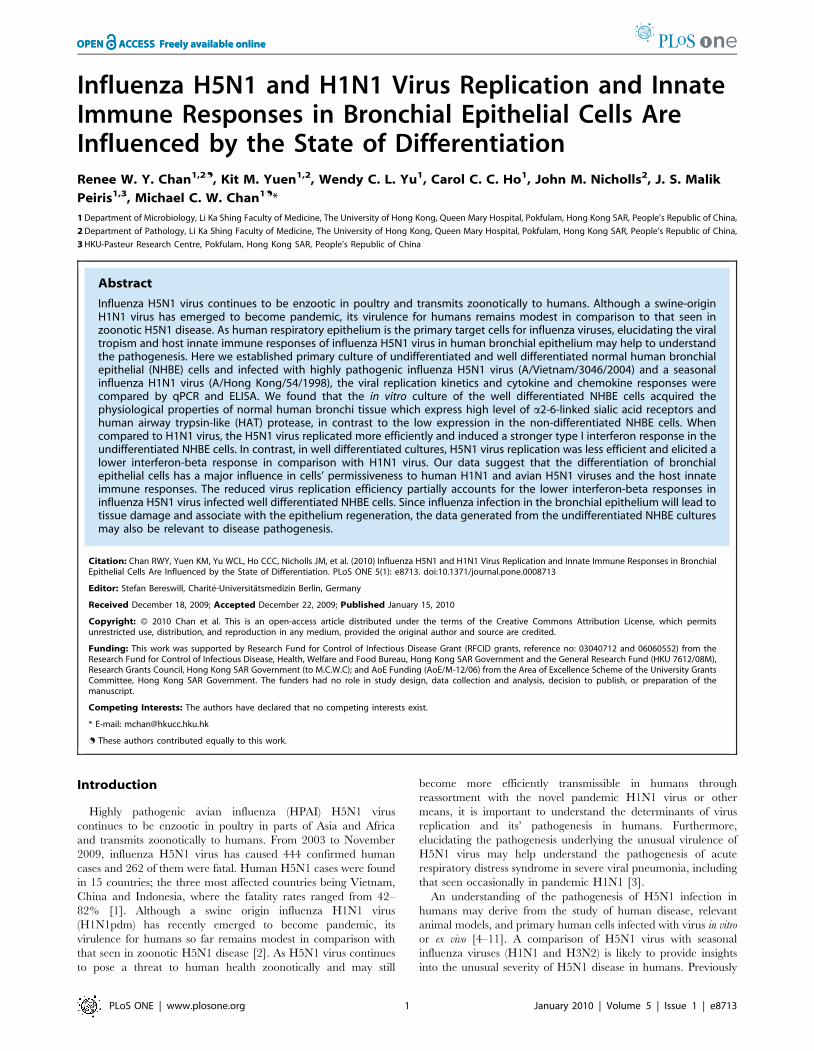
Cytokine and Chemokine mRNA Induction by InfluenzaVirus Infected NHBE Cells
We next investigated the influence of the differentiation state of
NHBE cells on the cytokine and chemokine responses induced by
HK98/H1N1 and VN04/H5N1 viruses. Specifically, we wanted
to determine whether the difference in NHBE cell differentiation
(ud-NHBE Vs wd-NHBE) led to qualitative or quantitative
differences in the profile of cytokines induced. Cytokine (TNF-a,
IFN-b) and chemokine (RANTES, IP-10) gene expression profiles
were evaluated by qPCR. TNF-a mRNA was not expressed in
either mock or both influenza A virus (HK98/H1N1 and VN04/
H5N1) infected ud- and wd-NHBE cells (data not shown). In
general, IFN-b, RANTES and IP-10 gene was induced following
infection with influenza H1N1 and H5N1 virus in both the ud-
NHBE and wd-NHBE cells. VN04/H5N1 virus led to significantly
higher IFN-b (Figure 6A, left panel) and IP-10 (Figure 6C,left panel) mRNA expression in ud-NHBE cells compared with
HK98/H1N1 or mock infection at both 6 h and 24 h post
infection. VN04/H5N1 virus induced a significantly higher
RANTES expression than HK98/H1N1 virus infected cells at
6 h post infection but not at 24 hours post infection (Figure 6B).
In wd-NHBE cells, VN04/H5N1 induced significantly higher
IFN-b (Figure 6A, right panel) and RANTES (Figure 6B,right panel) mRNA expression than HK98/H1N1 at 6 h post
infection. However, at 24 h post infection, RANTES (Figure 6B,left panel) and IP-10 (Figure 6C, right panel) mRNA
expression levels were comparable in VN04/H5N1 and HK98/
H1N1 viruses infected while IFN-b mRNA expression level was
significantly lower (p = 0.03) in VN04/H5N1 infected cells than in
HK98/H1N1 infected cells (Figure 6A, right panel). Inactiva-
tion of the virus by ultraviolet irradiation or high temperature
(70uC for 15 minutes) prior to infection of the ud- and wd-NHBE
cells abolished cytokine induction (data not shown). This suggests
that virus replication was required for cytokine induction and also
rules out the possibility that endotoxin contamination in virus
Figure 2. Lectin binding: MAL-I binds to Sia-a2-3Galb1-4GlcNAc (Panel A, D, G and J), MAL-II binds to Sia-a2-3Galb1-3GalNAc (PanelB, E, H and K) and SNA binds to Sia-a2-6-linkage (Panel C, F, I and L) to determine the Sias distribution on the ud- and wd-NHBEcells. MAL-I, MAL-II and SNA bindings presented on the (A-C) en face staining of ud-NHBE, (D-F) cross-section staining of ud-NHBE, (G-I) cross-sectionstaining of wd-NHBE cells in vitro cultures and (J-L) the human bronchial biopsy in reddish brown.doi:10.1371/journal.pone.0008713.g002
Figure 3. Influenza matrix (M) gene expression after infectionof influenza HK98/H1N1 virus (grey bars) and influenza VN04/H5N1 virus (black bars). Ud-NHBE supported M gene transcriptionfrom 3 h to 24 h post infection for both influenza viruses while wd-NHBE supported a better influenza HK98/H1N1 virus M genetranscription than influenza H5N1 virus. Bars represented the mean Mgene expressed per 105 b–actin house keeping gene and error barrepresent the standard error of mean from three independentexperiments. Asterisk indicates significant difference with p,0.05.doi:10.1371/journal.pone.0008713.g003
Flu in Human Bronchial Cells
PLoS ONE | www.plosone.org 5 January 2010 | Volume 5 | Issue 1 | e8713

stocks contributed to the observed cytokine responses. Besides, an
increase in the MOI up to 5 did not result in changes in the
expression profile of the cytokine and chemokine mRNA induced
by the influenza H1N1 and H5N1 viruses in both the ud- and wd-
NHBE cells (data not shown).
Secretion of Cytokine and Chemokine Protein of InfectedNHBE Cells
We next used ELISA to measure the secretion of cytokine and
chemokine proteins from ud- and wd-NHBE cell culture
supernatants infected by influenza H1N1 and H5N1 viruses.
The concentration of RANTES and IP-10 in the supernatants of
mock infected cells was below the detection limit of the ELISA kit
(31.25 rg/ml and 62.5 rg/ml, respectively). In parallel with the
gene expression profile, at 24 h post infection, VN04/H5N1 led to
significantly higher RANTES (p,0.001) (Figure 7A, left panel)and IP-10 (p,0.001) (Figure 7B, left panel) protein secretion in
ud-NHBE cells compared with HK98/H1N1. While VN04/
H5N1 induced significantly higher levels of RANTES secretion in
wd-NHBE cells than did HK98/H1N1 (p,0.01) (Figure 7A,right panel), the IP-10 protein secretion was comparable
between in HK98/H1N1 and VN04/H5N1 infected cells. We
Figure 4. Immunofluorescence staining of (A–C) ud-NHBE cells and (D–F) wd-NHBE cells at 16 h post infection with (A and D) mockinfection, (B and E) HK98/H1N1 and (C and F) VN04/H5N1. Influenza nucleoprotein and matrix protein was stained in green with FITC-conjugated mouse antibody.doi:10.1371/journal.pone.0008713.g004
Figure 5. Virus titer detected in the supernatant of influenza virus infected ud- and wd-NHBE cells. Virus titer of the (A) HK98/H1N1 and(B) VN04/H5N1 was determined after influenza viruses infected in the ud- and wd-NHBE cells from 1 h to 48 h post infection at MOI of 2. (C) Thecomparison of viral replication kinetic between influenza HK98/H1N1 and VN04/H5N1 viruses in ud- and wd-NHBE at 24 h post infection. The chartshowed the mean and the standard error of the virus titer pooled from three independent experiments. Single asterisk indicated statisticallysignificant difference of means with p,0.05, double asterisks indicated statistically significant differences of means with p,0.01. Dotted linerepresents the detection limit of the TCID50 assay.doi:10.1371/journal.pone.0008713.g005
Flu in Human Bronchial Cells
PLoS ONE | www.plosone.org 6 January 2010 | Volume 5 | Issue 1 | e8713
failed to detect any IFN b protein in the supernatant of the NHBE
cultures after influenza virus infection because of the poor
sensitivity of the IFN-b ELISA (detection limit 250 rg/ml).
Discussion
The presence of ciliated and goblet cells [21] within the
pseudostratified epithelium [22], the establishment of transepithe-
lial resistance [20] and the expression of HAT protease in the
bronchial epithelium are indicators of a well differentiated
bronchial epithelium. The fact that a low-pathogenic H1N1 virus
replicates efficiently in these cells without exogenous trypsin and
that addition of trypsin did not further enhance viral replication
suggests that the wd-NHBE cells make sufficient HAT to support
low pathogenic influenza viral replication, as occurs in vivo. Taken
together with the mucin producing ability, these findings provide
good evidence that wd-NHBE cell system had acquired the
physiological properties of a normal human bronchial epithelium
and provides a relevant model for the study of influenza virus
replication and pathogenesis.
We compared the distribution of Sia receptors in ud-NHBE and
wd-NHBE cell models with that seen in human bronchial
epithelium and found that the wd-NHBE has a comparable
profile to human tissue while the ud-NHBE does not. These
differences in Sia configurations and expression between ud-
NHBE and wd-NHBE cells may explain at least in part, the
different permissiveness to the human seasonal H1N1 and avian
H5N1 viruses. The higher expression of Sia-a2-6 in wd-NHBE
may account for the better replication of human influenza H1N1
virus which preferentially utilizes this receptor for entry. The lower
abundance of Sia-a2-3Galb1-3GalNAc in wd-NHBE may be
associated with the lower permissiveness of these cells for avian
influenza H5N1 virus (which has a higher binding affinity to Sia-
a2-3Galb1-3GalNAc). In addition to the degree of differentiation,
the complexity of wd-NHBE (i.e. three-dimensional structure with
a heterogeneous cell population vs. monolayer with single cell
type) could also account for such differences.
Cell tropism of influenza A virus in wd-human bronchial
epithelial cells was previously studied by Matrosovich et al.[23].
They found that human viruses preferentially infected non-ciliated
cells while the avian viruses infected ciliated cells in the first cycle
of infection. They correlated this observation with the localization
of Sia receptors on these two cell types, non-ciliated cells express
mainly Siaa2-6 receptors known to bind human influenza viruses
while ciliated cells predominantly express Siaa2-3, the receptor for
avian influenza viruses. Others have also reported that human
influenza virus replicates more efficiently compared to avian
influenza viruses in human differentiated tracheo-bronchial
epithelial cells [24]. Our data are in agreement with these findings
that the H5N1 virus replication in wd-NHBE cells was less
efficient when compared to H1N1 virus. In contrast, as both
Siaa2-3 and Siaa2-6 receptors were present in the ud-NHBE cells,
Figure 6. The (A) IFN-b, (B) RANTES and (C) IP-10 gene expression of the ud- and wd-NHBE cells at 6 h and 24 h post infection ofHK98/H1N1 and VN04/H5N1. The chart showed the mean and the standard error from three independent experiments. Single asterisk indicatedstatistically significant difference of means with p,0.05, double asterisks indicated statistically significant differences of means with p,0.01 and tripleasterisks indicated statistically significant differences of means with p,0.001.doi:10.1371/journal.pone.0008713.g006
Flu in Human Bronchial Cells
PLoS ONE | www.plosone.org 7 January 2010 | Volume 5 | Issue 1 | e8713

with homogenous distribution in the majority of basal epithelial
cells, both H1N1 and H5N1 viruses replicate well in these cells.
Mucin secreted by the goblet cells was present in wd-NHBE cell
cultures. Extracts of human bronchial mucin are found to contain
Sia primarily in the Sia-a2-3 linkage [25,26] and much less Sia-a2-
6 linkage [26]. Influenza virus inhaled to the conducting airway
would first encounter this soluble form of Sia receptor in mucin
before reaching the respiratory epithelial cell membrane with the
Sia receptors that the virus needs to attach to in order to initiate
infection. Secreted mucin with the Sia-a2-3 receptor has been
postulated to act as binding-decoy for influenza H5N1 virus,
reducing the possibility of the virus to bind to the bronchial
epithelium and achieve productive replication in the airway.
Matrosovich et al have noted that even after extensive washing of
wd-human airway epithelial cell cultures, there is still lectin
binding to the cell surface-associated mucins [23]. In addition, the
continuous secretion of mucin by goblets cells in the wd-NHBE
can interfere with multiple round of infection.
Bronchial epithelial cells play an important role in innate
defense and in the pathogenesis of the influenza virus infection.
They are target cells for infection and they secrete cytokines and
chemokines upon infection which mediate host defense and
potentially contribute to the inflammation of the bronchial
mucosa. Influenza A virus (H3N2) infection of undifferentiated
human bronchial epithelial cells NCI-H292 leads to induction of
RANTES, IL-6 and IL-8 secretions, but not granulocyte-
macrophage colony stimulating factor; these may be relevant in
host defense and pathogenesis [27,28]. There are however limited
studies of cytokine responses in well differentiated bronchial
epithelium. In our study of bronchial epithelial cells, influenza
virus infection induced IFN-b, RANTES and IP-10 mRNA in
these cells. Interestingly, a strikingly different cytokine and
chemokine expression was observed in the ud-NHBE and wd-
NHBE upon infection by the same virus. There were also
differences between H1N1 and H5N1 influenza viruses in the
profile of cytokine and chemokine release in ud-NHBE compared
with wd-NHBE. At 6 h post infection of ud-NHBE, VN04/H5N1
virus led to a more potent expression of IFN-b, RANTES and IP-
10 compared with HK98/H1N1 virus. This pattern is similar to
the human peripheral blood derived macrophages [6] and
pneumocytes [5].
On the contrary, at 24 h post infection, wd-NHBE cells had an
apparently reverse IFN-b mRNA profile with VN/04/H5N1 virus
inducing significantly lower levels than HK98/H1N1. Moreover,
VN04/H5N1 virus did not significantly increase expression of the
chemokine genes RANTES and IP-10 compared with HK98/
H1N1 (Figure 6). These may be associated with the lower levels of
H5N1 viral replication in wd-NHBE cells as evidenced by M gene
levels (Figure 3), imunofluorescent cells (Figure 4) and non-
productive replication (Figure 5B).
Zeng et al. [12] previously reported that IFN-b expression in
Calu-3 cells at 24 h post infection with H5N1 was lower and more
delayed than seen with seasonal influenza (H3N2). They found
that this delayed IFN-b response was associated with delayed
induction of interferon regulatory factor-3 nuclear translocation
and suggested that H5N1 evades the IFN-b innate host defenses
allowing it to replicate more efficiently. Our findings in NHBE
cells are in agreement with theirs at 24 h post infection but we find
this is associated with lower (not higher) levels of virus replication.
Furthermore we find that at 6 h post infection, H5N1 virus
induces more IFN-b than H1N1 virus. These apparently
contradictory findings may partly reflect the cell type (Calu-3 vs.
NHBE) and the extent of cell differentiation in the two
experimental models and the time of data collection. Calu-3 is a
transformed continuous cell line originally isolated from a human
pulmonary adenocarcinoma and they were differentiated in
transwell culture inserts for one week only. In contrast, our wd-
NHBE cells are primary cells derived from normal human
bronchi, and are cultured and differentiated for 21 days in ALI.
As shown in figure 1A, NHBE cell differentiation in our hands was
not completed after seven days of ALI cultures when compared to
day 21 (Figure 1B). Interestingly, studies on the pathogenesis of
severe acute respiratory syndrome coronavirus (SARS-CoV) using
polarized Calu-3 cells and differentiated human airway epithelial
cells [29,30] found that the latter supported a more productive
replication of SARs-CoV. Since SARS-CoV infects ciliated cells,
this difference was attributed to the lower numbers of ciliated
epithelial cells obtained in the polarized Calu-3 cell model [31]. It
is possible that the wd-NHBE cell model which recapitulates the
morphological and physiological features of the human conducting
airway in vivo would be a more representative model to study
respiratory infection.
Figure 7. The (A) RANTES and (B) IP-10 protein of the supernatant collected at the apical compartment of the influenza HK98/H1N1and VN04/H5N1 virus infected ud-NHBE (dark bars) and wd-NHBE (grey bars) cells at 24 h post infection. The chart showed the meanand the standard error from three independent experiments. Double asterisk indicated statistically significant difference of means with p,0.001 andtriple asterisks indicated statistically significant differences of means with p,0.0001. UD indicated the protein concentration of the sample is belowthe detection limit.doi:10.1371/journal.pone.0008713.g007
Flu in Human Bronchial Cells
PLoS ONE | www.plosone.org 8 January 2010 | Volume 5 | Issue 1 | e8713

In conclusion, our data shows that the origin and differentiation
of bronchial epithelial cells has a major impact to the
permissiveness of cells to influenza A virus replication and on
the host responses elicited by such infection. This reinforces the
importance of the use of physiologically relevant models for the
understanding of influenza pathogenesis. It is relevant to note that
a regenerating epithelium recovering from mucosal damage
caused by viral or other agents would have the equivalent of
undifferentiated epithelial cells present, thus the data from ud-
NHBE may still be physiologically relevant to such a situation.
Acknowledgments
We are grateful for the help of Joanne H.M. Fong and Lynsia L.S. Tang
with the cell culture, and molecular biology analysis, Mr. Kevin Fung for
the immunohistochemistry.
Author Contributions
Conceived and designed the experiments: RWYC JMN JSMP MCC.
Performed the experiments: RWYC KMY WCY CCH MCC. Analyzed
the data: RWYC JMN JSMP MCC. Contributed reagents/materials/
analysis tools: RWYC JMN JSMP MCC. Wrote the paper: RWYC JSMP
MCC.
References
1. WHO (2009) Cumulative Number of Confirmed Human Cases of Avian
Influenza A/(H5N1) Reported to WHO. November 2009 ed.
2. Dawood FS, Jain S, Finelli L, Shaw MW, Lindstrom S, et al. (2009) Emergenceof a novel swine-origin influenza A (H1N1) virus in humans. N Engl J Med 360:
2605–2615.3. Perez-Padilla R, de la Rosa-Zamboni D, Ponce de Leon S, Hernandez M,
Quinones-Falconi F, et al. (2009) Pneumonia and Respiratory Failure from
Swine-Origin Influenza A (H1N1) in Mexico. N Engl J Med.4. Chan RWY, Chan MCW, Wong ACN, Karamanska R, Dell A, et al. (2009)
DAS181 Inhibits H5N1 Influenza virus Infection of Human Lung Tissues.Antimicrob Agents Chemother: AAC. pp 00389–00309.
5. Chan MC, Cheung CY, Chui WH, Tsao SW, Nicholls JM, et al. (2005)Proinflammatory cytokine responses induced by influenza A (H5N1) viruses in
primary human alveolar and bronchial epithelial cells. Respir Res 6: 135.
6. Cheung CY, Poon LL, Lau AS, Luk W, Lau YL, et al. (2002) Induction ofproinflammatory cytokines in human macrophages by influenza A (H5N1)
viruses: a mechanism for the unusual severity of human disease? Lancet 360:1831–1837.
7. de Jong MD, Simmons CP, Thanh TT, Hien VM, Smith GJ, et al. (2006) Fatal
outcome of human influenza A (H5N1) is associated with high viral load andhypercytokinemia. Nat Med 12: 1203–1207.
8. Nicholls JM, Bourne AJ, Chen H, Guan Y, Peiris JS (2007) Sialic acid receptordetection in the human respiratory tract: evidence for widespread distribution of
potential binding sites for human and avian influenza viruses. Respir Res 8: 73.
9. Peiris JS, de Jong MD, Guan Y (2007) Avian influenza virus (H5N1): a threat tohuman health. Clin Microbiol Rev 20: 243–267.
10. Szretter KJ, Gangappa S, Lu X, Smith C, Shieh WJ, et al. (2007) Role of hostcytokine responses in the pathogenesis of avian H5N1 influenza viruses in mice.
J Virol 81: 2736–2744.11. Chan MC, Chan RW, Yu WC, Ho CC, Chui WH, et al. (2009) Influenza H5N1
virus infection of polarized human alveolar epithelial cells and lung
microvascular endothelial cells. Respir Res 10: 102.12. Zeng H, Goldsmith C, Thawatsupha P, Chittaganpitch M, Waicharoen S, et al.
(2007) Highly Pathogenic Avian Influenza H5N1 Viruses Elicit an AttenuatedType I Interferon Response in Polarized Human Bronchial Epithelial Cells.
J Virol 81: 12439–12449.
13. Suzuki Y (2005) Sialobiology of influenza: molecular mechanism of host rangevariation of influenza viruses. Biol Pharm Bull 28: 399–408.
14. Gray T, Guzman K, Davis C, Abdullah L, Nettesheim P (1996) MucociliaryDifferention of Serially Passaged Normal human Tracheobronchial Epithelial
Cells. American journal of respiratory cell and molecular biology 14: 104–112.15. LeSimple P, van Seuningen I, Buisine MP, Copin MC, Hinz M, et al. (2007)
Trefoil factor family 3 peptide promotes human airway epithelial ciliated cell
differentiation. Am J Respir Cell Mol Biol 36: 296–303.16. Atherton HC, Jones G, Danahay H (2003) IL-13-induced changes in the goblet
cell density of human bronchial epithelial cell cultures: MAP kinase andphosphatidylinositol 3-kinase regulation. Am J Physiol Lung Cell Mol Physiol
285: L730–739.
17. Danahay H, Atherton HC, Jackson AD, Kreindler JL, Poll CT, et al. (2006)
Membrane capacitance and conductance changes parallel mucin secretion in the
human airway epithelium. Am J Physiol Lung Cell Mol Physiol 290: L558–569.
18. Zhen G, Park SW, Nguyenvu LT, Rodriguez MW, Barbeau R, et al. (2007) IL-
13 and epidermal growth factor receptor have critical but distinct roles in
epithelial cell mucin production. Am J Respir Cell Mol Biol 36: 244–253.
19. Karber G (1931) 50% end-point calculation. Arch Exp Pathol Pharmak 162:
480–483.
20. Shu-Chih Chen-Quay KTEAWALNLSCQ (2008) Identification of tight
junction modulating lipids. Journal of Pharmaceutical Sciences 9999: n/a.
21. Park J, He F, Martin L, Li Y, Chorley B, et al. (2005) Human neutrophil elastase
induces hypersecretion of mucin from well-differenitated human bronchial
epithelial cells in vitro via a protein kinase Cd mediated mechanism. Am J Pathol
167: 651–661.
22. Jakiela B, Brockman-Schneider R, Amineva S, Lee WM, Gern JE (2008) Basal
cells of differentiate bronchial epithelium are more susceptible to rhinovirus
infection. Am J Respir Cell Mol Biol 38: 517–523.
23. Matrosovich MN, Matrosovich TY, Gray T, Roberts NA, Klenk HD (2004)
Human and avian influenza viruses target different cell types in cultures of
human airway epithelium. Proc Natl Acad Sci U S A 101: 4620–4624.
24. Thompson CI, Barclay WS, Zambon MC, Pickles RJ (2006) Infection of human
airway epithelium by human and avian strains of influenza a virus. J Virol 80:
8060–8068.
25. Couceiro JN, Paulson JC, Baum LG (1993) Influenza virus strains selectively
recognize sialyloligosaccharides on human respiratory epithelium; the role of the
host cell in selection of hemagglutinin receptor specificity. Virus Res 29:
155–165.
26. Baum LG, Paulson JC (1990) Sialyloligosaccharides of the respiratory epithelium
in the selection of human influenza virus receptor specificity. Acta Histochem
Suppl 40: 35–38.
27. Adachi M, Matsukura S, Tokunaga H, Kokubu F (1997) Expression of cytokines
on human bronchial epithelial cells induced by influenza virus A. Int Arch
Allergy Immunol 113: 307–311.
28. Matsukura S, Kokubu F, Noda H, Tokunaga H, Adachi M (1996) Expression of
IL-6, IL-8, and RANTES on human bronchial epithelial cells, NCI-H292,
induced by influenza virus A. J Allergy Clin Immunol 98: 1080–1087.
29. Tseng CT, Tseng J, Perrone L, Worthy M, Popov V, et al. (2005) Apical entry
and release of severe acute respiratory syndrome-associated coronavirus in
polarized Calu-3 lung epithelial cells. J Virol 79: 9470–9479.
30. Sims AC, Baric RS, Yount B, Burkett SE, Collins PL, et al. (2005) Severe acute
respiratory syndrome coronavirus infection of human ciliated airway epithelia:
role of ciliated cells in viral spread in the conducting airways of the lungs. J Virol
79: 15511–15524.
31. Sims AC, Burkett SE, Yount B, Pickles RJ (2008) SARS-CoV replication and
pathogenesis in an in vitro model of the human conducting airway epithelium.
Virus Res 133: 33–44.
Flu in Human Bronchial Cells
PLoS ONE | www.plosone.org 9 January 2010 | Volume 5 | Issue 1 | e8713

![VBA 1B VBA RWY 23 RWY 23 ARRIVALS D RASIN 2A OMA VBA 1A … · 2013. 3. 9. · RASIN 1B[RASI1B] VBA 1A 2 2 6 ^ CHANGES: RWY 23 ARRIVALS RWY 05 ARRIVALS RASIN 1B: 3500 M A X 2 5 0](https://img.pdfslide.us/doc/110x75/6103e8c30ce7341faa73a47d/vba-1b-vba-rwy-23-rwy-23-arrivals-d-rasin-2a-oma-vba-1a-2013-3-9-rasin-1brasi1b.jpg)