Embed Size (px)
Citation preview

International Journal of
Molecular Sciences
Article
Influence of the Type of Diet on the Incidence ofPathogenic Factors and Antibiotic Resistance inEnterococci Isolated from Faeces in Mice
Beatriz Sánchez 1, Antonio Cobo 1,†, Marina Hidalgo 1, Ana M. Martínez-Rodríguez 2 ,Isabel Prieto 3 , Antonio Gálvez 1 and Magdalena Martínez-Cañamero 1,*
1 Área de Microbiología, Departamento de Ciencias de la Salud, Universidad de Jaén,Paraje de Las Lagunillas s/n, 23071 Jaen, Spain
2 Departamento de Estadística e Investigación Operativa, Universidad de Jaén, Paraje de Las Lagunillas s/n,23071 Jaen, Spain
3 Área de Fisiología, Departamento de Ciencias de la Salud, Universidad de Jaén,Paraje de Las Lagunillas s/n, 23071 Jaen, Spain
* Correspondence: [email protected]† Current address: Departamento de Microbiología, Universidad de Granada, Campus de la Cartuja s/n,
18071 Granada, Spain.
Received: 30 July 2019; Accepted: 28 August 2019; Published: 2 September 2019�����������������
Abstract: A comparative study on potential risks was carried out in a collection of 50 enterococciisolated from faeces of mice fed a standard or a high-fat diet enriched with extra virgin olive oil,refined olive oil or butter, at the beginning, after six weeks and after twelve weeks of experiments.Strains were biochemically assessed and genetically characterized. E. faecalis and E. casseliflavus werethe most frequently isolated species in any diet and time points. Apart from the fact of not havingisolated any strain from the virgin olive oil group during the last balance, we found statisticallysignificant differences (p < 0.05) among the diets in the percentage of antibiotic resistance and inthe presence of the enterococcal surface protein gene (esp), as well as a tendency (p < 0.1) for thepresence of the tyrosine decarboxylase gene (tdc) to increase over time in the group of isolates fromthe standard diet. When the resistance of the strains to virgin or refined olive oil was studied, only thegroup of enterococci from high fat diets showed a significantly higher percentage of resistance torefined olive oil (p < 0.05), while both types of oil equally inhibited those isolated from the standarddiet (p > 0.05).
Keywords: enterococci; virulence; antibiotic resistance; olive oil; high fat diets
1. Introduction
The intestinal microbial diversity will be determined by the interactions among the organismsthat compose it, the genetics of the host and the diets applied [1]. Diet, in fact, has a marked influenceon the intestinal microbiota of the host [2] and much attention has been drawn specifically on high fatdiets (HFD) because of their influence on health [3]. While studying the dissimilar effect of fats withdifferent degrees of saturation, we have previously reported several studies comparing the influenceof virgin olive oil (EVOO) and butter (BT) on the intestinal microbiota of mice, both using genotypingmethods [4] and massive sequencing [5]. In these works, we presented evidence supporting a linkbetween specific diets, physiological parameters and some bacterial taxa. Moreover, by comparingEVOO and refined olive oil (ROO) diets, the possible effect of virgin olive oil polyphenols wasuncovered [6]. These new culture-independent technologies can, however, address only global taxa,giving no discriminative data on how the different strains of a certain bacterial group are evolving
Int. J. Mol. Sci. 2019, 20, 4290; doi:10.3390/ijms20174290 www.mdpi.com/journal/ijms
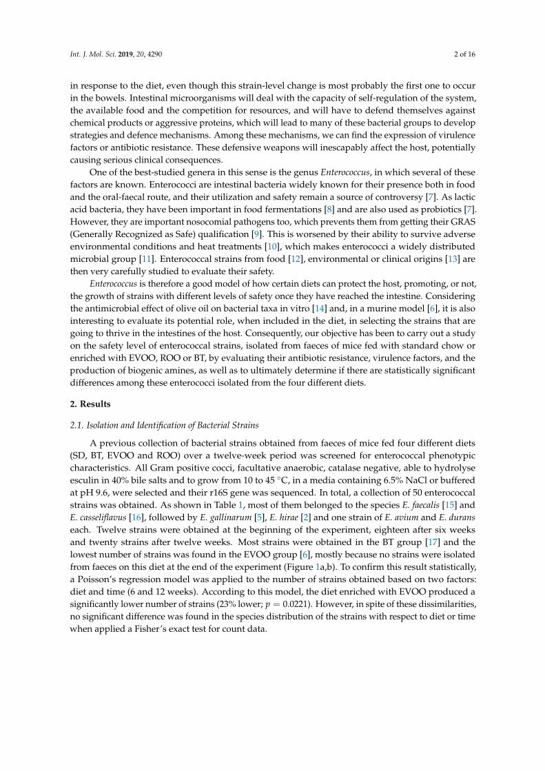
Int. J. Mol. Sci. 2019, 20, 4290 2 of 16
in response to the diet, even though this strain-level change is most probably the first one to occurin the bowels. Intestinal microorganisms will deal with the capacity of self-regulation of the system,the available food and the competition for resources, and will have to defend themselves againstchemical products or aggressive proteins, which will lead to many of these bacterial groups to developstrategies and defence mechanisms. Among these mechanisms, we can find the expression of virulencefactors or antibiotic resistance. These defensive weapons will inescapably affect the host, potentiallycausing serious clinical consequences.
One of the best-studied genera in this sense is the genus Enterococcus, in which several of thesefactors are known. Enterococci are intestinal bacteria widely known for their presence both in foodand the oral-faecal route, and their utilization and safety remain a source of controversy [7]. As lacticacid bacteria, they have been important in food fermentations [8] and are also used as probiotics [7].However, they are important nosocomial pathogens too, which prevents them from getting their GRAS(Generally Recognized as Safe) qualification [9]. This is worsened by their ability to survive adverseenvironmental conditions and heat treatments [10], which makes enterococci a widely distributedmicrobial group [11]. Enterococcal strains from food [12], environmental or clinical origins [13] arethen very carefully studied to evaluate their safety.
Enterococcus is therefore a good model of how certain diets can protect the host, promoting, or not,the growth of strains with different levels of safety once they have reached the intestine. Consideringthe antimicrobial effect of olive oil on bacterial taxa in vitro [14] and, in a murine model [6], it is alsointeresting to evaluate its potential role, when included in the diet, in selecting the strains that aregoing to thrive in the intestines of the host. Consequently, our objective has been to carry out a studyon the safety level of enterococcal strains, isolated from faeces of mice fed with standard chow orenriched with EVOO, ROO or BT, by evaluating their antibiotic resistance, virulence factors, and theproduction of biogenic amines, as well as to ultimately determine if there are statistically significantdifferences among these enterococci isolated from the four different diets.
2. Results
2.1. Isolation and Identification of Bacterial Strains
A previous collection of bacterial strains obtained from faeces of mice fed four different diets(SD, BT, EVOO and ROO) over a twelve-week period was screened for enterococcal phenotypiccharacteristics. All Gram positive cocci, facultative anaerobic, catalase negative, able to hydrolyseesculin in 40% bile salts and to grow from 10 to 45 ◦C, in a media containing 6.5% NaCl or bufferedat pH 9.6, were selected and their r16S gene was sequenced. In total, a collection of 50 enterococcalstrains was obtained. As shown in Table 1, most of them belonged to the species E. faecalis [15] andE. casseliflavus [16], followed by E. gallinarum [5], E. hirae [2] and one strain of E. avium and E. duranseach. Twelve strains were obtained at the beginning of the experiment, eighteen after six weeksand twenty strains after twelve weeks. Most strains were obtained in the BT group [17] and thelowest number of strains was found in the EVOO group [6], mostly because no strains were isolatedfrom faeces on this diet at the end of the experiment (Figure 1a,b). To confirm this result statistically,a Poisson’s regression model was applied to the number of strains obtained based on two factors:diet and time (6 and 12 weeks). According to this model, the diet enriched with EVOO produced asignificantly lower number of strains (23% lower; p = 0.0221). However, in spite of these dissimilarities,no significant difference was found in the species distribution of the strains with respect to diet or timewhen applied a Fisher’s exact test for count data.

Int. J. Mol. Sci. 2019, 20, 4290 3 of 16
(a) (b)
Figure 1. Bar plot of number of strains isolated within each species distributed according to time (a)and diet (b).
Table 1. Detection of virulence factors and biogenic amines gene products.
Virulence Factors Biogenic Amines
Species agg gelE cylA cylM cylB esp efaAfs efaAfm cob cpd ccf tdc
0S1-1 E. faecalis +0S1-2 E. casseliflavus +0S2-1 E. faecalis + +0B1-2 E. casseliflavus +0B2-4 E. faecalis +0B3-1 E. casseliflavus +0O5-2 E. faecalis + + +0O5-4 E. faecalis + +0O7-3 E. faecalis +0V3-1 E. casseliflavus +0V5-1 E. faecalis + +0V5-2 E. faecalis + + +6S3-3 E. casseliflavus + + +6S3-4 E. gallinarum +6S4-1 E. casseliflavus6S4-2 E. faecalis + +6S5-1 E. faecalis +6S5-3 E. faecalis + + +6B1-1 E. casseliflavus +6B1-2 E. gallinarum +6B1-3 E. hirae + +6B2-1 E. faecalis + +6B3-1 E. avium + +6B3-2 E. casseliflavus +6O5-2 E. faecalis + +6O5-3 E. faecalis +6O5-4 E. faecalis + +6O6-1 E. faecalis + +6O7-1 E. casseliflavus +6V3-1 E. casseliflavus +6V5-1 E. faecalis + +6V6-1 E. casseliflavus + +12S6-2 E. faecalis +12S6-3 E. casseliflavus + +12S7-2 E. casseliflavus +12S7-3 E. gallinarum + +12S7-4 E. casseliflavus + +12B1-3 E. hirae +12B1-4 E. faecalis + + +12B2-4 E. casseliflavus + +12B2-5 E. durans12B3-2 E. casseliflavus +12B3-4 E. faecalis +12B3-5 E. faecalis +12O5-1 E. gallinarum + +12O5-2 E. casseliflavus +12O5-3 E. gallinarum +12O6-1 E. casseliflavus + +12O6-3 E. casseliflavus +12O7-1 E. casseliflavus +

Int. J. Mol. Sci. 2019, 20, 4290 4 of 16
2.2. RAPD Classification
When subjected to RAPD-PCR genotyping, the fifty strains clustered in three different groups(Figure 2). Group 1 was deeply branched and included only five strains, all of which were isolatedfrom standard diet fed mice. Group 2 contained 40% of the isolates, all of them from the SD groupor from the other groups at t = 0. Finally, the third big cluster grouped the rest of the strains, 52%,most of them (21 out of 26) isolated from mice fed one of the three high fat diets (BT, EVOO or ROO).
Pearson correlation [0.0%-100.0%] RAPD-M13
100
95
90
85
80
75
70
65
60
55
50
45
40
35
12O5-2 12O6-3 12B2-5 12S6-2 12S6-3 12O5-3 12B3-4 12O5-1 6B1-3 6B2-1 6B1-1 6B3-1 6B3-2 0B1-2 6O5-3 6O5-4 6B1-2 0B2-4 6V6-1 12B1-4 12B2-4 12S7-4 12O6-1 12O7-1 12B3-5 12B1-3 6S5-3 0V5-2 0O5-2 6S5-1 0S1-1 0S2-1 0S1-2 6V3-1 0O7-3 0V3-1 6V5-1 0B3-1 0V5-1 6O5-2 6O6-1 6S3-2 6S3-3 6S3-1 6S4-2 12S7-3
G3-3
G3-2
G3-1
G3
G2
G1
Figure 2. Pearson coefficient-based analysis of the RAPD profiles of the strains isolated. The initialnumber indicates the week of isolation.
Int. J. Mol. Sci. 2019, 20, 4290 5 of 16
2.3. PCR Amplification of Virulence Factors
Specific PCR reactions were performed to detect the presence of genes related to virulence factors(Table 1). Thirty-six isolates presented at least one virulence-related gene, out of which, six strainsshowed positive for two virulence factors and only one isolate presented three. Five strains showedpositive for the aggregation substance gene (agg). Gelatinase gene (gelE) was amplified in only oneisolate. Sex pheromone genes (cpd and ccf) were present in one and four strains, respectively, while cobwas not detected. Of the cytolysin genes, only cylB was present, in only one strain, and genes related tocell wall adhesions (efaAfs and efaAfm) were detected in two strains each. In contrast, the gene codingfor the enterococcal surface protein (esp) was present in most of the strains (62%).
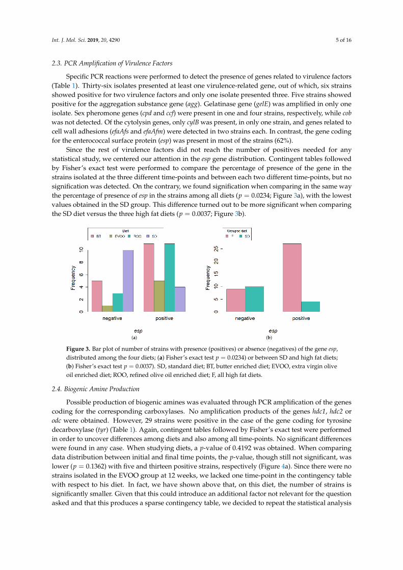
Since the rest of virulence factors did not reach the number of positives needed for anystatistical study, we centered our attention in the esp gene distribution. Contingent tables followedby Fisher’s exact test were performed to compare the percentage of presence of the gene in thestrains isolated at the three different time-points and between each two different time-points, but nosignification was detected. On the contrary, we found signification when comparing in the same waythe percentage of presence of esp in the strains among all diets (p = 0.0234; Figure 3a), with the lowestvalues obtained in the SD group. This difference turned out to be more significant when comparingthe SD diet versus the three high fat diets (p = 0.0037; Figure 3b).
(a) (b)
Figure 3. Bar plot of number of strains with presence (positives) or absence (negatives) of the gene esp,distributed among the four diets; (a) Fisher’s exact test p = 0.0234) or between SD and high fat diets;(b) Fisher’s exact test p = 0.0037). SD, standard diet; BT, butter enriched diet; EVOO, extra virgin oliveoil enriched diet; ROO, refined olive oil enriched diet; F, all high fat diets.
2.4. Biogenic Amine Production
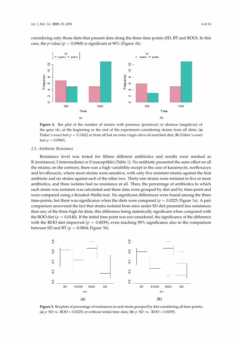
Possible production of biogenic amines was evaluated through PCR amplification of the genescoding for the corresponding carboxylases. No amplification products of the genes hdc1, hdc2 orodc were obtained. However, 29 strains were positive in the case of the gene coding for tyrosinedecarboxylase (tyr) (Table 1). Again, contingent tables followed by Fisher’s exact test were performedin order to uncover differences among diets and also among all time-points. No significant differenceswere found in any case. When studying diets, a p-value of 0.4192 was obtained. When comparingdata distribution between initial and final time points, the p-value, though still not significant, waslower (p = 0.1362) with five and thirteen positive strains, respectively (Figure 4a). Since there were nostrains isolated in the EVOO group at 12 weeks, we lacked one time-point in the contingency tablewith respect to his diet. In fact, we have shown above that, on this diet, the number of strains issignificantly smaller. Given that this could introduce an additional factor not relevant for the questionasked and that this produces a sparse contingency table, we decided to repeat the statistical analysis
Int. J. Mol. Sci. 2019, 20, 4290 6 of 16
considering only those diets that present data along the three time points (SD, BT and ROO). In thiscase, the p-value (p = 0.0968) is significant at 90% (Figure 4b).
(a) (b)
Figure 4. Bar plot of the number of strains with presence (positives) or absence (negatives) ofthe gene tdc, at the beginning or the end of the experiment considering strains from all diets; (a)Fisher’s exact test p = 0.1362) or from all but an extra virgin olive oil enriched diet; (b) Fisher’s exacttest p = 0.0968).
2.5. Antibiotic Resistance
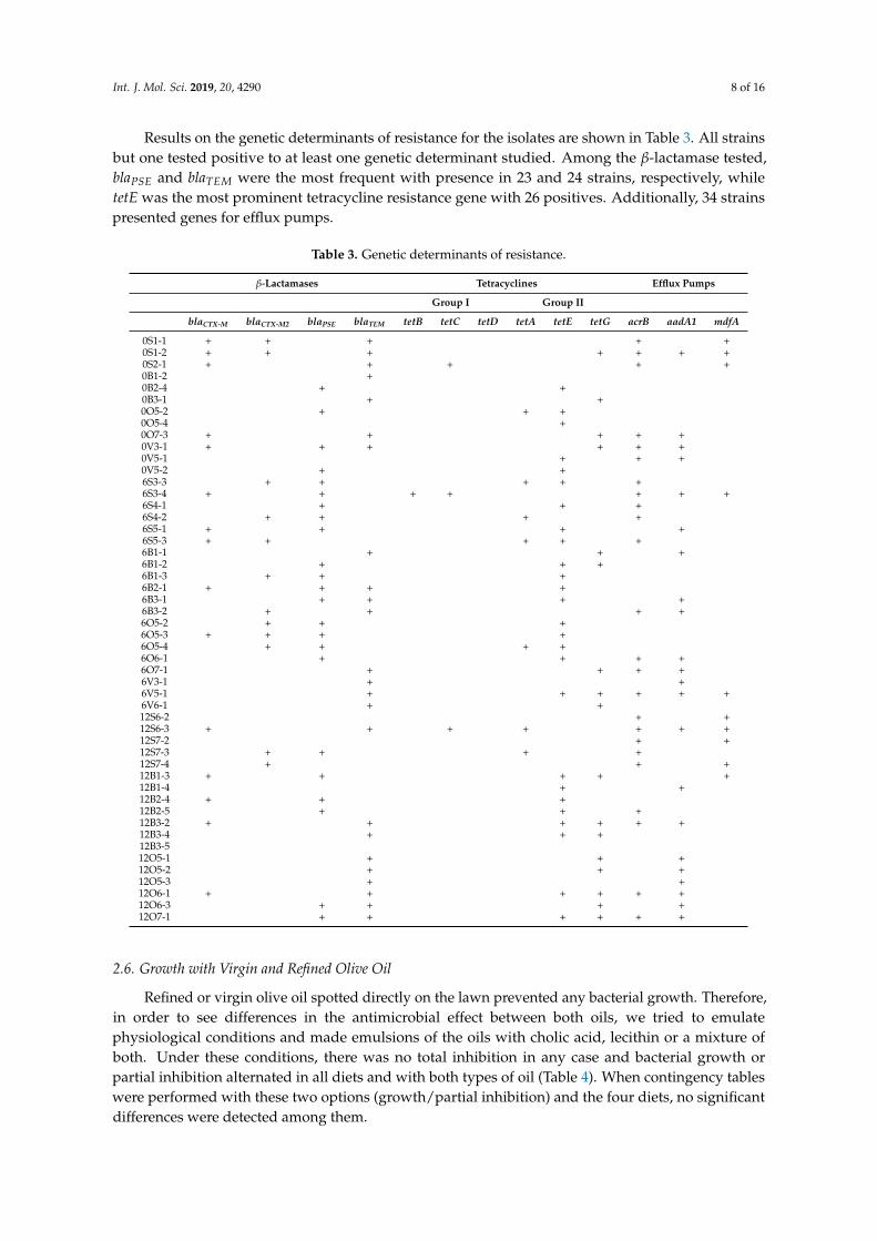
Resistance level was tested for fifteen different antibiotics and results were marked asR (resistance), I (intermediate) or S (susceptible) (Table 2). No antibiotic presented the same effect on allthe strains; on the contrary, there was a high variability except in the case of kanamycin, norfloxacynand levofloxacin, where most strains were sensitive, with only five resistant strains against the firstantibiotic and six strains against each of the other two. Thirty-one strains were resistant to five or moreantibiotics, and three isolates had no resistance at all. Then, the percentage of antibiotics to whicheach strain was resistant was calculated and these data were grouped by diet and by time-point andwere compared using a Kruskal–Wallis test. No significant differences were found among the threetime-points, but there was significance when the diets were compared (p = 0.0225; Figure 5a). A paircomparison uncovered the fact that strains isolated from mice under SD diet presented less resistancesthan any of the three high fat diets, this difference being statistically significant when compared withthe ROO diet (p = 0.0140). If the initial time point was not considered, the significance of the differencewith the ROO diet improved (p = 0.0039), even reaching 90% significance also in the comparisonbetween SD and BT (p = 0.0804; Figure 5b).
(a) (b)
Figure 5. Boxplots of percentage of resistances in each strain grouped by diet considering all time-points;(a) p SD vs. ROO = 0.0225) or without initial time data; (b) p SD vs. ROO = 0.0039).

Int. J. Mol. Sci. 2019, 20, 4290 7 of 16
Table 2. Antibiotic resistance. R: Resistant I: Intermediate S: Susceptible. Antibiotics. PENP: penicillin pneumo; PENS: penicillin strepto; AMPE: ampicillin; CTXP:cetofaxim pneumo; CTXS: cetoaxim strepto; IMIE: imipenem; KAHES: kanamycin; GEHES: gentamycin; FQPR: norfloxacin; MXFPS: moxifloxacin; LVXS: levofloxacinstrepto; LVXP: levofloxacin pneumo; ERYPS: erythromycin; TELPS: telithromycin; QDAE: quinupristin-dalfopristin; TEPS: tetracyclin; RFAPS: rifampicin; TSU:cotrimoxazol; LNZEP: linezolid pneumo; LNZS: linezolid strepto; FOSP: fosfomycin; FURES: nitrofurantoin; VAN: vancomycin; TEC: teicoplanin.
Antibiotic Resistance
PEN PENS AMPE CTXP CTXS/IMIE KAHES/GEHES FQPR/MXFPS LVXS/LVXP ERYPS/TELPS QDAE TETPS/RFAPS TSU/LNZEP LNZS FOSP7FURES VAN/TEC
0S1-1 R R I R I S S S R R R R R I R0S1-2 R R I R I S S S R R I I R S R0S2-1 R R R R I S S S R R R R R R R0B1-2 I S S R I S S S S S S S S S S0B2-4 R R R R I S S S R R R R R R R0B3-1 R R R I I S I S R R R R R R R0O5-2 R R R R R S S S R R R I R R R0O5-4 R I S R I S S I S I R S I I S0O7-3 R R I R I S S S R R R I R I R0V3-1 R R R I I S S S R R R I R R R0V5-1 R R R R I S S S R R R I R R R0V5-2 I I S R I S S S I R R S S S S6S3-3 R R I I I S S S I S S S S S S6S3-4 I S S I I S S S S S S S S S S6S4-1 R R I R I S S S R R R I R S R6S4-2 R R S R I S S S I S S S S S S6S5-1 I I S I I S S S I I I S S I S6S5-3 I I S S S S S S I I R S S I S6B1-1 R I S R I I S S I R R S S I S6B1-2 R R R R I S I S R R R I R R R6B1-3 I S S R I S S S I I I S I S S6B2-1 R R R R R R R R R S I I S S S6B3-1 R R R R I S R S R R R I R R R6B3-2 R I S R I S S S R R R S S I S6O5-2 R R R I S S S R R R R I R R R6O5-3 R R R R I S S S R R R I R R R6O5-4 R R R R R S S S R R R I R R R6O6-1 R R R R I S S S R R R I R R R6O7-1 R I S R I S S S I R R S S S S6V3-1 R R R R I S I S R R R I R R R6V5-1 I I S R I S S S R R R S S I S6V6-1 I I S I I S S S I R R S S S S12S6-2 R R I S S S S S R R R R R I R12S6-3 I I S R I S S S I I I S S I S12S7-2 I I S R I S S S I I I S S I S12S7-3 R R S I S S S S I S S I S S S12S7-4 R R I R I S S S R R R R R S R12B1-3 R R R R R R R R R S R I S S S12B1-4 R R R R R R R R R S R I S S S12B2-4 R I S R I S S S I R R S S R S12B2-5 R I S R I S S S I R R S S R S12B3-2 R R R R R R R R R S R I S S S12B3-4 R R R R R R R R R I R S S I I12B3-5 I I S I I S S S I S R S S I S12O5-1 R R S R I S S S R R R I R I S12O5-2 R R R R R S R I R R R I R R R12O5-3 I S S R I S S S S S R S S I S12O6-1 R R R R R S S S R R R I R R R12O6-3 R R R R I S S S R R R I R S R12O7-1 R R R R R S S S R R R I R R R
Int. J. Mol. Sci. 2019, 20, 4290 8 of 16
Results on the genetic determinants of resistance for the isolates are shown in Table 3. All strainsbut one tested positive to at least one genetic determinant studied. Among the β-lactamase tested,blaPSE and blaTEM were the most frequent with presence in 23 and 24 strains, respectively, whiletetE was the most prominent tetracycline resistance gene with 26 positives. Additionally, 34 strainspresented genes for efflux pumps.
Table 3. Genetic determinants of resistance.
β-Lactamases Tetracyclines Efflux Pumps
Group I Group II
blaCTX-M blaCTX-M2 blaPSE blaTEM tetB tetC tetD tetA tetE tetG acrB aadA1 mdfA
0S1-1 + + + + +0S1-2 + + + + + + +0S2-1 + + + + +0B1-2 +0B2-4 + +0B3-1 + +0O5-2 + + +0O5-4 +0O7-3 + + + + +0V3-1 + + + + + +0V5-1 + + +0V5-2 + +6S3-3 + + + + +6S3-4 + + + + + + +6S4-1 + + +6S4-2 + + + +6S5-1 + + + +6S5-3 + + + + +6B1-1 + + +6B1-2 + + +6B1-3 + + +6B2-1 + + + +6B3-1 + + + +6B3-2 + + + +6O5-2 + + +6O5-3 + + + +6O5-4 + + + +6O6-1 + + + +6O7-1 + + + +6V3-1 + +6V5-1 + + + + + +6V6-1 + +12S6-2 + +12S6-3 + + + + + + +12S7-2 + +12S7-3 + + + +12S7-4 + + +12B1-3 + + + + +12B1-4 + +12B2-4 + + +12B2-5 + + +12B3-2 + + + + + +12B3-4 + + +12B3-512O5-1 + + +12O5-2 + + +12O5-3 + +12O6-1 + + + + + +12O6-3 + + + +12O7-1 + + + + + +
2.6. Growth with Virgin and Refined Olive Oil
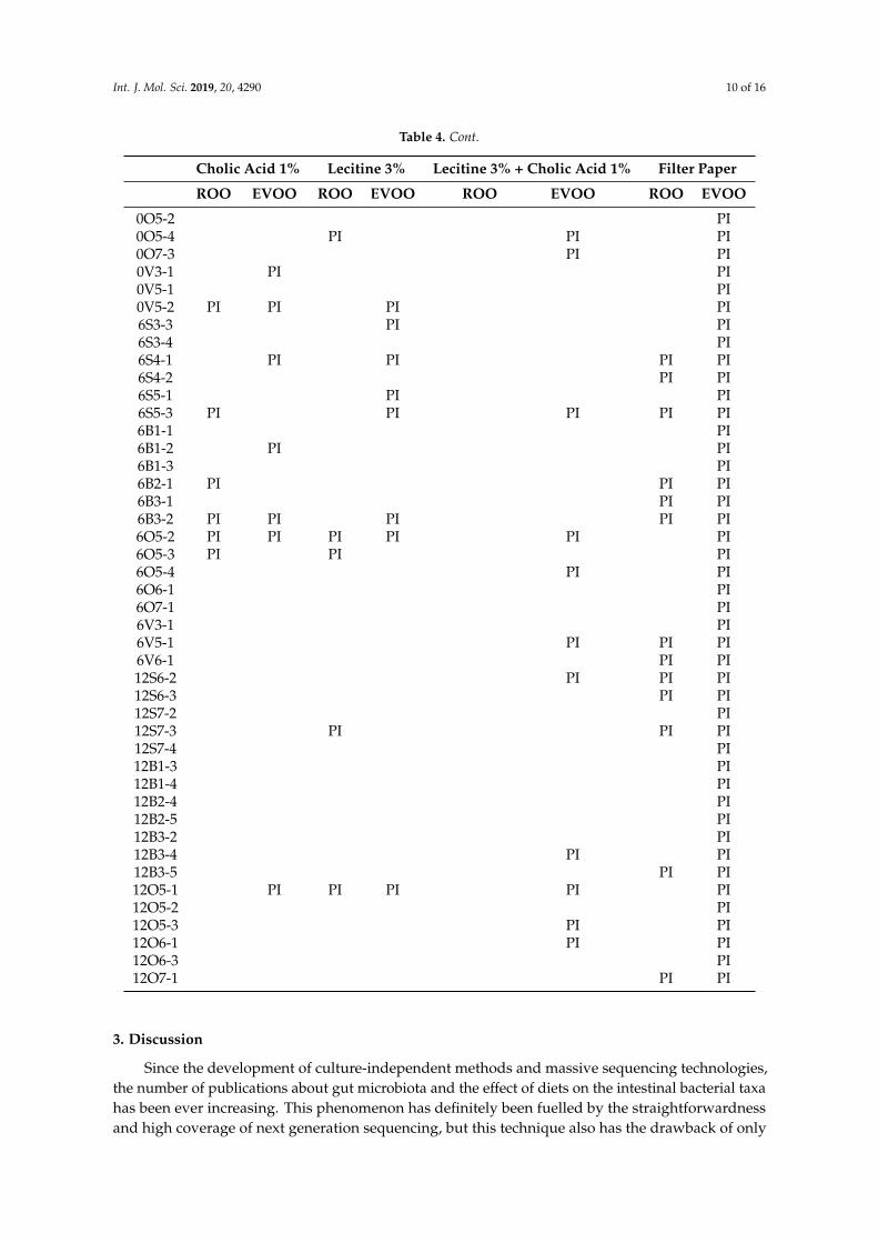
Refined or virgin olive oil spotted directly on the lawn prevented any bacterial growth. Therefore,in order to see differences in the antimicrobial effect between both oils, we tried to emulatephysiological conditions and made emulsions of the oils with cholic acid, lecithin or a mixture ofboth. Under these conditions, there was no total inhibition in any case and bacterial growth orpartial inhibition alternated in all diets and with both types of oil (Table 4). When contingency tableswere performed with these two options (growth/partial inhibition) and the four diets, no significantdifferences were detected among them.
Int. J. Mol. Sci. 2019, 20, 4290 9 of 16
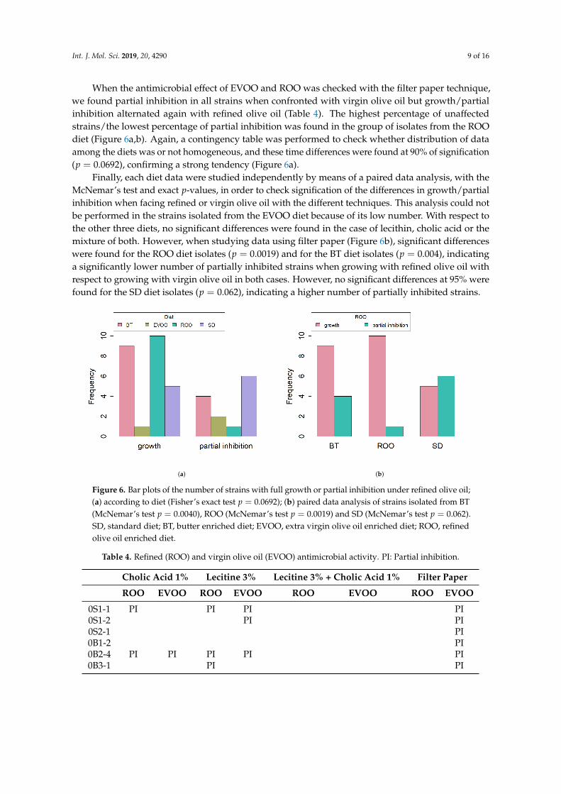
When the antimicrobial effect of EVOO and ROO was checked with the filter paper technique,we found partial inhibition in all strains when confronted with virgin olive oil but growth/partialinhibition alternated again with refined olive oil (Table 4). The highest percentage of unaffectedstrains/the lowest percentage of partial inhibition was found in the group of isolates from the ROOdiet (Figure 6a,b). Again, a contingency table was performed to check whether distribution of dataamong the diets was or not homogeneous, and these time differences were found at 90% of signification(p = 0.0692), confirming a strong tendency (Figure 6a).
Finally, each diet data were studied independently by means of a paired data analysis, with theMcNemar’s test and exact p-values, in order to check signification of the differences in growth/partialinhibition when facing refined or virgin olive oil with the different techniques. This analysis could notbe performed in the strains isolated from the EVOO diet because of its low number. With respect tothe other three diets, no significant differences were found in the case of lecithin, cholic acid or themixture of both. However, when studying data using filter paper (Figure 6b), significant differenceswere found for the ROO diet isolates (p = 0.0019) and for the BT diet isolates (p = 0.004), indicatinga significantly lower number of partially inhibited strains when growing with refined olive oil withrespect to growing with virgin olive oil in both cases. However, no significant differences at 95% werefound for the SD diet isolates (p = 0.062), indicating a higher number of partially inhibited strains.
(a) (b)
Figure 6. Bar plots of the number of strains with full growth or partial inhibition under refined olive oil;(a) according to diet (Fisher’s exact test p = 0.0692); (b) paired data analysis of strains isolated from BT(McNemar’s test p = 0.0040), ROO (McNemar’s test p = 0.0019) and SD (McNemar’s test p = 0.062).SD, standard diet; BT, butter enriched diet; EVOO, extra virgin olive oil enriched diet; ROO, refinedolive oil enriched diet.
Table 4. Refined (ROO) and virgin olive oil (EVOO) antimicrobial activity. PI: Partial inhibition.
Cholic Acid 1% Lecitine 3% Lecitine 3% + Cholic Acid 1% Filter Paper
ROO EVOO ROO EVOO ROO EVOO ROO EVOO
0S1-1 PI PI PI PI0S1-2 PI PI0S2-1 PI0B1-2 PI0B2-4 PI PI PI PI PI0B3-1 PI PI
Int. J. Mol. Sci. 2019, 20, 4290 10 of 16
Table 4. Cont.
Cholic Acid 1% Lecitine 3% Lecitine 3% + Cholic Acid 1% Filter Paper
ROO EVOO ROO EVOO ROO EVOO ROO EVOO
0O5-2 PI0O5-4 PI PI PI0O7-3 PI PI0V3-1 PI PI0V5-1 PI0V5-2 PI PI PI PI6S3-3 PI PI6S3-4 PI6S4-1 PI PI PI PI6S4-2 PI PI6S5-1 PI PI6S5-3 PI PI PI PI PI6B1-1 PI6B1-2 PI PI6B1-3 PI6B2-1 PI PI PI6B3-1 PI PI6B3-2 PI PI PI PI PI6O5-2 PI PI PI PI PI PI6O5-3 PI PI PI6O5-4 PI PI6O6-1 PI6O7-1 PI6V3-1 PI6V5-1 PI PI PI6V6-1 PI PI12S6-2 PI PI PI12S6-3 PI PI12S7-2 PI12S7-3 PI PI PI12S7-4 PI12B1-3 PI12B1-4 PI12B2-4 PI12B2-5 PI12B3-2 PI12B3-4 PI PI12B3-5 PI PI12O5-1 PI PI PI PI PI12O5-2 PI12O5-3 PI PI12O6-1 PI PI12O6-3 PI12O7-1 PI PI
3. Discussion
Since the development of culture-independent methods and massive sequencing technologies,the number of publications about gut microbiota and the effect of diets on the intestinal bacterial taxahas been ever increasing. This phenomenon has definitely been fuelled by the straightforwardnessand high coverage of next generation sequencing, but this technique also has the drawback of only
Int. J. Mol. Sci. 2019, 20, 4290 11 of 16
being reliable in reaching taxon levels of genera and above. However, many of the traits important infood safety and human health are species or even strain specific. Therefore, it is important to turn backto culture-dependent techniques and strain characterization in order to address how diet and otherpossible factors affect strain selection and population dynamics in an intestinal environment.
One of the best bacterial groups to study these influences in is the genus Enterococcus. Enterococciare intestine dwellers [10], able to survive in varied environmental conditions and widely presentin food and food fermentations [11]. They have coevolved with humans and are symbionts withus but can also bear important virulence factors and become dangerous nosocomial germs. It istherefore an interesting model to study how the organism copes to coexist with such a variety ofbeneficial and detrimental strains of the same species, presumably managing to select those that aremore advantageous.
In this research work, a collection of enterococci isolated from mice fed standard chow or threefat-enriched diets were studied and evaluated for safety. The most evident result was the inability toisolate any strain from the EVOO fed mice after twelve weeks of diet, which gave place to a statisticallysignificant lower total number of strains isolated from this group. This decrement did not correspond toa statistically different distribution of the species in any diet or time-point; we did not find differencesin the species distribution probably due to the fact that all but two of the species found in our studywere represented by a low number of isolates.
When studying the genetic diversity of the strains on the basis of a RAPD-PCR genotyping,three main groups of strains were found. Interestingly, all the strains isolated from HFD fed miceclustered together in G3. This group divided into three subgroups. Groups G3-1 and G3-3 includedonly strains isolated after twelve weeks of experiments, while G3-2 only had strains isolated aftersix weeks. Groups G1 and G2, on the contrary, comprised strains isolated from the SD fed groupor from the other three groups but at the beginning of the experiment, before HFD were applied.This clustering indicates already a clear genotypic separation between SD and HFD and, additionallywithin the latest, between six and twelve weeks of HFD application.
Most of the strains (31) were positive for the gene encoding the enterococcal surface protein (esp),which is not surprising in strains from a faecal origin, since this protein is known to promote theprimary attachment to biotic surfaces and to be involved in immune system evasion [18]. However,unexpectedly, the presence of the gene was significantly different in the various diets, this differencebeing greatest when comparing a standard diet against HFD fed mice. This could uncover the existenceof a selecting force in those intestines with a high fat input in such a way that only the enterocci withan increased efficiency in their attachment to the host mucosa can thrive. HFD would therefore proveto have promoted the presence of one virulence factor and, in any case, it is further evidence of howdiet can select the types of strains that live within the host.
The presence of decarboxylases is another undesired trait in enterococci in foods since theyproduce biogenic amines through amino acid decarboxylation [19] and ingestion of high amounts ofthese compounds can cause varied toxicological hazards [20]. In our strains, the only decarboxylasegene found was tyr, coding for tyrosine decarboxylase, which is very commonly found in foodcommodities [21,22]. No differences were found in the presence of tyr in the diverse diets, but therewas a strong tendency to increase with respect to time, with signification at 90%. This tendency isworth considering further since it could be a trait related to the age of the host, with implications forthe host’s health since tyramine has been linked to hypertension and to an increment in glucose inserum [23].
When we set up the collection of strains to grow under a filter paper soaked in virgin olive oil, all ofthem were partially inhibited. However, when refined olive oil was used instead, a variable number ofstrains grew at a normal pace, showing no inhibition. This allowed us to make comparisons amongthe four groups of isolates from the different diets, uncovering a slight difference in the growth/partialinhibition distribution between diets, the strains isolated from ROO-fed mice being the least inhibited.

Int. J. Mol. Sci. 2019, 20, 4290 12 of 16
However, the most interesting result was obtained after searching for statistical signification inthe growth behaviour of the strains within each diet. This would allow us to significantly state if thegroup of strains isolated from a certain diet was partially inhibited or not by refined olive oil, alwaysin comparison to the partial inhibition exerted by virgin olive oil in all strains. A paired data analysisclearly stated that strains isolated from ROO and BT fed mice grew better in the presence of refinedolive oil than in the presence of virgin olive oil, while strains isolated from standard fed animals wereequally inhibited by both oils. This result is noteworthy since it could indicate an adaptation of thefirst two groups of strains to a fat-enriched environment, an adaptation that is, however, less usefulwhen faced with virgin olive oil, where other antimicrobial factors are present in its unsaponifiablefraction, i.e., polyphenols [6,14,24].
To summarise, in this specific study, we have shown the influence of the type of diet on theselection of the virulence traits of the strains that thrive within the host. In the case we present, high fatdiets seem to enhance the presence of a virulence trait (esp), increase the number of antibiotic resistancesand improve the strain’s adaptation to grow in a fatty environment. However, virgin olive oil appearsonce more to behave differently probably due to the additional antimicrobial activity conferredby its minority components, which eventually might have an effect on the intestinal enterococci.Although framed in a very specific experimental model, these are interesting preliminary data thatopen new research lines. More studies using wider cohorts and categories will reinforce and delimitatethese results.
4. Materials and Methods
4.1. Isolation and Identification of Bacterial Strains
The bacterial strains used in this study belong to a collection obtained in a previous work [4].Briefly, as described previously [4], twelve male Swiss Webster ICR (CD-1) mice (Harlan Laboratories)were divided into four groups and fed for three months with a standard chow (SD, standard laboratorymice diet A04, 3% fat, Panlab, Barcelona, Spain) or one of three high fat diets (standard chowsupplemented with 20% of extra-organic virgin olive oil—EVOO, refined olive oil—ROO, or butter—BT).All experimental procedures were performed in accordance with the European Communities CouncilDirective 86/609/EEC and reviewed and approved by the Bioethics Committee of the University ofJaén. In the first, sixth and twelfth week of the experimental period, faecal samples were obtained andserial dilutions of faeces were used for plating on Tryptic Soy Agar (Scharlab, Barcelona, Spain) plus100 mg/L polymyxin B, and on Bile Esculin Agar (Scharlab), in order to select for enterococci colonies.
Putative enterococci were selected according to observation of colony characteristics and cellmorphology, Gram staining, catalase and oxidase production, growth at 10 ◦C and 45 ◦C, growth inthe presence of 6.5% NaCl, at pH 9.6 and growth and esculin hydrolysis on bile-esculin agar (Scharlab).Genetic identification at species level was done by species-specific PCR and 16S rDNA sequencing asdescribed elsewhere [25].
4.2. RAPD-PCR Amplification
Once the strain DNA was extracted, genotyping using randomly amplified PCR was carried outusing the primers M13 and AP4 as described elsewhere [8]. The RAPD patterns obtained with allstrains and each one of the primers were analyzed in a single dendrogram using BioNumerics softwareversion 2.5 (Applied Maths, Kortrijk, Belgium) as also indicated in the above reference.
4.3. PCR Amplification of Virulence Factors
Specific PCR reactions were performed in duplicate to detect the presence of genes involved inthe expression of cytolysin (cylA, cylB and cylM), the aggregation substance (agg), gelatinase (gelE),enterococcal surface protein (esp), cell wall adhesions (efaAfs and efaAfm), and sex pheromones (cpd, coband ccf), as established by Eaton and Gasson [17]. The positive control used was the DNA of E. faecalis

Int. J. Mol. Sci. 2019, 20, 4290 13 of 16
P9091, since it contains in its genome all the virulence factors. In order to prove primer specificity,possible amplicons were checked by PCR simulators using Enterococcus and Bacteria genomic databasesas templates (insilico.ehu.es and Primer-BLAST NCBI). This is also applicable to Sections 4.4 and 4.5.
4.4. Biogenic Amine Production
The potential for biogenic amine generation was detected by PCR amplification in duplicate ofthe amino-acid decarboxylase genes hdc (histidine decarboxylase), odc (ornithine decarboxylase) andtdc (tyrosine decarboxylase) as described before [26].
4.5. Antibiotic Resistance
The antibiotic susceptibility of isolates was determined using ATB ENTEROC 5 strips (BioMérieux,Marcy-l’Etoile, France), following the manufacturer’s instructions. The 50 isolates were also investigatedby PCR for the presence of genetic determinants of resistance as well as presence of efflux pumps.Extended-spectrum β-lactamase: blaCTX−M and blaCTX−M2 [27], blaPSE [28] and blaTEM [16], groups Iand II tetracycline resistance genes [15], and efflux pumps acrB, mdfA [29] and aadA1 [30] were tested.
4.6. Growth with Virgin and Refined Olive Oil
The antimicrobial effect of virgin and refined olive oil on the strains was measured by two methods.In the first one, the spot-on-a-lawn method, 150 µL of overnight culture was plated per triplicate on BrainHeart Infusion agar (Scharlab) and 5 µL axenic spots were added each containing one of the following:virgin olive oil; refined olive oil; a 50:50 mixture of 1% cholic acid and virgin or refined olive oil; a 50:50mixture of 3% lecithin and virgin or refined olive oil; a 50:50 mixture of 1% cholic acid-3% lecithin andvirgin or refined olive oil. Plates were incubated for 24 h at 37 ◦C and microbial growth was evaluated.For the second method, filter fragments were used. Again, 150 µL of overnight culture was plated pertriplicate on Brain Heart Infusion agar (Scharlab) and sterile 2 cm2 Whatman filter fragments soakedin saline solution, virgin olive oil or refined olive oil were laid on top. Plates were incubated for 24 hat 37 ◦C and microbial growth was evaluated underneath. Results were defined as “growth”, “partialinhibition” or “total inhibition”. Partial inhibition was defined as that situation in which the surfacecovered by the drop or the filter does not present a continuous lawn of bacterial growth.
4.7. Statistical Studies
The number of animals in this study (three per diet) was determined in a previous work [4] whereall animals on the same diet had been stabled in the same cage until the moment faeces were collected.In order to overcome the possibly low number of subjects, in this study, strains were taken as samplingunits and contingency tables were designed where each cage at a certain time-point was the basis of adifferent category of the variable “diet”.
To test whether or not the variable “species” has the same distribution in the different categoriesof the variable “diet”, and in the different categories of the variable “time”, the exact Fisher’s test forcontingency tables was performed as some of the expected counts were below 5 and therefore the χ2
test of homogeneity is not suitable. Similar analyses were carried out to identify whether there was aninfluence of “diet” or “time” in the variables tdc and esp, as well as to study whether “diet” had aninfluence on “growth/partial inhibition” on the different oils and procedures.
To investigate the effect of the variables “diet” and “time” on the number of enterococci strains,a Poisson regression model was fitted. As “diet” and “time” are categorical variables, dummy variableswere included in the model in order to evaluate the effect that the presence or absence of the differentlevels of the categorical variables may have on the number of strains.
To test equality of the distribution of the variable “% of antibiotic resistances” according the typeof diet we used the Kruskal–Wallis test as the Shapiro–Wilk test of normality rejected the normalityhypothesis. The same procedure was performed to study the equality of the distribution of the variable“% of antibiotic resistances” according to the variable “time”. Provided that significant differences

Int. J. Mol. Sci. 2019, 20, 4290 14 of 16
were detected, we applied post hoc Duncan tests for pairwise multiple comparisons with Bonferroni’sadjustment in the p-values.
To study whether or not there are differences in the antimicrobial effect of ROO and EVOOwithin each specific diet, only the growth/partial inhibition of the strains isolated from that group ofmice was observed. As the results are paired nominal data, the McNemar test was used to check thestatistical significance.
Author Contributions: Conceptualization, I.P. and M.M.-C.; Data curation, A.M.M.-R.; Funding acquisition,I.P. and M.M.-C.; Investigation, B.S., A.C. and M.H.; Methodology, A.M.M.-R., A.G. and M.M.-C.;Project administration, I.P. and M.M.-C.; Resources, A.G.; Supervision, A.C., A.G. and M.M.-C.; Validation,A.G. and M.M.-C.; Writing—Original draft, M.M.-C.; Writing—Review & editing, A.M.M.-R. and M.M.-C.
Funding: This research was funded by University of Jaén (PP2009/13/03) (to IP) and Junta de Andalucía(PI Excelencia_2010 AGR 6340) (to M.M.-C).
Acknowledgments: We would like to acknowledge Aceites Soler Romero for providing us with their extra virginolive oil and Peter Cassidy for revising the manuscript.
Conflicts of Interest: The authors declare no conflict of interest. The funders had no role in the design of thestudy; in the collection, analyses, or interpretation of data; in the writing of the manuscript, and in the decision topublish the results.
References
1. Zhang, C.; Zhang, M.; Wang, S.; Han, R., Cao, Y.; Hua, W. Interactions between gut microbiota, host geneticsand diet relevant to development of metabolic syndromes in mice. ISME J. 2010, 4, 232–241. [CrossRef][PubMed]
2. Arumugam, M.; Raes, J.; Pelletier, E.; Le Paslier. D.; Yamada, T.; Mende, D.R. Enterotypes of the humangut microbiome. Nature 2011, 473, 174–180. [CrossRef] [PubMed]
3. Albenberg, L.G.; Wu, G.D. Diet and the intestinal microbiome: associations, functions, and implications forhealth and disease. Gastroenterology 2014, 146, 1564–1572. [CrossRef] [PubMed]
4. Hidalgo, M.; Prieto, I.; Abriouel, H.; Cobo, A.; Benomar, N.; Gálvez, A.; Martínez-Cañamero, M. Effect ofvirgin and refined olive oil consumption on gut microbiota. Comparison to butter. Food Res. Int. 2014, 64,553–559. [CrossRef] [PubMed]
5. Prieto, I.; Hidalgo, M.; Segarra, A.B.; Martínez-Rodríguez, A.M.; Cobo, A.; Ramírez, M.; Abriouel, H.;Gálvez, A.; Martínez-Cañamero, M. Influence of a diet enriched in virgin olive oil or butter on mouse gutmicrobiota and its correlation to physiological and biochemical parameters related to metabolic syndrome.PLoS ONE 2018, 13, e0190368. [CrossRef] [PubMed]
6. Martínez, N.; Prieto, P.; Hidalgo, M.; Segarra, A.B.; Martínez-Rodríguez, A.M.; Cobo-Molinos, A.; Ramírez, M.;Gálvez, A.; Martínez-Cañamero, M. Refined versus extra virgin olive oil high fat diet impact on intestinalmicrobiota of mice and its relation to different physiological variables. Microorganisms 2019, 7, 61. [CrossRef][PubMed]
7. Franz, C.M.; Huch, M.; Abriouel, H.; Holzapfel, W.; Gálvez, A. Enterococci as probiotics and theirimplications in food safety. Int. J. Food Microbiol. 2011, 151, 125–140. [CrossRef] [PubMed]
8. Ben Omar, N.; Castro, A.; Lucas, R.; Abriouel, H.; Yousif, N.M.K.; Franz, C.M.A.P.; Holzapfel, W.H.;Pérez-Pulido, R.; Martínez-Cañamero, M.; Gálvez, A. Functional and safety aspects of enterococci isolatedfrom different Spanish foods. Syst. Appl. Microbiol. 2004, 27, 118–130. [CrossRef]
9. Ogier, J.C.; Serror, P. Safety assessment of dairy microorganisms: the Enterococcus genus. Int. J. Food Microbiol.2008, 126, 291–301. [CrossRef]
10. Tannock, G.W.; Cook, G. Enterococci as members of the intestinal microflora of humans. In The Enterococci:Pathogenesis, Molecular Biology and Antibiotic Resistance; Gilmore, M.S., Ed.; ASM Press: Washington, DC,USA, 2002; pp. 101–132.
11. Abriouel, H.; Ben Omar, N.; Cobo-Molinos, A.; Lucas López, R.; Grande, M.J.; Martínez-Viedma, P.; Ortega, E.;Martínez-Cañamero, M.; Gálvez, A. Comparative analysis of genetic diversity and incidence of virulencefactors and antibiotic resistance among enterococcal populations from raw fruit and vegetable foods, waterand soil, and clinical samples. Int. J. Food Microbiol. 2008, 123, 38–49. [CrossRef]
Int. J. Mol. Sci. 2019, 20, 4290 15 of 16
12. Pérez-Pulido, R.; Omar, N.B.; Lucas, R.; Abriouel, H.; Martínez-Cañamero, M.; Gálvez, A. Resistance toantimicrobial agents in lactobacilli isolated from caper fermentations. Antonie Van Leeuwenhoek 2005, 88,277–281. [CrossRef] [PubMed]
13. Cobo-Molinos, A.; Abriouel, H.; Omar, N.B.; López, R.L.; Galvez, A. Detection of ebp (endocarditis-and biofilm-associated pilus) genes in enterococcal isolates from clinical and non-clinical origin. Int. J.Food Microbiol. 2008, 126, 123–126. [CrossRef]
14. Medina, E.; de Castro, A.; Romero, C.; Brenes, M. Comparison of the concentrations of phenolic compoundsin olive oils and other plant oils: correlation with antimicrobial activity. J. Agric. Food Chem. 2006, 12,4954–4961. [CrossRef] [PubMed]
15. Ng, L.K.; Martin, I.; Alfa, M.; Mulvey, M. Multiplex PCR for the detection of tetracycline resistant genes.Mol. Cell. Probes. 2001, 15, 209–215. [CrossRef]
16. Sáenz, Y.; Briñas, L.; Domínguez, E.; Ruiz, J.; Zarazaga, M.; Vila, J.; Torres, C. Mechanisms of Resistancein Multiple-Antibiotic-Resistant Escherichia coli Strains of Human, Animal, and Food Origins. Antimicrob.Agents Chemother. 2004, 48, 3996–4001. [CrossRef] [PubMed]
17. Eaton, T.J.; Gasson, M.J. Molecular screening of Enterococcus virulence determinants and potential for geneticexchange between food and medical isolates. Appl. Environ. Microbiol. 2001, 67, 1628–1635. [CrossRef]
18. Toledo-Arana, A.; Valle, J.; Solano, C.; Arrizubieta, M.J.; Cucarella, C.; Lamata, M. The enterococcal surfaceprotein, esp, is involved in Enterococcus faecalis biofilm formation. Appl. Environ. Microbiol. 2001, 67,4538–4545. [CrossRef]
19. Gardini, F.; Martuscelli, M.; Caruso, M.C.; Galgano, F.; Crudele, M.A.; Favati, F.; Guerzoni, M.E.; Suzzi, G.Effects of pH, temperature and NaCl concentration on growth kinetics, proteolytic activity and biogenicamine production of Enterococcus faecalis. Int. J. Food Microbiol. 2001, 64, 105–117. [CrossRef]
20. Mariné-Font, A.; Vidal-Carou, M.C.; Izquierdo-Pulido, M.; Veciana-Noguès, M.T.; Hernández-Jover, T.Les amines biogènes dans les aliments: leur signification, leur analyse. Ann. Falsif. Expert Chim. Toxicol.1995, 88, 119–140.
21. Pérez-Pulido, R.; Abriouel, H.; Ben Omar, N.; Lucas, R.; Martínez-Cañamero, M.; Gálvez, A. Safety andpotential risks of enterococci isolated from traditional fermented capers. Food Chem. Toxicol. 2006, 44,2070–2077. [CrossRef]
22. Giraffa, G.; Carminati, D.; Neviani, E. Enterococci isolated from dairy products: A review of risks andpotential technological use. J. Food Protect. 1997, 60, 732–738. [CrossRef] [PubMed]
23. Shalaby, A.R. Significance of biogenic amines to food safety and human health. Food Res. Int. 1996, 29,675–690. [CrossRef]
24. Pacheco, Y. M.; López, S.; Bermúdez, B.; Abia, R.; Muriana, F.J. Extra-virgin vs. refined olive oil onpostprandial hemostatic markers in healthy subjects. J. Thromb. Haemost. 2006, 4, 1421–1422. [CrossRef][PubMed]
25. Abriouel, H.; Lucas, R.; Ben Omar, N.; Valdivia, E.; Maqueda, M.; Martínez-Cañamero, M.; Gálvez, A.Enterocin AS-48RJ: A variant of enterocin AS-48 chromosomally encoded by Enterococcus faecium RJ16isolated from food. Syst. Appl. Microbiol. 2005, 28, 383–397. [CrossRef] [PubMed]
26. Martín-Platero, A.M.; Valdivia, E.; Maqueda, M.; Martínez-Bueno, M. Characterization and safety evaluationof enterococci isolated from Spanish goats’ milk cheeses. Int. J. Food Microbiol. 2009, 132, 24–32. [CrossRef][PubMed]
27. Bertrand, S.; Weill, F.X.; Cloeckaert, A.; Vrints, M.; Mairiaux, E.; Praud, K.; Dierick, K.; Wildemauve, C.;Godard, C.; Butaye, P.; et al. Clonal emergence of extended-spectrum beta-lactamase (CTX-M-2)-producingSalmonella enterica serovar Virchow isolates with reduced susceptibilities to ciprofloxacin among poultry andhumans in Belgium and France (2000 to 2003). J. Clin. Microbiol. 2006, 44, 2897–2903. [CrossRef] [PubMed]
28. Chiu, CH.; Su, L.H.; Chu, CH.; Wang, M.H; Yeh, C.M.; Weill, F.X.; Chu, C. Detection of multidrug-resistantSalmonella enterica serovar Typhimurium phage types DT102, DT104, and U302 by multiplex PCR. J. Clin.Microbiol. 2006, 44, 2354–2358. [CrossRef]
Int. J. Mol. Sci. 2019, 20, 4290 16 of 16
29. Swick, M.C.; Morgan-Linnell, S.K.; Carlson, K.M.; Zechiedrich, L. Expression of multidrug efflux pumpgenes acrAB-tolC, mdfA, and norE in Escherichia coli clinical isolates as a function of fluoroquinolone andmultidrug resistance. Antimicrob. Agents Chemother. 2011, 55, 921–924. [CrossRef] [PubMed]
30. Guerra, B.; Soto, S.M.; Argüelles, J.M.; Mendoza, M.C. Multidrug resistance is mediated by large plasmidscarrying a class 1 integron in the emergent Salmonella enterica serotype [4,5,12:i:-]. Antimicrob. Agents Chemother.2001, 45, 1305–1308. [CrossRef]
c© 2019 by the authors. Licensee MDPI, Basel, Switzerland. This article is an open accessarticle distributed under the terms and conditions of the Creative Commons Attribution(CC BY) license (http://creativecommons.org/licenses/by/4.0/).