Embed Size (px)
Citation preview

NANO EXPRESS Open Access
Influence of the Shell Thickness and RatioBetween Core Elements on Photostabilityof the CdTe/CdS Core/Shell Quantum DotsEmbedded in a Polymer MatrixNataliia Doskaliuk, Yuriy Khalavka* and Petro Fochuk
Abstract
This paper reports a study of photooxidation and photomodification processes of the CdTe/CdS quantum dotsembedded in a polymer matrix under ambient condition. During the first few minutes of irradiation, the quasi-inverseincrease in photoluminescence intensity has been observed indicating the passivation of the nanocrystal surface traps bywater molecules. A prolonged irradiation of the polymer film containing CdTe/CdS quantum dots leads to a significantdecrease in the photoluminescence intensity together with the “blue shift” of the photoluminescence peak energyassociated with quantum dot photooxidation. The mechanisms of the CdTe/CdS core/shell quantum dot photooxidationand photomodification in a polymer matrix are discussed. We have found a correlation between the photostability of thequantum dots and the CdS shell thickness as well as the ratio of core elements.
Keywords: Quantum dots, Core/shell nanocrystals, Photoluminescence, Photooxidation, Layer-by-layer films
BackgroundSemiconductor nanocrystals or quantum dots (QDs)possess different optical, chemical, and electrical proper-ties from the bulk materials due to quantum confine-ment effects. QDs are highly luminescent materials withsize-dependent emission and absorption spectra [1–3].They can be used as active materials in optical and opto-electronic devices such as optical switches, sensors, andlasers [4–6]. The light-emitting diodes, lamps, anddisplays modified with QDs possess a significantly im-proved color quality [7–9]. A special attention should bepaid to solid composites of QDs which are functionalmaterials for luminescence solar light concentrators andspectrum converters [10–13] that are more effective andphotostable than those containing organic dyes. TheCdTe/CdS QDs are especially interesting in terms ofcreating luminescence light concentrators because theycan be prepared in large quantity in water and have ahigh fluorescence quantum yield [14]. In addition,
CdTe/CdS QDs in aqueous solution have a large effect-ive surface charge that allows their implementation in apolymer matrix with the use of highly flexible and inex-pensive layer-by-layer electrostatic assembly [15].Because of the fact that practical application of QDs
composites is mainly related to the interaction withlight, the investigation of irradiation influence on theoptical properties of QDs in polymer matrix is an im-portant task. Light interaction with QDs can affect thelatter in different ways. Very often, high-energy photonslaunch photoreactions on the surface of such nanocrys-tals. Under ambient conditions, there are two possibleways of such influence: (i) modification of nanocrystalsurface leading to the increase of luminescence intensityand (ii) photooxidation of nanocrystals resulting in deg-radation of the luminescence spectra. During prolongedirradiation, the combination of both effects is expected.The oxidation of semiconductors under the above-
band irradiation occurs according to the electron-activephotooxidation model which suggests the dissociation ofmolecular oxygen into more active atomic oxygen underthe influence of excited charge carriers of QDs [16, 17].The effect of quantum dot photooxidation has been
* Correspondence: [email protected] of Inorganic Chemistry of Solid State and Nanomaterials, YuriyFedkovych Chernivtsi National University, Kotsiubynskyi St, 2, Chernivtsi58012, Ukraine
© 2016 Doskaliuk et al. Open Access This article is distributed under the terms of the Creative Commons Attribution 4.0International License (http://creativecommons.org/licenses/by/4.0/), which permits unrestricted use, distribution, andreproduction in any medium, provided you give appropriate credit to the original author(s) and the source, provide a link tothe Creative Commons license, and indicate if changes were made.
Doskaliuk et al. Nanoscale Research Letters (2016) 11:216 DOI 10.1186/s11671-016-1428-3

studied mostly on the CdSe nanocrystals. Those studiesindicate the formation of CdSeOx (where x = 2 or 3) asthe photooxidation products [18–20]. Depending on thepresence of Cd-bound surface ligand, the physisorbedTeO2 layer or CdO and separate TeO2 phase can formdue to nanocrystals oxidation [21, 22]. The X-ray photo-electron spectroscopy measurement of the CdTe QDs inliving cells showed the additional peaks emerging andcorresponding to Te–O bonds in CdTeO3 after photo-oxidation [23]. Due to the small difference between theCd 3d peaks of CdTe and CdO (0.1 eV) with the limita-tions of the XPS resolution, the detected 0.2 eV shift tolower energy cannot give the clear evidence about theCdO formation. However, the CdO and TeO2 were sug-gested as products of CdTe QDs photooxidation [23].Despite the fact that the semiconductor nanocrystal
oxidation under irradiation is well known, approaches todesign photostable QDs have not been thoroughly ex-plored. In this study, we focus on the influence of passiv-ation efficiency of CdTe core by CdS shell and the coreelement ratio on the photostability of CdTe/CdS QDsembedded in the polymer matrix. The photomodifica-tion of the nanocrystals causing initial increase in photo-luminescence (Pl) intensity is also discussed briefly.
MethodsSynthesis of CdTe/CdS Colloidal SolutionThe CdTe QDs colloidal solutions were prepared ac-cording to a method proposed by Weller’s group [24].The method is based on the interaction between cad-mium thioglycolate and Te2− anions in an aqueousmedium. To prepare the initial precursor, aliquots of0.01 M 3CdSO4·8H2O solution and 98 % thioglycolicacid (TGA) from Sigma-Aldrich were mixed and the so-lution was titrated by 1 M sodium hydroxide until thepH became 11. As a Te2− source, we used electrolyticallygenerated hydrogen telluride which was bubbledthrough the preceding solution deaerated by argon flow.The final step of the synthesis was CdS shell formationby refluxing the colloid for several hours.The chemicals used in this study were сadmium sul-
fate 8/3-hydrate salt, ≥99.0 %; thioglycolic acid ≥98 %(catalog number T3758); sodium hydroxide ≥99 (S8045);and tellurium granular, 99.99 % trace metals basis(263303) all from Sigma-Aldrich.
Embedding CdTe/CdS QDs in a Polymer FilmDeposition of polymer films containing QDs was carriedout on a glass substrate using layer-by-layer assemblyproposed by Gero Decher [25]. This technique is basedon an electrostatic assembly of oppositely charged mate-rials which are polycation poly(dimethyldiallylammo-nium chloride) (PDDA) and TGA-stabilized CdTe/CdSQDs in our case. Every QD colloidal solution was diluted
to a constant concentration of nanoparticles (2 × 10⁻⁶ mol/l). The deposition of films was conducted auto-matically according to the scheme: “immersion of sub-strate in 0.125 % PDDA aqueous solution—washing indistilled water (1)—immersion in CdTe/CdS colloidalsolution—washing in distilled water (2).” One depositioncycle corresponds to the formation of single bilayer“PDDA—QDs” [26]. In this study, the films of 20 bilay-ers were prepared. We have obtained five series ofCdTe/CdS-PDDA multilayer films containing QDs withdifferent CdS shell thickness of three samples in eachand three series of polymer films containing QDs with adifferent core element ratio.The starting 20 wt% aqueous solution of poly(dial-
lyldimethylammonium chloride) with the average highmolecular weight 400.000–500.000 (409030) was fromSigma-Aldrich.
Film Irradiation and Photoluminescence MeasurementThe steady-state photoluminescence measurement wascarried out using OceanOptics USB2000 array spectro-photometer. Using SpectaSuite software, we collected Plspectra of the films and recorded the time dependenceof the integrated Pl intensity measured every 10 s. Plwas measured in the course of irradiation of the films bytwo low pressure mercury lamps with total power of8 W using optical fiber attached to the side of the films.
Results and DiscussionBoth photomodification and photooxidation processesoccur during irradiation of polymer films containingCdTe/CdS QDs with different shell thickness and coreelement ratio. The photomodification of QDs appears asthe significant increase of the Pl integral intensity whilethe photooxidation is accompanied by the decrease ofthe Pl intensity and “blue shift” of the Pl energy (Fig. 1).The first few minutes of irradiation lead to a 50 % in-
crease of the Pl integral intensity and a remarkable “blueshift” of the Pl maximum. There are two proposed ex-planations of the QDs Pl photoactivation mechanismunder short-term irradiation. Cordero et al. suggest thatsurface adsorbates, specifically water molecules, are re-sponsible for the initial Pl activation of CdSe QDs inmonolayer Langmuir film [27]. The irradiation of theQDs monolayer has the effect of quasi-inverse H2O mol-ecule physisorption on the nanocrystal surface and pas-sivation of surface traps similar to the enhancement ofthe luminescence QY observed upon adsorption ofelectron-donating molecules (Lewis bases) to bulk CdSesurfaces [28]. Wang et al. [29] hypothesized that the ini-tial Pl enhancement of CdSe QDs in densely packed filmis mainly due to the Foerster energy transfer process[30]. Because the photooxidation results in a decrease ofQDs size and increase of the overlap integral of the
Doskaliuk et al. Nanoscale Research Letters (2016) 11:216 Page 2 of 8

absorption and the Pl spectra, the coupling betweenQDs increases greatly, providing alternative radiativedecay channel for the excited carriers.We assume that the main difference between those
two mechanisms which can be figured out experimen-tally is the reversibility of the photomodification process.If the initial increase of the Pl intensity is associated withthe coupling strengthening due to QDs oxidation, thephotoactivated state should be irreversible. In contrast,reversible desorption of water molecules during irradi-ation should lead to the recovery of the initial value ofintensity. In order to verify the reversibility of the initialPl activation, we blocked the light from irradiationsource when Pl intensity reached its maximum and mea-sured Pl again, after storage of the sample in the darkfor 30 min. The increase of the Pl integral intensityunder short-term exposure was quasi-inverse in the caseof CdTe/CdS quantum dots in polymer matrix (Fig. 2a).
This observation supports the conclusion about passiv-ation of surface traps by physisorbed H2O molecules. Thepresence of a large amount of H2O in PDDA-CdTe/CdSfilms was confirmed by the observation of the band ofasymmetric stretching of OH group in the Fourier trans-form infrared spectroscopy (FTIR) spectra at 3440 sm−1
(Fig. 2b). With each subsequent cycle of short-term expos-ure, the Pl integral intensity increases indicating the for-mation of extra surface traps. In addition, the increase ofthe Pl energy maximum has been observed during photo-activation (Fig. 1). Therefore, we propose that the fast oxi-dation of the most active surface center accompany thesurface modification by H2O molecules. In our opinion,the most suitable candidates for such centers are theanion vacancies or the Cd dangling bonds. The anion va-cancies oxidation is the most thermodynamically favorableprocess in the system and can be described by the scheme:
CdTe1‐nVTe n=CdS þ O½ � → CdTe1‐nOn=CdS ð1Þ
The CdTeOx formation can create extra non-radiativelevels in the QDs band structure resulting in the quasi-inverse Pl intensity changes during brief irradiation. TheQDs surface modification by H2O molecules and thephotooxidation of nanocrystals are competing processesduring the long-term irradiation, but the second one be-gins to dominate when the critical concentration ofchemisorbed oxygen atoms is reached.Further irradiation of the sample for 5 h leads to the
Pl spectra degradation together with increase of the Plenergy maximum (Fig. 1). In addition, the increase inthe full width at a half maximum and the degradation ofthe absorption spectrum have been observed indicatingthe photooxidation of the nanocrystals (Fig. 3).In order to prevent such undesirable phenomena, en-
hancement of the passivation efficiency of the CdTe corewas proposed. As shown above, the photooxidation rate
200000
300000
400000
500000 5432
I(P
l), c
ou
nts
Irradiation time, min
1
0,0 0,5 1,0 1,5 2,0 2,5 3,0 500 1000 1500 2000 2500 3000 3500 4000
72
74
76
78
80
82
H
Wavelength, cm-1
HO
ba
Fig. 2 a Irradiation time dependence of the CdTe/CdS quantum dot photoluminescence integral intensity by each subsequent cycle ofphotoactivation from 1 to 5. After reaching the maximum of photoluminescence intensity, the irradiation source was turned off. Each followingcurve was recorded after 30 min of storage of the sample without irradiation. b FTIR spectrum of PDDA-CdTe/CdS multilayer film
Fig. 1 Typical time dependence of the photoluminescence integralintensity (filled squares) and the photoluminescence energymaximum (filled circle) of the CdTe/CdS quantum dots embedded inpolymer matrix during irradiation. The average data obtained fromindependent measurements of three different samples preparedwith the same quantum dots and plotted with error bars
Doskaliuk et al. Nanoscale Research Letters (2016) 11:216 Page 3 of 8
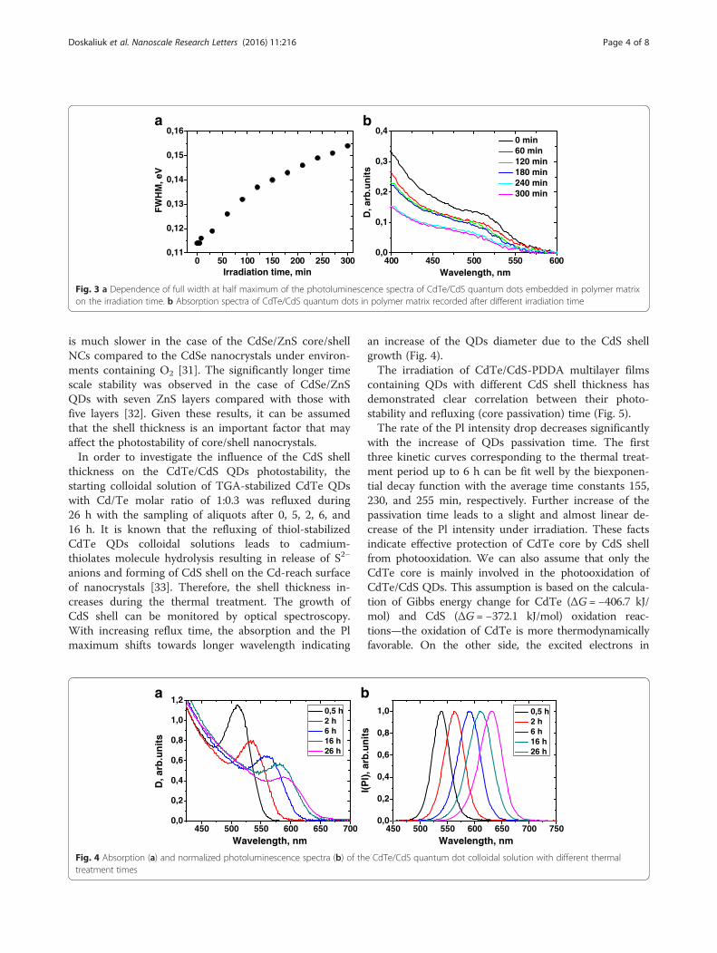
is much slower in the case of the CdSe/ZnS core/shellNCs compared to the CdSe nanocrystals under environ-ments containing O2 [31]. The significantly longer timescale stability was observed in the case of CdSe/ZnSQDs with seven ZnS layers compared with those withfive layers [32]. Given these results, it can be assumedthat the shell thickness is an important factor that mayaffect the photostability of core/shell nanocrystals.In order to investigate the influence of the CdS shell
thickness on the CdTe/CdS QDs photostability, thestarting colloidal solution of TGA-stabilized CdTe QDswith Cd/Te molar ratio of 1:0.3 was refluxed during26 h with the sampling of aliquots after 0, 5, 2, 6, and16 h. It is known that the refluxing of thiol-stabilizedCdTe QDs colloidal solutions leads to cadmium-thiolates molecule hydrolysis resulting in release of S2−
anions and forming of CdS shell on the Cd-reach surfaceof nanocrystals [33]. Therefore, the shell thickness in-creases during the thermal treatment. The growth ofCdS shell can be monitored by optical spectroscopy.With increasing reflux time, the absorption and the Plmaximum shifts towards longer wavelength indicating
an increase of the QDs diameter due to the CdS shellgrowth (Fig. 4).The irradiation of CdTe/CdS-PDDA multilayer films
containing QDs with different CdS shell thickness hasdemonstrated clear correlation between their photo-stability and refluxing (core passivation) time (Fig. 5).The rate of the Pl intensity drop decreases significantly
with the increase of QDs passivation time. The firstthree kinetic curves corresponding to the thermal treat-ment period up to 6 h can be fit well by the biexponen-tial decay function with the average time constants 155,230, and 255 min, respectively. Further increase of thepassivation time leads to a slight and almost linear de-crease of the Pl intensity under irradiation. These factsindicate effective protection of CdTe core by CdS shellfrom photooxidation. We can also assume that only theCdTe core is mainly involved in the photooxidation ofCdTe/CdS QDs. This assumption is based on the calcula-tion of Gibbs energy change for CdTe (ΔG = −406.7 kJ/mol) and CdS (ΔG = −372.1 kJ/mol) oxidation reac-tions—the oxidation of CdTe is more thermodynamicallyfavorable. On the other side, the excited electrons in
0,0
0,1
0,2
0,3
0,4
D, a
rb.u
nit
s
Wavelength, nm
0 min 60 min 120 min 180 min 240 min 300 min
400 450 500 550 6000 50 100 150 200 250 3000,11
0,12
0,13
0,14
0,15
0,16
FW
HM
, eV
Irradiation time, min
a b
Fig. 3 a Dependence of full width at half maximum of the photoluminescence spectra of CdTe/CdS quantum dots embedded in polymer matrixon the irradiation time. b Absorption spectra of CdTe/CdS quantum dots in polymer matrix recorded after different irradiation time
0,0
0,2
0,4
0,6
0,8
1,0
1,2
D, a
rb.u
nit
s
Wavelength, nm
0,5 h 2 h 6 h 16 h 26 h
450 500 550 600 650 700 450 500 550 600 650 700 7500,0
0,2
0,4
0,6
0,8
1,0
I(P
l), a
rb.u
nit
s
Wavelength, nm
0,5 h 2 h 6 h 16 h 26 h
ba
Fig. 4 Absorption (a) and normalized photoluminescence spectra (b) of the CdTe/CdS quantum dot colloidal solution with different thermaltreatment times
Doskaliuk et al. Nanoscale Research Letters (2016) 11:216 Page 4 of 8

CdTe/CdS QDs are delocalized throughout all hetero-structure due to a similar value of lower occupied molecu-lar orbital of the core and shell which may promote theshell oxidation. The long refluxing and increase in the shellthickness leads to the type II heterostructure formationwhen the excited electrons are localized in the shell[33]. The probability of its photooxidation should in-crease in this case. In contrast, we observed the sig-nificant increase in the CdTe/CdS QDs photostabilitywith increase in CdS shell thickness indicating thethermodynamic limitation of the photooxidation process.The change of the Pl energy maximum under irradi-
ation is also non-linear in all cases (Fig. 6a).
The best coefficient of determination was obtained bythe fitting of the experimental data with monomolecularexponential growth function:
y ¼ A 1−e−k x−xcð Þ� �
;
where k is the rate of the Pl energy maximum shift.The rate of the Pl energy maximum shift decreases twotimes with the increase in the passivation time for 6 hand then remains almost unchanged (Fig. 6b).It was suggested that the core photooxidation may be
possible due to oxygen diffusion through the shell incore/shell nanocrystals [32, 34]. Due to large lattice mis-match of zinc-blende CdTe and CdS (11.3 %), the for-mation of defects such as low-angle boundaries or grainboundaries is possible that significantly accelerates theoxygen diffusion to the core. The photooxidation prod-ucts may even rupture the shell resulting in the rapid re-duction of the QDs size. We propose that at the shortrefluxing times, incomplete passivation of the core oc-curs similarly to the oxidation observed for the CdSecore partially coated with ZnS [18]. According to our re-sults, a complete CdS shell is formed in 6 h throughrefluxing. A slight photooxidation of the complete pas-sivated nanocrystals is related to the oxygen diffusionthrough the shell. The combination of these two pro-cesses may predetermine the biexponential Pl decay andnonlinear change of the FWHM and the Pl energy max-imum of the QDs with refluxing time of up to 6 h. Thefirst time constant is one order less than the second inall cases. Therefore, we propose that it is related to fastphotooxidation of uncovered areas of the core which aremore able to the react with oxygen. At the same time,the formation of the CdO on top of those areas may pre-vent further photooxidation of QDs. The large second
0 50 100 150 200 250 300
0,2
0,4
0,6
0,8
1,0
Irradiation time, min
12
3
4
5
Fig. 5 Irradiation time dependence of the photoluminescenceintegral intensity of the CdTe/CdS quantum dots with differentpassivation times: 1–0.5, 2–2, 3–6, 4–16, and 5–26 h. Lines areaveraged through measurement of three different samples preparedwith the same quantum dots and are presented with error bars
5
6
7
8
9
10
Passivation time, hours0 5 10 15 20 250 50 100 150 200 250 300
1,90
1,95
2,00
2,05
2,10
2,15
2,20
2,25
2,30
54
3
2
Irradiation time, min
1
ba
Fig. 6 a Irradiation time dependence of photoluminescence energy maximum of the polymer composites containing CdTe/CdS quantum dotswith different passivation times: 1–0.5, 2–2, 3–6, 4–16, and 5–26 h. b Dependence of the Pl energy maximum shift rate on the passivation time ofquantum dots
Doskaliuk et al. Nanoscale Research Letters (2016) 11:216 Page 5 of 8

time constant can be attributed to the oxidation of thepassivated area of the core due to the slow oxygen diffu-sion through the shell. It should be emphasized that thefast oxidation of the anion vacancies is preceded or pre-cedes (with no “by” following it) by these two stages.Hence, we can propose the three-step photooxidationmodel of CdTe/CdS core/shell QDs embedded in poly-mer matrix (Fig. 7).During the first few minutes of irradiation, the fast
oxidation of anion vacancies accompanied by the H2Omolecules physisorption occurs. At the second stage, thebare CdTe core is oxidized resulting in a fast Pl decayand sufficient decrease of core radii due to TeO2 forma-tion. The third photooxidation stage caused by oxygendiffusion through the shell results in a slow Pl decay andQDs radii decrease.Taking into account that the uncovered area of
CdTe core is highly reactive in the photooxidationprocess but optical properties of such nanocrystalsare desirable in some cases, we propose to reduce thecore reactivity by decreasing its defectiveness. Bychanging the stoichiometric composition of CdTeclusters, we can obtain the nanocrystals with differentTe anion vacation concentration in the core. Whenthe other synthesis conditions including Cd/TGA ra-tio, refluxing time, and pH remain constant, thepassivation degree of this vacancies increases ap-proaching the stoichiometric Cd/Te ratio. The short-term refluxing, insufficient to complete passivation, isnecessary to obtain the different anion vacancy con-centrations in the prepared CdTe/CdS QDs.
Photoinduced Pl decay curves of the CdTe/CdS QDswith different Cd/Te ratio embedded in polymer filmsare shown in Fig. 8.Just like in the previous experiment, the dependence
of Pl intensity on irradiation duration can be fit well bybiexponential decay function with the average time con-stant 155 (curve 6), 230 (curve 7), and 255 min (curve8). Therefore, the decrease in the Cd/Te ratio and ap-proaching to the stoichiometric core composition resultsin a remarkable decrease of the photooxidation rate.This observation confirms that the anion vacancies areinvolved in the photooxidation of the CdTe/CdS QDs inaccordance with scheme 1. As has been discussed above,the vacancy oxidation occurs very fast during the firstminutes of irradiation, and therefore, it does not appearon the part of kinetic curves relating to the photooxida-tion. Probably, the dependence of the photooxidationrate on the vacancies concentration is caused by forma-tion of CdTe1−nOn fragments which acts as active cen-ters of further Te2− anions oxidation due to the shift ofthe electron density to oxygen and weakening of the Cd-Te bonds.
ConclusionsThe photooxidation and photomodification of theCdTe/CdS quantum dots embedded in the polymermatrix has been investigated. The quasi-inversephotoluminescence intensity increase has been ob-served during the first few minutes of UV irradiationindicating the surface trap passivation by H2O mole-cules accompanied by the anion vacancy oxidation.
Fig. 7 Schematic representation of the three-step photooxidation process of CdTe/CdS core/shell nanocrystal: 1—the anion vacancies oxidation,2—oxidation of bare CdTe core, and 3—oxidation of passivated area of the core due to oxygen diffusion through the CdS shell
Doskaliuk et al. Nanoscale Research Letters (2016) 11:216 Page 6 of 8

Further irradiation has led to quantum dot photooxi-dation manifested as a significant “blue shift” of thePl energy maximum and the decrease in photolumi-nescence intensity. The increase in the CdS shellthickness and decrease in Cd/Te ratio lead to a sig-nificant increase in the CdTe/CdS quantum dotphotostability. The three-step mechanism of CdTe/CdS quantum dot photooxidation including anion va-cancies oxidation, oxidation of the bare core area,and slow core oxidation via oxygen diffusion throughthe shell has been proposed.
AbbreviationsFTIR: Fourier transform infrared spectroscopy; FWHM: full width at halfmaximum; PDDA: poly(dimethyldiallylammonium chloride);Pl: photoluminescence; QDs: quantum dots; TGA: thioglycolic acid.
Competing InterestsThe authors declare that they have no competing interests.
Authors’ ContributionsND has conducted the synthesis of СdTe/CdS nanocrystals and preparation oflayer-by-layer films, performed the photooxidation experiments, analyzed anddiscussed the data, and wrote the manuscript. YK has contributed to the resultsdiscussion, photooxidation model suggestion, and the manuscript writing. PFhas contributed to the results discussion and improvement of the final text ofthe manuscript. All authors read and approved the final manuscript.
AcknowledgementsThe authors are grateful to Dr. Svitlana Filonenko for the assistance in theFTIR measurement.
FundingThis work was performed under the projects # 0115U003240 and "Opticallyactive materials based on metallic and semiconductor nanocrystalsembedded into the crystalline and amorphous matrix" with the financialsupport of the Ministry of Education and Science of Ukraine.
Received: 15 February 2016 Accepted: 13 April 2016
References1. Nenadovic MT, Micic RT (1985) Size quantization in small semiconductor
particles. J Phys Chem 89:397–92. Rossetti R, Ellison JL, Gibson JM, Brus LE (1984) Size effects in the excited
electronic states of small colloidal CdS crystallites. J Chem Phys 80:44643. Masumoto Y, Sonobe K (1997) Size-dependent energy levels of CdTe
quantum dots. Phys Rev B 56:97344. Sridharan D, Waks E (2011) All-optical switch using quantum-dot saturable
absorbers in a DBR microcavity. J Quantum Electron 47:31–95. Willard DM, Orden A (2003) Quantum dots: resonant energy-transfer sensor.
Nature Materials 2:575–66. Henini M, Bugajski M (2005) Advances in self-assembled semiconductor
quantum dot lasers. Microelectronics Journal 36:950–67. Sun Q, Wang YA, Li LS, Wang D, Zhu T, Xu J, Yang C, Li Y (2007) Bright,
multicoloured light-emitting diodes based on quantum dots. NaturePhotonics 1:717–22
8. Kim S, Imb SH, Kim SW (2013) Performance of light-emitting-diode basedon quantum dots. Nanoscale 5:5205
9. Yang X, Mutlugun E, Dang C, Dev K, Gao Y, Tan ST, Sun XW, Demir HV(2014) Highly flexible, electrically driven, top-emitting, quantum dotlightemitting stickers. ACS Nano 8:8224–31
10. Kamat PV (2008) Quantum dot solar cells. Semiconductor nanocrystals aslight harvesters. J Phys Chem C 112:18737–53
11. Kalytchuk S, Gupta S, Zhovtiuk O, Vaneski A, Kershaw SV, Fu H, Fan Z, KwokECH, Wang CF, Teoh WY, Rogach AL (2014) Semiconductor nanocrystals asluminescent down-shifting layers to enhance the efficiency of thin-filmCdTe/CdS and crystalline Si solar cells. J Phys Chem С 118:16393–400
12. Purcell-Milton F, Gun’ko YK (2012) Quantum dots for luminescent solarconcentrators. Mater Chem 22:16687
13. Bradshaw LR, Knowles KE, McDowall S, Gamelin DR (2015) Nanocrystals forluminescent solar concentrators. Nano Lett 15:1315–23
14. Liu YF, Yu JS (2009) Selective synthesis of CdTe and high luminescenceCdTe/CdS quantum dots: the effect of ligands. J Colloid Interf Sci 333:690–8
15. Li Z, Wan S, Xu W, Wang Y, Shah BR, Jin W, Chen Y, Li B (2015) Highlyluminescent film functionalized with CdTe quantum dots by layer-by-layerassembly. J Appl Polym Sci 132:41893
16. Young EM (1988) Electron-active silicon oxidation. Appl Phys A: Solids Surf47:259–69
17. Sato S, Nozaki S, Morisaki H (1997) Photo-oxidation of germanium nanostructuresdeposited by the cluster-beam evaporation technique. J Appl Phys 81:1518
18. Dabbousi BO, Rodriguez-Viejo J, Mikulec FV, Heine RJ, Mattoussi H, Ober R,Jensen KF, Bawendi MG (1997) (СdSe)ZnS core-shell quantum dots:synthesis and characterization of a size series of highly luminescentnanocrystallites. J Phys Chem B 101:9463–75
19. Hines MA, Guyot-Sionnest P (1996) Synthesis and characterization of stronglyluminescing ZnS-capped CdSe nanocrystals. J Phys Chem 100:468–71
20. Henglein A (1988) Mechanism of reactions on colloidal micro-electrodesand size quantization effects. Top Curr Chem 143:113–80
21. Bowen Katari JE, Colvin VL, Alivisatos AP (1994) X-ray photoelectronspectroscopy of CdSe nanocrystals with applications to studies of thenanocrystal surface. Phys Chem 98:4109–17
22. Hines DA, Becker MA, Kamat PV (2012) Photoinduced surface oxidation andits effect on the exciton dynamics of CdSe quantum dots. J Phys Chem C116:13452–57
23. Zhang Y, He J, Wang PN, Chen JY, Lu ZJ, Lu DR, Guo J, Wang CC, Yang WL(2006) Time-dependent photoluminescence blue shift of the quantum dotsin living cells: effect of oxidation by singlet oxygen. J Am Chem Soc 128:13396–401
24. Rogach AL, Katsikas L, Kornowski A, Su D, Eychmüller A, Weller H (1996)Synthesis and characterization of thiol-stabilzed CdTe nanocrystals. BerBunsenges Phys Chem 100:1772–8
25. Decher G (1997) Fuzzy nanoassemblies: toward layered polymericmulticomposites. Science 277:1232–37
26. Kotov NA, DCkiiny I, Fendler JH (1995) Layer-by-layer self-assembly ofpolyelectrolyte-semiconductor nanoparticle composite. J Phys Chem 99:13065–9
27. Cordero SR, Carson PJ, Estabrook RA, Strouse GF, Buratto SK (2000) Photo-activated luminescence of СdSe quantum dot monolayers. J Phys Chem B104:12137–42
28. Kepler KD, Lisensky GC, Patel M, Sigworth LA, Ellis AB (1995) Surface-boundcarbonyl compounds as lewis acids. Photoluminescence as a probe for the
0 50 100 150 200 250 300
0,2
0,4
0,6
0,8
1,0
Irradiation time, min
6
7
8
Fig. 8 Irradiation time dependence of photoluminescence integralintensity of CdTe/CdS quantum dots with different Cd/Te ratios: 6:20,7:4, 8:2. The refluxing time of the crude colloidal solution was 2 h
Doskaliuk et al. Nanoscale Research Letters (2016) 11:216 Page 7 of 8
binding of ketones and aldehydes to cadmium sulfide and cadmiumselenide surfaces. J Phys Chem 99:16011–7
29. Wang X, Zhang J, Nazzal A, Xiao M (2003) Photo-oxidation-enhanced couplingin densely packed CdSe quantum-dot films. App Phys Lett 83:162–4
30. Rogach AL, Klar TA, Lupton JM, Meijerinkd A, Feldmann J (2009) Energytransfer with semiconductor nanocrystals. J Mater Chem 19:1208–21
31. Nazzal AY, Wang X, Qu L, Yu W, Wang Y, Peng X, Xiao M (2004)Еnvironmental effects on photoluminescence of highly luminescent CdSeand CdSe/ZnS core/shell nanocrystals in polymer thin films. J Phys Chem B108:5507–15
32. Sark WGJHМ, Frederix PLTM, Heuvel DJ, Gerritsen HC (2001) Photooxidationand photobleaching of single CdSe/ZnS quantum dots probed by room-temperature time-resolved spectroscopy. J Phys Chem B 105:8281–4
33. Dai MQ, Zheng W, Huang Z, Yung LYL (2012) Aqueous phase synthesis ofwidely tunable photoluminescence emission CdTe/CdS core/shell quantumdots under a totally ambient atmosphere. J Mater Chem 22:16336–45
34. Sark WGJHM, Frederix PLTM, Bol AA, Gerritsen HC, Meijerink A (2002)Blueing, bleaching, and blinking of single CdSe/ZnS quantum dots.СhemPhysChem 3:871–9
Submit your manuscript to a journal and benefi t from:
7 Convenient online submission
7 Rigorous peer review
7 Immediate publication on acceptance
7 Open access: articles freely available online
7 High visibility within the fi eld
7 Retaining the copyright to your article
Submit your next manuscript at 7 springeropen.com
Doskaliuk et al. Nanoscale Research Letters (2016) 11:216 Page 8 of 8
![THICKNESS RATIO EFFECT ON THE DYNAMIC …facta.junis.ni.ac.rs/me/me2009/me2009-01.pdf · and airport runways (Fryba) [1]. However, ... Thickness Ratio Effect on the Dynamic Response](https://img.pdfslide.us/doc/110x75/5b3d19a47f8b9ace408dda29/thickness-ratio-effect-on-the-dynamic-factajunisniacrsmeme2009me2009-01pdf.jpg)