Embed Size (px)
Citation preview
© Horizon Scientific Press. Offprints from www.ciim.net
*For correspondence: [email protected]
Curr. Issues Intestinal Microbiol. 7: 35–52. Online journal at www.ciim.net
Influence of the Gastrointestinal Microbiota
on Development of the Immune System in
Young Animals
Eva Bauer1,2*, Barbara A. Williams2, Hauke
Smidt3, Martin W.A. Verstegen2, and Rainer
Mosenthin1
1Institute of Animal Nutrition, University of Hohenheim, Emil-Wolff-Str. 10, D-70599 Stuttgart, Germany2Animal Nutrition Group, Wageningen Institute of Animal Sciences, Wageningen University, Marijkeweg 40, 6709 PG Wageningen, The Netherlands3Laboratory of Microbiology, Wageningen University, Hesselink van Suchtelenweg 4, 6703 CT, Wageningen, The Netherlands
Abbreviations for scientific terms
Ang4: Angionin-4; APC: antigen-presenting cell; CD: cluster of differentiation; DC: dendritic cell; DGGE: denaturing gradient gel electrophoresis; FAE: follicle-associated epithelium; GALT: gut-associated-lymphoid tissue; GF: germ-free; GIT: gastrointestinal tract; IEL: intraepithelial lymphocytes; IFN: interferon; Ig: immunoglobulin; IL: interleukin; LPS: lipopolysaccharide; LTA: lipoteichoic acid; MHC: major histocompatibility complex; NDO: non-digestible oligosaccharides; NF: nuclear factor; NK: natural killer cell; PAMPs: pathogen-associated molecular patterns; PCR: polymerase chain reaction; PIgR: polymeric immunoglobulin receptor; PP: Peyer’s patches; SC: secretory component; SFB: segmented filamentous bacteria; sIgA: secretory immunoglobulin A; TGF: transforming growth factor; Th: T helper cell; TLR: Toll-like receptor; TNF: tumor necrosis factor.
Abstract
The gastrointestinal tract (GIT) of adult mammals is colonized by a complex and dynamic community of microorganisms. Most protection against potential pathogens occurs via a mucosal immune system involving mechanisms of innate immunity as well as a secondary lymphoid organ, the gut-associated lymphoid tissue (GALT). However, the bacterial community also supports its host against invasion by potential pathogens, by a mechanism called ‘colonization resistance’. Young animals need time to develop both a complex bacterial community and their immature GIT immune system, and until such developments have taken place, they are vulnerable to the presence of potential pathogens in their GIT. Initial protection against invading pathogens is provided by milk and colostrum, which contain antibodies and other bioactive components. At weaning, with the introduction of solid food and deprivation of the mother’s milk, the young must also cope with a rapidly changing microbiota. The colonizing microbiota not only provides colonization resistance to potentially pathogenic bacteria. It also has a major role in the development of
the intestinal immune system, both in terms of GALT development and mucosal immunity, and the induction of oral tolerance. Studies using gnotobiotic animal models have revealed that the presence of even limited numbers of the indigenous microbiota may influence the GIT immune system. Regulation of the composition of the GIT microbiota, e.g. by the use of pre- and probiotics, offers the possibility to influence the development of mucosal, and also systemic immunity.
Introduction
The bacterial community which inhabits the mammalian GIT is characterized by its high density and diversity. The colon contents support at least 400 different species, with numbers as high as 1010 and 1011 culturable bacteria/g of digesta (Savage, 1977; Mackie and White, 1997). These bacteria are constantly interacting with each other, and with the host, comprising a highly complex ecosystem of which comparatively little is known. It is only recently that new techniques in molecular biology are allowing the detection of microbial species that are either difficult or as yet impossible to culture (Vaughan et al., 2000; Tannock, 2001).
The resident microbiota confers many benefits to the intestinal physiology of the host and is, therefore, an example of a truly symbiotic relationship (Hooper and Gordon, 2001). Some of these benefits include the metabolism of nutrients and organic substrates, and the contribution to the phenomenon of colonization resistance. The latter is the ability of the GIT bacterial community to resist invasion of the host by exogenous microorganisms (Van der Waaij et al., 1971; Berg, 1996). Further benefits become visible in the period after birth, when the complexity of the intestinal environment increases considerably while changing from an exclusively milk-containing diet to an adult diet after weaning (Rumbo and Schiffrin, 2005). During this period, the intestinal microbiota plays a crucial inductive role in intestinal development. Studies in gnotobiotic animals have shown that association of germ-free rodents with a single bacterial species has a profound impact on the anatomical, physiological, and immunological development of the host. This includes microbicidal protein production, development of intestinal epithelium, vasculature and GALT (Shroff et al., 1995; Stappenbeck et al., 2002; Hooper, 2004). Bacterial colonization of the GIT is also essential for the development of oral tolerance, i.e. the systemic unresponsiveness to commensal bacteria and food proteins to avoid chronic inflammation. In the germ-free state, this mechanism does not exist, but develops rapidly after colonization (Sudo et al., 1997; Shi and Walker, 2004).
With the ban on dietary antibiotics as growth promoters within the European Union, animal nutritionists are seeking alternatives to these promoters, particularly for young animals. The beneficial effects of bacteria on
• MALDI-TOF Mass Spectrometry in Microbiology
Edited by: M Kostrzewa, S Schubert (2016) www.caister.com/malditof
• Aspergillus and Penicillium in the Post-genomic Era
Edited by: RP Vries, IB Gelber, MR Andersen (2016) www.caister.com/aspergillus2
• The Bacteriocins: Current Knowledge and Future Prospects
Edited by: RL Dorit, SM Roy, MA Riley (2016) www.caister.com/bacteriocins
• Omics in Plant Disease Resistance
Edited by: V Bhadauria (2016) www.caister.com/opdr
• Acidophiles: Life in Extremely Acidic Environments
Edited by: R Quatrini, DB Johnson (2016) www.caister.com/acidophiles
• Climate Change and Microbial Ecology: Current Research and Future Trends
Edited by: J Marxsen (2016) www.caister.com/climate
• Biofilms in Bioremediation: Current Research and Emerging Technologies
Edited by: G Lear (2016) www.caister.com/biorem
• Microalgae: Current Research and Applications
Edited by: MN Tsaloglou (2016) www.caister.com/microalgae
• Gas Plasma Sterilization in Microbiology: Theory, Applications, Pitfalls and New Perspectives
Edited by: H Shintani, A Sakudo (2016) www.caister.com/gasplasma
• Virus Evolution: Current Research and Future Directions
Edited by: SC Weaver, M Denison, M Roossinck, et al. (2016) www.caister.com/virusevol
• Arboviruses: Molecular Biology, Evolution and Control
Edited by: N Vasilakis, DJ Gubler (2016) www.caister.com/arbo
• Shigella: Molecular and Cellular Biology
Edited by: WD Picking, WL Picking (2016) www.caister.com/shigella
• Aquatic Biofilms: Ecology, Water Quality and Wastewater Treatment
Edited by: AM Romaní, H Guasch, MD Balaguer (2016) www.caister.com/aquaticbiofilms
• Alphaviruses: Current Biology
Edited by: S Mahalingam, L Herrero, B Herring (2016) www.caister.com/alpha
• Thermophilic Microorganisms
Edited by: F Li (2015) www.caister.com/thermophile
• Flow Cytometry in Microbiology: Technology and Applications
Edited by: MG Wilkinson (2015) www.caister.com/flow
• Probiotics and Prebiotics: Current Research and Future Trends
Edited by: K Venema, AP Carmo (2015) www.caister.com/probiotics
• Epigenetics: Current Research and Emerging Trends
Edited by: BP Chadwick (2015) www.caister.com/epigenetics2015
• Corynebacterium glutamicum: From Systems Biology to Biotechnological Applications
Edited by: A Burkovski (2015) www.caister.com/cory2
• Advanced Vaccine Research Methods for the Decade of Vaccines
Edited by: F Bagnoli, R Rappuoli (2015) www.caister.com/vaccines
• Antifungals: From Genomics to Resistance and the Development of Novel Agents
Edited by: AT Coste, P Vandeputte (2015) www.caister.com/antifungals
• Bacteria-Plant Interactions: Advanced Research and Future Trends
Edited by: J Murillo, BA Vinatzer, RW Jackson, et al. (2015) www.caister.com/bacteria-plant
• Aeromonas
Edited by: J Graf (2015) www.caister.com/aeromonas
• Antibiotics: Current Innovations and Future Trends
Edited by: S Sánchez, AL Demain (2015) www.caister.com/antibiotics
• Leishmania: Current Biology and Control
Edited by: S Adak, R Datta (2015) www.caister.com/leish2
• Acanthamoeba: Biology and Pathogenesis (2nd edition)
Author: NA Khan (2015) www.caister.com/acanthamoeba2
• Microarrays: Current Technology, Innovations and Applications
Edited by: Z He (2014) www.caister.com/microarrays2
• Metagenomics of the Microbial Nitrogen Cycle: Theory, Methods and Applications
Edited by: D Marco (2014) www.caister.com/n2
Caister Academic Press is a leading academic publisher of advanced texts in microbiology, molecular biology and medical research. Full details of all our publications at caister.com
Further Reading
Order from caister.com/order
36 Bauer et al.
the immune system have been proposed as one theory supporting the use of probiotic bacteria as an alternative to antibiotics in improving animal health and protection against infectious agents (Simon et al., 2003). Probiotics are live microorganisms that may beneficially affect the GIT balance, going far beyond their conventional nutritional effect (Fuller, 1992). A number of studies have reported immune-stimulating effects of different bacterial species, such as production of microbicidal proteins, or an enhanced antibody response (Forchielli and Walker, 2005). They may also lead to alleviation of intestinal inflammatory responses by regulation of cytokine production (Isolauri et al., 2001).
An improved understanding of the complex relation-ship between the indigenous intestinal microbiota and the host immune system will help to establish strategies to improve host health. This is of special importance in the period after birth, or at stressful moments in animal lives. For piglets, interest has focussed on the time of abrupt weaning, which has always been a source of economic losses for pig farmers.
Development of the intestinal microbiota from birth
After birth, the intestinal microbiota takes some time before developing a stable community (Gaskins, 2001). Colonization is a complex process of natural selection and ecological succession. It depends on various factors, some of which are of host origin, such as the genome and physiology of the animal, while others are of microbial origin, such as interactions between bacterial species (Konstantinov et al., 2004b).
During the first few weeks of life, microbial succession in the GIT of humans (Favier et al., 2002), pigs (Moughan et al., 1992), chickens (Barrow, 1992) and calves (Smith, 1965) is remarkably similar, even though animal species are usually exposed to greater numbers of bacteria from fecal and environmental sources, compared with humans. After birth, the germ-free GIT is rapidly colonized by anaerobic and facultative anaerobic bacteria. Culture studies have indicated that in general, humans are initially colonized by species showing a high reductive potential (e.g., Enterobacter). They metabolize oxygen, thus indirectly encouraging the growth of anaerobic bacteria including lactobacilli and bifidobacteria, Bacteroides and clostridia (Mackie et al., 1999; Teitelbaum and Walker, 2002). Favier et al. (2002) investigated the succession of bacterial communities in human neonates, by monitoring 16S rRNA gene diversity in fecal samples using PCR/DGGE. The first colonizers belonged to Escherichia coli or Clostridium, followed after a few days by Bifidobacterium, which then remained predominant in breast-fed infants. After weaning, Clostridium, Ruminococcus, Enterococcus and Enterobacter spp. appeared, and microbial DGGE profiles became more complex and also more stable with increasing age.
In suckling piglets, on the other hand, the population of fecal bifidobacteria seems to be numerically low (Mikkelsen et al., 2003), or even absent (Konstantinov et al., 2004a). Lactobacilli, however, establish early in the piglet’s intestine, and, although succession does occur throughout the pig’s lifetime, they remain a predominant member of the small intestinal microbiota (Tannock et al., 1990; Naito et al., 1995; Stewart, 1997). At weaning, when
it occurs early, the transition from milk to a solid diet leads to dramatic changes in the composition of the microbial community during the 7–14 days after weaning (Hillman, 2001). According to Ewing and Cole (1994), numbers of lactobacilli and other beneficial bacteria decrease in times of stress, as do their beneficial effects, allowing potential pathogens such as coliforms to increase. Franklin et al. (2002) found that lactobacilli populations in different GIT sections (jejunum, ileum, cecum) declined to lower levels in early-weaned pigs (17 days), compared with piglets weaned at 24 days.
Mucosal immunityThe mucosal immune system is directly exposed to the external environment and stimulated by antigens consisting of commensal and potentially pathogenic bacteria, dietary antigens, and viruses (Elson, 1985; Mayer, 1997; 2000; Nagler-Anderson and Shi, 2001). The basic mechanism of mucosal immunity is innate, non-specific immunity represented by processes that protect the host immediately after exposure to pathogens (Tlaskalova-Hogenova et al., 2004). Non-specific mechanisms include e.g. natural killer cells (NK) and activated macrophages, but also soluble mediators such as cytokines and ‘complement’, a system of proteolytic enzymes, which plays an important role in the elimination of microbes (Fearon, 2000; Janeway, 2001; Beutler, 2004).
The intestinal epithelium provides an essential part of innate immunity, as it must control the access of potential antigens and pathogens. It is supported by tight junctions which join intestinal epithelial cells, and restrict the passage of even very small (2 kDa) molecules (Madara, 1998). Further protection is provided by the generation of antimicrobial substances, including inorganic disinfectants (e.g. hydrogen peroxide and nitric oxide), large antimicrobial proteins (e.g. lysozyme and lactoferrin), or small antimicrobial peptides (e.g. defensins). The -defensins are synthesized by epithelial cells lining the mucous membranes of, e.g., the GIT. On the other hand, –defensins are produced in granules of neutrophils and Paneth cells of the small intestine (reviewed by Zhang et al., 2000; Zasloff, 2002; Lehrer, 2004). The present understanding of defensin antimicrobial mechanisms is that the peptides disable susceptible organisms by disrupting structural elements of the target cell membranes, and that permeabilization of the bacterial envelope is linked to microbicidal activity (Lehrer et al., 1989; Selsted and Ouellette, 2005).
A significant barrier to antigen entry is also provided by the mucin glycoproteins that line the surface epithelium in the GIT (McNabb and Tomasi, 1981; Deplancke and Gaskins, 2001). Bacteria and viruses become trapped in the mucus layer and are expelled by the peristaltic process of the gut, thereby preventing potential pathogens and antigens from gaining access to the underlying epithelium, a process called non-immune exclusion. Mucins also serve as a reservoir for secretory IgA (sIgA).
Gut-associated lymphoid tissue (GALT)The GALT is a secondary lymphoid organ, which can be divided into inductive and effector sites. The different lymphoid tissues found in the intestine are summarized in Table 1.
Gastrointestinal Microbiota and Immune Development 37
The inductive sites, mainly the Peyer’s patches (PP), consist of aggregations of lymphoid follicles. Their surface is covered by a unique epithelium (termed follicle-associated epithelium, FAE) which contains, interspersed between enterocytes, a specialized epithelial cell type, known as M cells (Brandtzaeg, 2003a). The brush border glycocalyx that characterizes villus enterocytes is absent from the apical surface of the M cell. It is replaced by microfolds (hence ‘M’ cells) that are more accessible to luminal antigens (Owen and Jones, 1974; Hathaway and Kraehenbuhl, 2000). M cells use trans-epithelial vesicular transport to carry antigens in the underlying GALT.
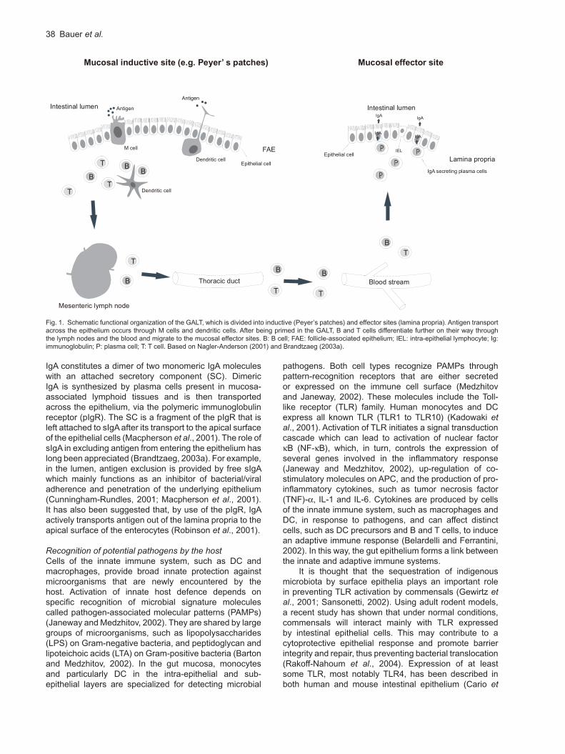
In the PP, antigens are presented by antigen-presenting cells (APCs, macrophages and dendritic cells) to both immature T and B cells (Kraehenbuhl and Neutra, 1992). Activated T cells preferentially differentiate into CD4+ helper cells which, aided by dendritic cells (DCs) and secretion of cytokines such as transforming growth factor (TGF)- and interleukin (IL)-10, induce the differentiation of antigen-specific B cells to predominantly IgA-committed plasmablasts (Brandtzaeg et al., 1999). The GALT-derived B cell blasts proliferate and differentiate further on their way through the mesenteric lymph nodes and the thoracic duct into the bloodstream. Then they migrate preferentially to the mucosal effector sites (i.e. lamina propria and intraepithelial regions, but not PP). Here they complete their terminal differentiation to IgA-producing plasma cells – a process called ‘homing’ (Kraehenbuhl and Neutra, 1992). A schematic depiction of the functional organization of the GALT with inductive and effector sites, is shown in Fig. 1.
However, there are also other cell types capable of transporting antigen across the epithelial barrier
(Rescigno et al., 2001). Dendritic cells may extend their dendritic-like processes through epithelial tight junctions and sample luminal antigen directly. However, the principal function of DC appears to be activation of T cells (Banchereau and Steinman, 1998). Circulating precursor DC enter peripheral tissues as immature DC, where they capture microbial or viral antigens, thereby acting as sentinels at the front line of host defence. Following antigen capture, the immature DC leave the tissues and migrate to lymphoid organs, where after maturation, they display antigen-derived peptides on their major histocompatibility complex (MHC) molecules, which in turn, select circulating antigen-specific T cells (Palucka and Banchereau, 1999).
Intestinal DC can retain small numbers of live commensals for several days. This allows DC to selectively induce IgA, which helps to protect against mucosal penetration by commensals. The commensal-loaded DC are restricted to the mucosal immune compartment by the mesenteric lymph nodes, which ensures that the immune responses to commensal bacteria are induced locally. This prevents any induction of either a systemic immune response or a damaging inflammatory response. So, IgA production, and probably intestinal T cell responses, can be selectively induced by DC loaded with commensal bacteria, and this increased local secretion of IgA limits the penetration of commensal bacteria (Macpherson and Uhr, 2004).
Secretory IgA (slgA)The most abundantly produced immunoglobulin in mammals is IgA, which is secreted mainly across mucous membranes, e.g., of the intestine. This ‘secretory’ form of
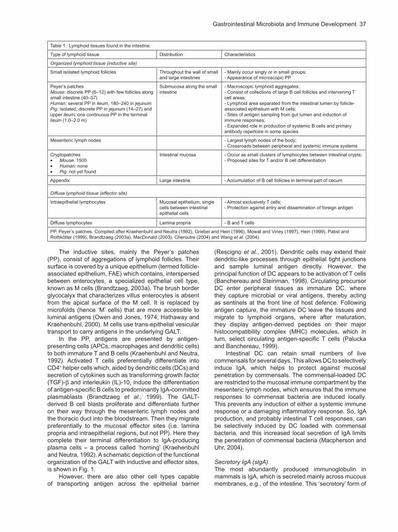
Table 1. Lymphoid tissues found in the intestine.
Type of lymphoid tissue Distribution Characteristics
Organized lymphoid tissue (inductive site)
Small isolated lymphoid follicles Throughout the wall of small and large intestines
- Mainly occur singly or in small groups;- Appearance of microscopic PP
Peyer’s patchesMouse: discrete PP (6–12) with few follicles along small intestine (40–57)Human: several PP in ileum, 180–240 in jejunumPig: Isolated, discrete PP in jejunum (14–27) and upper ileum; one continuous PP in the terminal ileum (1.0–2.0 m)
Submucosa along the small intestine
- Macroscopic lymphoid aggregates;- Consist of collections of large B cell follicles and intervening T cell areas;- Lymphoid area separated from the intestinal lumen by follicle- associated epithelium with M cells;- Sites of antigen sampling from gut lumen and induction of immune responses;- Expanded role in production of systemic B cells and primary antibody repertoire in some species
Mesenteric lymph nodes - Largest lymph nodes of the body;- Crossroads between peripheral and systemic immune systems
Cryptopatches Mouse: 1500 Human: none Pig: not yet found
Intestinal mucosa - Occur as small clusters of lymphocytes between intestinal crypts;- Proposed sites for T and/or B cell differentiation
Appendix Large intestine - Accumulation of B cell follicles in terminal part of cecum
Diffuse lymphoid tissue (effector site)
Intraepithelial lymphocytes Mucosal epithelium, single cells between intestinal epithelial cells
- Almost exclusively T cells;- Protection against entry and dissemination of foreign antigen
Diffuse lymphocytes Lamina propria - B and T cells
PP, Peyer’s patches. Compiled after Kraehenbuhl and Neutra (1992), Griebel and Hein (1996), Mowat and Viney (1997), Hein (1999), Pabst and Rothkötter (1999), Brandtzaeg (2003a), MacDonald (2003), Cheroutre (2004) and Wang et al. (2004).
38 Bauer et al.
IgA constitutes a dimer of two monomeric IgA molecules with an attached secretory component (SC). Dimeric IgA is synthesized by plasma cells present in mucosa-associated lymphoid tissues and is then transported across the epithelium, via the polymeric immunoglobulin receptor (pIgR). The SC is a fragment of the pIgR that is left attached to sIgA after its transport to the apical surface of the epithelial cells (Macpherson et al., 2001). The role of sIgA in excluding antigen from entering the epithelium has long been appreciated (Brandtzaeg, 2003a). For example, in the lumen, antigen exclusion is provided by free sIgA which mainly functions as an inhibitor of bacterial/viral adherence and penetration of the underlying epithelium (Cunningham-Rundles, 2001; Macpherson et al., 2001). It has also been suggested that, by use of the pIgR, IgA actively transports antigen out of the lamina propria to the apical surface of the enterocytes (Robinson et al., 2001).
Recognition of potential pathogens by the hostCells of the innate immune system, such as DC and macrophages, provide broad innate protection against microorganisms that are newly encountered by the host. Activation of innate host defence depends on specific recognition of microbial signature molecules called pathogen-associated molecular patterns (PAMPs) (Janeway and Medzhitov, 2002). They are shared by large groups of microorganisms, such as lipopolysaccharides (LPS) on Gram-negative bacteria, and peptidoglycan and lipoteichoic acids (LTA) on Gram-positive bacteria (Barton and Medzhitov, 2002). In the gut mucosa, monocytes and particularly DC in the intra-epithelial and sub-epithelial layers are specialized for detecting microbial
pathogens. Both cell types recognize PAMPs through pattern-recognition receptors that are either secreted or expressed on the immune cell surface (Medzhitov and Janeway, 2002). These molecules include the Toll-like receptor (TLR) family. Human monocytes and DC express all known TLR (TLR1 to TLR10) (Kadowaki et al., 2001). Activation of TLR initiates a signal transduction cascade which can lead to activation of nuclear factor B (NF- B), which, in turn, controls the expression of
several genes involved in the inflammatory response (Janeway and Medzhitov, 2002), up-regulation of co-stimulatory molecules on APC, and the production of pro-inflammatory cytokines, such as tumor necrosis factor (TNF)- , IL-1 and IL-6. Cytokines are produced by cells of the innate immune system, such as macrophages and DC, in response to pathogens, and can affect distinct cells, such as DC precursors and B and T cells, to induce an adaptive immune response (Belardelli and Ferrantini, 2002). In this way, the gut epithelium forms a link between the innate and adaptive immune systems.
It is thought that the sequestration of indigenous microbiota by surface epithelia plays an important role in preventing TLR activation by commensals (Gewirtz et al., 2001; Sansonetti, 2002). Using adult rodent models, a recent study has shown that under normal conditions, commensals will interact mainly with TLR expressed by intestinal epithelial cells. This may contribute to a cytoprotective epithelial response and promote barrier integrity and repair, thus preventing bacterial translocation (Rakoff-Nahoum et al., 2004). Expression of at least some TLR, most notably TLR4, has been described in both human and mouse intestinal epithelium (Cario et
Fig. 1. Schematic functional organization of the GALT, which is divided into inductive (Peyer’s patches) and effector sites (lamina propria). Antigen transport across the epithelium occurs through M cells and dendritic cells. After being primed in the GALT, B and T cells differentiate further on their way through the lymph nodes and the blood and migrate to the mucosal effector sites. B: B cell; FAE: follicle-associated epithelium; IEL: intra-epithelial lymphocyte; Ig: immunoglobulin; P: plasma cell; T: T cell. Based on Nagler-Anderson (2001) and Brandtzaeg (2003a).
Mucosal inductive site (e.g. Peyer’ s patches) Mucosal effector site
BB
B
T T
T
Thoracic duct Blood stream
Mesenteric lymph node
BB
TT
T
B
FAEM cell
Dendritic cell
Dendritic cell
Antigen
Antigen
Epithelial cell
Intestinal lumen
B
T
IgA
IgA
IgA
IgA
Intestinal lumen
IEL
IgA secreting plasma cells
Epithelial cellLamina propria
Figure 1. Schematic functional organization of the GALT, which is divided into inductive (Peyer’s
patches) and effector sites (lamina propria). Antigen transport across the epithelium occurs through M
cells and dendritic cells. After being primed in the GALT, B and T cells differentiate further on their way
through the lymph nodes and the blood and migrate to the mucosal effector sites. B: B cell; FAE:
follicle-associated epithelium; IEL: intra-epithelial lymphocyte; Ig: immunoglobulin; P: plasma cell; T: T
cell. Based on Nagler-Anderson (2001) and Brandtzaeg (2003a).
Gastrointestinal Microbiota and Immune Development 39
al., 2000; 2002; Ortega-Cava et al., 2003). Thus, TLR function as sensors of microbial infection, and are critical for the initiation of inflammatory and immune defence responses. However, they also seem to play a major role in maintaining intestinal epithelial homeostasis. However, it is not known to what extent this mechanism operates in newborns (Rumbo and Schiffrin, 2005).
Protection after birth
Role of milk and colostrumIn contrast to humans and rodents, which benefit from a transplacental passage of maternal serum antibodies during embryonic development, the multi-layered placenta in pigs prevents the transit of maternal Ig into the fetus (Butler, 1998; Salmon, 1999). Thus, first protection of the neonate piglet is provided by colostrum and milk.
In pigs, systemic humoral immunity is transmitted through colostrums conveying mainly IgG, and lower concentrations of IgA and IgM (Klobasa and Butler, 1987; Klobasa et al., 1987). Furthermore, a local humoral immunity, via sIgA, is transmitted mainly by milk (lactogenic immunity) until weaning. The specifity of these antibodies is believed to be acquired through the ‘entero-mammary’ pathway: during lactation, IgA+ B lymphocytes migrate from the intestine to the mammary glands, where they produce sIgA antibodies against the mother’s previous and present intestinal microbiota (Roux et al., 1977). Since the antigenic specifity of the antibodies reflects the maternal experience of environmental antigens, immunity acquired through colostrum and milk will protect the piglet against these antigens, but not against novel antigens (Rooke and Bland, 2002). In humans, it has been shown that free secretory component, which is abundant in breast milk, may, on its own, block epithelial adhesion, and thereby limit infection by enterotoxigenic E. coli (De Oliveira et al., 2001). Free SC is released into secretions when the polymeric Ig receptor is transcytosed without being bound to IgA (Mullock et al., 1980; Phalipon and Corthésy, 2003).
Porcine milk also contains other factors, which support resistance of the neonate against infections. These include maternal cells such as phagocytes, lymphocytes and epithelial cells, as well as antimicrobial substances, e.g. lactoferrin and lysozyme (reviewed by Wagstrom et al., 2000). Lactoferrin has a number of effects including microbicidal, immunostimulatory, and efficiently anti-inflammatory, by turning off the production of numerous pro-inflammatory cytokines such as IL-1 , IL-6, TNF- , and IL-8 (Mattsby-Baltzer et al., 1996; Håversen et al., 2000; Elass et al., 2002). During neonatal colonization and the subsequent expansion of the intestinal microbiota, it is particularly important to have considerable amounts of a protein such as lactoferrin in the gut, which is bactericidal and also prevents the induction of cytokines that cause clinical symptoms, energy consumption and inflammation (Hanson et al., 2003).
In sow’s milk, there are also considerable amounts of polyamines, which are of fundamental importance with regard to cell proliferation and differentiation (Johnson, 1988), but which also seem to be involved in the
maturation of the intestinal immune system. For example, oral administration of spermine to neonatal mice affected differentiation of the intraepithelial lymphocytes (Ter Steege et al., 1997). Furthermore, it has been suggested that polyamines are involved in the maturation of the glycosylation of the enterocytes (Biol-N’garagba and Louisot, 2003). Intestinal glycosylation is relevant to the implantation of the microbiota as the glycan structures at the enterocytes’ surface may determine the adhesion of specific pathogens (Jones and Freter, 1976).
Milk can also contain components such as glycoproteins, glycolipids, mucins and oligosaccharides (Newburg, 1999), some of which exhibit antimicrobial activity, but which may also act as growth promoters for bifidobacteria (Kunz and Rudloff, 1993). For example, the proliferation of Bifidobacterium bifidum and lactobacilli in the lower GIT is promoted by certain glycopeptides and glycoproteins, including caseins in human milk (Bezkorovainy and Topouzian, 1981; Liepke et al., 2002). Human milk oligosaccharides may also act as specific ‘bifidogenic factors’, supporting the survival of these bacteria (Beerens et al., 1980). They can also function as receptor analogues that inhibit the binding of enteric or respiratory bacterial pathogens, or their toxins, to epithelial cells (Kunz et al., 2000). Compared to other host species, human milk is considered to be unique in terms of its complex oligosaccharide content (Rudloff and Kunz, 1997). Although only present in small quantities in porcine milk, oligosaccharides may exert similar biological effects (Xu, 2003).
Development of the structure of the mucosal immune systemIn humans, PP and other structures of the mucosa-associated lymphoid tissue are already well developed at birth. However, they do not contain any secondary follicles with germinal centres signifying B cell activation, until some weeks after birth. This reflects their dependency on exogenous stimuli (Brandtzaeg et al., 1991; Brandtzaeg, 2003b). The lamina propria contains very few immunoglobulin-containing cells (Russell et al., 1990), which are mainly IgM+ and almost never IgA+ (Iwase et al., 1987; Russell et al., 1990). Fetal GIT contents or neonatal secretions contain no or only low levels of sIgA, but relatively more IgM (Petit et al., 1973; Gleeson et al., 1982; Mellander et al., 1984). This is due to the fact that IgM, in the absence of dimeric IgA, can bind to SC and be transported out into mucosal secretions (Hanson et al., 1999).
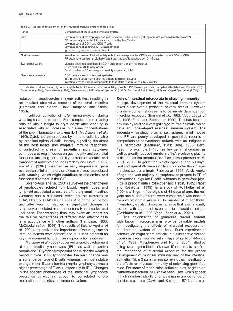
In the piglet, although considerable development of the systemic immune system has already taken place, the cells and structure of the mucosal immune system are almost completely absent, or immature, at birth (Gaskins, 1998). Four major phases of development of the mucosal immune system have been identified as shown in Table 2.
Effects of weaning on mucosal immunityWeaning is associated with alterations in intestinal morphology, such as villous atrophy and crypt hyperplasia in the small intestine (Kelly et al., 1992), together with a
40 Bauer et al.
reduction in brush-border enzyme activities, resulting in an impaired absorptive capacity of the small intestine (Hampson and Kidder, 1986; Hampson and Smith, 1986).
In addition, activation of the GIT immune system during weaning has been reported. For example, the decreasing ratio of villous height to crypt depth after weaning is associated with an increase in plasma concentrations of the pro-inflammatory cytokine IL-1 (McCracken et al., 1995). Cytokines are produced by immune cells, but also by intestinal epithelial cells, thereby signalling the onset of the host innate and adaptive immune responses. Uncontrolled synthesis of pro-inflammatory cytokines can have a strong influence on gut integrity and epithelial functions, including permeability to macromolecules and transport of nutrients and ions (McKay and Baird, 1999). Pié et al. (2004) observed an early response in gene expression of inflammatory cytokines in the gut associated with weaning, which might contribute to anatomical and functional disorders in the GIT.
Solano-Aguilar et al. (2001) surveyed the phenotypes of lymphocytes isolated from blood, lymph nodes, and lymphoid associated structures of the pig small intestine. Weaning had a significant effect on the numbers of CD4+, CD8+ or CD4+/CD8+ T cells. Age of the pig before and after weaning resulted in significant changes in lymphocytes isolated from mesenteric lymph nodes and ileal sites. That weaning time may exert an impact on the relative percentages of differentiated effector cells is in accordance with other authors (Hampson, 1986; McCracken et al., 1999). The results of Solano-Aguilar et al. (2001) emphasized the importance of weaning time on immune system development and thus their potential as key management factors in swine production systems.
Manzano et al. (2002) observed a rapid development of intraepithelial lymphocytes (IEL), as well as lamina propria and PP lymphocyte populations during the weaning period in mice. In PP lymphocytes the main change was a higher percentage of B cells, whereas the most notable change in the IEL and lamina propria lymphocytes was a higher percentage of T cells, especially in IEL. Changes in the specific phenotypes of the intestinal lymphocyte population at weaning appear to be related to the maturation of the intestinal immune system.
Role of intestinal microbiota in shaping immunity
In pigs, development of the mucosal immune system takes place over a period of several weeks. However, this development also seems to be largely dependent on microbial exposure (Bianchi et al., 1992; Vega-López et al., 1995; Pabst and Rothkötter, 1999). This has become obvious by studies investigating germ-free animals, which have an undeveloped mucosal immune system. The secondary lymphoid organs, i.e., spleen, lymph nodes and PP, are poorly developed in germ-free rodents in comparison to conventional rodents with an indigenous GIT microbiota (Bealmear, 1981; Berg, 1983; Berg, 1996). For example, PP contain few germinal centres, as well as greatly reduced numbers of IgA-producing plasma cells and lamina propria CD4+ T cells (Macpherson et al., 2001; 2003). In germ-free piglets aged 39 and 59 days, ileal and jejunal PP were significantly shorter than in age-matched control animals (Pabst et al., 1988). At six weeks of age, the vast majority of lymphocytes present in PP of conventional pigs are B cells, whereas in germ-free pigs, T cells predominate (Rothkötter and Pabst, 1989; Pabst and Rothkötter, 1999). In a study of Rothkötter et al. (1994), with germ-free piglets of 45 days of age, the cell yield and subset patterns were comparable with those in five-day old normal animals. The number of intraepithelial T lymphocytes also shows an increase that is significantly related with age and exposure to microbial antigen (Rothkötter et al., 1999; Vega-López et al., 2001).
The colonization of germ-free reared animals with known microorganisms provide valuable models for investigating the effects of microbial exposure on the immune system of the host. Such experimental colonization might seem artificial, but similar colonization occurs in every neonate within days of its birth (Mackie et al., 1999; Macpherson and Harris, 2004). Studies using such ‘gnotobiotic’ (‘known life’) animals confirm the importance of microbial exposure for the proper development of mucosal immunity and of the intestinal epithelia. Table 3 summarizes some studies investigating the effects on mucosal immunity of colonizing germ-free mice. For some of these colonization studies, segmented filamentous bacteria (SFB) have been used, which appear in high numbers shortly after weaning in a wide range of species e.g. mice (Davis and Savage, 1974), and pigs
Table 2. Phases of development of the mucosal immune system of the piglet.
Period Components of the mucosal immune system
Birth Low numbers of macrophage and granulocytes in villous and crypt regions (not yet functionally mature?)PP consist of primordial follicles surrounded by few T cellsLow numbers of CD4+ and CD8+ T cellsLow numbers of intestinal MHC class II+ cellsIg-containing cells are rare or absent
First two weeks Intestine becomes colonized with lymphoid cells (express the CD2 surface marker but not CD4 or CD8)PP begin to organize (a relatively ‘adult architecture’ is reached by 10–15 days)
Two to four weeks Mucosa becomes colonized by CD4+ cells (mainly in lamina propria)CD8+ cells are still largely absentSmall numbers of B cells appear, mainly expressing IgM
Five weeks onwards CD8+ cells appear in intestinal epitheliumIgA+ B cells appear (IgA becomes the predominant isotype)Intestinal architecture is comparable to that of the mature animal by 7 weeks
CD: cluster of differentiation; Ig: immunoglobulin; MHC: major histocompatibility complex; PP: Peyer’s patches. Compiled after Allen and Porter (1973), Butler et al. (1981), Bianchi et al. (1992), Stokes et al. (1992), Vega-López et al. (1995), Pabst and Rothkötter (1999) and Vega-López et al. (2001).
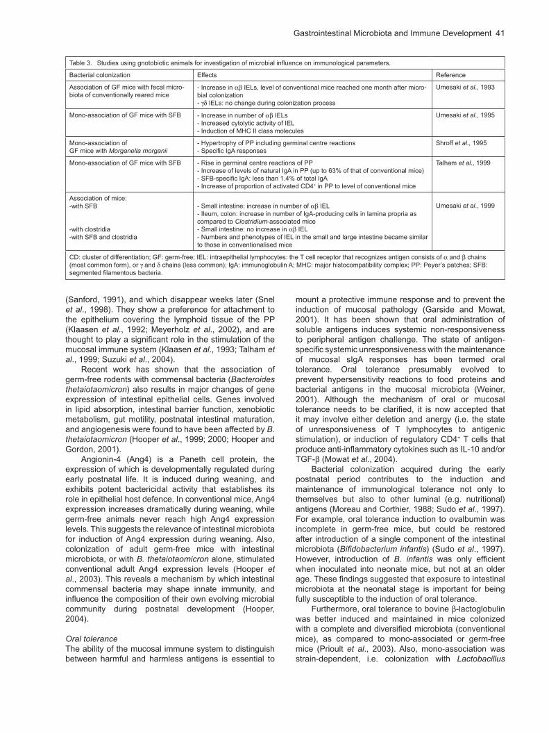
Gastrointestinal Microbiota and Immune Development 41
(Sanford, 1991), and which disappear weeks later (Snel et al., 1998). They show a preference for attachment to the epithelium covering the lymphoid tissue of the PP (Klaasen et al., 1992; Meyerholz et al., 2002), and are thought to play a significant role in the stimulation of the mucosal immune system (Klaasen et al., 1993; Talham et al., 1999; Suzuki et al., 2004).
Recent work has shown that the association of germ-free rodents with commensal bacteria (Bacteroides thetaiotaomicron) also results in major changes of gene expression of intestinal epithelial cells. Genes involved in lipid absorption, intestinal barrier function, xenobiotic metabolism, gut motility, postnatal intestinal maturation, and angiogenesis were found to have been affected by B. thetaiotaomicron (Hooper et al., 1999; 2000; Hooper and Gordon, 2001).
Angionin-4 (Ang4) is a Paneth cell protein, the expression of which is developmentally regulated during early postnatal life. It is induced during weaning, and exhibits potent bactericidal activity that establishes its role in epithelial host defence. In conventional mice, Ang4 expression increases dramatically during weaning, while germ-free animals never reach high Ang4 expression levels. This suggests the relevance of intestinal microbiota for induction of Ang4 expression during weaning. Also, colonization of adult germ-free mice with intestinal microbiota, or with B. thetaiotaomicron alone, stimulated conventional adult Ang4 expression levels (Hooper et al., 2003). This reveals a mechanism by which intestinal commensal bacteria may shape innate immunity, and influence the composition of their own evolving microbial community during postnatal development (Hooper, 2004).
Oral toleranceThe ability of the mucosal immune system to distinguish between harmful and harmless antigens is essential to
mount a protective immune response and to prevent the induction of mucosal pathology (Garside and Mowat, 2001). It has been shown that oral administration of soluble antigens induces systemic non-responsiveness to peripheral antigen challenge. The state of antigen-specific systemic unresponsiveness with the maintenance of mucosal sIgA responses has been termed oral tolerance. Oral tolerance presumably evolved to prevent hypersensitivity reactions to food proteins and bacterial antigens in the mucosal microbiota (Weiner, 2001). Although the mechanism of oral or mucosal tolerance needs to be clarified, it is now accepted that it may involve either deletion and anergy (i.e. the state of unresponsiveness of T lymphocytes to antigenic stimulation), or induction of regulatory CD4+ T cells that produce anti-inflammatory cytokines such as IL-10 and/or TGF- (Mowat et al., 2004).
Bacterial colonization acquired during the early postnatal period contributes to the induction and maintenance of immunological tolerance not only to themselves but also to other luminal (e.g. nutritional) antigens (Moreau and Corthier, 1988; Sudo et al., 1997). For example, oral tolerance induction to ovalbumin was incomplete in germ-free mice, but could be restored after introduction of a single component of the intestinal microbiota (Bifidobacterium infantis) (Sudo et al., 1997). However, introduction of B. infantis was only efficient when inoculated into neonate mice, but not at an older age. These findings suggested that exposure to intestinal microbiota at the neonatal stage is important for being fully susceptible to the induction of oral tolerance.
Furthermore, oral tolerance to bovine -lactoglobulin was better induced and maintained in mice colonized with a complete and diversified microbiota (conventional mice), as compared to mono-associated or germ-free mice (Prioult et al., 2003). Also, mono-association was strain-dependent, i.e. colonization with Lactobacillus
Table 3. Studies using gnotobiotic animals for investigation of microbial influence on immunological parameters.
Bacterial colonization Effects Reference
Association of GF mice with fecal micro-biota of conventionally reared mice
- Increase in IELs, level of conventional mice reached one month after micro-bial colonization- IELs: no change during colonization process
Umesaki et al., 1993
Mono-association of GF mice with SFB - Increase in number of IELs- Increased cytolytic activity of IEL- Induction of MHC II class molecules
Umesaki et al., 1995
Mono-association ofGF mice with Morganella morganii
- Hypertrophy of PP including germinal centre reactions- Specific IgA responses
Shroff et al., 1995
Mono-association of GF mice with SFB - Rise in germinal centre reactions of PP- Increase of levels of natural IgA in PP (up to 63% of that of conventional mice)- SFB-specific IgA: less than 1.4% of total IgA- Increase of proportion of activated CD4+ in PP to level of conventional mice
Talham et al., 1999
Association of mice:-with SFB
-with clostridia-with SFB and clostridia
- Small intestine: increase in number of IEL- Ileum, colon: increase in number of IgA-producing cells in lamina propria as compared to Clostridium-associated mice- Small intestine: no increase in IEL - Numbers and phenotypes of IEL in the small and large intestine became similar to those in conventionalised mice
Umesaki et al., 1999
CD: cluster of differentiation; GF: germ-free; IEL: intraepithelial lymphocytes: the T cell receptor that recognizes antigen consists of and chains (most common form), or and chains (less common); IgA: immunoglobulin A; MHC: major histocompatibility complex; PP: Peyer’s patches; SFB: segmented filamentous bacteria.
42 Bauer et al.
paracasei led to better suppression of immune response than mono-association with Bifidobacterium lactis or L. johnsonii.
Probiotics
In recent years, there has been interest in the use of living microorganisms (probiotics) as therapeutic agents. Bacteria so employed are usually members of the human intestinal microbiota, including Lactobacillus and Bifidobacterium (Alvarez-Olmos and Oberhelman, 2001). Within animal nutrition, they mostly belong to lactic acid bacteria naturally occurring in the GIT, or to the genus Bacillus with soil as its natural habitat (Simon et al., 2003). Of the possible mechanisms by which probiotics could beneficially influence intestinal health, one is the promotion of a non-immunologic defence barrier of the GIT, which is characterized by stabilization of the endogenous GIT microbiota (Salminen et al., 1998). However, another possible mechanism of probiotic therapy is improvement of the intestine’s immunologic barrier. For example, beneficial effects of probiotics may be related to the production of antimicrobial substances, or to an enhanced tight junction of the intestinal barriers to prevent intercellular bacterial invasion (Bourlioux et al., 2003; Forchielli and Walker, 2005). Several Lactobacillus strains have been reported to display stimulatory properties on cells of the innate immune system, thereby aiding in immune elimination. For example, increased macrophage phagocytic activity after oral administration of lactobacilli has been reported in mice (Perdigon et al., 1986), and in humans (Schiffrin et al., 1995). Also, increased IgA responses have been associated with the use of probiotic bacteria. For example, several lactic acid bacteria have been found to increase the number of IgA producing cells in the lamina propria (Vitini et al., 2000). Gnotobiotic rats colonized with E. coli and L. plantarum were shown to have increased serum IgA concentration in comparison to rats colonized with E. coli alone (Herias et al., 1999).
Most studies investigating the effects of probiotics on the immune system have been performed with rodents. However, there are also some studies reported for livestock and companion animals. For example, probiotic treatment using Bifidobacterium lactis reduced weaning diarrhoea associated with rotavirus and E. coli infection in a piglet model (Shu et al., 2001). In this study, the protective effect of probiotic treatment was associated with higher blood leukocyte phagocytic and T lymphocyte proliferative responses, and higher intestinal pathogen-specific antibody titres. A protective effect was also shown following dietary supplementation with L. sobrius to weaning piglets experimentally infected with enterotoxigenic E. coli K88 (ETEC) (Konstantinov, 2005). In this study, the presence of L. sobrius was accompanied by a significant reduction of ETEC prevalence in the porcine ileum, as well as an increase in body weight gain, when compared to piglets receiving a control diet. Furthermore, the piglets administered L. sobrius showed significantly higher saliva sIgA levels one week after the ETEC challenge, indicating a positive effect on the piglets’ immunity. However, administration of a probiotic Enterococcus faecium strain to pregnant sows and
their piglets showed no immuno-stimulatory effect, as measured by CD4+ and CD8+ T cell populations in the PP (Scharek et al., 2005).
Koenen et al. (2004a) investigated the effects of different probiotic Lactobacillus strains in meat-type and layer-type chicken. They found differences in serum IgG levels due to the different strains used, and due to administration dose. For broiler chickens, a stimulating effect of lactobacilli on humoral and cellular responses was found up to three weeks of age, and for high and continuously administrated doses. For layers, a lower dose was effective, as well as temporary administration (5 days) of the probiotic. This suggests that host factors such as age may also have an influence on probiotic effects.
In a study of Benyacoub et al. (2003), young dogs were administered with the probiotic Enterococcus faecium from weaning to one year of age. Fecal IgA tended to be higher for the probiotic group, and plasma IgA was significantly higher for the probiotic group. Also, the probiotic group showed higher levels of vaccine-specific (canine-distemper virus) IgG and IgA. There were no differences in the percentages of CD4+ and CD8+ T cells between the groups, but the proportion of mature B cells was higher in the probiotic group.
Furthermore, many probiotic effects are mediated through regulation of cytokine production, i.e. by control of the balance of pro-inflammatory and anti-inflammatory cytokines, which may lead to alleviation of intestinal inflammatory responses (Isolauri et al., 2001). A special focus has been placed on the skewing of the Th1/Th2-balance of the immune system. Th1 cells are a functional subset of helper T cells that secretes a particular set of cytokines, such as IL-2 and IFN- . They are involved in inducing a cell-mediated immune response aimed at protection against intracellular pathogens. Th2 cells are believed to emphasize protection against extra-cellular pathogens such as multi-cellular parasites. They secrete cytokines such as IL-4, IL-5, IL-10 and IL-13, and induce strong antibody-mediated immune responses. Their principal functions are to stimulate IgE production, and downregulate Th1 responses. On the negative side, the Th1 pathway is often portrayed as being the more aggressive of the two, and apparently, when it is over-reactive, can generate organ-specific autoimmune disease. The Th2 pathway is seen as underlying allergy and related IgE-based disease, and predisposing to systemic autoimmune disease (Singh et al., 1999; Kidd, 2003).
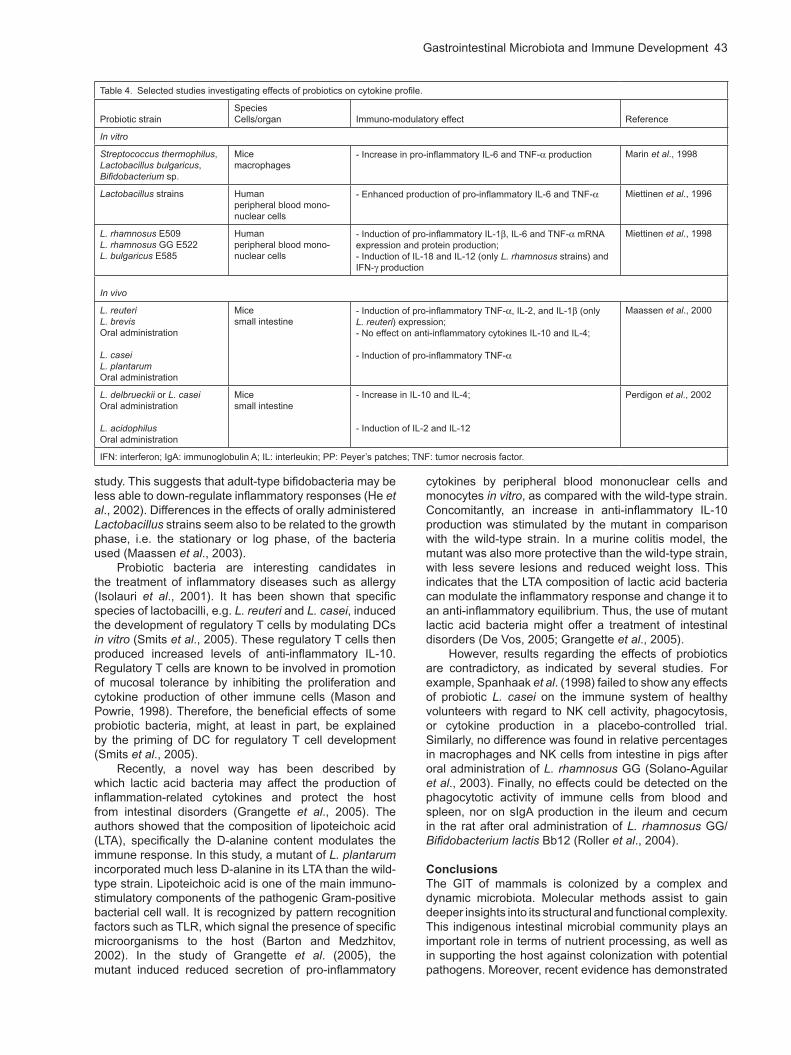
However, differences have been reported with regard to the immuno-modulatory effects of different probiotic bacteria (Table 4). For example, different lactic acid bacteria may induce distinct mucosal cytokine profiles suggesting their distinct effects on the Th1/Th2 balance (Perdigon et al., 2002). This emphasizes the importance of strain selection for immuno-therapeutic purposes. Adult-type bifidobacteria (B. adolescentis and B. longum) have been reported to induce more pro-inflammatory cytokines (IL-12 and TNF- ) in vitro in a murine macrophage-like cell line, than did infant-type bifidobacteria such as B. bifidum, B. breve or B. infantis. Furthermore, B. adolescentis did not stimulate the production of anti-inflammatory IL-10 as did the other bacteria tested in this
Gastrointestinal Microbiota and Immune Development 43
study. This suggests that adult-type bifidobacteria may be less able to down-regulate inflammatory responses (He et al., 2002). Differences in the effects of orally administered Lactobacillus strains seem also to be related to the growth phase, i.e. the stationary or log phase, of the bacteria used (Maassen et al., 2003).
Probiotic bacteria are interesting candidates in the treatment of inflammatory diseases such as allergy (Isolauri et al., 2001). It has been shown that specific species of lactobacilli, e.g. L. reuteri and L. casei, induced the development of regulatory T cells by modulating DCs in vitro (Smits et al., 2005). These regulatory T cells then produced increased levels of anti-inflammatory IL-10. Regulatory T cells are known to be involved in promotion of mucosal tolerance by inhibiting the proliferation and cytokine production of other immune cells (Mason and Powrie, 1998). Therefore, the beneficial effects of some probiotic bacteria, might, at least in part, be explained by the priming of DC for regulatory T cell development (Smits et al., 2005).
Recently, a novel way has been described by which lactic acid bacteria may affect the production of inflammation-related cytokines and protect the host from intestinal disorders (Grangette et al., 2005). The authors showed that the composition of lipoteichoic acid (LTA), specifically the D-alanine content modulates the immune response. In this study, a mutant of L. plantarum incorporated much less D-alanine in its LTA than the wild-type strain. Lipoteichoic acid is one of the main immuno-stimulatory components of the pathogenic Gram-positive bacterial cell wall. It is recognized by pattern recognition factors such as TLR, which signal the presence of specific microorganisms to the host (Barton and Medzhitov, 2002). In the study of Grangette et al. (2005), the mutant induced reduced secretion of pro-inflammatory
cytokines by peripheral blood mononuclear cells and monocytes in vitro, as compared with the wild-type strain. Concomitantly, an increase in anti-inflammatory IL-10 production was stimulated by the mutant in comparison with the wild-type strain. In a murine colitis model, the mutant was also more protective than the wild-type strain, with less severe lesions and reduced weight loss. This indicates that the LTA composition of lactic acid bacteria can modulate the inflammatory response and change it to an anti-inflammatory equilibrium. Thus, the use of mutant lactic acid bacteria might offer a treatment of intestinal disorders (De Vos, 2005; Grangette et al., 2005).
However, results regarding the effects of probiotics are contradictory, as indicated by several studies. For example, Spanhaak et al. (1998) failed to show any effects of probiotic L. casei on the immune system of healthy volunteers with regard to NK cell activity, phagocytosis, or cytokine production in a placebo-controlled trial. Similarly, no difference was found in relative percentages in macrophages and NK cells from intestine in pigs after oral administration of L. rhamnosus GG (Solano-Aguilar et al., 2003). Finally, no effects could be detected on the phagocytotic activity of immune cells from blood and spleen, nor on sIgA production in the ileum and cecum in the rat after oral administration of L. rhamnosus GG/Bifidobacterium lactis Bb12 (Roller et al., 2004).
Conclusions
The GIT of mammals is colonized by a complex and dynamic microbiota. Molecular methods assist to gain deeper insights into its structural and functional complexity. This indigenous intestinal microbial community plays an important role in terms of nutrient processing, as well as in supporting the host against colonization with potential pathogens. Moreover, recent evidence has demonstrated
Table 4. Selected studies investigating effects of probiotics on cytokine profile.
Probiotic strainSpeciesCells/organ Immuno-modulatory effect Reference
In vitro
Streptococcus thermophilus, Lactobacillus bulgaricus, Bifidobacterium sp.
Micemacrophages
- Increase in pro-inflammatory IL-6 and TNF- production Marin et al., 1998
Lactobacillus strains Humanperipheral blood mono-nuclear cells
- Enhanced production of pro-inflammatory IL-6 and TNF- Miettinen et al., 1996
L. rhamnosus E509L. rhamnosus GG E522L. bulgaricus E585
Humanperipheral blood mono-nuclear cells
- Induction of pro-inflammatory IL-1 , IL-6 and TNF- mRNA expression and protein production;- Induction of IL-18 and IL-12 (only L. rhamnosus strains) and IFN- production
Miettinen et al., 1998
In vivo
L. reuteriL. brevisOral administration
L. caseiL. plantarumOral administration
Micesmall intestine
- Induction of pro-inflammatory TNF- , IL-2, and IL-1 (only L. reuteri) expression;- No effect on anti-inflammatory cytokines IL-10 and IL-4;
- Induction of pro-inflammatory TNF-
Maassen et al., 2000
L. delbrueckii or L. caseiOral administration
L. acidophilusOral administration
Micesmall intestine
- Increase in IL-10 and IL-4;
- Induction of IL-2 and IL-12
Perdigon et al., 2002
IFN: interferon; IgA: immunoglobulin A; IL: interleukin; PP: Peyer’s patches; TNF: tumor necrosis factor.
44 Bauer et al.
that commensal bacteria also regulate intestinal development and function (Hooper et al., 2002). By use of gnotobiotic and germ-free animals, it has been shown that this complex microbial community exerts major influences on the maturation of the gut, e.g., in terms of regulating angiogenesis or production of microbicidal proteins. Furthermore, the indigenous microbiota influences the development of the GALT, mucosal immunity and induction of oral tolerance.
Understanding how the gut mucosal immune system generally responds to intestinal microbiota may be an important basis for targeting manipulation of the microbial composition. This might be of special interest at stressful times, such as weaning, when young animals have to cope with major changes in their diet and environment, and simultaneously face a change in the composition of their GIT microbiota. Weaning marks the time at which protective immunity shifts from passive maternal immunity to the active, adaptive immunity of the piglet. For example, in naturally suckled piglets, the lowest serum IgG concentrations are found at four weeks of age, after which, IgG levels rise as a result of de novo synthesis of Ig (Klobasa et al., 1986). Also, phenotypic studies suggest that the mucosal immune system remains relatively immature throughout the ‘normal’ weaning period (Stokes et al., 2004). The changes occurring in the immune system around weaning (e.g. as shown by altered percentages of T cell subsets, or cytokine production) emphasize the importance of weaning time in immune system development and thus potentially as key management factors in pig production systems. Weaning at an older age, and thus at a more advanced level of developmental maturity, might reduce weaning-induced inflammatory responses.
The strong influence of the indigenous microbiota on the GIT immune system might offer a possibility to beneficially steer the development of the GIT immune system, by manipulating microbial composition. This could be reached by the use of probiotics. Apart from their alleged properties to positively influence both development and stability of the GIT microbiota, these agents also may stimulate specific and non-specific components of the immune system. However, results are sometimes contradictory, and differences also exist due to specific bacterial strains. The use and further development of in vitro assays for careful pre-selection of possible probiotic strains would be useful. Recently, an in vitro assay for probiotic bacteria has been developed and validated by Koenen et al. (2004b). This in vitro assay is suitable for pre-selection of lactic acid bacteria in poultry, but it might also be useful for other species. However, it has also been shown that, despite similar in vitro properties, distinct probiotic strains may colonize the gut differently and generate divergent immune responses (Ibnou-Zekri et al., 2003).
It is also possible that effects on the immune system might not only be mediated by viable, but also by non-viable probiotic bacteria or bacterial DNA. This has been shown in a study of Rachmilewitz et al. (2004), who administered bacterial DNA (probiotic or E. coli) intra-gastrically to mice prior to induction of experimental colitis. Both DNA (probiotic and E. coli) ameliorated the severity
of experimental colitis, as did viable or irradiated non-viable probiotics. This is interesting for safety reasons, as irradiated probiotics could also be used in immuno-compromised hosts without the risk of bacteraemia.
It has been suggested that some bacteria species, e.g. B. thetaiotaomicron, downregulate the overall pro-inflammatory effect of enterobacteria (Kelly et al., 2004). Since neonatal gut colonization with such symbiotic ‘anti-inflammatory’ microorganisms depends on the environment and nutrients, it can be tightly controlled by the early diet. Supporting such colonization of the GIT by use of specific dietary components renders a further possibility to beneficially influence the development of the intestinal immune system. Also, dietary modulation of the GIT microbiota can result in an enhancement of colonization resistance against potential pathogens. For example Konstantinov et al. (2004a), by use of fermentable carbohydrates in weaning piglet diets, showed a stimulation of lactobacilli, with a concomitant suppression of Clostridium-like species. Such stimulation of the beneficial intestinal bacteria may also result from supplemental prebiotics. These are low molecular oligosaccharides, also referred to as non-digestible oligosaccharides (NDO) which are believed to enhance the beneficial activity of specific members of the microbiota, such as lactobacilli or bifidobacteria (Gibson and Roberfroid, 1995). They may be used alone, or alternatively, in combination with probiotics (synbiotic approach).
Until now, there have been few studies investigating the effects of microbial colonization on the immune system in livestock or companion animals. However, valuable information can be drawn from human studies and by the use of rodent models, particularly the gnotobiotic/germ-free model. These might contribute to the development of hypotheses to be validated in livestock and companion animals.
Acknowledgement
The writing of this review was supported by the Dutch Commodity Board for Animal Feedstuffs.
References
Allen, W.D., and Porter, P. (1973). The relative distribution of IgM and IgA cells in intestinal mucosa and lymphoid tissues of the young unweaned pig and their significance in ontogenesis of secretory immunity. Immunology 24, 493–501.
Alvarez-Olmos, M.I., and Oberhelman, R.A. (2001). Probiotic agents and infectious diseases: a modern perspective on a traditional therapy. Clin. Infect. Dis. 32, 1567–1576.
Banchereau, J., and Steinman, R.M. (1998). Dendritic cells and control of immunity. Nature 392, 242–252.
Barrow, P.A. (1992). Probiotics for chicken. In Probiotics: The Scientific Approach, R. Fuller, ed. (London, UK: Chapman and Hall), pp. 225–259.
Barton, G.M., and Medzhitov, R. (2002). Toll-like receptors and their ligands. Curr. Top. Microbiol. Immunol. 270, 81–92.
Bealmear, P.M. (1981). Host defense mechanisms in gnotobiotic animals. In Immunologic Defects in
Gastrointestinal Microbiota and Immune Development 45
Laboratory Animals, M.E. Gershwin, B. Merchant, eds. (New York: Plenum Press), pp. 261–350.
Beerens, H., Romond, C., and Neut, C. (1980). Influence of breast-feeding on the bifid flora of the newborn intestine. Am. J. Clin. Nutr. 33, 2434–2439.
Belardelli, F., and Ferrantini, M. (2002). Cytokines as a link between innate and adaptive antitumor immunity. Trends Immunol. 23, 201–208.
Benyacoub, J., Czarnecki-Maulden, G.L., Cavadini, C., Sauthier, T., Anderson, R.E., Schiffrin, E.J., and Von der Weid, T. (2003). Supplementation of food with Enterococcus faecium (SF68) stimulates immune functions in young dogs. J. Nutr. 133, 1158–1162.
Berg, R.D. (1983). Host immune response to antigens of the indigenous intestinal flora. In Human Intestinal Microflora in Health and Disease, D.J. Hentges, ed. (New York: Academic Press), pp. 101–126.
Berg, R.D. (1996). The indigenous gastrointestinal microflora. Trends Microbiol. 4, 430–435.
Beutler, B. (2004). Innate immunity: an overview. Mol. Immunol. 40, 845–859.
Bezkorovainy, A., and Topouzian, N. (1981). Bifidobacterium bifidus var. pennsylvanicus growth promoting activity of human milk casein and its derivatives. Int. J. Biochem. 13, 585–590.
Bianchi, A.T., Zwart, R.J., Jeurissen, S.H., and Moonen-Leusen, H.W. (1992). Development of the B and T cell compartments in porcine lymphoid organs from birth to adult life: an immunohistological approach. Vet. Immunol. Immunopathol. 33, 201–221.
Biol-N’garagba, M.,C., and Louisot, P. (2003). Regulation of the intestinal glycoprotein glycosylation during postnatal development: role of hormonal and nutritional factors. Biochimie 85, 331–352.
Bourlioux, P., Koletzko, B., Guarner, F., and Braesco, V. (2003). The intestine and its microflora are partners for the protection of the host: report on the Danone Symposium ‘The Intelligent Intestine’, held in Paris, June 14, 2002. Am. J. Clin. Nutr. 78, 675–683.
Brandtzaeg, P. (2003a). Role of secretory antibodies in the defence against infections. Int. J. Med. Microbiol. 293, 3–15.
Brandtzaeg, P. (2003b). Mucosal immunity: integration between mother and the breast-fed infant. Vaccine 21, 3382–3388.
Brandtzaeg, P., Nilssen, D.E., Rognum, T.O., and Thrane, P.S. (1991). Ontogeny of the mucosal immune system and IgA deficiency. Gastroenterol. Clin. North Am. 20, 397–439.
Brandtzaeg, P., Farstad, I.N., Johansen, F.-E., Morton, H.C., Norderhaug, I.N., and Yamanaka, T. (1999). The B-cell system of human mucosae and exocrine glands. Immunol. Rev. 171, 45–87.
Butler, J.E. (1998). Immunoglobulin diversity, B-cell and antibody repertoire development in large farm animals. Rev. Sci. Tech. 17, 43–70.
Butler, J.E., Klobasa, F., and Werhahn, E. (1981). The differential localization of IgA, IgM and IgG in the gut of suckled neonatal piglets. Vet. Immunol. Immunopathol. 1, 53–65.
Cario, E., Rosenberg, I.M., Brandwein, S.L., Beck, P.L., Reinecker, H.C., and Podolsky, D.K. (2000).
Lipopolysaccharide activates distinct signaling pathways in intestinal epithelial cell lines expressing Toll-like receptors. J. Immunol. 164, 966–972.
Cario, E., Brown, D., McKee, M., Lynch-Devaney, K., Gerken, G., and Podolsky, D.K. (2002). Commensal-associated molecular patterns induce selective toll-like receptor-trafficking from apical membrane to cytoplasmic compartments in polarized intestinal epithelium. Am. J. Pathol. 160, 165–173.
Cheroutre, H. (2004). Starting at the beginning: new perspectives on the biology of mucosal T cells. Annu. Rev. Immunol. 22, 217–246.
Cunningham-Rundles, C. (2001). Physiology of IgA and IgA deficiency. J. Clin. Immunol. 21, 303–309.
Davis, C.P., and Savage, D.C. (1974). Habitat, succession, attachment, and morphology of segmented, filamentous microbes indigenous to the murine gastrointestinal tract. Infect. Immun. 10, 948–956.
De Oliveira, I.R., Nascimento de Araújo, A., Nair Bao, S., and Giugliano, L.G. (2001). Binding of lactoferrin and free secretory component to enterotoxigenic Escherichia coli. FEMS Microbiol. Lett. 203, 29–33.
De Vos, W.M. (2005). Lipotechoic acid in lactobacilli: D-alanine makes the difference. Proc. Natl. Acad. Sci. USA 102, 10763–10764.
Deplancke, B., and Gaskins, H.R. (2001). Microbial modulation of innate defense: goblet cells and the intestinal mucus layer. Am. J. Clin. Nutr. 73 (Suppl.), 1131S-1141S.
Elass, E., Masson, M., Mazurier, J., and Legrand, D. (2002). Lactoferrin inhibits the lipopolysaccharide-induced expression and proteoglycan-binding ability of interleukin-8 in human endothelial cells. Infect. Immun. 70, 1860–1866.
Elson, C.O. (1985). Induction and control of the gastrointestinal immune system. Scand. J. Gastroenterol. 114 (Suppl.), 1–15.
Ewing, W.N., and Cole, D.J.A. (1994). The Living Gut (Dungannon, Ireland: Context).
Favier, C., Vaughan E.E., De Vos W.M., and Akkermans, A.D.L. (2002). Molecular monitoring of succession of bacterial communities in human neonates. Appl. Environ. Microbiol. 68, 219–226.
Fearon, D.T. (2000). Innate immunity – beginning to fulfil its promise? Nat. Immunol. 1, 102–103.
Forchielli, M.L., and Walker, W.A. (2005). The role of gut-associated lymphoid tissues and mucosal defence. Br. J. Nutr. 93 (Suppl.), S41–48.
Franklin, M.A., Mathew, A.G., Vickers, J.R., and Clift, R.A. (2002). Characterization of microbial populations and volatile fatty acids concentrations in the jejunum, ileum, and cecum of pigs weaned at 17 vs 24 days of age. J. Anim. Sci. 80, 2904–2910.
Fuller, R. (1992). History and development of probiotics. In Probiotics: The Scientific Basis (New York: Chapman and Hall), pp.1–8.
Garside, P., and Mowat, A.M. (2001). Oral tolerance. Semin. Immunol. 13, 177–185.
Gaskins, H.R. (1998). Immunological development and mucosal defense in the pig intestine. In Progress in Pig Science, J. Wiseman, M.A. Varley, J.P. Chadwick, eds. (UK: Nottingham University Press), pp. 81–101.
46 Bauer et al.
Gaskins, H.R. (2001). Intestinal bacteria and their influence on swine growth. In Swine Nutrition 2nd ed., A.J. Lewis, L.L. Southern, eds. (Florida: CRC Press LLC), pp. 585–608.
Gewirtz, A.T., Navas, T.A., Lyons, S., Godowski, P.J., and Madara, J.L. (2001). Cutting edge: bacterial flagellin activates basolaterally expressed TLR5 to induce epithelial proinflammatory gene expression. J. Immunol. 167, 1882–1885.
Gibson, G.R., and Roberfroid, M.B. (1995). Dietary modulation of the human colonic microbiota: introducing the concept of prebiotics. J. Nutr. 125, 1401–1412.
Gleeson, M., Cripps, A.W., Clancy, R.L., Husband, A.J., Hensley, M.J., and Leeder, S.R. 1982. Ontogeny of the secretory immune system in man. Austr. N. Z. J. Med. 12, 255–258.
Grangette, C., Nutten, S., Palumbo, E., Morath, S., Hermann, C., Dewulf, J., Pot, B., Hartung, T., Hols, P., and Mercenier, A. (2005). Enhanced antiinflammatory capacity of a Lactobacillus plantarum mutant synthesizing modified teichoic acids. Proc. Natl. Acad. Sci. USA 102, 10321–10326.
Griebel, P.J. and Hein, W.R. (1996). Expanding the role of Peyer’s patches in B cell ontogeny. Rev. Immunol. Today 17, 30–39.
Hampson, D.J. (1986). Alterations in piglet small intestine structure at weaning. Res. Vet. Sci. 40, 32–40.
Hampson, D.J., and Kidder, D.E. (1986). Influence of creep feeding and weaning on brush border enzyme activities in the piglet small intestine. Res. Vet. Sci. 40, 24–31.
Hampson, D.J., and Smith, W.C. (1986). Influence of creep feeding and dietary intake after weaning on malabsorption and occurrence of diarrhoea in the newly weaned pig. Res. Vet. Sci. 41, 63–69.
Hanson, L.Å., Dahlman-Höglund, A., Karlsson, M., Lundin, S., Dahlgren, U., and Telemo, E. (1999). Normal microbial flora of the gut and the immune system. In Nestlé Workshop Series 42, L.Å. Hanson, R.H. Yolken, eds. (Philadelphia: Vevey/Lippincott-Raven Publishers), pp. 217–228.
Hanson, L.Å., Korotkova, M., Lundin, S., Håversen, L., Silfverdal, S.A., Mattsby-Baltzer, I., Strandvik, B., and Telemo, E. (2003). The transfer of immunity from mother to child. Ann. N. Y. Acad.Sci. 987, 199–206.
Hathaway, L.J., and Kraehenbuhl, J.-P. (2000). The role of M cells in mucosal immunity. Cell. Mol. Life Sci. 57, 323–332.
Håversen, L.A., Engberg, I., Baltzer, L., Dolphin, G., Hanson, L.A., and Mattsby-Baltzer, I. (2000). Human lactoferrin and peptides derived from a surface-exposed helical region reduce experimental Escherichia coli urinary tract infection in mice. Infect. Immun. 68, 5816–5823.
He, F., Morita, H., Ouwehand, A.C., Hosoda, M., Hiramatsu, M., Kurisaki, J., Isolauri, E., Benno, Y., and Salminen, S. (2002). Stimulation of the secretion of pro-inflammatory cytokines by Bifidobacterium strains. Microbiol. Immunol. 46, 781–785.
Hein, W.R. (1999). Organization of mucosal lymphoid tissue. Curr. Top. Microbiol. Immunol. 236, 1–15.
Herias, M.V., Hessle, C., Telemo, E., Midtvedt, T., Hanson, L.A., and Wold, A.E. (1999). Immunomodulatory effects of Lactobacillus plantarum colonizing the intestine of gnotobiotic rats. Clin. Exp. Immunol. 116, 283–290.
Hillman, K. (2001). Bacteriological aspects of the use of antibiotics and their alternatives in the feed of non-ruminant animals. In Recent Advances in Animal Nutrition, P.C. Garnsworthy, J. Wiseman, eds. (UK: Nottingham University Press), pp. 107–134.
Hooper, L.V., Xu, J., Falk, P.G., Midtvedt, T., and Gordon, J.I. (1999). A molecular sensor that allows a gut commensal to control its nutrient foundation in a competitive ecosystem. Proc. Natl. Acad. Sci. USA 96, 9833–9838.
Hooper, L.V., Falk, P.G., and Gordon, J.I. (2000). Analyzing the molecular foundations of commensalism in the mouse intestine. Curr. Opin. Microbiol. 3, 79–85.
Hooper, L.V. (2004). Bacterial contributions to mammalian gut development. Trends Microbiol. 12, 129–134.
Hooper, L.V., and Gordon, J.I. (2001). Commensal host-bacterial relationships in the gut. Science 292, 1115–1118.
Hooper, L.V., Wong, M.H., Thelin, A., Hansson, L., Falk, P.G., and Gordon, J.I. (2002). Molecular analysis of commensal host-microbial relationships in the intestine. Science 291, 881–884.
Hooper, L.V., Stappenbeck, T.S., Hong, C.V., and Gordon, J.I. (2003). Angiogenins: a new class of microbicidal proteins involved in innate immunity. Nature Immunol. 4, 269–273.
Ibnou-Zekri, N., Blum, S., Schiffrin, E.J., and Von der Weid, T. (2003). Divergent patterns of colonization and immune response elicited from two intestinal Lactobacillus strains that display similar properties in vitro. Infect. Immun. 71, 428–436.
Isolauri, E., Sütas, Y., Kankaanpäa, P., Arvilommi, H., and Salminen, S. (2001). Probiotics: effects on immunity. Am. J. Clin. Nutr. 73 (Suppl.), 444–450.
Iwase, T., Moro, I., and Mestecky, J. (1987). Immunohistochemical study of the ontogeny of the secretory immune system. Ad. Exp. Med. Biol. 216B, 1359–1368.
Janeway, C.A. Jr. (2001). How the immune system works to protect the host from infection: a personal view. Proc. Natl. Acad. Sci. USA, 98, 7461–7468.
Janeway, C.A. Jr., and Medzhitov, R. (2002). Innate immune recognition. Annu. Rev. Immunol. 20, 197–216.
Johnson, L.R. (1988). Regulation of gastrointestinal mucosal growth. Physiol. Rev. 68, 456–502.
Jones, G.W., and Freter, R. (1976). Adhesive properties of Vibrio cholerae: nature of the interaction with isolated rabbit brush border membranes and human erythrocytes. Infect. Immun. 14, 240–245.
Kadowaki, N., Ho, S., Antonenko, S., Malefyt, R.W., Kastelein, R.A., Bazan, F., and Liu, Y.J. (2001). Subsets of human dendritic cell precursors express different toll-like receptors and respond to different microbial antigens. J. Exp. Med. 194, 863–869.
Kelly, D., Begbie, R., and King, T.P. (1992). Postnatal intestinal development. In Neonatal Survival and Growth, M.A. Varley, P.E.V. Williams, T.L.C. Lawrence,
Gastrointestinal Microbiota and Immune Development 47
eds. (British Society of Animal Production, Occasional Publication), 15, 63–79.
Kelly, D., Campbell, J.I., King, T.P., Grant, G., Jansson, E.A., Coutts, A.G., Pettersson, S., and Conway, S. (2004). Commensal anaerobic gut bacteria attenuate inflammation by regulating nuclear-cytoplasmic shuttling of PPAR-gamma and RelA. Nat. Immunol. 5, 104–112.
Kidd, P. (2003). Th1/Th2 balance: the hypothesis, its limitations, and implications for health and disease. Altern. Med. Rev. 8, 223–246.
Klaasen, H.L.B.M., Koopman, J.P., Poelma, F.G., and Beynen, A.C. (1992). Intestinal, segmented filamentous bacteria. FEMS Microbiol. Rev. 88, 165–180.
Klaasen, H.L.B.M., Van der Heijden, P.J., Stok, W., Poelma, F.G.J., Koopman, J.P., Van den Brink, M.E., Bakker, M.H., Eling, W.M.C., and Beynen, A.C. (1993). Apathogenic, intestinal, segmented, filamentous bacteria stimulate the mucosal immune system of mice. Infect. Immun. 61, 303–306.
Klobasa, F., Butler, J.E., Werhahn, E., and Habe, F. (1986). Maternal-neonatal immunoregulation in swine. II. Influence of multiparity on de novo immunglobulin synthesis by piglets. Vet. Immunol. Immunopathol. 11, 149–159.
Klobasa, F. and Butler, J.E. (1987). Absolute and relative concentrations of immunoglobulins G, M, and A, and albumin in the lacteal secretion of sows of different lactation numbers. Am. J. Vet. Res. 48, 176–182.
Klobasa, F., Werhahn, E., and Butler, J.E. (1987). Composition of sow milk during lactation. J. Anim. Sci. 64, 1485–1466.
Koenen, M.E., Kramer, J., Van der Hulst, R., Heres, L., Jeurissen, S.H., and Boersma, W.J. (2004a). Immunomodulation by probiotic lactobacilli in layer- and meat-type chickens. Br. Poult. Sci. 45, 355–366.
Koenen, M.E., Van der Hulst, R., Leering, M., Jeurissen, S.H., and Boersma, W.J. (2004b). Development and validation of a new in vitro assay for selection of probiotic bacteria that express immune-stimulating properties in chickens in vivo. FEMS Immunol. Med. Microbiol. 40, 119–127.
Konstantinov, S.R. (2005). Lactobacilli in the porcine intestine: from composition to functionality. PhD Thesis (The Netherlands, Wageningen University).
Konstantinov, S.R., Awati, A., Smidt, H., Williams, B.A., Akkermans, A.D.L., and De Vos, W.M. (2004a). Specific response of a novel and abundant Lactobacillus amylovorus-like phylotype to dietary prebiotics in the guts of weaning piglets. Appl. Environ. Microbiol. 70, 3821–3830.
Konstantinov, S.R., Favier, C.F., Zhu, W.Y., Williams, B.A., Klüss, J., Souffrant, W.B., De Vos, W.M., Akkermans, A.D.L., and Smidt, H. (2004b). Microbial diversity study of the porcine GI tract during the weaning transition. Anim. Res. 54, 317–24.
Kraehenbuhl, J.-P., and Neutra, M.R. (1992). Molecular and cellular basis of immune protection at mucosal surfaces. Physiol. Rev. 72, 853–879.
Kunz, C., and Rudloff, S. (1993). Biological functions of oligosaccharides in human milk. Acta Paediatr. 82, 903–912.
Kunz, C., Rudloff, S., Baier, W., Klein, N., and Strobel, S. (2000). Oligosaccharides in human milk: structural, functional, and metabolic aspects. Annu. Rev. Nutr. 20, 699–722.
Lehrer, R.I., Barton, A., Daher, K.A., Harwig, S.S., Ganz, T., and Selsted, M.E. (1989). Interaction of human defensins with Escherichia coli. Mechanism of bactericidal activity. J. Clin. Invest. 84, 553–561.
Lehrer, R.I. (2004). Primate defensins. Nat. Rev. Microbiol. 2, 727–738.
Liepke, C., Adermann, K., Raida, M., Magert, H.J., Forssmann, W.G., and Zucht, H.D. (2002). Human milk provides peptides highly stimulating the growth of bifidobacteria. Eur. J. Biochem. 269, 712–718.
Maassen, C.B., Van Holten-Neelen, C., Balk, F., Den Bak-Glashouwer, M.J., Lee, R.J., Laman, J.D., Boersma, W.J., and Claassen, E. (2000). Strain-dependent induction of cytokine profiles in the gut by orally administered Lactobacillus strains. Vaccine 18, 2613–2623.
Maassen, C.B., Boersma, W.J., Van Holten-Neelen, C., Claassen, E., and Laman, J.D. (2003). Growth phase of orally administered Lactobacillus strains differentially affects IgG1/IgG2a ratio for soluble antigens: implications for vaccine development. Vaccine 21, 2751–2757.
MacDonald, T.T. (2003). The mucosal immune system. Parasite Immunol. 25, 235–46.
Mackie, R.I., and White, B.A. (1997). Gastrointestinal Microbiology. Chapman and Hall Microbiology Series (New York: Chapman and Hall).
Mackie, R.I., Sghir, A., and Gaskins, H.R. (1999). Developmental microbial ecology of the neonatal gastrointestinal tract. Am. J. Clin. Nutr. 69, 1035–1045.
Macpherson, A.J., Hunziker, L., McCoy, K., and Lamarre, A. (2001). IgA responses in the intestinal mucosa against pathogenic and non-pathogenic microorgansims. Microbes Infect. 3, 1021–1035.
Macpherson, A.J., Martinic, M.M., and Harris, N. (2003). The functions of mucosal T cells in containing the indigenous flora of the intestine. Cell. Mol. Life Sci. 59, 2088–2096.
Macpherson, A.J., and Harris, N.L. (2004). Interactions between commensal intestinal bacteria and the immune system. Nature 4, 478–485.
Macpherson, A.J., and Uhr, T. (2004). Induction of protective IgA by intestinal dendritic cells carrying commensal bacteria. Science 303, 1662–1665.
Madara, J.L. (1998). Regulation of movement of solutes across tight junctions. Annu. Rev. Physiol. 60, 143–159.
Manzano, M., Abadía-Molina, A.C., García-Olivares, E., Gil, A., and Rueda, R. (2002). Absolute counts and distribution of lymphocyte subsets in small intestine of BALB/c mice change during weaning. J. Nutr. 132, 2757–2762.
Marin, M.L., Tejada-Simon, M.V., Lee, J.H., Murtha, J., Ustunol, Z., and Pestka, J.J. (1998). Stimulation of cytokine production in clonal macrophage and T-cell models by Streptococcus thermophilus: comparison with Bifidobacterium sp. and Lactobacillus bulgaricus. J. Food Prot. 61, 859–864.
48 Bauer et al.
Mason, D., and Powrie, F. (1998). Control of immune pathology by regulatory T cells. Curr. Opin. Immunol. 10, 649–655.
Mattsby-Baltzer, I., Roseanu, A., Motas, C., Elverfors, J., Engberg, I., and Hånson, L.A. (1996). Lactoferrin or a fragment thereof inhibits the endotoxin-induced interleukin-6 response in human monocytic cells. Pediatr. Res. 40, 257–262.
Mayer, L. (1997). Review article: local and systemic regulation of mucosal immunity. Aliment. Pharmacol. Ther. 11 (Suppl. 3), 81–88.
Mayer, L. (2000). Mucosal immunity and gastrointestinal antigen processing. J. Pediatr. Gastroenterol. Nutr. 30 (Suppl.), S4-S12.
McCracken, B.A., Gaskins, H.R., Ruwe-Kaiser, P.J., Klasing, K.C., and Jewell, D.E. (1995). Diet dependent and diet-independent metabolic responses underlie growth stasis of pigs at weaning. J. Nutr. 125, 2838–2845.
McCracken, B.A., Spurlock, M.E., Roos, M.A., Zuckermann, F.A., and Gaskins, H.R. (1999). Weaning anorexia may contribute to local inflammation in the piglet small intestine. J. Nutr. 129, 613–619.
McKay, D.M. and Baird, A.W. (1999). Cytokine regulation of epithelial permeability and ion transport. Gut 44, 283–289.
McNabb, P.C., and Tomasi, T.B. (1981). Host defense mechanisms at mucosal surfaces. Annu. Rev. Microbiol. 35, 477–496.
Medzhitov, R., and Janeway, C.A. Jr. (2002). Decoding the patterns of self and nonself by the innate immune system. Science 296, 298–300.
Mellander, L., Carlsson, B., and Hanson, L.Å. (1984). Appearance of secretory IgM and IgA antibodies to Escherichia coli in saliva during early infancy and childhood. J. Pediatr. 104, 564–568.
Meyerholz, D.K., Stabel, T.J., and Cheville, N.F. (2002). Segmented filamentous bacteria interact with intraepithelial mononuclear cells. Infect. Immun. 70, 3277–3280.
Miettinen, M., Vuopio-Varkila, J., and Varkila, K. (1996). Production of human tumor necrosis factor alpha, interleukin-6, and interleukin-10 is induced by lactic acid bacteria. Infect. Immun. 64, 5403–5405.
Miettinen, M., Matikainen, S., Vuopio-Varkila, J., Pirhonen, J., Varkila, K., Kurimoto, M., and Julkunen, I. (1998). Lactobacilli and streptococci induce interleukin-12 (IL-12), IL-18, and gamma interferon production in human peripheral blood mononuclear cells. Infect. Immun. 66, 6058–6062.
Mikkelsen, L.L., Bendixen, C., Jacobsen, M., and Jensen, B.B. (2003). Enumeration of bifidobacteria in gastrointestinal samples from piglets. Appl. Environ. Microbiol. 69, 654–658.
Moreau, M.C., and Corthier, G. (1988). Effect of the gastrointestinal microflora on induction and maintenance of oral tolerance to ovalbumin in C3H/HeJ mice. Infect. Immun. 56, 2766–2768.
Moughan, P.J., Birtles, M.J., Cranwell, P.J., Smith, W.C., and Pedraza, M. (1992). The piglet as a model animal for studying aspects of digestion and absorption in milk-fed human infants. In Nutritional Triggers for Health and
in Disease, A.P. Simopoulos, ed. (Basel: Karger), pp. 40–113.
Mowat, A.M. and Viney, J.L. (1997). The anatomical basis of intestinal immunity. Immunol. Rev. 156, 145–166.
Mowat, A.M., Parker, L.A., Beacock-Sharp, H., Millington, O.R., and Chirdo, F. (2004). Oral tolerance: overview and historical perspectives. Ann. N Y Acad. Sci. 1029, 1–8.
Mullock, B.M., Jones, R.S., and Hinton, R.H. (1980). Movement of endocytic shuttle vesicles from the sinusoidal to the bile canalicular face of hepatocytes does not depend on occupation of receptor sites. FEBS Lett. 113, 201–205.
Nagler-Anderson, C. (2001). Man the barrier! Strategic defences in the intestinal mucosa. Nat. Rev. Immunol. 1, 59–67.
Nagler-Anderson, C., and Shi, H.N. (2001). Peripheral non-responsiveness to orally administered soluble protein antigens. Crit. Rev. Immunol. 21, 121–132.
Naito, S., Hayashidani, H., Kaneko, K., Ogawa, M. and Benno, Y. (1995). Development of intestinal lactobacilli in normal piglets. J. Appl. Bacteriol. 79, 230–236.
Newburg, D.S. (1999). Human milk glycoconjugates that inhibit pathogens. Curr. Med. Chem. 6, 117–127.
Ortega-Cava, C.F., Ishihara, S., Rumi, M.A., Kawashima, K., Ishimura, N., Kazumori, H., Udagawa, J., Kadowaki, Y., and Kinoshita, Y. (2003). Strategic compartmentalization of Toll-like receptor 4 in the mouse gut. J. Immunol. 170, 3977–3985.
Owen, R.L., and Jones, A.L. (1974). Epithelial cell specialization within human Peyer’s patches: an ultrastructural study of intestinal lymphoid follicles. Gastroenterology 66, 189–203.
Pabst, R., Geist, M., Rothkötter, H.-J., and Fritz, F.J. (1988). Postnatal development and lymphocyte production of jejunal and ileal Peyer’s patches in normal and gnotobiotic pigs. Immunology 64, 539–544.
Pabst, R., and Rothkötter, H.J. (1999). Postnatal development of lymphocyte subsets in different compartments of the small intestine of piglets. Vet. Immunol. Immunopathol. 72, 167–173.
Palucka, K., and Banchereau, J. (1999). Dendritic cells: a link between innate and adaptive immunity. J. Clin. Immunol. 19, 12–25.
Perdigon, G., De Macias, M.E., Alvarez, S., Oliver, G., and De Ruiz Holgado, A.A. (1986). Effect of perorally administered lactobacilli on macrophage activation in mice. Infect. Immun. 53, 404–410.
Perdigon, G., Maldonado Galdeano, C., Valdez, J.C., and Medici, M. (2002). Interaction of lactic acid bacteria with the gut immune system. Eur. J. Clin. Nutr. 56, S21–26.
Petit, J.C., Galinha, A., and Salomon, J.C. (1973). Immunoglobulins in the intestinal content of the human fetus with special reference to IgA. Eur. J. Immunol. 3, 373–375.
Phalipon, A., and Corthésy, B. (2003). Novel functions of the polymeric Ig receptor: well beyond transport of immunoglobulins. Trends Immunol. 24, 55–58.
Pié, S., Lallès, J.P., Blazy, F., Laffitte, J., Sève, B., and Oswald, I.P. (2004). Weaning is associated with an upregulation of expression of inflammatory cytokines in the intestine of piglets. J. Nutr. 134, 641–647.
Gastrointestinal Microbiota and Immune Development 49
Prioult, G., Fliss, I., and Pecquet, S. (2003). Effect of probiotic bacteria on induction and maintenance of oral tolerance to beta-lactoglobulin in gnotobiotic mice. Clin. Diagn. Lab. Immunol. 10, 787–792.
Rachmilewitz, D., Katakura, K., Karmeli, F., Hayashi, T., Reinus, C., Rudensky, B., Akira, S., Takeda, K., Lee, J., Takabayashi, K., and Raz, E. (2004). Toll-like receptor 9 signaling mediates the anti-inflammatory effects of probiotics in murine experimental colitis. Gastroenterology 126, 520–528.
Rakoff-Nahoum, S., Paglino, J., Eslami-Varzaneh, F., Edberg, S., and Medzhitov, R. (2004). Recognition of commensal microflora by toll-like receptors is required for intestinal homeostasis. Cell 118, 229–241.
Rescigno, M., Urbano, M., Valzasina, B., Francolini, M., Rotta, G., Bonasio, R., Granucci, F., Kraehenbuhl, J.P., and Ricciardi-Castagnoli, P. (2001). Dendritic cells express tight junction proteins and penetrate gut epithelial monolayers to sample bacteria. Nature Immunol. 2, 361–367.
Robinson, J.K., Blanchard, T.G., Levine, A.D., Emancipator, S.N., and Lamm, M.E. (2001). A mucosal IgA-mediated excretory immune system in vivo. J. Immunol. 166, 3688–3692.
Roller, M., Rechkemmer, G., and Watzl, B. (2004). Prebiotic inulin enriched with oligofructose in combination with the probiotics Lactobacillus rhamnosus and Bifidobacterium lactis modulates intestinal immune functions in rats. J. Nutr. 134, 153–156.
Rooke, J.A., and Bland, I.M. (2002). The acquisition of passive immunity in the new-born piglet. Livest. Prod. Sci. 78, 13–23.
Rothkötter, H.J., and Pabst, R. (1989). Lymphocyte subsets in jejunal and ileal Peyer’s patches of normal and gnotobiotic minipigs. Immunology 67, 103–108.
Rothkötter, H.J., Kirchhoff, T., and Pabst, R. (1994). Lymphoid and non-lymphoid cells in the epithelium and lamina propria of intestinal mucosa of pigs. Gut 35, 1582–1589.
Rothkötter, H.J., Möllhoff, S., and Pabst, R. (1999). The influence of age and breeding conditions on the number and proliferation of intraepithelial lymphocytes in pigs. Scand. J. Immunol. 50, 31–38.
Roux, M.E., McWilliams, M., Phillips-Quagliata, J.M., Weisz-Carrington, P., and Lamm, M.E. (1977). Origin of IgA-secreting plasma cells in the mammary gland. J. Exp. Med. 146, 1311–1322.
Rudloff, S., and Kunz, C. (1997). Protein and nonprotein nitrogen components in human milk, bovine milk, and infant formula: quantitative and qualitative aspects in infant nutrition. J. Pediatr. Gastroenterol. Nutr. 24, 328–344.
Rumbo, M., and Schiffrin, E.J. (2005). Ontogeny of intestinal epithelium immune functions: developmental and environmental regulation. Cell. Mol. Life Sci. 62, 1288–1296.
Russell, G.J., Bhan, A.K., and Winter, H.S. (1990). The distribution of T and B lymphocyte populations and MHC class II expression in human fetal and postnatal intestine. Pediatr. Res. 27, 239–244.
Salminen, S., Bouley, C., Boutron-Ruault, M.C., Cummings, J.H., Franck, A., Gibson, G.R., Isolauri, E.,
Moreau, M.C., Roberfroid, M., and Rowland, I. (1998). Functional food science and gastrointestinal physiology and function. Br. J. Nutr. 80, S147–171.
Salmon, H. (1999). The mammary gland and neonate mucosal immunity. Vet. Immunol. Immunopathol. 72, 143–155.
Sanford, S.E. (1991). Light and electron microscopic observations of a segmented filamentous bacterium attached to the mucosa of the terminal ileum of pigs. J. Vet. Diagn. Invest. 3, 328–333.
Sansonetti, P. (2002). Host-pathogen interactions: the seduction of molecular cross talk. Gut 50 (Suppl. 3), 1113–1118.
Savage, D.C. (1977). Microbial ecology of the gastrointestinal tract. Ann. Rev. Microbiol. 31, 107–133.
Scharek, L., Guth, J., Reiter, K., Weyrauch, K.D., Taras, D., Schwerk, P., Schierack, P., Schmidt, M.F., Wieler, L.H., and Tedin, K. (2005). Influence of a probiotic Enterococcus faecium strain on development of the immune system of sows and piglets. Vet. Immunol. Immunopathol. 105, 151–161.
Schiffrin, E.J., Rochat, F., Link-Amster, H., Aeschlimann, J.M., and Donnet-Hughes, A. (1995). Immunomodulation of human blood cells following the ingestion of lactic acid bacteria. J. Dairy Sci. 78, 491–497.
Selsted, M.E., and Ouellette, A.J. (2005). Mammalian defensins in the antimicrobial immune response. Nat. Immunol. 6, 551–557.
Shi, H.N., and Walker, A. (2004). Bacterial colonization and the development of intestinal defences. Can. J. Gastroenterol. 18, 493–500.
Shroff, K.E., Meslin, K., and Cebra, J.J. (1995). Commensal enteric bacteria engender a self-limiting humoral mucosal immune response while permanently colonizing the gut. Infect. Immun. 63, 3904–3913.
Shu, Q., Qu, F., and Gill, H.S. (2001). Probiotic treatment using Bifidobacterium lactis HN019 reduces weanling diarrhea associated with rotavirus and Escherichia coli infection in a piglet model. J. Pediatr. Gastroenterol. Nutr. 33, 171–177.
Simon, O., Vahjen, W., and Scharek, L. (2003). Micro-organisms as feed additives – probiotics. In Proceedings of the 9th International Symposium on Digestive Physiology in Pigs Vol. 1, R.O. Ball, ed. (Canada, Banff: University of Alberta, Department of Agricultural, Food and Nutritional Science), pp. 295–318.
Singh, V.K., Mehrotra, S., and Agarwal, S.S. (1999). The paradigm of Th1 and Th2 cytokines: its relevance to autoimmunity and allergy. Immunol. Res. 20, 147–161.
Smith, H.W. (1965). Development of the flora of the alimentary tract in young animals. J. Pathol. Bacteriol. 90, 495–513.
Smits, H.H., Engering, A., Van der Kleij, D., De Jong, E.C., Schipper, K., Van Capel, T.M.M., Zaat, B.A.J., Yazdanbakhsh, M., Wierenga, E.A., Van Kooyk, Y., and Kapsenberg, M.L. (2005). Selective probiotic bacteria induce IL-10-producing regulatory T cells in vitro by modulating dendritic cell function through dendritic cell-specific intercellular adhesion molecule 3-grabbing nonintegrin. J. Allergy Clin. Immunol. 115, 1260–1267.
50 Bauer et al.
Snel, J., Hermsen, C.C., Smits, H.J., Bos, N.A., Eling, W.M.C., Cebra, J.J., and Heidt, P.J. (1998). Interactions between gut-associated lymphoid tissue and colonization levels of indigenous, segmented, filamentous bacteria in the small intestine of mice. Can. J. Microbiol. 44, 1177–1182.
Solano-Aguilar, G.I., Vengroski, K.G., Beshah, E., Douglass, L.W., and Lunney, J.K. (2001). Characterization of lymphocyte subsets from mucosal tissues in neonatal swine. Develop. Comp. Immunol. 25, 245–263.
Solano-Aguilar, G., Ledbetter, T., Dawson, H., Schoene, N., and Urban, J. Jr. (2003). The effect of dietary probiotic on the immune response of pigs. In Proceedings of the 9th International Symposium on Digestive Physiology in Pigs Vol. 2, R.O. Ball, ed. (Canada, Banff: University of Alberta, Department of Agricultural, Food and Nutritional Science), pp. 69–71.
Spanhaak, S., Havenaar, R., and Schaafsma, G. (1998). The effect of consumption of milk fermented by Lactobacillus casei strain Shirota on the intestinal microflora and immune parameters in humans. Eur. J. Clin. Nutr. 52, 899–907.
Stappenbeck, T.S., Hooper, L.V., and Gordon, J.I. (2002). Developmental regulation of intestinal angiogenesis by indigenous microbes via Paneth cells. Proc. Natl. Acad. Sci. USA 99, 15451–15455.
Stewart, C.S. (1997). Microorganisms in hindgut fermentors. In Gastrointestinal Microbiology, R.I. Mackie, B.A. White, R.E. Isaacson, eds. (New York: Chapman and Hall), pp. 142–186.
Stokes, C.R., Vega-López, M.A., Bailey, M., Telemo, E., and Miller, B.G. (1992). Immune development in the gastrointestinal tract of the pig. In Neonatal Survival and Growth, M.A. Varley, P.E.V. Williams, T.L.J. Williams, eds. (British Society of Animal Production, Occasional Publication) 15, 9–12.
Stokes, C.R., Bailey, M., Haverson, K., Harris, C., Jones, P., Inman, C., Pié, S., Oswald, I.P., Williams, B.A., Akkermans, A.D.L., Sowa, E., Rothkötter, H.-J., and Miller, B.G. (2004). Postnatal development of intestinal immune system in piglets: implications for the process of weaning. Anim. Res. 53, 325–334.
Sudo, N., Sawamura, S., Tanaka, K., Aiba, Y., Kudo, C., and Koga, Y. (1997). The requirement of intestinal bacterial flora for the development of an IgE production system fully susceptible to oral tolerance induction. J. Immunol. 159, 1739–1745.
Suzuki, K., Meek, B., Doi, Y., Muramatsu, M., Chiba T., Honjo, T., and Fagarsan, S. (2004). Aberrant expansion of segmented filamentous bacteria in IgA-deficient gut. Proc. Natl. Acad. Sci. USA 101, 1981–1986.
Talham, G.L., Jiang, H.Q., Bos, N.A., and Cebra, J.J. (1999). Segmented filamentous bacteria are potent stimuli of a physiologically normal state of the murine gut mucosal immune system. Infect. Immun. 67, 1992–2000.
Tannock, G.W., Fuller, R., and Pedersen, K. (1990). Lactobacillus succession in the piglet digestive tract demonstrated by plasmid profiling. Appl. Environ. Microbiol. 56, 1310–1316.
Tannock, G.W. (2001). Molecular assessment of intestinal microflora. Am. J. Clin. Nutr. 73, 410–414S.
Teitelbaum, J.E., and Walker, W.A. (2002). Nutritional impact of pre- and probiotics as protective gastrointestinal organisms. Annu. Rev. Nutr. 22, 107–138.
Ter Steege, J.C., Buurman, W.A., and Forget, P.P. (1997). Spermine induces maturation of the immature intestinal immune system in neonatal mice. J. Pediatr. Gastroenterol. Nutr. 25, 332–340.
Tlaskalova-Hogenova, H., Stepankova, R., Hudcovic, T., Tuckova, L., Cukrowska, B., Lodinova-Zadnikova, R., Kozakova, H., Rossmann, P., Bartova, J., Sokol, D., Funda, D.P., Borovska, D., Rehakova, Z., Sinkora, J., Hofman, J., Drastich, P., and Kokesova, A. (2004). Commensal bacteria (normal microflora), mucosal immunity and chronic inflammatory and autoimmune diseases. Immunol. Lett. 93, 97–108.
Umesaki, Y., Setoyama, H., Matsumoto, S., and Okada, Y. (1993). Expansion of alpha beta T-cell receptor bearing intestinal lymphocytes after microbial colonization in germ-free mice and its independence from thymus. Immunology 79, 32–37.
Umesaki, Y., Okada, Y., Matsumoto, S., Imaoka, A., and Setoyama, H. (1995). Segmented filamentous bacteria are indigenous bacteria that activate intraepithelial lymphocytes and induce MHC class II molecules and fucosyl asialo GM1 glycolipids on the small intestinal epithelial cells in the ex-germ-free mouse. Microbiol. Immunol. 39, 555–562.
Umesaki, Y., Setoyama, H., Matsumoto, S., Imaoka, A., and Itoh, K. (1999). Differential roles of segmented filamentous bacteria and clostridia in development of the intestinal immune system. Infect. Immun. 67, 3504–3511.
Van der Waaij, D., Berghuis-de Vries, J.M., and Lekkerkerk-van der Wees, J.E.C. (1971). Colonization resistance of the digestive tract in conventional and antibiotic-treated mice. J. Hyg. 69, 405–411.
Vaughan, E.E., Schut, F., Heilig, H.G., Zoetendal, E.G., De Vos, W.M., and Akkermans, A.D. (2000). A molecular view of the intestinal ecosystem. Curr. Issues Intest. Microbiol. 1, 1–12.
Vega-López, M.A., Bailey, M., Telemo, E., and Stokes, C.R. (1995). Effect of early weaning on the development of immune cells in the pig small intestine. Vet. Immunol. Immunopathol. 44, 319–327.
Vega-López, M.A., Arenas-Contreras, G., Bailey, M., Gonzalez-Pozos, S., Stokes, C.R., Ortega, M.G., and Mondragon-Flores, R. (2001). Development of intraepithelial cells in the porcine small intestine. Dev. Immunol. 8, 147–158.
Vitini, E., Alvarez, S., Medina, M., Medici, M., De Budeguer, M.V., and Perdigon, G. (2000). Gut mucosal immunostimulation by lactic acid bacteria. Biocell 24, 223–232.
Wagstrom, E.A., Yoon, K.J., and Zimmerman, J.J. (2000). Immune components in porcine mammary secretions. Viral. Immunol. 13, 383–397.
Wang, H.C., Montufar-Solis, D., Teng, B.B., and Klein, J.R. (2004). Maximum immunobioactivity of murine small intestinal intraepithelial lymphocytes resides in
Gastrointestinal Microbiota and Immune Development 51
a subpopulation of CD43+ T cells. J. Immunol. 173, 6294–6302.
Weiner, H.L. (2001). Induction and mechanism of action of transforming growth factor- -secreting Th3 regulatory cells. Immunol. Rev. 182, 207–214.
Xu, R.J. (2003). Composition of porcine milk. In The Neonatal Pig. Gastrointestinal Physiology and Nutrition,
R.J. Xu, P. Cranwell, eds. (UK: Nottingham University Press), pp. 213–246.
Zasloff, M. (2002). Trypsin, for the defense. Nature Immunol. 3, 583–590.
Zhang, G., Ross, C.R., and Blecha, F. (2000). Porcine antimicrobial peptides: new prospects for ancient molecules of host defense. Vet. Res. 31, 277–296.