Embed Size (px)
Citation preview

Brazilian Journal of Microbiology (2002) 33:178-184ISSN 1517-8382
178
INFLUENCE OF SUBINHIBITORY CONCENTRATIONS OF ANTIMICROBIALS ONHYDROPHOBICITY, ADHERENCE AND ULTRA-STRUCTURE OF
FUSOBACTERIUM NUCLEATUM
Ana C. Okamoto1; Elerson Gaetti-Jardim Jr.1; Victor E. Arana-Chavez2; Mario J. Avila-Campos1*
1 Departmento de Microbiologia, Instituto de Ciências Biomédicas, Universidade de São Paulo, São Paulo, SP, Brasil.2 Departamento de Histologia e Embriologia, Instituto de Ciências Biomédicas, Universidade de São Paulo, São Paulo, SP, Brasil
Submitted: July 06, 2001; Returned to authors for corrections: January 08, 2002; Approved: Junho 10, 2002
ABSTRACT
Fusobacterium nucleatum is considered a bridge organism between earlier and later colonizers in dentalbiofilms and a putative periodontopathogen. In Dentistry, antimicrobial agents are used for treatment andcontrol of infectious diseases associated with dental plaque. Antiseptics have been used in association withantibiotics to reduce infections after oral surgeries. In this study, the influence of subinhibitory concentrations(SC) of chlorhexidine, triclosan, penicillin G and metronidazole, on hydrophobicity, adherence to oral epithelialcells, and ultra-structure of F. nucleatum was examined. All isolates were susceptible to chlorhexidine, triclosan,and metronidazole; however, most of the isolates were susceptible to penicillin G, and all of them werehydrophilic when grown with or without antimicrobials. Adherence was decreased by all antimicrobials.Results suggest that adherence of F. nucleatum was influenced by adhesins because structures such asfimbries or capsule were not observed by transmission electronic microscope.
Key words: Fusobacterium nucleatum, hydrophobicity, adherence, antimicrobial, ultra-structure.
INTRODUCTION
The American Association of Public Health Dentists,Subcommittee of Preventive Periodontology (2) describes theperiodontal disease as the major cause of dental loss in adultpopulation, which is considered a serious health problemworldwide.
Gingivitis and periodontitis are associated with the dentalbiofilm, which is mediated by intrinsic and extrinsic factorssuch as the use of antimicrobial drugs, host�s susceptibilityor microorganism-host interactions (4,13). The humanperiodontophaties are associated with Gram-negative anaerobicand aerobic rods such as Porphyromonas gingivalis, Prevotellaintermedia, Fusobacterium nucleatum, Eikenella corrodens andWollinella recta, and these organisms are able to destroyperiodontal tissues by using different virulence factors such asvolatile compounds, enzymes, LPS or hemolysins (5,28).
* Corresponding author. Mailing address: Laboratório de Anaeróbios, Departamento de Microbiologia, ICB II � USP. Av. Prof. Lineu Prestes, 1374,Cidade Universitária, 05508-900, São Paulo, SP, Brasil. Phone: (+5511) 3091-7344, Fax: (+5511) 3091-7354. E-mail: [email protected]
Fusobacterium nucleatum is an important organism f7or itsrole as a bridge among earlier and later colonizers of the dentalbiofilm because of its ability of coaggregation with othermicrobial species (15,19,32).
Antimicrobial agents are used for treatment and control ofinfectious diseases associated to dental biofilm (17,29). Antisepticshave been used in association with antibiotics to reduceinfections after oral surgeries and also to help in treatingperiodontal infections (27). Chlorhexidine is a cationic bis-guanideagent used as an active bacterial plaque inhibitor and activeagainst Gram-positive and Gram-negative, aerobe or anaerobeorganisms, yeasts and virus (2). Triclosan is an active bis-phenolicand non-cationic agent, and it has been added to dentifrices andcolutories without side effects. However, this agent has a lowsubstantivity on oral mucosa but an effective action againstdifferent oral organisms has been observed (16,23). Also,antibiotics are used for treatment of infectious processes (17).
Inhibition of F. nucleatum
179
Considering F. nucleatum as a member of the humanautochthonous microbiota and for the importance in oral andnonoral infections, and considering the action of differentsubstances interfering with either bacterial virulence factors orwith the host-bacteria interaction, the goal of this study was toevaluate the influence of subinhibitory concentrations ofchlorhexidine, triclosan, penicillin G, and metronidazole, oncellular hydrophobicity, adherence on human oral epithelial cells,and ultra-structure of F. nucleatum.
MATERIALS AND METHODS
Patients and OrganismsTwenty periodontal patients (age ranged 18-40 years old)
without sex or race distinction were selected from the Clinic ofPeriodontology, Dental School, University of São Paulo. Allthe patients showed periodontal pockets deeper than 5 mm,and the bone loss was observed by radiographic examination.None of them used antibiotic three months prior to samplescollection. Subgingival samples were collected by using threesterilized paper points (EndoPoints Ind. Co. Ltd., São Paulo, SP,Brazil) introduced into periodontal pocket for 60 seconds andtransported in 2.0 ml of Ringer-PRAS solution. After dilutions,aliquots of 0.1 ml were transferred to Omata and Disraely agar,and then, incubated in atmosphere of 90% N2 + 10% CO2, at37ºC, for 4 days. Twenty F. nucleatum isolates were identifiedby conventional biochemical tests (31). Reference strains F.nucleatum ATCC 10953 and ATCC 25586, were also included.In this study, one isolate from each patient was isolated.
Antimicrobial SusceptibilityThe minimal inhibitory concentration (MIC) was determined
by using a macrodilution method (NCCLS, 1995) in brain heartinfusion (BHI, Difco Laboratories, SP, Brazil) supplemented with0.5% yeast extract (Difco). The cellular standardization (108 cells/ml) was performed using a 0.5 McFarland scale. Two-fold serialdilutions of chlorhexidine (Sigma Chemical Co. USA), triclosan(Ciba-Geigy, SP, Brazil), penicillin G (Wyeth Laboratories, SP,Brazil) and metronidazole (Rhodia Farma Ltd., SP, Brazil) wereused. 5.0 ml of BHI with and without antimicrobials wereinoculated with bacteria, in duplicate, and incubated inanaerobiosis, at 37ºC, for 48 h. The MIC was defined as thelowest concentration of the drug able to inhibit the bacterialgrowth. The subinhibitory concentration (SC) was determinedtaking 50% MIC values.
Cellular HydrophobicityThe cellular hydrophobicity was performed using a
modification of the Gibbons and Etherden (10) method. Briefly,100 µl of each bacterial inoculum (108 cells/ml) were grown in 10ml of BHI with antimicrobial in SC. Also, BHI without drug wasused as control. All the tubes were incubated in anaerobiosis at
37ºC, 48 h. Then, cells were harvested, washed and resuspendedwith 3.0 ml of phosphate urea magnesium (PUM) buffer andthen, 400 µl of n-hexadecane (Sigma Chemical Co., USA) wereadded. All the tubes were incubated at 37ºC, for 10 minutes, andthen, vortexed (twice for 30 seconds) and, two phases wereobserved. Hydrophobicity values were obtained using aspectrophotometer A550 nm (Coleman Instruments Corporation,Mod. 6/35, USA). Isolates were considered hydrophobic when50% cells were bounded to n-hexadecane, and hydrophilic whencells were observed in PUM buffer. All the tests were performedin triplicate.
Bacterial adherenceAdherence on oral epithelial cells (OEC) was performed by a
Childs and Gibbons (3) method. Briefly, 100 µl of bacterialinoculum (108 cells/ml) was transferred to 1.9 ml of BHI with orwithout antimicrobials in SC. All the tubes were added of 5 mCi/ml H3-methyl-tymidine (Amersham Life Science, USA) tocomplete 2 ml, and incubated in anaerobiosis, at 37ºC, for 48 h.Then, bacterial were harvested, washed and resuspended inphosphate-buffered saline (PBS, 0.01 M) to give the finalconcentration of 107 cells/ml (A550 nm). OEC were obtained fromjugal region from a donor male, healthy, and blood type A Rh-positive, by using a wood piece and resuspended in PBS andwashed three times (1.000 x g, 5 minutes). Cellular suspensionwas adjusted to 2 x 10⁵ cells/ml using a Neubauer chamber. 75 µlof each bacterial suspension and 75 µl of OEC was mixed byrotation (6 rpm, 2 h, at room temperature) (Cole PalmerInstruments Co., Mod. 7637, USA). The mixture was transferredto Ultra-clear tubes (Beckman Instuments, USA) containing 4ml of 50% percoll (Sigma Chemical Co., USA), and centrifugedat 31.000 x g, 4ºC, for 20 minutes in a SW-50.1 rotor (BeckmanInstruments, USA). The upper layer (1 ml) was removed anddiluted in 3 ml of PBS and filtered in cellulose ester membrane(0.65 mm) (Millipore, USA). 75 µl of bacterial suspension withoutantimicrobials and bacteria grown in SC of antimicrobial plus 5mCi/ml de H3-methyl-tymidine were used as control. Adherencevalues were obtained using a scintillation machine LS-5000 TD(Beckman Instruments, USA).
Ultra-structureBacteria were grown in 5 ml BHI with or without each
antimicrobial in SC and incubated in anaerobiosis, at 37ºC, for48 h. After centrifugation, the pellet was fixed in 2% glutaraldehyde,2.5% formaldehyde and 0.1 M sodium cacodilate buffer (pH 7.4)and maintained in rotation (6 x g, 2h, at room temperature). Themixture was post-fixed in 1% osmium tetraoxide. Samples weretreated with 70 % alcohol, at 4ºC, overnight, and a resin/acetatemixture (3:1 and 1:1) was used. Finally, a pure resin was addedand maintained in rotation (6 x g, overnight). Samples wereincluded in Spurr resin (Electron Microscopy Sciences, FortWashington, USA) and these blocks were cut by using

180
A.C. Okamoto et al.
ultramicrotom Sorvall MT2-B with glass-knifes and stained with0.25% toluidin blue. Small pieces were placed on cupper supports(200-300 meshes) and 2% uranyl acetate and 0.5% lead citratesolution was used as contrast. Samples were photographedwith a transmission electronic microscope (TEM) JEOL 100 CXII (Japan).
Statistic AnalysisA non-parametric test was used. The null hypothesis value
was fixed in 0.05 or 0.5% (P = 0.05) for all the tests. A Wilcoxontest for two non-independents samples was used to comparethe hydrophobicity and adhesion values, respectively, i.e.,control group (bacteria plus PUM) with bacteria plus PUMplus n-hexadecane, for hydrophobicity and; bacteria plus H3-methyl-tymidine (control) with bacteria plus H3-methyl-tymidine plus OEC, for adherence assay. Also, the varianceanalysis of Kruskal-Wallis (30) to compare the hydrophobicityor adhesion values between both groups with and withoutantimicrobials was used.
RESULTS
Table 1 shows the MIC values to each antimicrobial agent.Chlorhexidine and triclosan showed MIC values of 2 to 8 µg/ml, penicillin G, < 0.03 to 512 µg/ml and, metronidazole, 0.25µg/ml to 1 µg/ml. Isolates with MIC values to penicillin G <0.03 µg/ml were not tested for hydrophobicity or adherence inSC of drugs.
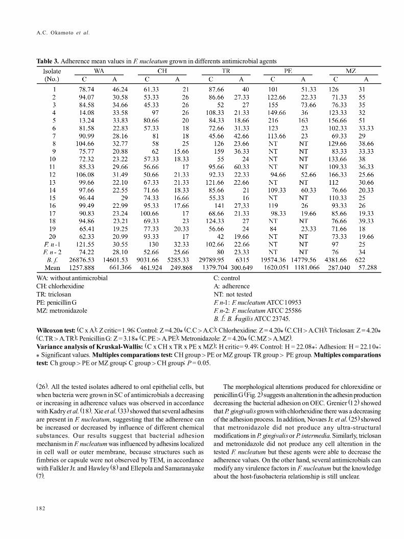
Most of the tested isolates and reference strains werehydrophilic when grown without antimicrobial agents or in SCof chlorhexidine, triclosan, or metronidazole. On the other hand,isolates 1, 2 and 12, were hydrophobic (< 50% values) (Table 2).Isolates grown with or without antimicrobials were capable toadhere on oral epithelial cells. Mean values of adherence fromcontrol group and bacteria treated with antimicrobials areshowed in Table 3. By variance analysis was observed thatcontrol group showed better adherence than bacteria treatedwith antimicrobials. Then, we can observe, for the adherenceprocess in bacteria grown in SC of antimicrobial: bacteria pluschlorhexidine > Bacteria plus triclosan > bacteria plus penicillinG or metronidazole. However, nine bacteria treated withmetronidazole increased their adherence on OEC. Also, sevenisolates treated with triclosan and four with penicillin increasedtheir adherence process (Table 3).
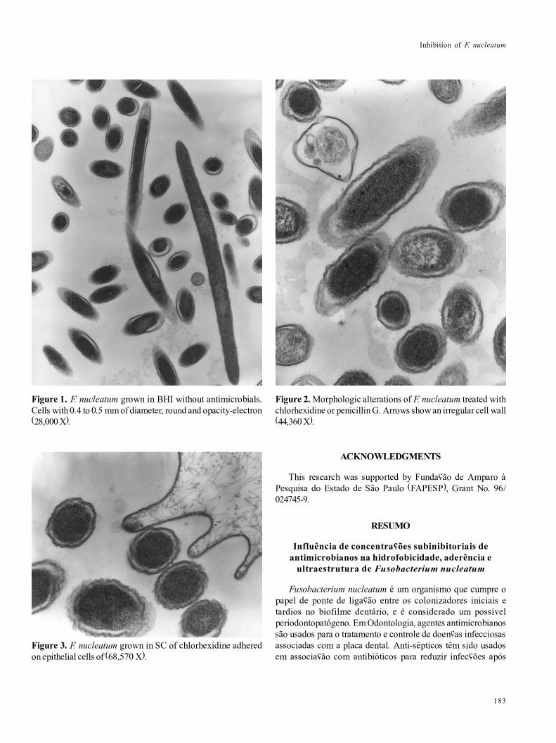
TEM showed that F. nucleatum grown without agents were0.4 to 0.5 µm of diameter, round and with opacity-electron(Fig. 1). Bacteria treated with chlorhexidine or penicillin Gshowed an irregular cell wall, but no alteration in diameter wasobserved (Fig. 2), and isolates grown in SC of triclosan ormetronidazole did not show any alteration in cell wall or diameter.In Fig. 3 can be observed the F. nucleatum grown in SC ofchlorhexidine adhered to epithelial cells.
DISCUSSION
Periodontal disease is a result of the bacterial virulencefactors action in a susceptible host (13). Also, virulence factorssuch as hydrophobicity and adherence are considered animportant prerequisite for colonization and subsequent infection(7,32). Chemical compounds such as chlorhexidine or triclosan,penicillin G or metronidazole, have been used in patients withdifficulties to maintain a good oral clean or with refractory orrapid progression periodontopathies (11).
The antimicrobial inhibitory concentrations or SC may altereither the bacterial virulence factors or the microorganism-hostrelationship (6,14,18). Although, few antimicrobials in inhibitoryconcentrations are able to remain for long period into the oralcavity, because of diluents of the saliva and by cleansing actionof the oral musculature (22). All the F. nucleatum isolates weresusceptible to chlorhexidine or triclosan with MIC valuesranged from 4 to 8 µg/ml. It is known that chlorhexidine showsa higher substantivity than triclosan on oral mucosa (1,23).However, both of agents showed similar action on testedisolates.
Table 1. MIC values of chlorhexidine, triclosan, penicillin G andmetronidazole, to 20 F. nucleatum isolates and reference strains.
Breakpoint: Chlorhexidine and Triclosan, 4 µg/ml; Penicillin Gand Metronidazole, 8 µg/ml.

Inhibition of F. nucleatum
181
Penicillins are still considered drugs of choice in dentistryfor treatment of different oral infections, but recently, anamoxicillin/metronidazole association has been observed inliterature (9,21). Most of F. nucleatum isolates were susceptibleto penicillin G, however three isolates (15%) were resistant (MICranged from 128 to 512 µg/ml) (Table 1) in accordance withKononen et al. (20). The susceptibility to metronidazole is acharacteristic of the genus Fusobacterium and all the testedisolates were susceptible to this drug (MIC ranged from 0.25 to1 µg/ml) in accordance with Feres et al. (19).
It is known that cellular hydrophobicity is closely associatedwith bacterial virulence and, microorganisms adsorbing to the
n-hexadecane are also able to adhere to the acquired dentalpellicle, epithelia or prosthetic denture (7,10). All the F.nucleatum grown without antimicrobials and the most of theisolates grown in SC showed a hydrophilic character. However,three-tested F. nucleatum grown in SC of penicillin G werehydrophobic, and it suggests that any factor might have alteredthe surface. In addition, it is known that chlorhexidine alter theouter membrane and cytoplasm of Gram-negative bacteria (7).In this study, chlorhexidine was not able of modifying the cellularhydrophobicity of F. nucleatum.
The hydrophobicity in F. nucleatum was not associated toadhesion on host�s oral cells, in accordance with Okuda et al.
Table 2. Mean values of the hydrophobic or hydrophilic characteristic of F. nucleatum, measured in spectrophotometer A550 nm
C: controlH: hydrophobicityNT: not testedF. n-1: F. nucleatum ATCC 10953;F. n-2: F. nucleatum ATCC 25586B. f.: B. fragilis ATCC 23745.
Wilcoxon test: (C x H); Z critic= 1.96; Control: Z = 3.74* (C.C > H.C); Chlorhexidine: Z = 3.88* (C.CH > H.CH); Triclosan: Z = 3.92*(C.TR > H. TR); Penicillin G: Z = 2.75* (C.PE > H.PE); Metronidazole: Z = 4.06* (C.MZ > H.MZ).Variance analysis of Kruskal-Wallis: (C x CH x TR x PE x ME); H critic = 9.49; Control: H = 19.69*; Hydrophobicity: H = 7.35;* Significant values.Multiples comparations test: TR group > PE or MZ group; P = 0.05.
WA: without antimicrobialCH: chlorhexidineTR: triclosanPE: penicillin GMZ: metronidazole
182
A.C. Okamoto et al.
Table 3. Adherence mean values in F. nucleatum grown in differents antimicrobial agents
WA: without antimicrobialCH: chlorhexidineTR: triclosanPE: penicillin GMZ: metronidazole
C: controlA: adherenceNT: not testedF. n-1: F. nucleatum ATCC 10953F. n-2: F. nucleatum ATCC 25586B. f.: B. fragilis ATCC 23745.
Wilcoxon test: (C x A); Z critic= 1.96; Control: Z =4.20* (C.C > A.C); Chlorhexidine: Z = 4.20* (C.CH > A.CH); Triclosan: Z = 4.20*(C.TR > A.TR); Penicillin G: Z = 3.18* (C.PE > A.PE); Metronidazole: Z = 4.20* (C.MZ > A.MZ).Variance analysis of Kruskal-Wallis: (C x CH x TR x PE x MZ); H critic= 9.49; Control: H = 22.08*; Adhesion: H = 22.10*;* Significant values. Multiples comparations test: CH group > PE or MZ group; TR group > PE group. Multiples comparationstest: Ch group > PE or MZ group; C group > CH group; P = 0.05.
(26). All the tested isolates adhered to oral epithelial cells, butwhen bacteria were grown in SC of antimicrobials a decreasingor increasing in adherence values was observed in accordancewith Kadry et al. (18). Xie et al. (33) showed that several adhesinsare present in F. nucleatum, suggesting that the adherence canbe increased or decreased by influence of different chemicalsubstances. Our results suggest that bacterial adhesionmechanism in F. nucleatum was influenced by adhesins localizedin cell wall or outer membrane, because structures such asfimbries or capsule were not observed by TEM, in accordancewith Falkler Jr. and Hawley (8) and Ellepola and Samaranayake(7).
The morphological alterations produced for chlorexidine orpenicillin G (Fig. 2) suggests an alteration in the adhesin productiondecreasing the bacterial adhesion on OEC. Grenier (12) showedthat P. gingivalis grown with chlorhexidine there was a decreasingof the adhesion process. In addition, Novaes Jr. et al. (25) showedthat metronidazole did not produce any ultra-structuralmodifications in P. gingivalis or P. intermedia. Similarly, triclosanand metronidazole did not produce any cell alteration in thetested F. nucleatum but these agents were able to decrease theadherence values. On the other hand, several antimicrobials canmodify any virulence factors in F. nucleatum but the knowledgeabout the host-fusobacteria relationship is still unclear.
Inhibition of F. nucleatum
183
ACKNOWLEDGMENTS
This research was supported by Fundação de Amparo àPesquisa do Estado de São Paulo (FAPESP), Grant No. 96/024745-9.
RESUMO
Influência de concentrações subinibitoriais deantimicrobianos na hidrofobicidade, aderência e
ultraestrutura de Fusobacterium nucleatum
Fusobacterium nucleatum é um organismo que cumpre opapel de ponte de ligação entre os colonizadores iniciais etardios no biofilme dentário, e é considerado um possívelperiodontopatógeno. Em Odontologia, agentes antimicrobianossão usados para o tratamento e controle de doenças infecciosasassociadas com a placa dental. Anti-sépticos têm sido usadosem associação com antibióticos para reduzir infecções após
Figure 1. F. nucleatum grown in BHI without antimicrobials.Cells with 0.4 to 0.5 mm of diameter, round and opacity-electron(28,000 X).
Figure 2. Morphologic alterations of F. nucleatum treated withchlorhexidine or penicillin G. Arrows show an irregular cell wall(44,360 X).
Figure 3. F. nucleatum grown in SC of chlorhexidine adheredon epithelial cells of (68,570 X).

184
A.C. Okamoto et al.
cirurgias orais. Neste estudo, foi avaliada a influência deconcentrações subinibitórias de clorexidina, triclosan, penicilinaG e metronidazol, sobre a hidrofobicidade, aderência às célulasepiteliais bucais, e a ultra-estrutura de F. nucleatum. Todos osisolados foram susceptíveis a clorexidina, triclosan, emetronidazol. A maioria deles foi sensível à penicilina G.Também, a maioria dos isolados, foi hidrofílica quando cresceramcom ou sem antimicrobianos. A aderência foi diminuída pelaação dos antimicrobianos usados. Os resultados sugerem quea aderência de F. nucleatum foi influenciada por adesinasdevido a que estruturas como fímbrias ou cápsula não foramobservadas pela microscopia eletrônica de transmissão.
Palavras-chave: Fusobacterium nucleatum, hidrofobicidade,aderência, antimicrobianos, ultra-estrutura.
REFERENCES
1. Albandar, J.M.; Gjermo, P.; Preus, H.R. Chlorhexidine use after twodecades of over-the-counter availability. J. Periodontol., 65: 109-112, 1994.
2. American Association of Public Health Dentist. Subcommittee ofPreventive Periodontics. Periodontal Disease in Americans: Apersonal and national tragedy. Dent. Hyg., 58: 10-18, 1984.
3. Childs III, W.C.; Gibbons, R.J. Use of Percoll density gradients forstudying the attachment of bacteria to oral epithelial cells. J. Dent.Res., 67: 826-30, 1988.
4. Clarck, W.B.; Löe, H. Mechanisms of initiation and progression ofperiodontal disease. Periodontology 2000, 2: 72-82, 1993.
5. Duerden, B.I. Virulence factors in anaerobes. Clin. Infect. Dis., 18:253-259, 1994.
6. Di Martino. Effects of antibiotics on adherence of Pseudomonasaeruginosa and Pseudomonas fluorescens to human fibronectin.Chemotherapy, 47: 344-349, 2001.
7. Ellepola, A.N.B.; Samaranayake, L.P. The effect of limited exposureto antimycotics on the relative cell-surface hydrophobicity and theadhesion of oral Candida albicans to buccal epithelial cells. ElsevierScien., 43: 879-887, 1998.
8. Falkler Jr.; W.A.; Hawley, C.A. Hemagglutination activity ofFusobacterium nucleatum. Infect. Immun., 15: 230- 238, 1977.
9. Feres, M.; Haffajee, A.D.; Allard, K.; Som, S.; Socransky, S.S. Changein subgingival microbial profiles in adult periodontitis subjectsreceiving either systemically-administered amoxicillin or metonidazole.J. Clin. Periodontol., 28: 597-609, 2001.
10. Gibbons, R.J.; Etherden, I. Comparative hydrophobicities of oralbacteria and their adherence to salivary pellicles. Infect. Immun.,41: 1190-1196, 1983.
11. Goodson, J.M. Antimicrobiol strategies for treatment of periodontaldiseases. Periodontology 2000, 5: 142-168, 1994.
12. Grenier, D. Effect of chlorhexidine on the adherence properties ofPorphyromonas gingivalis. J. Clin. Periodontol., 23: 140-142, 1996.
13. Han, Y.W.; Shi, W.; Huang, G.T.J.; Haake, S.K.; Park, N.H.; Kuramitsu,H.; Genco, R.J. Interations between periodontal bacteria and humanoral epithelia cells: Fusobacterium nucleatum adheres to and invadesepithelial cells. Infect. Immun., 68: 3140-3146, 2000.
14. Held, T.K.; Adamczik, C.; Trautmann, M.; Cross, A.S. Effects ofMICs of antibiotics on production of capsular polysaccharide of
Klebsiella pneumoniae. Antimicrob. Agents Chemother., 39: 1093-1096, 1995.
15. Jabra-Rizk, M.A.; Falkler Jr., W.A.; Merz, W.G.; Meiller, T.F. Newassay for measuring cell surface hydrophobicities of Candidadubliniensis and Candida albicans. Clin. and Diag. Lab. Immunol.,8: 585-587, 2001.
16. Jenkins, S.; Addy, M.; Newcombe, R. Triclosan and sodium laurylsulphate mouthwashes. (I) Effects on salivary bacterial counts. J.Clin. Periodontol., 18: 140-144, 1991.
17. Jorgensen, M.G.; Slots, J. Responsible use of antimicrobials inperiodontics. J. Calf. Dent. Assoc., 28: 185-93, 2000.
18. Kadry, A.A.; Tawfik, A.; Abu El-Asrar, A.A.; Shibl, A.M. Reductionof mucoid Staphylococcus epidermidids adherence in intraocularlenses by selected antimicrobial agents. Chemotherapy., 45: 56-60,1999.
19. Kolenbrander, P.E.; London, J. Adhere today, here tomorrow: oralbacterial adherence. J. Bacteriol., 175: 3247-3252, 1993.
20. Könönen, E.; Kanervo, A.; Salminen, K.; Jousimies-Somer, H. β-lactamase production and antimicrobial susceptibility of oralheterogeneous Fusobacterium nucleatum populations in youngchildren. Antimicrob. Agents Chemother., 43: 1270-1273, 1999.
21. Kuriyama, T.; Karasawa, T.; Nakagawa, K.; Yamamoto, E.;Nakamura, S. Incidence of b-lactamase production and antimicrobialsusceptibility of anaerobic gram-negative rods isolated from pusspecimens of orofacial odontogenic infections. Oral Microbiol.Immunol., 16: 10-15, 2001.
22. Martin, M.V. Antifungical agents. In: Samaranayake, L.P.;MacFarlane, T.W. (ed.). Oral candidosis. Wrught, London, 1990,pp.238-255.
23. Moran, J.; Addy, M.; Newcombe, R.G.; Marlow, I. A study to assessthe plaque inhibitory action of a new formulated triclosan toothpaste.J. Clin. Periodontol., 28: 86-89, 2001.
24. National Committee for Clinical Laboratory Standard. Referenceagar dilution procedure for antimicrobial susceptibility testing ofanaerobic bacteria approved standard, M-11 A. National Committeefor Clinical Laboratory Standard, Villanova, Pa, 1995.
25. Novaes Jr., A.B.; Uzeda, M.; Fonseca, M.E.F.; Feitosa, A.C.R. Theeffect of subinhibitory concentrations of metronidazole andtetracycline on the ultrastructure of periodontopathic bacteria. J.Antimicrob. Chemother., 28: 151-154, 1991.
26. Okuda, K.; Kato, T.; Ishihara, K.; Naito, Y. Adherence to experimentalpellicle of rough-type lipopolysaccharides from subgingival plaquebacteria. Oral Microbiol. Immun., 6: 241-5, 1991.
27. Rahn, R.; Schneider, S.; Diehl, O.; Schäfer, V.; Shah, P.M. Preventingpost-treatment bacteremia: comparing topical povidone-iodine andchlorhexidine. JADA, 126: 1145-1148, 1995.
28. Robets, G.L. Fusobacterial infections: an underestimated threat. B.J. Biomed., 57: 156-162, 2000.
29. Sbordone, L.; Barone, A.; Ramaglia, L.; Ciaglia, R.N.; Iacono, V.J.Antimicrobial susceptibility of periodontopathic bacteria associatedwith failing implants. J. Periodontol., 66: 69-74, 1995.
30. Siegel, S.; Castellani Jr., N.J. Nonparametric statistics. 2a.ed., McGraw-Hill Int. ed.- New York, 1988, 399p.
31. Slots, J. Selective medium for isolation of Actinobacillusactinomycetemcomitans. J. Clin. Microbiol., 15: 606-609, 1982.
32. Weiss, E.I.; Shaniztki, B.; Dotan, M.; Ganesshkumar, N.;Kolenbrander, P.E.; Metzger, Z. Attachment of Fusobacteriumnucleatum PK1594 to mammalian cells and its coaggregation withperiodontopathogenic bacteria are mediated by the same galactose-binding adhesin. Oral Microbiology Immunol., 15: 371-377, 2000.
33. Xie, H.; Gibbons, R.J.; Hay, D.I. Adhesive properties of strains ofFusobacterium nucleatum of the subspecies nucleatum, vincentiiand polymorphum. Oral Microbiol. Immun., 6: 257-263, 1991.