Embed Size (px)
Citation preview

Full Terms & Conditions of access and use can be found athttp://www.tandfonline.com/action/journalInformation?journalCode=iddi20
Download by: [LIU Libraries] Date: 05 May 2016, At: 07:09
Drug Development and Industrial Pharmacy
ISSN: 0363-9045 (Print) 1520-5762 (Online) Journal homepage: http://www.tandfonline.com/loi/iddi20
Influence of spray drying and dispersing agent onsurface and dissolution properties of griseofulvinmicro and nanocrystals
Dhaval A. Shah, Manan Patel, Sharad B. Murdande & Rutesh H. Dave
To cite this article: Dhaval A. Shah, Manan Patel, Sharad B. Murdande & Rutesh H. Dave(2016): Influence of spray drying and dispersing agent on surface and dissolution propertiesof griseofulvin micro and nanocrystals, Drug Development and Industrial Pharmacy, DOI:10.1080/03639045.2016.1178770
To link to this article: http://dx.doi.org/10.1080/03639045.2016.1178770
Accepted author version posted online: 14Apr 2016.Published online: 04 May 2016.
Submit your article to this journal
Article views: 13
View related articles
View Crossmark data

RESEARCH ARTICLE
Influence of spray drying and dispersing agent on surface and dissolutionproperties of griseofulvin micro and nanocrystals
Dhaval A. Shaha, Manan Patela, Sharad B. Murdandeb and Rutesh H. Davea
aArnold & Marie Schwartz College of Pharmacy and Health Sciences, Long Island University, Brooklyn, NY, USA; bDrug Product Design, PfizerWorldwide R&D, Groton, CT, USA
ABSTRACTThe purpose for the current research is to compare and evaluate physiochemical properties of spray-dried(SD) microcrystals (MCs), nanocrystals (NCs), and nanocrystals with a dispersion agent (NCm) from a poorlysoluble compound. The characterization was carried out by performing size and surface analysis, interfacialtension (at particle moisture interface), and in-vitro drug dissolution rate experiments. Nanosuspensionswere prepared by media milling and were spray-dried. The SD powders that were obtained were character-ized morphologically using scanning electron microscopy (SEM), polarized light microscopy (PLM), andFlowchem. Solid-state characterization was performed using X-ray powder diffraction (XRPD), Fourier trans-fer infrared spectroscopy (FT-IR), and differential scanning calorimetry (DSC) for the identification of thecrystalline nature of all the SD powders. The powders were characterized for their redispersion tendency inthe water and in pH 1.2. Significant differences in redispersion were noted for both the NCs in both dissol-ution media. The interfacial tension for particle moisture interface was determined by applying the BET(Braunauer–Emmett–Teller) equation to the vapor sorption data. No significant reduction in the interfacialtension was observed between MCs and NCs; however, a significant reduction in the interfacial tension wasobserved for NCm at both 25 �C and 35 �C temperatures. The difference in interfacial tension and redisper-sion behavior can be attributed to a difference in the wetting tendency for all the SD powders. The dissol-ution studies were carried out under sink and under non-sink conditions. The non-sink dissolutionapproach was found suitable for quantification of the dissolution rate enhancement, and also for providingthe rank order to the SD formulations.
ARTICLE HISTORYReceived 29 December 2015Revised 30 March 2016Accepted 7 April 2016Published online 3 May 2016
KEYWORDSGriseofulvin; interfacialtension; nanocrystals; non-sink dissolution;redispersion; spray dryer
Introduction
It has been reported that more than half of the identified potentialdrug candidates have challenges related to aqueous solubility and/or the dissolution rate1. A majority of these compounds belong tothe BCS class II and IV categories2,3. The rate and the extent ofabsorption of the Class II compounds are highly dependent on theperformance of the formulated product4–6. Several formulationapproaches have been suggested and reported to overcome theseproblems. The formulation of nanocrystals (NCs; suspension orsolid) is one of the preferred approaches that has been used forthe drugs which belong to the above BCS classes7,8. NCs offer sev-eral advantages, including enhancement in bioavailability, reduc-tion in therapeutic dose, and elimination of the food effect9–14.The solubility behavior of crystalline nanoparticles can beexplained by the Ostwald–Frendulich equation. Based on the pub-lished reports, the thermodynamic solubility enhancement of nano-crystalline particles should be approximately 10–15%8,15,16. On theother hand, nanosuspensions have a very high surface-to-volumeratio and high Gibbs energy, which indicates the instability of thesystem. They tend to form aggregates due to Ostwald’s ripen-ing13,17. Nanosuspensions can be stabilized and their shelf life canbe enhanced by converting them into solid powders by spray dry-ing and/or freeze drying10,18.
Upon solidification, the surface properties of dried particles (i.e.size, shape) may tend to change significantly, which will have a dir-ect impact on the wetting and dissolution performance of the
formulation19. Hence, it is crucial to assess the surface and theinterfacial properties of the dried particles20,21. It is also known thatthese suspensions (micro/nano) may form reversible or nonreversi-ble aggregates upon solidification. Therefore, aggregation isanother key product quality parameter that needs to be assessedfor effectiveness with the dried particles22,23. During spray drying,the particle size distribution is governed by the nozzle size24. It ispossible to have different dissolution behavior for formulationswith the same particle size due to change in surface wetting prop-erty25. Therefore, it will be important to determine the role of theinterfacial tension (at particle to moisture interface) in governingthe redispersion and the kinetic solubility (initial dissolution rate)of NCs and micro crystals (MCs).
In the current study, we have formulated spray-dried (SD) nano-crystalline powder (with and without the bulking agent mannitol)and MCs of a poorly soluble drug. Griseofulvin (a BCS Class II com-pound) was selected as a model compound and docusate sodium(ionic stabilizer) and HPC-SL (Hydroxypropyl cellulose-low viscositygrade; polymeric stabilizer) were utilized to prevent agglomerationof nanosuspensions before spray drying, while the mannitol wasused as a dispersing agent for spray drying. The SD powders wereevaluated for surface characterization (size and shape analysis),solid-state characterization (XRPD, DSC, and FT-IR), interfacial ten-sion (DVSO), redispersion, and in-vitro dissolution rates (sink andnon-sink). This study shows the effect of interfacial tension in gov-erning the dissolution of NCs and quantitates the dissolution rate
CONTACT Rutesh H. Dave [email protected] Arnold & Marie Schwartz College of Pharmacy and Health Sciences, Long Island University, Brooklyn, NY 11201,USA.� 2016 Informa UK Limited, trading as Taylor & Francis Group
DRUG DEVELOPMENT AND INDUSTRIAL PHARMACY, 2016http://dx.doi.org/10.1080/03639045.2016.1178770
Dow
nloa
ded
by [
LIU
Lib
rari
es]
at 0
7:09
05
May
201
6

enhancement for the SD powders using non-sink conditions toprovide them in rank order based on their in-vitro performance.
Materials
Griseofulvin was obtained from Spectrum Chemicals (NewBrunswick, NJ). The yttrium-stabilized zirconium oxide beads wereobtained from Glen Mills Inc. (Clifton, NJ). Hydroxypropylcellulose(HPC)–SL was obtained from Nisso America Inc. (New York, NY).Other laboratory chemicals, such as mannitol, docusate sodiumUSP, acetonitrile, and trifluoroacetic acid (TFA), were purchasedfrom Spectrum Chemicals (New Brunswick, NJ) and were of analyt-ical grades. All the excipients utilized in this study were of highpurity. The chemical structure of API and excipients are presentedin Figure 1.
Methods
Wet milling
Griseofulvin (50 mg/mL) was suspended in an aqueous stabilizersolution which is a mixture of 1.25% (w/v) HPC–SL (stearic stabil-izer) and 0.05% (w/v) docusate sodium (ionic stabilizer). The sus-pension (500 mL) was milled using the yttrium stabilized zirconiumoxide beads (YTZ beads) at a milling speed of 1000 rpm in the con-tinuous model8. The suspension formulation was milled for 4 h,and the particle size was measured during and following the mill-ing process. The temperature of the sample was maintained below25 �C using the cold water that surrounded the milling chamber tocompensate for the heat generated by high-speed milling. The sus-pension was harvested by filtration through a coarse sintered-glassfunnel filter (75 mm). The nanosuspension was divided into twoequal parts, and in one part the mannitol was dissolved in a 1:2ratio (Griseofulvin: mannitol). The suspension was then stirred forat least 30 min and then prepared for spray drying. The microcrys-talline suspension was prepared using the same method and thesame stabilizer concentration with the absence of the millingvehicle (i.e. YTZ beads).
Spray drying
Nano (with and without the mannitol) and microcrystalline suspen-sions were spray-dried using a B-290 spray dryer (BuchiLabortechhnik AG, Flawil, Switzerland). The spray dryer was equili-brated using distilled water at 128 �C inlet temperature, 3 mL/minfeed rate, and 100% aspiration rate. The outlet temperature wasapproximately 75 �C. The aforementioned SD parameters weredetermined from some preliminary experiments. Once the spraydryer was equilibrated, the suspension formulations were subjectedto spray drying with continuous stirring using a magnetic stirrer.SD powders were collected from the collection chamber andimmediately analyzed for particle size, process yield, andcrystallinity.
Particle size, surface and shape analysis
A dynamic light scattering (DLS) instrument (Malvern InstrumentsInc., Malvern, UK) was used to measure particle size and the sizedistribution of suspensions. All three SD powders were subjectedto particle size and shape analysis. Particle size measurements ofSD powders were performed using a Sympatec GmbH System-Partikel-Technik (Sympatec Inc., Pennington, NJ). Briefly, around100 mg of a sample was subjected for analysis. Compressed airwas used to disperse the powder, which is blown through the laser
beam and collected by suction after the analysis. Polarized lightmicroscopy (PLM; Model BX60FS; Olympus BX60, Center Valley, PA)and scanning electron microscopy (SEM, ASPEX Scanning ElectronMicroscope model PSEM 3025 SEM/EDS, Fei Inc., Hillsboro, OR)were conducted to observe the surface morphology and texture ofthe pure materials and SD powders. The polarized microscopicimages were taken using a camera from Diagnostic InstrumentsInc. (Sterling Heights, MI), and the analysis was performed with thehelp of SPOTTM imaging advanced software. For the SEM imaging,the samples were mounted on double-sided carbon tape. The lowvacuum (LV) mode was used to prevent the samples from charg-ing. The energy beam and filament drive were adjusted to 20 kevand 72%, respectively. The samples were analyzed under approxi-mately 60% contrast and 30% brightness in secondary electronmode. Particle shape and further morphological characterizationwere performed using the flow cam machine (Fluid ImagingTechnologies, Scarborough, ME). Here, the sample was prepared bydispersing the SD powders into the water and then subjected toanalysis. The machine captures particle images at up to 22 framesper second. The analysis was performed by keeping the flow rateat 0.3 mL/min and viewed under 4� optical lens. The morpho-logical measurements (i.e. shape) of particles were calculated usingthe Visual spread sheetVR Particle analysis software.
Differential scanning calorimetry
Modulated differential scanning calorimetric (mDSC) analysis wasperformed using a Q200 (TA Instruments, New Castle, DE)equipped with a cooling system. A sample (5–10 mg) was weighedinto an aluminum pan with a pin hole and hermitically sealed. TheDSC parameters used were: a sample equilibration at 30 �C, modu-lated for ±0.318 �C every 60 s, isothermal for 5 min and heated to230 �C with a ramp of 2 �C/min. The DSC cell was purged withnitrogen gas at a flow rate of 50 mL/min. The data obtained wereanalyzed with the Universal Analysis Software (TA Instruments,New Castle, DE).
Powder X-ray diffraction
Powder X-ray diffraction was done using a scanning diffractometer(Advanced Diffraction System, Scintag Inc. Model XI, Cupertino,CA), controlled by a computer with diffraction management systemsoftware for Windows NT. The radiation used was generated by acopper Ka filter, with a wavelength of 1.54 A� at 45 kV and 40 mA.High energy mixture patterns obtained by the X-ray were
Figure 1. Chemical structures of a model compound and excipients.
2 D. A. SHAH ET AL.
Dow
nloa
ded
by [
LIU
Lib
rari
es]
at 0
7:09
05
May
201
6

compared to a reference drug to confirm crystallinity26. Sampleswere scanned over a range of 5–40�degrees, using a scan rate of1�/min and a step size of 0.02. X-ray diffraction patterns wereobtained for pure griseofulvin, excipients, physical mixtures, andformulated SD powders. Data analysis for recorded patterns wasdone using X’Pert Data Viewer software.
Infrared spectroscopy (FT-IR)
FTIR spectra for all the abovementioned samples were obtained byNicolet-FTIR equipped with smart ATR (Thermo Fisher Scientific Inc.Madison, WI), and a DTGS-KBr (Deuterated triglycine sulfate)detector was used for background and baseline corrections. Smallamounts of the sample were placed on the diamond crystal of theATR and then the ATR knob was closed. Finally, the spectra werecollected from 400 to 4000 cm�1 with 264 accumulations at a reso-lution of 4 cm�1.
Gravimetric vapor sorption and interfacial energy (DVS)
Moisture sorption profiles were obtained for the SD powders usinga SGA-100 Sorption Analyzer (VTI Corporation, Hialeah, FL). Themeasurements were taken at 25 �C (storage temperature) and35 �C (close to physiological temperature) using a sample size of10–20 mg. All SD samples were exposed to an automated compu-terized sequence of increasing relative humidity (RH) in steps of10% RH. The RH range was from 0% RH to 95% RH. The vaporsorption analysis was carried out to calculate the interfacial tensionand water uptake capacity of all three powders as described in theliterature27. The adsorption of moisture onto multilayer surfacesis believed to follow the Brunauer–Emmett–Teller (BET) model.Similarly, we made the assumption that adsorption of moistureonto SD powder particles would follow the BET multilayer model.The BET constants (m0 and C) were determined from the initial lin-ear region of the moisture sorption adsorption isotherm (<0.35%w/w). The interfacial tension at particle moisture interface wasdetermined by calculating the binding energy (from BET constantC) and the average surface area of a water molecule (Appendix A).
HPLC analytical method
The quantitative analysis of model compounds used in the solubil-ity studies was performed by a reverse-phase gradient HPLC usingan Agilent 1100 System (Agilent Technologies, Palo Alto, CA)equipped with a 4.6� 75 mm, 2.7 mm C18 column (AdvancedMaterials Technology, Wilmington, DE). The temperature of the col-umn was kept at 45 �C, and the wavelength of detection was setto 210 nm. The mobile phase consisted of a binary gradient ofSolvent A (acetonitrile) and Solvent B (HPLC grade water with0.05% TFA). The linear gradient started at 5% A and increased to95% A in 8.5 min, followed by a return to the starting conditionwithin 1.6 min and equilibrated at the starting condition for 2 min.The injection volume was 10 mL. The flow rate was 1.0 mL/min, andthe total run time was set for 13.10 min. This reverse-phase gradi-ent method provided a baseline resolution and excellent peakcharacteristics for the griseofulvin. Quantification of drug concen-trations was performed by analyzing the peak area at retentionusing Agilent ChemstationVR analytical software.
Redispersion analysis
The nanosuspensions have tendency to form loose or hard aggre-gates when spray-dried. Sometimes these aggregates are reversibleand sometime they are not reversible. To evaluate this behavior,we carried out the redispersion study of SD NCs (with and without
mannitol) in the DI water and in the simulated gastric media (pH1.2). The study was performed in a conventional USP-type – II dis-solution apparatus at 100 rpm and 37 �C temperature. Here, sam-ples were withdrawn at different time intervals and subjected tothe particle size analysis using the zetasizer28.
In vitro dissolution rate determination
A dissolution apparatus (Distek 2100C; Distek Inc., North Brunswick,NJ) equipped with a USP-type-II (paddle) was used to measure thedissolution rates of crystalline SD dispersions. The dissolution studywas carried out by comparing the initial dissolution rates of MCsand NCs (with and without mannitol). Each dissolution study wascarried out at 37 ± 0.5 �C under a 100-rpm paddle speed in a USP-type II apparatus for 2 h. The dissolution was carried out undersink condition (sink condition factor 5.7�), and under non-sinkconditions (at 20%, 50%, and 90% saturation level of API in dissol-ution media) with a 50 mg loading of griseofulvin. A 10 mL of a dis-solution sample was withdrawn at intervals of 0, 2, 5, 7, 10, 20, 30,45, 60, 90, and 120 min and replaced with the same volume of dis-solution media (pre-equilibrated at 37 ± 0.5�). Samples were filteredthrough 0.1-mm (MCs) and 0.02-mm (NCs) anotop syringe filters(WhatmanTM; GE Healthcare, Piscataway, NJ) and subjected toHPLC analysis. The first few milliliters of each sample were dis-carded to minimize losses because of adsorption. The discardedvolume selected for each compound was determined by a separatefilter validation experiment (data not shown). All studies were per-formed in triplicate29–35.
Results and discussion
Morphological characterization
The particle size of the suspensions before the spray dying wasmeasured using DLS. The particle sizes of the SD powders weremeasured using the Sympatec GmbH System-Partikel-Technik(HELOS & RODOS). The particle size and shape analysis data arepresented in Table 1. The particle size of micronized SD powderupon drying is approximately 14 mm, while the particle size ofboth SD nanosuspension powders upon drying are around 6 mm.The particle size of nano powders are significantly lower than theparticle size of the micronized powder, which is explainable dueto difference in particle size before spray drying. There is no sig-nificant difference in particle size between nanosuspensionswith and without mannitol. Morphological characterization byPLM and SEM (Figures 2 and 3) shows the particle size differencebetween MCs and NCs. Moreover, the NCs in both formulationsbecame hollow upon particle size reduction while MCs appearcylindrical shaped. The SEM image of nanocrystals with mannitol(NCm) also shows the presence of mannitol on the surface ofNCs. Initial observation from SEM images of the NCs with andwithout the mannitol showed formation of aggregates uponspray drying. Therefore, redispersion analysis was carried out forfurther characterization of the aggregation property (reversible orirreversible). The FLOWCHEM machine has the ability to capture30 unique particle measurements. The morphological measure-ments for SD particles were calculated using Visual spread sheetVR
particle analysis software. The NCm were found to be spherical inshape as compared to NCs without the mannitol and the MCs(Table 1).
Solid-state characterization
It is known that changes in the crystalline state can occur duringboth milling and spray drying. To ensure that the changes are due
DRUG DEVELOPMENT AND INDUSTRIAL PHARMACY 3
Dow
nloa
ded
by [
LIU
Lib
rari
es]
at 0
7:09
05
May
201
6

to particle size reduction and that no amorphous griseofulvin wasformed, MDSC, XRPD, and FT-IR were performed on the SDsamples.
Griseofulvin showed distinct crystalline peaks for 2h at 11� and17� (Figure 4(a)) in all the SD powders. As shown by the XRPD pat-terns (Figure 4(a)), the reduction in the intensity and broadening of
Figure 2. PLM images of (a) pure griseofulvin, (b) MC, (c) NCs, and (d) NCm.
Figure 3. SEM images of (a) HPC-SL, (b) DOSS, (c) mannitol, (d) pure griseofulvin, (e) MC, (f) NCs, and (g) NCm.
Table 1. Particle size and shape analysis.
FormulationAvg. particle size
before spray drying PDIAvg. particle size
after SD (lm) Span(D90–D10)/D50
Circularity(Hu)
Aggregation basedon SEM/PLM
MC 11 lm – 14.9 2.3 0.81 YesNCs 210 nm 0.210 6.64 1.85 0.82 YesNanocrystal-mannitol 205 nm 0.192 6.42 1.82 0.95 Yes
4 D. A. SHAH ET AL.
Dow
nloa
ded
by [
LIU
Lib
rari
es]
at 0
7:09
05
May
201
6

the peaks is observed for the nanoparticles. XPRD of SD powdersshowed a small hump in between 20 and 30 at 2h scale representspossible minor amorphous conversion. Figure 4(b) shows the MDSCthermograms for pure griseofulvin and the SD formulations. Thethermograms were compared to confirm the crystallinity upon mill-ing and spray drying. The onset melting temperature of griseofulvinand mannitol were detected at 219.0 �C and 168.4 �C, respectively.After milling and spray drying, a reduction in the onset of the melt-ing temperature (2–4 �C) and the heat of fusion was observed forboth griseofulvin and mannitol. The melting peaks of griseofulvinshifted in the SD formulations (Figure 4(b)). The observed meltingpoint reduction and peak shift have been reported to be in agree-ment with the particle size reduction. It can also be explained bythe Gibbs–Thomson equation36, which explains that, the meltingtemperature of a material is proportional to its cohesive energy.Since the atoms at the surface have reduced cohesive energy com-pared to bulk material due to physical milling and spray drying.Therefore, they require less energy to free from the solid phase (i.e.melting), which results in the reduction of melting point. Based onthe MDSC and XRPD data, it can be concluded that the wet-mediamilling and spray drying might have affected the crystalline struc-ture of the model compound by partially (minor) converting it in tothe amorphous form. For further clarification of the crystalline struc-ture, FT-IR spectroscopy was conducted on the SD samples (Datanot shown). The SD formulation with mannitol showed intense peakat 3288�1 cm and 3388�1 cm (–OH stretching) suggesting the pres-ence of mannitol in the formulation. These transmissions wereabsent in the IR spectra of MCs and NCs without the mannitol. TheIR spectra of all three SD formulations are same except the increasein the –OH and –CH stretching intensity due to the presence ofmannitol in the SD formulation with mannitol.
Interfacial energy and water uptake tendency
The dynamic vapor sorption analysis was performed to evaluate themoisture uptake tendency of the SD powders. The water uptake
tendency of NCm was found to be higher compared to both theMCs and NCs. For a detailed evaluation of this, we calculated theinterfacial energy as described in Appendix A, and the results arepresented in Table 2. For the colloidal dispersion systems, the inter-facial energy plays an important role. For different compounds, thesolubility and, eventually, the dissolution rate increased as the sol-id–liquid interfacial tension decreases37. The interfacial tension val-ues for the MC and NC do not have any significant difference ateither temperature (both at 25 �C and 35 �C). The NCm showed lessinterfacial tension values compared to MCs and NCs without manni-tol at both temperatures. Here, mannitol has a characteristic prop-erty to act as a solubilizing and dispersing agent. Mannitol acts byforming capillaries in contact with aqueous solutions and generatesrapid dispersion and dissolution38. Based on these studies, we canconclude that the dissolution and redispersion behavior for NCmwill be significantly higher due to the enhancement of the wettingproperty and a reduction in interfacial tension. Crisp39 and his col-leagues have also reported that the dissolution of NCs is moredependent on interfacial tension than the surface area.
Redispersion analysis
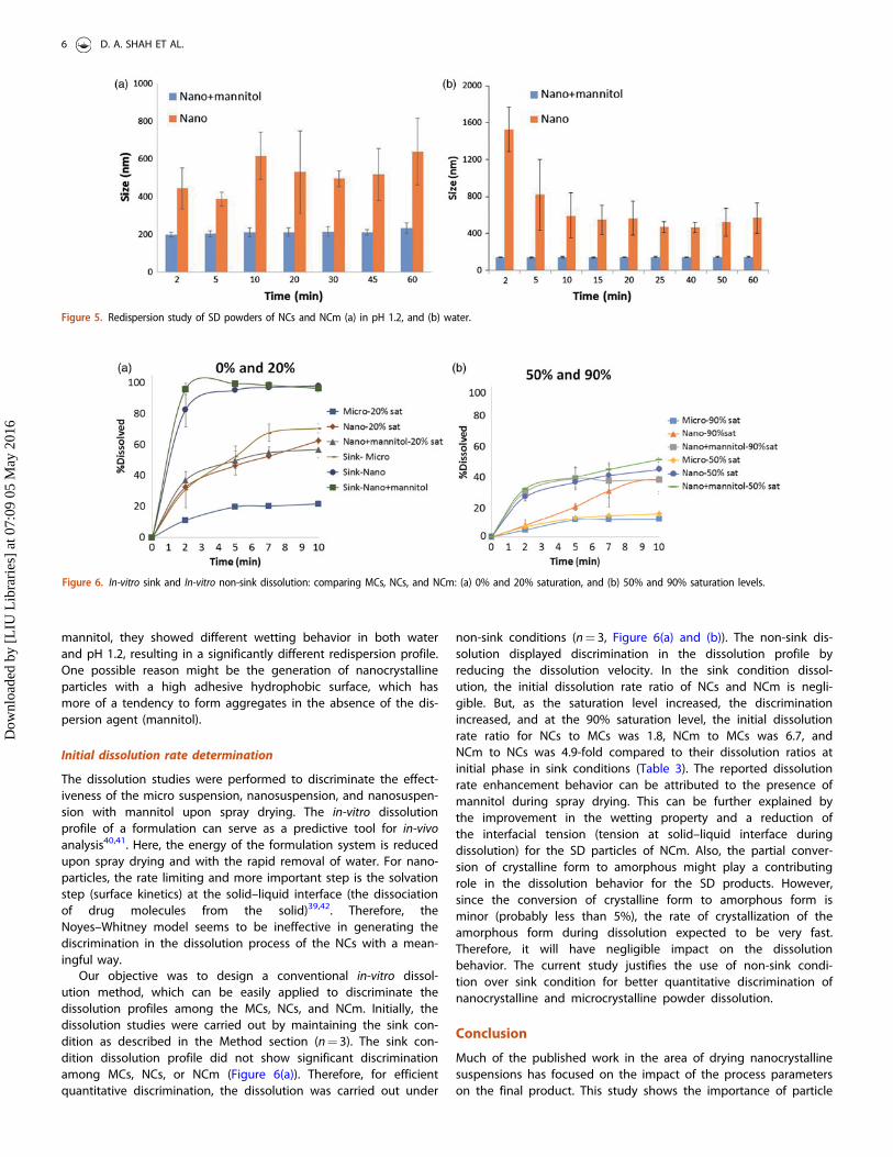
The nanosuspensions have a tendency to form loose or hardaggregates when SD. Nanoparticle aggregation was visuallyobserved for SD powders in SEM images. Sometimes these aggre-gates are reversible and sometime they are not reversible. In orderto assess their aggregation property, the redispersion analysis wascarried out. The SD powder of NCm redispersed quickly during theredispersion study in water and in pH 1.2 (Figure 5(a) and (b)). TheNCm quickly reached the initial particle size. On the other hand,the NCs without the mannitol were not able to achieve the initialparticle size (200–300 nm) when spray-dried. The dispersion behav-ior of particles in the liquid phase is mainly affected by the inter-action between the particles and the dispersion media. Therefore,the presence of mannitol aids in wetting the particles quickly byforming reversible aggregates. In the case of the NCs without the
Figure 4. Solid-state characterization and comparison of pure API, physical mixture, MCs, NCs, and NCm (a) XRPD, and (b) DSC.
Table 2. Determination of BET constants and interfacial tension.
Formulation Particle size (lm)
C(binding constant)
m0
(monolayer g/gsolids)
DE (J/mole) (sur-face binding
energy)
c(mN/m) (interfacial
tension)
25 �C 35 �C 25 �C 35 �C 25 �C 35 �C 25 �C 35 �C
MC 14.9 �20.58 �20.43 12.15 1.77 7492 7726 138.2 142.5NCs 6.64 �17.00 �17.26 19.60 24.07 7681 7790 133 134Nanocrystals-mannitol 6.42 �11.48 �11.24 32.26 21.19 6047 6195 111.6 114.3
DRUG DEVELOPMENT AND INDUSTRIAL PHARMACY 5
Dow
nloa
ded
by [
LIU
Lib
rari
es]
at 0
7:09
05
May
201
6
mannitol, they showed different wetting behavior in both waterand pH 1.2, resulting in a significantly different redispersion profile.One possible reason might be the generation of nanocrystallineparticles with a high adhesive hydrophobic surface, which hasmore of a tendency to form aggregates in the absence of the dis-persion agent (mannitol).
Initial dissolution rate determination
The dissolution studies were performed to discriminate the effect-iveness of the micro suspension, nanosuspension, and nanosuspen-sion with mannitol upon spray drying. The in-vitro dissolutionprofile of a formulation can serve as a predictive tool for in-vivoanalysis40,41. Here, the energy of the formulation system is reducedupon spray drying and with the rapid removal of water. For nano-particles, the rate limiting and more important step is the solvationstep (surface kinetics) at the solid–liquid interface (the dissociationof drug molecules from the solid)39,42. Therefore, theNoyes–Whitney model seems to be ineffective in generating thediscrimination in the dissolution process of the NCs with a mean-ingful way.
Our objective was to design a conventional in-vitro dissol-ution method, which can be easily applied to discriminate thedissolution profiles among the MCs, NCs, and NCm. Initially, thedissolution studies were carried out by maintaining the sink con-dition as described in the Method section (n¼ 3). The sink con-dition dissolution profile did not show significant discriminationamong MCs, NCs, or NCm (Figure 6(a)). Therefore, for efficientquantitative discrimination, the dissolution was carried out under
non-sink conditions (n¼ 3, Figure 6(a) and (b)). The non-sink dis-solution displayed discrimination in the dissolution profile byreducing the dissolution velocity. In the sink condition dissol-ution, the initial dissolution rate ratio of NCs and NCm is negli-gible. But, as the saturation level increased, the discriminationincreased, and at the 90% saturation level, the initial dissolutionrate ratio for NCs to MCs was 1.8, NCm to MCs was 6.7, andNCm to NCs was 4.9-fold compared to their dissolution ratios atinitial phase in sink conditions (Table 3). The reported dissolutionrate enhancement behavior can be attributed to the presence ofmannitol during spray drying. This can be further explained bythe improvement in the wetting property and a reduction ofthe interfacial tension (tension at solid–liquid interface duringdissolution) for the SD particles of NCm. Also, the partial conver-sion of crystalline form to amorphous might play a contributingrole in the dissolution behavior for the SD products. However,since the conversion of crystalline form to amorphous form isminor (probably less than 5%), the rate of crystallization of theamorphous form during dissolution expected to be very fast.Therefore, it will have negligible impact on the dissolutionbehavior. The current study justifies the use of non-sink condi-tion over sink condition for better quantitative discrimination ofnanocrystalline and microcrystalline powder dissolution.
Conclusion
Much of the published work in the area of drying nanocrystallinesuspensions has focused on the impact of the process parameterson the final product. This study shows the importance of particle
Figure 5. Redispersion study of SD powders of NCs and NCm (a) in pH 1.2, and (b) water.
Figure 6. In-vitro sink and In-vitro non-sink dissolution: comparing MCs, NCs, and NCm: (a) 0% and 20% saturation, and (b) 50% and 90% saturation levels.
6 D. A. SHAH ET AL.
Dow
nloa
ded
by [
LIU
Lib
rari
es]
at 0
7:09
05
May
201
6

size reduction and the incorporation of mannitol as a dispersingagent during the spray drying of nanocrystalline suspension ofpoorly soluble compounds (BCS class II/IV). Upon spray drying, thefundamental properties (surface and in-vitro dissolution under non-sink condition) change drastically. The interfacial tension at theparticle moisture interface and redispersion behavior and non-sinkdissolution profile of griseofulvin–mannitol nanocrystalline suspen-sions show superior performance as compared to the micron/un-milled and nanocrystalline suspension formulations. The presentstudy provides an in-depth understanding to a formulation scien-tist who can quantitate the change in the interfacial tension (atparticle moisture interface) and in the in-vitro dissolution rateenhancement during initial phase. This provides a platform toapply a practical approach to screen and rank order the SD formu-lation with different initial particle size (before drying) with manni-tol as a dispersing agent.
Disclosure statement
The authors report no declaration of interests.
References
1. Lipinski CA. Drug-like properties and the causes of poor solu-bility and poor permeability. J Pharmacol Toxicol Methods2000;44:235–49.
2. Hywel D, Williams NLT, Charman SA, et al. Strategies toaddress low drug solubility in discovery and development.Pharmacol Rev 2013;65:315–499.
3. Shegokar R, Muller RH. Nanocrystals: industrially feasiblemultifunctional formulation technology for poorly solubleactives. Int J Pharm 2010;399:129–39.
4. Serajuddin ATM. Solid dispersion of poorly water-solubledrugs: early promises, subsequent problems, and recentbreakthroughs. J Pharm Sci 1999;88:1058–66.
5. Dave RH, Shah DA, Patel PG. Development and evaluation ofhigh loading oral dissolving film of aspirin and acetamino-phen. J Pharm Sci Pharmacol 2014;1:112–22.
6. Mou D, Chen H, Wan J, et al. Potent dried drug nanosuspen-sions for oral bioavailability enhancement of poorly solubledrugs with pH-dependent solubility. Int J Pharm2011;413:237–44.
7. Merisko-Liversidge E, Liversidge GG, Cooper ER. Nanosizing: aformulation approach for poorly-water-soluble compounds.Eur J Pharm Sci 2003;18:113–20.
8. Murdande SB, Shah DA, Dave RH. Impact of nanosizing onsolubility and dissolution rate of poorly soluble pharmaceuti-cals. J Pharm Sci 2015;104:2094–102.
9. Shah DA, Murdande SB, Dave RH. A review: pharmaceuticaland pharmacokinetic aspect of nanocrystalline suspensions. JPharm Sci 2016;105:10–24.
10. Kumar S, Jog R, Shen J, et al. Formulation and performanceof danazol nano-crystalline suspensions and spray dried pow-ders. Pharm Res 2015;32:1694–703.
11. Song J, Wang Y, Song Y, et al. Development and character-isation of ursolic acid nanocrystals without stabilizer havingimproved dissolution rate and in vitro anticancer activity.AAPS PharmSciTech 2014;15:11–9.
12. M€uller RH, Peters K. Nanosuspensions for the formulation ofpoorly soluble drugs: I. Preparation by a size-reduction tech-nique. Int J Pharm 1998;160:229–37.
13. Rabinow BE. Nanosuspensions in drug delivery. Nat Rev DrugDiscov 2004;3:785–96.
14. Cooper ER. Nanoparticles: a personal experience for formulat-ing poorly water soluble drugs. J Control Release2010;141:300–2.
15. Van Eerdenbrugh B, Vermant J, Martens JA, et al. Solubilityincreases associated with crystalline drug nanoparticles:methodologies and significance. Mol Pharm 2010;7:1858–70.
16. Anhalt K, Geissler S, Harms M, et al. Development of a newmethod to assess nanocrystal dissolution based on light scat-tering. Pharm Res 2012;29:2887–901.
17. Ghosh I, Bose S, Vippagunta R, Harmon F. Nanosuspensionfor improving the bioavailability of a poorly soluble drug andscreening of stabilizing agents to inhibit crystal growth. Int JPharm 2011;409:260–8.
18. Chaubal MV, Popescu C. Conversion of nanosuspensions intodry powders by spray drying: a case study. Pharm Res2008;25:2302–8.
19. Zuo B, Sun Y, Li H, et al. Preparation and in vitro/in vivo evalu-ation of fenofibrate nanocrystals. Int J Pharm 2013;455:267–75.
20. Isa L, Lucas F, Wepf R, Reimhult E. Measuring single-nanopar-ticle wetting properties by freeze-fracture shadow-castingcryo-scanning electron microscopy. Nat Commun 2011;2:438.doi:10.1038/ncomms1441.
21. Van Eerdenbrugh B, Froyen L, Van Humbeeck J, et al. Dryingof crystalline drug nanosuspensions – the importance of sur-face hydrophobicity on dissolution behavior upon redisper-sion. Eur J Pharm Sci 2008;35:127–35.
22. Beck C, Sievens-Figueroa L, G€artner K, et al. Effects of stabil-izers on particle redispersion and dissolution from polymerstrip films containing liquid antisolvent precipitated griseoful-vin particles. Powder Technol 2013;236:37–51.
23. Hu J, Ng WK, Dong Y, et al. Continuous and scalable processfor water-redispersible nanoformulation of poorly aqueoussoluble APIs by antisolvent precipitation and spray-drying. IntJ Pharm 2011;404:198–204.
24. Niwa T, Danjo K. Design of self-dispersible dry nanosuspen-sion through wet milling and spray freeze-drying for poorlywater-soluble drugs. Eur J Pharm Sci 2013;50:272–81.
25. Bojnanska E, Kalina M, Parizek L, et al. Determination of crit-ical parameters of drug substance influencing dissolution: acase study. BioMed Res Int 2014;2014. doi:http://dx.doi.org/10.1155/2014/929248.
Table 3. Quantitation of initial dissolution rate ratios.
ConditionsDissolved in 2 min (%) Initial dissolution rate ratios
MC NC Nanocrystals-mannitol NCa/MCb NCmc/MC NCm/NC
Sink 31 83 96 2.7 3.1 1.120% 11 33 37 3.0 3.3 1.150% 6 13 30 2.16 5.0 2.390% 4 7 28 1.75 7.0 4.0aNC spray-dried, bmicro SD powder, cnanocrystal with mannitol spray-dried.
DRUG DEVELOPMENT AND INDUSTRIAL PHARMACY 7
Dow
nloa
ded
by [
LIU
Lib
rari
es]
at 0
7:09
05
May
201
6

26. Sahoo NG, Kakran M, Shaal LA, et al. Preparation and charac-terization of quercetin nanocrystals. J Pharm Sci 2011;100:2379–90.
27. Okoye PC. To systemically characterize magnesium stearatepolymorphs and practically evaluate their effect on physico-chemical properties of naproxen (BCS Class II) and acet-aminophen (BCS Class III) as model drugs. Edited byPharmaceutical Sciences. Brooklyn (NY): Long IslandUniversity;2013:190.
28. Prajapati H, Dalrymple DM, Serajuddin AT. In vitro dispersiontest can serve as a predictive method for assessing perform-ance of lipid-based drug delivery systems. J Excipient FoodChem 2013;4:111–25.
29. Sun J, Wang F, Sui Y, et al. Effect of particle size on solubility,dissolution rate, and oral bioavailability: evaluation usingcoenzyme Q(10) as naked nanocrystals. Int J Nanomed2012;7:5733–44.
30. Chidambaram N, Burgess DJ. A novel in vitro release methodfor submicron-sized dispersed systems. AAPS PharmSci1999;1:32–40
31. Kocbek P, Baumgartner S, Kristl J. Preparation and evaluationof nanosuspensions for enhancing the dissolution of poorlysoluble drugs. Int J Pharm 2006;312:179–86.
32. Jinno J, Kamada N, Miyake M, et al. Effect of particle sizereduction on dissolution and oral absorption of a poorlywater-soluble drug, cilostazol, in beagle dogs. J ControlRelease 2006;111:56–64.
33. Ma Q, Sun H, Che E, et al. Uniform nano-sized valsartan fordissolution and bioavailability enhancement: Influence of par-ticle size and crystalline state. Int J Pharm 2013;441:75–81.
34. Liversidge GG, Cundy KC. Particle size reduction for improve-ment of oral bioavailability of hydrophobic drugs: I. Absoluteoral bioavailability of nanocrystalline danazol in beagle dogs.Int J Pharm 1995;125:91–7.
35. Korang-Yeboah M, Rahman Z, Shah D, et al. Impact of formu-lation and process variables on solid-state stability of theo-phylline in controlled release formulations. Int J Pharm2016;499:20–8.
36. Wu W, Nancollas G. A new understanding of the relationshipbetween solubility and particle size. J Solut Chem 1998;27:521–31.
37. Kaur J, Aggarwal G, Singh G, Rana AC. Improvement of drugsoluility using solid dispersion. Int J Pharm Pharm Sci2012;4:47–53
38. Li X, Vogt FG, Hayes D, Mansour HM. Design, characteriza-tion, and aerosol dispersion performance modeling ofadvanced spray-dried microparticulate/nanoparticulate man-nitol powders for targeted pulmonary delivery as dry powderinhalers. J Aeros Med Pulm Drug Deliv 2014;27:81–93.
39. Crisp MT, Tucker CJ, Rogers TL, et al. Turbidimetric measure-ment and prediction of dissolution rates of poorly solubledrug nanocrystals. J Control Release 2007;117:351–9.
40. Heng D, Cutler DJ, Chan HK, et al. What is a suitable dissol-ution method for drug nanoparticles? Pharm Res2008;25:1696–701.
41. Kataoka M, Itsubata S, Masaoka Y, et al. In vitro dissolution/permeation system to predict the oral absorption of poorlywater-soluble drugs: effect of food and dose strength on it.Biol Pharm Bull 2011;34:401–7.
42. Shekunov BY, Chattopadhyay P, Seitzinger J, Huff R.Nanoparticles of poorly water-soluble drugs prepared bysupercritical fluid extraction of emulsions. Pharm Res2006;23:196–204.
Appendix A
The adsorption of moisture onto multilayer surfaces is believed tofollow the Brunauer–Emmett–Teller (BET) model. Similarly, we makethe assumption that adsorption of moisture onto spray-dried pow-der particles follows the BET multilayer model. In this assignment,an attempt is made to relate surface energy from BET to interfacialtension based on moisture adsorption onto powder particles.
Determination of BET parameters
The BET equation is given by:
mm0¼ CXð1� XÞ½1þ ðC � 1ÞX� (A1)
From thermodynamics, the ratio of partial pressure of waterand that of pure substance is represented by:
aw ¼p
p0¼ X (A2)
We can now rearrange Equation (A1) to obtain the multilayermodel of BET as shown by:
m ¼ aw m0Cð1� awÞ½1þ awðC � 1Þ� (A3)
where m is the moisture content in g/g solids; aw , is wateractivity; m0, is the moisture content of monolayer in g/g solids;
C ¼ eDE = RT
, the BET model is typically applicable up to 0.5aw .The experimental application of the BET multilayer model involvesobtaining some initial values of water activity, aw , with correspond-ing moisture content values, m. The analytical method involves lin-earization of Equation (A3) to obtain:
½1þ awðc � 1Þ�m0c
¼ aw
ð1� awÞm(A4)
Finally,
aw
ð1� awÞm¼ aw
c � 1m0c
� �þ 1
m0c(A5)
where a plot of awð1�awÞm versus aw gives a straight line with slope as
c�1m0c
h iand the intercept as 1
m0c. The BET parameters, m0, and C wereexperimentally obtained as m0 ¼ 1
slopeþintercept and C ¼ 1intercept�m0
.
Determination of the cross-sectional area of the water molecule
Assume monolayer saturable sites with each molecule of waterspreading across the spray-dried powder particle. From quantumthermodynamics, the spatial separation between two water mole-cules is given by:
VN
� �1=3
18x10�6m3
6x10²³
� �1=3
¼ ð30x10�30 m3Þ1=3 ¼ 3x10�10 m (A6)
8 D. A. SHAH ET AL.
Dow
nloa
ded
by [
LIU
Lib
rari
es]
at 0
7:09
05
May
201
6

3x10�10 m � 3x10�10 m ¼ 9x10�20 m2 ¼ 0:9x10�19 m2
The cross-sectional area of a water molecule is then derived as:Note: The actual cross-sectional area of a water molecule:
1.06� 10�19 m2.
Determination of interfacial tension (c) between particle andmoisture
The molecular relationship between solid–liquid interface may bedescribed by the changes in surface energy (DE). The deposition ofmonolayer of moisture on the powder molecules creates an inter-facial tension that is expressed based on Young–Laplace expression:
DE ¼ cA
where DE is the surface or binding energy of the monolayer; c is
the molecular interfacial tension; and A is the molecular cross sec-tional area.
If we assume non-interactive particle–particle behavior asespoused by BET, then the molecular interfacial tension betweenpowder and water molecules could be estimated from the bindingor surface energy at the saturable sites as given by:
c ¼ DENAvogadro
� 1AH20
(A7)
where DE is monolayer binding or surface energy; NAvogadro is theAvogadro’s number,
(6:02x1023molecules=mol); AH20 is the cross-sectional area ofwater molecule (1:06x10�19m2). In conclusion, the BET modelallowed the estimation of powder-moisture interfacial tensionthrough the construct of surface energy of saturablemonolayer.
DRUG DEVELOPMENT AND INDUSTRIAL PHARMACY 9
Dow
nloa
ded
by [
LIU
Lib
rari
es]
at 0
7:09
05
May
201
6