Embed Size (px)
Citation preview

Accepted Manuscript
Title: Influence of peptide dendrimers and sonophoresis on thetransdermal delivery of ketoprofen
Authors: Jyothsna Manikkath, Aswathi R. Hegde, GuruprasadKalthur, Harendra S. Parekh, Srinivas Mutalik
PII: S0378-5173(17)30083-2DOI: http://dx.doi.org/doi:10.1016/j.ijpharm.2017.02.002Reference: IJP 16406
To appear in: International Journal of Pharmaceutics
Received date: 2-1-2017Revised date: 28-1-2017Accepted date: 1-2-2017
Please cite this article as: Manikkath, Jyothsna, Hegde, Aswathi R., Kalthur,Guruprasad, Parekh, Harendra S., Mutalik, Srinivas, Influence of peptide dendrimersand sonophoresis on the transdermal delivery of ketoprofen.International Journal ofPharmaceutics http://dx.doi.org/10.1016/j.ijpharm.2017.02.002
This is a PDF file of an unedited manuscript that has been accepted for publication.As a service to our customers we are providing this early version of the manuscript.The manuscript will undergo copyediting, typesetting, and review of the resulting proofbefore it is published in its final form. Please note that during the production processerrors may be discovered which could affect the content, and all legal disclaimers thatapply to the journal pertain.

1
Influence of peptide dendrimers and sonophoresis on the transdermal delivery of
ketoprofen
Jyothsna Manikkath1, Aswathi R Hegde1, Guruprasad Kalthur2, Harendra S Parekh3,
Srinivas Mutalik1*
1 Department of Pharmaceutics, Manipal College of Pharmaceutical Sciences, Manipal
University, Manipal 576104, Karnataka State, India
2 Department of Clinical Embryology, Kasturba Medical College, Manipal University, Manipal
576104, Karnataka State, India
3 School of Pharmacy, Pharmacy Australia Centre of Excellence (PACE), The University of
Queensland, Brisbane, QLD 4072, Australia
* Corresponding Author:
Dr. Srinivas Mutalik
Department of Pharmaceutics
Manipal College of Pharmaceutical Sciences
Manipal University, Manipal 576104, Karnataka, India
Email: [email protected]
Phone: +91-820-2922482
Fax: +91-820-2571998

2
Graphical abstract
ABSTRACT
The aim of this study was to determine the individual and combined effects of peptide
dendrimers and low frequency ultrasound on the transdermal permeation of ketoprofen.
Arginine terminated peptide dendrimers of varying charges (4+, 8+ and 16+, named as A4. A8
and A16 respectively) were synthesized and characterized. Ketoprofen was subjected to
passive, peptide dendrimer-assisted and sonophoretic permeation studies (with and without
dendrimer application) across Swiss albino mouse skin, both in vitro and in vivo. The studies
revealed that the synthesized peptide dendrimers considerably increased the transdermal
permeation of ketoprofen and displayed enhancement ratios of up to 3.25 (with A16
dendrimer), compared to passive diffusion of drug alone in vitro. Moreover, the combination
of peptide dendrimer treatment and ultrasound application worked in synergy and gave
enhancement ratios of up to 1369.15 (with ketoprofen-A16 dendrimer complex). In vivo studies
demonstrated that dendrimer and ultrasound-assisted permeation of drug achieved much higher

3
plasma concentration of drug, compared to passive diffusion. Comparison of transdermal and
oral absorption studies revealed that transdermal administration of ketoprofen with A8
dendrimer showed comparable absorption and plasma drug levels with oral route. The excised
mouse skin after in vivo permeation study with dendrimers and ultrasound did not show major
toxic reactions. This study demonstrates that arginine terminated peptide dendrimers combined
with sonophoresis can effectively improve the transdermal permeation of ketoprofen.
Keywords: Transdermal, Peptide dendrimers, Sonophoresis, Ketoprofen, Ultrasound.

4
1. INTRODUCTION
Peptide dendrimers are radially branched macromolecules that contain a peptidyl
branching core and/ or peripheral peptide chains (Sadler and Tam, 2002). Being dendrimers,
they are nanostructures with precise architecture and low polydispersity. As they can be
tailored to therapeutic needs, they are ideal carriers for drugs and biomolecules (Svenson,
2009). Compared to other polymers used in drug delivery, dendrimers offer a host of
advantages including narrow polydispersity, nanometre size range (which makes them easily
cross biological barriers) (Nanjwade et al., 2009), nanoscale container properties (i.e.
encapsulation of drug) and nanoscaffolding properties (i.e. attachment or surface adsorption of
drug) (Svenson, 2009). Peptide dendrimers have been used as protein mimetics, immunogens,
in vaccine delivery, reaction catalysts, biomedical diagnostic reagents, anticancer and antiviral
agents (Sadler and Tam, 2002) and therapeutic agents per se (Mignani et al., 2013). Peptide
dendrimers have many advantages including formation of non-toxic metabolites, cost-effective
bulk synthesis, easy purification by RP-HPLC and monodispersity when synthesised by solid
phase peptide synthesis (SPPS) (Mutalik et al., 2009a).
Dendrimers have been tried in different routes of drug delivery including intravenous,
intraperitoneal, transmucosal, oral, transdermal and ocular routes (Cheng et al., 2008).
Transdermal delivery, which is one of the most important routes for chronic administration, is
the non-invasive method of permeating drugs through the skin for systemic delivery.
Transdermal delivery systems can avoid peaks and troughs of drug levels in plasma and provide
consistent plasma drug concentrations. This simplifies the dosing regimen and improves
compliance. Sustained/ prolonged drug delivery and bypassing of hepatic first pass metabolism
and chemical degradation in the gastrointestinal tract are some of the major advantages of
transdermal drug delivery. Transdermal delivery comes with its own limitations of low

5
permeation especially for drug molecules that are large in size (>500 Da), have low partition
coefficient and require high doses (Prausnitz 2004, Mutalik et al., 2013a).
Dendrimers have been successfully shown to improve transdermal drug delivery, but
most of the reports available are on PAMAM dendrimers (Chauhan et al., 2003, Cheng, 2007;
Chauhan, 2015). Some of the widely used dendrimers including PAMAM dendrimers have
been reported to be toxic in many studies (Jevprasesphant et al., 2003; Duncan and Izzo, 2005;
Kolhatkar et al., 2007). Although not completely devoid of toxicity, peptide dendrimers were
synthesized as an alternative to their more toxic PAMAM and PPI (polypropylene imine)
counterparts (Jain et al., 2010; Shah et al., 2014). Eggimann et al. (2014) reported negligible
cytotoxicity of peptide dendrimers compared to linear peptides.
The reports on the use of peptide dendrimers for transdermal delivery are limited to our
research group. These studies reported the absence of perceptible permeation of peptide
dendrimers themselves across skin without aid of external physical agents like sonophoresis
and iontophoresis (Mutalik et al., 2009a, 2012, 2013a and 2014).
Sonophoresis is the use of ultrasound energy to transport molecules into and across skin
(Mutoh et al., 2003, Polat et al., 2011). Although ultrasound can be classified into low
frequency ultrasound or LFU (20–100 kHz) and therapeutic frequency ultrasound (1–3 MHz),
it is the former that has been shown to improve transdermal permeation (Mitragotri and Kost,
2000; Tezel et al., 2002a; Boucaud et al., 2002;, Mitragotri and Kost 2004; Mitragotri et al.,
2005). The main mechanism behind ultrasound-assisted transdermal permeation enhancement
is thought to be acoustic cavitation, i.e., formation and oscillation of microbubbles in the
coupling medium (Tang et al., 2002, Tezel et al., 2002a, Ueda et al., 2009). Using LFU, the
extent of skin perturbation and the resulting skin permeability enhancement can be controlled,
by varying the ultrasound application parameters. For transdermal permeation enhancement,
sonophoresis has been used both alone, and in combination with chemical enhancers.

6
Synergistic skin permeability enhancement could occur with the combination of multiple skin
penetration enhancers, both chemical and physical (Polat et al., 2010). Not only that, this
combination is believed to reduce the severity of chemical enhancers required to achieve target
permeation rate (Mutalik et al., 2013b).
There are currently no reports on the combined use of peptide dendrimers and low
frequency ultrasound in transdermal permeation enhancement. This work attempts to synergise
the effects of both these enhancers to improve the transdermal permeation of a model drug,
ketoprofen.
2. MATERIALS AND METHODS
Fmoc amino acids O-(1H benzotriazol-1-yl)-1,1,3,3- tetramethyluronium
hexafluorophosphate (HBTU), (Fmoc-Gly-OH, Fmoc-Lys(Fmoc)-OH, Fmoc-Arg(Pbf)-OH
and Rink amide resin (0.70 mmol/g) were purchased from Merck Biosciences, Darmstadt,
Germany. Dichloromethane (DCM), acetonitrile and N,N-dimethylformamide (DMF) were
obtained from RCI Labscan, Samutsakorn, Thailand. N,N-diisopropylethylamine (DIEA),
trifluoroacetic acid (TFA), triisopropyl silane (TIPS), piperidine, 4-(2-hydroxyethyl)-1-
piperazineethanesulfonic acid (HEPES) and diethyl ether were obtained from Sigma-Aldrich,
St. Louis, MO, USA. Ketoprofen was purchased from T&T Pharma Care Pvt. Ltd. Thane,
India. All other chemicals used were of analytical grade.
2.1. Synthesis of peptide dendrimers:
Arginine terminated peptide dendrimers of varying charge (4+, 8+ and 16+) were
synthesised by Fmoc SPPS (Mutalik et al., 2013a, Parekh et al., 2006). Initially, rink amide
resin was swollen using DMF. Fmoc removal was brought about by piperidine (20% v/v) in
DMF. Fmoc-Gly-OH, activated with HBTU and DIEA was coupled to the rink amide resin.
The resultant product was treated with piperidine (20% v/v) in DMF and the next amino acid
coupling was performed in a similar manner. This process was continued in sequence until the

7
dendrimer of the desired generation was obtained. At every amino acid coupling step, the
efficiency was established by the ninhydrin test and the next amino acid was coupled only after
achieving at least 99% coupling of the previous amino acid. Once the required dendrimer was
synthesized, its Fmoc groups were removed. This was followed by flow washing using DMF
and DCM and drying of the resin in vacuo. Cleaving of the dendrimer from the resin was
effected by stirring in a mixture of TFA, DCM, water and TIPS (90:5:2.5:2.5) for 3 h. TFA
was removed in vacuo and the resulting product was azeotroped using toluene and then
triturated in ice-cold diethyl ether. This was followed by lyophilization in deionised water.
Purification of the dendimers was done using a preparative HPLC system (Waters, Milford,
MA, USA). Characterization was done by ESI+-MS (2000 QTRAP Nano spray, MDS Sciex,
Ontario, Canada) for the molecular ion [M+H]+. Analytical RP-HPLC was then performed to
ensure single peak purity.
2.2. Solubility Studies:
Solubility of ketoprofen was determined in water and in HEPES buffer solutions of
varying pH 4.5, 7.4 and 9.2, according to the reported method (Higuchi and Connors, 1965).
Excess of ketoprofen was added to 10 mL vials with water/ HEPES buffer solution (pH 4.5,
7.4 and 9.2) and kept on stirring for 24 h at room temperature. The dispersions were then
filtered through 0.45 µm membrane filter and the amount of the drug dissolved was determined
by HPLC. The solubility of ketoprofen was also determined in the presence of peptide
dendrimers.
2.3. Determination of partition coefficient:
The oil/ water partition coefficient values of ketoprofen were determined in the
presence and absence of dendrimers in n-octanol/ water system. Oil phase (3 mL of n-octanol)
was added to an equal volume of saturated drug solution (in Milli-Q water) and kept on a
shaking water bath at 25 °C for 24 h. After the study, the aqueous layer was separated and

8
clarified by centrifugation (10,000 rpm, 5 min). Both initial and final concentrations of drug
were determined by HPLC. Partition coefficient (K or Ko/w) was calculated using following
equation:
K= (concentration of drug)oil / (concentration of drug)aqueous
2.4. Degradation of Ketoprofen in Skin:
Stability of ketoprofen in different extracts viz., epidermal, dermal and homogenized
skin extracts was determined to rule out enzymatic degradation of drug in the skin. Freshly
excised mouse skin was placed in the vertical Franz diffusion cell, with the stratum corneum
facing the donor compartment and dermis facing the receptor compartment. Both
compartments were filled with HEPES buffer solution (pH 7.4) and stirred for 8 h at 400 rpm.
After 8 h, the donor (epidermal) and receptor (dermal) extracts were collected separately.
Freshly excised mouse skin (area 1 cm2) was homogenized in 10 mL of buffer solution for 10
min in an ice bath, to prepare skin extract. The homogenate was centrifuged at 3000 rpm for
20 min and the supernatant was collected. 200 µL of drug solution in HEPES buffer (pH 7.4)
was spiked with equal quantity of these extracts separately. The samples were stirred for 8 h
(30 rpm) at room temperature. The concentration of drug in each solution was analyzed at
different time intervals by HPLC. Drug solution in pH 7.4 HEPES buffer was used as the
control (Morris et al., 2009).
2.5. HPLC Estimation of Ketoprofen:
Analysis was performed by reverse phase HPLC (Ahmed and Fatahalla 2007). The
HPLC system (Shimadzu, Kyoto, Japan) consisted of a pump (LC-10 AD), variable-
wavelength UV/Vis detector (SPD-10A), degasser (DGU 20As Prominence, Shimadzu),
system controller (CBM-20A Prominence, Shimadzu), auto-injector (SIL-10AXL) and LC
Solution software. The HPLC column was a reverse phase C18 column (particle size 5 µm;

9
250 x 4.6 mm), maintained at 25 °C. Acetonitrile and potassium dihydrogen phosphate buffer
(pH 3, 10 mM) mixture (55:45) was used as the mobile phase at a flow rate of 0.8 mL/min. The
injection volume was 100 µL and the wavelength of detection was 260 nm. The method was
validated with respect to limit of quantification (5.21 ng/mL), limit of detection (1.72 ng/mL),
calibration curve (R 2 > 0.9994), precision of area (RSD values ranging between 0.05 and
1.60%) and accuracy (between 99 and 101% at different concentrations).
2.5.1. HPLC Analysis of Ketoprofen in Plasma:
Extraction of drug from plasma: Protein precipitation using chilled acetonitrile was used for
extracting the drug from plasma. Lercanidipine hydrochloride was used as the internal
standard. Plasma (90 µL) was mixed with 10 µL of lercanidipine hydrochloride (IS) standard
solution (50 µg/mL in acetonitrile: water 50:50) and vortexed for 2 min. Chilled acetonitrile
(300 µL; precipitating agent) was added (extraction ratio was 1:3) and the mixture was again
vortexed for 2 min. This was followed by centrifugation at 10000 rpm for 10 min in a cooling
centrifuge (Sigma Laborzentrifugen GmbH, Germany) at 4 °C. The supernatant was then
collected separately and injected into the HPLC. Analysis was performed as mentioned above,
with the differences being the mobile phase ratio (50:50), flow rate (1.0 mL/min) and injection
volume (50 µL). This bioanalytical method was validated with respect to precision, accuracy
and recovery.
2.6. Drug Dendrimer Complexation and Characterization:
Ketoprofen (25.43 mg) was dissolved in 20 mL of methanol. A16 dendrimer (55.55
mg) was dissolved in 20 mL of Milli-Q water, added to the dendrimer solution and stirred
overnight on a magnetic stirrer at 600 rpm (molar ratio of drug and dendrimer was 1:0.25). The
solvent was evaporated under vacuum in a rotary flash evaporator. Milli-Q water (15 mL) was
added and the mixture was stirred for 12 h to extract the drug-dendrimer complex. The solution
was filtered through a 0.45 µm syringe filter. The filtrate was dialyzed and lyophilized to get

10
the complex in powder form (Devarakonda et al., 2005 and 2007).The prepared drug-polymer
complexes were characterized with respect to Differential Scanning Calorimetry, FTIR
spectroscopy and zeta potential (Caminade et al., 2005, Mutalik et al., 2014).
2.7. In vitro Skin Permeation Studies:
2.7.1. Animals:
Male Swiss Albino mice, 6-8 weeks old, weighing 20-25 g were used for the
experiments. The animals were housed at 24-26°C, exposed to a daily 12:12 h light: dark cycle
and had unrestricted access to standard mice chow and water. The mice were handled daily for
1 week prior to experimentation to reduce the stress associated with the experimental
procedure. The animal ethical protocol was approved by the Institutional Animal
Ethical Committee, KMC, Manipal (Approval No.: IAEC/KMC/25/2012).
2.7.2. Preparation of skin for permeation studies:
Skin was obtained from the abdominal portion of the mice. The mice were sacrificed
using excess of ketamine (i.p.). Abdominal fur was removed using an electric clipper and the
skin was excised. Subcutaneous fat and adhering tissue were removed and the skin was washed
under running water. Membrane integrity was determined using a digital multimeter.
Epidermal membranes that showed a resistance of >20 kΩ only were used in the study. At the
end of each permeation study, the resistance of the sample skin was measured again to ascertain
that its integrity had been maintained throughout the experiments.
2.7.3. Passive diffusion studies:
Vertical type Franz diffusion cells (diffusion area of 1 cm2 and receptor compartment
capacity of 3.5 mL) were used for the in vitro skin permeation studies. Freshly excised skin
was sandwiched between the donor and receptor compartments, with the stratum corneum
facing the donor compartment. HEPES buffer of pH 7.4 were used in both the donor and
receptor compartments. The selection of pH 7.4 was to avoid irritation or damage to the skin

11
at lower and higher pH values. The receptor phase was stirred using a magnetic bead at 600
rpm. After hydrating the skin for one hour, 2 mL of drug suspension (5 mg/mL of ketoprofen
in HEPES buffer pH 7.4; excess of ketoprofen was added based on solubility study) was added
to the donor compartment and plain HEPES buffer pH 7.4 was added to the receptor
compartment. Samples (1 mL) were collected from the receptor compartment at regular time
intervals up to 24 h and analyzed for ketoprofen using HPLC. Samples from the receptor
compartment were replaced with fresh receptor solution with each sampling to maintain
appropriate volume (Mutalik et al., 2012; Panus et al., 1997).
2.7.4. Permeation studies with dendrimer treatment:
The permeation studies were conducted in a manner similar to the above, with the
addition of dendrimer. Experiments using dendrimers were conducted either by (a)
simultaneous application of both drug and dendrimer or (b) pretreatment of skin with
dendrimers 2 h before application of drug solution (Mutalik et al., 2014). For the simultaneous
mode of application, saturated drug solution was initially prepared in HEPES buffer solution
of pH 7.4 and dendrimer was stirred (for 3 h) and dissolved in this solution. Then, the drug was
again added to make it a suspension (5 mg/mL) and this was used as the donor solution. In
studies involving pretreatment, the skin was treated with 25 mg/mL (1 mL) of dendrimer in pH
7.4 HEPES buffer for a time of 2h. Then the dendrimer was completely removed by washing
and permeation studies were conducted as mentioned earlier.
Permeation studies were also conducted with drug-dendrimer complex using both
simultaneous application and pretreatment techniques. In simultaneous application, an amount
of the complex equivalent to give 5 mg/mL of ketoprofen was dispersed in HEPES buffer pH
7.4. The dispersion (2 mL) was added to the diffusion cell and the permeation study was
conducted as mentioned above. In pretreatment method, the skin was treated with the respective
dendrimer solution on the SC (stratum corneum) side for a specified period of time (2 h). After

12
washing the skin, permeation study was carried out in a similar manner, dispersing an amount
of complex equivalent to give 5 mg/mL of ketoprofen.
2.7.5. Application of ultrasound:
2.7.5.1. In the absence of dendrimer:
These experiments were conducted in a manner similar to the previous, with low
frequency ultrasound applied to the donor compartment with a probe sonicator (VibraCell, VC
130, Sonics and Materials, Newton, CT, USA). The tip of the probe was maintained at a
distance of 3-5 mm from the skin.
Sonication Parameters: Following are the sonication parameters (Mutalik et al., 2012) -Time:
30 min; Amplitude: ~30; Output wattage: 7-8/ cm2; On/off cycle: 1 s ‘on’, followed by 1 s
‘off’.
The on/off cycle was maintained to prevent drastic increase in temperature during
sonication. Sample collection was performed at 10th min up to 30 min. Replacement of donor
solution at every 2 min was also done to prevent increase in temperature and ultrasound was
not applied during this changing time. Samples of 1 mL were collected from the receptor
compartment and an equal volume of buffer was replaced. The drug content in the samples was
determined by HPLC.
2.7.5.2. In the presence of dendrimers:
Permeation studies with ultrasound in the presence of dendrimers were carried out by
(a) simultaneous application of both drug and dendrimer or (b) pretreatment of skin with
dendrimers 2 h before application of drug solution (c) complexation of drug and dendrimer,
followed by simultaneous application or pretreatment. Ultrasound was applied as explained in
the previous section (2.7.5.1).

13
2.8. In vivo Permeation Studies:
The abdominal fur of the mice was removed using an electric clipper, 24 h prior to
experimentation. The mice were anesthetized by intraperitoneal administration of ketamine
hydrochloride. Additional doses, each comprising 1/3rd of the initial dose, were given every 30
min, if required. After about 10 min into anesthesia, the mice were fixed on their backs and the
donor compartment of the Franz diffusion cell was positioned on the animal’s abdomen.
Adhesive tape was also used to prevent leakage of the drug solution from the diffusion cell.
2.8.1. Passive diffusion study:
The donor compartment was filled with ketoprofen solution (5 mg/mL) in HEPES
buffer pH 7.4. At different time intervals, blood (about 300 µL) from the mice was withdrawn
by retro orbital puncture, into heparinized tubes. The blood samples were immediately
centrifuged to separate the plasma and the latter was stored at -20°C. The drug content in the
samples was then determined by HPLC.
2.8.2. Permeation studies in the presence of dendrimers:
The dendrimer that showed highest permeation enhancement in the in vitro experiments
was selected for this study. The in vivo experiments were conducted in a manner similar to the
in vitro experiments. Ketoprofen dispersions (5 mg/mL) were prepared by stirring (30 rpm, 24
h) in HEPES buffer pH 7.4. The dendrimer was added in a concentration similar to the in vitro
studies and stirred for 3 h. This was taken in the donor compartment and the permeation study
was continued. Only simultaneous application was performed.
2.8.3. Permeation study in the presence of sonophoresis alone:
The experiment was conducted in a manner similar to the above method. Ultrasound
was applied by immersing the transducer in the donor solution. The location of the centre of
the diffusion cell was chosen to avoid damage to blood capillaries. The tip of the probe was
maintained at a distance of 3-5 mm from the skin. Ultrasound was applied for of 30 min. at an

14
amplitude of 30, to achieve output wattage of 7-8/ cm2 (Mutalik et al 2012). Sample collection
was at the 15th and 30th min. The ultrasound application procedure was similar to the in vitro
experiments. At the end of 15 min and 30 min, blood (about 300 µL) was withdrawn by retro
orbital puncture, into heparinized tubes. The blood samples were immediately centrifuged to
separate the plasma and the latter was stored at -20°C. The drug content in the samples was
then determined by HPLC.
2.8.4. Permeation studies in the presence of both dendrimers and sonophoresis:
The experiment conducted in a manner similar to the previous, adding the respective
concentrations of dendrimer/ drug-dendrimer complex in the ketoprofen dispersion. Only
simultaneous application was performed. The animals were divided into 6 groups of 6 animals
each. In vivo transdermal permeation was also compared with oral absorption of ketoprofen in
mice. The oral dose of ketoprofen was 20 mg/ kg (Rhee et al., 1999).
2.8.5. Histopathological studies of the treated skin for toxic reactions:
Following the in vivo permeation studies, the treated skin was excised out and subjected
for histopathological evaluation after staining with hemotoxilin and eosin. The skin sections
were observed under optical microscope for skin reactions such as necrosis, inflammation,
cavitation, cellular atypia and degeneration.
2.9. Statistical analysis:
One-way analysis of variance (ANOVA) followed by Dunnet’s post hoc-test (in case of
comparison of results with control) and Student’s t-test (to compare two groups) were used to
analyze the results (GraphPad Prism). A ‘p’ value less than 0.05 was considered statistically
significant.

15
3. RESULTS AND DISCUSSION:
3.1. Dendrimer Synthesis and Characterization:
Arginine terminated peptide dendrimers having varying positive charge and molecular
weights (4+, 8+ and 16+; 515.8, 1084.4 & 2222.5 Da) were synthesized by SPPS. The purity of
each dendrimer subjected to skin permeation experiments was >95%. All the synthesized
peptide dendrimers were purified by preparative RP-HPLC (semi-automated) and were
collected within the first 10 min of the total run time. All the dendrimers showed the desired
molecular ion ([M+H]+) in ESI-MS, which was in agreement with the theoretically calculated
molecular weights of each dendrimer. All the peptide dendrimers also presented single peak
profiles in analytical RP-HPLC. Therefore, the formation of the target dendrimers and
chromatographic purity were confirmed.
3.2. Solubility Studies:
The solubility of ketoprofen in water and HEPES buffers (pH 4.5, 7.4 and 9.2) was
determined. Ketoprofen showed pH-dependent solubility; solubility increased with increase in
pH and was highest in pH 9.2 (2764.87±18.99 µg/mL), followed by pH 7.4 (2273.10±131.36
µg/mL). Solubility was lower in pH 4.5 (160.80±1.10 µg/mL) and Milli-Q water (66.13±0.21
µg/mL). Based on the results of the solubility study and favourable physiological conditions to
avoid skin irritation, pH 7.4 was selected for further permeability studies (Singh and Jayaswal,
2008).
Results of solubility with peptide dendrimers at pH 7.4 showed that the solubility of
ketoprofen decreased in the presence of dendrimers. Further, ketoprofen solubility decreased
with increase in molecular weight of the dendrimer (1491.80±20.36 µg/mL and 1074.62±14.68
µg/mL with A4 and A16 dendrimers respectively). Solubility of ketoprofen varied when the
concentration of dendrimer in solution was increased (381.43±9.23 µg/mL, 1491.80±20.36
µg/mL and 1461.20±26.42 µg/mL with 5 mg/mL, 10 mg/mL and 20 mg/mL of A4 dendrimer).

16
3.3. Determination of partition coefficient:
The partition coefficient value of ketorpfen in n-octanol/ water system was determined
to be 12.89 (log P= 1.11). This value is in close agreement with previous reports (Schmitt and
Guentert, 1990; www.accessdata.fda.gov).The corresponding value in the presence of A8
dendrimer was found to be much higher (52.50; log P= 1.72). This increased value of partition
coefficient indicates that the peptide dendrimers could increase the partitioning of ketoprofen
into the lipid rich stratum corneum (Venuganti and Perumal, 2008).
3.4. Stability of ketoprofen in skin:
The skin has metabolic activity, so stability study of ketoprofen in different skin
extracts was determined (Banga, 2006). The stability of ketoprofen was determined in
epidermal, dermal and whole skin extracts up to 8 h. Ketoprofen displayed reasonable stability
in all tested media, with values of 83.64±1.27%, 91.2±2.13% and 80.99±1.04% (mean±SD;
n=3), in the epidermal, dermal and skin extracts respectively, compared to 87.25±2.56% in
HEPES buffer pH 7.4 (control). These results indicate that ketoprofen has reasonable stability
in the skin and is suitable for transdermal administration.
3.5. Characterization of drug-dendrimer complex:
Ketoprofen-A16 complex was prepared by coprecipitation method. Formation of
complex between the drug and dendrimer was confirmed by Differential Scanning Calorimetry
(DSC), Fourier Transform Infrared Spectroscopy (FTIR) and zeta potential measurements.
Differential Scanning Calorimetry: If a complex of drug is formed with the dendrimer,
melting point, enthalpy and entropy of fusion of the complex may be different from that of the
plain drug (Devarakonda et al., 2005). If interaction occurs, there could be a shift in the value
of melting endotherm of the plain drug or complete masking of the endothermic peak of drug.
If interactions are absent, the thermograms of the mixtures would show endothermic peak with
intensity corresponding to that of plain drug. If interaction occurs, this is indicated in the

17
thermogram of the mixture by a shift in the value of melting endotherm of the pure drug or
masking of the endothermic peak of drug. The DSC thermograms of ketoprofen, A16
dendrimer, ketoprofen-A16 complex and the mixture of ketoprofen+A16 from the donor
solution with 3 h of stirring are given in Fig. 1A-1D. Thermograms of ketoprofen (Fig. 1A)
and A16 dendrimer (Fig. 1B) exhibited endothermic peaks at 94 °C and 264 °C respectively
corresponding to the melting point of ketoprofen and terminal amino acid (arginine) in the
dendrimer. However, ketoprofen-A16 dendrimer complex (Fig. 1C) did not exhibit any
prominent peaks. This ascertains association between drug and dendrimer. Also, the mixture
of ketoprofen+A16 from the donor solution did not exhibit any prominent peak (Fig. 1D). This
could imply that during the 3 h of stirring, the drug could be getting complexed to the dendrimer
in the donor solution.
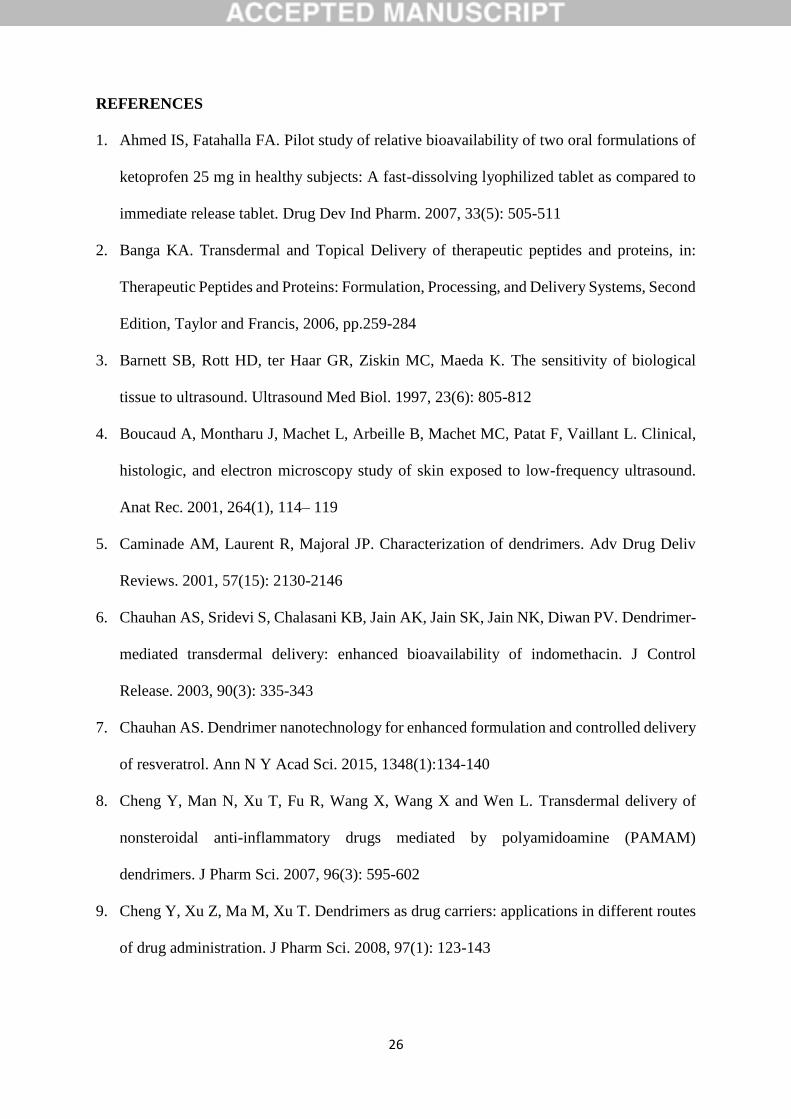
Fourier Transform Infrared Spectroscopy: The samples that were analyzed by FTIR were
ketoprofen, A16 dendrimer, ketoprofen-A16 complex, ketoprofen-A16 mixture in the donor
solution with 3 h stirring and their corresponding FTIR spectra are given in Fig. 2A-2D. The
FTIR spectrum of ketoprofen (Fig. 2A) exhibited characteristic peaks at 2982 cm-1 (CH alkane
stretch), 2935 cm-1 (CH3 group), 1697 cm-1 (C=O group), 1651.12 cm-1 (alkenyl C=C stretch),
3740 cm-1 (OH group) and 2359 cm-1 (carboxylic acid group). In Fig. 2C and Fig. 2D, the C=O
group (1750- 1690 cm-1) is not evident. Therefore, probably, the C=O group of ketoprofen is
involved in linkage with –NH2 of peptide dendrimer which results in formation of CONH
linkage with a prominent peak at 3732 cm-1. This along with the DSC results supports the
formation of complex between ketoprofen and A16 dendrimer.
Zeta potential measurement: Zeta potential was measured to measure the change in the charge
upon complexation. The zeta potential of A16 dendrimer in HEPES buffer pH 7.4 was
4.73±0.71 mV. On the other hand, the zeta potential of ketoprofen-A16 complex in HEPES
buffer pH 7.4 was much higher (6.73±0.89 mV). This difference in zeta potential of the

18
complex and the plain dendrimer clearly suggest the formation of complexes. The high positive
zeta potential of the complex (discussed in later sections) could be one of the reasons for
increased permeation, as at pH above the isoelectric point (pI), skin shows cation
permselectivity (Banga, 2006).
3.6. In vitro permeation studies:
The in vitro skin permeation of ketoprofen was studied under different conditions of
dendrimer and ultrasound application viz., differing charge of dendrimer, different modes of
dendrimer application (simultaneous application, pretreatment and complexation), ultrasound
application alone, ultrasound application with dendrimer treatment (with simultaneous
application and pretreatment and complexation).
Permeation studies of ketoprofen with dendrimers alone: The effect of differing peptide
dendrimer charge on the permeation of ketoprofen with simultaneous application was
investigated and the results are shown in Fig. 3 and Table 1. All the dendrimers significantly
(p<0.05) increased the cumulative amount of drug permeated in 24 h (Q24). Accordingly flux
values were also observed in the similar manner. As the charge increased from 4+ to 8+, drug
permeation also increased, but further increase in charge (16+) decreased both the flux of the
drug and Q24. The permeation parameters observed were highest with A8 dendrimer (Q24:
174.81±11.01 µg; Jss 9.68±1.23 µg/cm2/h; enhancement ratio (ER): 2.93, compared to Q24:
51.88±5.07 µg; Jss: 3.30±0.24 µg/cm2/h observed with passive diffusion).
Permeation studies were also conducted by pretreating the skin with dendrimer for 2 h
before application of ketoprofen dispersion in pH 7.4 buffer solution. The pretreatment studies
were carried out to understand the interaction of dendrimers with the skin. All the tested
dendrimers considerably increased the skin permeation of ketoprofen in the pretreatment
studies (Table 1 and Fig. 4). The highest permeation enhancement was found with A16
dendrimer (Q24: 212.12±12.15 µg; Jss: 10.75±0.40 µg/cm2/h and enhancement ratio: 3.25).

19
The pretreatment studies indicate that the dendrimers penetrate and alter the skin
barrier. The skin is negatively charged at physiological pH; therefore cations have
greater affinity for the skin. Interaction of dendrimers having amine termination
with lipid bilayers and consequent enhancement of transport of molecules has been shown in
model lipid bilayers and cell cultures. Reports are available on the interaction of
dendrimers with the negatively charged phosphate head groups of model phospholipids and
consequent fluidization of the lipid bilayers. Also, dendrimers can interact with skin ceramides
and free fatty acids. Dendrimers have also reported to cause alterations in lipid stretching peaks
in FTIR spectrum of skin (Venuganti and Perumal, 2008). Cationic dendrimers (like full
generation PAMAM dendrimers) have been found to induce hole-formation in model lipid
bilayers and cell membranes (Mecke et al., 2004).
The partition coefficient experiments with the peptide dendrimers (presented above)
also indicated that they improve partitioning of ketoprofen into the stratum corneum. Apart
from increasing the oil/water partition coefficient of the drug, peptide dendrimers may also
interact with proteins and lipids in the viable cells of skin, thus, enhancing the transdermal
transport of drug molecules (Mutalik et al., 2014).
In both simultaneous as well as pretreatment modes, ketoprofen-A16 dendrimer
complex did not show higher permeation for ketoprofen. This could be due to larger size of the
complex which hinders the permeation across the skin. When simultaneous application and
pretreatment modes were compared with respect to ketoprofen-A16 complex, pretreatment
mode showed higher Q24 value. This could be due to the interaction between A16 dendrimer
and skin during pretreatment period that disrupts the skin integrity (ketoprofen-A16 complex
alone: Q24, Jss and ER were 98.71±9.77 µg, 7.07±0.87 µg/cm2/h and 2.14, respectively;
ketoprofen-A16 complex after pretreatment with A16: Q24, Jss and ER were182.81±6.67 µg,
10.55±0.72 µg/cm2/h and 3.19, respectively).

20
Permeation studies of ketoprofen with dendrimers + ultrasound:
When the skin permeation experiments were conducted with ultrasound application, a
host of surprising results were derived. With ultrasound application alone, the amount of
ketoprofen that permeated in just 30 min (Q0.5: 118.67±15.28 µg) was more than that permeated
in 24 h in passive diffusion (51.88±5.07 µg).
In the second set of experiments, when ultrasound was combined with simultaneous
application of dendrimers, again it was found that the amount of ketoprofen permeated in 30
min using sonophoresis and dendrimer application, was significantly (p<0.05) higher than that
permeated in 24 h without sonophoresis, for all dendrimers tested (Table 1 and Fig. 5). This
clearly demonstrates the permeation enhancing effect produced by the combination of
dendrimer and ultrasound, which is not achievable by either of them alone. The results
indicated that, with simultaneous mode of application, ketoprofen-A16 dendrimer complex
showed the highest cumulative drug permeated in 30 min (829.44±73.34 µg). A4, A8 and A16
dendrimers combined with ultrasound produced permeation of 182.95±15.81, 650.05±34.76
and 593.46±67.80 µg of ketoprofen in 30 min, respectively.
With pretreatment mode of dendrimer application, combining ultrasound with
dendrimers produced very high permeation rates of ketoprofen (Table 1 and Fig. 6). Like the
earlier experiments, ketoprofen-A16 dendrimer complex produced highest permeation
(1922.88±240.55 µg) followed by A16 dendrimer (761.47±89.34 µg), A8 dendrimer
(673.09±73.20 µg) and A4 dendrimer (364.47±28.70 µg) in 30 min.
Low frequency ultrasound (LFU) has been shown to improve the transdermal
permeation of drugs and macromolecules (Boucaud et al., 2002; Han and Das, 2013; Mitragotri
and Kost, 2000, Mitragotri and Kost, 2004; Polat et al., 2011; Tezel et. al., 2002a). LFU causes
acoustic cavitation, or creation of microbubbles in water and tissue, which forms water
channels in lipid bilayers. On the one hand, while sonophoresis has been found to enhance

21
transdermal permeation, it has been found that this enhancement depends on the
physicochemical properties of the drug, including the n-octanol/water partition coefficient
(Mitragotri 1997) expressed in the equation below:
𝑒~𝐾𝑜/𝑤0.75
(4 × 10⁴)𝑃𝑃
where ‘e’ is the relative sonophoretic transdermal transport, Ko/w is the drug octanol-water
partition coefficient and PP is the passive skin permeability of the drug (cm/h). If the
permeability coefficient is unchanged, higher partition coefficient would result in higher
permeation enhancement with ultrasound. Although the above equation was developed for
studies utilizing therapeutic frequency ultrasound (1-3 MHz), perhaps the same can be
extended to low frequency ultrasound also. Peptide dendrimers were found to considerably
increase the n-octanol/water partition coefficient of ketoprofen (mentioned in section 3.3).
Therefore, this improvement in partition coefficient could also contribute greatly to the
observed improvement in permeation when peptide dendrimers are combined with ultrasound
treatment. This can also be confirmed by the observation that if both sonophoresis and
dendrimer treatment were acting alone, the permeation enhancement observed should be equal
to that produced by the enhancing treatment, which in this case is sonophoresis.
Synergistic enhancement in transdermal drug delivery has been reported when LFU
was combined with chemical enhancers like citral in ethanol (Mutalik et al., 2009), Tween-20,
dimethyl formamide, propylene glycol, polyethylene glycol and ethanol (Johnson et al., 1996,
Shetty et al., 2013), porous resins (Terahara et al., 2002) and a host of surfactants (Tezel,
2002b). But this is the first report of synergistic effects on permeation produced by LFU and
peptide dendrimers.
Tezel et al (2002b) reported that the synergistic effect of surfactants and LFU could be
due to i) increase in rate of surfactant entry into the skin and ii) dispersion of surfactant within

22
the skin, both induced by ultrasound. Mutalik et al (2012) also reported the increase in uptake
of peptide dendrimers across skin by LFU. Therefore, the observed synergy between LFU and
peptide dendrimers in transdermal permeation of ketoprofen with simultaneous application of
drug and dendrimer could be mediated by these mechanisms. On the other hand, with
pretreatment of dendrimer on skin, the permeation enhancement of drug could be due to
fluidization of skin lipid bilayers further accentuated by ultrasound.
When the permeation studies were conducted without the aid of ultrasound, the
permeation produced by the drug-dendrimer complex was lesser than that produced by drug
and dendrimer individually. However, with the introduction of ultrasound, drug-dendrimer
complex clearly gave much higher permeation enhancement than with the components used
individually. A reason for this could be that transient microbubbles formed in the lipid bilayers
upon ultrasound application (Tang et al., 2002) could be large enough to allow the transport of
the entire drug and dendrimer complex. Also, the greater positive charge of the drug-dendrimer
complex compared to plain dendrimer (section 3.5) could be bringing about the increased
permeation. However, the underlying mechanism for this phenomenon needs to be explored in
further detail.
The target skin permeation rate for ketoprofen was calculated using the following
equation from the available pharmacokinetic data (Mutalik & Udupa, 2002; Shetty et al., 2013;
www.accessdata.fda.gov):
Jss.A=Cl.Cp.W
where, Jss is the flux (µg/cm2/h), A is the area of application (cm2), Cl is the clearance
rate (0.08 L/h/kg), Cp is plasma concentration (0.4 µg/mL) and W is the weight of subject (65
kg). The target permeation rate for ketoprofen as calculated from the above equation was found
to be 2.08 mg/h. The flux values obtained with the aid of ultrasound and dendrimers indicate

23
that the target permeation rate for ketoprofen can be achieved within an appreciable range of
application area.
3.7.In vivo skin permeation experiments:
In vivo skin permeation studies were performed with dendrimer application and
sonophoresis. With passive diffusion, the concentration of ketoprofen in plasma was found to
be 226.05±31.19 ng/ mL at the end of 30 min. Sonophoresis alone produced permeation of
481.45±63.38 ng/mL of ketoprofen in plasma. Skin permeation studies of ketoprofen
performed with A8 dendrimer, A16 dendrimer and ketoprofen-A16 dendrimer complex
produced 1753.31±142.61, 1245.79±54.61 and 859.45±38.05 ng/ mL of ketoprofen in plasma
in 30 min. respectively, which were significantly different (p<0.05) from the passive diffusion
value at the respective time intervals (Table 2). The same combinations of ultrasound and
dendrimer application when tested for in vivo permeation resulted in 3660.86±199.79,
2760.52±103.47 and 2650.20±223.79 ng/mL of ketoprofen in plasma respectively (Table 2).
These results revealed a clear correlation between the in vitro and in vivo experiments, with
respect to dendrimer treatment (± ultrasound application). In both in vitro and in vivo
experiments without ultrasound, A8 dendrimer resulted in highest increase in permeation. In
the studies involving the use of ultrasound, ketoprofen-A16 complex produced the greatest
permeation enhancement.
3.8. Comparison of transdermal and oral administration:
Swiss albino mice were administered with 20 mg/kg of ketoprofen. Oral administration
of drug suspension produced 3626.56±143.55 ng/mL and 8406.56±265.37 ng/mL of
ketoprofen in the respective time intervals. Nevertheless, combination of ultrasound and A8
dendrimer with ketoprofen produced 2176.32±166.81 ng/mL and 3660.86±199.79 ng/mL of
ketoprofen concentration in plasma in 15 and 30 min. respectively. This indicates that although
oral absorption of ketoprofen is more rapid, the above transdermal administration produced

24
about ≈ 44% of plasma levels compared to oral route. The results of this study are encouraging
because with little increase in the skin application area, the drug concentration as obtained with
oral route can easily be achieved.
3.9. Histopathological studies of the treated skin for toxic reactions:
After in vivo permeation studies, the excised mouse skin was subjected to
histopathological evaluation to observe if the synergy between ultrasound and peptide
dendrimers also translated to increased skin irritation. Ultrasound alone showed slight necrosis,
inflammation and cavitation in treated skin, when compared to control. Similar skin reactions
were observed in previous studies when ultrasound was applied to skin (Barnett et al., 1997;
Nyborg 2001). Skin of the animals treated with A16 dendrimer (with and without ultrasound)
showed slight/ negligible cellular atypia, degeneration, necrosis, inflammation and cavitation.
This shows the appreciable dermal safety and non-toxicity of the tested peptide dendrimers.
4. CONCLUSIONS
All the tested peptide dendrimers considerably increased the transdermal permeation of
ketoprofen. Application of ultrasound alone for 30 min was found to produce a similar skin
permeation of ketoprofen as obtained with passive diffusion in 24 h. Remarkable enhancement
in skin permeation of ketoprofen was observed when ultrasound was applied along with
dendrimer treatment. Dendrimer and sonophoresis-assisted transdermal delivery was found to
produce plasma levels of drug comparable to oral administration. Histopathological evaluation
of skin upon different treatments did not reveal considerable dermal toxicity. The study reveals
that combined approach of dendrimer treatment (chemical enhancement) along with low
frequency ultrasound application (physical permeation enhancement technique) is an attractive
approach for achieving high transdermal permeation rates for ketoprofen and this approach
could be further extended to other drugs.

25
ACKNOWLEDGEMENTS
The authors are also thankful to Manipal College of Pharmaceutical Sciences, Manipal
University, Manipal, India and School of Pharmacy (PACE), The University of Queensland,
Brisbane, Australia for providing the necessary facilities for the study.
26
REFERENCES
1. Ahmed IS, Fatahalla FA. Pilot study of relative bioavailability of two oral formulations of
ketoprofen 25 mg in healthy subjects: A fast-dissolving lyophilized tablet as compared to
immediate release tablet. Drug Dev Ind Pharm. 2007, 33(5): 505-511
2. Banga KA. Transdermal and Topical Delivery of therapeutic peptides and proteins, in:
Therapeutic Peptides and Proteins: Formulation, Processing, and Delivery Systems, Second
Edition, Taylor and Francis, 2006, pp.259-284
3. Barnett SB, Rott HD, ter Haar GR, Ziskin MC, Maeda K. The sensitivity of biological
tissue to ultrasound. Ultrasound Med Biol. 1997, 23(6): 805-812
4. Boucaud A, Montharu J, Machet L, Arbeille B, Machet MC, Patat F, Vaillant L. Clinical,
histologic, and electron microscopy study of skin exposed to low-frequency ultrasound.
Anat Rec. 2001, 264(1), 114– 119
5. Caminade AM, Laurent R, Majoral JP. Characterization of dendrimers. Adv Drug Deliv
Reviews. 2001, 57(15): 2130-2146
6. Chauhan AS, Sridevi S, Chalasani KB, Jain AK, Jain SK, Jain NK, Diwan PV. Dendrimer-
mediated transdermal delivery: enhanced bioavailability of indomethacin. J Control
Release. 2003, 90(3): 335-343
7. Chauhan AS. Dendrimer nanotechnology for enhanced formulation and controlled delivery
of resveratrol. Ann N Y Acad Sci. 2015, 1348(1):134-140
8. Cheng Y, Man N, Xu T, Fu R, Wang X, Wang X and Wen L. Transdermal delivery of
nonsteroidal anti-inflammatory drugs mediated by polyamidoamine (PAMAM)
dendrimers. J Pharm Sci. 2007, 96(3): 595-602
9. Cheng Y, Xu Z, Ma M, Xu T. Dendrimers as drug carriers: applications in different routes
of drug administration. J Pharm Sci. 2008, 97(1): 123-143

27
10. Devarakonda B, Hill RA, Liebenberg W, Brits M, de Villers MM. Comparison of the
aqueous solubilization of practically insoluble niclosamide by polyamidoamine (PAMAM)
dendrimers and cyclodextrins. Int J Pharm. 2005, 304(1-2): 193-209
11. Devarakonda B, Ottoa DP, Judefeinda A, Hill RA, de Villiers MM. Effect of pH on the
solubility and release of furosemide from polyamidoamine (PAMAM) dendrimer
complexes. Int J Pharm. 2007, 345(1-2): 142-153
12. Duncan R, Izzo L. Dendrimer biocompatibility and toxicity. Adv Drug Deliv Rev. 2005,
57(15):2215-2237
13. Eggimann GA, Blattes E, Buschor S, Biswas R, Kammer SM, Darbre T, Reymond J-L.
Designed cell penetrating peptide dendrimers efficiently internalize cargo into cells. Chem.
Commun. 2014, 50, 7254-7257
14. Han T and Das DB. Permeability enhancement for transdermal delivery of large molecule
using low-frequency sonophoresis combined with microneedles. J Pharm Sci. 2013,
102(10): 3614-3122
15. Higuchi T and Connors KA. Phase solubility techniques. Adv. Anal. Chem. Instrum.1965,
4: 117–212
16. Jevprasesphant R, Penny J, Jalal R, Attwood D, McKeown N, D’Emanuele A. The
influence of surface modification on the cytotoxicity of PAMAM dendrimers. Int. J. Pharm.
2003, 252: 263–266
17. Johnson ME, Mitragotri S, Patel A, Blankschtein D, Langer R. Synergistic effects of
chemical enhancers and therapeutic ultrasound on transdermal drug delivery. J Pharm Sci.
1996, 85(7): 670-679
18. Kolhatkar RB, Kitchens KM, Swaan PW, Ghandehari H. Surface acetylation of
polyamidoamine (PAMAM) dendrimers decreases cytotoxicity while maintaining
membrane permeability. Bioconj. Chem. 2007, 18(6): 2054–2060

28
19. Mecke A, Uppuluri S, Sassanella TM, Lee DK, Ramamoorthy A, Baker JR Jr, Orr BG,
Holl MM. Direct observation of lipid bilayer disruption by poly(amidoamine) dendrimers.
Chem Phys Lipids. 2004, 132(1): 3–14
20. Mignani S, Kazzouli El S, Bousmina M, Majoral JP. Expand classical drug administration
ways by emerging routes using dendrimer drug delivery systems: a concise overview. Adv
Drug Deliv Rev. 2013, 65(10), 1316-1330
21. Mitragotri S and Kost J. Low frequency sonophoresis: a non-invasive method of drug
delivery and diagnostics. Biotechnol Prog. 2000, 16(3): 488-492
22. Mitragotri S and Kost J. Low-frequency sonophoresis: a review. Adv Drug Deliv Rev.
2004, 56(5): 589-601
23. Mitragotri S, Blankschtein D, Langer R. An Explanation for the Variation of the
Sonophoretic Transdermal Transport Enhancement from Drug to Drug. J Pharm Sci. 1997,
86(10): 1190-1192
24. Mitragotri S, Edwards D, Blankschtein D, Langer R. A mechanistic study of ultrasonically-
enhanced transdermal drug delivery. J. Pharm. Sci.1995, 84(6): 697–706
25. Morris AP, Brain KR, Heard CM. Skin permeation and ex vivo skin metabolism of O-acyl
haloperidol ester prodrugs. Int J Pharm. 2009, 367(1-2), 44-50
26. Mutalik S, Hewavitharana AK, Shaw PN, Anissimov YG, Roberts MS, Parekh HS.
Development and validation of a reversed-phase high-performance liquid chromatographic
method for quantification of peptide dendrimers in human skin permeation experiments. J
Chromatogr B Analyt Technol Biomed Life Sci. 2009, 877(29): 3556-3562
27. Mutalik S, Parekh HS, Anisisimov YG, Grice JE, Roberts MS. Iontophoresis-Mediated
Transdermal Permeation of Peptide Dendrimers across Human Epidermis. Skin
Pharmacology and Physiology. 2013, 26:127-138

29
28. Mutalik S, Parekh HS, Davies NM, Udupa N. A combined approach of chemical enhancers
and sonophoresis for the transdermal delivery of tizanidine hydrochloride. Drug Deliv.
2009, 16(2): 82-91
29. Mutalik S, Shetty PK, Kumar A, Kalra R, Parekh HS. Enahncement in permeation and
deposition of 5-fluorouracil through human epidermis assisted by peptide dendrimers. Drug
Deliv. 2014, 21(1): 44-54
30. Mutalik S, Udupa N. Transdermal delivery of glibenclamide and glipizide: in vitro
permeation studies through mouse skin. Pharmazie. 2002, 57(12):838-841
31. Mutalik, S, Nayak, UY, Kalra R, Kumar A, Kulakarni RV, Parekh HS. Sonophoresis-
mediated permeation and retention of peptide dendrimers across human epidermis. Skin
Res Technol. 2012, 18(1): 101-107
32. Mutoh M, Ueda H, Nakamura Y, Hirayama K, Atobe M, Kobayashi D, Morimoto Y.
Characterization of transdermal solute transport induced by low-frequency ultrasound in
the hairless rat skin. J Control Release. 2003, 92(1-2), 137–146.
33. Nanjwade BK, Bechra HM, Derkar GK, Manvi FV, Nanjwade VK. Dendrimers: Emerging
polymers for drug-delivery systems. European Journal of Pharmaceutical Sciences. 2009,
38: 185(3)-196
34. Nyborg WL. Biological effects of ultrasound: development of safety guidelines. Part II:
general review. Ultrasound Med Biol. 2001, 27(3): 301-333
35. Panus PC, Campbell J, Kulkarni SB, Herrick RT, Ravis WR, Banga AK. Transdermal
iontophoretic delivery of ketoprofen through human cadaver skin and in humans. J. Control
Release. 1997, 44(2-3): 113–121
36. Parekh HS, Marano RJ, Rakoczy EP, Blanchfield J, Toth I. Synthesis of a library of
polycationic lipid core dendrimers and their evaluation in the delivery of an oligonucleotide
with hVEGF inhibition. Bioorg Med Chem. 2006, 14(14): 4775–4780

30
37. Polat BE, Blankschtein D, Langer R. Low-frequency sonophoresis: application to the
transdermal delivery of macromolecules and hydrophilic drugs. Expert Opin Drug Deliv.
2010, 7(12): 1415-1432
38. Polat BE, Hart D, Langer R, Blankschtein D. Ultrasound mediated transdermal drug
delivery: mechanisms, scope and emerging trends. J Control Release. 2011, 152(3): 330-
348
39. Prausnitz MR, Mitragotri S, Langer R. Current status and future potential of transdermal
drug delivery. Nat Rev Drug Discov. 2004, 3(2):115–124
40. Rhee GJ, Woo JS, Hwang SJ, Lee YW and Lee CH. Topical oleo-hydrogel preparation of
ketoprofen with enhanced skin permeability. Drug Dev Ind Pharm. 1999, 25(6): 717-726
41. Sadler K and Tam JP. Peptide dendrimers: applications and synthesis. J Biotechnol. 2002,
90(3-4):195-229
42. Shah ND, Parekh HS, Steptoe RJ. Asymmetric Peptide Dendrimers are Effective Linkers
for Antibody-Mediated Delivery of Diverse Payloads to B Cells in Vitro and in Vivo.
Pharm Res. 2014. 31(11):3150-3160
43. Schmitt M, Guentert TW. Biopharmaceutical evaluation of ketoprofen following
intravenous, oral, and rectal administration in dogs. J Pharm Sci. 1990, 79(7):614-616.
44. Shetty PK, Suthar NA, Menon J, Deshpande PB, Avadhani K, Kulkarni RV, Mutalik S.
Transdermal delivery of lercanidipine hydrochloride: effect of chemical enhancers and
ultrasound. Curr Drug Deliv. 2013, 10(4):427-434
45. Singh BN and Jayaswal SB. Iontophoretic delivery of 5-fluorouracil through excised
human stratum corneum. Drug Discov Ther. 2008, 2(2): 128-135
46. Svenson S. Dendrimers as versatile platform in drug delivery applications. Eur J Pharm
Biopharm. 2009, 71(3): 445-462

31
47. Tang H, Blankschtein D, Langer R. Effects of low frequency ultrasound on the transdermal
permeation of mannitol: comparative studies with in vivo and in vitro skin. J Pharm Sci.
2002, 91(8): 1776-1794
48. Terahara T, Mitragotri S, Langer R. Porous resins as a cavitation enhancer for low-
frequency sonophoresis. J. Pharm Sci, 2002, 91(3): 753-759
49. Tezel A, Sens A, Mitragotri S. Investigations of the role of cavitation in low-frequency
sonophoresis using acoustic spectroscopy. J Pharm Sci. 2002, 91(2): 444–453
50. Tezel A, Sens A, Tuchscherer J. Mitragotri S. Synergistic effect of low frequency
ultrasound and surfactants on skin permeability. J Pharm Sci. 2002, 91(1): 91-100
51. Ueda H, Mutoh M, Seki T, Kobayashi D, Morimoto Y. Acoustic cavitation as an enhancing
mechanism of low-frequency sonophoresis for transdermal drug delivery. Biol Pharm Bull.
2009, 32: 916–920.
52. Venuganti VVK and Perumal OP. Effect of poly(amidoamine) (PAMAM) dendrimer on
skin permeation of 5-fluorouracil. Int J Pharm. 2008. 361(1-2), 230-238
53. www.accessdata.fda.gov/drugsatfda_docs/label/2006/018754s027,019816s010lbl.pdf.
Accessed on 27-01-2017

32
Fig.1. DSC thermograms
A) Ketoprofen B) A16 peptide dendrimer C) ketoprofen-A16 dendrimer complex (1:0.25
molar ratio) and D) ketoprofen-A16 mixture after 3 h of stirring from donor solution.
A B
C D
33
Fig. 2. FTIR spectra
A) ketoprofen B) A16 peptide dendrimer C) ketoprofen-A16 dendrimer complex (1:0.25 molar
ratio) and D) ketoprofen-A16 mixture after 3 h of stirring from donor solution.
75015002250300037501/cm
75
82.5
90
%T
38
61
.62
37
40
.10
36
72
.59
34
23
.76
30
95
.85
30
32
.20
29
20
.32 28
50
.88
23
60
.95
23
35
.87
21
29
.48
20
61
.97
16
49
.19
15
54
.68
15
14
.17
14
54
.38
13
92
.65
13
61
.79
12
84
.63
10
20
.38
71
9.4
76
73
.18
E
75015002250300037501/cm
60
70
80
90
%T
37
34
.31
34
37
.26
30
84
.28
29
66
.62
29
26
.11
23
60
.95
16
49
.19
15
62
.39
14
52
.45
13
88
.79
13
59
.86
12
82
.71
12
03
.62 1
06
8.6
01
02
2.3
1 96
0.5
8 87
9.5
78
27
.49
78
3.1
37
17
.54
64
6.1
7
D7501500225030003750
1/cm
40
60
80
%T
37
28
.53
34
44
.98
29
58
.90
29
26
.11
28
52
.81
23
60
.95 2
33
5.8
7
16
80
.05
16
51
.12
15
60
.46
14
56
.30
13
94
.58
13
19
.35
12
82
.71
12
01
.69
11
38
.04
10
47
.38
99
1.4
4
83
9.0
68
02
.41
76
7.6
97
21
.40
68
6.6
85
94
.10
C
A B
C D

34
Fig. 3. In vitro skin permeation profiles of ketoprofen after simultaneous application of
peptide dendrimers
K-A16= Ketoprofen-A16 peptide dendrimer complex
0
20
40
60
80
100
120
140
160
180
200
0 4 8 12 16 20 24
Cu
mu
lati
ve d
rug
per
me
ate
d (
µg/
cm2)
Time (h)
Passive diffusion
A4
A8
A16
K-A16 complex

35
Fig. 4. In vitro skin permeation profiles of ketoprofen after pretreatment with peptide
dendrimers
K-A16= Ketoprofen-A16 peptide dendrimer complex
0
50
100
150
200
250
0 4 8 12 16 20 24
Cu
mu
lati
ve d
rug
per
me
ate
d (
µg/
cm2 )
Time (h)
Passive diffusion
A4
A8
A16
K-A16 complex

36
Fig. 5. In vitro skin permeation profiles of ketoprofen with simultaneous
application of peptide dendrimers and sonophoresis
US=Ultrasound; K-A16= Ketoprofen-A16 peptide dendrimer complex
0
100
200
300
400
500
600
700
800
900
1000
0 10 20 30
Cu
mu
lati
ve d
rug
per
me
ate
d (
µg/
cm2 )
Time (min)
Ultrasound alone
A4+US
A8+US
A16+US
K-A16 complex+US

37
Fig. 6. In vitro skin permeation profiles of ketoprofen with pretreatment with
peptide dendrimers followed by sonophoresis
US= Ultrasound; K-A16= Ketoprofen-A16 peptide dendrimer complex
0
500
1000
1500
2000
2500
0 10 20 30
Cu
mu
lati
ve d
rug
per
me
ate
d (
µg/
cm2 )
Time (min)
Ultrasound alone
A4+US
A8+US
A16+US
K-A16 complex+US

38
Table 1. In vitro skin permeation studies of ketoprofen with different treatments and
modes of application
Treatments
Mode of
application
Q24# OR Q0.5
$
(µg/cm2)
Jss
(µg/cm2/h)
ER
Passive diffusion (drug alone) NA 51.88±5.07# 3.30±0.24 NA
Ketoprofen + A4 dendrimer
Simultaneous
application
75.97±7.79#* 3.66±0.21 1.01
Ketoprofen + A8 dendrimer 142.43 ±8.61#* 10.29±0.79* 3.12
Ketoprofen + A16 dendrimer 118.71±6.03#* 7.35±0.75* 2.23
Ketoprofen-A16 complex 98.71±9.77#* 7.07±0.87* 2.14
Ketoprofen + A4 dendrimer
Pretreatment
108.04±5.77#* 6.92±0.82* 2.10
Ketoprofen + A8 dendrimer 174.81±11.01#* 9.68±1.23* 2.93
Ketoprofen + A16 dendrimer 212.12±12.15#* 10.75±0.40* 3.25
Ketoprofen-A16 complex 182.81±6.67#* 10.55±0.72* 3.19
US+ Ketoprofen NA 118.67±15.28$ 172.00±19.76* 52.08
US+ Ketoprofen + A4 dendrimer
Simultaneous
application
182.95±15.81$* 265.44±18.41* 80.37
US+ Ketoprofen + A8 dendrimer 650.05±34.76$*£ 1063.11±36.42*£ 321.90
US+ Ketoprofen + A16 dendrimer 593.46±67.80$*£ 933.38±98.74*£ 282.62
US+ Ketoprofen-A16 complex 829.44±73.34$*£ 1644.94±26.79*£ 498.06
US+ Ketoprofen + A4 dendrimer
Pretreatment
364.37±28.70$*£ 766.23±55.30*£ 232.00
US+ Ketoprofen + A8 dendrimer 673.09±73.20$*£ 944.97±79.64*£ 286.12
US+ Ketoprofen + A16 dendrimer 761.47±89.34$*£ 840.90±83.11*£ 254.61
US+ Ketoprofen-A16 complex 1922.88±240.55$*£ 4521.86±363.92*£ 1369.15
US= Ultrasound application; Jss=Steady state flux; ER= Enhancement ratio;
Values are represented as Mean ± SD; n=3.
* Significantly different (p<0.05) compared to Passive diffusion (drug alone); £ Significantly
different (p<0.05) compared to US+ Ketoprofen.
39
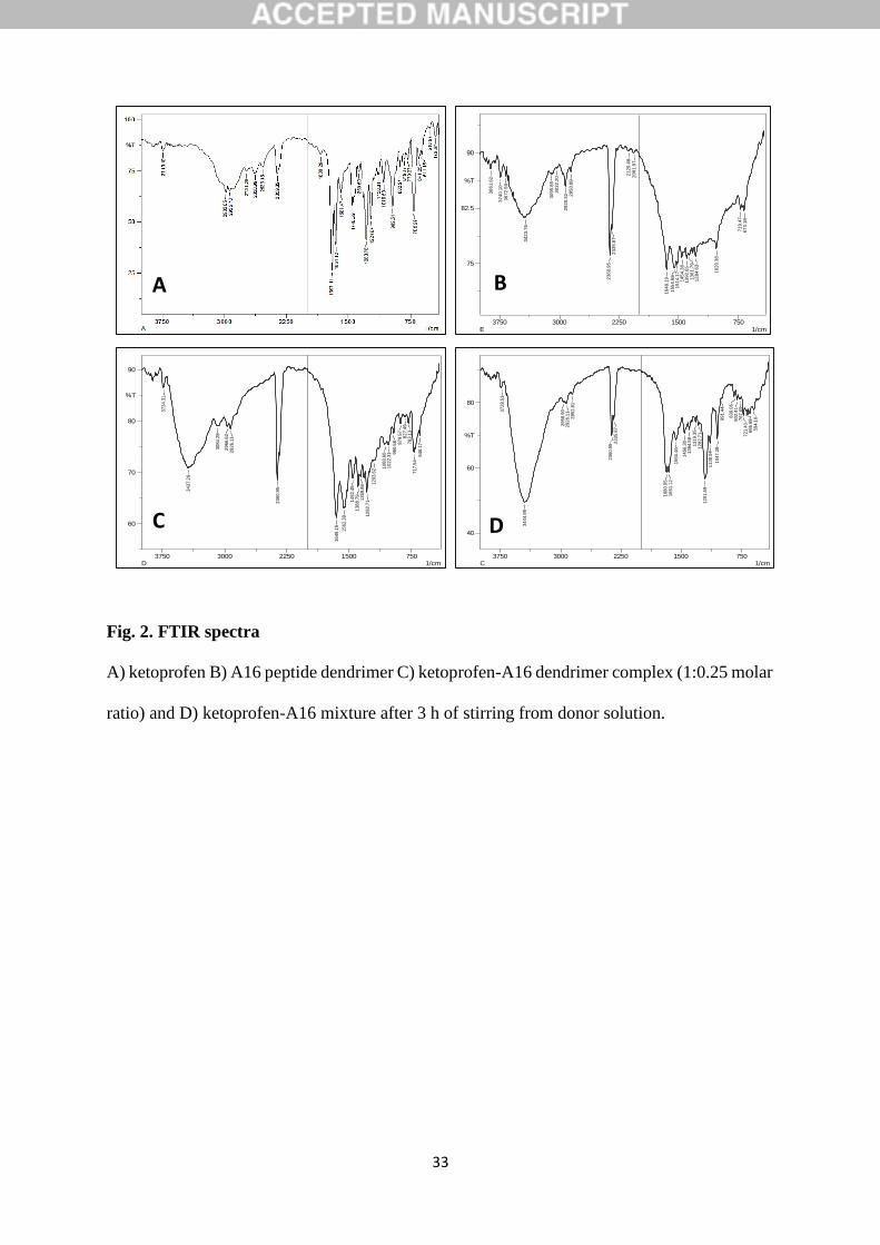
Table 2. In vivo permeation studies of ketoprofen with different treatments
Treatment
Plasma concentration of drug (ng/mL)*
15 min 30 min
Passive diffusion (drug alone) 4.47±1.01 226.05±31.19
Ketoprofen + A8 dendrimer 1364.33±117.56* 1753.31±142.61*
Ketoprofen + A16 dendrimer 1177.67±95.63* 1245.79±54.61*
Ketoprofen-A16 complex 267.67±31.13* 859.45±38.05*
US + Ketoprofen 216.59±14.39 481.45±63.38
US + Ketoprofen + A8 dendrimer 1634.92±244.23*# 2650.20±223.79*#
US + Ketoprofen + A16 dendrimer 1826.78±87.91*# 2760.52±103.47*#
US + Ketoprofen-A16 complex 2176.32±166.81*# 3660.86±199.79*#
US= Ultrasound application; All the values are represented as Mean ± SD, n=3.
* Significantly (p<0.05) different compared to passive diffusion (drug alone); # Significantly
(p<0.05) different compared to US+ Ketoprofen.