Embed Size (px)
Citation preview
1
1
2
Running head: Inflorescence Branching Effector of S. reilianum 3
4
5
6
Corresponding author: 7
Prof. Dr. Jan Schirawski 8
RWTH Aachen University 9
Microbial Genetics, Institute of Applied Microbiology 10
Aachen Biology and Biotechnology 11
Worringerweg 1 12
52074 Aachen 13
Germany 14
15
Tel.: +49-241-80-26616 16
e-mail: [email protected] 17
18
19
20
Research area: Genes, Development and Evolution 21
22
Plant Physiology Preview. Published on November 9, 2015, as DOI:10.1104/pp.15.01347
Copyright 2015 by the American Society of Plant Biologists
www.plantphysiol.orgon August 25, 2018 - Published by Downloaded from Copyright © 2015 American Society of Plant Biologists. All rights reserved.
2
SUPPRESSOR OF APICAL DOMINANCE 1 of Sporisorium reilianum Modulates 23
Inflorescence Branching Architecture in Maize and Arabidopsis1 24
25
26
Hassan Ghareeb$,*, Frank Drechsler$, Christian Löfke**, Thomas Teichmann, 27
Jan Schirawski*** 28
29
Georg-August-Universität Göttingen, Molecular Biology of Plant-Microbe Interactions, 30
Albrecht-von-Haller Institute of Plant Sciences, Julia-Lermontowa-Weg 3, 37077 31
Göttingen, Germany (H.G., J.S.) 32
Max Planck Institute for Terrestrial Microbiology, Organismic Interactions, Karl-von-33
Frisch Straße 10, 35043 Marburg, Germany (H.G., J.S.) 34
Georg-August-Universität Göttingen, Plant Cell Biology, Albrecht-von-Haller Institute 35
of Plant Sciences, Julia-Lermontowa-Weg 3, 37077 Göttingen, Germany (C.L., T.T.) 36
RWTH Aachen University, Microbial Genetics, Institute of Applied Microbiology, 37
Aachen Biology and Biotechnology, Worringerweg 1, 52074 Aachen, Germany (F.D., 38
J.S.) 39
National Research Centre, Plant Biotechnology Department, Giza, Egypt (H.G.) 40
41 42
One-Sentence Summary 43
A fungal protein increases plant inflorescence branching both when delivered by the 44
fungus in maize and when stably expressed in Arabidopsis, suggesting a conserved 45
pathway for function. 46
47
www.plantphysiol.orgon August 25, 2018 - Published by Downloaded from Copyright © 2015 American Society of Plant Biologists. All rights reserved.
3
1This work was supported by the Max Planck Society, the Georg-August-University 48
through the German Initiative of Excellence (DFG ZUK45/1) (J.S.), and the RWTH 49
Aachen University. Funding via the IMPRS for Environmental, Cellular and Molecular 50
Microbiology (H.G.) and the Göttingen Graduate School for Neurosciences, 51
Biophysics, and Molecular Biosciences (GGNB) (H.G.) is gratefully acknowledged. 52
53 $ These authors contributed equally to this work. 54
55
* Present Address: Georg-August-Universität Göttingen, Plant Cell Biology, Albrecht-56
von-Haller Institute of Plant Sciences, Julia-Lermontowa-Weg 3, 37077 Göttingen, 57
Germany 58
** Present Address: University of Natural Resources and Life Sciences Vienna, 59
Institute of Applied Genetics and Cell Biology, Muthgasse 18, 1190 Vienna 60
61
*** Corresponding author: Jan Schirawski e-mail: [email protected] 62
63
64
www.plantphysiol.orgon August 25, 2018 - Published by Downloaded from Copyright © 2015 American Society of Plant Biologists. All rights reserved.
4
ABSTRACT 65
The biotrophic fungus Sporisorium reilianum causes head smut of maize following 66
systemic plant colonization. Symptoms include the formation of multiple female 67
inflorescences at subapical nodes of the stalk due to loss of apical dominance. By 68
deletion analysis of cluster 19-1, the largest genomic divergence cluster in S. 69
reilianum, we identified a secreted fungal effector responsible for S. reilianum-70
induced loss of apical dominance, which we named SUPPRESSOR OF APICAL 71
DOMINANCE1 (SAD1). SAD1 transcript levels were highly up-regulated during 72
biotrophic fungal growth in all infected plant tissues. SAD1-GFP fusion proteins 73
expressed by recombinant S. reilianum localized to the extracellular hyphal space. 74
Transgenic A. thaliana expressing GFP-SAD1 displayed an increased number of 75
secondary rosette-leaf branches. This suggests that SAD1 manipulates inflorescence 76
branching architecture in maize and A. thaliana via a conserved pathway. Using a 77
yeast two-hybrid library of S. reilianum-infected maize tissues, we identified potential 78
plant interaction partners that had a predicted function in ubiquitination, signaling and 79
nuclear processes. Presence of SAD1 led to an increase of the transcript levels of 80
the auxin transporter PIN1 in the root and a reduction of the branching regulator TB1 81
in the stalk. This indicates a role of SAD1 in regulation of apical dominance by 82
modulation of branching through increasing transcript levels of the auxin transporter 83
PIN1 and derepression of bud outgrowth. 84
85
www.plantphysiol.orgon August 25, 2018 - Published by Downloaded from Copyright © 2015 American Society of Plant Biologists. All rights reserved.
5
INTRODUCTION 86
Plants are known to exhibit a broad range of different morphologies under different 87
environmental conditions. In addition to changing height or leaf area, they are also 88
able to alter their morphological appearance and develop leaves instead of floral 89
organs or change their bud outgrowth pattern. Pathological changes in the 90
developmental program are often caused by plant infection with biotrophic 91
pathogens. Sporisorium reilianum, a biotrophic fungal pathogen of maize and 92
sorghum, interferes with regular development of inflorescences of its host plants and 93
leads to phyllody (Matheussen et al., 1991) that is caused by changes in floral organ 94
identity, floral meristem identity, and floral meristem determinacy (Matheussen et al., 95
1991; Ghareeb et al., 2011). In addition, the fungus triggers suppression of apical 96
dominance, which leads to a higher number of female inflorescences (ears) of maize 97
(Ghareeb et al., 2011) and increased tillering of sorghum (Matheussen et al., 1991). 98
In fact, the inflorescence tillering symptom resembles the crazy top disease of maize 99
that is induced by the downey mildew fungus Sclerophthora macrospora (Malaguti et 100
al., 1977). Broom-like structures on its host plants are also induced by Moniliophthora 101
perniciosa that induces hyperplasia and hyperproliferation of axillary shoots, resulting 102
in witches’ broom disease of cacao (Meinhardt et al., 2008). Similarly, infection of a 103
whole range of dicotyledonous plants with biotrophic phytopathogenic bacteria of the 104
taxon Candidatus Phytoplasma asteris, leads to the reversion of floral organs into 105
leaves or the emergence of a dense cluster of branches resembling broom as a 106
consequence of lack of apical dominance (Hoshi et al., 2009). 107
Branching architecture is mainly regulated by auxin, strigolactone, and cytokinin, that 108
affect axillary meristem initiation and outgrowth (McSteen, 2009; Gallavotti, 2013). 109
Axillary meristem initiation is preceded by a depletion of auxin as shown for 110
Arabidopsis thaliana and Solanum lycopersicum (Wang et al., 2014). After meristem 111
initiation, the emerging axillary meristem is characterized by a local accumulation of 112
auxin, as shown in maize (Gallavotti et al., 2008a). Basipetal auxin transport from the 113
apex towards the root suppresses axillary meristem outgrowth leading to apical 114
dominance (Davies et al., 1966). Outgrowth of axillary buds is also inhibited by 115
strigolactones synthesized in the shoots directly, or long-distance transported from 116
the root to the shoot (Turnbull et al., 2002; Crawford et al., 2010). On the other hand, 117
bud outgrowth is promoted by cytokinins produced in the nodal stem (Nordström et 118
www.plantphysiol.orgon August 25, 2018 - Published by Downloaded from Copyright © 2015 American Society of Plant Biologists. All rights reserved.
6
al., 2004; Tanaka et al., 2006). Cytokinins also modulate branching indirectly through 119
regulation of meristem size (Shani et al., 2006). Although many details about the 120
contribution of particular hormones to regulation of axillary meristem development 121
have been elucidated, the exact mode of hormone action is still ambiguous (Cheng et 122
al., 2013; Barazesh and McSteen, 2008). 123
Increasing knowledge about the mode of action of auxins, cytokinins and 124
strigolactones led to the proposal of two models, the “auxin transport canalization 125
model” and the “second messenger model”, that explain apical dominance 126
mechanistically (Domagalska and Leyser, 2011). In the auxin transport canalization 127
model, polar auxin transport (PAT) plays the central role in apical dominance, 128
regardless of the auxin concentration. During basipetal movement, auxin flux is 129
canalized gradually into a tight thread of cells, the auxin canal, resulting in high PAT 130
(Sachs, 1981). To be activated, the bud must also generate PAT, and export auxin to 131
the auxin canal in the stem (Domagalska and Leyser, 2011). Strigolactones also 132
impact on bud activity since they reduce PAT (Prusinkiewicz et al., 2009). In the 133
second messenger model, auxin regulates cytokinins and strigolactones (second 134
messengers), that migrate to the axillary bud and control its activity (Domagalska and 135
Leyser, 2011). Several studies have been conducted that support either of the two 136
models (Domagalska and Leyser, 2011). 137
Axillary meristem initiation and outgrowth are also under genetic control of 138
transcription factors and signaling components. In maize, determination of axillary 139
meristems is influenced by DELAYED FLOWERING1 (DLF1) and Zea 140
FLORICAULA/LEAFY1 (ZFL1)and FLORICAULA/LEAFY2 (ZFL2)transcription factors 141
(Coe et al., 1988; Muszynski et al., 2006; Sheridan, 1988; Bomblies et al. 2006). The 142
outgrowth of these axillary meristems is controlled by TEOSINTE BRANCHED1 143
(TB1), a suppressor of axillary branches (Hubbard et al., 2002). In addition, the 144
serine/threonine-protein kinase BARREN INFLORESCENCE2 (BIF2) is involved in 145
auxin signaling, and disruption leads to a defect in inflorescence branch meristem 146
initiation (McSteen and Hake, 2001; McSteen et al., 2007). The auxin efflux carrier 147
PIN1 also plays a regulating role in axillary meristem outgrowth (McSteen, 2009). 148
Both PIN1 and BIF2 act on polar auxin transport and thereby control bud outgrowth 149
(Bennetzen and Hake, 2009). 150
www.plantphysiol.orgon August 25, 2018 - Published by Downloaded from Copyright © 2015 American Society of Plant Biologists. All rights reserved.
7
In rare cases, mechanisms have been proposed to explain pathogen-mediated 151
changes in plant branching architectures. Infection with S. reilianum decreased the 152
gibberellin content of sorghum, suggesting that low gibberellin concentration is the 153
reason for increased tillering of infected plants (Matheussen et al., 1991). A 154
misregulation of gibberellin biosynthesis genes was observed in S. reilianum-155
colonized maize inflorescences with early signs of floral reversion (Ghareeb et al., 156
2011). Additionally, misregulated genes were homologous to genes involved in auxin 157
and cytokinin mobilization in other plants (Ghareeb et al., 2011). On the other hand, 158
some examples exist where the architecture of the plant is altered by pathogen 159
effector proteins that are secreted during plant colonization. SAP54 was identified as 160
a secreted protein from Candidatus Phytoplasma asteris that induces leafy flower 161
development when expressed in A. thaliana (MacLean et al., 2011). Likewise, the 162
TENGU effector of Phytoplasma was shown to induce witches’ broom when 163
expressed in A. thaliana, and the TENGU-expressing transgenic plants showed 164
down-regulation of auxin-responsive genes. This suggests that TENGU inhibits 165
auxin-related pathways, which in turn influence plant development (Hoshi et al., 166
2009). 167
The maize smut fungus Ustilago maydis is capable of auxin biosynthesis, and fungal 168
auxin contributes to the total auxin content of colonized tumorous leaf tissues 169
(Reineke et al., 2008). Interestingly, the fungal capacity of auxin formation did not 170
affect the nature of the symptoms induced on its host plant maize (Reineke et al., 171
2008) suggesting that symptom formation may instead be influenced by fungal 172
effector proteins secreted into the colonized tissues. 173
Analysis of the U. maydis genome sequence revealed the existence of gene clusters 174
encoding secreted proteins with an effect on virulence (Kämper et al., 2006). After 175
sequence elucidation of the S. reilianum genome, a genomic comparison identified 176
divergence clusters encoding proteins weakly conserved between U. maydis and S. 177
reilianum (Schirawski et al., 2010). The largest divergence cluster (19-1; Schirawski 178
et al., 2010) corresponded to the U. maydis gene cluster 19A, whose deletion 179
resulted in the lack of U. maydis-specific tumor formation in seedling leaves (Kämper 180
et al., 2006) but not in inflorescences (Skibbe et al., 2010). 181
In this study, we identified one gene (sr10077, SUPPRESSOR OF APICAL 182
DOMINANCE1, SAD1) of cluster 19-1 of S. reilianum as responsible for the loss of 183
www.plantphysiol.orgon August 25, 2018 - Published by Downloaded from Copyright © 2015 American Society of Plant Biologists. All rights reserved.
8
apical dominance phenotype induced by S. reilianum wild type strains. SAD1 is 184
highly up-regulated upon fungal colonization of maize and potentially interacts with a 185
large number of intracellular maize proteins. Lack of SAD1 in S. reilianum leads to 186
decreased transcript levels of the polar auxin transporter PIN1 in roots and increased 187
levels of the bud-outgrowth regulator TB1 in stalks of infected maize. When 188
expressed heterologously as a SAD1-GFP fusion protein in A. thaliana, SAD1 leads 189
to an increase in secondary inflorescence branches. This suggests that SAD1 affects 190
apical dominance via a conserved mechanism that includes regulation of gene 191
transcription and modulation of auxin transport. 192
193
194
www.plantphysiol.orgon August 25, 2018 - Published by Downloaded from Copyright © 2015 American Society of Plant Biologists. All rights reserved.
9
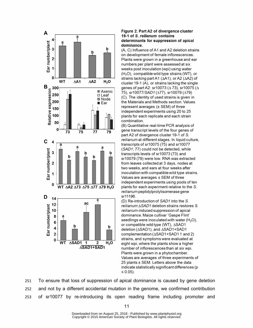
RESULTS 195
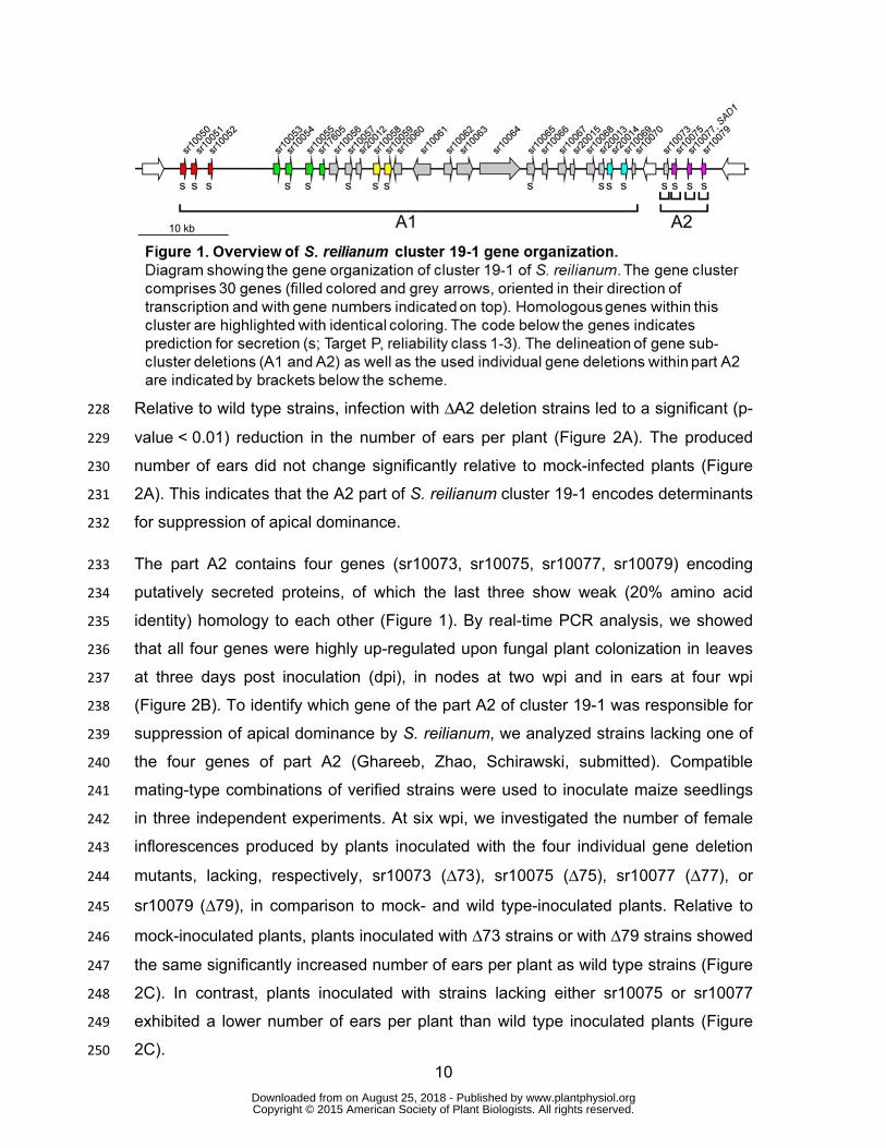
Cluster 19-1 Harbors a Suppressor of Apical Dominance 196
The divergence cluster 19-1 was identified as the largest region of genomic 197
difference between S. reilianum and U. maydis (Schirawski et al., 2010). Since 198
deletion of the gene cluster in U. maydis resulted in the loss of the U. maydis-specific 199
symptom of tumor formation on seedling leaves (Kämper et al., 2006), we 200
investigated whether the corresponding region in S. reilianum also encoded symptom 201
specificity determinants. 202
Cluster 19-1 of S. reilianum covers about 55 kb and is split in two parts by the 203
presence of a highly conserved gene (sr10071, Figure 1) encoding a predicted 204
protein with strong sequence identity to tubulin beta chains of different fungi. 205
Replacement of the first part (A1) with the phleomycin resistance cassette and of the 206
second part (A2) with the hygromycin resistance cassette, resulted in strains with 207
reduced virulence on the maize cultivar 'Gaspe Flint' (Ghareeb, Zhao, Schirawski, 208
submitted). We used compatible mating-type combinations of deletion strains for 209
inoculation experiments of maize to analyze loss of the S. reilianum-specific symptom 210
formation. Each inoculation experiment was done in three biological replicates with 211
about 25 plants inoculated per experiment and strain combination. 212
In all experiments, wild type and deletion strains lacking either part A1 (ΔA1 strains) 213
or part A2 (ΔA2 strains) were able to cause smutted ears and ears showing phyllody 214
(Ghareeb, Zhao, Schirawski, submitted). In addition, we carefully dissected all ears 215
by removal of the surrounding husk leaves (Supporting Figure S1). Outgrowth at 216
subapical nodes of the shank was unwrapped and counted as additional ears if the 217
structures could be macroscopically identified as ears (Supporting Figure S1D). 218
Numbers of apical and subapical ears were added to calculate the number of ears 219
per plant. Outgrowth of ears at subapical nodes is controlled by apical 220
dominance.Suppression of apical dominance caused by S. reilianumoccurred only 221
with wild type and ΔA1 strains but not with ΔA2 strains (Figure 2A). Loss of apical 222
dominance occurred predominantly at subapical nodes of a branch carrying an 223
infected female inflorescence at the apex (Ghareeb et al., 2011) (Supporting Figure 224
S1B). At six weeks post inoculation (wpi), at which the phenotype can be clearly 225
observed, wild type-inoculated plants showed a significantly increased (3.3 ± 0.14; p-226
value < 0.05) number of ears per plant relative to mock-inoculated plants (2.6 ± 0.18). 227
www.plantphysiol.orgon August 25, 2018 - Published by Downloaded from Copyright © 2015 American Society of Plant Biologists. All rights reserved.
10
Relative to wild type strains, infection with ΔA2 deletion strains led to a significant (p-228
value < 0.01) reduction in the number of ears per plant (Figure 2A). The produced 229
number of ears did not change significantly relative to mock-infected plants (Figure 230
2A). This indicates that the A2 part of S. reilianum cluster 19-1 encodes determinants 231
for suppression of apical dominance. 232
The part A2 contains four genes (sr10073, sr10075, sr10077, sr10079) encoding 233
putatively secreted proteins, of which the last three show weak (20% amino acid 234
identity) homology to each other (Figure 1). By real-time PCR analysis, we showed 235
that all four genes were highly up-regulated upon fungal plant colonization in leaves 236
at three days post inoculation (dpi), in nodes at two wpi and in ears at four wpi 237
(Figure 2B). To identify which gene of the part A2 of cluster 19-1 was responsible for 238
suppression of apical dominance by S. reilianum, we analyzed strains lacking one of 239
the four genes of part A2 (Ghareeb, Zhao, Schirawski, submitted). Compatible 240
mating-type combinations of verified strains were used to inoculate maize seedlings 241
in three independent experiments. At six wpi, we investigated the number of female 242
inflorescences produced by plants inoculated with the four individual gene deletion 243
mutants, lacking, respectively, sr10073 (Δ73), sr10075 (Δ75), sr10077 (Δ77), or 244
sr10079 (Δ79), in comparison to mock- and wild type-inoculated plants. Relative to 245
mock-inoculated plants, plants inoculated with Δ73 strains or with Δ79 strains showed 246
the same significantly increased number of ears per plant as wild type strains (Figure 247
2C). In contrast, plants inoculated with strains lacking either sr10075 or sr10077 248
exhibited a lower number of ears per plant than wild type inoculated plants (Figure 249
2C). 250
www.plantphysiol.orgon August 25, 2018 - Published by Downloaded from Copyright © 2015 American Society of Plant Biologists. All rights reserved.
11
To ensure that loss of suppression of apical dominance is caused by gene deletion 251
and not by a different accidental mutation in the genome, we confirmed contribution 252
of sr10077 by re-introducing its open reading frame including promoter and 253
www.plantphysiol.orgon August 25, 2018 - Published by Downloaded from Copyright © 2015 American Society of Plant Biologists. All rights reserved.
12
terminator regions in two compatible Δ77 deletion strains. Successful integration of 254
sr10077 at a known ectopic locus (the MIG1 locus, see Materials and Methods) was 255
verified by PCR and Southern blot. Southern blot analysis showed that all obtained 256
complementation strains contained multiple copies of sr10077. By quantitative RT-257
PCR we detected 11-19 copies of the introduced gene in each complementation 258
strain (not shown). The complementation strains were tested for their ability to 259
suppress apical dominance in maize. Since we noticed that the phenotype becomes 260
more severe with time, we changed the evaluation time point from six wpi to eight 261
wpi. Inoculation of maize with the complementation strains led to a significant 262
increase (p-value < 0.001) in the number of ears per plant in comparison to the 263
deletion strains and to the mock control (Figure 2D). In addition, maize inoculation 264
with the complementation strains containing multiple copies of sr10077 led to an 265
even higher number of ears per plant than inoculation with wild type strains (Figure 266
2D). These results indicate that suppression of apical dominance imposed by wild 267
type strains of S. reilianum is associated with sr10077. Therefore, we named the 268
gene SUPPRESSOR OF APICAL DOMINANCE1 (SAD1). 269
270
SAD1 Is Secreted from Fungal Hyphae in planta 271
Analysis of the SAD1 amino acid sequence with the program SignalP 4.1 predicted 272
the presence of a secretion signal peptide that is likely cleaved between amino acid 273
positions 24 and 25 of SAD1 (probability = 0.833; Petersen et al., 2011) (Supporting 274
Figure S2). Protein secretion of pathogen effectors is a prerequisite for an interaction 275
of the effector with plant proteins. To validate predicted SAD1 secretion, a construct 276
expressing a C-terminal fusion of SAD1 with GFP, SAD1-GFP, under the control of 277
SAD1 promoter was integrated at the MIG1 locus in two compatible S. reilianum 278
strains lacking SAD1 (∆SAD1 deletion strains) to generate the complementation 279
strains (∆SAD1+SAD1-GFP). Southern blot analysis showed that the strains had 280
integrated multiple copies of the SAD1-GFP construct at the MIG1 locus. 281
www.plantphysiol.orgon August 25, 2018 - Published by Downloaded from Copyright © 2015 American Society of Plant Biologists. All rights reserved.
13
To test whether the SAD1-GFP fusion protein is secreted from plant tissue-colonizing 282
fungal hyphae, we collected female inflorescences from plants inoculated with the 283
∆SAD1+SAD1-GFP complementation strains and with control strains, expressing 284
GFP under control of the SAD1 promoter (PSAD1:GFP). Fluorescence microscopic 285
www.plantphysiol.orgon August 25, 2018 - Published by Downloaded from Copyright © 2015 American Society of Plant Biologists. All rights reserved.
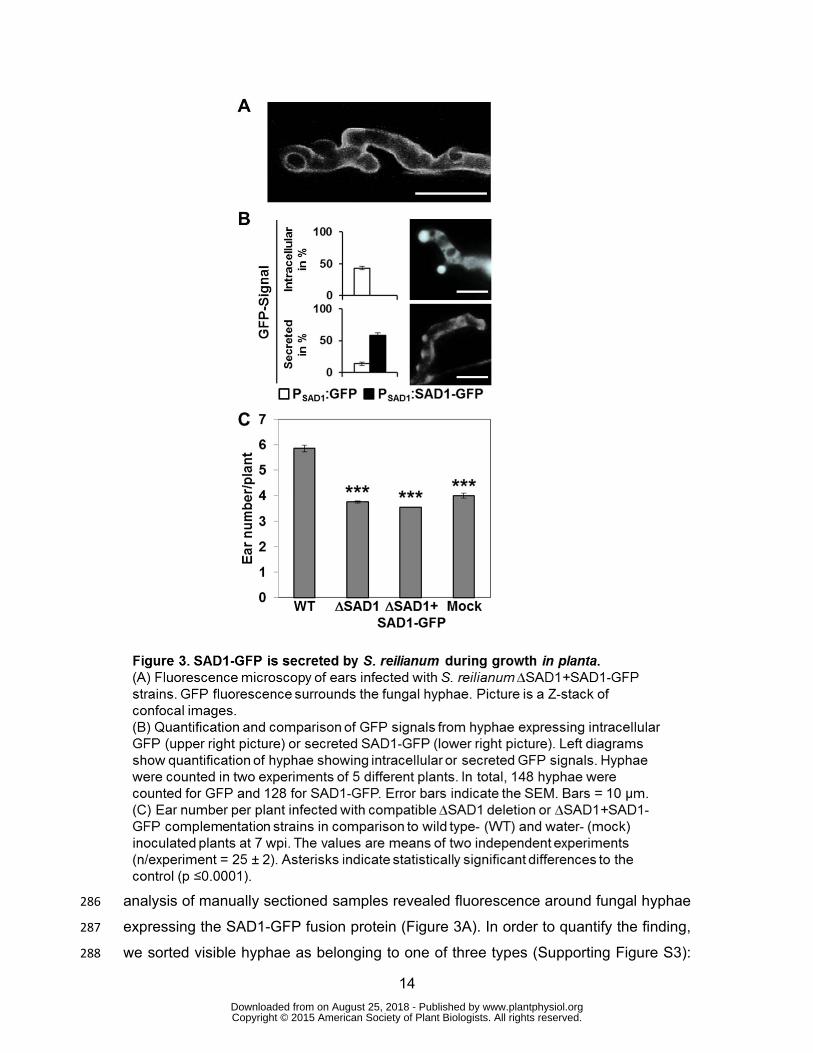
14
analysis of manually sectioned samples revealed fluorescence around fungal hyphae 286
expressing the SAD1-GFP fusion protein (Figure 3A). In order to quantify the finding, 287
we sorted visible hyphae as belonging to one of three types (Supporting Figure S3): 288
www.plantphysiol.orgon August 25, 2018 - Published by Downloaded from Copyright © 2015 American Society of Plant Biologists. All rights reserved.
15
Hyphae of type 1 had a weak GFP signal with a low signal to noise ratio that was 289
associated to very slim hyphae (Supporting Figure S3A, left panel); hyphae of type 2 290
had a strong GFP signal around fungal hyphae and at hyphal tips (Supporting Figure 291
S3A, middle panel) that co-localized with the cell membrane staining dye FM 4-64 292
(Supporting Figure S4); and hyphae of type 3 had a strong signal within the 293
boundaries of the fungal hyphae (Supporting Figure S3A, right panel). A GFP signal 294
corresponding to very slim hyphae was present for nearly 50% of all hyphae detected 295
for both strain combinations expressing either GFP or SAD1-GFP (Figure 3B). A 296
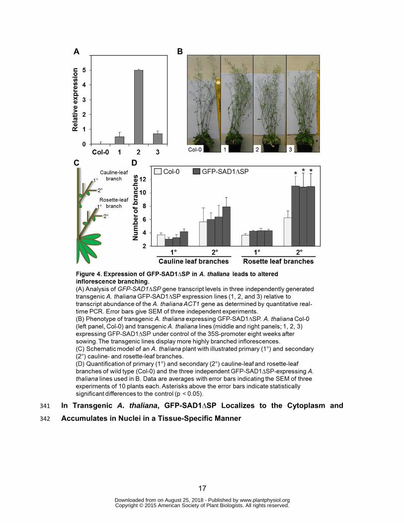
strong GFP signal around fungal hyphae and at hyphal tips could be observed for a 297
small fraction of the strains expressing GFP (Figure 3B, GFP) and for about 50% of 298
the hyphae of ∆SAD1 deletion strains expressing the SAD1-GFP fusion protein 299
(Figure 3B, SAD1-GFP). In contrast, an intracellular GFP signal was present in about 300
60% of hyphae expressing GFP, while no such GFP signals were observed with 301
strains expressing the SAD1-GFP fusion protein (Figure 3B). The differential 302
localization pattern of GFP and SAD1-GFP fluorescence confirmed that the SAD1-303
GFP fusion protein does not localize to the fungal cytoplasm and is secreted by the 304
fungal hyphae during plant colonization. 305
Symptom evaluation at eight weeks post inoculation revealed that the ∆SAD1+SAD1-306
GFP complementation strains led to the same low number of ears per plant as mock- 307
and ∆SAD1-inoculated plants (Figure 3C), indicating that the SAD1-GFP fusion 308
protein expressed in S. reilianum is not functional. To test the hypothesis that the 309
protein is non-functional due to the increased size we also tested smaller tags. 310
Infection of maize plants with S. reilianum strains ∆SAD1+MYC-SAD1 and 311
∆SAD1+SAD1-HA showed no increased number of ears per plant and thus no 312
complementation of SAD1 (Supporting Figure S5). The inoperativeness of the fusion 313
protein precludes further functional analysis. 314
315
Heterologous expression of SAD1 in A. thaliana Changes Inflorescence 316
Branching Architecture 317
To examine whether the SAD1 protein had an impact on plant development that is 318
independent of the presence of S. reilianum, we generated homozygous transgenic 319
A. thaliana plants that expressed an intracellular GFP-SAD1 fusion protein, i.e. 320
www.plantphysiol.orgon August 25, 2018 - Published by Downloaded from Copyright © 2015 American Society of Plant Biologists. All rights reserved.
16
without the predicted secretion signal peptide (see below) under control of the 35S 321
promoter (P35S:GFP-SAD1∆SP). The reason for expressing an N-terminal GFP-322
SAD1∆SP fusion was based on the observation that expression of a SAD1-GFP 323
fusion in S. reilianum leads to a non-functional SAD1 protein. Non-functionality could 324
be due to either GFP covering an active domain of SAD1 at the C-terminus, or the 325
interference of the tag with uptake of the fusion protein into the plant cells. 326
We verified production of SAD1 transcripts in the transgenic lines via real-time PCR 327
(Figure 4A). The plants grew without any obvious phenotype. However, at flowering 328
time, their inflorescences were more branched than those of the progenitor plants 329
(Figure 4B). To quantify the changes that might occur in the branching pattern of the 330
transgenic plants (Figure 4C), the primary and secondary cauline-leaf branches, and 331
the primary and secondary rosette-leaf branches were counted at the end of the 332
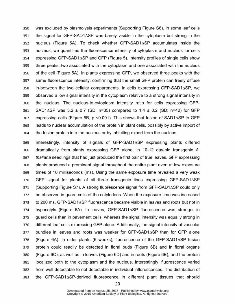
flowering period (eight weeks). A. thaliana lines expressing intracellular GFP-333
SAD1∆SP had the same number of primary and secondary cauline-leaf branches 334
and primary rosette-leaf branches but displayed a significant (p-value <0.05) increase 335
in the number of secondary rosette-leaf branches (Figure 4D). This shows that the 336
intracellularly expressed GFP-SAD1∆SP fusion protein affects the inflorescence 337
branching pattern of transgenic A. thaliana. 338
339
340
www.plantphysiol.orgon August 25, 2018 - Published by Downloaded from Copyright © 2015 American Society of Plant Biologists. All rights reserved.
17
In Transgenic A. thaliana, GFP-SAD1∆SP Localizes to the Cytoplasm and 341
Accumulates in Nuclei in a Tissue-Specific Manner 342
www.plantphysiol.orgon August 25, 2018 - Published by Downloaded from Copyright © 2015 American Society of Plant Biologists. All rights reserved.
18
To know the subcellular localization of the functional GFP-SAD1∆SP fusion protein 343
expressed in A. thaliana, we compared localization of GFP-SAD1∆SP and GFP, both 344
expressed under control of the cauliflower mosaic virus 35S promoter. GFP 345
fluorescence was observed inside the cytoplasm and the nucleus for both constructs 346
www.plantphysiol.orgon August 25, 2018 - Published by Downloaded from Copyright © 2015 American Society of Plant Biologists. All rights reserved.
19
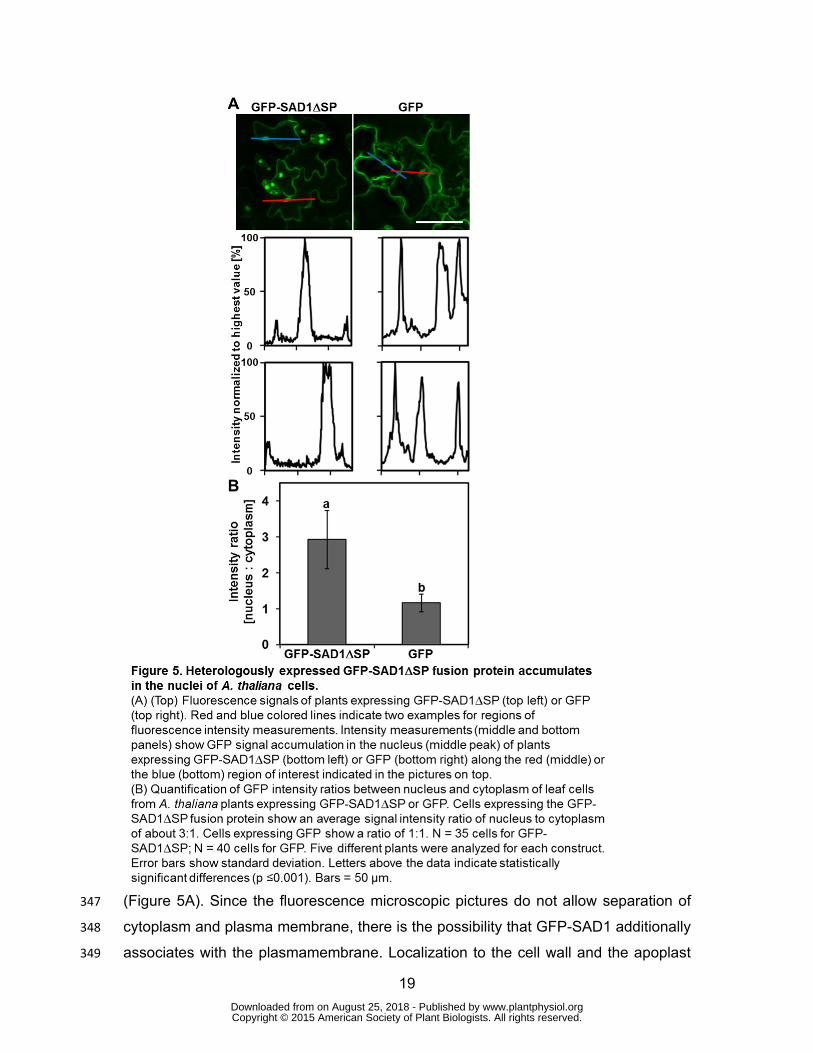
(Figure 5A). Since the fluorescence microscopic pictures do not allow separation of 347
cytoplasm and plasma membrane, there is the possibility that GFP-SAD1 additionally 348
associates with the plasmamembrane. Localization to the cell wall and the apoplast 349
www.plantphysiol.orgon August 25, 2018 - Published by Downloaded from Copyright © 2015 American Society of Plant Biologists. All rights reserved.
20
was excluded by plasmolysis experiments (Supporting Figure S6). In some leaf cells 350
the signal for GFP-SAD1∆SP was barely visible in the cytoplasm but strong in the 351
nucleus (Figure 5A). To check whether GFP-SAD1∆SP accumulates inside the 352
nucleus, we quantified the fluorescence intensity of cytoplasm and nucleus for cells 353
expressing GFP-SAD1∆SP and GFP (Figure 5). Intensity profiles of single cells show 354
three peaks, two associated with the cytoplasm and one associated with the nucleus 355
of the cell (Figure 5A). In plants expressing GFP, we observed three peaks with the 356
same fluorescence intensity, confirming that the small GFP protein can freely diffuse 357
in-between the two cellular compartments. In cells expressing GFP-SAD1∆SP, we 358
observed a low signal intensity in the cytoplasm relative to a strong signal intensity in 359
the nucleus. The nucleus-to-cytoplasm intensity ratio for cells expressing GFP-360
SAD1∆SP was 3.2 ± 0.7 (SD; n=35) compared to 1.4 ± 0.2 (SD; n=40) for GFP 361
expressing cells (Figure 5B, p <0.001). This shows that fusion of SAD1∆SP to GFP 362
leads to nuclear accumulation of the protein in plant cells, possibly by active import of 363
the fusion protein into the nucleus or by inhibiting export from the nucleus. 364
Interestingly, intensity of signals of GFP-SAD1∆SP expressing plants differed 365
dramatically from plants expressing GFP alone. In 10-12 day-old transgenic A. 366
thaliana seedlings that had just produced the first pair of true leaves, GFP expressing 367
plants produced a prominent signal throughout the entire plant even at low exposure 368
times of 10 milliseconds (ms). Using the same exposure time revealed a very weak 369
GFP signal for plants of all three transgenic lines expressing GFP-SAD1∆SP 370
(Supporting Figure S7). A strong fluorescence signal from GFP-SAD1∆SP could only 371
be observed in guard cells of the cotyledons. When the exposure time was increased 372
to 200 ms, GFP-SAD1∆SP fluorescence became visible in leaves and roots but not in 373
hypocotyls (Figure 6A). In leaves, GFP-SAD1∆SP fluorescence was stronger in 374
guard cells than in pavement cells, whereas the signal intensity was equally strong in 375
different leaf cells expressing GFP alone. Additionally, the signal intensity of vascular 376
bundles in leaves and roots was weaker for GFP-SAD1∆SP than for GFP alone 377
(Figure 6A). In older plants (6 weeks), fluorescence of the GFP-SAD1∆SP fusion 378
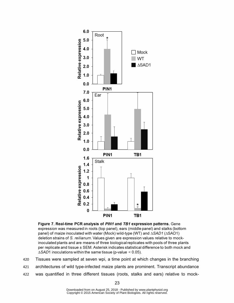
protein could readily be detected in floral buds (Figure 6B) and in floral organs 379
(Figure 6C), as well as in leaves (Figure 6D) and in roots (Figure 6E), and the protein 380
localized both to the cytoplasm and the nucleus. Interestingly, fluorescence varied 381
from well-detectable to not detectable in individual inflorescences. The distribution of 382
the GFP-SAD1∆SP-derived fluorescence in different plant tissues that should 383
www.plantphysiol.orgon August 25, 2018 - Published by Downloaded from Copyright © 2015 American Society of Plant Biologists. All rights reserved.
21
express the protein in every cell suggest that in A. thaliana expression or stability of 384
the fusion protein is translationally or post-translationally controlled. 385
386
www.plantphysiol.orgon August 25, 2018 - Published by Downloaded from Copyright © 2015 American Society of Plant Biologists. All rights reserved.
22
SAD1 Can Activate Transcription of Reporter Genes in Yeast 387
To investigate whether SAD1 has the potential to activate gene transcription, we 388
expressed SAD1 lacking its signal peptide as a fusion to the GAL4 DNA-binding 389
domain (BD-SAD1∆SP) in the strain Y2HGold of the Matchmaker™ Gold Yeast Two-390
Hybrid System expressing the GAL4 activation domain (AD). In cell extracts of this 391
strain, presence of a 41 kDa fusion protein corresponding to BD-SAD1∆SP could 392
clearly be detected (Supporting Figure S8). In contrast to control strains that only 393
expressed AD and the Gal4 DNA-binding domain (BD) and in which no reporter 394
genes were activated, two of the four reporter genes were activated in the strains 395
expressing AD and BD-SAD1∆SP. Colonies grew on medium containing 396
Aureobasidin A resulting from activation of AUR1-C, and led to the formation of a 397
blue color on plates containing α-X-GAL resulting from activation of MEL1 398
(Supporting Figure S9A). In contrast, cells were unable to grow on media lacking 399
adenine or histidine, showing that ADE2 and HIS3 were not activated (Supporting 400
Figure S9B). Since expression of HIS3 and ADE2 is driven by the G1 and G2 401
promoter elements, respectively, whereas the expression of both AUR1-C and MEL1 402
is driven by the M1 promoter element, this indicates that BD-SAD1∆SP is able to 403
auto-activate the transcription of the M1 promoter but not that of the G1 and G2 404
promoters, showing that SAD1 has the potential to activate gene expression in yeast. 405
Control experiments where SAD1 lacking its signal peptide was fused to the GAL4 406
activation domain (AD-SAD1ΔSP) showed that AD-SAD1ΔSP is not able to auto 407
activate MEL1 expression (Supporting Figure S10). This likely indicates that SAD1 408
can influence nuclear processes including activation of gene transcription not by 409
direct binding to DNA but by interaction with nuclear proteins. 410
411
SAD1 Modulates Transcription of Plant Genes Involved in Auxin Transport and 412
Branching Control 413
To find out whether SAD1 regulates nuclear processes in maize by modulating plant 414
gene expression, we analyzed the transcript level pattern of TB1, a dosage-415
dependent inhibitor of axillary bud outgrowth in maize, and PIN1, encoding an auxin 416
efflux transporter (Hubbard et al., 2002; Gallavotti et al., 2008a). Transcript levels 417
were determined by quantitative RT-PCR in different tissues of maize plants 418
inoculated either with water (mock), S. reilianum wild type or ∆SAD1 deletion strains. 419
www.plantphysiol.orgon August 25, 2018 - Published by Downloaded from Copyright © 2015 American Society of Plant Biologists. All rights reserved.
23
Tissues were sampled at seven wpi, a time point at which changes in the branching 420
architectures of wild type-infected maize plants are prominent. Transcript abundance 421
was quantified in three different tissues (roots, stalks and ears) relative to mock-422
www.plantphysiol.orgon August 25, 2018 - Published by Downloaded from Copyright © 2015 American Society of Plant Biologists. All rights reserved.
24
inoculated plants. For quantification of PIN1 transcripts, a pair of primers was used 423
that amplified part of the first exon of ZmPIN1a, ZmPIN1b, and ZmPIN1c (Supporting 424
Figure S11; Carraro et al., 2006; Forestan et al., 2010). In roots of wild type-425
inoculated maize plants, PIN1 transcript levels were significantly increased (p-value 426
<0.05), which was not the case for plants inoculated with strains lacking SAD1 427
(Figure 7, top panel). In ears, no significant difference could be detected in 428
abundance of PIN1 or TB1 transcripts in wild type- or ∆SAD1-inoculated plants 429
(Figure 7, middle panel). In stalks, where an altered plant gene expression should 430
have the most imminent effect on plant morphology, the level of PIN1 transcripts was 431
significantly reduced (p-value <0.05) in both wild type- and ΔSAD1-inoculated plants, 432
indicating a SAD1-independent effect. In contrast, the transcript level of TB1 was 433
significantly (p-value <0.05) decreased in the stalks of wild type- but not for ΔSAD1- 434
or mock-inoculated plants (Figure 7, lower panel). These results show that presence 435
of SAD1 in S. reilianum influences expression of the maize auxin transporter in roots 436
and the main repressor of bud outgrowth in stalks of ears. 437
438
Interaction Partners of SAD1 Are Involved in Development, Transcription, 439
Ubiquitination and Signaling 440
To get a better insight into the role of SAD1 in planta, we aimed to identify the 441
protein's interaction partners. We generated a normalized yeast two-hybrid library 442
with cDNA generated from axenically-grown S. reilianum and different S. reilianum-443
colonized maize tissues that results in C-terminal fusions to the Gal4 AD domain. 444
Transformation of the library into the yeast strain Y187 resulted in 1.3 x 10⁹ 445
independent clones. The library-containing strains were mated to the pGBKT7-SAD1-446
containing Y2HGold strain of the Matchmaker™ Gold Yeast Two-Hybrid System 447
expressing BD-SAD1∆SP, which resulted in 5 x 10⁵ independent diploid colonies 448
auxotrophic for tryptophan and leucine and thus containing bait and prey plasmids. 449
Of these, 434 diploids grew on medium also lacking adenine and histidine (see 450
Supporting Figure S12) indicating that they contained plasmids encoding proteins 451
that interacted with SAD1. The plasmid insert sequences of 384 clones were 452
determined and the sequences were compared among each other. This resulted in 453
179 non-redundant sequences encoding potential SAD1 interaction partners. We 454
verified 139 interactions by re-introduction of representative plasmids of each group 455
www.plantphysiol.orgon August 25, 2018 - Published by Downloaded from Copyright © 2015 American Society of Plant Biologists. All rights reserved.
25
into Y187 followed by mating with the Y2HGold strain containing pBKT7-SAD1. Of 456
the 139 potential interaction partners, 74 were recovered more than once. Of these, 457
only one (found three times) was of fungal origin, excluding a possible general 458
stickiness of the bait protein. By comparison to nucleotide databases (BLASTN, 459
NCBI) (Altschul et al., 1990) we were able to assign a putative function to 45 of the 460
identified proteins. Interestingly, more than half of these annotated interaction 461
partners have a putative function in development, transcription, ubiquitination or 462
signaling (Table 1). These results suggest that SAD1 can interact with multiple 463
intracellular maize proteins with a putative cytoplasmic or nuclear localization. 464
465
466
www.plantphysiol.orgon August 25, 2018 - Published by Downloaded from Copyright © 2015 American Society of Plant Biologists. All rights reserved.
26
DISCUSSION 467
Comparison of the S. reilianum and U. maydis smut fungal genomes revealed the 468
existence of divergence regions, often correlating to previously identified regions 469
encoding proteins with a predicted secretion signal and no known enzymatic function 470
(Kämper et al., 2006; Schirawski et al., 2010). Deletion of the largest virulence 471
effector-encoding region in U. maydis resulted in strains that were unable to induce 472
tumors on seedling leaves (Kämper et al., 2006) and had thus lost one prominent U. 473
maydis-specific symptom. This prompted us to investigate whether the corresponding 474
region in S. reilianum encoded determinants for S. reilianum-specific symptom 475
formation. In comparison to U. maydis, S. reilianum has an extended endophytic 476
phase in which it lives and spreads inside the plant without causing obvious damage 477
(Schirawski et al., 2010). Symptoms include the replacement of individual flowers or 478
complete inflorescences by spore-filled sori, the presence of floral reversion leading 479
to phyllody, and the occurrence of multiple female inflorescences at subapical nodes, 480
which is caused by suppression of apical dominance (Ghareeb et al., 2011). Gene 481
deletion strains lacking the left and larger (ΔA1) or the right and smaller part (ΔA2) of 482
cluster 19-1 were still able to induce spores and phyllody in inflorescences of maize 483
(Ghareeb, Zhao, Schirawski, submitted). However, the ΔA2 deletion strains did not 484
lead to suppression of apical dominance. Individual gene deletion of each of the four 485
genes present in the 19A2 region (Ghareeb, Zhao, Schirawski, submitted) followed 486
by symptom analysis showed that absence of SAD1 led to absence of the loss of 487
apical dominance phenotype in infected maize plants. Re-introduction of SAD1 in 488
multiple copies in the ΔSAD1 deletion strains resulted in a severe increase in the 489
number of female inflorescences per plant, suggesting that SAD1 functions as a 490
suppressor of apical dominance in maize. Thus, SAD1 is a fungal effector that affects 491
the branching architecture of maize and represents a molecular link between 492
pathogen proteins and symptom formation. 493
To affect development of host tissues, SAD1 should be expressed and secreted 494
during colonization of maize by S. reilianum. Both requirements are met, since SAD1 495
is highly expressed in leaves, nodes and ears of colonized plants (Figure 2B). In 496
addition, the functionality of the bioinformatically predicted secretion signal peptide 497
(Supporting Figure S2) was indicated by localization around fungal hyphae of a 498
SAD1-GFP fusion protein expressed in S. reilianum, and by co-localization with the 499
www.plantphysiol.orgon August 25, 2018 - Published by Downloaded from Copyright © 2015 American Society of Plant Biologists. All rights reserved.
27
cell membrane in maize inflorescences (Figure 3, Supporting Figure S3and 500
Supporting Figure S4). Thus, SAD1 fulfills the requirements to influence plant 501
development by directly interacting with components of the plant cell. 502
Yeast two-hybrid analysis revealed intracellular plant proteins as potential interaction 503
partners. While some interaction partners likely occur in the plant cell cytoplasm (i.e. 504
those involved in signaling or ubiquitination; Table 1), others are likely located in the 505
plant cell nucleus (i.e. those involved in nuclear processes; Table 1). This strongly 506
suggests that SAD1 functions inside plant cells and might have a decisive role inside 507
plant cell nuclei. Experiments involving heterologous expression of SAD1 fusion 508
proteins in A. thaliana support this suggestion. A GFP-SAD1∆SP fusion protein 509
expressed without secretion signal peptide could be detected in the cytoplasm and 510
accumulated in plant cell nuclei when stably expressed in A. thaliana (Figure 4 and 511
Figure 5). In addition, expression in yeast of SAD1 as a fusion to the GAL4-BD led to 512
transcriptional activation of reporter genes (Supporting Figure S9), and interaction of 513
SAD1 with the DNA-directed RNA polymerase II subunit J of maize in a yeast two-514
hybrid screen indicated that the protein may act as a transcriptional activator. Since 515
SAD1 was not able to substitute for the GAL4 DNA binding domain (Supporting 516
Figure S11), promoter activation might be through protein-protein interaction. Since 517
we did not use a split-ubiquitin assay for protein-protein interaction detection we 518
cannot rule out that SAD1 interacts with membrane-bound plant proteins inside or 519
even outside the plant cell and functions by activating downstream signaling 520
pathways. 521
Heterologous expression of SAD1 as a GFP-SAD1∆SP fusion in stable transgenic A. 522
thaliana resulted in plants that formed an increased number of secondary rosette leaf 523
branches (Figure 4C). This is remarkable, because it shows that SAD1 alone without 524
the help of any other fungal proteins is able to modify inflorescence branching 525
architecture in A. thaliana. In addition, this indicates that the fusion protein is 526
functional when localized to plant intracellular compartments. The increased 527
secondary branching that occurs in both A. thaliana expressing SAD1 and in S. 528
reilianum-infected maize, indicates a function of SAD1 via a pathway conserved in 529
the monocot maize and the dicot plant A. thaliana. 530
Function of SAD1 may depend on its transfer to the plant cell nucleus since the 531
concentration of the GFP-SAD1∆SP fusion protein was much higher in nuclei than in 532
www.plantphysiol.orgon August 25, 2018 - Published by Downloaded from Copyright © 2015 American Society of Plant Biologists. All rights reserved.
28
the cytoplasm (Figure 5). Reasons for the nuclear accumulation could be increased 533
cytoplasmic degradation, retention of the protein in nuclei, or specific translocation 534
from the cytoplasm to the nucleus in plant cells. Further experimentation is needed to 535
find out which scenario is valid for SAD1. Intracellular localization of SAD1 might be 536
dependent on posttranslational modification such as phosphorylation. Examples 537
include the proline-rich tyrosine kinase Pyk2 of neurons that localizes to the nucleus 538
of PC12 cells upon phosphorylation (Faure et al., 2013), and the extracellular signal-539
regulated kinase (ERK) where phosphorylation by a casein kinase 2 promotes 540
nuclear import (Plotnikov et al., 2011). Bioinformatic analysis predicted several 541
phosphorylation sites within SAD1, two phosphorylation sites for protein kinase C and 542
one for casein kinase 2 (Supporting Figure S2). In addition, three putative 543
serine/threonine-protein kinases, one putative casein kinase II, and one putative 544
phosphatase of maize were found to interact with SAD1 in the yeast two-hybrid assay 545
(Table 1). At this point it remains speculative whether phosphorylation is involved in 546
regulation of activity, stability or localization of SAD1. 547
Although it is unclear, if and how the SAD1 protein secreted from fungal hyphae 548
enters plant cells, the fact that we find intracellular plant proteins as interaction 549
partners and that an inflorescence branching phenotype is observed when the protein 550
is expressed in A. thaliana strongly hint at an intracellular function of SAD1 in maize. 551
Once inside the plant cell, SAD1 accumulates in the nucleus and could affect apical 552
dominance by altering transcription of genes involved in biosynthesis, signaling or 553
transport of auxin, cytokinin, strigolactones or gibberellins. Measurement of auxin 554
concentrations in emerging inflorescences at 4 wpi, a time point at which the 555
appearance of multiple ears per branch is initiated (F. Drechsler and J. Schirawski, 556
unpublished), showed a 30% increase in auxin concentration of infected 557
inflorescences (Ghareeb et al., 2011). Since auxin transport rather than absolute 558
auxin concentration is the main contributor to apical dominance, we investigated 559
transcript levels of the auxin efflux transporter PIN1. In ears, PIN1 transcripts were 560
slightly but not significantly increased by the presence of SAD1. PIN1 transcript 561
levels in the stalk were down-regulated regardless of the presence of SAD1 (Figure 562
7). This shows that reduction of PIN1 transcripts in the aerial parts of the plant is 563
SAD1 independent. In contrast, presence of SAD1 during infection with S. reilianum 564
leads to increased PIN1 transcript abundance in roots (Figure 7). Increased PIN1 565
transcript abundance in the root may increase auxin sink strength in the stalk and 566
www.plantphysiol.orgon August 25, 2018 - Published by Downloaded from Copyright © 2015 American Society of Plant Biologists. All rights reserved.
29
thus enable axillary buds to establish a polar auxin transport stream and continue 567
bud outgrowth as predicted by the auxin canalization model (Domagalska and 568
Leyser, 2011). To determine whether SAD1 has an effect on polar auxin transport, 569
PIN1 protein distribution in roots of infected plants needs to be verified. 570
In addition to PIN1, we measured transcript levels of the bud outgrowth inhibitor TB1. 571
TB1 has been identified as a maize domestication gene that contributes to the 572
increased apical dominance in maize in comparison to its highly branched ancestor, 573
teosinte (Studer et al., 2011). TB1 suppresses axillary bud outgrowth in maize, and 574
TB1 deletion mutants show excessive side branching (Doebley et al., 1997). In our 575
experiments, TB1 transcripts were reduced in stalks of wild type-inoculated plants in 576
comparison to mock- or ∆SAD1-inoculated plants (Figure 7). Down-regulation of TB1 577
in the presence of SAD1 explains the observed increase in subapical inflorescence 578
bud outgrowth in infected maize. In A. thaliana, a homologue of TB1 was identified as 579
BRANCHED1 (BRC1) that is a central regulator of branching (Aguilar-Martínez et al., 580
2007). Expression of BRC1 was shown to be enhanced by overproduction of auxin in 581
A. thaliana 35S:YUCCA plants leading to a lower amount of branches compared to 582
wild type plants (Finlayson 2007). In contrast, it was shown in maize that mutation of 583
the auxin biosynthetic genes SPI1 encoding a flavin monooxygenase or VT2 584
encoding a tryptophan aminotransferase leads to reduced auxin levels (Phillips et al., 585
2011), and fewer tillers are produced in spi1 and spi1/tb1 mutant plants (Gallavotti et 586
al., 2008b). This indicates that inactivation of auxin biosynthesis genes leads to 587
opposite effects in A. thaliana and maize, i. e. increased branching in Arabidopsis 588
and decreased tillering in maize (Galavotti, 2013), while in both maize and 589
Arabidopsis, branching is dependent on TB1/BRC1 (Finlayson, 2007; Doebley et al., 590
1997). Since expression of SAD1 results in an increase in bud outgrowth in both 591
maize and Arabidopsis, SAD1 likely plays a role in bud outgrowth that is independent 592
of auxin. 593
Other factors could also contribute to SAD1-dependent bud outgrowth, such as the 594
timing of the local accumulation of auxin, strigolactone and cytokinin (Müller and 595
Leyser, 2011). Reduction of strigolactone or increase in cytokinin concentrations 596
would activate bud outgrowth (Cheng et al., 2013) and lead to the observed increase 597
in the number of ears per plant. In A. thaliana it was shown that strigolactone 598
decreases PIN1 levels by two to six fold, which is sufficient to alter apical dominance 599
www.plantphysiol.orgon August 25, 2018 - Published by Downloaded from Copyright © 2015 American Society of Plant Biologists. All rights reserved.
30
(Shinohara et al., 2013). Recently, effector proteins of a plant pathogenic 600
phytoplasma bacterium were identified that influence different phases of meristem 601
development in tomato (Wei et al., 2013). Additionally to the tested transcription 602
factor TB1 there are multiple other transcription factors that are involved in 603
inflorescence architecture. One very interesting example is DELAYED FLOWERING1 604
(DLF1; Muszynski et al., 2006) a basic leucine zipper protein. Maize B73 with a DLF1 605
deletion exhibit the same branching phenotype in ears as we observed for Gaspe 606
Flint infected with S. reilianum. We could not detect DLF1 in the yeast two hybrid 607
screen and it is unknown whether SAD1 has an effect on transcription of DLF1. It will 608
be challenging to unravel the exact target and mechanism of how the SAD1 effector 609
influences plant branching architecture. 610
CONCLUSION 611
Apical dominance in maize is broken by infection with S. reilianum. We have 612
identified a fungal protein responsible for this disease symptom that we named 613
SUPPRESSOR OF APICAL DOMINANCE 1 (SAD1). SAD1 was found to be 614
secreted from fungal hyphae. SAD1 transcripts were not detectable when the fungus 615
was growing as a saprotroph but were heavily abundant as soon as the fungus 616
entered the maize plant and spread through leaves, nodes and inflorescences. 617
Several factors indicate that SAD1 mediates suppression of apical dominance. 618
Firstly, S. reilianum strains lacking SAD1 no longer lead to suppression of apical 619
dominance although virulence is unaffected. Secondly, re-introduction of SAD1 in the 620
S. reilianum ΔSAD1 deletion strain in multiple copies restored and even increased 621
the suppression of apical dominance phenotype. Thirdly, stable heterologous 622
expression of the SAD1 protein as a fusion to GFP and lacking its secretion signal 623
peptide in A. thaliana resulted in increased inflorescence branching. 624
The mechanism of how SAD1 affects apical dominance in maize and A. thaliana is 625
less clear. It seems likely, that the factor functions within plant cells. It is secreted 626
from fungal hyphae, it has an effect when expressed within A. thaliana cells, it 627
localizes to the cytoplasm and the nucleus in A. thaliana and it can interact with 628
maize cytoplasmic and nuclear proteins. If and how the factor – once secreted from 629
fungal hyphae – ends up in maize cells is an open question, as well as what exactly 630
its effect is, once it has entered. However, we could detect a positive influence of the 631
presence of SAD1 on the PIN1 transcript level in roots of infected maize plants, 632
www.plantphysiol.orgon August 25, 2018 - Published by Downloaded from Copyright © 2015 American Society of Plant Biologists. All rights reserved.
31
which suggests increased auxin flow to the root as mechanism supporting bud 633
outgrowth. In addition, we observed SAD1-dependent down-regulation of the 634
branching inhibitor TB1, which is well in line with the observed phenotype of 635
increased branching induced by SAD1. Circumstantial evidence points to a role of 636
SAD1 in bud outgrowth that is independent of auxin: While SAD1 has the same effect 637
in maize and A. thaliana, disruption of auxin biosynthesis has an opposite effect on 638
branching in maize and A. thaliana. Elucidating the exact mechanism of how this 639
interesting protein mediates suppression of apical dominance is one of the 640
challenges that lie ahead. 641
642
MATERIALS AND METHODS 643
Plant Lines, Growth Conditions and Inoculation Experiments 644
The maize cultivar ‘Gaspe Flint’, an early-flowering dwarf maize line, was cultivated 645
and used for inoculation with S. reilianum as previously described (Ghareeb et al., 646
2011). Sterilized A. thaliana Col-0 seeds were kept at 4°C for 1-3 days prior to 647
sowing. Plants were grown in soil (Fruhstofer, Type T25) under long day conditions at 648
22°C, 160 µmol, 65% humidity in a phytochamber (Johnson Controls). Sterilized A. 649
thaliana seeds were kept for 2 days prior to plating. Plants were grown on 0.5 x MS 650
medium including vitamins (Duchefa-biochemie) supplemented with 1% sucrose. The 651
Col-0 lines expressing GFP-SAD1∆SP were 970.1 (1), 975.2 (2) and 979.4 (3). 652
S. reilianum strains were prepared as stocks for storage in 25% glycerol at - 80°C, 653
strains were freshly streaked on PD plates, and single colonies were used for 654
inoculation of precultures in YEPSlight (yeast extract, 10 g/l, peptone, 10 g/l, sucrose, 655
10 g/l). For plant inoculation experiments, precultures of the compatible S. reilianum 656
wild type strains SRZ1_5-2 and SRZ2_5-1 (Schirawski et al., 2005; Zuther et al., 657
2012) and their deletion derivatives were used to inoculate potato dextrose broth 658
(Difco, Heidelberg, Germany), and were grown at 28°C under constant shaking to an 659
OD600 of 0.5 to 0.8. Cell pellets were resuspended in water to reach an OD600 of 2.0. 660
Cultures of compatible strains carrying the same deletion were mixed in a 1:1 ratio, 661
and the mixture was used to inoculate seven-day old maize seedlings via syringe-662
assisted leaf whorl apposition (Gillissen et al., 1992). 663
www.plantphysiol.orgon August 25, 2018 - Published by Downloaded from Copyright © 2015 American Society of Plant Biologists. All rights reserved.
32
664
Generation of Plasmids, Knockout, Complementation and Localization 665
Constructs 666
For generating the expression vectors for p35S:GFP-SAD1∆SP to be expressed in A. 667
thaliana, the primers forA and revA were used to amplify a 0.5 kb fragment containing 668
the SAD1 ORF lacking the signal peptide (SAD1∆SP). The fragment was fused to 669
GFP by cloning into a binary vector containing GFP using NotI and EcoRI. The fusion 670
construct was moved as an AscI and EcoRI-fragment to the binary plant expression 671
vector pAMPAT-MCS (accession: AY436765). For yeast two-hybrid analysis, 672
SAD1∆SP was amplified with oHG210 and oHG211. The 0.5 kb PCR amplicon was 673
digested with EcoRI and BamHI and cloned into the bait vector pGBKT7 (Clontech). 674
For complementation studies, a strategy to integrate the genes of interest at the mig1 675
locus (intergenic region between the genes sr14222 and sr14223) was established. 676
For generation of the SAD1 complementation construct, right and left flanks of the 677
MIG1 locus and the SAD1 gene including promoter and terminator regions were 678
amplified using primers listed in Table 2. The MIG1 flanks, SAD1 and the carboxin 679
resistance cassette from the plasmid pMF1-c (Brachmann et al., 2004) were digested 680
with SfiI and ligated. The ligation product was excised from gel and used to amplify 681
the complementation construct using oligonucleotides listed in Table 2. 682
683
Transformation of S. reilianum and Generation of Recombinant Strains 684
The complementation and localization constructs were used to transform S. reilianum 685
via protoplast transformation. Protoplast preparation and transformation of 686
S. reilianum was performed according to the protocols used for U. maydis (Schulz et 687
al., 1990; Gillissen et al., 1992) with some modifications. A single fungal colony was 688
used to inoculate 2 ml YEPS-light liquid medium and incubated at 28°C for 8 – 12 h. 689
This culture was used to inoculate 100 ml of YEPS-light, which was incubated at 690
28°C with constant shaking at 200 rpm. Cells were grown to an OD600nm of 0.6–0.8 691
and subsequently pelleted by centrifugation at 3500 g (Beckmann Biofuge) for 5 min. 692
The supernatant was discarded and the pellet resuspended in 50 ml SCS buffer (20 693
mM Na-citrate, pH 5.8,1 M sorbitol and sterile filtered). Cells were centrifuged at 3500 694
www.plantphysiol.orgon August 25, 2018 - Published by Downloaded from Copyright © 2015 American Society of Plant Biologists. All rights reserved.
33
g for 10 min and the supernatant was discarded. Protoplasts were produced by 695
resuspending the cells in 2 ml of Novozyme (2.5 mg/ml) solution (Novo Nordisc, 696
Copenhagen, Denmark) and incubating at room temperature for 5-10 min until ~50% 697
of the cells produced protoplasts. The formation of protoplasts was confirmed by 698
microscopy. Protoplasting was stopped by adding 20 ml SCS buffer and centrifuging 699
the solution at 2300 g for 15 min. The supernatant was discarded and the pellet was 700
washed twice with SCS and once with STC buffer (10 mM Tris-HCl, pH 7.5, 100 mM 701
CaCl2, 1 M sorbitol and sterile filtered), respectively, by carefully resuspending the 702
pellet in 20 ml of either buffers and centrifuging at 2300 g for 15 min. Finally, 703
protoplasts were resuspended in 500 μl ice-cold STC buffer and dispensed into 70 μl 704
aliquots and either used directly for transformation or stored at -80°C. 705
For transformation of the protoplasts, a 70 μl aliquot of protoplasts was mixed with ~5 706
μg DNA and 1.5 μl heparin sodium sulfate (50 mg/ml) and kept on ice for 10 min. 707
Protoplasts were carefully mixed with 500 μl of cold STC containing 40% PEG4000 708
solution and incubated on ice for a further 15 min on ice. The entire mixture was 709
plated onto regeneration medium (10 g/l tryptone, 10 g/l yeast extract, 10 g/l sucrose, 710
182.2 g/l sorbitol and 20 g/l agar) supplemented with the appropriate antibiotic 711
(phleomycin 1 µg/ml, carboxin 5 µg/ml or hygromycin 150 µg/ml). Plates were 712
incubated at 28°C for 4-6 days or until distinct colonies appeared. Single colonies 713
were picked using sterile toothpicks and streaked onto PD or regeneration medium-714
plates supplemented with the appropriate antibiotic to obtain single colonies. Putative 715
transformants were selected and verified by PCR using primers listed in Table 2, and 716
by Southern blot using the deletion construct as probe. 717
For each mutant, 1-3 different strains in each SRZ1_5-2 and SRZ2_5-1 background 718
were used to inoculate maize plants. Strains used in this study for ∆19A1 mutants 719
were (1, JS747 and JS751; 2, JS748 and JS752), for ∆19A2 mutants were (1, HG125 720
and HG127; 2, HG126 and HG128), for ∆sr10073 mutants were HG109 and HG113, 721
for ∆sr10075 mutants were HG80 and HG84, for ∆sr10077 mutants were HG95 and 722
HG99, for ∆sr10079 mutants were HG89 and HG92 (Ghareeb, Zhao, Schirawski, 723
submitted), for ∆SAD1+SAD1 mutants were (1, HG163 and HG167; 2, HG164 and 724
HG168), for PSAD1:GFP containing mutants were (YF2 5-1#1 and YF2 5-2#1), and for 725
∆SAD1+SAD1-GFP mutants were (1, HG183 and HG186; 2, HG185 and HG187). 726
www.plantphysiol.orgon August 25, 2018 - Published by Downloaded from Copyright © 2015 American Society of Plant Biologists. All rights reserved.
34
For generation of S. reilianum strains expressing cytoplasmic GFP, protoplasts of the 727
compatible S. reilianum wild type strains SRZ2_5-1 and SRZ1_5-2 were transformed 728
with pHSP70-SG (Spellig et al., 1996). 729
730
Arabidopsis Transformation 731
Seeds of A. thaliana ecotype Col-0 were grown on soil in growth chambers (York) at 732
22-25°C under a long day regime of 16 h exposure to light and 8 h darkness and at 733
approximately 60% humidity. Recombinant DNA constructs were introduced into A. 734
thaliana plants by Agrobacterium tumefaciens-mediated transformation, using A. 735
tumefaciens strain EHA105 (Hood et al., 1993) and the floral-dip method (Clough and 736
Bent, 1998). Independent transformants were selected according to resistance 737
against aerosolic glufosinate ammonium (Bayer). 738
739
RNA Isolation and qRT-PCR Analysis 740
Transcript levels of cluster 19A2 genes (as indicated in Figure 2B) were measured by 741
isolating RNA from sporidia grown in liquid culture to an OD600nm of 0.5, as well as by 742
isolating RNA from infected plant samples. Plant samples were collected from a 2-cm 743
piece below the injection hole of the third leaf at three dpi (10 days after sowing 744
(das)), developing nodes of the main stem at 15 dpi (21 das), and ears including 745
stalks with a length of 1-3 cm at 7 wpi (56 das) from mock- or S. reilianum-inoculated 746
plants. The samples were collected from at least 20 plants for each experiment and 747
sampling time point and the experiment was done three independent times. 748
For qRT-PCR analysis of the ZmPIN1 and TB1 genes (indicated in Figure 7), three 749
plant tissues were analyzed: roots, ears of a size of 1-3 cm excluding stalks, and 750
stalks after removal of the ears or axillary buds. For each biological replicate, 751
samples were collected from five different S. reilianum- or water-inoculated plants at 752
7 wpi (56 das). RNA was extracted with Trizol (Invitrogen, Karlsruhe, Germany), 753
DNase treatment was performed using RNase-Free DNase Set (Qiagen; Hilden; 754
Germany), and RNA was purified with RNeasy Plant Mini Kit (Qiagen; Hilden; 755
Germany). Gene transcript levels were determined relative to the 756
peptidylprolylisomerase (ppi, sr11196) transcript level from S. reilianum for analysis 757
www.plantphysiol.orgon August 25, 2018 - Published by Downloaded from Copyright © 2015 American Society of Plant Biologists. All rights reserved.
35
of cluster 19A2 genes, and relative to the ACT1 gene from maize for analysis of 758
maize gene transcription. Since the family members of ZmPIN1 share high 759
conservation of amino acid sequences and exon/intron structure (Carraro et al., 760
2006; Forestan et al., 2010; Forestan et al., 2012), we designed oligonucleotide 761
primers that targeted exon1 of the three family members ZmPIN1a, ZmPIN1b and 762
ZmPIN1c (Supporting Figure S11). Primers are listed in Table 2. 763
764
Statistical Data Analysis 765
For statistical analysis, all data was tested for normal distribution. The statistical 766
differences between different treatments were analyzed by one way ANOVA and 767
Tukey test using the R software. 768
769
Yeast cDNA Library Construction 770
RNA for normalized cDNA library construction was obtained from an axenically grown 771
mixture of the two compatible S. reilianum wild type strains SRZ1_5-2 and SRZ2_5-1 772
and from three different maize tissues infected with S. reilianum; inoculated leaves (2 773
cm below injection hole) at three dpi, infected nodes at 15 dpi and infected ears at 4 774
wpi (smaller than 2 cm). The tissues were collected from three independent 775
experiments, which each included 20 plants. The quality and purity of RNA from each 776
individual experiment and tissue was checked by Bioanalyzer 2100 (Agilent) and 777
NanoDrop ND-1000 (Thermo Fisher Scientific). Equal RNA amounts from the pooled 778
RNA of each tissue were mixed to make the RNA sample for cDNA library 779
construction. A normalized cDNA library was constructed by Bio S&T, Lachine, QC, 780
Canada. Briefly, 100 µg of total RNA were used for mRNA extraction and cDNA 781
synthesis followed by cDNA normalization using a modified SMART cDNA synthesis 782
method (Clontech). The cDNA was amplified, purified, digested with SfiI for 783
directional cloning into the pGADT7 (modified to include a SfiI site at the multiple 784
cloning site), ligated and transformed into E. coli. Cells were plated into LB solid 785
medium plates with ampicillin and incubated at 37°C overnight. Colonies were 786
collected from plates with 10% glycerol and help of glass beads. The E. coli cells 787
were kept at -80°C in 1-ml aliquots. A portion of the E. coli-containing cDNA library 788
was grown overnight in LB liquid medium with ampicillin. The pGADT7 containing the 789
www.plantphysiol.orgon August 25, 2018 - Published by Downloaded from Copyright © 2015 American Society of Plant Biologists. All rights reserved.
36
cDNA library was isolated and used for transformation of yeast to construct the yeast 790
two-hybrid library. 791
792
Yeast Two-Hybrid Library Generation 793
The yeast two-hybrid library was constructed according to Make Your Own Mate and 794
Plate Yeast Two-Hybrid Library System and Yeastmaker™ Yeast Transformation 795
System 2 (Clontech, Saint-Germain-en-Laye, France) with some modifications. A 2-3 796
mm single colony of the yeast strain Y187 was inoculated in 3 ml YPDA liquid 797
medium and incubated at 30°C with 250 rpm constant shaking for 8-12 h. From this 798
culture 5 µl were used to inoculate 50 ml YPDA liquid medium and incubated as 799
previously indicated for 16-20 h until OD600nm = 0.15-0.3 is reached. The culture was 800
centrifuged at 700 g for 5 min. The cell pellet was resuspended in 100 ml YPDA liquid 801
medium and left to grow at 30°C with 250 rpm constant shaking for 3-5 h until 802
OD600nm = 0.4-0.5 is reached. The culture was centrifuged at 700 g for 5 min. The cell 803
pellet was resuspended in 30 ml water and centrifuged once again. The cell pellet 804
was resuspended in 1.2 ml TE buffer. The cells were used immediately for 805
transformation of cDNA library. 806
For transformation, the 600 µl cell suspension was mixed with 15 µg cDNA library, 20 807
µl denatured DNA carrier (10 mg/ml; Clontech, Saint-Germain-en-Laye, France) and 808
2.5 ml PEG/LiAc solution. The transformation mixture was incubated at 30°C for 45 809
min with gentle mixing every 15 min. Thereafter 160 µl DMSO was added to the 810
mixture and then incubated in a water bath at 42°C for 20 min with mixing every 10 811
min. The cells were centrifuged at 700 g for 5 min, resuspended in 3 ml YPD Plus 812
Medium (Clontech, Saint-Germain-en-Laye, France) and incubated at 30°C with 250 813
rpm shaking for 90 min. The transformed culture was centrifuged once again. This 814
transformation was performed 10 times individually and the cell pellets were pooled 815
and resuspended in 30 ml 0.9% NaCl. The cell suspensions (150 µl per plate) were 816
plated on 200 SD/-Leucine solid medium. Plates were incubated at 30°C for 4 days, 817
and then chilled at 4°C for 1 day. The colonies in each plate were collected using 5 818
ml freezing medium (YPDA with 25% glycerol and 50 µg/ml kanamycin) and glass 819
rod. Cell suspensions were collected in one container, well mixed and distributed in 820
1-ml aliquots for direct use or 50-ml aliquots for long term storage. Aliquots were 821
stored at -80°C until used. Cell density of the library was calculated using a 822
www.plantphysiol.orgon August 25, 2018 - Published by Downloaded from Copyright © 2015 American Society of Plant Biologists. All rights reserved.
37
hemocytometer and titering 10-4, 10-5, 10-6 and 10-7 dilutions of the library on SD/-823
Leucine (SD/-Leu) solid plates. 824
825
Autoactivation Test and Yeast Two-Hybrid Screening 826
The yeast two-hybrid screening was performed according to Matchmaker™ Gold 827
Yeast Two-Hybrid System (Clontech, Saint-Germain-en-Laye, France). To prepare 828
for the yeast two-hybrid screening with SAD1 the SAD1 gene without the signal 829
peptide was cloned into pGBKT7 (pGBKT7–SAD1) so that it fuses with the GAL4 830
DNA binding domain. The plasmid was transformed into the yeast strain Y2HGold. 831
As controls, the empty plasmid pGBKT7 and pGBKT7-53 (containing p53 coding 832
sequence), and pGADT7 (empty prey plasmid), pGADT7-T (containing T antigen 833
coding sequence) and pGADT7-Lam (containing the Lam coding sequence) were 834
transformed into the Y2HGold and Y187 strains, respectively. The Y2HGold and 835
Y187 strains can mate. 836
To test whether SAD1 has an auto-activation effect on the selectable markers of the 837
yeast strain, the strain Y2HGold containing pGBKT7-SAD1 was mated with the strain 838
Y187 containing pGADT7 and as controls the strain Y2HGold containing pGBKT7-53 839
was mated with the stain Y187 containing either pGADT7-T (resulting in positive 840
interaction) pGADT7-Lam (resulting in negative interaction). The mating was 841
performed by mixing one 2-3 mm single colony from each strain in 2X YPDA liquid 842
medium. The mixture was incubated at 30°C with 50 rpm shaking for 20-24 h. The 843
mating events were selected on SD/-Leu/-Tryptophan (Trp) double dropout (DDO). 844
The auto-activation of the selectable markers was analyzed by growing the zygotes 845
on DDO with 125-300 ng Aureobasidin A and X-α-Gal (DDO/A/X) or DDO additionally 846
lacking adenine and histidine (quadruple dropout, QDO) solid medium. 847
To screen for the interaction partners of SAD1, a single colony of the Y2HGold strain 848
containing pGBKT7-SAD1 was inoculated in 70 ml SD/-Trp liquid medium and 849
incubated at 30°C with 250 rpm shaking until an OD600nm of 0.8 was reached. The 850
culture was centrifuged at 1000 g for 5 min, then the cell pellet was resuspended in 4 851
ml SD/-Trp liquid medium and combined with 200 µl of Y187 containing the cDNA 852
library (see above) in a sterile 2-L flask. To allow mating, 45 ml of 2X YPDA liquid 853
medium supplemented with 50 μg/ml kanamycin was added, and the culture was 854
www.plantphysiol.orgon August 25, 2018 - Published by Downloaded from Copyright © 2015 American Society of Plant Biologists. All rights reserved.
38
incubated at 30°C with 50 rpm shaking for 24 h. The mated culture was centrifuged 855
at 1000 g for 10 min and then resuspended in 10 ml 0.5X YPDA (with 50 μg/ml 856
kanamycin). Each 200-µl aliquot of mating suspension was spread on high stringency 857
medium (QDO). Additionally, dilution serials 10-3, 10-5 and 10-6 were prepared and 858
100 µl of each dilution was spread on SD/-Leu, SD/-Trp and DDO plates to estimate 859
the mating efficiency and to calculate the total number of screened clones. The plates 860
were incubated at 30°C for 5 days. 861
The growing colonies were re-purified twice on QDO with 300 ng/ml Aureobasidin 862
(QDO/A). Colonies that survived were used to isolate plasmids, which were 863
individually used to transform E. coli, and then plasmids were isolated from E. coli 864
and sequenced. The sequences were grouped and a representative plasmid 865
(pGADT7-PREY) for each group was retransformed in the Y187 strain to generate 866
Y187+pGADT7-PREY. The Y187+pGADT7-PREY strains were grown in 150 µl liquid 867
SD/-Leu medium in 96 well microtiter plates at 30°C and shaking at 280 rpm for two 868
days. Meanwhile, the Y2HGold strains containing either pGBKT7-SAD1 or pGBKT7 869
were grown in 20 ml SD/-Trp medium at 30°C and 250 rpm until an OD600nm of 5-6 870
was reached. To verify the interaction, 75 µl of Y187+pGADT7-PREY were mixed 871
with 75 µl Y2HGold strains containing either pGBKT7-SAD1 or pGBKT7 in 96 well 872
microtiter plates and incubated overnight at 30°C with shaking at 50 rpm. The cell 873
mixtures were printed on DDO and QDO plates using 96-prong replicator and 874
incubated at 30°C for 3-4 days. 875
876
Staining and Confocal Microscopy 877
Samples for microscopy were freshly collected and prepared by mounting ~1 cm-878
pieces of infected leaves or thin sections of infected ears on a glass slide and 879
immediately fixing the material on the slide with the help of a drop of water and a 880
cover slip. The samples were immediately observed using a TCS-SP5 confocal 881
microscope (Leica, Germany), Zeiss Axioplan II or Axio Observer.Z1 microscopes 882
(Zeiss, Jena, Germany), or a AF 6000LX fluorescence microscope (Leica, Germany). 883
For fluorescence microscopy of GFP the FITC filter (excitation at 450-490 nm and 884
emission at 515-565 nm) was used. Image processing was performed using the 885
imaging software MetaMorph v6.2 (Universal Imaging, Downing Town, PA, USA) or 886
www.plantphysiol.orgon August 25, 2018 - Published by Downloaded from Copyright © 2015 American Society of Plant Biologists. All rights reserved.
39
AxioVision v4.3 (Zeiss, Jena, Germany), or Leica Application Suite Advanced 887
Fluorescence v4.0 (Leica, Germany). For confocal microscopy of SAD1-GFP fusion 888
proteins in planta, the samples were laser-excited at 488 nm and the emission signal 889
detected at 500–530 nm. For staining plant and fungal cell membranes, FM4-64 890
(Molecular Probes, Invitrogen, Karlsruhe) was used. The infected ears were manually 891
thin-sectioned and dipped in FM4-64 staining solution (17 µM FM4-64 in water) and 892
incubated in the dark and on ice for 30 min. The FM4-64 was excited at 561 nm and 893
signal was detected at 640-750 nm. Images were processed using LAS AF v1.8 and 894
v 4.0 (Leica, Bensheim, Germany). Deconvolution of Z-stacks was performed using 895
the LAS AF v4.0 (Leica, Germany) using 10 iterations with a refractive index of 1.33 896
for the HCX PL APO 40x/1.10 WATER objective, and the “blind” mode. 897
898
ACKNOWLEDGEMENTS 899
We thank Regine Kahmann and Thomas Brefort for stimulating discussions. We are 900
grateful to Jennifer Krüger for technical assistance and help with identification of 901
homozygous A. thaliana lines expressing GFP-SAD1ΔSP, Elmar Meyer for help with 902
generation of cytoplasmic GFP expressing S. reilianum strains, and Yufei Wang for 903
generation of the PSAD1:GFP construct. We thank Ivo Feussner (Göttingen) and 904
Regine Kahmann (Marburg) for greenhouse space and Volker Lipka (Göttingen) for 905
use of the confocal microscope. 906
907
AUTHOR CONTRIBUTIONS 908
H.G., F.D., J.S. designed experiments, H.G., F.D., C.L. performed experiments, C. L., 909
T.T. helped in generation of recombinant Arabidopsis lines, H.G., F.D., J.S. created 910
digital artwork, H.G., F.D., J.S. wrote the paper, H.G., F.D., C.L., T.T., J.S. discussed 911
results and proofread the paper. 912
913
www.plantphysiol.orgon August 25, 2018 - Published by Downloaded from Copyright © 2015 American Society of Plant Biologists. All rights reserved.
40
Table 1. List of SAD1 interaction partners putatively involved in development, 914
transcription, ubiquitination or signaling. 915
Name IP Frequencya Accession Annotation
Development SAD1-IP4 5 NM_0011118 Zea mays AGAMOUS 1 (ZAG1) SAD1-IP1 4 AF467541 Putative aldehydedehydrogenase MIS1 (MIS1) SAD1-IP2 3 AY883559 Teosinte glume architecture 1 (TGA1) SAD1-IP3 2 NM_0011771 Fimbriata-like protein Nuclear processes SAD1-IP17 5 NM_0011750 Nucleolar complex protein 2 homolog SAD1-IP5 4 NM_0011123 Helicase RH2 protein-like SAD1-IP6 4 NM_0011582 Polypyrimidine tract-binding protein SAD1-IP7 3 NM_0011552 Arginine/serine-rich splicing factor 10 SAD1-IP8 3 EU955917 RNA-binding protein-like SAD1-IP9 3 EU962463 Zinc finger protein-like 1 mRNA SAD1-IP10 2 NM_0011544 Zinc finger, C3HC4 type family protein SAD1-IP11 2 NM_0011751 Transcription factor PIF3 SAD1-IP12 2 EU964022 DNA-directed RNA polymerase II subunit J Ubiquitination SAD1-IP22 7 EU965267 Similar to SAM domain of Anks family protein SAD1-IP23 4 NM_0011478 Probable E3 ubiquitin-protein ligase SAD1-IP24 2 NM_0011555 Probable E3 ubiquitin-protein ligase SAD1-IP25 2 U29161 MubG7 ubiquitinfusionproteingene SAD1-IP26 2 BT066423 Ubiquitin-associated protein SAD1-IP27 2 NM_0011527 E3 ubiquitin-protein ligase RGLG2 SAD1-IP28 2 EU940814.1 E3 ubiquitin-protein ligase KEG SAD1-IP29 2 BT067186 S-phase kinase-associated protein 1-interacting Signaling SAD1-IP33 6 NM_0011533 Probable phosphatase 2C SAD1-IP34 2 NM_0011114 Pyruvatedehydrogenase (lipoamide) kinase1 (pdlk1) SAD1-IP35 2 EU954821 Casein kinase II subunit beta-4 SAD1-IP36 2 NM_0011587 Kinase APK1B
aThe frequency of recovered sequences.
916
917
918
www.plantphysiol.orgon August 25, 2018 - Published by Downloaded from Copyright © 2015 American Society of Plant Biologists. All rights reserved.
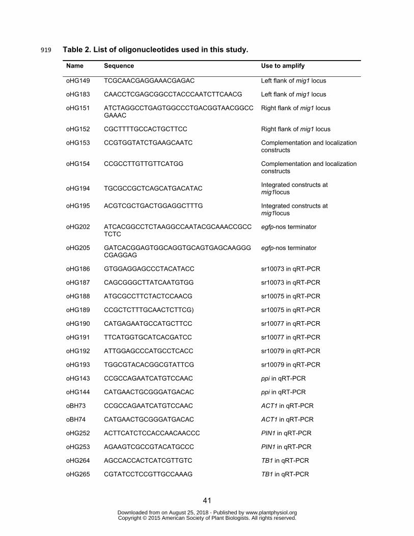
41
Table 2. List of oligonucleotides used in this study. 919
Name Sequence Use to amplify
oHG149 TCGCAACGAGGAAACGAGAC Left flank of mig1 locus
oHG183 CAACCTCGAGCGGCCTACCCAATCTTCAACG Left flank of mig1 locus
oHG151 ATCTAGGCCTGAGTGGCCCTGACGGTAACGGCCGAAAC
Right flank of mig1 locus
oHG152 CGCTTTTGCCACTGCTTCC Right flank of mig1 locus
oHG153 CCGTGGTATCTGAAGCAATC Complementation and localization constructs
oHG154 CCGCCTTGTTGTTCATGG Complementation and localization constructs
oHG194 TGCGCCGCTCAGCATGACATAC Integrated constructs at mig1locus
oHG195 ACGTCGCTGACTGGAGGCTTTG Integrated constructs at mig1locus
oHG202 ATCACGGCCTCTAAGGCCAATACGCAAACCGCCTCTC
egfp-nos terminator
oHG205 GATCACGGAGTGGCAGGTGCAGTGAGCAAGGGCGAGGAG
egfp-nos terminator
oHG186 GTGGAGGAGCCCTACATACC sr10073 in qRT-PCR
oHG187 CAGCGGGCTTATCAATGTGG sr10073 in qRT-PCR
oHG188 ATGCGCCTTCTACTCCAACG sr10075 in qRT-PCR
oHG189 CCGCTCTTTGCAACTCTTCG) sr10075 in qRT-PCR
oHG190 CATGAGAATGCCATGCTTCC sr10077 in qRT-PCR
oHG191 TTCATGGTGCATCACGATCC sr10077 in qRT-PCR
oHG192 ATTGGAGCCCATGCCTCACC sr10079 in qRT-PCR
oHG193 TGGCGTACACGGCGTATTCG sr10079 in qRT-PCR
oHG143 CCGCCAGAATCATGTCCAAC ppi in qRT-PCR
oHG144 CATGAACTGCGGGATGACAC ppi in qRT-PCR
oBH73 CCGCCAGAATCATGTCCAAC ACT1 in qRT-PCR
oBH74 CATGAACTGCGGGATGACAC ACT1 in qRT-PCR
oHG252 ACTTCATCTCCACCAACAACCC PIN1 in qRT-PCR
oHG253 AGAAGTCGCCGTACATGCCC PIN1 in qRT-PCR
oHG264 AGCCACCACTCATCGTTGTC TB1 in qRT-PCR
oHG265 CGTATCCTCCGTTGCCAAAG TB1 in qRT-PCR
www.plantphysiol.orgon August 25, 2018 - Published by Downloaded from Copyright © 2015 American Society of Plant Biologists. All rights reserved.
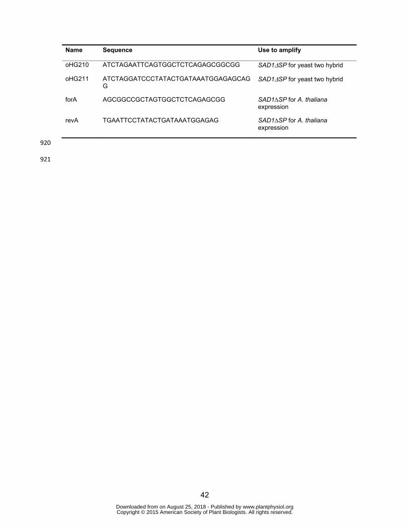
42
Name Sequence Use to amplify
oHG210 ATCTAGAATTCAGTGGCTCTCAGAGCGGCGG SAD1ΔSP for yeast two hybrid
oHG211 ATCTAGGATCCCTATACTGATAAATGGAGAGCAGG
SAD1ΔSP for yeast two hybrid
forA AGCGGCCGCTAGTGGCTCTCAGAGCGG SAD1∆SP for A. thaliana expression
revA TGAATTCCTATACTGATAAATGGAGAG SAD1∆SP for A. thaliana expression
920
921
www.plantphysiol.orgon August 25, 2018 - Published by Downloaded from Copyright © 2015 American Society of Plant Biologists. All rights reserved.
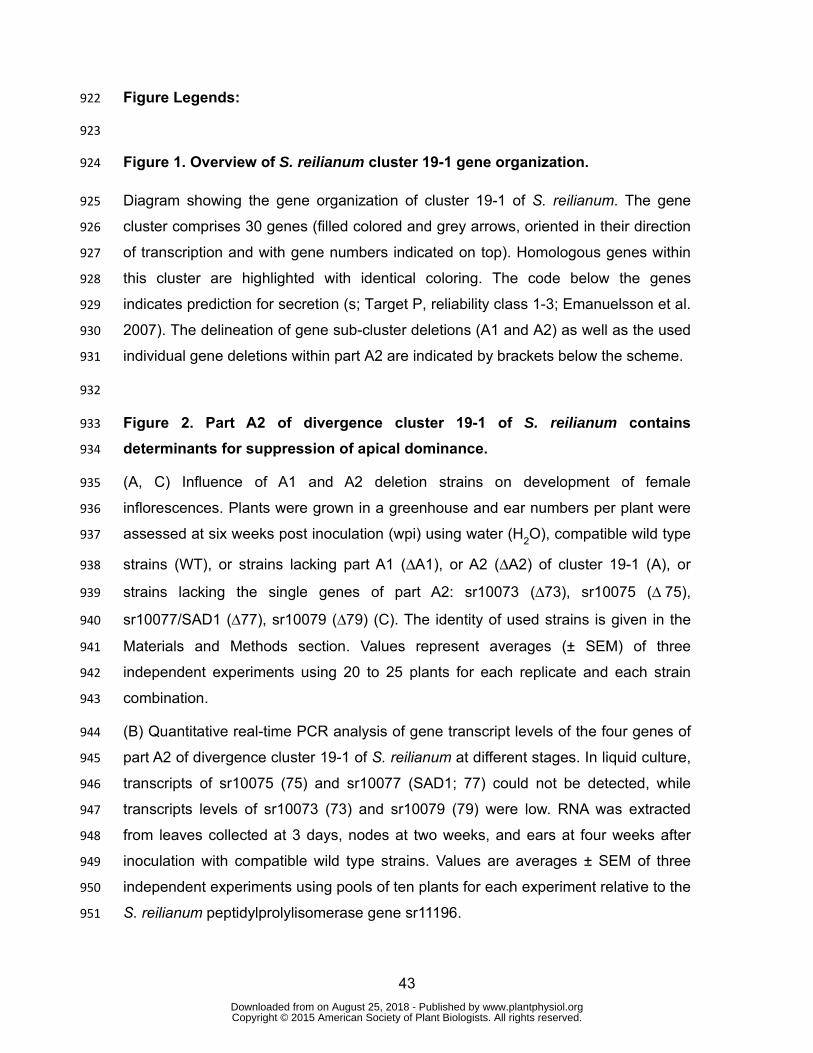
43
Figure Legends: 922
923
Figure 1. Overview of S. reilianum cluster 19-1 gene organization. 924
Diagram showing the gene organization of cluster 19-1 of S. reilianum. The gene 925
cluster comprises 30 genes (filled colored and grey arrows, oriented in their direction 926
of transcription and with gene numbers indicated on top). Homologous genes within 927
this cluster are highlighted with identical coloring. The code below the genes 928
indicates prediction for secretion (s; Target P, reliability class 1-3; Emanuelsson et al. 929
2007). The delineation of gene sub-cluster deletions (A1 and A2) as well as the used 930
individual gene deletions within part A2 are indicated by brackets below the scheme. 931
932
Figure 2. Part A2 of divergence cluster 19-1 of S. reilianum contains 933
determinants for suppression of apical dominance. 934
(A, C) Influence of A1 and A2 deletion strains on development of female 935
inflorescences. Plants were grown in a greenhouse and ear numbers per plant were 936
assessed at six weeks post inoculation (wpi) using water (H2O), compatible wild type 937
strains (WT), or strains lacking part A1 (ΔA1), or A2 (ΔA2) of cluster 19-1 (A), or 938
strains lacking the single genes of part A2: sr10073 (Δ73), sr10075 (Δ 75), 939
sr10077/SAD1 (Δ77), sr10079 (Δ79) (C). The identity of used strains is given in the 940
Materials and Methods section. Values represent averages (± SEM) of three 941
independent experiments using 20 to 25 plants for each replicate and each strain 942
combination. 943
(B) Quantitative real-time PCR analysis of gene transcript levels of the four genes of 944
part A2 of divergence cluster 19-1 of S. reilianum at different stages. In liquid culture, 945
transcripts of sr10075 (75) and sr10077 (SAD1; 77) could not be detected, while 946
transcripts levels of sr10073 (73) and sr10079 (79) were low. RNA was extracted 947
from leaves collected at 3 days, nodes at two weeks, and ears at four weeks after 948
inoculation with compatible wild type strains. Values are averages ± SEM of three 949
independent experiments using pools of ten plants for each experiment relative to the 950
S. reilianum peptidylprolylisomerase gene sr11196. 951
www.plantphysiol.orgon August 25, 2018 - Published by Downloaded from Copyright © 2015 American Society of Plant Biologists. All rights reserved.
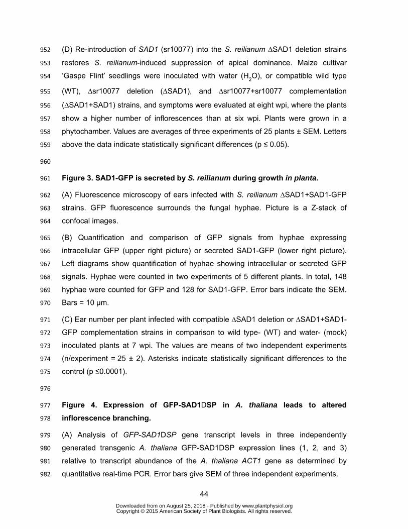
44
(D) Re-introduction of SAD1 (sr10077) into the S. reilianum ΔSAD1 deletion strains 952
restores S. reilianum-induced suppression of apical dominance. Maize cultivar 953
‘Gaspe Flint’ seedlings were inoculated with water (H2O), or compatible wild type 954
(WT), Δsr10077 deletion (ΔSAD1), and Δsr10077+sr10077 complementation 955
(ΔSAD1+SAD1) strains, and symptoms were evaluated at eight wpi, where the plants 956
show a higher number of inflorescences than at six wpi. Plants were grown in a 957
phytochamber. Values are averages of three experiments of 25 plants ± SEM. Letters 958
above the data indicate statistically significant differences (p ≤ 0.05). 959
960
Figure 3. SAD1-GFP is secreted by S. reilianum during growth in planta. 961
(A) Fluorescence microscopy of ears infected with S. reilianum ∆SAD1+SAD1-GFP 962
strains. GFP fluorescence surrounds the fungal hyphae. Picture is a Z-stack of 963
confocal images. 964
(B) Quantification and comparison of GFP signals from hyphae expressing 965
intracellular GFP (upper right picture) or secreted SAD1-GFP (lower right picture). 966
Left diagrams show quantification of hyphae showing intracellular or secreted GFP 967
signals. Hyphae were counted in two experiments of 5 different plants. In total, 148 968
hyphae were counted for GFP and 128 for SAD1-GFP. Error bars indicate the SEM. 969
Bars = 10 µm. 970
(C) Ear number per plant infected with compatible ∆SAD1 deletion or ∆SAD1+SAD1-971
GFP complementation strains in comparison to wild type- (WT) and water- (mock) 972
inoculated plants at 7 wpi. The values are means of two independent experiments 973
(n/experiment = 25 ± 2). Asterisks indicate statistically significant differences to the 974
control (p ≤0.0001). 975
976
Figure 4. Expression of GFP-SAD1DSP in A. thaliana leads to altered 977
inflorescence branching. 978
(A) Analysis of GFP-SAD1DSP gene transcript levels in three independently 979
generated transgenic A. thaliana GFP-SAD1DSP expression lines (1, 2, and 3) 980
relative to transcript abundance of the A. thaliana ACT1 gene as determined by 981
quantitative real-time PCR. Error bars give SEM of three independent experiments. 982
www.plantphysiol.orgon August 25, 2018 - Published by Downloaded from Copyright © 2015 American Society of Plant Biologists. All rights reserved.

45
(B) Phenotype of transgenic A. thaliana expressing GFP-SAD1DSP. A. thaliana Col-0 983
(left panel, Col-0) and transgenic A. thaliana lines (middle and right panels; 1, 2, 3) 984
expressing GFP-SAD1DSP under control of the 35S-promoter eight weeks after 985
sowing. The transgenic lines display more highly branched inflorescences. 986
(C) Schematic model of an A. thaliana plant with illustrated primary (1°) and 987
secondary (2°) cauline- and rosette-leaf branches. 988
(D) Quantification of primary (1°) and secondary (2°) cauline-leaf and rosette-leaf 989
branches of wild type (Col-0) and the three independent GFP-SAD1DSP-expressing 990
A. thaliana lines used in B. Data are averages with error bars indicating the SEM of 991
three experiments of 10 plants each. Asterisks above the error bars indicate 992
statistically significant differences to the control (p < 0.05). 993
994
995
Figure 5. Heterologously expressed GFP-SAD1ΔSP fusion protein accumulates 996
in the nuclei of A. thaliana cells. 997
(A) (Top) Fluorescence signals of plants expressing GFP-SAD1∆SP (top left) or GFP 998
(top right). Red and blue colored lines indicate two examples for regions of 999
fluorescence intensity measurements. Intensity measurements (middle and bottom 1000
panels) show GFP signal accumulation in the nucleus (middle peak) of plants 1001
expressing GFP-SAD1∆SP (bottom left) or GFP (bottom right) along the red (middle) 1002
or the blue (bottom) region of interest indicated in the pictures on top. 1003
(B) Quantification of GFP intensity ratios between nucleus and cytoplasm of leaf cells 1004
from A. thaliana plants expressing GFP-SAD1∆SP or GFP. Cells expressing the 1005
GFP-SAD1∆SP fusion protein show an average signal intensity ratio of nucleus to 1006
cytoplasm of about 3:1. Cells expressing GFP show a ratio of 1:1. N = 35 cells for 1007
GFP-SAD1∆SP; N = 40 cells for GFP. Five different plants were analyzed for each 1008
construct. Error bars show standard deviation. Letters above the data indicate 1009
statistically significant differences (p ≤0.001). Bars = 50 µm. 1010
1011
Figure 6. Microscopic overview of GFP-SAD1ΔSP distribution in transgenic A. 1012
thaliana plants. 1013
www.plantphysiol.orgon August 25, 2018 - Published by Downloaded from Copyright © 2015 American Society of Plant Biologists. All rights reserved.
46
(A) 12-day old A. thaliana plants expressing GFP (left) or GFP-SAD1∆SP (right). 1014
Pictures were taken with an exposure time of 10 ms (left) or 200 ms (right). Plants 1015
expressing GFP show a fluorescence signal in every part of the plant. GFP-1016
SAD1∆SP was not detected in hypocotyls. 1017
(B) Fluorescence microscopic picture of three floral buds (arrows) of A. thaliana 1018
expressing GFP-SAD1∆SP. 1019
(C-E) GFP fluorescence of GFP-SAD1DSP expressed in petal cells of the flower (C), 1020
leaves (D), and root cells (E) of six week-old plants. Fluorescence is observed in the 1021
nucleus and in the cytoplasm. Bars, 100 µm (A,B) and 25 µm (C-E). 1022
1023
Figure 7. Real-time PCR analysis of PIN1 and TB1 transcript abundance. 1024
Gene expression was measured in roots (top panel), ears (middle panel) and stalks 1025
(bottom panel) of maize inoculated with water (Mock) wild-type (WT) and ∆SAD1 1026
(ΔSAD1) deletion strains of S. reilianum. Values given are expression values relative 1027
to mock-inoculated plants and are means of three biological replicates with pools of 1028
three plants per replicate and tissue ± SEM. Asterisk indicates statistical difference to 1029
both mock and ΔSAD1 inoculations within the same tissue (p-value < 0.05). 1030
1031
1032
www.plantphysiol.orgon August 25, 2018 - Published by Downloaded from Copyright © 2015 American Society of Plant Biologists. All rights reserved.
Parsed CitationsAguilar-Martínez JA, Poza-Carrión C, Cubas P (2007) Arabidopsis BRANCHED1 acts as an integrator of branching signals withinaxillary buds. Plant Cell 19: 458-472
Pubmed: Author and TitleCrossRef: Author and TitleGoogle Scholar: Author Only Title Only Author and Title
Altschul SF, Gish W, Miller W, Myers EW, Lipman DJ (1990) Basic local alignment search tool. J MolBiol 215: 403-410Pubmed: Author and TitleCrossRef: Author and TitleGoogle Scholar: Author Only Title Only Author and Title
Barazesh S, McSteen P (2008) Hormonal control of grass inflorescence development. Trends Plant Sci 13: 656-662Pubmed: Author and TitleCrossRef: Author and TitleGoogle Scholar: Author Only Title Only Author and Title
Bennetzen JL, Hake SC (2009) Handbook of maize: its biology. Springer, New YorkPubmed: Author and TitleCrossRef: Author and TitleGoogle Scholar: Author Only Title Only Author and Title
Bomblies K, Doebley JF (2006) Pleiotropic effects of the duplicate maize FLORICAULA/LEAFY genes zfl1 and zfl2 on traits underselection during maize domestication. Genetics 172: 519-531
Pubmed: Author and TitleCrossRef: Author and TitleGoogle Scholar: Author Only Title Only Author and Title
Bos JIB, Armstrong MR, Gilroy EM, Boevink PC, Hein I, Taylor RM, Zhendong T, Engelhardt S, Vetukuri RR, Harrower B (2010)Phytophthora infestans effector AVR3a is essential for virulence and manipulates plant immunity by stabilizing host E3 ligaseCMPG1. Proc Natl Acad Sci USA 107: 9909-9914
Pubmed: Author and TitleCrossRef: Author and TitleGoogle Scholar: Author Only Title Only Author and Title
Brachmann A, König J, Julius C, Feldbrügge M (2004) A reverse genetic approach for generating gene replacement mutants inUstilago maydis. Mol Genet Genom 272: 216-226
Pubmed: Author and TitleCrossRef: Author and TitleGoogle Scholar: Author Only Title Only Author and Title
Brunoud G, Wells DM, Oliva M, Larrieu A, Mirabet V, Burrow AH, Beeckman T, Kepinski S, Traas J, Bennett MJ, Vernoux T (2012) Anovel sensor to map auxin response and distribution at high spatio-temporal resolution. Nature 482: 103-106
Pubmed: Author and TitleCrossRef: Author and TitleGoogle Scholar: Author Only Title Only Author and Title
Carraro N, Forestan C, Canova S, Traas J, Varotto S (2006) ZmPIN1a and ZmPIN1b encode two novel putative candidates for polarauxin transport and plant architecture determination of maize. Plant Physiology 142: 254-264
Pubmed: Author and TitleCrossRef: Author and TitleGoogle Scholar: Author Only Title Only Author and Title
Chen Z, Agnew JL, Cohen JD, He P, Shan L, Sheen J, Kunkel BN (2007) Pseudomonas syringae type III effector AvrRpt2 altersArabidopsis thaliana auxin physiology. Proc Natl Acad Sci USA 104: 20131-20136
Pubmed: Author and TitleCrossRef: Author and TitleGoogle Scholar: Author Only Title Only Author and Title
Cheng X, Ruyter-Spira C, Bouwmeester H (2013) The interaction between strigolactones and other plant hormones in theregulation of plant development. Front Plant Sci. 4: 199.
Pubmed: Author and TitleCrossRef: Author and TitleGoogle Scholar: Author Only Title Only Author and Title
Choi J, Huh SU, Kojima M, Sakakibara H, Paek KH, Hwang I (2010) The cytokinin-activated transcription factor ARR2 promotesplant immunity via TGA3/NPR1-dependent salicylic acid signaling in Arabidopsis. Dev Cell 19: 284-295
Pubmed: Author and TitleCrossRef: Author and TitleGoogle Scholar: Author Only Title Only Author and Title
Cline M, Wesse T, Iwamura H (1997) Cytokinin/auxin control of apical dominance in Ipomoea nil. Plant Cell Physiol 38: 659-667Pubmed: Author and TitleCrossRef: Author and TitleGoogle Scholar: Author Only Title Only Author and Title
Clough SJ, Bent AF (1998) Floral dip: a simplified method for Agrobacterium-mediated transformation of Arabidopsis thaliana. PlantJ 16: 735-743
Pubmed: Author and Title www.plantphysiol.orgon August 25, 2018 - Published by Downloaded from Copyright © 2015 American Society of Plant Biologists. All rights reserved.
CrossRef: Author and TitleGoogle Scholar: Author Only Title Only Author and Title
Coe EH, Neuffer MG, Hoisington DA (1988) The genetics of corn. American Society of Agronomy, MadisonPubmed: Author and TitleCrossRef: Author and TitleGoogle Scholar: Author Only Title Only Author and Title
Crawford S, Shinohara N, Sieberer T, Williamson L, George G, Hepworth J, Müller D, Domagalska MA, Leyser O (2010)Strigolactones enhance competition between shoot branches by dampening auxin transport. Development 137: 2905-2913
Pubmed: Author and TitleCrossRef: Author and TitleGoogle Scholar: Author Only Title Only Author and Title
Davies CR, Seth AK, Wareing PF (1966) Auxin and kinetin interaction in apical dominance. Science 151: 468-469Pubmed: Author and TitleCrossRef: Author and TitleGoogle Scholar: Author Only Title Only Author and Title
Doebley J, Stec A, Hubbard L (1997) The evolution of apical dominance in maize. Nature 386: 485-488Pubmed: Author and TitleCrossRef: Author and TitleGoogle Scholar: Author Only Title Only Author and Title
Domagalska MA, Leyser O (2011) Signal integration in the control of shoot branching. Nature Reviews: Mol Cell Biol 12: 211-221Pubmed: Author and TitleCrossRef: Author and TitleGoogle Scholar: Author Only Title Only Author and Title
Emanuelsson O, Brunak S, Heijne G, Nielsen H (2007) Locating proteins in the cell using TargetP, SignalP, and related toolsNature Protocols 2: 953-971
Pubmed: Author and TitleCrossRef: Author and TitleGoogle Scholar: Author Only Title Only Author and Title
Faure C, Ramos M, Girault JA (2013) Pyk2 cytonuclear localization: mechanisms and regulation by serine dephosphorylation. CellMol Life Sci 70: 137-152
Pubmed: Author and TitleCrossRef: Author and TitleGoogle Scholar: Author Only Title Only Author and Title
Finlayson A, (2007) Arabidopsis TEOSINTE BRANCHED1-LIKE 1 regulates axillary bud outgrowth and is homologous to monocotTEOSINTE BRANCHED1. Plant Cell Physiol 48: 667-677
Pubmed: Author and TitleCrossRef: Author and TitleGoogle Scholar: Author Only Title Only Author and Title
Forestan C, Farinati S, Varotto S (2012) The maize PIN gene family of auxin transporters. Front. Plant Sci. 3Pubmed: Author and TitleCrossRef: Author and TitleGoogle Scholar: Author Only Title Only Author and Title
Forestan C, Meda S, Varotto S (2010) ZmPIN1-mediated auxin transport is related to cellular differentiation during maizeembryogenesis and endosperm development. Plant Physiology 152: 1373-1390
Pubmed: Author and TitleCrossRef: Author and TitleGoogle Scholar: Author Only Title Only Author and Title
Gallavotti A (2013) The role of auxin in shaping shoot architecture. J Exp Bot 64: 2593-2608Pubmed: Author and TitleCrossRef: Author and TitleGoogle Scholar: Author Only Title Only Author and Title
Gallavotti A, Yang Y, Schmidt RJ, Jackson D (2008a) The relationship between auxin transport and maize branching. Plant Physiol147: 1913-1923
Pubmed: Author and TitleCrossRef: Author and TitleGoogle Scholar: Author Only Title Only Author and Title
Gallavotti A, Barazesh S, Malcomber S, Hall D, Jackson D, Schmidt RJ, McSteen P. (2008b) Sparse inflorescence1 encodes amonocot-specific YUCCA-like gene required for vegetative and reproductive development in maize. Proc Natl Acad Sci U S A105:15196-15201
Pubmed: Author and TitleCrossRef: Author and TitleGoogle Scholar: Author Only Title Only Author and Title
Ghareeb H, Becker A, Iven T, Feussner I, Schirawski J (2011) Sporisorium reilianum infection changes inflorescence andbranching architectures of maize. Plant Physiol 156: 2037-2052
Pubmed: Author and TitleCrossRef: Author and TitleGoogle Scholar: Author Only Title Only Author and Title www.plantphysiol.orgon August 25, 2018 - Published by Downloaded from
Copyright © 2015 American Society of Plant Biologists. All rights reserved.
Gillissen B, Bergemann J, Sandmann C, Schroeer B, Bölker M, Kahmann R (1992) A two-component regulatory system for self/non-self recognition in Ustilago maydis. Cell 68: 647-657
Pubmed: Author and TitleCrossRef: Author and TitleGoogle Scholar: Author Only Title Only Author and Title
Hood EE, Gelvin SB, Melchers LS, Hoekema A (1993) New Agrobacterium helper plasmids for gene transfer to plants. TransgenicRes 2: 208-218
Pubmed: Author and TitleCrossRef: Author and TitleGoogle Scholar: Author Only Title Only Author and Title
Hoshi A, Oshima K, Kakizawa S, Ishii Y, Ozeki J, Hashimoto M, Komatsu K, Kagiwada S, Yamaji Y, Namba S (2009) A uniquevirulence factor for proliferation and dwarfism in plants identified from a phytopathogenic bacterium. Proc Natl Acad Sci USA 106:6416-6421
Pubmed: Author and TitleCrossRef: Author and TitleGoogle Scholar: Author Only Title Only Author and Title
Hubbard L, McSteen P, Doebley J, Hake S (2002) Expression patterns and mutant phenotype of teosinte branched1 correlate withgrowth suppression in maize and teosinte. Genetics 162: 1927-1935
Pubmed: Author and TitleCrossRef: Author and TitleGoogle Scholar: Author Only Title Only Author and Title
Kämper J (2004) A PCR-based system for highly efficient generation of gene replacement mutants in Ustilago maydis. Mol GenetGenom 271: 103-110
Pubmed: Author and TitleCrossRef: Author and TitleGoogle Scholar: Author Only Title Only Author and Title
Kämper J, Kahmann R, Bölker M, Ma LJ, Brefort T, Saville BJ, Banuett F, Kronstad JW, Gold SE, Müller O, Perlin MH, Wösten HAB,de Vries R, Ruiz-Herrera J, Reynaga-Pena CG, Snetselaar K, McCann M, Perez-Martin J, Feldbrugge M, Basse CW, Steinberg G,Ibeas JI, Holloman W, Guzman P, Farman M, Stajich JE, Sentandreu R, Gonzalez-Prieto JM, Kennell JC, Molina L, Schirawski J,Mendoza-Mendoza A, Greilinger D, Munch K, Rossel N, Scherer M, Vranes M, Ladendorf O, Vincon V, Fuchs U, Sandrock B, MengS, Ho ECH, Cahill MJ, Boyce KJ, Klose J, Klosterman SJ, Deelstra HJ, Ortiz-Castellanos L, Li WX, Sanchez-Alonso P, Schreier PH,Hauser-Hahn I, Vaupel M, Koopmann E, Friedrich G, Voss H, Schluter T, Margolis J, Platt D, Swimmer C, Gnirke A, Chen F,Vysotskaia V, Mannhaupt G, Guldener U, Munsterkotter M, Haase D, Oesterheld M, Mewes HW, Mauceli EW, DeCaprio D, WadeCM, Butler J, Young S, Jaffe DB, Calvo S, Nusbaum C, Galagan J, Birren BW (2006) Insights from the genome of the biotrophicfungal plant pathogen Ustilago maydis. Nature 444: 97-101
Pubmed: Author and TitleCrossRef: Author and TitleGoogle Scholar: Author Only Title Only Author and Title
MacLean AM, Sugio A, Makarova OV, Findlay KC, Grieve VM, Tóth R, Nicolaisen M, Hogenhout SA (2011) Phytoplasma effectorSAP54 induces indeterminate leaf-like flower development in Arabidopsis plants. Plant Physiol 157: 831-841
Pubmed: Author and TitleCrossRef: Author and TitleGoogle Scholar: Author Only Title Only Author and Title
Malaguti G, Fernandez B, Nass H (1977) Downy mildew or crazy top of maize in Venezuela. Agronomia Tropical 27: 103-129Pubmed: Author and TitleCrossRef: Author and TitleGoogle Scholar: Author Only Title Only Author and Title
Matheussen AM, Morgan PW, Frederiksen RA (1991) Implication of gibberellins in head smut (Sporisorium reilianum) of Sorghumbicolor. Plant Physiol 96: 537-544
Pubmed: Author and TitleCrossRef: Author and TitleGoogle Scholar: Author Only Title Only Author and Title
McSteen P (2009) Hormonal regulation of branching in grasses. Plant Physiol 149: 46-55Pubmed: Author and TitleCrossRef: Author and TitleGoogle Scholar: Author Only Title Only Author and Title
McSteen P, Hake S (2001) barren inflorescence2 regulates axillary meristem development in the maize inflorescence.Development 128: 2881-2891
Pubmed: Author and TitleCrossRef: Author and TitleGoogle Scholar: Author Only Title Only Author and Title
McSteen P, Malcomber S, Skirpan A, Lunde C, Wu X, Kellogg E, Hake S (2007) barren inflorescence2 encodes a co-ortholog of thePINOID serine/threonine kinase and is required for organogenesis during inflorescence and vegetative development in maize.Plant Physiol 144: 1000-1011
Pubmed: Author and TitleCrossRef: Author and TitleGoogle Scholar: Author Only Title Only Author and Title
Meinhardt LW, Rincones J, Bailey BA, Aime MC, Griffith GW, Zhang D, Pereira GAG (2008) Moniliophthora perniciosa, the causal www.plantphysiol.orgon August 25, 2018 - Published by Downloaded from Copyright © 2015 American Society of Plant Biologists. All rights reserved.
agent of witches' broom disease of cacao: what's new from this old foe? Mol Plant Pathol 9: 577-588Pubmed: Author and TitleCrossRef: Author and TitleGoogle Scholar: Author Only Title Only Author and Title
Mukhtar MS, Carvunis AR, Dreze M, Epple P, Steinbrenner J, Moore J, Tasan M, Galli M, Hao T, Nishimura MT, Pevzner SJ,Donovan SE, Ghamsari L, Santhanam B, Romero V, Poulin MM, Gebreab F, Gutierrez BJ, Tam S, Monachello D, Boxem M, HarbortCJ, McDonald N, Gai L, Chen H, He Y, European Union Effectoromics C, Vandenhaute J, Roth FP, Hill DE, Ecker JR, Vidal M,Beynon J, Braun P, Dangl JL (2011) Independently evolved virulence effectors converge onto hubs in a plant immune systemnetwork. Science 333: 596-601
Pubmed: Author and TitleCrossRef: Author and TitleGoogle Scholar: Author Only Title Only Author and Title
Muszynski MG, Dam T, Li B, Shirbroun DM, Hou Z, Bruggemann E, Archibald R, Ananiev EV, Danilevskaya ON (2006) delayedflowering1 Encodes a basic leucine zipper protein that mediates floral inductive signals at the shoot apex in maize. Plant Physiol142:1523-1536
Pubmed: Author and TitleCrossRef: Author and TitleGoogle Scholar: Author Only Title Only Author and Title
Müller D, Leyser O (2011) Auxin, cytokinin and the control of shoot branching. Ann Bot 107: 1203-1212Pubmed: Author and TitleCrossRef: Author and TitleGoogle Scholar: Author Only Title Only Author and Title
Nordström A, Tarkowski P, Tarkowska D, Norbaek R, Astot C, Dolezal K, Sandberg G. (2004) Auxin regulation of cytokininbiosynthesis in Arabidopsis thaliana: a factor of potential importance for auxin-cytokinin-regulated development. Proc Natl AcadSci U S A 101: 8039-8044
Pubmed: Author and TitleCrossRef: Author and TitleGoogle Scholar: Author Only Title Only Author and Title
Navarro L, Dunoyer P, Jay F, Arnold B, Dharmasiri N, Estelle M, Voinnet O, Jones JDG (2006) A plant miRNA contributes toantibacterial resistance by repressing auxin signaling. Science 312: 436-439
Pubmed: Author and TitleCrossRef: Author and TitleGoogle Scholar: Author Only Title Only Author and Title
Petersen TN, Brunak S, von Heijne G, Nielsen H (2011) SignalP 4.0: discriminating signal peptides from transmembrane regions.Nature Methods 8: 785-786
Pubmed: Author and TitleCrossRef: Author and TitleGoogle Scholar: Author Only Title Only Author and Title
Phillips KA, Skirpan AL, Liu X, Christensen A, Slewinski TL, Hudson C, Barazesh S, Cohen JD, Malcomber S, McSteen P (2011)Vanishing tassel2 encodes a grass-specific tryptophan aminotransferase required for vegetative and reproductive developmentin maize. Plant Cell 23: 550-566
Pubmed: Author and TitleCrossRef: Author and TitleGoogle Scholar: Author Only Title Only Author and Title
Plotnikov A, Chuderland D, Karamansha Y, Livnah O, Seger R (2011) Nuclear extracellular signal-regulated kinase 1 and 2translocation is mediated by casein kinase 2 and accelerated by autophosphorylation. Mol Cell Biol 31: 3515-3530
Pubmed: Author and TitleCrossRef: Author and TitleGoogle Scholar: Author Only Title Only Author and Title
Prusinkiewicz P, Crawford S, Smith RS, Ljung K, Bennett T, Ongaro V, Leyser O (2009) Control of bud activation by an auxintransport switch. Proc Natl Acad Sci USA 106: 17431-17436
Pubmed: Author and TitleCrossRef: Author and TitleGoogle Scholar: Author Only Title Only Author and Title
Reineke G, Heinze B, Schirawski J, Buettner H, Kahmann R, Basse CW (2008) Indole-3-acetic acid (IAA) biosynthesis in the smutfungus Ustilago maydis and its relevance for increased IAA levels in infected tissue and host tumour formation. Mol Plant Pathol 9:339-355
Pubmed: Author and TitleCrossRef: Author and TitleGoogle Scholar: Author Only Title Only Author and Title
Robert-Seilaniantz A, Grant M, Jones JDG (2011) Hormone crosstalk in plant disease and defense: more than just jasmonate-salicylate antagonism. Ann Rev Phytopathol 49: 317-343
Pubmed: Author and TitleCrossRef: Author and TitleGoogle Scholar: Author Only Title Only Author and Title
Sachs T (1981) The control of the patterned differentiation of vascular tissues. Adv Bot Res 9: 151-262Pubmed: Author and TitleCrossRef: Author and Title www.plantphysiol.orgon August 25, 2018 - Published by Downloaded from
Copyright © 2015 American Society of Plant Biologists. All rights reserved.
Google Scholar: Author Only Title Only Author and Title
Sachs T, Thimann KV (1967) The role of auxins and cytokinins in the release of buds from dominance. Am J Bot 54: 136-144Pubmed: Author and TitleCrossRef: Author and TitleGoogle Scholar: Author Only Title Only Author and Title
Schirawski J, Heinze B, Wagenknecht M, Kahmann R (2005) Mating type loci of Sporisorium reilianum: novel pattern with three aand multiple b specificities. Eukaryotic Cell 4: 1317-1327
Pubmed: Author and TitleCrossRef: Author and TitleGoogle Scholar: Author Only Title Only Author and Title
Schirawski J, Mannhaupt G, Münch K, Brefort T, Schipper K, Doehlemann G, Di Stasio M, Rössel N, Mendoza-Mendoza A, PesterD, Müller O, Winterberg B, Meyer E, Ghareeb H, Wollenberg T, Münsterkötter M, Wong P, Walter M, Stukenbrock E, Güldener U,Kahmann R (2010) Pathogenicity determinants in smut fungi revealed by genome comparison. Science 330: 1546-1548
Pubmed: Author and TitleCrossRef: Author and TitleGoogle Scholar: Author Only Title Only Author and Title
Schulz B, Banuett F, Dahl M, Schlesinger R, Schäfer W, Martin T, Herskowitz I, Kahmann R (1990) The b alleles of U. maydis,whose combinations program pathogenic development, code for polypeptides containing a homeodomain-related motif. Cell 60:295-306
Pubmed: Author and TitleCrossRef: Author and TitleGoogle Scholar: Author Only Title Only Author and Title
Shani E, Yanai O, Ori N (2006) The role of hormones in shoot apical meristem function. Curr Opin Plant Biol 9: 484-489Pubmed: Author and TitleCrossRef: Author and TitleGoogle Scholar: Author Only Title Only Author and Title
Sheridan WF (1988) Maize developmental genetics: genes of morphogenesis. Ann Rev Genet 22: 353-385Pubmed: Author and TitleCrossRef: Author and TitleGoogle Scholar: Author Only Title Only Author and Title
Shinohara N, Taylor C, Leyser O (2013) Strigolactone can promote or inhibit shoot branching by triggering rapid depletion of theauxin efflux protein PIN1 from the plasma membrane. PLoS Biology 11: e1001474
Pubmed: Author and TitleCrossRef: Author and TitleGoogle Scholar: Author Only Title Only Author and Title
Skibbe DS, Doehlemann G, Fernandes J, Walbot V (2010) Maize tumors caused by Ustilago maydis require organ-specific genes inhost and pathogen. Science 328: 89-92
Pubmed: Author and TitleCrossRef: Author and TitleGoogle Scholar: Author Only Title Only Author and Title
Spellig T, Bottin A, Kahmann R (1996) Green fluorescent protein (GFP) as a new vital marker in the phytopathogenic fungusUstilago maydis. Mol Gen Genet 252: 503-509
Pubmed: Author and TitleCrossRef: Author and TitleGoogle Scholar: Author Only Title Only Author and Title
Studer A, Zhao Q, Ross-Ibarra J, Doebley J (2011) Identification of a functional transposon insertion in the maize domesticationgene tb1. Nature Genetics 43: 1160-1163
Pubmed: Author and TitleCrossRef: Author and TitleGoogle Scholar: Author Only Title Only Author and Title
Tanaka M, Takei K, Kojima M, Sakakibara H, Mori H (2006) Auxin controls local cytokinin biosynthesis in the nodal stem in apicaldominance. Plant J. 45: 1028-1036
Pubmed: Author and TitleCrossRef: Author and TitleGoogle Scholar: Author Only Title Only Author and Title
Turnbull CG, Booker JP, Leyser HM (2002) Micrografting techniques for testing long-distance signalling in Arabidopsis. Plant J. 32:255-262
Pubmed: Author and TitleCrossRef: Author and TitleGoogle Scholar: Author Only Title Only Author and Title
Wang Q, Kohlen W, Rossmann S, Vernoux T, Theres K (2014) Auxin Depletion from the Leaf Axil Conditions Competence forAxillary Meristem Formation in Arabidopsis and Tomato. Plant Cell 26: 2068-2079
Pubmed: Author and TitleCrossRef: Author and TitleGoogle Scholar: Author Only Title Only Author and Title
Wei W, Davis RE, Nuss DL, Zhao Y (2013) Phytoplasmal infection derails genetically preprogrammed meristem fate and alters plantarchitecture. Proc Natl Acad Sci USA 111: 19149-19154 www.plantphysiol.orgon August 25, 2018 - Published by Downloaded from
Copyright © 2015 American Society of Plant Biologists. All rights reserved.
Pubmed: Author and TitleCrossRef: Author and TitleGoogle Scholar: Author Only Title Only Author and Title
Yan Y, Christensen S, Isakeit T, Engelberth J, Meeley R, Hayward A, Emery RJN, Kolomiets MV (2012) Disruption of OPR7 andOPR8 reveals the versatile functions of jasmonic acid in maize development and defense. Plant Cell 24: 1420-1436
Pubmed: Author and TitleCrossRef: Author and TitleGoogle Scholar: Author Only Title Only Author and Title
Yao C, Wu Y, Nie H, Tang D (2012) RPN1a, a 26S proteasome subunit, is required for innate immunity in Arabidopsis. Plant J 71:1015-1028
Pubmed: Author and TitleCrossRef: Author and TitleGoogle Scholar: Author Only Title Only Author and Title
Zuther K, Kahnt J, Utermark J, Imkampe J, Uhse S, Schirawski J (2012) Host specificity of Sporisorium reilianum is tightly linked togeneration of the phytoalexin luteolinidin by Sorghum bicolor. Mol Plant-Microbe Interact 25: 1230-1237
Pubmed: Author and TitleCrossRef: Author and TitleGoogle Scholar: Author Only Title Only Author and Title
www.plantphysiol.orgon August 25, 2018 - Published by Downloaded from Copyright © 2015 American Society of Plant Biologists. All rights reserved.