Embed Size (px)
Citation preview

Inflammatory Disorders
By Nancy Jenkins
Updated Spring 2010 by John Nation -
Overview of Today’s Lecture
A & P Review Endocarditis- infection of the endocardial surface
of the heart
Myocarditis- a focal or diffuse inflammation of the myocardium
Pericarditis- inflammation of the pericardial sac (the pericardium)
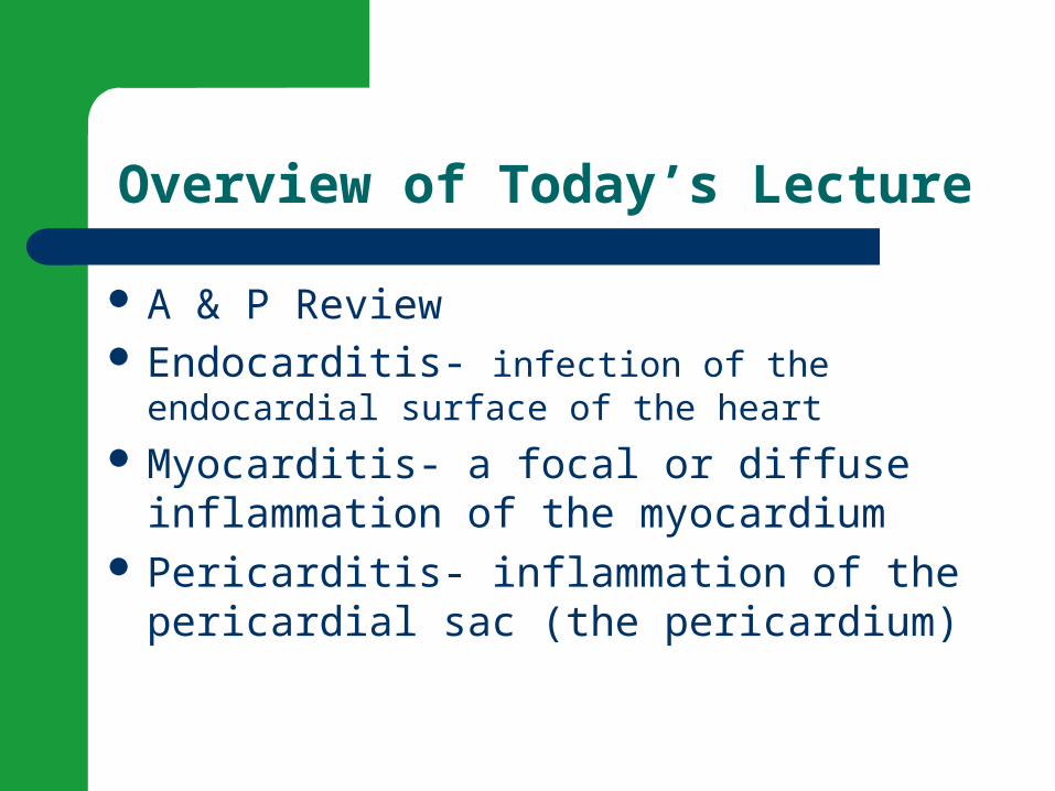
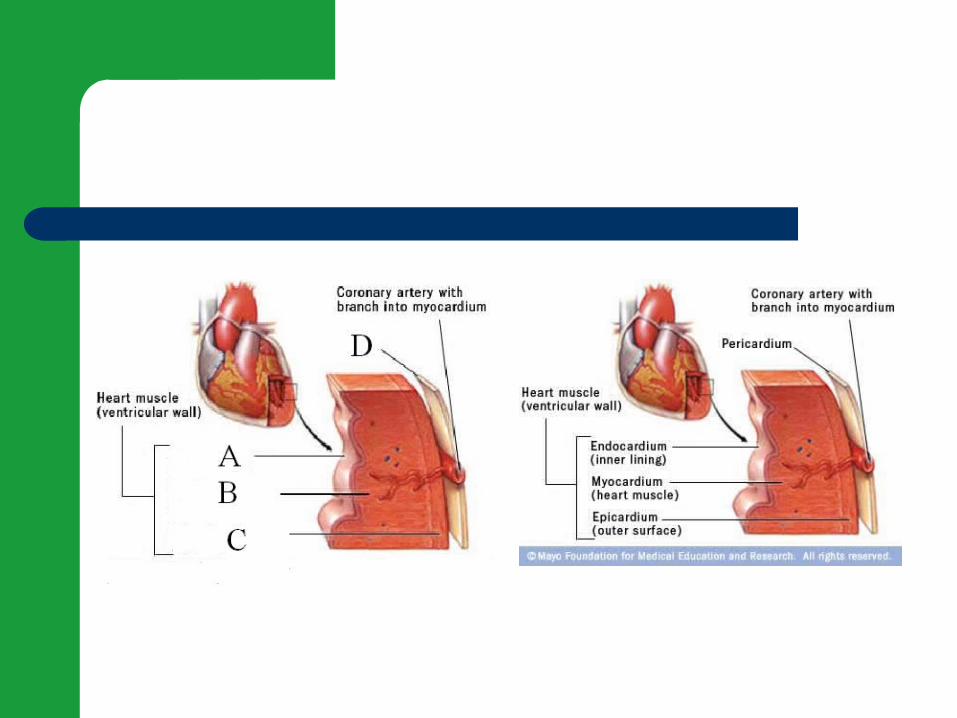
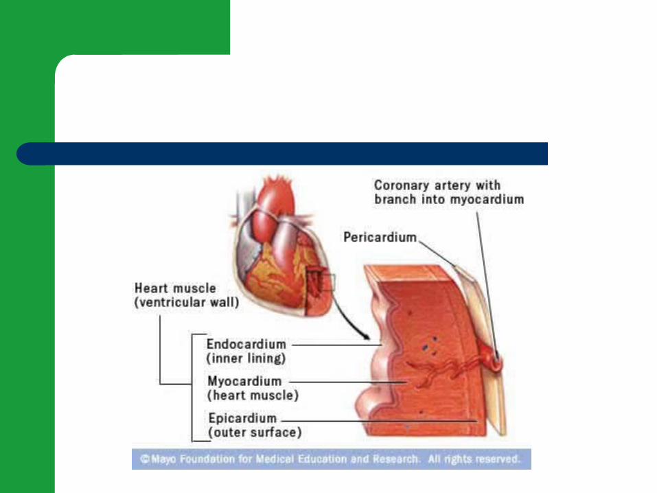
Layers of the Heart Muscle
Anatomy and Physiology!
How the heart works!
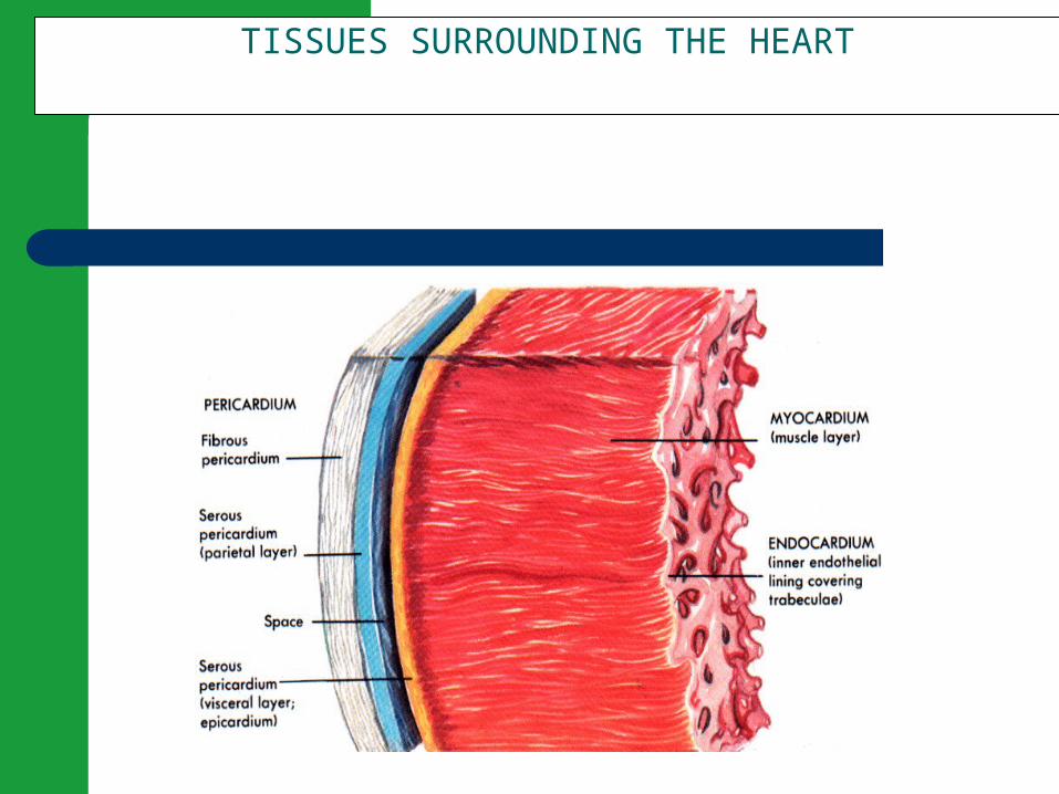
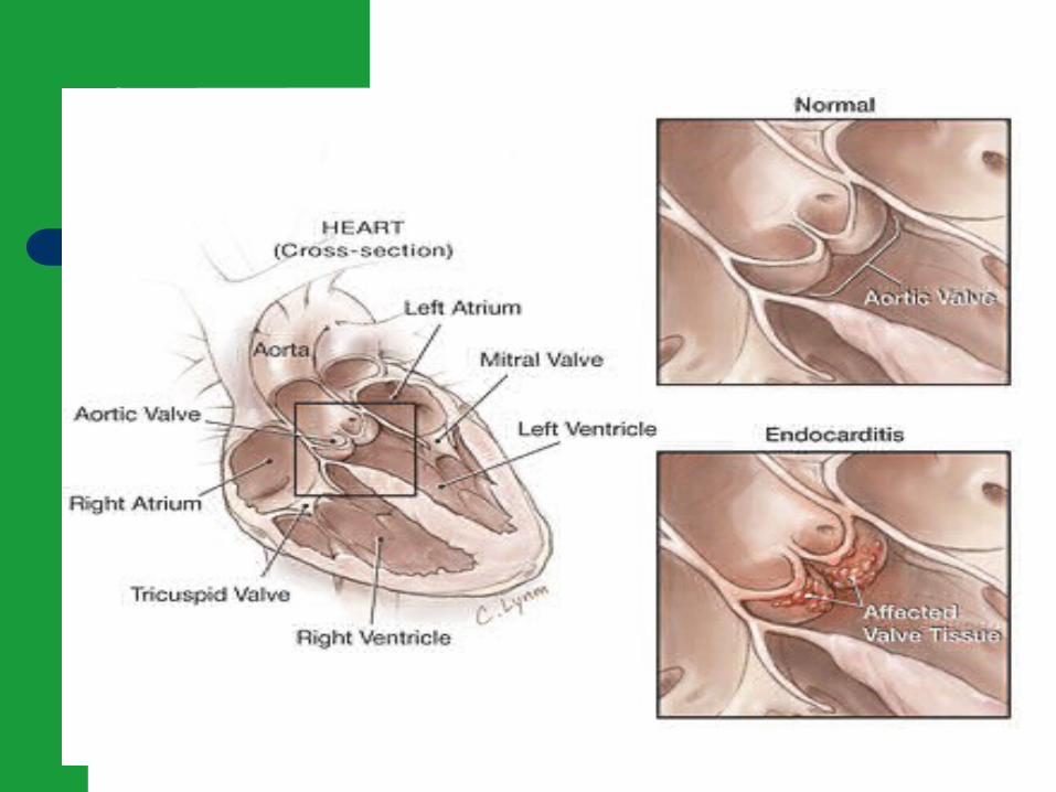
TISSUES SURROUNDING THE HEART

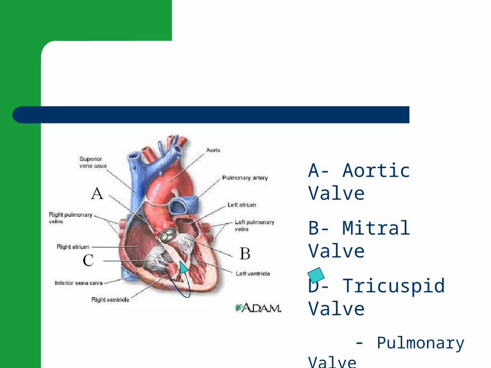
A- Aortic Valve
B- Mitral Valve
D- Tricuspid Valve
- Pulmonary Valve
Anatomy and Physiology Review
Blood enters the right atrium and moves through the _______ into the right ventricle.
Blood then moves from the right ventricle into the pulmonary artery via the _________.
A- Aortic Valve
B- Mitral Valve
C- Pulmonary Valve
D- Tricuspid Valve
Anatamy and Physiology Review (Cont’d)
After entering the left atrium via the pulmonary veins, blood moves through the _____ into the left ventricle.
Finally, it travels through the _____ and out of the heart.
A- Aortic Valve
B- Mitral Valve
C- Pulmonary Valve
D- Tricuspid Valve
Infective Endocarditis
• Infection of the inner layer of the heart
• Usually affects the cardiac valves
• Was almost always fatal until
development of penicillin
• Around 15,000 cases diagnosed
annually in the U.S.
Causative Organisms
Causative organisms Streptococcus viridans Staphylococcus aureus Viruses Fungi
Etiology and Pathophysiology
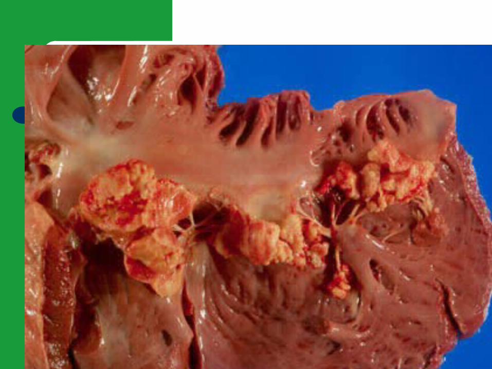
Vegetation – Fibrin, leukocytes, platelets, and microbes– Adhere to the valve or endocardium – Embolization of portions of vegetation into
circulation
Etiology and Pathophysiology
Occurs when blood turbulence within heart allows causative agent to infect previously damaged valves or other endothelial surfaces

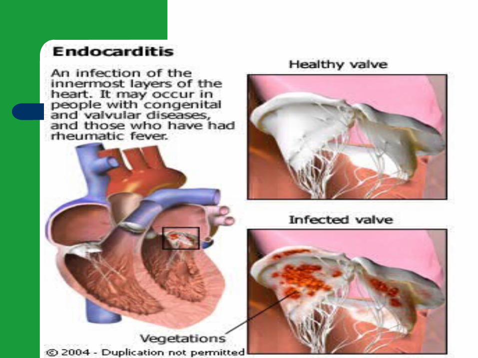
Endocarditis
Infection of the innermost layers of the heart May occur in people with congenital and
valvular heart disease May occur in people with a history of
rheumatic heart disease May occur in people with normal valves with
increased amounts of bacteria
Etiology/Pathophysiology
Endocarditis– When valve damaged, blood is slowed down and
forms a clot– Bacteria get into blood stream – Bacterial or fungal vegetative growths deposit on
normal or abnormal heart valves
Classifications of Endocarditis
Acute Infective Endocarditis– Abrupt onset– Rapid course– Staph Aureus
Subacute Infective Endocarditis SBE– Gradual onset– Systemic manifestations
Prosthetic Valve Endocarditis
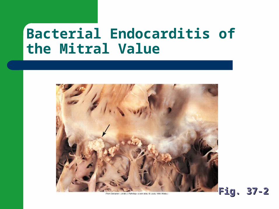
Bacterial Endocarditis of the Mitral Value
Fig. 37-2Fig. 37-2

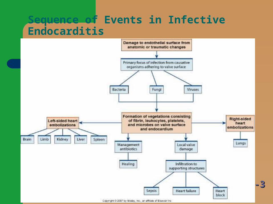
Sequence of Events in Infective Endocarditis
Fig. 37-3Fig. 37-3
Risk Factors- endocarditis
Hx of rheumatic fever or damaged heart valve Prior history of endocarditis Invasive procedures- (introduce bacteria into blood
stream) (surgery, dental, etc) Recent Dental Surgery Permanent Central Venous Access IV drug users Valve replacements
Nursing Assessment
Subjective Data– History of valvular, congenital, or syphilitic cardiac
disease– Previous endocarditis – Staph or strep infection– Immunosuppressive therapy– Recent surgeries and procedures
Nursing Assessment
Functional health patterns– IV drug abuse– Alcohol abuse– Weight changes– Chills

Nursing Assessment
– Diaphoresis– Bloody urine– Exercise intolerance – Generalized weakness– Fatigue – Cough
Nursing Assessment
– Dyspnea on exertion – Night sweats – Chest, back, abdominal pain

Collaborative Care
Fungal and prosthetic valve endocarditis– Responds poorly to antibiotics– Valve replacement is adjunct procedure
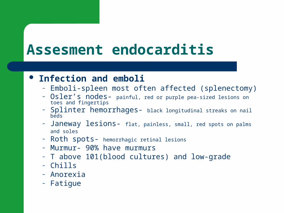
Assesment endocarditis
Infection and emboli– Emboli-spleen most often affected (splenectomy)– Osler’s nodes- painful, red or purple pea-sized lesions on toes and fingertips
– Splinter hemorrhages- black longitudinal streaks on nail beds
– Janeway lesions- flat, painless, small, red spots on palms and soles
– Roth spots- hemorrhagic retinal lesions
– Murmur- 90% have murmurs– T above 101(blood cultures) and low-grade– Chills– Anorexia– Fatigue
Clinical Manifestations
Murmur in most patients Heart failure in up to 80% with aortic valve
endocarditis Manifestations secondary to embolism
Heart Sounds Assessment Video

Auscultating Heart Sounds
The aortic area or right sternal border (RSB) is at the right 2nd intercostal space, just under and to the right of the angle of Louis (sternal angle)
The pulmonic area or left upper sternal border (LUSB) is at the left 2 nd intercostal space
The tricuspid area or left lower sternal border (LLSB) is at the left fourth intercostal space
The mitral area or apex is at the PMI -- the 5 th intercostal space in midclavicular line

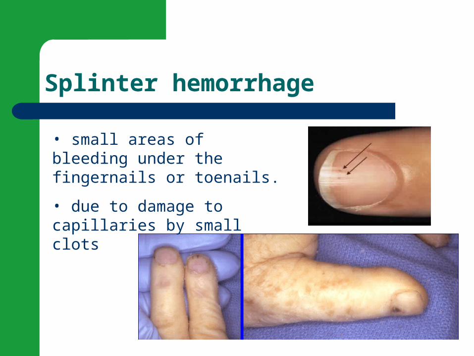
Splinter hemorrhage
• small areas of bleeding under the fingernails or toenails.
• due to damage to capillaries by small clots
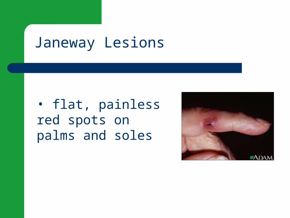
Janeway Lesions
• flat, painless red spots on palms and soles
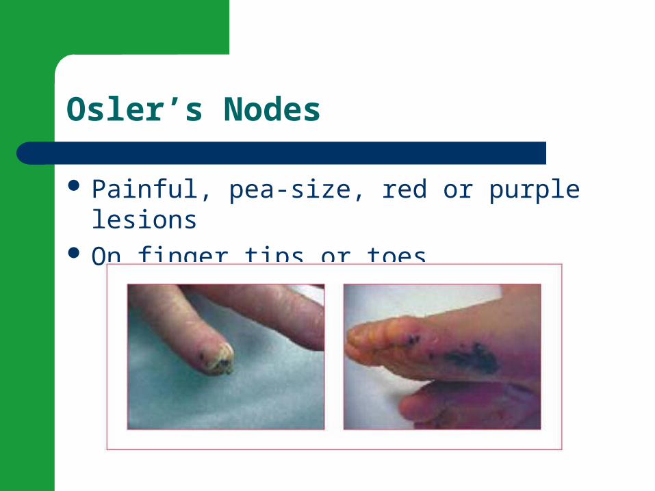
Osler’s Nodes
Painful, pea-size, red or purple lesions On finger tips or toes
Roth spotsOsler’s nodes
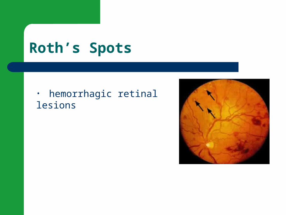
Roth’s Spots
• hemorrhagic retinal lesions
Diagnostic Tests
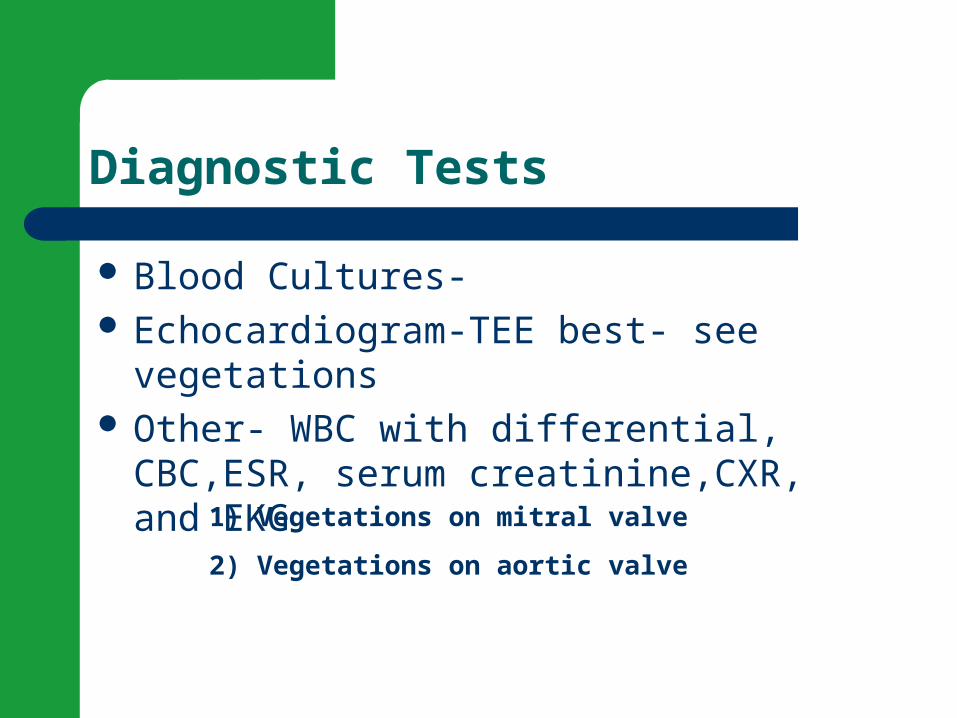
Blood Cultures- Echocardiogram-TEE best- see vegetations Other- WBC with differential, CBC,ESR,
serum creatinine,CXR, and EKG
1) Vegetations on mitral valve
2) Vegetations on aortic valve
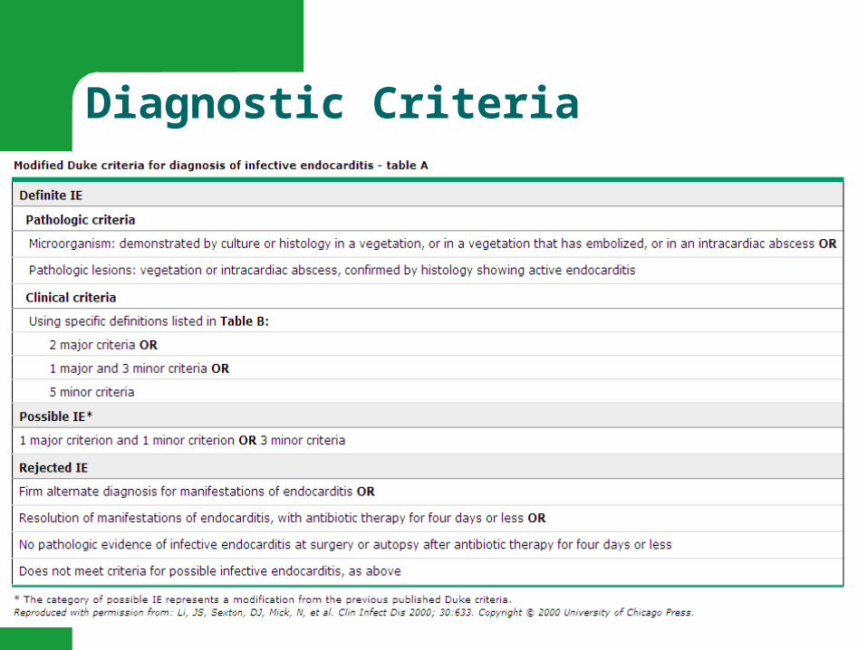
Diagnostic Criteria
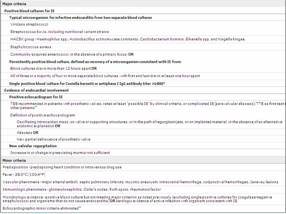
Diagnostic Criteria
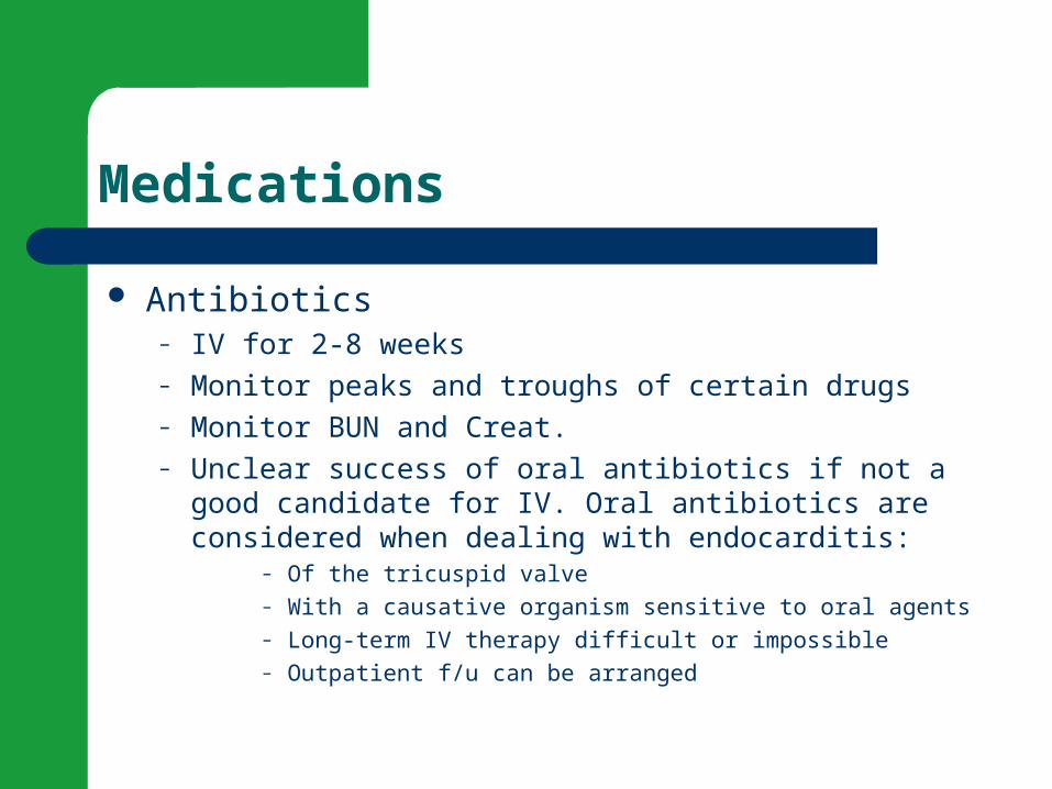
Medications
Antibiotics– IV for 2-8 weeks– Monitor peaks and troughs of certain drugs– Monitor BUN and Creat.– Unclear success of oral antibiotics if not a good candidate
for IV. Oral antibiotics are considered when dealing with endocarditis:
– Of the tricuspid valve– With a causative organism sensitive to oral agents– Long-term IV therapy difficult or impossible– Outpatient f/u can be arranged
Nursing Diagnoses
Risk for Imbalanced Body Temperature Risk for Ineffective Tissue Perfusion-emboli Ineffective Health Maintenance
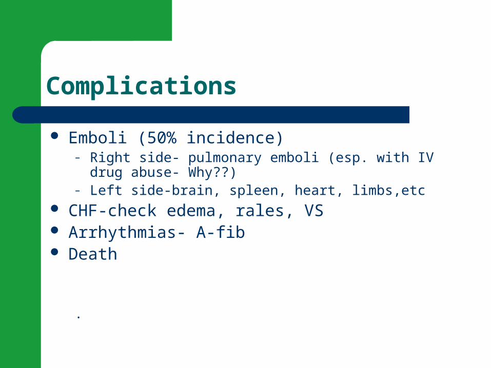
Complications
Emboli (50% incidence)– Right side- pulmonary emboli (esp. with IV drug abuse-
Why??)– Left side-brain, spleen, heart, limbs,etc
CHF-check edema, rales, VS Arrhythmias- A-fib Death
.
Prevention
Eliminate risk factors Patient teaching
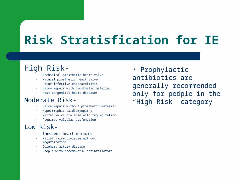
Risk Stratisfication for IE
High Risk- – Mechanical prosthetic heart valve– Natural prosthetic heart valve– Prior infective endocardititis– Valve repair with prosthetic material– Most congenital heart diseases
Moderate Risk- – Valve repair without prosthetic material– Hypertrophic cardiomyopathy– Mitral valve prolapse with regurgitation– Acquired valvular dysfunction
Low Risk-– Innocent heart murmurs– Mitral valve prolapse without regurgitation– Coronary artery disease– People with pacemakers/ defibrillators
• Prophylactic antibiotics are generally recommended only for people in the “High Risk” category
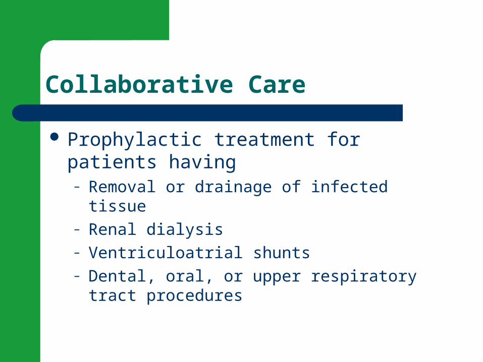
Collaborative Care
Prophylactic treatment for patients having– Removal or drainage of infected tissue– Renal dialysis– Ventriculoatrial shunts– Dental, oral, or upper respiratory tract procedures

Video Review- Endocarditis
Livestrong Endocarditis Video
To diagnose the causative organism in endocarditis, the nurse
should anticipate the doctor ordering which test?
Ches
t x-ra
y
Ech
ocard
iogra
m
Blo
od cultu
res
CBC
25% 25%25%25%
1. Chest x-ray
2. Echocardiogram
3. Blood cultures
4. CBC
Which assessment finding is characteristic of endocarditis?
Per
ipher
al e
dema
Jau
ndice
Bra
dycar
dia
Hea
rt M
urmur
25% 25%25%25%
1. Peripheral edema
2. Jaundice
3. Bradycardia
4. Heart Murmur

A common complication of endocarditis of the mitral valve is pulmonary embolism.
Tru
e
Fal
se
50%50%
1. True
2. False

Layers of the Heart Muscle
Myocarditis
Myocarditis is an uncommon inflammation of the heart muscle (myocardium). This inflammation can be caused by infectious agents, toxins, drugs or for unknown reasons. It may be localized to one area of the heart, or it may affect the entire heart.
Etiology/Pathophysiology
Myocarditis– Virus, toxin or autoimmune response causes necrosis of the
myocardium– Most often caused by viral infection– Frequently caused by Coxsackie B virus– Frequently follows an upper respiratory infection or viral
illness– Get decreased contractility– Can become chronic and lead to dilated cardiomyopathy-
heart transplant or death
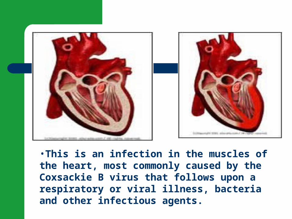
•This is an infection in the muscles of the heart, most commonly caused by the Coxsackie B virus that follows upon a respiratory or viral illness, bacteria and other infectious agents.
Risk factor-myocarditis
Hx of upper respiratory infection Toxic or chemical effects (radiation, alcohol) Autoimmune or immunosuppresents- 10%
HIV develop it Metabolic-lupus Heat stroke or hypothermia
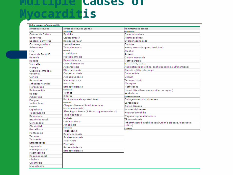
Multiple Causes of Myocarditis
Assessment myocarditis
Infection and CHF– Fatigue,DOE– Tachycardia– Arrhythmias- PVCs, PACs, Atrial Tachycardias, – Chest pain– Signs of heart failure (S3, etc.) – Pericarditis frequently occurs with myocarditis-
check friction rub
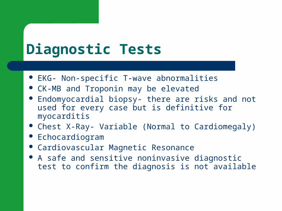
Diagnostic Tests
EKG- Non-specific T-wave abnormalities CK-MB and Troponin may be elevated Endomyocardial biopsy- there are risks and not used
for every case but is definitive for myocarditis Chest X-Ray- Variable (Normal to Cardiomegaly) Echocardiogram Cardiovascular Magnetic Resonance A safe and sensitive noninvasive diagnostic test to
confirm the diagnosis is not available

Chest X-Ray in Myocarditis

MRI in Acute Myocarditis
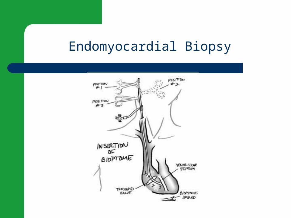
Endomyocardial Biopsy
Biopsy Video
Heart Biopsy Video
Possible Medications
Antibiotics (rarely) Antiviral with interferon-a IVIG- experimental trials Corticosteroids or immunosuppressents HF drugs- ACE, diuretics, beta blockers etc Antiarrhythmics Anticoagulants- Why??
Other Treatments
Bedrest and activity restrictions- Why important??
**Activities may be limited for 6 months- 1 yr. O2
GOAL- Decrease workload of the heart so it can heal
Nursing Diagnoses
Activity Intolerance Decreased CO Anxiety Excess fluid Volume

Your client with myocarditis develops heart failure; his BP is 80/40; (MAP is 53.3).Which of the following medications should the nurse anticipate administering to improve myocardial contractility?
Lis
inop
ril
Las
ix IV
Dobut
amin
e IV
Am
iodar
one IV
25% 25%25%25%
MAP = Systolic BP + 2 (Diastolic BP)
divided by 3
1. Lisinopril
2. Lasix IV
3. Dobutamine IV
4. Amiodarone IV
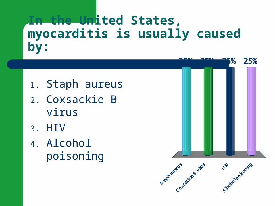
In the United States, myocarditis is usually caused by:
Sta
ph aure
us
Coxs
acki
e B v
irus
HIV
Alc
ohol pois
oning
25% 25%25%25%
1. Staph aureus
2. Coxsackie B virus
3. HIV
4. Alcohol poisoning

Break!
!
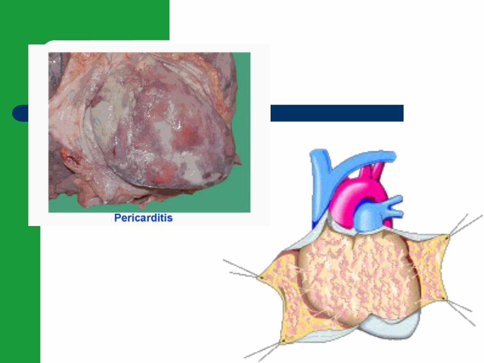
Pericarditis
Pericarditis is an inflammation of the pericardium, the thin, fluid-filled sac surrounding the heart. It can cause severe chest pain (especially upon taking a deep breath) and shortness of breath.

Etiology/Pathophysiology
Pericarditis– bacterial, fungal or viral infection– Heart loses natural lubrication(10-30cc’s) and
layers roughen and rub– Inflammatory process causes lymphatic fluid
build-up- if sudden may have cardiac tamponade– Pericardial Effusion- usually 250 cc’s before show
up on x-ray. Can have 1000cc’s.
Risk Factors/pericarditis
Post MI (Dressler’s syndrome) Radiation Infection Trauma Cancer Drugs and toxins Rheumatic diseases Trauma or cardiac surgery Can be chronic disorder-pericardium becomes rigid
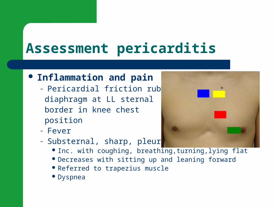
Assessment pericarditis
Inflammation and pain– Pericardial friction rub- diaphragm at LL sternal border in knee chest position – Fever– Substernal, sharp, pleuritic chest pain
Inc. with coughing, breathing,turning,lying flat Decreases with sitting up and leaning forward Referred to trapezius muscle Dyspnea
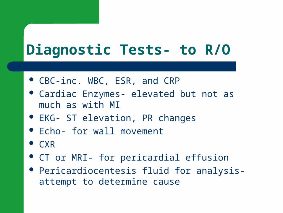
Diagnostic Tests- to R/O
CBC-inc. WBC, ESR, and CRP Cardiac Enzymes- elevated but not as much as with
MI EKG- ST elevation, PR changes Echo- for wall movement CXR CT or MRI- for pericardial effusion Pericardiocentesis fluid for analysis- attempt to
determine cause
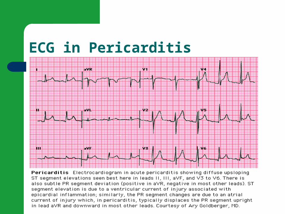
ECG in Pericarditis

Medications
ASA or tylenol NSAIDS- ibuprofen Corticosteroids
Pericarditis Video Review
Livestrong Pericarditis Video

Complications of Pericarditis
Pericardial Effusion- an accumulation of excess fluid in the pericardium
Cardiac Tamponade- an increase in intracardial pressure caused by pericardial effusion that results in compession of the heart
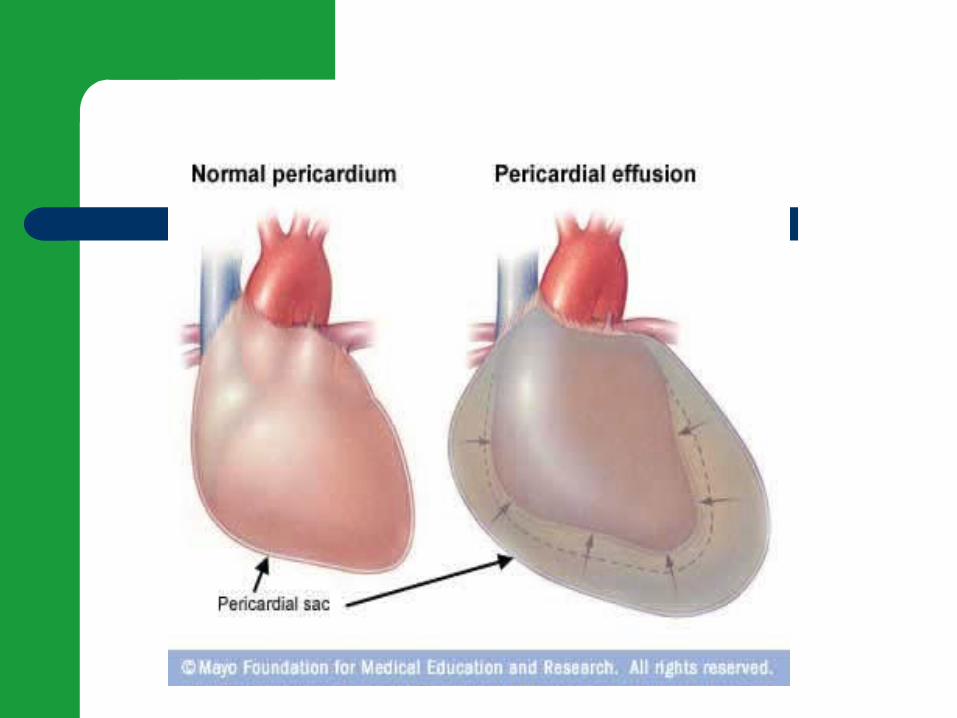
Pericardial Effusion
Can occur rapidly or slowly Pulmonary compression-cough, dyspnea,
and tachypnea Phrenic nerve involvement- hiccups Laryngeal nerve- hoarseness
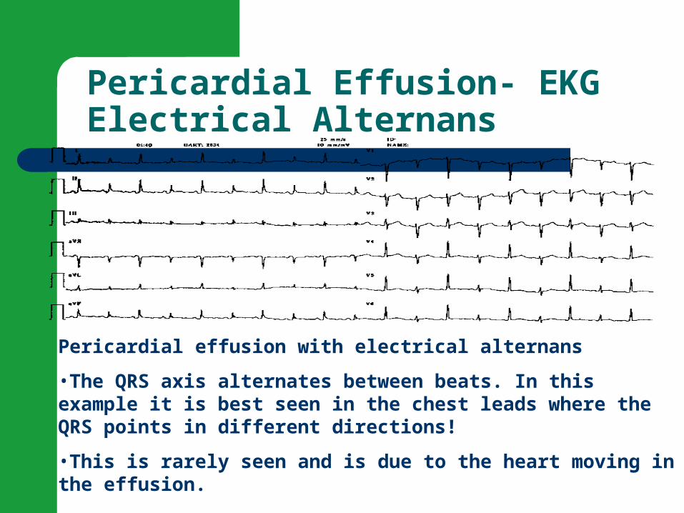
Pericardial Effusion- EKGElectrical Alternans
Pericardial effusion with electrical alternans
•The QRS axis alternates between beats. In this example it is best seen in the chest leads where the QRS points in different directions!
•This is rarely seen and is due to the heart moving in the effusion.

Cardiac Tamponade
Compression of the heart Can occur acutely (trauma) or sub-acutely
(malignancy) Symptoms- chest pain, confusion, anxious and
restless Later- tachypnea, tachycardia, and decreased CO,
NVD (neck vein distention) and pulsus paradoxus present
With slow onset dyspnea may be only symptom
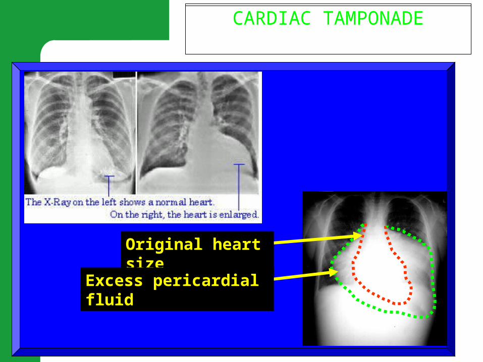
PERICARDIUMCARDIAC TAMPONADE
Original heart size
Excess pericardial fluid

Cardiac tamponade
Physiology- Paradoxical pulse is a pulse that markedly decreases in amplitude during inspiration. On inspiration, more blood is pooled in the lungs and so decreases the return to the left side of the heart; this affects the consequent stroke volume.
Definition- a decrease in systolic BP with inspirations that is exaggerated in cardiac tamponade
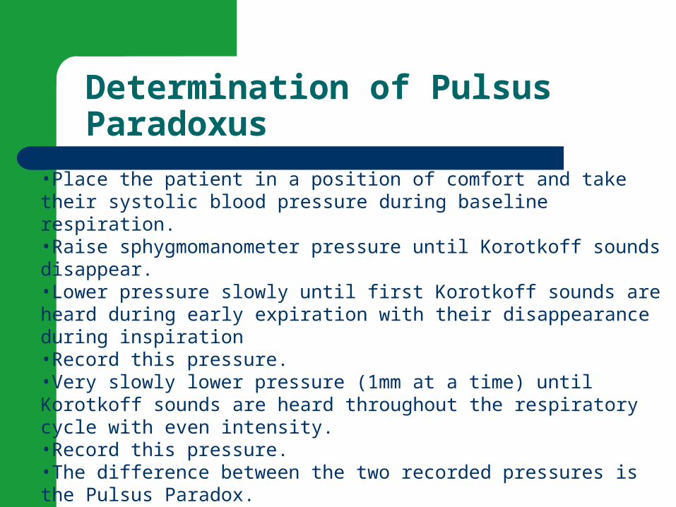
Determination of Pulsus Paradoxus
•Place the patient in a position of comfort and take their systolic blood pressure during baseline respiration. •Raise sphygmomanometer pressure until Korotkoff sounds disappear. •Lower pressure slowly until first Korotkoff sounds are heard during early expiration with their disappearance during inspiration •Record this pressure. •Very slowly lower pressure (1mm at a time) until Korotkoff sounds are heard throughout the respiratory cycle with even intensity. •Record this pressure. •The difference between the two recorded pressures is the Pulsus Paradox. •Hemodynamically significant pulsus paradox is greater than or equal to 10 but we look at trends. People with COPD may have a paradox due to increased thoracic pressures.
Surgical/invasive Interventions
Pericardiocentesis– Hook needle to V lead- guided by EKG and echo– Look for ST elevation– Withdraw fluid– Afterward watch for cardiac tamponade (PP), arrhythmias,
and pneumothorax
Pericardiectomy Pericardial window Sclerosing agent- tetracycline (Bonds layers
together)

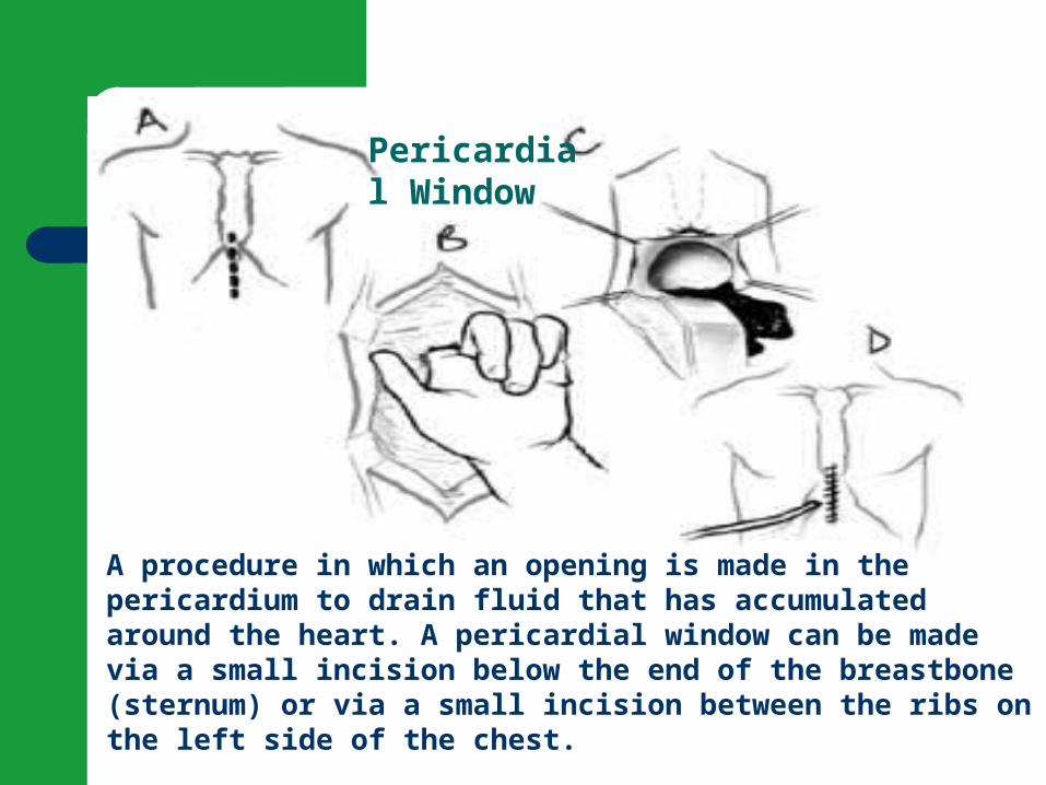
A procedure in which an opening is made in the pericardium to drain fluid that has accumulated around the heart. A pericardial window can be made via a small incision below the end of the breastbone (sternum) or via a small incision between the ribs on the left side of the chest.
Pericardial Window

Cardiac Tamponade and treatment
You tube- Cardiac Tamponade
Chronic Constrictive Pericarditis
Starts with acute then scarring and fibrosis occur
See signs of HF and cor pulmonale; most relate to decreased cardiac output
Most prominent finding is jugular vein distention (JVD)
Treatment of choice pericardectomy- with use of cardiopulmonary bypass
Nursing Diagnoses for Pericarditis
Acute Pain Ineffective Breathing Pattern Risk for Decreased Cardiac Output Activity Intolerance
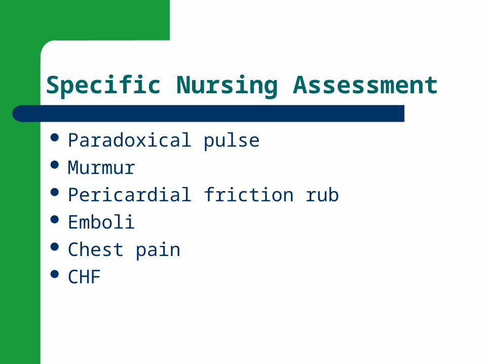
Specific Nursing Assessment
Paradoxical pulse Murmur Pericardial friction rub Emboli Chest pain CHF

Comfort Measures
O2 Bedrest Positioning Prevent complications of immobility Psychological support
Case study
http://www.austincc.edu/adnlev4/rnsg2331online/module06/mr_a_case_study.htm

Case Studies
http://intmedweb.wfubmc.edu/grand_rounds/1999/tamponade.html - CASE%20PRESENTATION
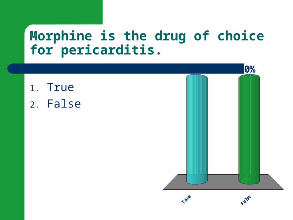
Morphine is the drug of choice for pericarditis.
Tru
e
Fal
se
50%50%
1. True
2. False

You have just received change of shift report about these clients on the coronary step-down unit. Which one will you assess first?
1. A 26 year old with heart failure caused by congenital mitral stenosis who is scheduled for balloon valvuloplasty later today.
2. A 45 year old with constrictive cardiomyopathy who has developed acute dyspnea and agitation.
3. A 56 year old who had a coronary angioplasty and stent placement yesterday and has complained of occasional chest pain since the procedure.
4. A 77 year old who transferred from intensive care 2 days ago after coronary artery bypass grafting and has a temperature of 100.6F.
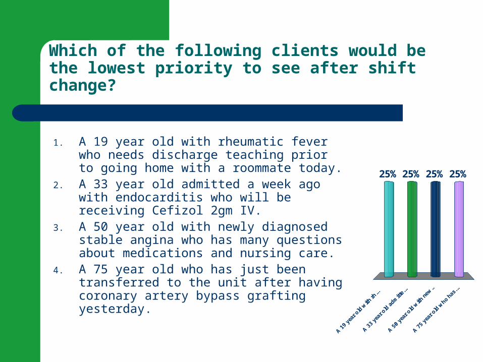
Which of the following clients would be the lowest priority to see after shift change?
A 1
9 ye
ar o
ld w
ith rh
...
A 3
3 ye
ar o
ld a
dmitt
e...
A 5
0 ye
ar o
ld w
ith n
ew..
A 7
5 ye
ar o
ld w
ho ha
s...
25% 25%25%25%
1. A 19 year old with rheumatic fever who needs discharge teaching prior to going home with a roommate today.
2. A 33 year old admitted a week ago with endocarditis who will be receiving Cefizol 2gm IV.
3. A 50 year old with newly diagnosed stable angina who has many questions about medications and nursing care.
4. A 75 year old who has just been transferred to the unit after having coronary artery bypass grafting yesterday.

The End!