Embed Size (px)
Citation preview

MICROBIOLOGY
Immunology lec. 3
Lect. Dr. Dheyaa S.A. Al-Hissnawy
Third year
Inflammation1. Inflammation: local defensive response
resulted by damage to body tissue.
1.Causative agents: microbial infection
physical agents (heat, radiant agents,
electricity, and sharp objectives) chemical
agents (acids, basis, and gases).
2.Signs: redness pain heat swelling
3. (and sometimes) loss of function.
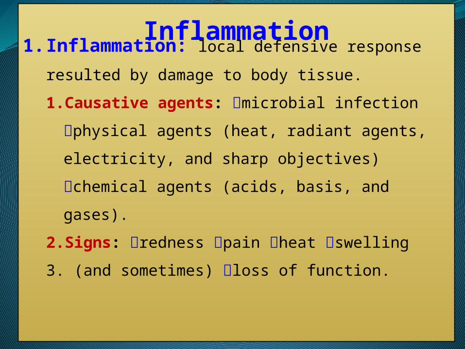
Inflammation3.Functions of inflammation:
To destroy the injurious agent, if possible,
and to remove it and its by-products from the
body.
If destruction is not possible, to limit the
effects on the body by confining or walling off
the injurious agent and it's by products.
To repair or replace tissue damaged by the
injurious agent and it's by products.
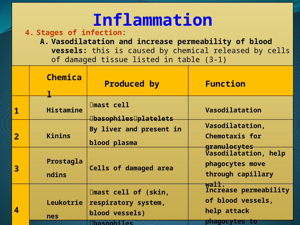
Inflammation4. Stages of infection:
A. Vasodilatation and increase permeability of blood vessels: this is caused by chemical released by cells of damaged tissue listed in table (3-1)
Chemical Produced by Function
1 Histamine mast cell basophilesplatelets Vasodilatation
2 KininsBy liver and present in blood
plasma
Vasodilatation, Chemotaxis for granulocytes
3Prostaglandi
nsCells of damaged area
Vasodilatation, help phagocytes move through capillary wall.
4 Leukotrienes
mast cell of (skin, respiratory system, blood vessels) basophiles
Increase permeability of blood vessels, help attack phagocytes to pathogen
5
Cytokines: IL-1, IL-6, IL-8, IL-12, and TNF-α
Macrophages (fig. 3-1) Vasodilatation, increase permeability of blood vessels, Chemotactic factors
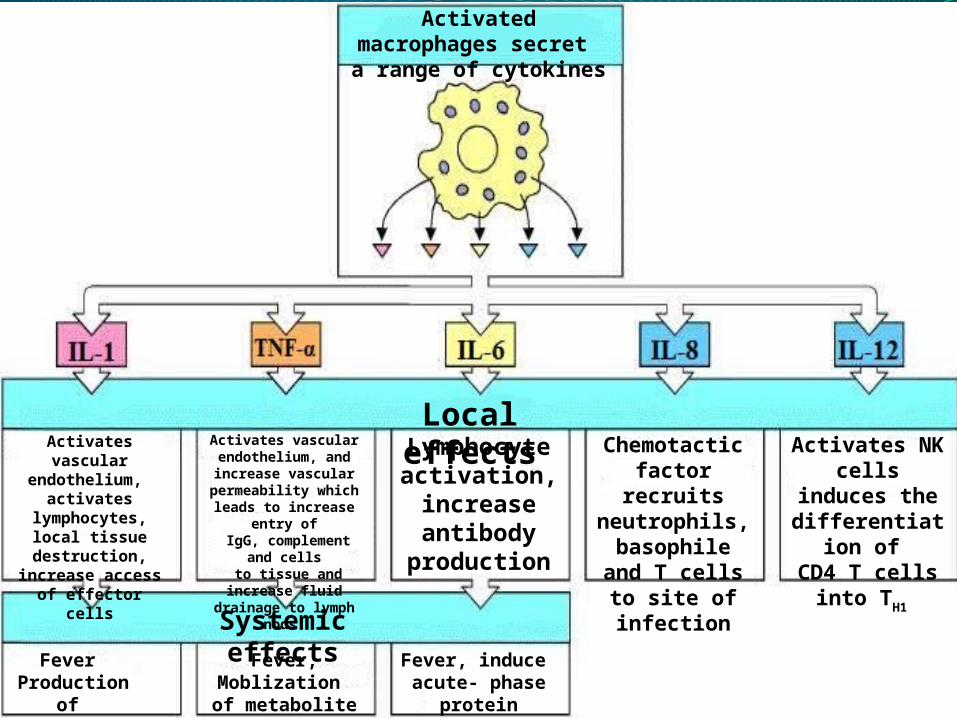
Activates vascular endothelium,
activates lymphocytes,local tissue destruction,
increase access of effector cells
Local effectsActivates vascular
endothelium, and increase vascular permeability
which leads to increase entry of
IgG, complement and cells to tissue and increase fluid
drainage to lymph nods
Chemotactic factor recruits neutrophils,
basophile and T cells to site of
infection
Lymphocyte activation,
increase antibody
production
Activates NK cells induces the
differentiation of CD4 T cells into
TH1
Systemic effectsFever
Production of IL-6
Fever, Moblization of metabolite shoks
Fever, induce acute- phase protein
production
Activated macrophages secret a range of cytokines
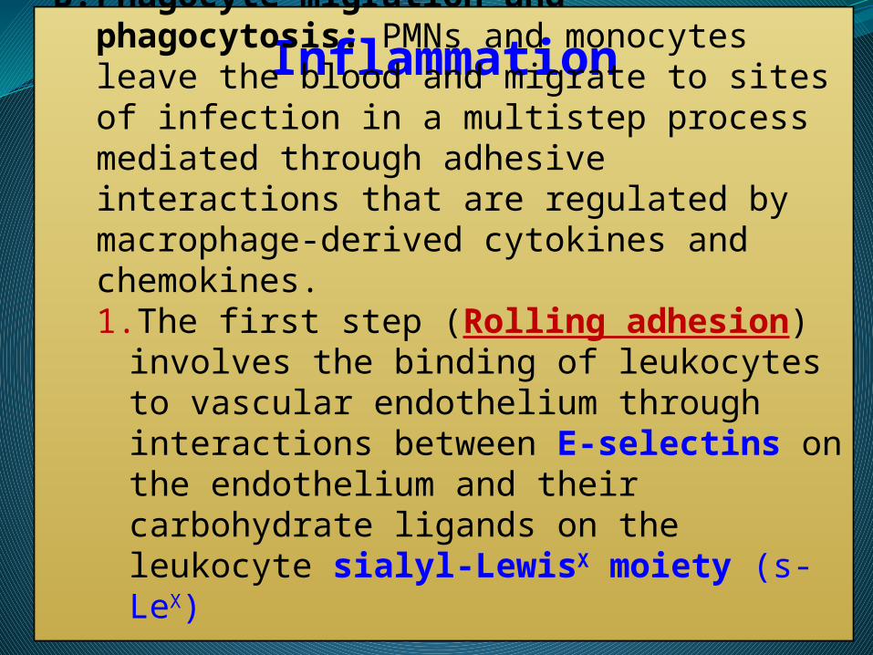
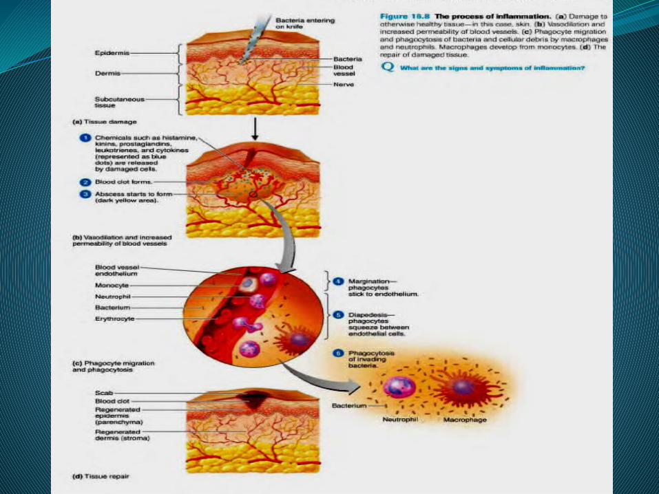
InflammationB. Phagocyte migration and phagocytosis: PMNs and
monocytes leave the blood and migrate to sites of infection in a multistep process mediated through adhesive interactions that are regulated by macrophage-derived cytokines and chemokines.1. The first step (Rolling adhesion) involves the
binding of leukocytes to vascular endothelium through interactions between E-selectins on the endothelium and their carbohydrate ligands on the leukocyte sialyl-LewisX moiety (s-LeX)
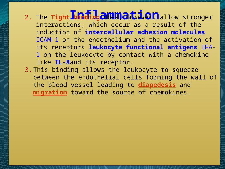
Inflammation2. The Tight binding does, however, allow stronger interactions, which
occur as a result of the induction of intercellular adhesion molecules ICAM-1 on the endothelium and the activation of its receptors leukocyte functional antigens LFA-1 on the leukocyte by contact with a chemokine like IL-8and its receptor.
3. This binding allows the leukocyte to squeeze between the endothelial cells forming the wall of the blood vessel leading to diapedesis and migration toward the source of chemokines.
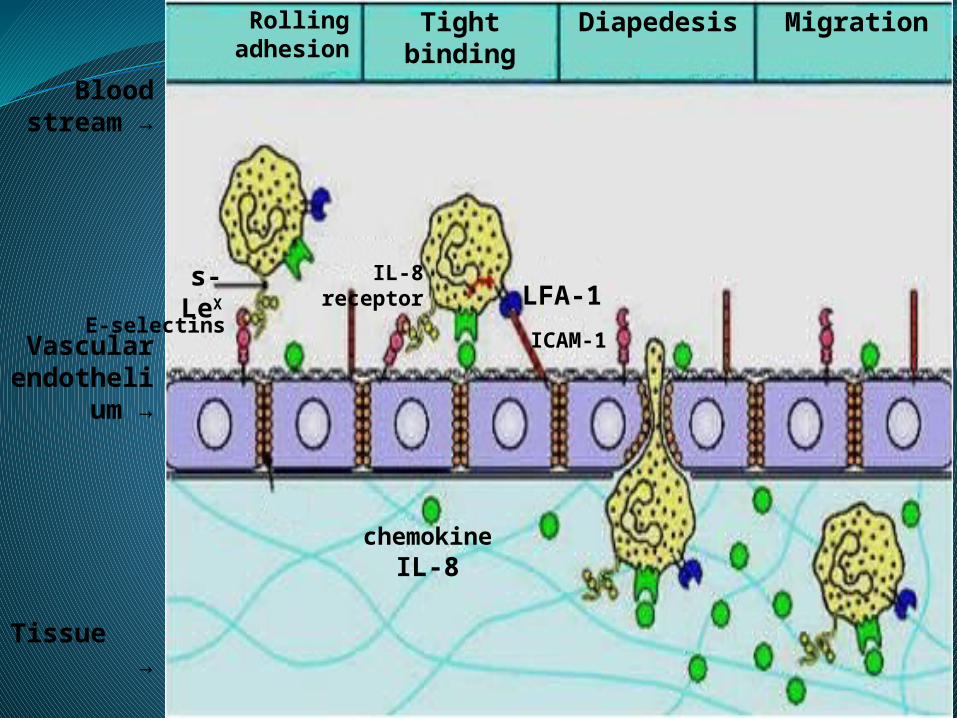
Blood stream →
Vascular endothelium
→
Tissue
→
Rolling adhesion Tight binding Diapedesis Migration
LFA-1
ICAM-1
IL-8 receptor
E-selectins
s-LeX
chemokineIL-8
Inflammation
4.The invading microbes were eliminated by
phagocytosis of PMNs other phagocytes start
establishes for adaptive immune response.
5.The pain is caused by
nerve damage
pressure of edema
irritation by toxins.

Inflammation3. Tissue repair: the process in which tissue replace dead or
damaged tissue.
1. Skin has a high capacity for regeneration, whereas, cardio
muscular does not.
2. Some microbes evade phagocytosis and cause chronic
inflammation like Mycobacterium tuberculosis, such away
lead to continuous production of cytokines and induce
fibroblast in the site of infection to synthesis collagen fiber
which leads to fibrosis.
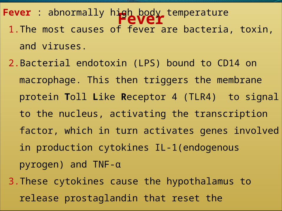
Fever
Fever : abnormally high body temperature
1. The most causes of fever are bacteria, toxin, and viruses.
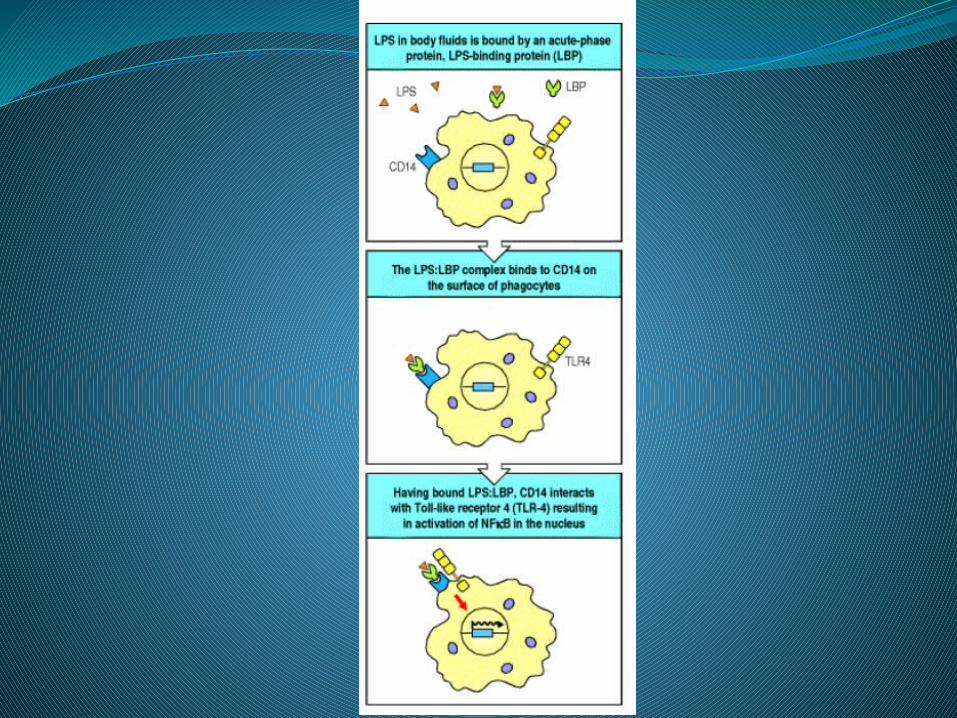
2. Bacterial endotoxin (LPS) bound to CD14 on macrophage. This then
triggers the membrane protein Toll Like Receptor 4 (TLR4) to
signal to the nucleus, activating the transcription factor, which in
turn activates genes involved in production cytokines IL-
1(endogenous pyrogen) and TNF-α
3. These cytokines cause the hypothalamus to release prostaglandin
that reset the hypothalamic thermostat at high temperature, thereby
causing fever.
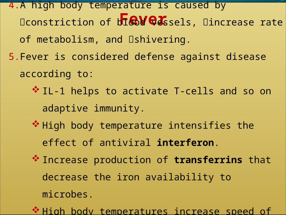
Fever4. A high body temperature is caused by constriction of blood
vessels, increase rate of metabolism, and shivering.
5. Fever is considered defense against disease according to:
IL-1 helps to activate T-cells and so on adaptive immunity.
High body temperature intensifies the effect of antiviral
interferon.
Increase production of transferrins that decrease the iron
availability to microbes.
High body temperatures increase speed of body reactions and
help tissue repair.
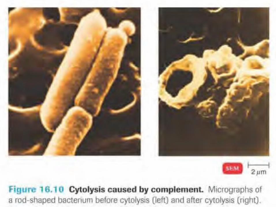
Antimicrobial substancesAntimicrobial substances: Complement system.
Complement is a system of more than 30 plasma
proteins that activates a cascade of proteolytic
reactions on microbial surfaces but not on host
cells, coating these surfaces with fragments that
are recognized and bound by phagocytic
receptors on macrophages. The cascade of
reactions also releases small peptides that
contribute to inflammation and cytolysis (fig. 3-3).
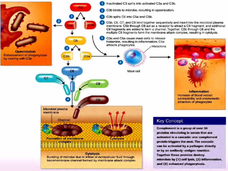

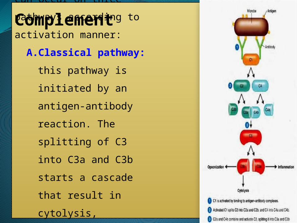
ComplementComplement activation can
occur on three pathways
according to activation manner:
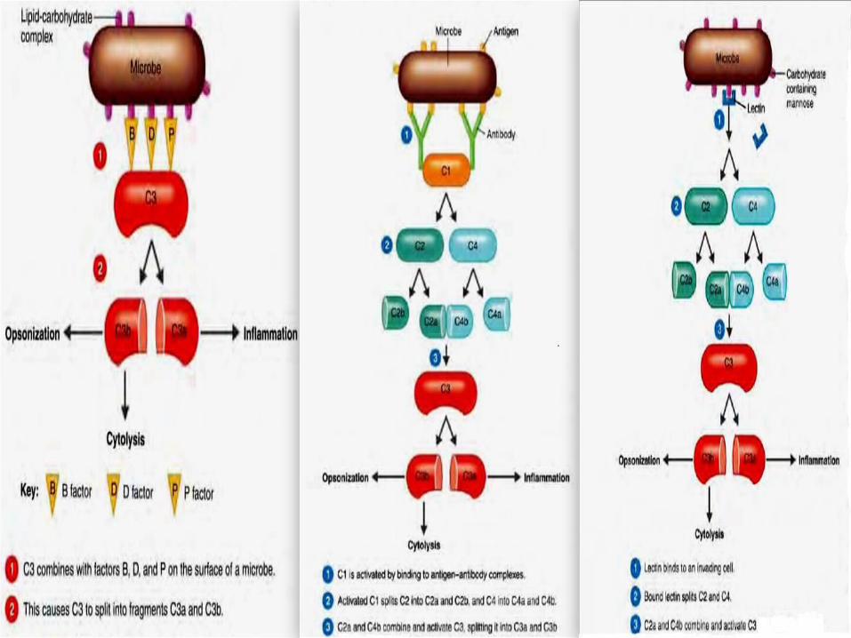
A. Classical pathway: this
pathway is initiated by an
antigen-antibody reaction.
The splitting of C3 into C3a
and C3b starts a cascade that
result in cytolysis,
inflammation, and
opsonization. (fig 3-4)
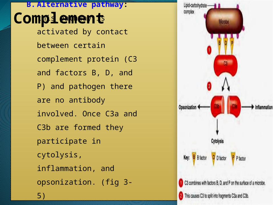
ComplementB. Alternative pathway: this
pathway is activated by contact
between certain complement
protein (C3 and factors B, D,
and P) and pathogen there are no
antibody involved. Once C3a
and C3b are formed they
participate in cytolysis,
inflammation, and opsonization.
(fig 3-5)
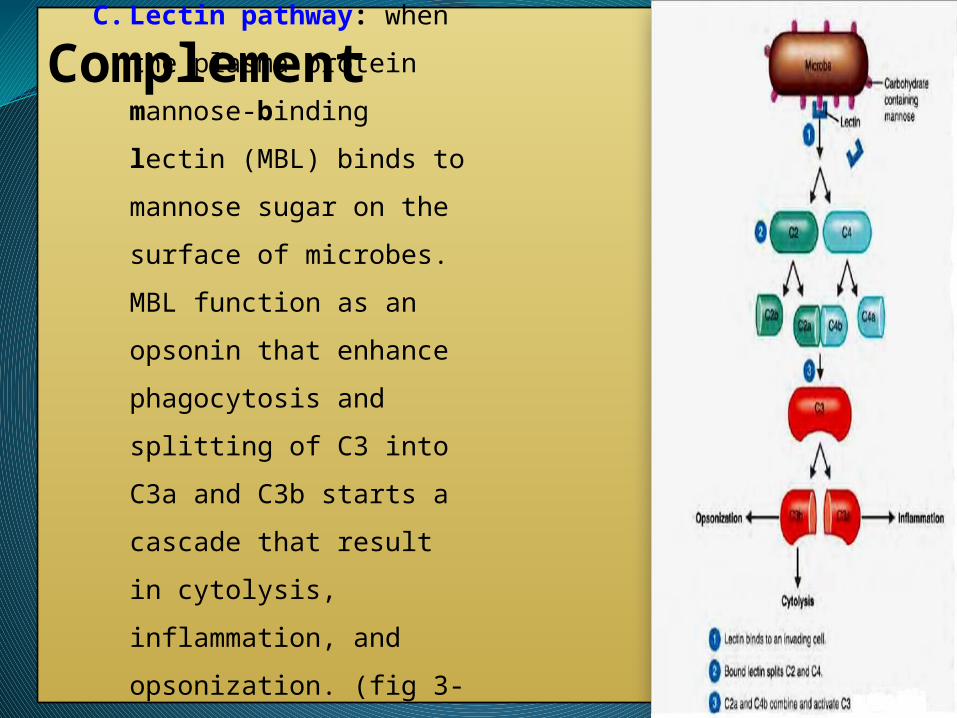
ComplementC. Lectin pathway: when the
plasma protein mannose-
binding lectin (MBL) binds to
mannose sugar on the surface of
microbes. MBL function as an
opsonin that enhance
phagocytosis and splitting of C3
into C3a and C3b starts a
cascade that result in cytolysis,
inflammation, and opsonization.
(fig 3-6)

Thank you for lesson