Embed Size (px)
Citation preview

Behavioral/Systems/Cognitive
Inferior Frontal Gyrus Activation Predicts IndividualDifferences in Perceptual Learning of Cochlear-ImplantSimulations
Frank Eisner,1 Carolyn McGettigan,1 Andrew Faulkner,2 Stuart Rosen,2 and Sophie K. Scott1
1Institute of Cognitive Neuroscience, University College London, WC1N 3AR, London, United Kingdom, and 2Speech, Hearing, and Phonetic Sciences,Division of Psychology and Language Sciences, University College London, WC1N 1PF, London, United Kingdom
This study investigated the neural plasticity associated with perceptual learning of a cochlear implant (CI) simulation. Normal-hearinglisteners were trained with vocoded and spectrally shifted speech simulating a CI while cortical responses were measured with functionalmagnetic resonance imaging (fMRI). A condition in which the vocoded speech was spectrally inverted provided a control for learnabilityand adaptation. Behavioral measures showed considerable individual variability both in the ability to learn to understand the degradedspeech, and in phonological working memory capacity. Neurally, left-lateralized regions in superior temporal sulcus and inferior frontalgyrus (IFG) were sensitive to the learnability of the simulations, but only the activity in prefrontal cortex correlated with interindividualvariation in intelligibility scores and phonological working memory. A region in left angular gyrus (AG) showed an activation pattern thatreflected learning over the course of the experiment, and covariation of activity in AG and IFG was modulated by the learnability of thestimuli. These results suggest that variation in listeners’ ability to adjust to vocoded and spectrally shifted speech is partly reflected indifferences in the recruitment of higher-level language processes in prefrontal cortex, and that this variability may further depend onfunctional links between the left inferior frontal gyrus and angular gyrus. Differences in the engagement of left inferior prefrontal cortex,and its covariation with posterior parietal areas, may thus underlie some of the variation in speech perception skills that have beenobserved in clinical populations of CI users.
IntroductionCochlear implants (CIs) can restore hearing after sensorineuralhearing loss, or provide auditory input to children born deaf.These prostheses deliver tonotopically distributed electrical stim-ulation to the auditory nerve via an electrode array that is insertedinto the cochlea. CIs provide a limited degree of spectral resolu-tion, sufficient for good speech intelligibility in quiet (Moore andShannon, 2009), but lose much detail of the original signal.Acoustic cues that are important for decoding verbal and non-verbal information may thus be weakened or lost. Postlinguallydeafened, adult CI users commonly report that speech and thesensation of pitch sound very different from their previous memo-ries, and it can take some time for users to adapt to the new input(Tyler et al., 1997; Reiss et al., 2007; Moore and Shannon, 2009). Inaddition to the limited availability of acoustic cues, the placement ofthe electrode array can further affect the intelligibility of the speechsignal (Skinner et al., 2002; Finley et al., 2008). If the array has arelatively shallow insertion into the cochlea, the spectral features ofspeech may be signaled at more apical places than in normal hearing.
Thereby, the speech signal is in effect shifted up in frequency (Dor-man et al., 1997; Shannon et al., 1998; Rosen et al., 1999).
Some CI users learn to use their device exceedingly well, andare even able to use the telephone with ease. There is, however,considerable interindividual variability in outcome, for which themain predicting factors include age of implantation, duration ofdeafness, and residual speech perception levels before implanta-tion (UKCISG, 2004). Cognitive factors, such as verbal learningand phonological working memory, have also been implicated inimplantation outcomes, both in adults (Heydebrand et al., 2007)and children (Fagan et al., 2007). However, no currently knownset of factors can account for all of the interindividual variabilitythat is observed clinically.
The adaptation to the novel stimulation from a CI is mediatedby plasticity in the ascending auditory pathway (Fallon et al.,2008) and the cortex. Functional imaging of CI users usingpositron emission tomography (PET) has identified neural cor-relates of postimplant sound and speech perception in primaryand secondary auditory cortex, prefrontal and parietal cortex,and visual cortex (Wong et al., 1999; Giraud et al., 2000, 2001;Giraud and Truy, 2002; Green et al., 2005). Variation in speechperception is associated with activity in temporal and prefrontalcortex (Mortensen et al., 2006; Lee et al., 2007). Technical limi-tations of functional magnetic resonance imaging (fMRI) withimplant devices, and radiation exposure limits with PET, haveprevented the imaging of neural changes associated with initialperceptual and linguistic processing of the novel-sounding CI
Received Aug. 18, 2009; revised Oct. 9, 2009; accepted Dec. 9, 2009.F.E., C.M., and S.K.S. were funded by Wellcome Trust Grant WT074414MA. We thank Joe Devlin, Rob Leech, Fred
Dick, and Marty Sereno at the Birkbeck-UCL Centre for Neuroimaging for technical advice, and we gratefully ac-knowledge helpful comments on the manuscript by D. Sauter and two anonymous reviewers.
Correspondence should be addressed to Frank Eisner, University College London, Institute of Cognitive Neuro-science, 17 Queen Square, London WC1N 3AR, UK. E-mail: [email protected].
DOI:10.1523/JNEUROSCI.4040-09.2010Copyright © 2010 the authors 0270-6474/10/307179-08$15.00/0
The Journal of Neuroscience, May 26, 2010 • 30(21):7179 –7186 • 7179
input. The current study used fMRI and asimulation of the spectral resolution andspectral shifting of a cochlear implant. Itsaim was to identify cortical changes in na-ive, hearing listeners as they learnt to un-derstand this novel input over the courseof a training session, and to relate thesechanges to their capacity in phonologicalworking memory.
Materials and MethodsNoise-vocoding was used to simulate thespectral resolution and spectral shifting of acochlear implant (Fig. 1). The number ofspectral channels and degree of shift of the CIsimulation was set to a level that was suffi-ciently difficult to understand initially, yetallowed learning to occur on a time scale thatcan be tracked in a functional imaging study(i.e., minutes, rather than seconds or hours).A condition in which the spectral informa-tion in the speech signal was inverted suchthat low frequencies in the speech input became high and vice versa,served as a control for learnability as well as for the overall acousticproperties of the stimuli. To examine the relationship betweenintelligibility-related neural activity and memory systems, a battery ofphonological working memory and vocabulary tests was adminis-tered before scanning (see supplemental materials, available atwww.jneurosci.org).
Participants. Twenty right-handed native speakers of English (10male, mean age 25 years, range 19 –31 years) participated in the ex-periment, and a further five (3 male, mean age 24, range 22–25) tookpart in a pretest. None reported having a history of hearing disorderor neurological illness, taking medication, or having prior experiencewith CI simulations. All volunteers gave informed written consentand were paid for their participation. The study was approved bythe University College London Department of Psychology EthicsCommittee.
Materials and methods. Stimulus materials were created from record-ings of sentence lists (Bench et al., 1979) which comprise 336 syntacticallyand semantically simple sentences. Recordings were made in a sound-damped room by a male native English speaker, recorded to MiniDVtape (Bruel & Kjaer 4165 microphone, digitized at a 48 kHz samplingrate with 16 bit quantization) and edited using Final Cut Pro (AppleInc.) and Matlab (MathWorks) software.
The sentences for the “learnable” condition were individually manip-ulated using noise-vocoding (Shannon et al., 1995) and spectral shifting(Dorman et al., 1997; Shannon et al., 1998; Rosen et al., 1999). Thesetechniques simulate two critical aspects of how stimulation produced bya cochlear implant may differ from that of normal hearing; respectively,a coarse spectral resolution resulting from the limited number of effec-tive frequency channels, and a misalignment of the frequencies deliveredto the implant’s electrode array with the tonotopy of the basilar mem-brane. Noise-vocoding involves dividing the frequency spectrum intoanalysis bands, extracting the amplitude envelope from each band, andmultiplying the envelope with a noise-excited carrier band whose centerfrequency and cutoffs are matched to its respective analysis band. Theamplitude-modulated carrier bands are then added together. Spectralshifting additionally alters the cutoff frequencies of the carrier bands by afactor that reflects a given misalignment of cochlear place according toGreenwood’s frequency–position function (Greenwood, 1990). Our ma-nipulations simulated eight effective frequency channels, and a basal-ward shift of 4.8 mm from the apex of the basilar membrane. The filtercutoff frequencies that were used for the analysis- and noise-bands areshown in Table 1; otherwise the signal processing procedure followedRosen et al. (1999). Stimuli for the “inverted” control condition wereprocessed identically, except that the mapping from analysis bands tocarrier bands was inverted in the frequency domain; that is, the ampli-
tude envelope from the lowest analysis band was mapped to the highestcarrier band, the second lowest analysis band mapped to the secondhighest carrier band, etc. This produced stimuli with acoustic character-istics that were highly matched to the learnable condition, but were un-intelligible and could not be understood even with training. Examples ofthe learnable and inverted stimuli are in supplemental materials (avail-able at www.jneurosci.org).
Pretest. A behavioral pilot study was conducted to ensure that thespectrally inverted stimuli were indeed unintelligible and could not beunderstood after training. The design and procedure of the pretestwas identical to that of the fMRI experiment, except that instead ofcollecting verbal responses during test phases, listeners were asked totype on a computer keyboard what they heard after the first presen-tation of each stimulus; after this they saw the sentence presented onthe screen. The responses to “vocoded” and “vocoded-inverted” stim-uli were binned in four blocks of 25 and scored in terms of the per-centage of correctly reported key words. In the vocoded condition,the average scores from blocks one to four were 39.3, 47.0, 62.8, and65.2; representing an average improvement of 26%. The correspond-ing scores in the vocoded-inverted condition were 1.0, 0.3, 1.3, and1.0, indicating that, as expected, listeners are unable to adjust to thespectral inversion manipulation within the context and time frame ofthis experiment.
Training design and procedure. The training materials comprised of100 vocoded-shifted and 100 vocoded-inverted sentences. On eachtrial, subjects would first hear a sentence while the instruction “Lis-ten” was displayed on the screen. This was then followed by a secondpresentation at which, simultaneously, a written version of the sentence wasshown on-screen. The mean interstimulus interval between these “listening”and “listen � feedback” trials was 5 s (jittered up to �1 s). The order oflearnable and inverted trials was pseudo-randomized such that not morethan three trials of one type could occur in a row. The training session was
Figure 1. Spectrograms of the sentence “The sweet shop was empty” in the learnable (A) and spectrally inverted (B) conditions.In both conditions the original speech signal was noise-vocoded and spectrally shifted upward. Stimuli in the inverted condition arenot intelligible, and listeners cannot adapt to this distortion.
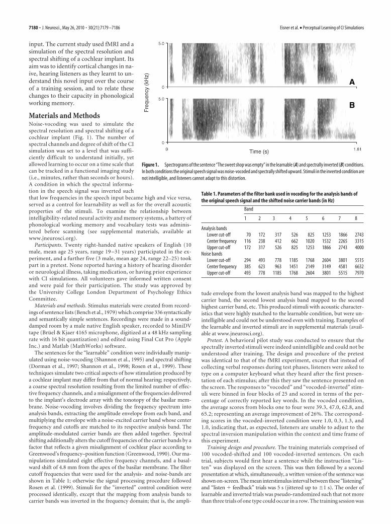
Table 1. Parameters of the filter bank used in vocoding for the analysis bands ofthe original speech signal and the shifted noise carrier bands (in Hz)
Band
1 2 3 4 5 6 7 8
Analysis bandsLower cut-off 70 172 317 526 825 1253 1866 2743Center frequency 116 238 412 662 1020 1532 2265 3315Upper cut-off 172 317 526 825 1253 1866 2743 4000
Noise bandsLower cut-off 294 493 778 1185 1768 2604 3801 5515Center frequency 385 623 963 1451 2149 3149 4581 6632Upper cut-off 493 778 1185 1768 2604 3801 5515 7970
7180 • J. Neurosci., May 26, 2010 • 30(21):7179 –7186 Eisner et al. • Perceptual Learning of CI Simulations

broken up into four blocks of 50 sentences (25 vocoded and 25 vocoded-inverted in each; Fig. 2). In between the training blocks, and at the be-ginning and end of the experiment, there were five test phases whichconsisted of 10 sentences each. During a test phase, subjects were asked torepeat aloud what they could understand after having heard a vocodedsentence once, while the instruction “Repeat” was displayed on-screen.These verbal responses were recorded and scored off-line as the per-centage of key words that were repeated correctly in each test phase.For each individual subject, the items that made up the “learnable,”“inverted,” and “test” conditions were drawn at random from thecorpus of 336 sentences, such that a particular sentence would notoccur more than once in the experiment. A repeated-measuresANOVA with the factors “test phase” and “percentage of correct key-words,” with a planned t-contrast comparison of block 1 and block 5,was used to test for a change in intelligibility over the course of theexperiment.
fMRI data acquisition and analysis. Stimuli were delivered using Matlabwith the Psychophysics Toolbox extension (Brainard, 1997) and re-sponses recorded in Audacity (audacity.sourceforge.net). Subjects wore aheadset with electrodynamic headphones and an optical microphone(MR Confon GmbH). Whole-brain functional and structural magneticresonance imaging (MRI) data were acquired on a Siemens Avanto 1.5tesla scanner (Siemens AG) with a 12-channel birdcage head coil. Agradient-echo echo-planar imaging sequence was used for the functionalscans (repetition time � 3 s, echo time � 50 ms, flip angle � 90°, isotropicvoxel size � 3 mm3, 35 axial slices, 805 volumes; total duration � 40.3 min).A T1-weighted anatomical scan was acquired after the functional run (high-
resolution magnetization-prepared rapid-acqui-sition gradient echo, voxel size � 1 mm3, 160sagittal slices).
MRI data were analyzed using SPM5 (Well-come Department of Imaging Neuroscience,London, UK) with the MarsBaR extension forregion-of-interest analyses (Brett et al., 2002).Functional MRI volumes that had been ac-quired during the test phases were discarded.The remaining 668 fMRI volumes were re-aligned, slice-timing corrected, coregisteredwith the structural scan, segmented, normal-ized to a standard stereotactic space (MontrealNeurological Institute) on the basis of the seg-mentation parameters, and smoothed with anisotropic Gaussian kernel of 6 mm full-width athalf-maximum. Statistical whole-brain analy-ses were conducted in the context of the gen-eral linear model, and included four effects ofinterest at the single-subject level (learnable vsinverted, each under listening and listening �feedback conditions). Event-related hemody-namic responses for each event type were mod-eled as a canonical hemodynamic (gammatype) response function.
ResultsEffect of learnabilityWe first compared the hemodynamic response elicited by listen-ing to learnable cochlear-implant simulations to the responseelicited by listening to spectrally inverted control stimuli. Arandom-effects group analysis was used to compare the learnableand inverted conditions on listening trials. This included a co-variate for the mean repetition test scores from each subject,averaged across the session. The statistical threshold was set atp � 0.001 at the voxel level (uncorrected for multiple compari-sons) with a cluster extent threshold k � 30 voxels. This analysisrevealed greater activation for learnable stimuli in the left supe-rior temporal sulcus (STS) and the left inferior frontal gyrus (IFG;Table 2). The average percentage of signal change across the ses-sion was obtained for each condition and for each subject in thetwo clusters of significantly activated voxels. These regionally av-eraged percentages of signal change confirmed that the overalleffect of learnability was present at both sites, both in the trialswhere participants initially listened to a stimulus and in the trialswhere they received simultaneous written feedback during stim-ulus presentation (Fig. 3).
Individual differences in learningBehavioral data collected over five test blocks indicated that par-ticipants improved significantly in the identification of wordswithin sentences in the learnable condition, albeit with consider-able variability of scores over time (Fig. 4). While nearly all par-ticipants repeated fewer than 10% of keywords correctly in thefirst test phase, some improved considerably (by �60%) over thecourse of the experiment, while others barely improved at all (by�5%). The neural correlates of this variability were investigatedas follows. Voxels showing interindividual variation in the effectof learnability were defined by masking inclusively (at a heightthreshold of p � 0.001) the effect of overall learnability and theeffect of a covariate coding the improved intelligibility over thecourse of the experiment for each subject (calculated as the dif-ference between the first and last test phase). This revealed an areaon the inferior frontal gyrus in which the neural learnability ef-fect, that is, the difference between the responses to the learnable
Figure 2. The fMRI session consisted of five test phases and four training phases (A). During each training phase (B), listenerswere presented with 50 pairs of listening and feedback trials, half of which were potentially learnable. Listeners always heard thespoken version of a sentence first, followed by a second auditory presentation with simultaneous written feedback. During the testphases, participants heard 10 sentences without feedback, and repeated back what they understood after each sentence (C).
Table 2. Summary of activation clusters in the random-effects group analyses
LocationCoordinates(MNI space) Z-score
Extent(mm 3)
Learnability effectLeft IFG �52 16 18 4.24 1125Left STS �52 �48 2 4.12 234
Individual variation in learningLeft IFG �46 26 20 3.44 36
Individual variation in working memoryLeft IFG �44 14 26 4.26 645
Increase with learningLeft AG �36 �58 50 4.01 396Left SMG �66 �28 32 3.63 243
Stereotactic coordinates refer to peak voxels. MNI, Montreal Neurological Institute.
Eisner et al. • Perceptual Learning of CI Simulations J. Neurosci., May 26, 2010 • 30(21):7179 –7186 • 7181

and inverted conditions, correlated signifi-cantly with individual participants’ behav-ioral learning scores. From these voxels,regionally averaged percentages of signalchange were obtained as an index of effectsize in each condition for each subject(Fig. 5).
The analysis was repeated with a predic-tor that, instead of intelligibility, coded indi-vidual composite working memory scores,measured using a battery of phonologicalworking memory tests (see supplementalmaterials, available at www.jneurosci.org).This, again, revealed an area of the inferiorfrontal gyrus in which the overall neural ef-fect of learnability correlated positively withindividual working memory scores (Fig. 5).This area partially overlapped with the oneidentified on the basis of intelligibility. Incontrast, neither the intelligibility scores northe phonological working memory scorescorrelated significantly with activation inthe superior temporal lobe.
Neural basis of intelligibility changesover timeTo identify brain areas which change overtime as a function of learning, a covariateof interest was included at single-subjectlevel in addition to the four experimentalconditions, which represented the indi-vidual learning curve. The learning curvewas derived by cubic interpolation of thefive behavioral test scores over the courseof the experiment. A random-effectsgroup analysis ( p � 0.001, uncorrected; k � 30) of this covariaterevealed significant activations in two regions in the inferior pa-rietal lobe, specifically the left supramarginal gyrus (SMG), andthe left angular gyrus (AG), in which the fMRI signal intensitycorrelated with the participants’ intelligibility scores over thecourse of the experiment (Fig. 6). For these two regions we againobtained the percentages of signal change in each condition andfor each subject (Fig. 6). A repeated-measures ANOVA with thefactors area, trial type, and learnability showed that there was nosignificant difference in these regions between the learnable andinverted stimuli, during either the listening or listening � feed-back trials, although there was a trend for the learnable stimuli toactivate the angular gyrus more, over time, during the listening �feedback trials ( p � 0.07).
Patterns of functional connectivity, as indexed by correlationsof the average signal strength over time in a particular region,were investigated between the two parietal regions, and the re-gions in left IFG and STS that had been identified on the basis ofthe effect of overall learnability. This was done by calculating thecorrelations in regional signal change in each of the four regions,separately for the learnable and inverted conditions (two-tailedPearson’s product-moment correlations; � level set to 0.004 tocorrect for multiple comparisons). This analysis showed signifi-cant correlations of responses between STS and IFG, and betweenAG and SMG, for both learnable and inverted conditions. Therewas also a significant correlation between responses in IFG andAG in the learnable condition only. No other connections be-tween the four regions were significantly correlated (Fig. 6B).
DiscussionA left-lateralized system in IFG and STS was sensitive to the learn-ability of the cochlear implant simulations, showing greater ac-tivity when listening to learnable sentences than when listening to
Figure 3. Overall effect of learnability. Two regions, on the left IFG and left STS, were sensitive to the learnability of the stimuli (A).Results are shown for a t-contrast of the learnable and the inverted conditions in a random-effects group analysis (height threshold p �0.001; cluster extent threshold k�30 voxels). The bar graphs show the pattern of mean signal change in the two regions for the learnableand inverted conditions, separately for trials in which listeners initially heard a sentence, and for when they received feedback (B). Error barsrepresent SEM. Stereotactic coordinates of the peak activations are given in the Montreal Neurological Institute system. L, Left hemisphere.
Figure 4. Box plot showing the behavioral learning effect. Over the course of the training,listeners reported more keywords correctly during the test phases (main effect of test phase,F(4,19) � 26.17, p � 0.001; planned comparison of test phase 1 and 5, t(1,19) � 7.65, p �0.001). Red lines indicate the median, blue edges represent 25 th and 75 th percentiles, dashedlines cover the range of data points excluding outliers, and red crosses mark individual outliers.
7182 • J. Neurosci., May 26, 2010 • 30(21):7179 –7186 Eisner et al. • Perceptual Learning of CI Simulations

spectrally inverted sentences. The STS has consistently been im-plicated in the processing and representation of intelligiblespeech, including in studies that used noise-vocoding as a way ofmanipulating the intelligibility of speech (Scott et al., 2000, 2006;Davis and Johnsrude, 2003; Obleser et al., 2007a, 2008), conse-quent to acoustic-phonetic processing in the superior temporalgyrus (STG) (Jacquemot et al., 2003; Obleser et al., 2007b;Obleser and Eisner, 2009). Many of these studies have revealed asensitivity in the left and right STS to the number of channels inthe vocoded speech signal (Davis and Johnsrude, 2003; Scott etal., 2006; Obleser et al., 2008). Consistent with this work, wepropose that in the present study, the left STS is responding to thespeech characteristics that are preserved in the cochlear implantsimulation.
In contrast to STS, the left IFG has typically been implicated inhigher-order language processes, and activity in this area hasbeen described in a number of previous functional imaging stud-ies of passive speech perception (Davis and Johnsrude, 2003;Friederici et al., 2003; Rodd et al., 2005; Obleser et al., 2007a). Forexample, activation has been observed in the left IFG when par-ticipants heard sentences containing ambiguous words (Hoenigand Scheef, 2009), during aspects of syntax processing (Tetta-manti et al., 2009), and when the semantic predictability of asentence supported comprehension (Obleser et al., 2007a). LeftIFG has been proposed to act as a unification space, integratinglinguistic information across the phonological, semantic, andsyntactic levels (Hagoort, 2005). Prefrontal regions receive pro-jections from both the anterior and posterior auditory streams ofprocessing (Romanski et al., 1999; Scott and Johnsrude, 2003;
Rauschecker and Scott, 2009). In thepresent study, the functionality of the leftIFG could thus include the specific use ofthe simultaneous written feedback to en-hance comprehension of the speech, ormore generally in the use of linguisticknowledge—lexical, syntactic, or contex-tual—to support comprehension.
The training paradigm of this studyaimed to elicit relatively rapid perceptuallearning of the type that may be targetedin computer-based rehabilitation pro-grams (Fu and Galvin, 2007; Stacey andSummerfield, 2007). The profile of behav-iorally measured intelligibility effects (Fig.4) shows that the biggest differences in in-telligibility occurred over the first trials,and performance continued to improvemore slowly across the rest of the session.This pattern is consistent with other audi-tory learning paradigms, which havefound that the biggest improvements inperformance often occur at the start oftraining (Wright and Fitzgerald, 2001;Hawkey et al., 2004). The improvement inthe current study was far from reachingceiling, and previous behavioral trainingstudies suggest that learning would likelycontinue with further training sessions(Rosen et al., 1999; Fu and Galvin, 2007;Stacey and Summerfield, 2007). Fittingthe individual learning profiles to the neu-ral activity revealed two regions in leftSMG and AG in which intelligibility-
related change was correlated with change in activation over thecourse of the training session. This suggests that, alongside theactivation in left STS and IFG—which was associated with in-creases in the intelligibility of spectrally degraded speech—neuralactivity in the left inferior parietal lobe underlies the behavioraladaptation to the stimuli. In AG, the time-dependent effect wasbroadly modulated by intelligibility during the perception trialsin which written feedback was provided. In contrast, activity inthe SMG did not differ between the learnable and inverted trials,nor between trials in which the subjects listened to the stimuli andthose in which written feedback was presented simultaneouslywith the CI simulation. This may imply that the AG is implicatedin the specific use of other linguistic information to supportlearning, while the SMG may be more generally sensitive to time-dependent exposure to the stimuli, rather than to the linguisticcontent.
The increase in speech intelligibility over time suggests thatthe effects of learnability and adaptation should be related. Therewas indeed a significant correlation in activation between the AGand the IFG for only the learnable trials, which may be subservedby a bidirectional anatomical connection between these regionsvia the superior longitudinal fasciculus (Frey et al., 2008). In-creased functional connectivity between the left IFG and angulargyrus has been demonstrated when context helps the compre-hension of noise vocoded speech (Obleser et al., 2007a), andwhen subjects are making overt semantic decisions about noisevocoded speech (Sharp et al., 2009). Both regions have been im-plicated in other learning processes in speech perception, such aslearning to perceive a non-native phonemic contrast (Golestani
Figure 5. Effects of individual variation. The learnability effect was modulated by interindividual differences in the amount oflearning (left) and in working memory capacity (right). These effects were observed in the left inferior frontal gyrus, but not insuperior temporal cortex, in a whole-brain analysis. Stereotactic coordinates of the peak activations are given in the MontrealNeurological Institute system. L, Left hemisphere.
Eisner et al. • Perceptual Learning of CI Simulations J. Neurosci., May 26, 2010 • 30(21):7179 –7186 • 7183

and Zatorre, 2004), which supports thepossibility that they are part of a moregeneralized learning mechanism whichnot only applies to spectrally degradedspeech, but also to other listening situa-tions where perceptual processing is ef-fortful. We suggest that in the presentstudy, the functional connectivity be-tween the left IFG and AG may have a spe-cific role in the task of mapping betweenthe written sentence information and theheard CI simulation when the simulationis learnable.
An extensive network of brain regionsthus underlies adaptation to a CI simula-tion. Among recipients of CIs there is con-siderable variation in outcome, and thisvariation can only in part be explained byknown predictive factors such as the pre-implantation level of residual hearing. CIusers’ neural capacity for adaptation maybe facilitated by cognitive functions whichare not primarily part of the central audi-tory system (Moore and Shannon, 2009).In our study, we observed a wide range ofscores in participants’ comprehension ofCI simulations, which were correlatedwith activity in the left IFG. In contrast tothe IFG, the response in STS did not varywith individual differences in intelligibil-ity. This pattern of results is consistentwith claims that the basic speech percep-tion system, as represented by the activityin the STS, works to its fullest extent withwhat it can process from the incoming sig-nal, and is not modulated by feedbackprojections from high-order languageareas (Norris et al., 2000). The lack ofassociation between activity in STS andindividual differences in learning, in con-trast with the strong association betweencomprehension and left IFG, suggests thatvariation in successful processing of CIsimulations can depend on high-level, lin-guistic and cognitive factors that go be-yond relatively early, acoustic-phoneticprocesses.
One candidate higher-order cognitivefactor that has been implicated in the suc-cessful use of CIs is phonological workingmemory (pWM). Several behavioral stud-ies have found that phonological workingmemory scores are positively correlatedwith successful perception of speech fol-lowing cochlear implantation in children (Pisoni and Geers,2000; Cleary et al., 2001; Pisoni and Cleary, 2003; Dillon et al.,2004). More generally, developmental language disorderssuch as specific language impairment or dyslexia often involvea deficit in working memory (Bishop, 2006; Ramus andSzenkovits, 2008). For the participants in the current study, acomposite measure of phonological working memory corre-lated with activity in the left IFG. Functional imaging studieshave outlined a network of brain areas underlying phonolog-
ical working memory, including the inferior parietal lobe andthe inferior frontal gyrus (Buchsbaum and D’Esposito, 2008),and one study specifically linked IFG activation in a pWM taskwith the encoding, maintenance, and response elements ofpWM (Strand et al., 2008). The current study shows a neurallink between variation in pWM capacity and variation inspeech intelligibility, which thus represents a potential func-tional anatomical basis for the variation that is observed in theresponsiveness to postimplant rehabilitation.
Figure 6. Areas of the brain that change over time with learning. Two regions in inferior parietal cortex (in blue) show anincrease in activation over time which reflects learning curves modeled at a single-subject level (A). The bar graphs show thepatterns of mean signal change in these two regions for the learnable and inverted conditions. Error bars represent SEM. Theseareas exhibit differential correlation patterns with activity in the superior temporal sulcus and inferior frontal gyrus, which weresensitive to overall intelligibility (B). Solid lines indicate significant correlations ( p � 0.004) in signal change in the learnable andinverted listening conditions; dashed lines indicate nonsignificant correlations. Stereotactic coordinates of the peak activations inA are given in the Montreal Neurological Institute system. L, Left hemisphere.
7184 • J. Neurosci., May 26, 2010 • 30(21):7179 –7186 Eisner et al. • Perceptual Learning of CI Simulations

It is possible that the use of sentences as stimuli has empha-sized individual differences in higher-order language processing(Hervais-Adelman et al., 2008), and this may interact with pWMprocesses. Neuropsychological studies have reported patientswho had deficits in pWM and who made errors processing pho-nemes in sentences, but not in isolated words (Jacquemot et al.,2006). This possibility should be tested with further studies usingadaptation to single-word or sublexical stimuli.
Our results suggest that individual variation in the compre-hension of a cochlear implant simulation is at least in part deter-mined by differences in the employment of higher-orderlanguage processes to help decode the speech sequences, andfunctional connectivity between the frontal and parietal lobes,rather than differences in the quality of acoustic-phonetic pro-cessing or representations in the dorsolateral temporal lobes.Furthermore, the results show that variation in phonologicalworking memory scores shares an anatomical location in left IFGwith individual variability in the learning of the CI simulations.Problems with speech and language processing commonly co-occur with problems in phonological working memory tasks. Wesuggest that one of the linguistic properties of the left IFG is to actas an interface for the interaction of speech perception and pho-nological working memory when processing spoken sentences,and that activation differences in this area across individuals areassociated with differences in the successful adaptation to a CIsimulation.
ReferencesBench J, Kowal A, Bamford J (1979) The BKB (Bamford-Kowal-Bench)
sentence lists for partially-hearing children. Br J Audiol 13:108 –112.Bishop DVM (2006) Developmental cognitive genetics: how psychology
can inform genetics and vice versa. Q J Exp Psychol 59:1153–1168.Brainard DH (1997) The Psychophysics Toolbox. Spat Vis 10:433– 436.Brett M, Anton J, Valabregue A, Poline J (2002) Region of interest analysis
using an SPM toolbox. In: Proceedings of the 8th International Conferenceon Functional Mapping of the Human Brain, June 2–6, 2002. Sendai, Japan.Available on CD-ROM in Neuroimage, Vol 16, No 2.
Buchsbaum BR, D’Esposito M (2008) The search for the phonological store:from loop to convolution. J Cogn Neurosci 20:762–778.
Cleary M, Pisoni DB, Geers AE (2001) Some measures of verbal and spatialworking memory in eight- and nine-year-old hearing-impaired childrenwith cochlear implants. Ear Hear 22:395– 411.
Davis MH, Johnsrude IS (2003) Hierarchical processing in spoken languagecomprehension. J Neurosci 23:3423–3431.
Dillon CM, Burkholder RA, Cleary M, Pisoni DB (2004) Nonword repeti-tion by children with cochlear implants: Accuracy ratings from normal-hearing listeners. J Speech Lang Hear Res 47:1103–1116.
Dorman MF, Loizou PC, Rainey D (1997) Simulating the effect of cochlear-implant electrode insertion depth on speech understanding. J Acoust SocAm 102:2993–2996.
Fagan MK, Pisoni DB, Horn DL, Dillon CM (2007) Neuropsychologicalcorrelates of vocabulary, reading, and working memory in deaf childrenwith cochlear implants. J Deaf Stud Deaf Educ 12:461– 471.
Fallon JB, Irvine DR, Shepherd RK (2008) Cochlear implants and brainplasticity. Hear Res 238:110 –117.
Finley CC, Holden TA, Holden LK, Whiting BR, Chole RA, Neely GJ, HullarTE, Skinner MW (2008) Role of electrode placement as a contributor tovariability in cochlear implant outcomes. Otol Neurotol 29:920 –928.
Frey S, Campbell JS, Pike GB, Petrides M (2008) Dissociating the humanlanguage pathways with high angular resolution diffusion fiber tractogra-phy. J Neurosci 28:11435–11444.
Friederici AD, Ruschemeyer SA, Hahne A, Fiebach CJ (2003) The role of leftinferior frontal and superior temporal cortex in sentence comprehension:localizing syntactic and semantic processes. Cereb Cortex 13:170 –177.
Fu QJ, Galvin JJ 3rd (2007) Perceptual learning and auditory training incochlear implant recipients. Trends Amplif 11:193–205.
Giraud AL, Truy E (2002) The contribution of visual areas to speech com-prehension: a PET study in cochlear implants patients and normal-hearing subjects. Neuropsychologia 40:1562–1569.
Giraud AL, Truy E, Frackowiak RJS, Gregoire MC, Pujol JF, Collet L (2000)Differential recruitment of the speech processing system in healthy sub-jects and rehabilitated cochlear implant patients. Brain 123:1391–1402.
Giraud AL, Price CJ, Graham JM, Truy E, Frackowiak RS (2001) Cross-modal plasticity underpins language recovery after cochlear implanta-tion. Neuron 30:657– 663.
Golestani N, Zatorre RJ (2004) Learning new sounds of speech: reallocationof neural substrates. Neuroimage 21:494 –506.
Green KM, Julyan PJ, Hastings DL, Ramsden RT (2005) Auditory corticalactivation and speech perception in cochlear implant users: effects ofimplant experience and duration of deafness. Hear Res 205:184 –192.
Greenwood DD (1990) A cochlear frequency-position function for severalspecies—29 years later. J Acoust Soc Am 87:2592–2605.
Hagoort P (2005) On Broca, brain, and binding: a new framework. TrendsCogn Sci 9:416 – 423.
Hawkey DJ, Amitay S, Moore DR (2004) Early and rapid perceptual learn-ing. Nat Neurosci 7:1055–1056.
Hervais-Adelman A, Davis MH, Johnsrude IS, Carlyon RP (2008) Percep-tual learning of noise vocoded words: effects of feedback and lexicality.J Exp Psychol Hum Percept Perform 34:460 – 474.
Heydebrand G, Hale S, Potts L, Gotter B, Skinner M (2007) Cognitive pre-dictors of improvements in adults’ spoken word recognition six monthsafter cochlear implant activation. Audiol Neurootol 12:254 –264.
Hoenig K, Scheef L (2009) Neural correlates of semantic ambiguity process-ing during context verification. Neuroimage 45:1009 –1019.
Jacquemot C, Pallier C, LeBihan D, Dehaene S, Dupoux E (2003) Phono-logical grammar shapes the auditory cortex: a functional magnetic reso-nance imaging study. J Neurosci 23:9541–9546.
Jacquemot C, Dupoux E, Decouche O, Bachoud-Levi AC (2006) Misper-ception in sentences but not in words: speech perception and the phono-logical buffer. Cogn Neuropsychol 23:949 –971.
Lee HJ, Giraud AL, Kang E, Oh SH, Kang H, Kim CS, Lee DS (2007) Corticalactivity at rest predicts cochlear implantation outcome. Cereb Cortex17:909 –917.
Moore DR, Shannon RV (2009) Beyond cochlear implants: awakening thedeafened brain. Nat Neurosci 12:686 – 691.
Mortensen MV, Mirz F, Gjedde A (2006) Restored speech comprehensionlinked to activity in left inferior prefrontal and right temporal cortices inpost-lingual deafness. Neuroimage 31:842– 852.
Norris D, McQueen JM, Cutler A (2000) Merging information in speechrecognition: feedback is never necessary. Behav Brain Sci 23:299 –370.
Obleser J, Eisner F (2009) Pre-lexical abstraction of speech in the auditorycortex. Trends Cogn Sci 13:14 –19.
Obleser J, Wise RJ, Alex Dresner M, Scott SK (2007a) Functional integrationacross brain regions improves speech perception under adverse listeningconditions. J Neurosci 27:2283–2289.
Obleser J, Zimmermann J, Van Meter J, Rauschecker JP (2007b) Multiplestages of auditory speech perception reflected in event-related fMRI.Cereb Cortex 17:2251–2257.
Obleser J, Eisner F, Kotz SA (2008) Bilateral speech comprehension reflectsdifferential sensitivity to spectral and temporal features. J Neurosci28:8116 – 8123.
Pisoni DB, Cleary M (2003) Measures of working memory span and verbalrehearsal speed in deaf children after cochlear implantation. Ear Hear24:106S–120S.
Pisoni DB, Geers AE (2000) Working memory in deaf children with co-chlear implants: correlations between digit span and measures of spokenlanguage processing. Ann Otol Rhinol Laryngol Suppl 185:92–93.
Ramus F, Szenkovits G (2008) What phonological deficit? Q J Exp Psychol61:129 –141.
Rauschecker JP, Scott SK (2009) Maps and streams in the auditory cortex:nonhuman primates illuminate human speech processing. Nat Neurosci12:718 –724.
Reiss LA, Turner CW, Erenberg SR, Gantz BJ (2007) Changes in pitch witha cochlear implant over time. J Assoc Res Otolaryngol 8:241–257.
Rodd JM, Davis MH, Johnsrude IS (2005) The neural mechanisms of speechcomprehension: fMRI studies of semantic ambiguity. Cereb Cortex15:1261–1269.
Romanski LM, Tian B, Fritz J, Mishkin M, Goldman-Rakic PS, RauscheckerJP (1999) Dual streams of auditory afferents target multiple domains inthe primate prefrontal cortex. Nat Neurosci 2:1131–1136.
Rosen S, Faulkner A, Wilkinson L (1999) Adaptation by normal listeners to
Eisner et al. • Perceptual Learning of CI Simulations J. Neurosci., May 26, 2010 • 30(21):7179 –7186 • 7185
upward spectral shifts of speech: Implications for cochlear implants.J Acoust Soc Am 106:3629 –3636.
Scott SK, Johnsrude IS (2003) The neuroanatomical and functional organi-zation of speech perception. Trends Neurosci 26:100 –107.
Scott SK, Blank CC, Rosen S, Wise RJ (2000) Identification of a pathway forintelligible speech in the left temporal lobe. Brain 123:2400 –2406.
Scott SK, Rosen S, Lang H, Wise RJ (2006) Neural correlates of intelligibilityin speech investigated with noise vocoded speech—a positron emissiontomography study. J Acoust Soc Am 120:1075–1083.
Shannon RV, Zeng FG, Kamath V, Wygonski J, Ekelid M (1995) Speechperception with primarily temporal cues. Science 270:303–304.
Shannon RV, Zeng FG, Wygonski J (1998) Speech recognition with al-tered spectral distribution of envelope cues. J Acoust Soc Am 104:2467–2476.
Sharp DJ, Turkheimer F, Bose S, Scott SK, Wise RJS (2009) Increased fronto-parietal integration after stroke and cognitive recovery. Ann Neurol. Advanceonline publication. Retrieved Jan. 5, 2010. doi:10.1002/ana.21866.
Skinner MW, Ketten DR, Holden LK, Harding GW, Smith PG, Gates GA,Neely JG, Kletzker GR, Brunsden B, Blocker B (2002) CT-derived esti-mation of cochlear morphology and electrode array position in relation toword recognition in Nucleus-22 recipients. J Assoc Res Otolaryngol3:332–350.
Stacey PC, Summerfield AQ (2007) Effectiveness of computer-based audi-tory training in improving the perception of noise-vocoded speech.J Acoust Soc Am 121:2923–2935.
Strand F, Forssberg H, Klingberg T, Norrelgen F (2008) Phonological work-ing memory with auditory presentation of pseudo-words—an event re-lated fMRI study. Brain Res 1212:48 –54.
Tettamanti M, Rotondi I, Perani D, Scotti G, Fazio F, Cappa SF, Moro A(2009) Syntax without language: neurobiological evidence for cross-domain syntactic computations. Cortex 45:825– 838.
Tyler RS, Parkinson AJ, Woodworth GG, Lowder MW, Gantz BJ (1997)Performance over time of adult patients using the Ineraid or nucleuscochlear implant. J Acoust Soc Am 102:508 –522.
UKCISG [UK Cochlear Implant Study Group] (2004) Criteria of candidacyfor unilateral cochlear implantation in postlingually deafened adults II:cost-effectiveness analysis. Ear Hear 25:336 –360.
Wong D, Miyamoto RT, Pisoni DB, Sehgal M, Hutchins GD (1999) PETimaging of cochlear-implant and normal-hearing subjects listening tospeech and nonspeech. Hear Res 132:34 – 42.
Wright BA, Fitzgerald MB (2001) Different patterns of human discrimina-tion learning for two interaural cues to sound-source location. Proc NatlAcad Sci U S A 98:12307–12312.
7186 • J. Neurosci., May 26, 2010 • 30(21):7179 –7186 Eisner et al. • Perceptual Learning of CI Simulations
![ALDENHAM PSYCHOLOGY · Web viewPrep: Casey Textbook questions page 241. State what behaviour is linked to the inferior frontal gyrus. [1] State what behaviour is linked to the ventral](https://img.pdfslide.us/doc/110x75/5fa80570eef99a25e00aaa4a/aldenham-web-view-prep-casey-textbook-questions-page-241-state-what-behaviour.jpg)