Embed Size (px)
Citation preview
GEOLOGICAL SURVEY CIRCULAR 848-D
Infectious Waterborne Diseases

Infectious Waterborne Diseases
By Phillip E. Greeson
Briefing Papers on Water Quality
GEOLOGICAL SURVEY CIRCULAR 848-D
1981
United States Department of the Interior JAMES G. WATT, Secretary
Geological Survey
Doyle G. Frederick, Acting Director
Library of Congress Cataloging in Publication Data
Greeson, Phillip E. Infectious waterborne diseases.
(Briefing papers on water quality) (Geological Survey circular; 848-D) Bibliography: p. Supt. of Docs. no.: I 19.4/2:848-D 1. Waterborne infection. I. Title. II. Series. III. Series: Geological Survey circular ; 848-D. QE75.C5 no. 848-D [RA642.W3] 557.3s 81-607881 [614.4] AACR2
Free on application to Distribution Branch, Text Products Section, U. S. Geological Survey, 604 South Pickett Street, Alexandria, VA 22304
FOREWORD
In August 1974, the Water Resources Division of the U.S. Geological Survey introduced the first of a series of briefing papers that were designed to increase the understanding of its employees of the significance of various aspects of water quality. Numerous briefing papers have been prepared by the Quality of Water Branch. Others will be prepared as the need arises. Each paper addresses a separate topic and is written in a nontechnical, easy-to-understand manner for distribution within the organization.
Because of the favorable reception that the papers have received and their apparent effectiveness in accomplishing the objectives stated above, it would appear that their wider distribution would serve a useful purpose. It is hoped that a wide range of persons, including those interested in the quality of our Nation's water resources but who have little or no technical training, will find value in reading the papers. Furthermore, it is hoped that the papers will be suitable for supplemental reading in secondary education programs and in beginning college-level courses.
The U.S. Geological Survey plans to publish several U.S. Geological Survey Circulars that contain briefing papers on particular aspects of water quality. This fourth Circular contains a paper discussing infectious waterborne diseases.
III
Philip Cohen Chief Hydrologist

Foreword -------------------------------------Introduction ------------------------------------
Bacteria ---------------------------------------Aerobic bacteria -----------------------------
Spirochete ---------------------------------Rickettsia -----------------------------------
Viruses ----------------------------------------
CONTENTS
Page
III D1
3
3
6
6
6
Parasites --------------------------------------Protozoans _________________________________ _ Nematodes _________________________________ _
Trematodes --------------------------------Cestodes -----------------------------------
Multiple infectious agents -------------------------Conclusion ------------------------------------References -------------------------------------
ILLUSTRATIONS
Page
FIGURE 1. General life history of waterborne disease-causing protozoans__________________ D8 2. Infectious route of A ngiostrongylus cantonensis (rat lungworm)---------------- 10 3. Infectious route of Dracunculus medinensis (guinea worm)-------------------- 10 4. Infectious route of Capillaria hepactica and C. philippinensis (whip worms)______ 11 5. Infectious route of Gnathostoma spinigerum ------------------------------- 11 6. Generalized life cycle of trematodes--------------------------------------- 12 7. Life cycle of Diphyllobothrium latum (broad or fish tapeworm)----------------- 13 8. Infectious route of Echinococcus spp. ------------------------------------- 13
TABLES
Page
TABLE 1. Composite list of waterborne diseases and their infectious agents--------------- D2 2. Infectious agents of waterborne diseases ---------------------------------- 3
v
Page
D8 8 9
11 13 13 14 14

BRIEFING PAPERS ON WATER QUALITY
Infectious Waterborne Diseases
By Phillip E. Greeson
ABSTRACT
This is the fourth in a series of briefing papers on water quality prepared by the U.S. Geological Survey. Each briefing paper is prepared in a simple, nontechnical, easy-to-understand manner. This U.S. Geological Survey Circular contains a paper on "Infectious Waterborne Diseases," which discusses 36 infectious diseases of man that can be or are strongly suspected of being transmitted by water. Of the 36 diseases, 12 are caused by bacteria, 4 are caused by viruses, 19 are caused by parasites, and 1 has numerous infectious agents.
INTRODUCTION
A principal concept of the understanding of infectious diseases was formulated in 1914 by Theobald Smith. He wrote: "Infectious disease is a manifestation of parasitism. The dynamics of infectious disease in human communities [can be] interpreted as expressions of the internal struggle of living things for food by parasitism, for shelter, and for opportunity to propagate their kind. As a result of host wanderings, mutation, and selective adaptation, certain [organisms] have been successful in maintaining their propagation in the biologic orbit of man."
What Smith was saying is that certain organisms have adapted their needs to the environment of man's body. Those organisms that function in or need man's body to the detriment of man are disease producers. This is the basic ecological concept of parasitism, which can be defined as the interaction of two organisms in which one population is benefited, whil~ the other is affected; therefore, the infectious agent is benefited and man is affected.
This paper is concerned with the infections and diseases of man that are transmitted by water. Specifically, it will discuss those infections and diseases that are caused by the ingestion of water containing an infectious agent, by the ingestion of an aquatic organism (for example, fish or shellfish) that contains an agent infectious to man, and by physical contact with water containing an infec-
D1
tious agent. It must be noted that man can become infected by an agent, but not display the clinical symptoms of disease. In other words, an infection can be subclinical or asymptomatic in nature. Water, in the context of this paper, includes all native (or untreated) waters, both surface water and ground water; public water supplies; sewage effluents; and swimming pools.
The paper will discuss only those diseases that are caused by an infectious agent, specifically those diseases caused by bacteria, viruses, and parasites. These diseases are referred to as communicable diseases. The paper will not discuss those physiological or metabolic malfunctions or illnesses that are caused by the ingestion of water containing abiotic (nonliving) substances, for example, trace metals, pesticides, manufactured organic compounds, or other toxicants.
Table 1 is a composite list of waterborne diseases and their infectious agents. There now appears to be 36 known diseases of man that can be or are strongly suspected of being transmitted by water; many also have other means of transmission, such as person-to-person contact and so on. The 36 diseases are listed in capital letters in the table. The nomenclature of the diseases generally follows that of the 8th revision of the "International Classification of Diseases." The names in lowercase letters are synonyms, which include common names, local names, and so forth. Of the 36 diseases, 12 are caused by bacteria, 4 are caused by viruses, 19 are caused by parasites, and 1 has numerous etiological or infectious agents.
The diseases that are listed in table 1 exist somewhere on Earth; however, some of them are highly restricted to remote areas, while others are ubiquitous. Many of the diseases are not indigenous to the United States, but with jet travel eliminating time barriers and opening new areas
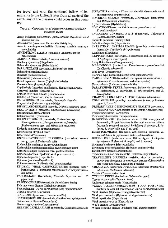
for travel and with the continual inflow of immigrants to the United States from all parts of the earth, any of the diseases could occur in this country.
TABLE !.-Composite list of waterborne diseases and their infectious agents
Acute infectious nonbacterial gastroenteritis (Epidemic viral gastroenteritis)
AMEBIASIS (protozoan, Entamoeba histolytica) Amebic meningoencephalitis (Primary amebic meningo
encephalitis) ANGIOSTRONGYLIASIS (nematode, A ngiostrongylus
cantonensis) ANISAKIASIS (nematode, A nisalcis marina) Bacillary dysentery (Shigellosis) Balantidial dysentery (Balantidiasis) BALANTIDIASIS (protozoan, Balantidium coli) Balantidiosis (Balantidiasis) Bilharzia (Schistosomiasis) Bilharziasis (Schistosomiasis) Broad tapeworm disease (Diphyllobothriasis) Canicola fever (Leptospirosis) Capillariasis (Intestinal caP.illariasis, Hepatic capillariasis) Catarrhal jaundice (Hepatitis A) Chinese liver fluke disease (Clonorchiasis) CHOLERA (bacterium, Vibrio cholerae) CLONORCHIASIS (trematode, Clonorchis sinensis) Conjuctivitis (Inclusion conjunctivitis) DIPHYLLOBOTHRIASIS (cestode, Diphyllobothrium latum) DRACONTIASIS (nematode, Dracunculus medinensis) Dracunculiasis (Dracontiasis) Echinococcosis (Hydatidosis) ECHINOSTOMIASIS (trematode, Echinostoma spp.,
Euparyphium spp., Paryphostomum sufrartyfex, Echinochasmus spp., and Himasthia muhlensi)
Endemic hemoptysis (Paragonimiasis) Enteric fever (Typhoid fever) Enterocolitis (Y ersiniosis) ENTEROPATHOGENIC DIARRHEA (bacterium, several
serogroups of Escherichia coli) Eosinophilic meningitis (Angiostrongyliasis) Eosinophilic meningoencephalitis (Angiostrongyliasis) Epidemic collapse (Epidemic viral gastroenteritis) Epidemic diarrhea (Epidemic viral gastroenteritis) Epidemic hepatitis (Hepatitis A) Epidemic jaundice (Hepatitis A) Epidemic nausea (Epidemic viral gastroenteritis) EPIDEMIC VIRAL GASTROENTERITIS (possibly water
borne) (virus, 2-3 probable serotypes of a 27 -nm parvoviruslike agent)
FASCIOLIASIS (trematode, Fasciola hepatica and F. gigantica)
FASCIOLOPSIASIS (trematode, Fasciolopsis buski) Fish tapeworm disease (Diphyllobothriasis) Food poisoning (Vibrio parahaemolyticus food poisoning) Giardia enteritis (Giardiasis) GIARDIASIS (protozoan, Giardia Lamblia) GNATHOSTOMIASIS (nematode, Gnathostoma spinigerum) Guinea worm disease (Dracontiasis) Hemorrhagic jaundice (Leptospirosis) HEPATIC CAPILLARIASIS (nematode, Capillaria hepatica)
HEPATITIS A (virus, a 27-nm particle with characteristics of an enterovirus or parvovirus)
HETEROPHYIDIASIS (trematode, Heterophyes heterophyes and Metagonimus yokogawai)
Hydatid disease (Hydatidosis) HYDATIDOSIS (cestode, Echinococcus granulosus and
E. multilocularis) INCLUSION CONJUNCTIVITIS (bacterium, Chlamydia
(Bedsonia) trachomatis) Infantile paralysis (Poliomyelitis) Infectious hepatitis (Hepatitis A) INTESTINAL CAPILLARIASIS (possibly waterborne)
(nematode, Capillaria philippinensis) Lambliasis (Giardiasis) LEPTOSPIROSIS (bacteruim, 18 serogroups and 170 serotypes
of Leptospira interrogans) Lung fluke disease (Paragonimiasis) MELIOIDOSIS (bacterium, Pseudomonas pseudomallei
(Loefjlerella whitmori)) Mud fever (Leptospirosis) Norwalk type disease (Epidemic viral gastroenteritis) PARAGONIMIASIS (trematode, Paragonimus westermani, P.
africanus, P. szechuanensis, and other species) Paratrachoma (Inclusion conjunctivitis) PARATYPHOID FEVER (bacterium, Salmonella paratyphi,
S. choleraesuis, S. enteritidis, S. schottimuelleri, and S. hirschfeldii)
PHARYNGOCONJUNCTIV AL FEVER (virus, adenovirus) POLIOMYELITIS (possibly waterborne) (virus, poliovirus
types 1, 2, and 3) PRIMARY AMEBIC MENINGOENCEPHALITIS (protozoan,
Naegleriafowleri and species of the genus Acanthamoeba) Pseudotuberculosis (Y ersiniosis) Pulmonary distomiasis (Paragonimiasis) SALMONELLOSIS (bacterium, about 1,500 serotypes of
Salmonella; S. typhimurium is the most common, others frequently reported include S. heidelburg, S. newport, S. infantis, S. enteritidis, and S. st. paul)
SCHISTOSOMIASIS (tremode, Schistosoma mansoni, S. haematobium, S. y'aponicum, and S. intercalatum)
SHIGELLOSIS (bacterium, over 100 serotypes of Shigella dysenteriae, S. flexneri, S. boydii, and S. sonnet)
Swimmer's itch (see Schistosomiasis) Swimming pool conjunctivitis (Inclusion conjunctivitis) Swineherd's disease (Leptospirosis) Trachoma-inclusion conjunctivitis (Inclusion conjunctivitis) TRA YELLER'S DIARRHEA (variable, virus or bacterium,
parvovirus-like agents to enterotoxic strains of Escherichia coli; other undefined agents are possible)
TULAREMIA (bacterium, Francisella tularensis (Pasteurella tularensis, Bacterium tularense)
Turista (Traveler's diarrhea) TYPHOID FEVER (bacterium, Salmonella typht) Typhus abdominalis (Typhoid fever) Unilocular echinococcosis (Hydatidosis) VIBRIO PARAHAEMOLYTICUS FOOD POISONING
(bacterium, over 60 serotypes of Vibrio parahaemolyticus) Viral diarrhea (Epidemic viral gastroenteritis) Viral gastroenteritis (Epidemic viral gastroenteritis) Viral hepatitides (Hepatitis A) Viral hepatitis type A (Hepatitis A) Weil's disease (Leptospirosis) Winter vomiting disease (Epidemic viral gastroenteritis)
D2
YERSINIOSIS (possibly waterborne) (bacterium, numerous serotypes of Y ersinia pseudotuberculosis and Y. enterocol itica)
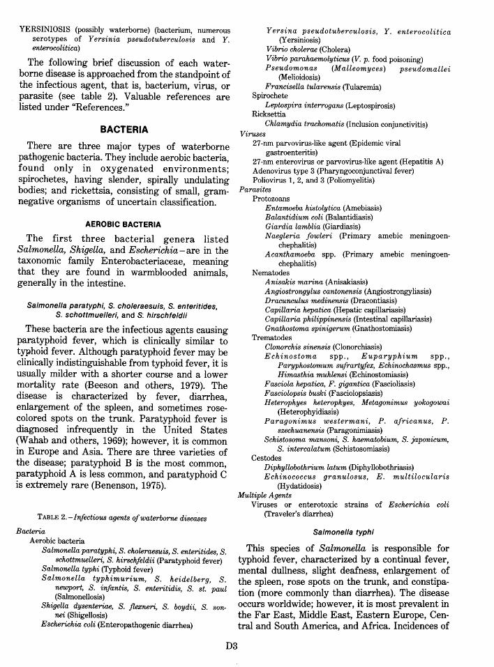
The following brief discussion of each waterborne disease is approached from the standpoint of the infectious agent, that is, bacterium, virus, or parasite (see table 2). Valuable references are listed under "References."
BACTERIA
There are three major types of waterborne pathogenic bacteria. They include aerobic bacteria, found only in oxygenated environments; spirochetes, having slender, spirally undulating bodies; and rickettsia, consisting of small, gramnegative organisms of uncertain classification.
AEROBIC BACTERIA
The first three bacterial genera listed Salmonella, Shigella, and Escherichia-are in the taxonomic family Enterobacteriaceae, meaning that they are found in warmblooded animals, generally in the intestine.
Salmonella paratyphi, S. choleraesuis, S. enteritides, S. schottmuelleri, and S: hirschfeldii
These bacteria are the infectious agents causing paratyphoid fever, which is clinically similar to typhoid fever. Although paratyphoid fever may be clinically indistinguishable from typhoid fever, it is usually milder with a shorter course and a lower mortality rate (Beeson and others, 1979). The disease is characterized by fever, diarrhea, enlargement of the spleen, and sometimes rosecolored spots on the trunk. Paratyphoid fever is diagnosed infrequently in the United States (Wahab and others, 1969); however, it is common in Europe and Asia. There are three varieties of the disease; paratyphoid B is the most common, paratyphoid A is less common, and paratyphoid C is extremely rare (Benenson, 1975).
TABLE 2.-lnfectious agents ofwaterborne diseases
Bacteria Aerobic bacteria
Salmonella paratyphi, S. choleraesuis, S. enteritides, S. schottmuelleri, S. hirschfeldii (Paratyphoid fever)
Salmonella typhi (Typhoid fever) Salmonella typhimurium, S. heidelberg, S.
newport, S. infantis, S. enteritidis, S. st. paul (Salmonellosis)
Shigella dysenteriae, S. jlexneri, S. boydii, S. sonnei (Shigellosis)
Escherichia coli (Enteropathogenic diarrhea)
D3
Yersina pseudotuberculosis, Y. enterocolitica (Y ersiniosis)
Vibrio cholerae (Cholera) Vibrio parahaemolyticus (V. p. food poisoning) Pseudomonas (Malleomyces) pseudomallei
(Melioidosis) Francisella tularensis (Tularemia)
Spirochete Leptospira interrogans (Leptospirosis)
Ricksettia Chlamydia trachomatis (Inclusion conjunctivitis)
Viruses 27-nm parvovirus-like agent (Epidemic viral
gastroenteritis) 27 -nm enterovirus or parvovirus-like agent (Hepatitis A) Adenovirus type 3 (Pharyngoconjunctival fever) Poliovirus 1, 2, and 3 (Poliomyelitis)
Parasites Protozoans
Entamoeba histolytica (Amebiasis) Balantidium coli (Balantidiasis) Giardia lamblia (Giardiasis) N aegleria fowleri (Primary amebic meningoen
chephalitis) Acanthamoeba spp. (Primary amebic meningoen
chephalitis) Nematodes
Anisakis marina (Anisakiasis) Angiostrongylus cantonensis (Angiostrongyliasis) Dracunculus medinensis (Dracontiasis) Capillaria hepatica (Hepatic capillariasis) Capillaria philippinensis (Intestinal capillariasis) Gnathostoma spinigerum (Gnathostomiasis)
Trematodes Clonorchis sinensis (Clonorchiasis) Echinostoma spp., Euparyphium spp.,
Paryphostomum sufrartyfex, Echinochasmus spp., Himasthia muhlensi (Echinostomiasis)
Fasciola hepatica, F. gigantica (Fascioliasis) Fasciolopsis buski (Fasciolopsiasis) Heterophyes heterophyes, Metagonimus yokogowai
(Heterophyidiasis) Paragonimus westermani, P. africanus, P.
szech uanensis (Paragonimiasis) Schistosoma mansoni, S. haematobium, S. japonicum,
S. intercalatum (Schistosomiasis) Cestodes
Diphyllobothrium latum (Diphyllobothriasis) Echinococcus granulosus, E. multilocularis
(Hydatidosis) Multiple Agents
Viruses or enterotoxic strains of Escherichia coli (Traveler's diarrhea)
Salmonella typhi
This species of Salmonella is responsible for typhoid fever, characterized by a continual fever, mental dullness, slight deafness, enlargement of the spleen, rose spots on the trunk, and constipation (more commonly than diarrhea). The disease occurs worldwide; however, it is most prevalent in the Far East, Middle East, Eastern Europe, Central and South America, and Africa. Incidences of
typhoid fever gradually have increased in Mexico since 1972 (Benenson, 1975). The number of cases reported annually in the United States has remained relatively constant, at about 500 cases (Wicks and others, 1971), for the past 10 to 20 years. Occurrence is greatest in young people. Mortality from the disease varies from 1 to 5 percent.
The disease is transmitted primarily by food or water contaminated by feces or urine of a carrier (Woodward and Smadel, 1964). It can be transmitted by improperly cooked starchy foods, raw fruits and vegetables, and in milk. Shellfish, in contaminated waters, have been shown as a source of the disease. S. typhi is very hardy and can survive for extended periods in polluted waters.
Salmonella typhimurium, S. heidelburg, S. newport, S. in fa ntis, S. enteritidis, and S. st. paul
Numerous serotypes of Salmonella are the agents of salmonellosis, which result in gastroenteritis, abdominal cramps, diarrhea, nausea, vomiting, dehydration, and fever. The disease is common worldwide and is reported extensively in North America and Europe. In 1975, Salmonella was isolated from 23,445 persons in the United States (Center for Disease Control, 1976). It often is called or is associated with food poisoning. The bacteria are found in virtually all foods. Salmonella is detected frequently in the polluted water environment (Geldreich, 1972). Epidemics generally are traced to commercially processed meat products, inadequately cooked poultry, raw sausage, eggs, unpasteurized milk, and even to contaminated eating utensils. A significant portion (1 to 58 percent) of raw meat purchased in retail markets is contaminated with Salmonella (Beeson and others, 1979). An individual can become infected by inhalation of the bacteria (Bennett and Hook, 1959).
In 1967, a severe epidemic of salmonellosis in Riverside, California, which produced more than 15,000 cases, resulted from contamination by sewage of an unchlorinated public ground-water supply. In the United States, about 0.2 percent of the population is an asymptomatic carrier of the bacteria at any one time (Beeson and other, 1979). Clinical symptoms of salmonellosis vary seasonally, with the largest number of infections occurring during July through November (Hornick, 1973). Shigella dysenteriae, S. flexneri, S. boydii, and S. sonnei
Shigellosis, or more commonly bacillary dysentery, is caused by many serotypes of Shigella.
It is a specific acute bacterial infection of the intestinal tract of man. The bacteria are the most commonly identified cause of diarrheal disease in the United States (Geldriech, 1972). The most common species in the United States, Western Europe, and Japan is S. sonnei: Prior to 1965, S. flexneri was predominant in this Country (Carpenter, 1976b), but remains as the most common form in developing nations. Symptoms vary from mild transitory diarrhea to acute attacks accompanied by fever, vomiting, and profuse bloody stools. The mortality in untreated cases may exceed 20 percent. The disease is found worldwide; it is endemic in certain custodial institutions, and among those populations where sanitation and personal hygiene are substandard. Over 60 percent of the cases occur in children under 10 years of age.
In 1969, the disease became pandemic in Central America, spreading from Guatemala and El Salvador. The infectious agent was S. dysenteriae (Neisman and others, 1973). It generally has summer and early autumn peaks of occurrence, but it can occur the year around. About 0.46 percent of the United States' population is a carrier at any one time. Shigellosis is transmitted by a direct or indirect fecal-oral route in food and water. It also can be transmitted by person-to-person contact. Survival time of the bacteria in water varies from 20 days in cold ground water to 30 minutes in warm, highly aerated streams.
Escherichia coli
Serogroups of the bacterium, E. coli, found in the coliform group are mostly nonpathogenic; however, about 140 pathogenic serotypes have been identified. They are the infectious agents of enteropathogenic diarrhea. Symptoms of the disease vary from two clinical types of the bacterium. The invasive strains produce symptoms similar to shigellosis -diarrhea, fever, and abdominal cramps. The enterotoxic strains produce symptoms similar to cholera-profuse watery diarrhea, abdominal cramps, dehydration, and fever. The disease occurs worldwide. It is the most serious form of diarrhea in children under 5 years of age, particularly in newborns since it is common in hospital nurseries. Mortality in infected newborns may approach 40 percent (Benenson, 1975). The bacterium is one of the many causes of traveler's diarrhea. The disease is transmitted in fecally contaminated food and water, and from mother to infant during delivery.
D4
Yersinia pseudotuberculosis and Y. enterocolitica
Y ersiniosis, erroneously called pseudotuberculosis, is caused by about 100 serotypes of Yersinia. The disease is characterized by diarrhea, cramps similar to appendicitis, fever, headache, sore throat, vomiting, arthritis, and skin ulcerations and abscesses. It is found worldwide; however, man is an accidental host, since the bacteria are found mostly in domesticated birds and mammals. Y ersiniosis is a rather newly recognized disease. In 1966, only 23 cases of yersiniosis had been reported, whereas more than 4,000 cases were reported by 1977 (Highsmith and others, ·1977). The first recognized epidemic of yersiniosis in the United States occurred in 1973 among 4 rural families; 16 of 21 persons in the families became ill (Benenson, 1975). It frequently is reported in Western Europe and Scandinavian countries. It is sporadically reported in the United States. The actual means of transmission has not been defined, but transmission is suspected to be by contaminated food and water (Highsmith and others, 1977).
Vibrio cholerae
Cholera, the most virulent and, when untreated, probably the most fatal of the waterborne diseases, is caused by the single species of bacterium, Vibrio cholerae. It is a serious acute intestinal disease characterized by sudden onset, profuse watery stools, vomiting, rapid dehydration, subnormal body temperature, and circulatory collapse. Death may occur within a few hours of onset. In untreated cases, fatality may exceed 50 percent. The disease results from an enterotoxin produced by the bacterium, which is transmitted almost exclusively by way of contaminated water. The bacterium survives for long periods in seawater, but only for a short time in freshwater (Carpenter, 1979a).
Epidemics of cholera are limited to areas of poor sanitation. During the 19th century, pandemic cholera repeatedly spread from its traditional source in India. In the early 20th century, the disease was largely confined to Asia, although a severe epidemic occurred in Egypt in 1947. The disease has caused seven worldwide pandemics since 1817, and three major North American epidemics in 1832, 1848, and 1867 (Carpenter, 1979a). Between 1961 and 1976, extension of the seventh pandemic of cholera spread from a focal
point in Indonesia through most of Asia, the Middle East, Eastern Europe, and Africa. Cholera remains endemic in the common delta of the Ganges and Brahmaputra Rivers of the Indian subcontinent (Barua and Burrows, 1974). There are no reports of indigenous cholera in the Western Hemisphere. In 1973, a case of unknown origin was confirmed in Texas. Fewer than 70,000 cases of cholera were reported in 1977; this represented a marked decrease, from 148,000 reported at the peak of the seventh pandemic in 1971 (Carpenter, 1979a).
Vibrio parahaemolyticus
Several serotypes are responsible for Vibrio parahaemolyticus food poisoning, a rarely fatal disease. The disease is an intestinal disorder characterized by watery diarrhea, abdominal cramps, nausea, vomiting, fever, and headache. It primarily occurs during the warm months. The disease is found worldwide, but it is most common in Japan, Southeast Asia, and the United States (Barker and Gangarosa, 1974). It is the most common cause of infective food poisoning in Japan, accounting for 59 percent of 22,000 cases of identifiable cause in 1963 (Lambert, 1979). The bacterium has been implicated as the cause of illness from eating steamed crabs in Maryland and boiled shrimp in Louisiana (Dadisman and others, 1972). Vibrio parahaemolyticus is primarily a marine organism, and as such is responsible for illness as a result of eating raw or inadequately cooked s'"'afood. This is the principal means of transmission.
Pseudomonas (Malleomyces) pseudomallei
The bacterium, Pseudomonas pseudomallei, also referred to as Whitmore's bacillus, is the cause of melioidosis, an uncommon disease (Khaira and others, 1959). Its symptoms vary from a mild circulatory infection to rapidly fatal septicemia (blood poisoning). Frequently, the initial symptoms of melioidosis are cutaneous abscesses, diarrhea, and pneumonia (Spotnitz and others, 1967). Cases have been reported in Southeast Asia, Iran, Australia, Ecuador, Panama, New Guinea, Guam, and Aruba. The disease is transmitted by intimate contact (for example, swimming or bathing) with contaminated water or soil or ingestion of contaminated water (Benenson, 1975).
D5
Francisella tularensis
The bacterium, Francisella tularensis, is the infectious agent of tularemia, a disease primarily found in rodents and lagomorphs (for example, rabbits). In man, the disease is characterized by the sudden and dramatic onset of chills and fever and swelling of the lymph nodes. Pneumonia occurs in about 30 percent of the cases (Woodward, 1979). Mortality in untreated cases varies from 5 to 30 percent, depending upon the clinical manifestation (Forshay, 1950). Tularemia occurs through most of North America, most of continental Europe, and Japan. Since 1967, fewer than 200 cases have been reported annually in the United States (Woodward, 1979). It occurs year around, but generally its incidence is higher during early winter during rabbit-hunting season. Tularemia generally is transmitted by contact with an infected animal; following contact, the bacterium penetrates the skin. It also is transmitted by bites from infected deerflies, ticks, mosquitos, and in aerosols (McCrumb, 1961).
Epidemics of tularemia resulting from contaminated water have been reported in Montana, Oregon (Benenson, 1975), and Vermont (Young, 1969). During World War II, an epidemic (25,000 persons) in the Soviet army resulted through use of a contaminated well.
SPIROCHETE Leptospira interrogans
Numerous serotypes of Leptospira are the cause of leptospirosis, also called W eil's disease or mud fever. The bacteria primarily produce acute infections of the kidneys; however, infections of the liver and central nervous system also may occur. Common symptoms include fever, headache, chills, vomiting, depression, muscular aches, and renal (kidney) insufficiency. Mortality may approach 10 percent. It is primarily a disease of teenagers and young adults, occurs predominantly in males, and develops most frequently in hot weather (Heath and others, 1965). The nesting sites of the bacteria in hosts (primarily rats, but also commonly foxes, opossums,skunks, and raccoons) are in kidney tubules; therefore, they are excreted with urine. Infection is by contact with water or wet soils contaminated with urine of carriers. The bacteria gain access to the body through skin abrasions, the nasopharynx, or the eye (Alexander, 1974). The disease is found worldwide, but it is most common in Europe, the Near East, North America, Central America, and the Far East. Leptospirosis,
although uncommon, has been reported from all regions of the United States. Between 1960 and 1976, 50 to 90 cases were reported annually (Sanford, 1979). It is commonly acquired by swimming or bathing in contaminated waters; most of the recognized common source outbreaks have been traced to contaminated water (Wong and others, 1977).
RICKETISIA
Chlamydia trachomatis
The organism, Chlamyd~a trachomatis, is the infectious agent of inclusion conjunctivitis, commonly called "sticky eye" in this country. Even though infections typically involve the eye, urethra, and cervix, it is primarily a venereal disease (Jawetz, 1979). Eye infection in a newborn is by direct contact with an infected cervix. Numerous outbreaks have been recorded from swimming in nonchlorinated pools, presumably contaminated with genitourinary discharges (Benenson, 1975). The disease is found worldwide; however, small epidemics have been reported in North America, Europe, and Japan.
VIRUSES
Viruses probably are responsible for more diseases, both recognized and unrecognized, ~n man than are all other types of infectious agents combined. Undoubtedly, many viral diseases can be transmitted in water (see Fox, 1976), particularly the enteroviruses such as polioviruses, coxsackieviruses, and echoviruses. Until the state-ofthe-understanding increases about viral diseases, in general, and their transmission, specifically, it is difficult to discuss them in a brief paper of this type. There are, however, four waterborne enteric viral diseases that are universally recognized. They are discussed below.
Viruses consist of either desoxyribonucleic acid (DNA) or ribonucleic acid (RNA), which are genetic materials and which serve as the infectious elements. The nucleic acid is enclosed within a protein sheath, called the capsid. A virus affects a living cell by obstructing the cell's own genetic system by replacing it with the viral DNA or RNA. The cell then functions according to its newly acquired genetic message. The response varies with the type of virus present and with the type of cell infected.
For additional information on viruses, refer to Cookson (1974), Berg and Bodily (1976), Evans (1976), or Martin (1978).
D6
Epidemic viral gastroenteritis
The disease is usually mild and self-limited. It often occurs in distinct outbreaks with explosive patterns of spread and is characterized by a variety of symptoms, including nausea, vomiting, diarrhea, abdominal pain, and low-grade fever. The disease usually lasts about 24 to 48 hours. It occurs worldwide, most frequently from September to March in the Northern Hemisphere. The infectious nucleic acid is a 27-nm, parvovirus-like, Norwalklike agent (Kapikian and others, 1972; Appleton and others, 1977).
Hepatitis A
Hepatitis A is characterized by an abrupt onset with fever, headache, nausea, vomiting, and abdominal discomfort, followed in a few days by jaundice . It generally is nonfatal. It may last from 1 to 2 weeks to several months. The disease occurs worldwide; most cases are observed during autumn and winter. It frequently is transmitted by person-to-person contact; however, epidemics generally are related to contaminated water and food.
The first description of hepatitis is attributed to Hippocrates; however, detailed descriptions of the disease did not appear until the 17th and 18th centuries. The infectious character of the disease was first described in the early 1900's. A large scale epidemic of hepatitis in 1955 was reported in New Delhi when a temporary diversion of a river resulted in the sewage contamination of the public water supply; over 30,000 cases were reported. Mosely (1967) suggested that the incidence of waterborne infectious hepatitis is grossly underreported, but estimated that less than 1 percent of the cases of the disease results from· transmission by water. The disease is caused by a 27-nm, enterovirus or parvovirus-like agent. It is highly resistant to chlorination, disinfectants, heat, and ultraviolet irradiation.
Pharyngoconjunctival fever
The disease, acute and self-limited, is characterized by pharyngitis, conjunctivitis, and fever which frequently peaks and subsides. Secondary symptoms include diarrhea, nasopharyngeal discharge, and swelling of the tonsils and lymph nodes of the neck. Either one or both of the eyes may become inflamed. The disease, observed in Europe and the United States in the 1920's was recognized formally in 1955 (Bell and others, 1961). It is primarily an epidemic disease found in family groups or in closed-contact groups, such as in organized camps.
Almost invariably, the disease can be related to contact with water, especially unsatisfactorily chlorinated swimming pools and small lakes (Foy and others, 1968). Parrott and others (1954) suggest that transmission is by direct contact with water containing the virus, which enters the eye or upper respiratory tract. The infectious adenovirus type 3 is susceptible to common chlorination practices.
Poliomyelitis
Poliomyelitis, often called polio or infantile paralysis, is an acute illness with a range of severity, from subclinical manifestations to paralysis. Only about 1 percent of infections result in a recognized clinical illness (Melnick, 1976). Symptoms generally include fever, headache, gastrointestinal upset, and stiffness of the back and neck. The site of paralysis, if it occurs, depends upon the location of nerve destruction. Polio is found worldwide, but large-scale epidemics now are limited to developing countries. In the United States, the first significant epidemic occurred in 1894 in Vermont; 132 cases were reported. Caverly's (1896) observation that many cases had occurred in the Otter Creek Valley suggested that the disease might be. waterborne. Epidemics of poliomyelitis have ben reduced markedly since the development of the polio vaccine in 1955. In the early 1950's, prior to the vaccine, about 38,000 cases were reported annually in the United States. In the early 1960's, an average of 570 cases were reported annually. During 1962-1963, the live attenuated vaccine was administered widely in this country, resulting in further reductions in the disease. By 1974, there were only 14 cases of poliomyelitis reported in the United States. Presently, the disease is virtually nonexistent in this country.
Polioviruses belong to the enterovirus group, which also includes coxsackieviruses and echoviruses, all of which inhabit the human alimentary tract. Numerous studies have reported the presence of enteroviruses in contaminated streams, in sewage, and in sewage effluents (Bloom and others 1959; Clarke and Kabler, 1964;
· Grinstein and others, 1970). Therefore, it is to be assumed that the multitude of clinical illnesses caused by enteroviruses can be related to the ingestion of contaminated water. The viruses are believed to enter the body through the alimentary tract by way of the mouth (Melnick, 1976). Enteroviruses are resistant to all known antibiotics and chemotherapeutic agents, as well as
D7
to standard laboratory disinfectants,, In fact, they are resistant to the standard methods used for s'ewage treatment. The density of enteroviruses in receiving waters generally is proportional to the extent of infection of the local populace.
PARASITES
There are four types of animal parasites that are significant infectious agents of waterborne diseases in man. They include: (1) protozoans, which are single-celled organisms of the Phylum Protozoa; (2) nematodes or round worms of the Phylum Nemathelminthes; (3) trematodes, flukes, or flat worms of the Phylum Platyhelminthes; ( 4) cestodes or tapeworms, also of the Phylum Platyhelminthes.
PROTOZOANS
There are five protozoans that are waterborne, disease-causing agents. They include:
) Entamoeba histolytica ) cause dysentary-type
) di~eases Balantidium coli ) (inhabit intestine).
) Giaradia lamblia )
) }{ aegleria fowleri )
) Acanthamoeba )
spp. ) )
causes neurological diseases
(inhabit central nervous system).
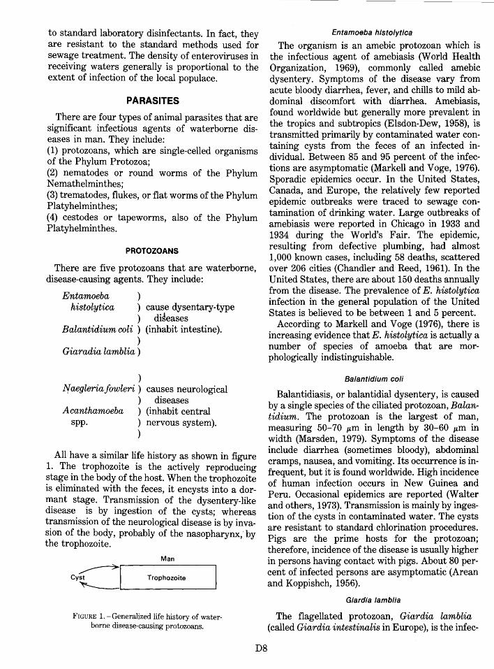
All have a similar life history as shown in figure 1. The trophozoite is the actively reproducing stage in the body of the host. When the trophozoite is eliminated with the feces, it encysts into a dormant stage. Transmission of the dysentery-like disease is by ingestion of the cysts; whereas transmission of the neurological disease is by invasion of the body, probably of the nasopharynx, by the trophozoite.
Man
Cy~~L----T_r_op_h_o_zo-it __ e __ ~
FIGURE 1.-Generalized life history of waterborne disease-causing protozoans.
D8
Entamoeba histolytica
The organism is an amebic protozoan which is the infectious agent of amebiasis (World Health Organization, 1969), commonly called amebic dysentery. Symptoms of the disea_se vary. from acute bloody diarrhea, fever, ~nd chills to mil? ~bdominal discomfort with diarrhea. Amebiasis, found worldwide but generally more prevalent in the tropics and subtropics (Elsdon-Dew, 1958), is transmitted primarily by contaminated water containing cysts from the feces of an infected individual. Between 85 and 95 percent of the infections are asymptomatic (Markell and Voge, 1976). Sporadic epidemics occur. In the United States, Canada, and Europe, the relatively few reported epidemic outbreaks were traced to sewage contamination of drinking water. Large outbreaks of amebiasis were reported in Chicago in 1933 and 1934 during the World's Fair. The epidemic, resulting from defective plumbing, had almost 1,000 known cases, including 58 deaths, scattered over 206 cities (Chandler and Reed, 1961). In the United States there are about 150 deaths annually from the dise~se. The prevalence of E. histolytica infection in the general population of the United States is believed to be between 1 and 5 percent.
According to Markell and Voge (1976), there is increasing evidence that E. histolytica is actually a number of species of amoeba that are morphologically indistinguishable.
Balantidium coli
Balantidiasis, or balantidial dysentery, is caused by a single species of the ciliated protozoan, Balantidium. The protozoan is the largest of rna~, measuring 50-70 I'm in length by 30-60 I'm In width (Marsden, 1979). Symptoms of the dis~ase include diarrhea (sometimes bloody), abdominal cramps, nausea, and vomiting. Its occ~rr~nc~ is infrequent, but it is found worldwide. High ~ncidence of human infection occurs in New Gmnea and Peru. Occasional epidemics are reported (Walter and others, 1973). Transmission is mainly by ingestion of the cysts in contaminated water. The cysts are resistant to standard chlorination procedures. Pigs are the prime hosts for . the proto~oan; therefore, incidence of the disease IS usually higher in persons having contact with pigs. Abo~t 80 percent of infected persons are asymptomatic (Arean and Koppishch, 1956).
Giardia Iamblia
The flagellated protozoan, Giardia lamblia (called Giardia intestinalis in Europe), is the infec-
tious agent of giardiasis, an infection of the small intestine (Jakubowski and Hoff, 1979). The disease is characterized by chronic diarrhea, which is often pale, greasy, or malodorous, abdominal cramps, bloating, excessive fatigue, and weight loss. Giardia is transmitted by water containing the cysts, which are resistant to standard chlorination procedures. The disease is found worldwide. Tour groups to Russia, particularly to Leningrad, frequently become infected (Brodsky and others, 1974; Gendel, 1974; Center for Disease Control, 1974; Craun, 1979). In certain parts of the United States, up to 20 percent of the population may be carriers.
An epidemic of giardiasis was recorded during the 1965-66 ski season at Aspen, Colorado, where 11 percent of all skiers had an acute infection (Moore and others, 1969). The epidemic increased the awareness of physicians of this country to the seriousness of the disease. Since the Aspen epidemic, there has been a tremendous increase in incidence in campers and backpackers in Idaho, California, Utah, Colorado, Oregon, and Kentucky. The disease has become so common that it has been termed, "backpacker's diarrhea." Since the epidemic in Aspen during the mid-1960's, 23 waterborne epidemics, affecting 7,009 persons, have been confirmed in this country. Among these, one of the most notable involved 4,800 individuals in E,ome, New York, during 1974-75 (Shaw and others, 1977). Four waterborne outbreaks of giardiasis, affecting 1,012 persons, were recorded in the United States during 1977 (Center for Disease Control, 1979). Of these, the largest outbreak, affecting 750 persons, occurred in New Hampshire. The infectious agerit was transmitted in a chlorinated municipal water supply.
Naegleria fow/eri Acanthamoeba spp.
The genera, Naegleria and Acanthamoeba, both free-living amebic protozoans, are responsible for the newly recognized disease, primary amebic meningoencephalitis (Butt, 1966; Carter, 1972). Naegleria is an amoeboflagellate in that it has an amoeboid phase alternating with a biflagellate phase. The disease is very rare, but usually rapidly fatal. It is characterized by inflammation of the brain, severe frontal headache, nausea, vomiting, and severe hemorrhaging from nasal passages. Death generally occurs in the fifth or sixth day. The disease was first reported in Australia in 1965 (Fowler and Carter, 1965); subsequently, ·cases have been diagnosed in Czechoslovakia, Britain, Ireland, Belgium, New Zealand, India, and Mrica.
In the United States, cases have been reported in Florida, Texas, Virginia, Georgia, Pennsylvania, New York, and California. Most cases have occurred in persons who had been swimming in warm fresh- or brackish-water, particularly in warm mineral springs, swimming pools, and in children who had played in mudholes. The body is invaded by the trophozoite, probably through invasion of the nasopharyngeal area.
Nemoatodes
Anisakis marina
Anisakiasis, caused by larvae of Anisakis marina, is characterized by fever, acute nausea, vomiting, and abscesses or granulomae in the walls of the stomach or small intestine. The life cycle of the nematode is unknown, but probably involves marine mammals. As far as is known, the larvae do not mature in man (Markell and Voge, 1976). The newly recognized disease is believed to be worldwide; numerous cases have been reported in Japan (Oshima, 1972) and along both coasts of the United States (Little and Most, 1973; Chitwood, 1975). It is acquired by the ingestion of raw or pickled marine fishes, particularly slightly salted raw herring.
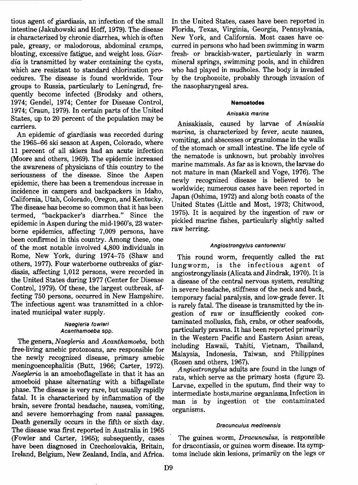
Angiostrongylus cantonenisi
This round worm, frequently called the rat lungworm, is the infectious agent of angiostrongyliasis (Alicata and Jindrak, 1970). It is a disease of the central nervous system, resulting in severe headache, stiffness of the neck and back, temporary facial paralysis, and low-grade fever. It is rarely fatal. The disease is transmitted by the ingestion of raw or insufficiently cooked contaminated mollusks, fish, crabs, or other seafoods, particularly prawns. It has been reported primarily in the Western Pacific and Eastern Asian areas, including Hawaii, Tahiti, Vietnam, Thailand, Malaysia, Indonesia, Taiwan, and Philippines (Rosen and others, 1967).
A ngiostrongylus adults are found in the lungs of rats, which serve as the primary hosts (figure 2). Larvae, expelled in the sputum, find their way to intermediate hosts,marine organisms. Infection in man is by ingestion ot the contaminated organisms.
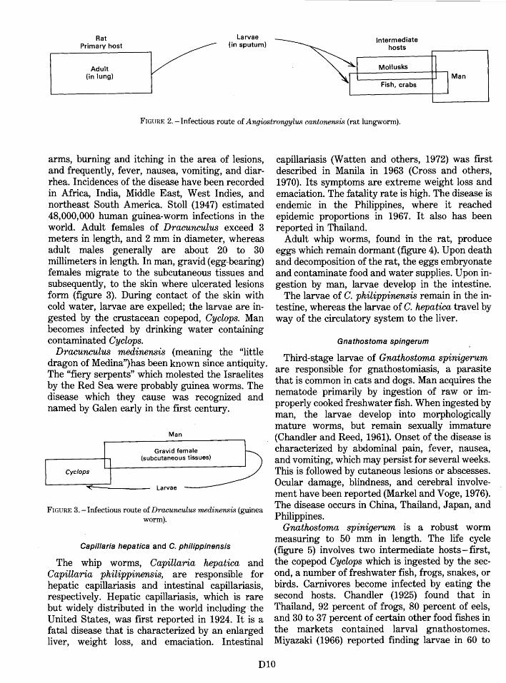
Dracunculus medinensis
The guinea worm, Dracunculus, is responsible for dracontiasis, or guinea worm disease. Its symptoms include skin lesions, primarily on the legs or
D9
Rat Primary host
Adult (in lung)
Larvae (in sputum)
Intermediate hosts
Mollusks
Fish, crabs Man
FIGURE 2.- Infectious route of A ngiostrongylus cantonensis (rat lungworm).
arms, burning and itching in the area of lesions, and frequently, fever, nausea, vomiting, and diarrhea. Incidences of the disease have been recorded in Africa, India, Middle East, West Indies, and northeast South America. Stoll (194 7) estimated 48,000,000 human guinea-worm infections in the world. Adult females of Dracunculus exceed 3 meters in length, and 2 mm in diameter, whereas adult males generally are about 20 to 30 millimeters in length. In man, gravid (egg-bearing) females migrate to the subcutaneous tissues and subsequently, to the skin where ulcerated lesions form (figure 3). During contact of the skin with cold water, larvae are expelled; the larvae are ingested by the crustacean copepod, Cyclops. Man becomes infected by drinking water containing contaminated Cyclops.
Dracunculus medinensis (meaning the "little dragon of Medina") has been known since antiquity. The "fiery serpents" which molested the Israelites by the Red Sea were probably guinea worms. The disease which they cause was recognized and named by Galen early in the first century.
Man
Gravid female (subcutaneous tissues)
Larvae
FIGURE 3.- Infectious route of Dracunculus medinensis (guinea worm).
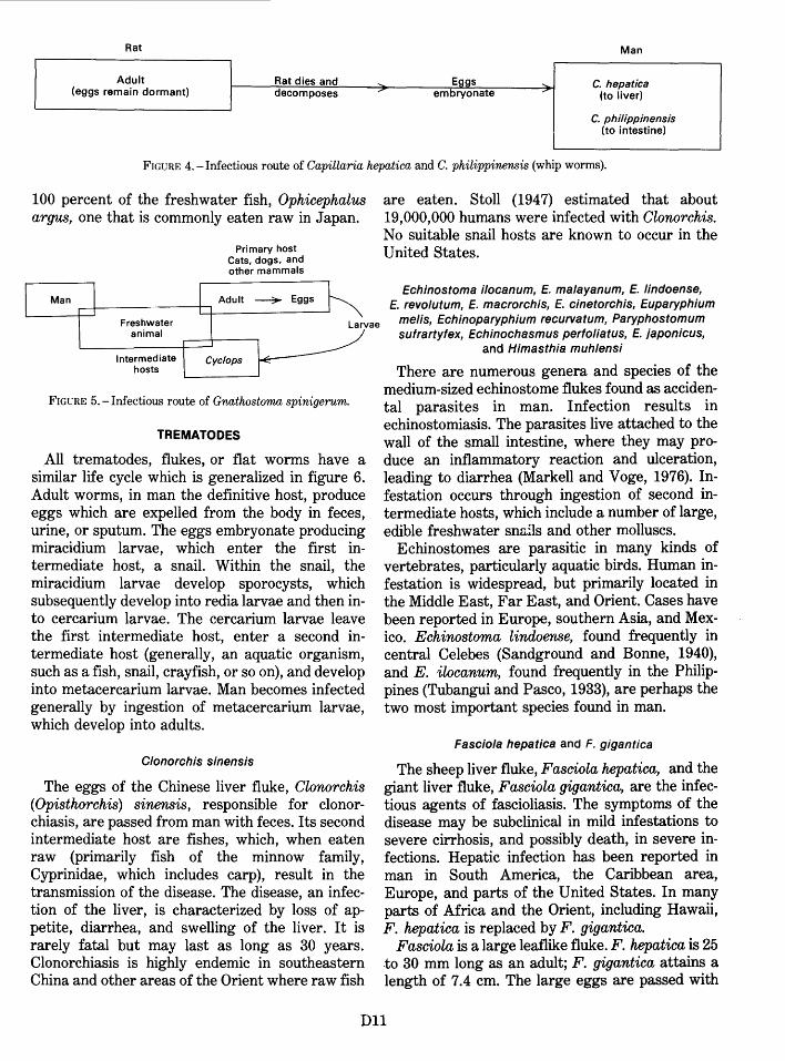
Capillaria hepatica and C. philippinensis
The whip worms, Capillaria hepatica and Capillaria philippinensis, are responsible for hepatic capillariasis and intestinal capillariasis, respectively. Hepatic capillariasis, which is rare but widely distributed in the world including the United States, was first reported in 1924. It is a fatal disease that is characterized by an enlarged liver, weight loss, and emaciation. Intestinal
capillariasis (W atten and others, 1972) was first described in Manila in 1963 (Cross and others, 1970). Its symptoms are extreme weight loss and emaciation. The fatality rate is high. The disease is endemic in the Philippines, where it reached epidemic proportions in 1967. It also has been reported in Thailand.
Adult whip worms, found in the rat, produce eggs which remain dormant (figure 4). Upon death and decomposition of the rat, the eggs embryonate and contaminate food and water supplies. Upon ingestion by man, larvae develop in the intestine.
The larvae of C. philippinensis remain in the intestine, whereas the larvae of C. hepatica travel by way of the circulatory system to the liver.
Gnathostoma spingerum
Third-stage larvae of Gnathostoma spinigerum are responsible for gnathostomiasis, a parasite that is common in cats and dogs. Man acquires the nematode primarily by ingestion of raw or improperly cooked freshwater fish. When ingested by man, the larvae develop into morphologically mature worms, but remain sexually immature (Chandler and Reed, 1961). Onset of the disease is characterized by abdominal pain, fever,· nausea, and vomiting, which may persist for several weeks. This is followed by cutaneous lesions or abscesses. Ocular damage, blindness, and cerebral involvement have been reported (Markel and Voge, 1976). The disease occurs in China, Thailand, Japan, and Philippines.
Gnathostoma spinigerum is a robust worm measuring to 50 mm in length. The life cycle (figure 5) involves two intermediate hosts- first, the copepod Cyclops which is ingested by the second, a number of freshwater fish, frogs, snakes, or birds. Carnivores become infected by eating the second hosts. Chandler (1925) found that in Thailand, 92 percent of frogs, 80 percent of eels, and 30 to 37 percent of certain other food fishes in the markets contained larval gnathostomes. Miyazaki (1966) reported finding larvae in 60 to
DlO
Rat Man
Adult Rat dies and Eggs ..., C. hepatica (eggs remain dormant) decomposes / embryonate ,.-
(to liver)
C. philippinensis (to intestine)
FIGURE 4.- Infectious route of Capillaria hepatica and C. philippinensis (whip worms).
100 percent of the freshwater fish, Ophicephalus argus, one that is commonly eaten raw in Japan.
Freshwater animal
Intermediate hosts
Primary host Cats, dogs, and other mammals
~Eggs
FIGURE 5.- Infectious route of Gnathostoma spinigerum.
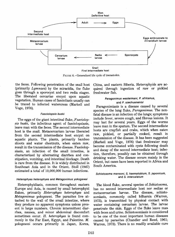
TREMATODES
All trematodes, flukes, or flat worms have a similar life cycle which is generalized in figure 6. Adult worms, in man the definitive host, produce eggs which are expelled from the body in feces, urine, or sputum. The eggs embryonate producing miracidium larvae, which enter the first intermediate host, a snail. Within the snail, the miracidium larvae develop sporocysts, which subsequently develop into redia larvae and then into cercarium larvae. The cercarium larvae leave the first intermediate host, enter a second intermediate host (generally, an aquatic organism, such as a fish, snail, crayfish, or so on), and develop into metacercarium larvae. Man becomes infected generally by ingestion of metacercarium larvae, which develop into adults.
Clonorchis sinensis
The eggs of the Chinese liver fluke, Clonorchis (Opisthorchis) sinensis, responsible for clonorchiasis, are passed from man with feces. Its second intermediate host are fishes, which, when eaten raw (primarily fish of the minnow family, Cyprinidae, which includes carp), result in the transmission of the disease. The disease, an infection of the liver, is characterized by loss of appetite, diarrhea, and swelling of the liver. It is rarely fatal but may last as long as 30 years. Clonorchiasis is highly endemic in southeastern China and other areas of the Orient where raw fish
are eaten. Stoll (1947) estimated that about 19,000,000 humans were infected with Clonorchis. No suitable snail hosts are known to occur in the United States.
Echinostoma ilocanum, E. malayanum, E. lindoense, E. revolutum, E. macrorchis, E. cinetorchis, Euparyphium
me/is, Echinoparyphium recurvatum, Paryphostomum sufrartyfex, Echinochasmus perfoliatus, E. japonicus,
and Himasthia muhlensi
There are numerous genera and species of the medium-sized echinostome flukes found as accidental parasites in man. Infection results in echinostomiasis. The parasites live attached to the wall of the small intestine, where they may produce an inflammatory reaction and ulceration, leading to diarrhea (Markell and Voge, 1976). Infestation occurs through ingestion of second intermediate hosts, which include a number of large, edible freshwater snails and other molluscs.
Echinostomes are parasitic in many kinds of vertebrates, particularly aquatic birds. Human infestation is widespread, but primarily located in the Middle East, Far East, and Orient. Cases have been reported in Europe, southern Asia, and Mexico. Echinostoma lindoense, found frequently in central Celebes (Sandground and Bonne, 1940), and E. ilocanum, found frequently in the Philippines (Tubangui and Pasco, 1933), are perhaps the two most important species found in man.
Fasciola hepatica and F. gigantica
The sheep liver fluke, Fasciola hepatica, and the giant liver fluke, Fasciola gigantica, are the infectious agents of fascioliasis. The symptoms of the disease may be subclinical in mild infestations to severe cirrhosis, and possibly death, in severe infections. Hepatic infection has been reported in man in South America, the Caribbean area, Europe, and parts of the United States. In many parts of Africa and the Orient, including Hawaii, F. hepatica is replaced by F. gigantica.
Fasciola is a large leaflike fluke. F. hepatica is 25 .to 30 mm long as an adult; F. gigantica attains a length of 7.4 em. The large eggs are passed with
D11
Man Definitive host
~~L...---Adu-lt ->-Eg-gs ~~ Second
intermediate host
Metacercari um larvae
~ Cercarium Redia ./
larvae ""' larvae
Snail
~ Sporocysts .......
Eggs emb ryonate to m larvae miracidiu
v First intermediate host
FIGURE 6. -Generalized life cycle of trematodes.
the feces. Following penetration of the snail host (primarily Lymnaea) by the miracidia, the fluke goes through a sporocyst and two redia stages. The liberated cercariae encyst upon aquatic vegetation. Human cases of fascioliasis usually can be traced to infected watercress (Markell and Voge, 1976).
Fasciolopsis buski
The eggs of the giant intestinal fluke, Fasciolopsis buski, the infectious agent of fasciolopsiasis, leave man with the feces. The second intermediate host is the snail. Metacercarium larvae liberated from the second intermediate host encyst on aquatic plants. The plants, primarily bamboo shoots and water chestnuts, when eaten raw, result in the transmission of the disease. Fasciolopsiasis, an infection of the small intestine, is characterized by alternating diarrhea and constipation, vomiting, and intestinal blockage. Death is rare from the disease. It is widely distributed in Southeast Asia and in the Orient. Stoll (1947) estimated a total of 10,000,000 human infections.
Heterophyes heterophyes and Metagonimus yokogawai
Heterophyidiasis, common throughout eastern Europe and Asia, is caused by small heterophyid flukes, primarily Heterophyes heterophyes and Metagonimus yokogawai. The parasites live attached to the wall of the small intestine, where they produce no apparent symptoms unless present in large numbers. Chronic intermittent diarrhea, nausea, and minor abdominal discomfort sometimes occur. H. heterophyes is found commonly in the Far East, Egypt, and Palestine. M. yokogawai occurs primarily in Japan, Korea,
China, and eastern Siberia. Heterophyids are acquired through ingestion of raw or pickled freshwater fish.
Paragonimus westermani, P. africanus, and P. szechuanenisi
Paragonimiasis is a disease caused by several species of the lung fluke, Paragonimus. The nonfatal disease is an infection of the lungs; symptoms include fever, severe cough, and fibrous lesions. It may last for several years. Eggs of the worms leave man in the sputum. The second intermediate hosts are crayfish and crabs, which when eaten raw, pickled, or partially cooked, result in transmission of the disease. It has been suggested (Markell and Voge, 1976) that freshwater may become contaminated with cysts following death and decay of the second intermediate host; infection, therefore, possibly can be obtained through drinking water. The disease occurs mainly in the Orient, but cases have been reported in Mrica and South America.
Schistosoma mansoni, S. haematobium, S. japonicum, and S. intercalatum
The blood fluke, several species of Schistosoma, has no second intermediate host nor rediae or metacercarium larvae. The disease, schistosomiasis, commonly called bilharzia (Warren, 1973), is transmitted by physical contact with water containing cercarium larvae. The larvae penetrate the skin. Eggs of the fluke leave man with feces and urine. Schistosomiasis is considered to be one of the most important human diseases caused by parasites (Chandler and Reed, 1961; Warren, 1973). There is no readily available cure
D12
for the disease, nor easy means of control of the intermediate host.
The long-lasting rarely fatal disease is one of the circulatory system. Minute scars are produced in organs where the adults lodge. S. mansoni, S. japonicum, and S. intercalatum are responsible for intestinal clinical symptoms, whereas S. haematobium results in urinary tract infections. The disease occurs worldwide: S. mansoni is found primarily in Mrica, the Brazilian peninsula, and in eastern South America and the Caribbean; S. hematobium occurs in Africa and the Middle East; S. japonicum occurs in the Orient; and S. intercalatum occurs in western and central Africa.
Man probably has been infected with Schistosoma for thousands of years. The early Egyptians left recorded accounts in hieroglyphics of bloody urine, a telltale sign of S. haematobium infestation. Ruffer (1910) noted the presence of calcified eggs of Schistosoma in Egyptian mummies from 1250-1000 B.C. The parasite was observed and described by Theodor Bilharz in 1851. Stoll (1947) estimated that there were 114 million persons infected with schistosomes in the world, 46 million of them were in the Orient.
A related disease, called swimmer's itch, is common in the United States. It is caused by larvae of bird and mammal schistosomes (Trichobilharzia spp., Gigantobilharzia spp., Schistosomatium douthitti, and Microbilharzia spp.) attempting to penetrate the skin. The result is short-termed dermatitis, sometimes complicated by pustules.
CESTODES
Diphyllobothrium latum
The. broad or fish tapeworm is the largest tapeworm found in man. It commonly will reach 9 meters in length, be 2.5 centimeters in width, and contain over 4,000 "segments." One woman was reported to be infected with four worms having a total length of over 88 meters (290 ft) (Chandler and Reed, 1961). The disease of Diphyllobothrium is diphyllobothriasis, an intestinal disease of long duration. Symptoms are generally trivial (for example, slight anemia) or absent. The disease is endemic worldwide in temperate zones, where eating raw or partly cooked fish is popular. It is common in the United States, .since pike and perch are common second intermediate hosts.
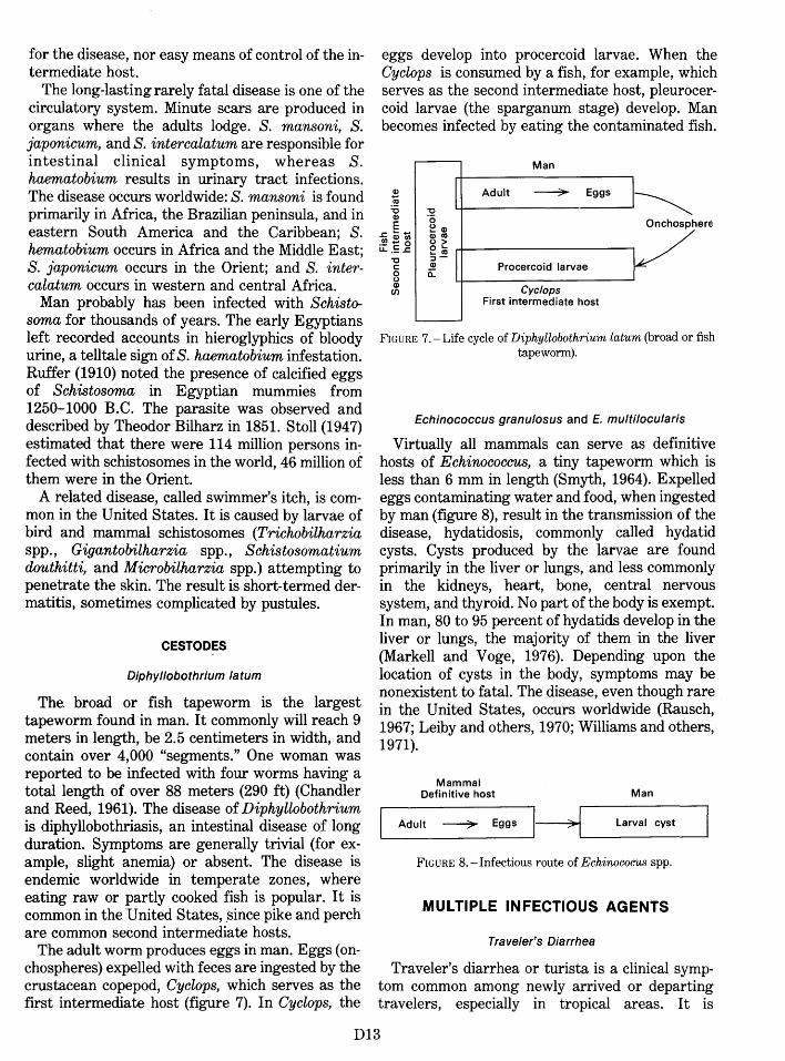
The adult worm produces eggs in man. Eggs (onchospheres) expelled with feces are ingested by the crustacean copepod, Cyclops, which serves as the first intermediate host (figure 7). In Cyclops, the
eggs develop into procercoid larvae. When the Cyclops is consumed by a fish, for example, which serves as the second intermediate host, pleurocercoid larvae (the sparganum stage) develop. Man becomes infected by eating the contaminated fish.
Man
Adult ~ Eggs ---..._ ! (II
4--------' ~ :0 "C Q) ·a E ~ Q)
~Ciiti Q)«J
ii: .'g _g e ~ r-1-----------. "C ::J-c: Q)
0 a: u Q) (/)
Procercoid larvae
Cyclops First intermediate host
Onchosphere
FIGURE 7.- Life cycle of Diphyllobothrium latum (broad or fish tapeworm).
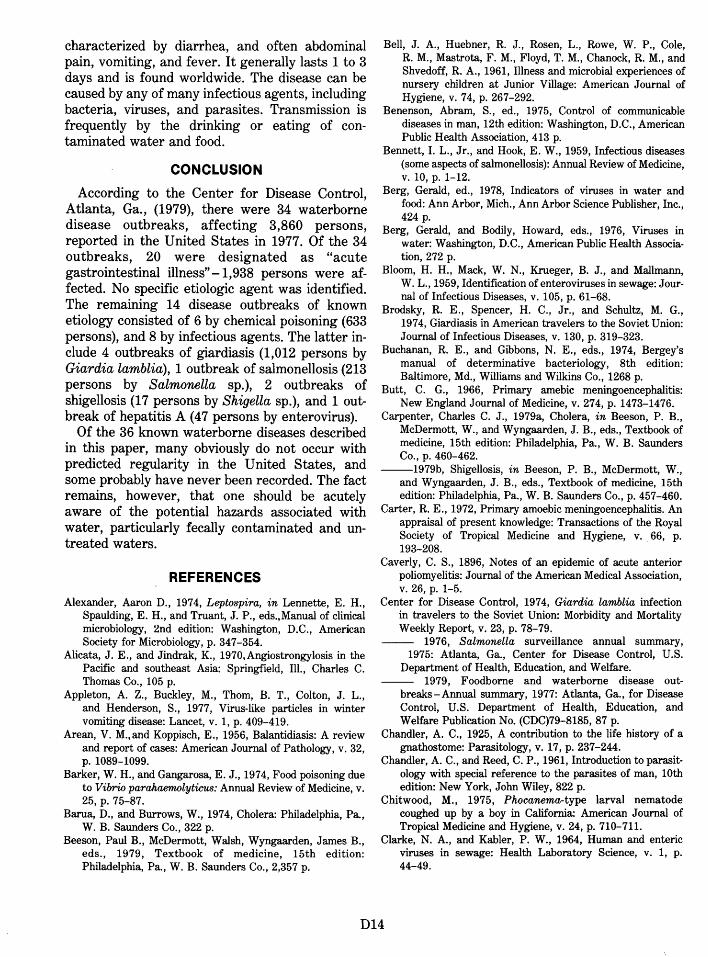
Echinococcus granulosus and E. multilocularis
Virtually all mammals can serve as definitive hosts of Echinococcus, a tiny tapeworm which is less than 6 mm in length (Smyth, 1964). Expelled eggs contaminating water and food, when ingested by man (figure 8), result in the transmission of the disease, hydatidosis, commonly called hydatid cysts. Cysts produced by the larvae are found primarily in the liver or lungs, and less commonly in the kidneys, heart, bone, central nervous system, and thyroid. No part of the body is exempt. In man, 80 to 95 percent of hydatids develop in the liver or lungs, the majority of them in the liver (Markell and Voge, 1976). Depending upon the location of cysts in the body, symptoms may be nonexistent to fatal. The disease, even though rare in the United States, occurs worldwide (Rausch, 1967; Leiby and others, 1970; Williams and others, 1971).
Mammal Definitive host Man
Adult ------7 Eggs I FIGURE 8.- Infectious route of Echirwcocus spp.
.MULTIPLE INFECTIOUS AGENTS
Traveler's Diarrhea
Traveler's diarrhea or turista is a clinical symptom common among newly arrived or departing travelers, especially in tropical areas. It is
D13
characterized by diarrhea, and often abdominal pain, vomiting, and fever. It generally lasts 1 to 3 days and is found worldwide. The disease can be caused by any of many infectious agents, including bacteria, viruses, and parasites. Transmission is frequently by the drinking or eating of contaminated water and food.
CONCLUSION
According to the Center for Disease Control, Atlanta, Ga., (1979), there were 34 waterborne disease outbreaks, affecting 3,860 persons, reported in the United States in 1977. Of the 34 outbreaks, 20 were designated as "acute gastrointestinal illness" -1,938 persons were affected. No specific etiologic agent was identified. The remaining 14 disease outbreaks of known etiology consisted of 6 by chemical poisoning (633 persons), and 8 by infectious agents. The latter include 4 outbreaks of giardiasis (1,012 persons by Giardia lamblia), 1 outbreak of salmonellosis (213 persons by Salmonella sp.), 2 outbreaks of shigellosis (17 persons by Shigella sp.), and 1 outbreak of hepatitis A ( 4 7 persons by enterovirus).
Of the 36 known waterborne diseases described in this paper, many obviously do not occur with predicted regularity in the United States, and some probably have never been recorded. The fact remains, however, that one should be acutely aware of the potential hazards associated with water, particularly fecally contaminated and untreated waters.
REFERENCES
Alexander, Aaron D., 1974, Leptospira, in Lennette, E. H., Spaulding, E. H., and Truant, J. P., eds.,Manual of clinical microbiology, 2nd edition: Washington, D.C., American Society for Microbiology, p. 347-354.
Alicata, J. E., and Jindrak, K., 1970,Angiostrongylosis in the Pacific and southeast Asia: Springfield, Ill., Charles C. Thomas Co., 105 p.
Appleton, A. Z., Buckley, M., Thorn, B. T., Colton, J. L., and Henderson, S., 1977, Virus-like particles in winter vomiting disease: Lancet, v. 1, p. 409-419.
Arean, V. M.,and Koppisch, E., 1956, Balantidiasis: A review and report of cases: American Journal of Pathology, v. 32, p. 1089-1099.
Barker, W. H., and Gangarosa, E. J., 1974, Food poisoning due to Vibrio parahaemolyticus: Annual Review of Medicine, v. 25, p. 75-87.
Barua, D., and Burrows, W., 1974, Cholera: Philadelphia, Pa., W. B. Saunders Co., 322 p.
Beeson, Paul B., McDermott, Walsh, Wyngaarden, James B., eds., 1979, Textbook of medicine, 15th edition: Philadelphia, Pa., W. B. Saunders Co., 2,357 p.
Bell, J. A., Huebner, R. J., Rosen, L., Rowe, W. P., Cole, R. M., Mastrota, F. M., Floyd, T. M., Chanock, R. M., and Shvedoff, R. A., 1961, Illness and microbial experiences of nursery children at Junior Village: American Journal of Hygiene, v. 74, p. 267-292.
Benenson, Abram, S., ed., 1975, Control of communicable diseases in man, 12th edition: Washington, D.C., American Public Health Association, 413 p.
Bennett, I. L., Jr., and Hook, E. W., 1959, Infectious diseases (some aspects of salmonellosis): Annual Review of Medicine, v. 10, p. 1-12.
Berg, Gerald, ed., 1978, Indicators of viruses in water and food: Ann Arbor, Mich., Ann Arbor Science Publisher, Inc., 424 p.
Berg, Gerald, and Bodily, Howard, eds., 1976, Viruses in water: Washington, D.C., American Public Health Association, 272 p.
Bloom, H. H., Mack, W. N., Krueger, B. J., and Mallmann, W. L., 1959, Identification of enteroviruses in sewage: Journal of Infectious Diseases, v. 105, p. 61-68.
Brodsky, R. E., Spencer, H. C., Jr., and Schultz, M. G., 1974, Giardiasis in American travelers to the Soviet Union: Journal of Infectious Diseases, v. 130, p. 319-323.
Buchanan, R. E., and Gibbons, N. E., eds., 1974, Bergey's manual of determinative bacteriology, 8th edition: Baltimore, Md., Williams and Wilkins Co., 1268 p.
Butt, C. G., 1966, Primary amebic meningoencephalitis: New England Journal of Medicine, v. 274, p. 1473-1476.
Carpenter, Charles C. J., 1979a, Cholera, in Beeson, P. B., McDermott, W., and Wyngaarden, J. B., eds., Textbook of medicine, 15th edition: Philadelphia, Pa., W. B. Saunders Co., p. 460-462.
--1979b, Shigellosis, in Beeson, P. B., McDermott, W., and Wyngaarden, J. B., eds., Textbook of medicine, 15th edition: Philadelphia, Pa., W. B. Saunders Co., p. 457-460.
Carter, R. E., 1972, Primary amoebic meningoencephalitis. An appraisal of present knowledge: Transactions of the Royal Society of Tropical Medicine and Hygiene, v. 66, p. 193-208.
Caverly, C. S., 1896, Notes of an epidemic of acute anterior poliomyelitis: Journal of the American Medical Association, v. 26, p. 1-5.
Center for Disease Control, 1974, Giardia lamblia infection in travelers to the Soviet Union: Morbidity and Mortality Weekly Report, v. 23, p. 78-79.
-- 1976, Salmonella surveillance annual summary, 1975: Atlanta, Ga., Center for Disease Control, U.S.
Department of Health, Education, and Welfare. -- 1979, Foodborne and waterborne disease out
breaks-Annual summary, 1977: Atlanta, Ga., for Disease Control, U.S. Department of Health, Education, and Welfare Publication No. (CDC)79-8185, 87 p.
Chandler, A. C., 1925, A contribution to the life history of a gnathostome: Parasitology, v. 17, p. 237-244.
Chandler, A. C., and Reed, C. P., 1961, Introduction to parasitology with special reference to the parasites of man, lOth edition: New York, John Wiley, 822 p.
Chitwood, M., 1975, Phocanema-type larval nematode coughed up by a boy in California: American Journal of Tropical Medicine and Hygiene, v. 24, p. 710-711.
Clarke, N. A., and Kabler, P. W., 1964, Human and enteric viruses in sewage: Health Laboratory Science, v. 1, p. 44-49.
D14
Cookson, John T., Jr., 1974, Virus and water supply: Journal of the American Water Works Association, December, p. 707-711.
Craun, G. F., 1979, Waterborne outbreaks of giardiasis, in Jakubowski, W., and Hoff, J. C., eds., Waterborne transmission of giardiasis: Cincinnati, Ohio, U.S. Environmental Protection Agency, EPA-600/9-79-001, p. 127-149.
Cross, J. H., Banzon, T., Murrell, K. D., Watten, R. H., and Dizon, J. J., 1970, A new epidemic diarrheal disease caused by the nematode Capillaria philippinensis: Industry and Tropical Health, v. 7, p. 129-151.
Dadisman, T. A., Nelson, R., Molenda, J. R., and Garber, H. J., 1972, Vibrio parahaemolyticus in Maryland. 1. Clinical and epidemiological aspects: American Journal of Epidemiology, v. 96, p. 414-426.
Elsdon-Dew, R., 1958, Factors influencing the pathogenicity of Entarrweba histolytica: Proceedings of the World Congress on Gastroenterology, v. II, p. 770-773.
Evans, Alfred S., ed., 1976, Viral infections of humansEpidemiology and control: New York, Plenum Medical Book Co., 584 p.
Foshay, L., 1950, Tularemia: Annual Review of Microbiology, v. 4, p. 313-334.
Fowler, M., and Carter, R. F., 1965,Acute pyogenic meningitis probably due to Acantharrweba sp.: A preliminary report: British Medical Journal, v. 2, p. 740-742.
Fox, John P., 1976, Human-associated viruses in water, in Berg, G., and Bodily, H., eds., Viruses in water: Washington D.C., American Public Health Association, p. 39-49.
Foy, H. M., Cooney, M. K., and Hatten, J. B., 1968, Adenovirus type 3 epidemic associated with intermittent chlorination of a swimming pool: Archives of Environmental Health, v. 17, p. 795-802.
Geldreich, Edwin E., 1972, Water-borne pathogens, in Mitchell, Ralph, ed., Water pollution microbiology: New York, WileyInterscience, p. 207-241.
Gendel, E., 1974, Giardiasis in Russia: New England Journal of Medicine, v. 290, p. 286.
Grinstein, S., Melnick, J. L., and Wallis, C., 1970, Virus isolations from sewage and from a stream receiving effluents of sewage treatment plants: Bulletin of the World Health Organization, v. 42, p. 291-296.
Heath, C. W., Jr., Alexander, A. D., and Galton, M. M., 1965, Leptospirosis in the United States. Analysis of 483 cases in man. 1949-1961: New England Journal of Medicine, v. 273, p. 857-865.
Highsmith, A. K., Feeley, J. C., and Morris, G. K., 1977, Isolation of Y ersina enterocolitica from water, in Hoadley, A. W., and Dutka, B. J., eds., Bacterial indicators-Health hazards associated with water: Philadelphia, Pa., American Society for Testing and Materials, ASTM Special Technical Publication 635, p. 265-274.
Hoadley, A.W., and Dutka, B. J., eds., 1977, Bacterial indicators/health hazards associated with water: Philadelphia, Pa., American Society for Testing and Materials, ASTM Special Technical Publication 635, 356 p.
Hornick, R. B., 1973, Salrrwnella infection-Newer perspectives of an old infection: Transactions of American Clinical Climatological Association, v. 85, p. 164-176.
Jakubowski, W., and Hoff, J. C., eds., 1979, Waterborne transmission of giardiasis: Cincinnati, Ohio, U.S. Environmental Protection Agency, EPA 600/9-79-001,306 p.
Jawetz, Ernest, 1979, Trachoma and inclusion conjunctivitis, in Beeson, P. B., McDermott, W., and Wyngaarden, J. B., eds., Textbook of medicine, 15th edition: Philadelphia, Pa., W. B. Saunders Co., p. 332-335.
Kapikian, A. Z., Wyatt, R. G., Dolin, R., Thornhill T. S., Kalica, A. R., and Chanock, R. M., 1972, Visualization of a 27 -mn particle associated with acute infectious nonbacterial gastroenteritis: Journal of Virology, v. 10, p. 1075-1079.
Khaira, B.S., Young, W. B., and Hart, P. D., 1959, Melioidosis: British Medical Journal, v. 1, p. 949-961.
Lambert, Harold P., 1979, Food poisoning, in Beeson, P. B., McDermott, W., and Wyngaarden, J. B., eds., Textbook of medicine, 15th edition: Philadelphia, Pa., W. B. Saunders Co., p. 64-71.
Leiby, P. D., Carney, W. P., and Woods, C. E., 1970, Studies on sylvatic echinococcosis. III. Host occurrence and geographic distribution of Echinococcus multilocularis in the North Central United States: Journal of Parasitology, v. 56, p. 1141-1150.
Lenette, E. H., Spaulding, E. H., and Truant, J.P., eds., 1974, Manual of clinical microbiology, 2nd edition: Washington, D.C., American Society for Microbiology, 970 p.
Little, M. D., and Most, H., 1973, Anisakid larva from the throat of a woman in New York: American Journal of Tropical Medicine and Hygiene, v. 22, p. 609-612.
Malina, J. F., and Sagik, B. P., eds., 1974, Virus survival in water and wastewater systems: Austin, Tex., Center of Research in Water Resouces, Water Resources Symposium No.7, 264 p.
Markell, Edward K., and Voge, Marietta, 1976, Medical parasitology, 4th edition: Philadelphia, Pa., W. B. Saunders Co., 393 p.
Marsden, Philip D., 1979, Balantidiasis, in Beeson, P. B., McDermott, W., and Wyngaarden, J. B., eds., Textbook of medicine, 15th edition: Philadelphia, Pa., W. B. Saunders Co., p. 603.
Martin, S. J., 1978, The biochemistry of viruses: New York, Cambridge University Press, 145 p.
McCrumb, F. R., 1961, Aerosol infection of man with Pastuerella tularensis: Bacterial Review, v. 25, p. 262-270.
Melnick, J. L., 1957, A water-borne urban epidemic of hepatitis, in Hartman, F. W., ed., Hepatitis frontiers: Boston, Little, Brown, and Co., p. 211-225.
---1976, Enteroviruses, in Evans, A. S., ed., Viral infections of humans-Epidemiology and control: New York, Plenum Medical Book Co., p. 163-207.
Mitchell, Ralph, ed., 1972, Water pollution microbiology, v. 1: New York, Wiley-lnterscience, 416 p.
--1972b, Water pollution microbiology, v. 2: New York, Wiley-Interscience, 442 p.
Miyazaki, J., 1966, Gnathostoma and gnathostomiasis in Japan. Progress of medical parasitology in Japan: Meguro Parasitological Museum, v. 3, p. 531-586.
Moore, G. T., Cross, W. M., McGuire, D., Mollohan, D. S., Gleason, N. N., Healy, G. R., and Newton, L. H., 1969, Epidemic giardiasis at a ski resort: New England Journal of Medicine, v. 281, p. 402-407.
Mosley, J. W., 1967, Transmission of viral diseases by drinking water, in Berg, G., ed., Transmission of viruses by. the water route: New York, Interscience Publishers, p. 5-23.
National Research Council, 1977, Drinking water and health: Washington, D.C., National Academy of Sciences, 939 p.
Neisman, J. B., Martin, K. 1., Lewis, J. N., Freidman, C. T. H., and Gangarosa, E. J., 1973, Impact in the United States of
D15
the Shiga dysentery pandemic of Central America and Mexico-A review of the surveillance data through 1972: Journal of Infectious Diseases, v. 128, p. 574-576.
Oshima, T., 1972, Anisakis and anisakiasis in Japan and adjacent area: Meguro Parasitological Museum, v. 4, p. 303-393.
Parrott, R. H., Rowe, W. P., Huebner, R. J., Bernton, H. W., and McCullough, N. B., 1954, Outbreaks of febrile pharyngitis and conjunctivitis associated with type 3 adenoidal-pharyngeal-conjunctival virus infection: New England Journal of Medicine, v. 251, p. 1087-1090.
Pipes, Wesley 0., ed., 1978, Water quality and health significance of bacterial indicators of pollution: Philadelphia, Pa., Drexel University, 228 p.
Rausch, R. L., 1967, On the occurrence and distribution of Echinococcus spp. (Cestoda: Taeniidae), and characteristics of their development in the intermediate host: Annuals of parasitology, v. 42, p. 19-63.
Rosen, L., Loison, G., Laigret, J., and Wallace, G. D., 1967, Studies on eosinophilic meningitis. 3. Epidemiologic and clinical observations on Pacific islands and the · possible etiologic role of A ngiostrongylus cantonensis: American Journal of Epidemiology, v. 85, p. 17-44.
Ruffer, M. A., 1910, Notes on the presence of Bilharzia haematobia in Egyptian mummies of the twentieth dynasty, 1220-1000 B.C.: British Medical Journal, v. 1, p. 16.
Sandground, J. H., and Bonne, C., 1940, Echinostoma lindoensis, n. sp., a new parasite of man in the Celebes with an account of its life history and epidemiology: American Journal of Tropical Medicine, v. 20, p. 511-535.
Sanford, Jay P., 1979, Leptospirosis, in Beeson, P. B., McDermott, W., and Wyngaarden, J. B., eds., Textbook of Medicine, 15th edition: Philadelphia, Pa., W. B. Saunders Co., p. 532-535.
Shaw, P. K., Brodsky, R. E., and Lyman, D. 0., 1977, A communitywide outbreak of giardiasis with evidence of transmission by a municipal water supply: Annals of Internal Medicine, v. 87, p. 426-432.
Smyth, J. D., 1964, The biology of the hydatid organism, in Dawes, B., ed., Advances in parasitology: New York, Academic Press, p. 169-219.
Spotnitz M., Rudnitsky, A., and Rambaud, J. J., 1967, Melioidosis pneumonitis: Journal of the American Medical Association, v. 202, p. 950-956.
Stoll, N. R., 1947, This wormy world: Journal of Parasitology, v. 33, p. 1-8.
Tubangui, M. A., and Pasco, A. M., 1933, The life history of the human intestinal fluke Euparyphium ilocanum (Garrison, 1908): Philippine Journal of Science, v. 51, p. 581-606.
Wahab, M.F.A., Robertson, R. P., and Raasch, F. 0., 1969, Paratyphoid A fever: Annals of Internal Medicine, v. 70, p. 913-925.
Walter, P. D., Judson, F. N., Murphy, K. B., Healy, G. R., English, D. K., and Schultz, M. G., 1973, Balantidiasis outbreak in Truk: American Journal of Tropical Medicine and Hygiene, v. 22, p. 33-41.
Warren, Kenneth S., 1973, Schistosomiasis: The evolution of a medical literature: Cambridge, Mass., MIT Press, 1307 p.
Watten, R. H., Beckner, W. M., Cross, J. H., Gunning, J. J., and Jarimillo, J., 1972, Clinical studies of capillariasis philippinensis: Transactions of the Royal Society of Tropical Medicine and Hygiene, v. 66, p. 828-834.
Wicks, C. B., Holmes, G. S., and Davidson, L., 1971, Endemic typhoid fever: Quarterly Journal of Medicine, v. 159, p. 341-353.
Williams, J. F., Lopez-Adaros, H., and Trajos, A., 1971, Current prevalence and distribution of hydatidosis with special reference to the Americas: American Journal of Tropical Medicine and Hygiene, v. 20, p. 224-236.
Wong, M. L., Kaplan, D., Dunkle, L. M., Strechenberg, B. W., and Feigin, R. D., 1977, Leptospirosis: A childhood disease: Journal of Pediatrics, v. 90, p. 532.
Woodward, Theodore E., 1979, Tularemia, in Beeson, P. B., McDermott, W., and Wyngaarden, J. B., eds., Textbook of medicine, 15th edition: Philadelphia, Pa., W. B. Saunders Co., p. 465-468.
Woodward, T. E., and Smadel, J. E., 1964, Management of typhoid fever and its complications: Annals of Internal Medicine, v. 60, p. 144-156.
World Health Organization, 1969, Amoebiasis: Report of a WHO expert committee: World Health Organization, Technical Report Series, no. 421, 52 p.
Young, L. S., 1969, Tularemia epidemic, Vermont, 1968. Fortyseven cases linked to contact with muskrats: New England Journal of Medicine, v. 280, p. 1253-1265.
1:r U.S. GOVERNMENT PRINTING OFFICE: 1981- 341-614/239
D16