Embed Size (px)
Citation preview

5/6/2010
1
INFECTIOUS DISEASES OF
53rd Pathology of Laboratory AnimalsVirginia Beach, VA
June, 2010
INFECTIOUS DISEASES OF LABORATORY RATS
Charles B. Clifford, DVM, PhD, DACVP
Rat Serology ResultsAgent/Assay # tested % positive
NS-1 63,101 2.3692%
H-1 81,764 1.6120%
RPV 88,399 1.6018%
KRV 88,667 1.5101%
RMV 44,075 1.4475%
RTV 34,970 1.2325%
CARB 25,220 0.2617%CARB 25,220 0.2617%
SDAV 82,375 0.2428%
MPUL 81,648 0.1727%
PVM 79,957 0.1438%
ECUN 22,190 0.1217%
HANT 22,846 0.0438%
MAV1,2 34,096 0.0293%
SEND 80,839 0.0247%
REO 73,482 0.0082%
LCMV 36,297 0.0000%

5/6/2010
2
Plus
• “Rat Respiratory Virus”
• Clostridium piliforme
• Corynebacterium kutscheri
• Coronavirus: common in conventional rats (enveloped ss RNA virus) Formerly called SDAV or SDAV/RCV
– Many strains with varying predilection for salivary gland (most common) to upper respiratory tract to lower respiratory tract
Rat Coronavirus (SDAV/RCV)
common), to upper respiratory tract, to lower respiratory tract
• Host range: rats only
• The virus has short incubation time and is highly contagious
• Transmitted by aerosol, contact, fomites
• Rapidly reaches high prevalence in infected colonies housed in open-top caging
• Very high morbidity: swollen cervical areaalmost diagnostic, porphyria very nonspecific
• Gross lesions:– Swollen edematous salivary glands
Rat Coronavirus (SDAV/RCV)
– Cervical lymph node enlargement– Rhinitis and possibly interstitial pneumonia– Occasional ophthalmologic lesions
(keratoconjunctivitis, corneal opacities, megaloglobus, hypopyon, hyphema, etc.)
5/6/2010
3
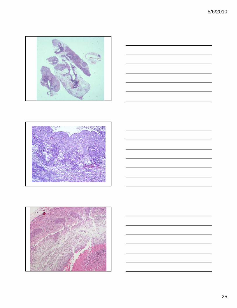
courtesy of Dr. Dean Percy
5/6/2010
4
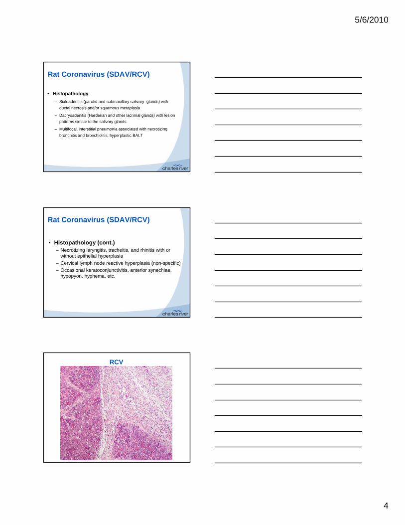
• Histopathology
– Sialoadenitis (parotid and submaxillary salivary glands) with
ductal necrosis and/or squamous metaplasia
– Dacryoadenitis (Harderian and other lacrimal glands) with lesion
Rat Coronavirus (SDAV/RCV)
y ( g )
patterns similar to the salivary glands
– Multifocal, interstitial pneumonia associated with necrotizing
bronchitis and bronchiolitis; hyperplastic BALT
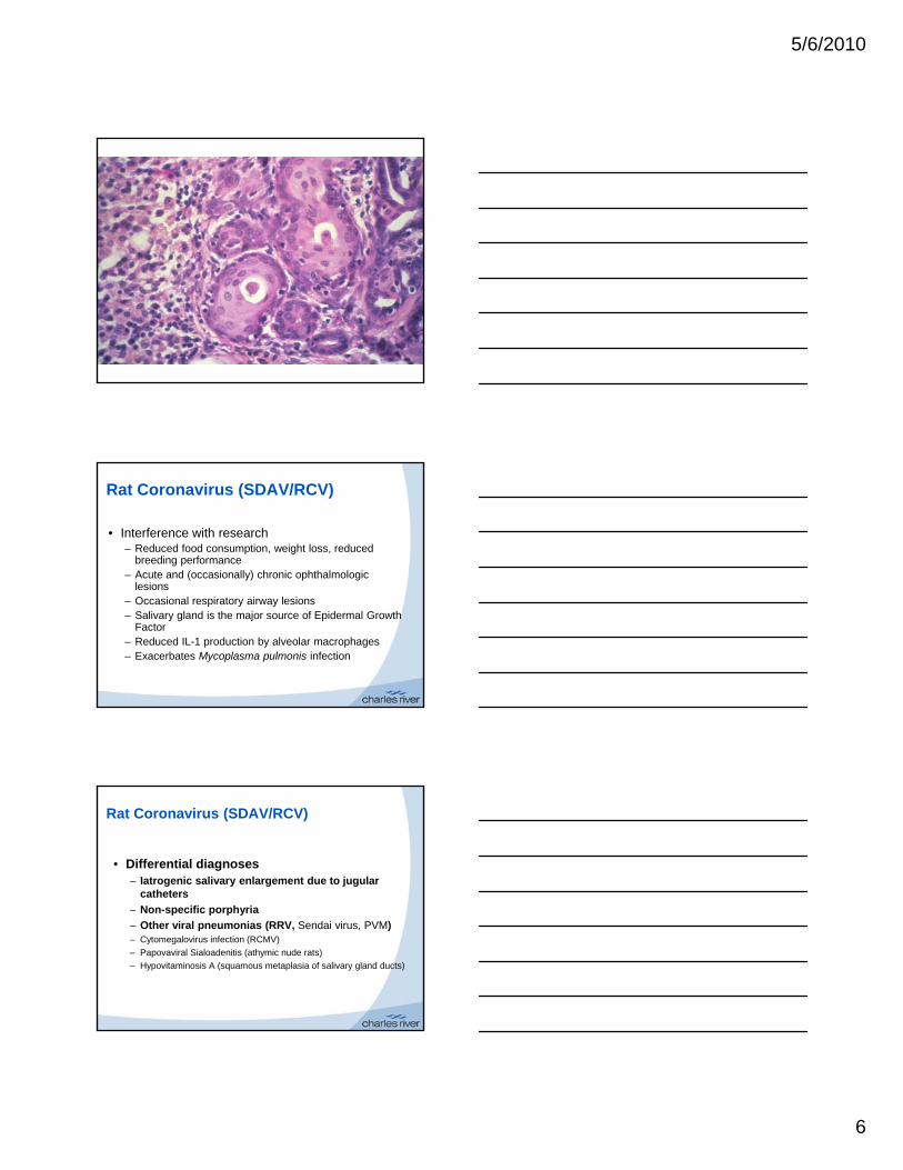
• Histopathology (cont.)– Necrotizing laryngitis, tracheitis, and rhinitis with or
without epithelial hyperplasia
Cervical lymph node reactive hyperplasia (non specific)
Rat Coronavirus (SDAV/RCV)
– Cervical lymph node reactive hyperplasia (non-specific)
– Occasional keratoconjunctivitis, anterior synechiae, hypopyon, hyphema, etc.
RCV

5/6/2010
5
courtesy of Dr. Dean Percy
Experimental SDA @8 days picourtesy of Dr. Dean Percy
5/6/2010
6
• Interference with research– Reduced food consumption, weight loss, reduced
breeding performance– Acute and (occasionally) chronic ophthalmologic
lesions
Rat Coronavirus (SDAV/RCV)
lesions– Occasional respiratory airway lesions– Salivary gland is the major source of Epidermal Growth
Factor– Reduced IL-1 production by alveolar macrophages– Exacerbates Mycoplasma pulmonis infection
• Differential diagnoses– Iatrogenic salivary enlargement due to jugular
catheters
Non specific porphyria
Rat Coronavirus (SDAV/RCV)
– Non-specific porphyria
– Other viral pneumonias (RRV, Sendai virus, PVM)– Cytomegalovirus infection (RCMV)
– Papovaviral Sialoadenitis (athymic nude rats)
– Hypovitaminosis A (squamous metaplasia of salivary gland ducts)

5/6/2010
7
• Diagnosis
– Pathology and clinical signs - first week
– PCR – Early in infection
Rat Coronavirus (SDAV/RCV)
– Serology - later (after 7-10 days)
• Good cross-reaction among all known strains
– Immunohistochemistry or PCR on paraffin-embedded
tissue
Parvoviruses
• ssDNA, (5.4 kb genome), non-enveloped– Virus remains active in environment
• Resistant to desiccation, non-oxidizing disinfectants
• Most are common in lab rats and mice
• Require cells in S phase of mitosis– Triggers production of nonstructural proteins, NS1 and NS2,
which direct viral replication and assembly and are responsible for cytotoxicity.
• Very low or no morbidity
• Cause persistent infection
• Different serotypes not very cross-reactive on ELISA/MFIA
• RV - Rat Virus (previously KRV, Kilham Rat Virus)
– Natural infections usually asymptomatic, but persistent
– Infects rapidly growing cells: Vascular endothelium, lymphoreticular
and hematopoietic tissues, developing cerebellum and liver
– Rare epizootic disease in fetal/neonatal rats: Cerebellar hypoplasia,
Parvoviruses of Rats
p yp p ,
anemia, thrombocytopenia
– Very rare disease in older rats: Hemorrhagic disease

5/6/2010
8
Rat Virus
Rat Virus
• Long-term infection, especially if infected as young rats.
– May cause persistent infection (6 months or more)
– May have prolonged shedding (10 weeks or more)
• Research Effects:• Research Effects:
– RV induced diabetes in DR BB rats (Guberski et al., 1991)
• Possibly due to imbalance in Th1 and Th2 responses (Jun and
Yoon, 2001)
• Few studies in literature, very difficult to isolate
– Multiple strains exist
– No clinical disease reported
– Research effects: Suppression of LGL lymphoid tumor growth in vivo in F344 rats: RPV-1a
Rat Parvovirus
– RV NS protein induced epigenetic modification in thymic lymphoma line, causing reversion to benignancy (Iseki H , 2005)
– RPV does not infect mice

5/6/2010
9
• H-1 (Toolan’s H-1)- no natural disease
– Significance through research interference: liver
Parvoviruses of Rats
– Current interest (and historic) in possible use treating human tumors
• Rat Minute Virus (RMV)
– Almost nothing in literature
– Serologically and genetically more similar to RV than to RPV
•• SerologySerology – Best for screening– MFIA or ELISA
– Use panel of antigens for each serotype, plus the generic NS 1 antigen
Detection of Parvoviruses
generic NS-1 antigen• Rats - RV, H-1, RPV, RMV and NS-1
– IFA – Good follow-up assay for positive/equivocal MFIA/ELISA
Rat Serology for parvoviruses
Agent/Assay # tested % positive
NS-1 63,101 2.3692%
H-1 81,764 1.6120%
RPV 88,399 1.6018%RPV 88,399 1.6018%
KRV 88,667 1.5101%
RMV 44,075 1.4475%

5/6/2010
10
•• PCRPCR
– Can be strain-specific (VP2) or generic (NS-1)
– Mesenteric LN stay positive indefinitely
Detection of Parvoviruses
y p y
– PCR of fecal samples valuable to detect shedding (can pool fecal samples. Beware of fecal inhibitors of PCR)
– Valuable for testing biologicals and cell cultures
– Applicable to environmental swabs
• Pneumonia first observed in F-344 rats mid-1990s– Idiopathic pneumonitis
• Reported in:
“Rat Respiratory Virus (RRV)”
Reported in:
– Inhalation Toxicology in 1997, Gilbert, B.E, et al.
– Toxicologic Pathology in 1997, Elwell, MR, et al.
– Toxicologic Pathology in 1998, Slaoui, M, et al.
– Veterinary Pathology in 2009, Albers, TM, et al.
• Prevalence: Common (~5-6% by histopathology in external sample stream at Charles River)– True prevalence likely higher if serology or PCR were available
• Biology
Rat Respiratory Virus (RRV)
– Agent not identified. Discussed as probably viral, but keep an open mind. Rat Respiratory “Agent”?
– Published abstracts implicating a hantavirus are not widely accepted and (in presenter’s opinion) are erroneous.
• Based on cross-reactions seen by one diagnostic lab using a Hantaviral IFA, which is allegedly more prone to false positives than many tests. No confirmation of these reports by culture or molecular techniques in several years since initial suggestion.
5/6/2010
11
• Epidemiology– Host range - rats are the only known host, all strains
susceptible
– Cases detected in North America Europe Asia
Rat Respiratory Virus (RRV)
Cases detected in North America, Europe, Asia
– Transmitted by aerosol and/or dirty bedding
– Additional fomite transmission likely
– Lesions most prevalent (=>50%) and most pronounced in naïve colony (epizootic form)
– Lesions have low prevalence (<=20%) and low severity in endemic colony (enzootic form)
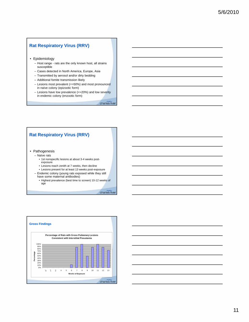
• Pathogenesis– Naïve rats
• 1st nonspecific lesions at about 3-4 weeks post-
Rat Respiratory Virus (RRV)
exposure• Lesions reach zenith at 7 weeks, then decline• Lesions present for at least 13 weeks post-exposure
– Endemic colony (young rats exposed while they still have some maternal antibodies)
• Highest prevalence (best time to screen) 10-12 weeks of age
Gross Findings
Percentage of Rats with Gross Pulmonary Lesions Consistent with Interstitial Pneumonia
80%90%
100%
0%10%20%30%40%50%60%70%
1 2 3 4 5 6 7 8 9 10 11 12 13
Weeks of Exposure
Pe
rce
nta
ge
0 1 2

5/6/2010
12
Microscopic Findings
Percentage of Rats Exhibiting Microscopic Lesions Consistent with RRV
70%80%90%
100%
e
0%10%20%30%40%50%60%70%
1 2 3 4 5 6 7 8 9 10 11 12 13
Weeks of Exposure
Per
cen
tag
e
0 1 2
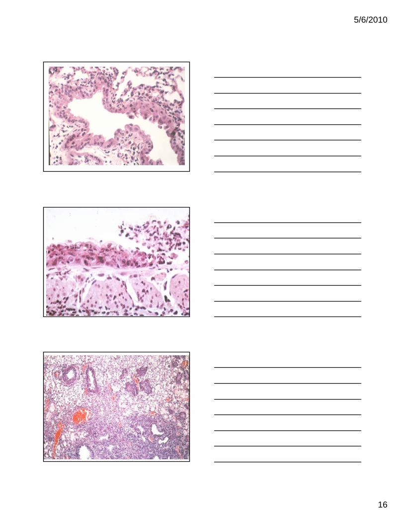
• Diagnosis– Gross Lesions: Scattered brown to grey areas on pleural
surface, suggestive of interstitial pneumonia.• RRV is currently the only common cause of interstitial
Rat Respiratory Virus (RRV)
• RRV is currently the only common cause of interstitial pneumonia in rats
–– Histopathology is diagnosticHistopathology is diagnostic• Prominent perivascular cuffs distributed in lungs• Interstitial pneumonia (lymphohistiocytic)• Syncytial cells (occasional)
• Lesions graded minimal to moderate
– No Serology or PCR available

5/6/2010
13

5/6/2010
14
RRV
• Control
– Eliminate by Rederivation
Duration of shedding? No definite answer– Duration of shedding? -No definite answer
– Disinfection - Unknown

5/6/2010
15
RRV
• Research interference
– Nothing demonstrated.
– Anecdotal reports suggest increased mortality under– Anecdotal reports suggest increased mortality under anesthesia, failed ex vivo lung studies, and confounded pulmonary histopathology assessment of inhalation studies.
Sendai Virus Infection
• Etiology: Sendai virus, Parainfluenza virus type I (PI-1)– Sendai is not the only PI-1 virus. Rats may also be susceptible to
other PI viruses, such as PI-3.
Host range• Host range – Mice
– Rats
– Hamsters
– Guinea pigs: usually non-specific serological reactions with other parainfluenza viruses
• Prevalence – rare in lab rodents (0.003% in mice, 0.024% in rats)
5/6/2010
16

5/6/2010
17
Sendai Virus Infection
• Histopathology– Reparative stage: Proliferation and regeneration of
target epithelium• Epithelial hyperplasia and dysplasia in upper and lower
airways and alveolar septa
• May see squamous metaplasia, polypoid masses in bronchiolar lumina
Sendai Virus Infection
• Histopathology– Recovery stage: Either a return to normal or
persistent scars• Fibrosis
• Cholesterol clefts
• Dilated airways containing inspissated secretions
• Peribronchial, peribronchiolar, and perivascular mononuclear cell cuffs and aggregates
Diagnosis of Sendai Virus Infection
• Serology: MFIA, ELISA, IFA, HAI– Use sentinel mice to screen for cross-reacting antibodies in GP
• PCR
• Pathology– Lesions not specific but inclusions in airway cells and– Lesions not specific, but inclusions in airway cells and
syncytia are very suggestive of Sendai virus infection
• Virus isolation
• Immunohistochemistry and immunofluorescence of tissues

5/6/2010
18
Rat Theilovirus (RTV)
• Discovery
– Serologic titers have long been detected in rats using antigen from the GD-VII strain of TMEV
• Some colonies were positive, others negative, suggesting the presence of a virus related to TMEV
http://phene.cpmc.columbia.edu/WIntkey/Images/RENDER/1tmf.jpg
suggesting the presence of a virus related to TMEV.
• Since the rat virus did not appear to transfer to mice, and vice versa, the rat virus was thought probably distinct from TMEV.
– The virus in rats has been now sequenced, the taxonomy of picornaviruses has been adjusted, and the virus is now referred to as rat theilovirus (RTV)
Rat Theilovirus (RTV)
• Agent– Family: Picornaviridae, Genus: Cardiovirus, Species:
Theilovirus, Serotype: Rat theilovirus..• There are three serotypes in the theilovirus species:
TMEV, RTV (or Theiler’s-like virus of rats), Vilyuisk , ( ), yhuman encephalomyelitis virus, Saffold virus.
– RTV and TMEV are small non-enveloped, RNA viruses.
• Moderate environmental persistence and resistance to disinfection are expected.
Rat Theilovirus (RTV)
• Epizootiology– Prevalence – moderate. The CR diagnostic
laboratory finds about 2% of rats serum samples from external sources are positive for RTV
– The host species range is unknown but there isThe host species range is unknown, but there is evidence against natural spread to mice
– Infected rats have been reported to shed RTV for at least 13.5 weeks

5/6/2010
19
Rat Theilovirus (RTV)
• Disease– No disease resulting from natural infection has been
reported– Experimental Disease (IC inoculation of sucklings with
material from rat intestine)• Ohsawa, et al. – no diseaseOhsawa, et al. no disease• Rodrigues, et al. – flaccid paralysis, tremor, death
– No histopathology. Demonstrated virus in brain. No HM on “donor” rats, and did not check for other agents in affected sucklings
• Henderson, et al. – No neurologic disease. “Possible” wasting in nude rats after oral gavage
– Conclusion – at this time potential pathogenicity, or variation in virulence among strains is not known
Rat Theilovirus (RTV)
• Research Effects– None reported
Rat Theilovirus (RTV)
• Diagnosis– Serology –
• MFIA of ELISA
• IFA
– PCR – virus shed for long periods, PCR may be the preferred method to screen animals in quarantine
– Soiled bedding should be adequate exposure for sentinels

5/6/2010
20
Rat Theilovirus (RTV)
• Management– Rederivation by embryo transfer or caesarian section
should be successful
– Success at early cross-fostering not reported• Reported as successful for most litters for TMEV• Reported as successful for most litters for TMEV
– Pest control. TMEV reported from wild mice. RTV status of wild rats is not known.
– Environmental disinfection should be as for other nonenveloped viruses, e.g., parvoviruses
• Oxidizing disinfectants
Rat Bacteriology ResultsAgent # tested # pos. % pos.
Helicobacter bilis 8,031 111 1.3821%
any Helicobacter 7,968 636 7.9819%
Helicobacter hepaticus 8,031 35 0.4358%
B. bronchiseptica 6,477 0 0.0000%
Beta Strep sp 6,505 1 0.0154%
Beta Strep Grp B 6,447 221 3.4280%
Beta Strep Grp G 6,447 1 0.0155%p p ,
C. kutscheri 6,492 0 0.0000%
M pulmonis 3,594 2 0.0556%
P. multocida 6,409 0 0.0000%
P. pneumotropica 6,409 340 5.3050%
other Pasteurella 6,357 24 0.3775%
Ps aeruginosa 12,931 301 2.3277%
Salmonella 6,430 0 0.0000%
Staphylococcus aureus 6,492 1,550 23.8755%
Strep. pneumoniae 6,484 0 0.0000%
Rat Parasitology Results
Agent # tested # pos. % pos.
A. tetraptera 8,350 4 0.0479%
S. muris 8,350 139 1.6647%
S. obvelata 8,350 1 0.0120%
All pinworms 8,350 144 1.7246%
Lice 7,307 0 0.0000%
Mites* 7,310 0 0.0000%
Giardia 6,957 0 0.0000%
Spironucleus 6,957 15 0.2156%
"other" flagellates 6,957 500 7.1870%
Entamoeba 6,957 191 2.7454%
* * -- Outbreaks of Outbreaks of Ornithonyssus bacotiOrnithonyssus bacoti reported in some reported in some facilities in southern, southwestern, and eastern USfacilities in southern, southwestern, and eastern US

5/6/2010
21
Mycoplasma pulmonis infection
• Host Range– Rats
– Mice
– Guinea pigs, Hamsters and Rabbits (culture evidence but no disease reported)but no disease reported)
• Prevalence – Infrequent to rare– Very common in pet rats
• Clinical signs (disease of older animals)– Usually clinically silent in young, non-specific in older
• Rales and dyspnea, snuffling/chattering• Ocular and nasal discharge as well as
Mycoplasma pulmonis infection
gchromodacryorrhea
• Rubbing of eyes• Head tilt• Rats spin when held up by tail • Decreased reproductive efficiency (rats)
Pathogenesis of Mycoplasmosis
• Transmission– Horizontal transmission (aerosol or in utero exposure,
rats only)
– Venereal transmission (?)Venereal transmission (?)
• Note: Mycoplasmas that can commonly infect cell cultures are not M. pulmonis. Many can be eliminated by passaging the cell lines through rodents. However, M. arginini has been found in cell cultures and can cause arthritis in mice.

5/6/2010
22
• Disease outcome depends on interaction of:– Host factors
• Age
Pathogenesis of Mycoplasmosis
g
• Strain (BALB/c more susceptible than C57BL/6, SD > Lewis, F344)
• Immune status, concurrent infections, nutritional status (e.g., vitamin A and E deficiencies)
• M. pulmonis possibly damages host cells by:– "Ciliostasis and ciliolysis”
• Probably responsible for exudate accumulation, opportunistic bacterial infections, and impaired transport of ova (infertility).
Pathogenesis of Mycoplasmosis
– Competing for the host cells' metabolites– Toxic metabolites (e.g., peroxides)– Production of nonspecific mitogens >> autoreactive
clones of lymphocytes >> immune-mediated damage– M. pulmonis may also cause damage indirectly
through bystander effect from host leukocytes
• Infection persists – Disease primarily in older rats
Gross Lesions of Mycoplasmosis
• Upper respiratory tract (young and adults)– Suppurative: rhinitis, otitis media, laryngitis, tracheitis
• Lung"Cobblestone" lung (older adults primarily rare)– Cobblestone lung (older adults primarily, rare)
• Suppurative bronchopneumonia with or without abscesses
• Atelectasis
• Bronchiectasis and/or bronchiolectasis

5/6/2010
23

5/6/2010
24
Gross Lesions of MRM
• Arthritis (occasionally)• Genital tract
– Usually no lesion observedF l t– Female rat
• Partially resorbed fetuses• Suppurative salpingitis
Histopathology of MRM
• Airway lesions in the respiratory tract are usually characterized by– Suppurative exudatepp
– Hyperplasia of the mucosal epithelium
– Hyperplasia of the bronchial associated lymphoid tissue
5/6/2010
25

5/6/2010
26
Histopathology of Mycoplasmosis
• Other respiratory tract lesions related to gross lesions – Squamous metaplasia of airway epithelia
– Pseudoglandular hyperplasia of nasal epithelium (chronic)
– Peribronchial alveolar type-II pneumocyte hyperplasia
– CAR bacillus and/or secondary bacterial pneumonias
– Syncytia may be observed on the surface of nasal and bronchial mucosa (mice)
– Loss of cilia
• Lesions in the female genital tract (rats)– Suppurative oophoritis
– Hydrosalpingitis or suppurative salpingitis
Histopathology of Mycoplasmosis
y p g pp p g
– Suppurative endometritis or pyometra; maybe epithelial hyperplasia and squamous metaplasia
• Differential diagnoses
– Cilia-Associated Respiratory (CAR) Bacillus infection
Diagnosis of Mycoplasmosis
infection
– Iatrogenic pneumonia– Bacterial infections (Pseudotuberculosis, Streptococcosis, B. hinzii
in mice)
– Viral infections (RRV, Sendai virus, PVM, etc.)
– Mycotic pneumonia

5/6/2010
27
Mycoplasma pulmonis infection
• Diagnosis– Culture: Especially exudates in the upper respiratory
tract and middle ears. More sensitive than serology for early infections. Culture takes 2 weeks.
S l B t f i l f l i i– Serology – Best for screening large, freely-mixing populations
– PCR – Specific (not generic – cross-reactions).
– Pathology– Immunofluorescence or immunohistochemistry of tissue or
exudates
Cilia-associated (CAR) bacillus
• Cause – Gliding bacterium, similar to Flavobacterium and Flexibacter
• Prevalence – Rare (< 0.2% rats, 0.0% mice)
http://www.zerberus-online.de/Problematik/Flexibacter-2.jpg
• Natural lab animal host range of CAR bacillus – Rats– Mice– Rabbits
• Clinical signs of CAR bacillus infection– Sometimes nonspecific respiratory signs (dyspnea)– Sometimes weight loss
CAR bacillus
• Pathogenesis of CAR bacillus infection– Transmission probably via direct contact with infected
animals, contaminated fomites (soiled bedding) and aerosol not important
– CAR bacillus may act in synergy with other respiratory– CAR bacillus may act in synergy with other respiratory agents to produce chronic respiratory disease
• Interference with research (unknown)– Effects on mucociliary clearance and immune function
speculated, not demonstrated

5/6/2010
28
CAR bacillus
• Gross lesions of CAR bacillus infection– Resemble those of the primary infections, e.g.,
Mycoplasmosis, Sendaiy p ,
– Rarely, uncomplicated infections may produce bronchiectasis, mucus accumulation in bronchioles, and lymphoid hyperplasia
• Inflammation can be neutrophilic, but less suppurative than with mycoplasmosis
• Bronchial epithelium is preserved, or hyperplastic
• Cilia prominent, not lost as with M. pulmonis
CAR bacillus
• Histopathology of CAR bacillus infection– Cilia on respiratory epithelium may appear slightly
basophilic with H&E
– Long, slender bacilli among the cilia at any level of respiratory epithelium (nasal cavity to bronchioles) -observed in silver stained sections
– Hyperplastic BALT
– Rarely, there may also be suppurative bronchopneumonia

5/6/2010
29

5/6/2010
30
Bordetella hinzii
CAR bacillus
• Differential diagnoses for CAR bacillus infection– Mycoplasma pulmonis (very often co-infection)Mycoplasma pulmonis (very often co infection)
– Other bacteria (i.e., Bordetella hinzii, S. pneumoniae, C. kutscheri, etc.)
– Mycotic pneumonias (i.e., aspergillosis, mucormycosis, etc.)
– Viral pneumonia (RRV, Sendai virus, PVM, etc.)
CAR bacillus
• Diagnosis of CAR bacillus infection– Serology – MFIA or ELISA
– PCR – Lung wash, lung tissue, feces
– Histopathologyp gy• Warthin-Starry silver stain
• Grocott's methenamine silver stain– Isolation in embryonated eggs or tissue culture
– Electron microscopy
– Immunofluorescence (tissue)

5/6/2010
31
Tyzzer’s Disease
• Etiology: Clostridium piliforme• Hosts (some evidence of partial species-specificity of
strains)strains)– Rodents (virtually all, Mongolian gerbil very susceptible)– Rabbits– Carnivores (cat, dog)– Horses– Non-human primates– Humans (Infection has been reported in one HIV+ patient to date, but
seroconversion, always suspect, has been reported in many)
Tyzzer’s Disease
• Prevalence: Tyzzer’s Disease is infrequent, although the organism may be widespread
• Clinical signs– Usually absent
Overt disease mostly in young recently weaned animals– Overt disease mostly in young recently weaned animals• Acute death with or without clinical signs• Diarrhea with or without mucus and blood• Distended abdomen (rat)• Anorexia, Lethargy, Emaciation, Ruffled fur
• May be widespread in nature
• Vegetative form survives only inside of cells– Epithelium (small and large intestine, gall bladder, bile
Pathogenesis of Tyzzer’s Disease
p ( g , g ,duct)
– Hepatocytes
– Myocardial fibers
– Smooth muscle of small and large intestine

5/6/2010
32
• Transmission– Horizontal transmission
• Ingestion of spores in
Pathogenesis of Tyzzer’s Disease
Ingestion of spores in
– Feces
– Contaminated feed and bedding
– Carcasses (cannibalism)
• Proposed sequence of infection
– Spores ingested >> produce the vegetative form,
Pathogenesis of Tyzzer’s Disease
actively phagocytosed by epithelial cells overlying the
GALT >> vegetative form escapes phagosome >>
multiples in intestinal mucosal epithelial cells and
possibly RE cells in Peyer's patches
• Proposed sequence of infection (cont.)– Most infections appear to be cleared at this point, and
animals stop shedding spores within about 2 weeks.
Pathogenesis of Tyzzer’s Disease
animals stop shedding spores within about 2 weeks. – If infection extends past GI tract - Vegetative form
reaches liver by one or more routes• Portal circulation (most likely)• Lymphatics• Common bile duct (the vegetative form is motile)

5/6/2010
33
• Proposed sequence of infection (cont.)
– Vegetative form infects and multiples in the hepatocytes, then may do one or more things depending how long the
Pathogenesis of Tyzzer’s Disease
then may do one or more things depending how long the animal survives
• Enter into the blood stream or lymphatics to colonize the myocardium
• Possibly enter into epithelium of biliary tree to multiply and eventually be shed into bile to re-infect intestine and liver (auto-infection)
• Factors which influence infection and outcome– Host factors
• Age (recently weaned most susceptible)• Genotype (CBA/N mice supposedly very susceptible,
Pathogenesis of Tyzzer’s Disease
yp ( pp y y p ,C57BL/6 more resistant than DBA/2)
• Immune function– Latent infection may be activated by:
» Stress, Drugs (cortisone, cyclophosphamide, etc.), Leukocyte injection
– Nutritional status (Fasted mice resistant to overt disease)– Gnotobiotic status
» Escherichia coli reportedly potentiates C. piliforme in rabbits
• Factors which influence infection and outcome– Bacterial factors
• Strain
Pathogenesis of Tyzzer’s Disease
S a– Some species-specificity– Some strains produce a high-molecular weight, cytotoxic
protein. Pathogenicity seems dependent on this. Some strains may be non-pathogenic.
• Dose

5/6/2010
34
• Factors which influence infection and outcome– Environmental factors
• Increased environmental temperatures and humidity
Pathogenesis of Tyzzer’s Disease
p y– May precipitate a latent infection (stress)
– May increase number or viability of spores >> increasing exposure
• Damp feed and poor husbandry– May also increase number of spores in environment
• Overcrowding– Stress and increased spores in environment
• Interference with research– Direct effects, especially in immunosuppressed
animals
Pathogenesis of Tyzzer’s Disease
animals
– Reported to alter hemostatic parameters and cytokines
Gross Lesions of Tyzzer’s Disease
• Perianal fecal staining may be present
• LiverMultiple disseminated pinpoint or larger pale foci– Multiple, disseminated, pinpoint or larger, pale foci (necrosis) within and on the surface of the liver
– The liver may only be swollen and mottled

5/6/2010
35
Gross Lesions of Tyzzer’s Disease
• Intestine– Megaloileitis (rat)
• Greatly dilated fairly flaccid hyperemic small intestinesGreatly dilated, fairly flaccid, hyperemic small intestines (ileum)
– Hyperemia, edema, hemorrhage, and possibly ulceration of any part of the intestines, but especially the terminal ileum, cecum, and colon

5/6/2010
36
Gross Lesions of Tyzzer’s Disease
• Heart– Pale, circumscribed, sometimes raised foci may be
present on the surfacep
– Pale linear streaks near the apex of the heart
• Enlarged, hyperemic and edematous mesenteric lymph nodes

5/6/2010
37
Histopathology of Tyzzer’s Disease
• Intestine– May see nothing even if lesions in liver and heart– Necrotizing enteritis, typhlitis, and colitis with or without
• Edema (common)• Edema (common)• Blunted and fused villi• Crypt epithelial hyperplasia• Ulceration• Hemorrhage• Cellular debris in crypts and lymphatics
Histopathology of Tyzzer’s Disease
• Liver– Coagulative necrosis (frequently periportal) with or
without• Inflammation (neutrophils, mononuclear cells, histiocytes,
and rare multinucleated giant cells)
– Hemorrhage
– Dystrophic calcification
– Fibrosis

5/6/2010
38
Histopathology of Tyzzer’s Disease
• Heart– Myocardial degeneration with or without
• NecrosisNecrosis
• Mixed inflammatory cells
• Dystrophic calcification
Histopathology of Tyzzer’s Disease
• Diagnostic if characteristic bacilli seen– Sometimes visible with H&E, but usually need special
stains • Warthin-Starry silver stain (best)• Immunoperoxidase stain
– Probably excellent, but not commercially available• Giemsa and methylene blue stains
– Tissues or smears• Brown & Brenn stain
– Organism is gram-negative but stains very poorly

5/6/2010
39
Histopathology of Tyzzer’s Disease
• Liver– Organisms are most often observed in surviving
hepatocytes at the periphery or within lesions– May be in hepatocytes not associated with a lesionMay be in hepatocytes not associated with a lesion
• Intestine– Normal gut flora within mucosal crypts and
superimposed upon the mucosal epithelial cells may complicate evaluation.
Histopathology of Tyzzer’s Disease
• Vegetative form of C. pilforme is 8.0 to 20.0 x 0.3 to 0.5 microns bacillus. (long and thin, piliform)– One or usually more bacilli are present in cells in
either a jumbled array (pickup stick) or parallel arrangement depending on the shape of the cell
• Hepatocytes, epithelial cells, • neurons: Pickup-stick arrangement• Smooth muscle and myocardial fibers: Parallel
arrangement

5/6/2010
40
Tyzzer’s Disease
• Differential diagnoses– Bacteremia (Streptococcus, others)
– Adynamic ileus due to chloral hydrate (rat)y y ( )
– Yersinia tuberculosis (guinea pig)
– Hepatic coccidiosis (rabbit)
– Alflatoxicosis
– Others
Diagnosis of Tyzzer’s Disease
• Pathology– Cytology or histopathology with the identification
of intracellular long bacilli is diagnostic• Warthin-Starry silver stain (tissue)
• Giemsa or methylene blue stain (smear or tissue)
• PCR on paraffin-embedded tissue
• Immunohistochemistry (tissue)
• Immunofluorescent staining of tissues

5/6/2010
41
Diagnosis of Tyzzer’s Disease
• Provocation tests to provoke latent infections. Some doubt as to efficacy, but may distinguish infections with potentially pathogenic strains. Must select correct animals to immunosuppress.
• Cyclophosphamide
• Cortisone
– Sentinel animals placed on soiled bedding (not foolproof)
• Gerbil
• CBA/N mice
Diagnosis of Tyzzer’s Disease
• Serology (does not distinguish between pathogenic and non-pathogenic strains)– MFIA, ELISA, IFA– Positive finding should be confirmed by pathology
• PCR – Feces (if shedding) can be hard to extract DNA
form spores– Tissue - should be positive if lesions are due to
Tyzzer’s• Isolation of the organism (not practical)
– Cell culture– Embryonated eggs
Pseudotuberculosis
• Etiology: Corynebacterium kutscheri
• Hosts– RatsRats
– Mice
– Guinea pig, hamster (culture evidence, no disease)
• Prevalence - Rare

5/6/2010
42
C. kutscheri infection
• Clinical signs– Infections are frequently clinically silent
– Nonspecific (sick rat) clinical signs may be observed, p ( ) g y ,death in 1 to 7 days
• Porphyrin and mucopurulent ocular and nasal discharges
• Respiratory rales and dyspnea
• Lameness
Pathogenesis of C. kutscheri infection
• Latent infections are currently rare in laboratory rats and mice. However, infected animals are usually clinically normal. In these, the organism may be cultured from:– Submaxillary (cervical) lymph nodes– Oral cavity– Nasal cavity– Middle ears– Preputial gland abscesses
Pathogenesis of C. kutscheri infection
• Factors which may precipitate latent infections include age and conditions which immunosuppress the host– Stress (poor husbandry, overcrowding, shipping, etc.)
– Concurrent infections
I di ti– Irradiation
– Immunosuppressive drugs (steroids, cyclophosphamide, etc.)
– Malnutrition (e.g., pantothenic acid and biotin deficiencies)

5/6/2010
43
Pathogenesis of C. kutscheri infection
• Transmission is probably through direct contact and/or oronasal exposure.
• Septic emboli become trapped in organs orSeptic emboli become trapped in organs or tissues with either a large capillary network (lung, liver, and kidney) and/or responsible for filtering blood (synovia and glomeruli). This accounts for the distribution of the lesions
Pathogenesis of C. kutscheri infection
• Although any or all organs and tissues may be
involved, the frequency of lesion distribution
varies with the speciesvaries with the species
– Rat: pulmonary involvement
– Mouse: hepatic and renal involvement
C. kutscheri infection
• Gross– Lung: 1 or more randomly distributed abscesses +/-
hemorrhage and pleuritis (fibrinous or fibrous)– Liver: Solitary or multiple abscesses and/or necrosis– Kidney: Solitary or multiple abscesses and/or– Kidney: Solitary or multiple abscesses and/or
pyelonephritis– Preputial gland: Abscess– Joints: Suppurative arthritis– Skin: Abscess(es), ulcerations, fistulous tracts,
pododermatitis– Middle ear: Suppurative otitis media

5/6/2010
44

5/6/2010
45
C. kutscheri infection
• Histopathology (related to gross findings)– Lung
• Abscesses predominately in the interstitium due to the hematogenous seeding of the lung with bacteriag g g
• May see caseous necrosis
• Epithelioid macrophages and multinucleated giant cells may be present in the abscesses
• Bronchi and bronchioles may contain suppurative exudate

5/6/2010
46
C. kutscheri infection
• Histopathology (cont.)– Liver
• May see caseous necrosisy
– Kidney• Septic embolic glomerulitis
• Abscesses with or without pyelonephritis
– May see lesions in any tissue (e.g., brain, skin, joints)

5/6/2010
47
C. kutscheri infection
• Differential diagnoses– Localized or disseminated opportunistic bacterial
infections: Staphylococcus spp., Streptococcus spp., Salmonella spp., etc.
– Mycoplasmal diseases– Mycotic pneumonia (Aspergillosis, Mucormycosis,
etc.)– Tyzzer's Disease – Viral pneumonia– Streptobacillus moniliformis
C. kutscheri infection
• Diagnosis– Bacteriology
• Best culture site probably submandibular lymph nodesp y y p
• May also be in oral cavity, cecum, colon and rectum
– PCR
– Pathology• May see characteristic configuration of G+ coryneforms
in sections or impression smears
C. kutscheri infection
• Diagnosis (cont.)– Cortisone stress (provocation) test - obsolete
• To activate latent infections and also possibly p yPneumocystis carinii and Tyzzer's disease
– Serology• May see false positives and false negatives
• Should be confirmed by PCR, culture