Embed Size (px)
Citation preview

1
Infectious Diseases in Pediatrics
Kevin Coulter MDDepartment of PediatricsUniversity of California, Davis Medical Center
Pneumococcal Vaccine 2010
� Pneumococcal Conjugate Vaccine (PCV13)– All children 2-59 months– A single revaccination after 5 years of age
for children with asplenia or other chronic diseases or immunocompromising conditions (eg, chronic lung disease, cochlear implants, HIV)• These children should also receive PPSV

2
Influenza Vaccine 2010
� Administer annually to children/adolescents 6 months through 18 years of age. (trivalent inactivated vaccine)– Live attenuated vaccine for children over 2
years of age (but not for 2-4 year olds who have been wheezing in the past year)
Influenza
� 6 yo with cough and high fever
Meningococcal Vaccine 2010
� Administer at 11-12 years of age (catch up unvaccinated 13-18 year olds) .
� Administer to previously unvaccinated college freshmen if living in a dormitory
� Administer to children 2-10 years of age with special conditions; (eg, persistent complement deficiencies, asplenia)
U.S. rates of meningococcal disease by age

3
General vaccine recommendations
� All vaccines can be administered at the same visit as all other vaccines.
� If not given at the same visit, live parenteral vaccines or live intranasal influenza vaccine should be separated by at least 4 weeks.
General vaccine recommendations
� Increasing the interval between doses of a multidose vaccine does not diminish the effectiveness of the vaccine.
� Decreasing the interval may interfere with antibody response
General vaccine recommendations
� Live vaccine should not be administered to severely immunosuppressed patients.
� Inactivated vaccines are safe for immunosuppressed patients.
Common Vaccine Questions
� Can you give live virus vaccine to children taking corticosteroids?– No: if taking >2 mg/kg/day or >20 mg/day
of prednisone for greater than 14 days.– Yes: if taking lower daily doses, on
alternate day dosage, on systemic steroids for less than 14 days, on inhaled or topical steroids

4
Live attenuated vaccines for patients with HIV
– Vaccine Asympt. Sympt– Varicella yes no– MMR yes no– LAIV no no– Rotavirus no no
Children with anaphylactic egg allergy
� MMR is okay
� Influenza vaccines are not okay
Common Vaccine Questions
� How long should children wait to get vaccinated after receiving immunoglobulins?– Depends on the immunoglobulin
Common Vaccine Questions
� What are contraindications to further immunization with pertussis vaccine?– Anaphylactic reaction to the vaccine– Encephalopathy developing within 7 days
of the vaccine
5
Common Vaccine Questions
� What are precautions to further vaccinations with pertussis vaccine?– Fever >40.5 within hours of a previous
dose– Persistent inconsolable crying for >3 hours– Collapse or shock-like state– Seizure within 3 days of previous vaccine
Invalid contraindications to vaccination
– Mild illness– Antimicrobial therapy– Pregnant or immunosuppressed person in
the home– Breastfeeding– Premature birth– Tuberculin skin test
Tuberculosis in Children
� Epidemiology– Case rates highest in urban, poor children– Infants are at increased risk of progression
of disease– Tuberculosis in a child is a sentinel event– Children usually not contagious– Incubation period 2-12 weeks
Tuberculosis in Children
� Clinical Manifestations– Usually asymptomatic with normal chest
xray– Early manifestations include fever, cough,
chills, night sweats– Radiographic findings: hilar or mediastinal
adenopathy, atalectasis, pleural effusion

6
Tuberculosis in Children
� Diagnosis– Isolation of organism best achieved with
gastric aspirates
Tuberculosis in Children
� Tuberculin testing– Use Mantoux Method ( 5 tuberculin units
administered intradermally)– Test those children at increased risk for
disease
Tuberculin Testing
� Immediate TSTs– Contacts of confirmed or suspicious cases– Children with clinical findings of disease– Children immigrating from or with recent
travel to endemic countries and/or significant contact with indigenous persons from such countries.
Tuberculin Testing
� Annual TST’s– Children infected with HIV or living in
household with HIV infected persons– Incarcerated adolescents

7
Tuberculin Testing
� TST testing every 2-3 years– Children exposed to following persons:
• HIV infected• Homeless• Residents of nursing homes
• Institutionalized or incarcerated adolescents or adults
• Users of illicit drugs• Migrant farm workers
Tuberculin Testing
� TST at 4-6 years and 11-16 years– Children whose parents immigrated from
endemic countries– Children without specific risk factors who
reside in high prevalence areas
Tuberculin Testing
� Definition of positive TST– >5mm
• Children in close contact with active disease
– >10mm• Children at increased risk of dissemination
– >15mm• Children older than 4 years with no risk factors
Tuberculosis in Children
� Treatment for positive TST, normal CXR– INH 10 mg/kg/d for 9 months– Don’t routinely monitor LFTs

8
Tuberculosis in Children
� Treatment for children who are contacts of contagious disease– 1. Do TST– 2. Treat for 3 months with INH if TST
negative– 3. Repeat TST; if negative, stop treatment
Pulmonary tuberculosis
� 16 year old female with history of worsening cough and weight loss for one month

9
Mycobacterial Infections
� Axillary adenitis after BCG vaccination
TST and BCG
� AAP recommends using same criteria for interpreting TST results in children who have been previously immunized with BCG.
Pharyngitis in Children
� Etilogy– Most cases are viral infections:
• Adenovirus• Rhinovirus• Parainfluenza
• Influenza• EBV
Pharyngitis in Children
� Etiology– Grp A Streptococcus accounts for 15% of
all cases– Uncommon causes – Grp C and G beta
hemolytic streptococcus, N gonorrhea, tularemia
– Mycoplasma infections of the upper respiratory tract are also associated with pharyngitis

10
Pharyngitis in Children
� Bacterial vs. Viral– Scarlet fever rash– Fever, headache, abdominal pain– Sore throat in absence of viral symptoms– Tonsillar erythema, exudate– Palatal petechiae– Cervical lymphadenitis– Children >2 years old
Pharyngitis in Children
� Throat Cultures– Laboratory confirmation of infection
recommended as clinical identification not reliable
– Throat culture remains the gold standard– Rapid antigen tests are highly specific, but
have variable sensitivity (negative antigen tests should be followed up with culture)
Pharyngitis in Children
� Who NOT to do throat cultures on:– Children <3 years old– Children with coryza, conjunctivitis, cough,
hoarseness, anterior stomatitis, discreet ulcerations
– Post-treatment
GAS Pharyngitis – Why Treat?
� Suppurative sequelae– Peritonsillar abscess, cervical adenitis,
otitis media
� Nonsuppurative sequelae– Acute rheumatic fever, post-streptococcal
glomerulonephritis

11
GAS Pharyngitis - Treatment
� Penicillin V– 250 mg BID for children for 10 days– 500 mg BID for teens for 10 days
� Benzathine penicillin G– 600,000 units IM for kids <60 lbs– 1.2 million units for everyone >60 lbs
GAS Pharyngitis - Treatment
� Amoxicillin– Single daily dose, 50 mg/kg/d for 10 days
� Macrolides– Erythromycin, azithromycin, clarithromycin
� First generation cephalosporin– Keflex 20-50 mg/kg/d
Streptococcal Pharyngitis
� Palatal petechiae
Streptococcal scarlet fever
� Typical facial rash with erythema of cheeks and perioral pallor

12
Streptococcal Scarlet Fever
� Sandpaper exanthem of scarlet fever
Streptococcal Scarlet Fever
� Peeling of palms and soles 1-2 weeks after the illness
Streptococcal Infections
� Perianal streptococcal cellulitis– Diagnosis
confirmed by culture of rectal swab
– Treat like strep pharyngitis
Otitis Media
� Leading cause of physician visits among children
� Peak incidence rates occur at 6-18 months and 5-6 years of age
� 2/3 of all children have at least one episode of otitis media and 1/3 have 3 or more episodes per year

13
Acute Otitis Media
� Bacteriology– S. pneumo– H. flu (non-typable)– M. catarrhalis– Grp A Strep– S. aureus
Acute Otitis Media
� Def.: fluid in the middle ear in association with signs or symptoms of local or systemic illness
� Without treatment 80% resolve
� With treatment 95% resolve
Acute Otitis Media
� Elements of the definition of AOM are all of the following:– Recent, usually abrupt, onset of signs and
symptoms– Presence of MEE that is indicated by any of the
following:• Bulging of the TM, decreased TM mobility, air fluid level
behind the TM, otorrhea
– Signs or symptoms of middle-ear inflammation• Distinct otalgia; distinct erythema of the TM
Acute Otitis Media
� Severe illness– Moderate to severe otalgia or temperature
greater than 39 C
� Nonsevere illness– Mild otalgia and temperature less than 39
C

14
Acute Otitis Media
� Treatment Recommendations– Infants younger than 6 months should
receive antibiotics– Children 6 months – 2 years old: should
receive antibiotics if diagnosis is certain. If diagnosis uncertain and illness nonsevere, observation for 48-72 hours can be considered
Acute Otitis Media
� Children 2 years and older:– Should receive antibiotics if diagnosis
certain and disease severe– Observation is an option when diagnosis
uncertain or if diagnosis is certain and disease nonsevere
Acute Otitis Media
� Antibiotic Treatment– Amoxicillin 80-90 mg/kg/d– Treat all children < 6 years of age or those
with severe disease for 10 days– Children 6 years and older with nonsevere
disease can be treated for 5-7 days
Acute Otitis Media
� Antibiotic Treatment– For patients with severe disease, first line
treatment should be augmentin (90 mg/kg/d amoxicillin, 6.4 mg/kg/d clavulanate)

15
Acute Otitis Media
� Alternative first line therapy for penicillin allergic patients
� For severe disease– Ceftriaxone, 1-3 days
� For nonsevere disease– Non-type 1 allergy: cefdinir, cefpodoxime,
cefuroxime– Type 1 allergy: azithromycin, clarithromycin
Acute Otitis Media
� Antibiotic options for treatment failure after 48-72 hours initial therapy– Nonsevere disease – augmentin
• If has non-type penicillin allergy, ceftriaxone for 3 days
• For type 1 penicillin allergy, clindamycin for 10 days
Acute Otitis Media
� Antibiotic treatment for clinical failure after 48-72 hours initial therapy– Severe disease: Ceftriaxone for 3 days
• If penicillin allergic, clindamycin (consider tympanocentesis)
Otitis Media
� Opacification of the tympanic membrane with loss of normal landmarks

16
Sinusitis in Children
� Development of Sinuses in Children– Ethmoid and maxillary sinuses present at
birth– Frontal sinuses begin to develop at 2 years
old but not fully developed until 6 years of age
– Sphenoid sinus developed by 6 years of age
Sinusitis
� Diseases predisposing patients to sinusitis– Viral rhinitis– Allergic rhinitis– Ciliary dysmotility
• Kartagener’s Syndrome
– Cystic Fibrosis– Asthma
Sinusitis
� Clinical diagnosis– Nonspecific signs and symptoms
(rhinorrhea, sore throat, cough) for >10 days
– More severe signs and symptoms (fever, facial pain, facial swelling)
Sinusitis
� Diagnosis– Clinical diagnosis– Transillumination– Xray, CT scan, MRI

17
Sinusitis in Children
� Radiologic diagnosis– Consider sinus xrays, CT scan, MRI for
recurrent sinusitis, suspected complications of sinusitis (especially orbital involvement), or unclear diagnosis
Sinusitis in Children
� Bacterial etiology of acute sinusitis– Strep pneumoniae, H.influenza,
M.catarrhalis, S.aureus
Sinusitis in Children
� Treatment (10-14 days)– Amoxicillin– Augmentin– Second generation cephalosporin– Azithromycin
Sinusitis
� Orbital cellulitis secondary to extension of ethmoid sinusitis

18
Sinusitis
� Pott’s Puffy Tumor- osteomyelitis secondary to frontal sinusitis
Pneumonia Syndromes
� Infants – febrile, ill appearing, lobar consolidation– Etiology – s.pneumonia, h.influenza,
s.aureus– Mgt - CBC, blood culture, consider LP– Treatment – ceftriaxone, cefuroxime,
vancomycin plus ceftriaxone if considering s.aureus
Penumonia Syndromes
� Infants – afebrile, well appearing, interstitial infiltrates– Etiology –chlamydia trachomatis, RSV,
influenza, adenovirus, parainfluenza, pertussis
– Mgt – CBC, chest xray, hospitalize for respiratory distress, apnea
– Treatment - erythromycin
Chlamydia Trachomatis
� Pneumonia in infants– Afebrile illness 2-19 weeks after birth– Staccato cough, tachypnea, rales– Chest xray - bilateral diffuse infiltrates,
hyperinflation– Occasionally severe– Untreated disease can linger or recur– Elevated C trachomatis-specific IgM

19
Chlamydia Pneumonia
� Chest xray of 3 month old infant with 2 week history of cough and rales
� History of neonatal conjunctivitis
� Elevated chlamydia IgM
Pneumonia Syndromes
� Toddlers/preschoolers/adolescents –febrile, ill, lobar consolidation– Etiology – strep pneumoniae, staph aureus– Treatment
• Outpatient – IM ceftriaxone, augmentin, amoxicillin
• Inpatient – Ceftriaxone, Vancomycin
Pneumonia in Children
� 8 year old boy with 1 week history of abdominal pain, fever, vomiting
Pneumonia in Children
� 8 year old boy; WBC 28,000, right upper lobe consolidation, worsening respiratory distress

20
Pneumonia in Children
� 8 year old boy after two week IV antibiotic treatment for pneumonia/empyema
Pneumonia in Children
� 13 month old boy with history of recurrent pneumonias and 2 weeks of cough
� At bronchoscopy found to have coin and peanut in proximal esophagus
Things children swallow
� 3 year old boy ingested fishing sinker
Things children swallow
� Fishing sinker ingestion as a cause of lead toxicity

21
Pneumonia Syndromes
� Toddlers/preschoolers/adolescents –+/- fever, well appearing, diffuse crackles, wheezing– Etiology – RSV, influenza in the winter;
parainfluenza in the Fall– Pertussis, mycoplasma pneumoniae– Treatment – supportive, erythromycin if
suspect mycoplasma or pertussis
Mycoplasma pneumoniae
� 8 yo female with 10 day history of fever and cough
Croup
� Self-limited subglottic viral infection– Parainfluenza, RSV
� Barking cough, hoarseness, stridor� Predominance in Fall and Winter
� Usually in children <2 years old
� Spasmodic croup
Croup
� Diagnosis– History and physical examination– AP and lateral neck films

22
Croup
� Lateral neck film– Normal epiglottis– Increased air in the
hypopharynx
Differential Diagnosis of Croup
� Infectious– Epiglottitis– Bacterial tracheitis– Diphtheria– Peritonsillar abscess– Retropharyngeal abscess
Differential Diagnosis of Stridor
� Epiglottitis– Lateral neck film
(positive thumb sign)
Differential Diagnosis of Croup
� Anatomic– Foreign body– Laryngeal papillomas– Tracheomalacia– Subglottic webs, vascular rings– Psychogenic stridor

23
Croup
� Treatment– Oxygen– Nebulized saline– Racemic epinephrine
• 0.5ml of 2.25% solution in normal saline
– Steroids• Decadron 0.6 mg IM or po• Prednisone 2mg/kg/d for 3 days
– Hospital admission
UTIs in Infants and Children
� Prevalence of UTIs in febrile children– <2 year old without a “source” – 5%– Girls <1 year old – 6.3%– Girls 1-2 years old – 8.1%– Boys <1 year old – 3.3%– Boys 1-2 years old – 1.9%– Circumcised boys – 0.2-0.4%– Uncircumcised boys – 5-20 times higher
Urinary Tract Infections in Children
� Neonatal UTIs– Often associated with bacteremia (21-33%)– As compared to:
• 1-3 month olds – 18%• 4-8 month olds – 6%
Urinary Tract Infections
� Microbiology:– E Coli (90%)– Proteus (males)– Staph saprophyticus (sexually active
teens)– Enterococcus– Klebsiella– Enterobacter, pseudomonas
24
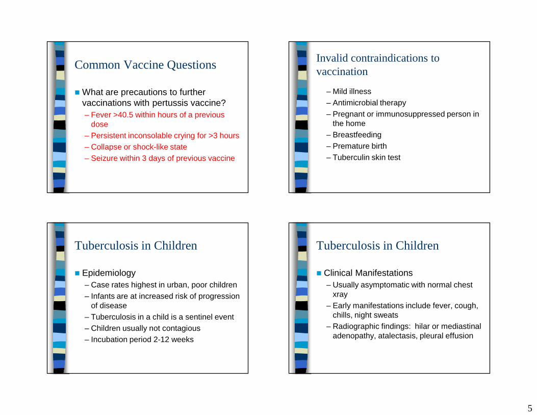
Urinary Tract Infections
� Distinguishing lower tract from upper tract disease:– Fever, CVA tenderness– CBC, sed rate, CRP– Radioisotope scan
UTIs in Children
� Making the diagnosis– Supra-pubic aspiration– Catheterization– Perineal collecting bags– Best possible clean catch
UTIs in Children
� Interpretation of urinalysis– Leucocyte esterase– Nitrite– Pyuria– Bacteruria on unspun gram stain
UTIs in Children
� Outpatient treatment– Empiric antibiotic therapy is directed
against E Coli– Cephalexin first line therapy for 7-14 days– Alternative po antibiotics
• TMP-SMX • Sulfisoxazole• Cefpodoxime (vantin)• Amoxicillin

25
UTIs in Children
� Inpatient therapy for children who are toxic, dehydrated, or unable to take po fluids– Ceftriaxone 75 mg/kg/d– Cefotaxime 150 mg/kg/d divided q6h– Cefazolin 50 mg/kg/d divided q8h– Gentamicin 7.5 mg/kg/d divided q8h
UTI’s in Children
� Further evaluation of children with UTI– Follow-up cultures not necessary unless
child not clinically responding– Duration of antibiotic therapy should be 7-
14 days– Children who will need radiologic imaging
of the urinary tract should be on prophylactic antibiotics until imaging completed
Urinary Tract Imaging
� Indications for Imaging– Acute Pyelonephritis– First UTI in a boy– First UTI in girl < 3 yo– UTI in a child with
• Urinary tract abnormalities• Voiding abnormalities• Hypertension• Poor growth
Urinary Tract Imaging
� Ultrasound– Hydronephrosis, dilatation of distal
ureters,hypertrophy of bladder, ureterocele
� Voiding Cystourethrogram– Vesicoureteral reflux, posterior urethral
valves
� Radionuclide Renal Scans– Reflux, renal scarring

26
Bacterial Meningitis
� The Bugs– 0-3 months: Grp B Strep, Listeria, E Coli– 1-3 months: the above and S pneumoniae,
N meningitidis, H influenzae– 3-36 months: Strep pneumoniae, N
meningitidis, H influenzae, M tuberculosis
Meningitis in Children
� Viral meningitis– Enterovirus ( coxsackie, echo)– Mumps– HSV– VZV– EBV– Adenovirus
Meningitis in Children
� Treatment– < 30 days; ampicillin and gentamicin or
cefotaxime– > 30 days; vancomycin and ceftriaxone or
cefotaxime– Dexamethasone
• Should be considered as adjunctive treatment for H flu and S pneumo meningitis
– Prophylaxis of contacts
Pneumococcal Meningitis
� 6 month old infant with pneumococcal meningitis
� MRI showing multiple brain emboli.

27
Kawasaki’s Disease
� An acute multisystem vasculitis of unknown etiology
� A leading cause of acquired heart disease in children
Kawasaki’s Disease
� Diagnostic Criteria:– Fever for 5 or more days– Bilateral nonexudative bulbar conjunctivitis– Polymorphous exanthem with perineal
accentuation– Red cracked lips, strawberry tongue,
pharyngeal erythema– Erythema and induration of hands and feet– Cervical adenopathy present in 50% of
cases
Kawasaki’s Disease
� Coronary artery dilatation or aneurysms will develop in 15-25% of untreated patients
� Risk factors for coronary artery aneurysms– Male– < 1 year old– Long duration of fever (> 10 days)– Elevated sedimentation rate– Elevated band count– Hgb < 10, thrombocytopenia,hypoalbuminemia
Kawasaki’s Disease
� Differential Diagnosis– Measles– Scarlet fever– Steven’s-Johnson syndrome– Staphylococcal scalded skin– Toxic shock syndrome– JRA

28
Kawasaki’s Disease
� Peak age of occurrence between 18 months and 2 years
� 80% of patients less than 5 years old
� Incidence is highest in Asians
Kawasaki’s Disease
� Associated findings– Urethritis with sterile pyuria– Hepatic dysfunction– Arthritis, arthralgia– Aseptic meningitis– Pericardial effusion– Myocarditis with CHF– Gallbladder hydrops
Kawasaki’s Disease
� Treatment/Management– IVIG– Aspirin– Echocardiography– Immunizations
Kawasaki’s Disease
� Dry, cracked lips

29
Kawasaki’s Disease
� Bulbar, nonexudative conjunctivitis
Kawasaki’s Disease
� Erythema and tender induration of hand
Common Pediatric Viral Infections
� Erythema infectiosum– Parvovirus B19– Low grade fever– Parvovirus affects red
blood cell precursors– Decrease in
reticulocyte count
Common Viral Infections
� Coxsackie virus infection– Hand-foot-mouth
syndrome– Summer and Fall– Lesions on hands
and feet are usually vesicular
– May be associated with aseptic meningitis

30
Common Viral Infections
� Coxsackie virus – herpangina
Common Viral Infections
� Roseola– Human Herpesvirus 6– High fever for 1-5 days– Rash follows fever– Post-Occipital
adenopathy– Common cause of
febrile seizures
Scabies in Babies
� Predilection for axilla
Scabies in Babies
� Commonly associated with nodular lesions
� May involve the face� Caused by the mite,
Sarcoptes scabiei� Elimite

31
Scabies
� 3 month old infant with typical lesions of scabies including papules and burrows
![Lethal complication in Pott's Puffy tumor : A case report · rare complication, called Pott’s puffy tumor [2, 3], has been reported, which is a painful forehead soft tissue tumor](https://img.pdfslide.us/doc/110x75/5f084dc67e708231d421585f/lethal-complication-in-potts-puffy-tumor-a-case-report-rare-complication-called.jpg)