Embed Size (px)
Citation preview
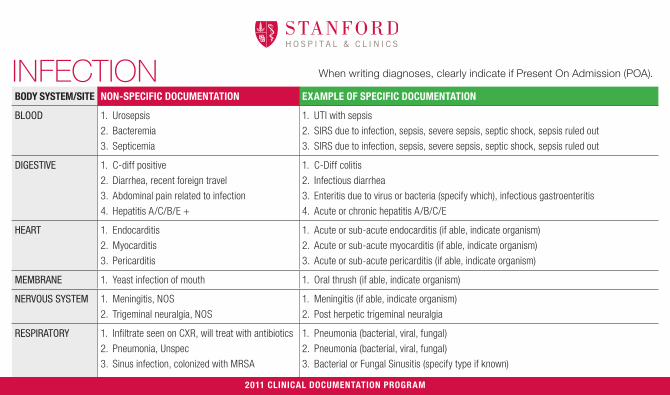
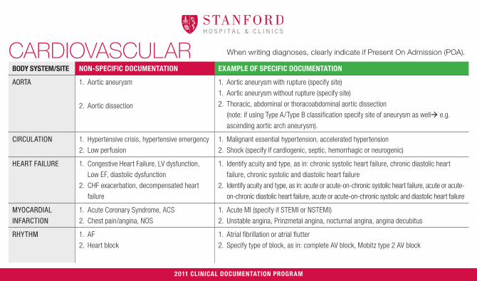
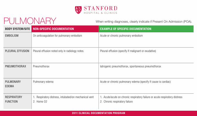
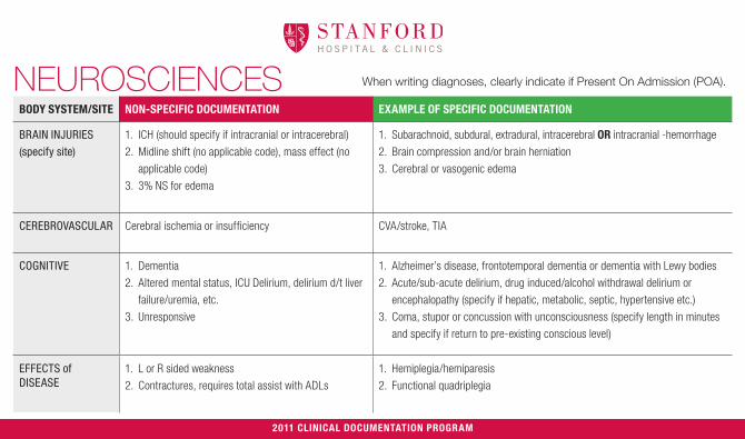
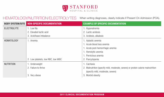
When writing diagnoses, clearly indicate if Present On Admission (POA).
BODY SYSTEM/SITE NON-SPECIFIC DOCUMENTATION EXAMPLE OF SPECIFIC DOCUMENTATION
BLOOD 1. Urosepsis
2. Bacteremia
3. Septicemia
1. UTI with sepsis
2. SIRS due to infection, sepsis, severe sepsis, septic shock, sepsis ruled out
3. SIRS due to infection, sepsis, severe sepsis, septic shock, sepsis ruled out
DIGESTIVE 1. C-diff positive
2. Diarrhea, recent foreign travel
3. Abdominal pain related to infection
4. Hepatitis A/C/B/E +
1. C-Diff colitis
2. Infectious diarrhea
3. Enteritis due to virus or bacteria (specify which), infectious gastroenteritis
4. Acute or chronic hepatitis A/B/C/E
HEART 1. Endocarditis
2. Myocarditis
3. Pericarditis
1. Acute or sub-acute endocarditis (if able, indicate organism)
2. Acute or sub-acute myocarditis (if able, indicate organism)
3. Acute or sub-acute pericarditis (if able, indicate organism)
MEMBRANE 1. Yeast infection of mouth 1. Oral thrush (if able, indicate organism)
NERVOUS SYSTEM 1. Meningitis, NOS
2. Trigeminal neuralgia, NOS
1. Meningitis (if able, indicate organism)
2. Post herpetic trigeminal neuralgia
RESPIRATORY 1. Infiltrate seen on CXR, will treat with antibiotics
2. Pneumonia, Unspec
3. Sinus infection, colonized with MRSA
1. Pneumonia (bacterial, viral, fungal)
2. Pneumonia (bacterial, viral, fungal)
3. Bacterial or Fungal Sinusitis (specify type if known)
INFECTION
2011 CLINICAL DOCUMENTATION PROGRAM

DISEASE DEFINITION (FROM ICD-9-CM FOR HOSPITALS, 2011, UNLESS OTHERWISE INDICATED)
Acute and sub-acute endocarditis Bacterial inflammation of the endocardium
Acute and sub-acute myocarditis Acute inflammation of the muscular walls of the heart
Acute and sub-acute pericarditis Inflammation of the pericardium
Bacteremia Lab finding of bacteria in the blood in the absence of two or more signs of sepsis; transient in nature, progresses to
septicemia with severe infectious process.
Colitis Inflammation of mucous membranes of large intestine
Enteritis Inflammation of mucous membranes of the small intestine
Gastroenteritis Inflammation of mucous membrane of stomach and intestines
Sepsis SIRS due to infection
Septic shock Circulatory failure associated with severe sepsis
Severe sepsis Sepsis with associated acute organ dysfunction
SIRS Generally refers to the systemic response to infection, trauma/burns or other insult (such as cancer) with symptoms including
fever, tachycardia, tachypnea and leukocytosis
INFECTION
2011 CLINICAL DOCUMENTATION PROGRAM
When writing diagnoses, clearly indicate if Present On Admission (POA).
BODY SYSTEM/SITE NON-SPECIFIC DOCUMENTATION EXAMPLE OF SPECIFIC DOCUMENTATION
AORTA 1. Aortic aneurysm
2. Aortic dissection
1. Aortic aneurysm with rupture (specify site)
1. Aortic aneurysm without rupture (specify site)
2. Thoracic, abdominal or thoracoabdominal aortic dissection
(note: if using Type A/Type B classification specify site of aneurysm as wellà e.g.
ascending aortic arch aneurysm).
CIRCULATION 1. Hypertensive crisis, hypertensive emergency
2. Low perfusion
1. Malignant essential hypertension, accelerated hypertension
2. Shock (specify if cardiogenic, septic, hemorrhagic or neurogenic)
HEART FAILURE 1. Congestive Heart Failure, LV dysfunction,
Low EF, diastolic dysfunction
2. CHF exacerbation, decompensated heart
failure
1. Identify acuity and type, as in: chronic systolic heart failure, chronic diastolic heart
failure, chronic systolic and diastolic heart failure
2. Identify acuity and type, as in: acute or acute-on-chronic systolic heart failure, acute or acute-
on-chronic diastolic heart failure, acute or acute-on-chronic systolic and diastolic heart failure
MYOCARDIAL
INFARCTION
1. Acute Coronary Syndrome, ACS
2. Chest pain/angina, NOS
1. Acute MI (specify if STEMI or NSTEMI)
2. Unstable angina, Prinzmetal angina, nocturnal angina, angina decubitus
RHYTHM 1. AF
2. Heart block
1. Atrial fibrillation or atrial flutter
2. Specify type of block, as in: complete AV block, Mobitz type 2 AV block
CARDIOVASCULAR
2011 CLINICAL DOCUMENTATION PROGRAM
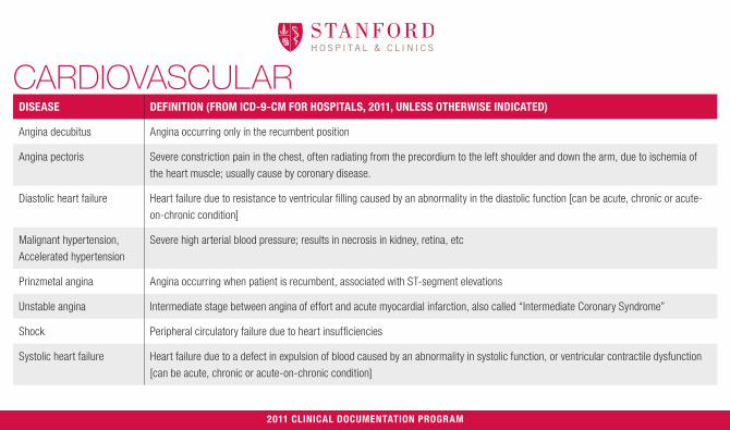
DISEASE DEFINITION (FROM ICD-9-CM FOR HOSPITALS, 2011, UNLESS OTHERWISE INDICATED)
Angina decubitus Angina occurring only in the recumbent position
Angina pectoris Severe constriction pain in the chest, often radiating from the precordium to the left shoulder and down the arm, due to ischemia of
the heart muscle; usually cause by coronary disease.
Diastolic heart failure Heart failure due to resistance to ventricular filling caused by an abnormality in the diastolic function [can be acute, chronic or acute-
on-chronic condition]
Malignant hypertension,
Accelerated hypertension
Severe high arterial blood pressure; results in necrosis in kidney, retina, etc
Prinzmetal angina Angina occurring when patient is recumbent, associated with ST-segment elevations
Unstable angina Intermediate stage between angina of effort and acute myocardial infarction, also called “Intermediate Coronary Syndrome”
Shock Peripheral circulatory failure due to heart insufficiencies
Systolic heart failure Heart failure due to a defect in expulsion of blood caused by an abnormality in systolic function, or ventricular contractile dysfunction
[can be acute, chronic or acute-on-chronic condition]
CARDIOVASCULAR
2011 CLINICAL DOCUMENTATION PROGRAM
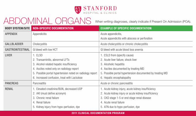
When writing diagnoses, clearly indicate if Present On Admission (POA).
BODY SYSTEM/SITE NON-SPECIFIC DOCUMENTATION EXAMPLE OF SPECIFIC DOCUMENTATION
APPENDIX Appendicitis Acute appendicitis,
Acute appendicitis with abscess or perforation
GALLBLADDER Cholecystitis Acute cholecystitis or chronic cholecystitis
GASTROINTESTINAL GI bleed with low HCT GI bleed with acute blood loss anemia
LIVER 1. ELSD
2. Transaminitis, abnormal LFTs
3. Alcohol related hepatic insufficiency
4. Ascites noted only on radiology report
5. Possible portal hypertension noted on radiology report
6. Increased confusion, treat with Lactulose
1. ESLD from (specify cause)
2. Acute liver failure, shock liver
3. Alcoholic hepatitis
4. Ascites documented by treating MD
5. Possible portal hypertension documented by treating MD
6. Hepatic encephalopathy
PANCREAS Pancreatitis Acute or chronic pancreatitis
RENAL 1. Elevated creatinine/BUN, decreased UOP
2. AKI (must define acronym)
3. Chronic renal failure
4. Renal failure
5. Kidney injury from hypo-perfusion, dye
1. Acute kidney injury, acute kidney insufficiency
2. Acute kidney injury or acute kidney insufficiency
3. CKD stage 1-5 or end stage renal disease
4. Acute renal failure
5. ATN due to hypo-perfusion, dye
ABDOMINAL ORGANS
2011 CLINICAL DOCUMENTATION PROGRAM
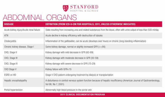
DISEASE DEFINITION (FROM ICD-9-CM FOR HOSPITALS, 2011, UNLESS OTHERWISE INDICATED)
Acute kidney injury/Acute renal failure State resulting from increasing urea and related substances from the blood, often with urine output of less than 500 ml/day
ATN Acute decline in kidney efficiency with destruction of tubules
Cholecystitis Inflammation of the gallbladder, can be acute (develops over hours) or chronic (long standing inflammation)
Chronic kidney disease, Stage I Some kidney damage, normal or slightly increased GFR (>=90)
CKD, Stage II Kidney damage with mild decrease in GFR (60-89)
CKD, Stage III Kidney damage with moderate decrease in GFR (30-59)
CKD, Stage IV Kidney damage with severe decrease in GFR (15-29)
CKD Stage V Kidney failure with GFR<15
ESRD on HD Stage V CKD patient undergoing treatment by dialysis or transplantation
Hepatic encephalopathy A disturbance in central nervous system function because of hepatic insufficiency (American Journal of Gastroenterology,
Vol 96, No 7, 2001)
Portal hypertension Abnormally high blood pressure in the portal vein
ABDOMINAL ORGANS
2011 CLINICAL DOCUMENTATION PROGRAM
When writing diagnoses, clearly indicate if Present On Admission (POA).
BODY SYSTEM/SITE NON-SPECIFIC DOCUMENTATION EXAMPLE OF SPECIFIC DOCUMENTATION
EMBOLISM On anticoagulation for pulmonary embolism Acute or chronic pulmonary embolism
PLEURAL EFFUSION Pleural effusion noted only in radiology notes. Pleural effusion (specify if malignant or exudative)
PNEUMOTHORAX Pneumothorax Iatrogenic pneumothorax, spontaneous pneumothorax
PULMONARY
EDEMA
Pulmonary edema Acute or chronic pulmonary edema (specify if cause is cardiac)
RESPIRATORY
FUNCTION
1. Respiratory distress, intubated/on mechanical vent
2. Home O2
1. Acute/acute on chronic respiratory failure or acute respiratory distress
2. Chronic respiratory failure
PULMONARY
2011 CLINICAL DOCUMENTATION PROGRAM
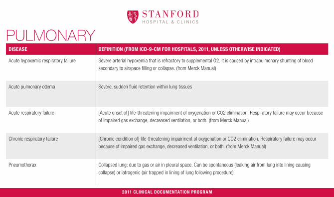
DISEASE DEFINITION (FROM ICD-9-CM FOR HOSPITALS, 2011, UNLESS OTHERWISE INDICATED)
Acute hypoxemic respiratory failure Severe arterial hypoxemia that is refractory to supplemental O2. It is caused by intrapulmonary shunting of blood
secondary to airspace filling or collapse. (from Merck Manual)
Acute pulmonary edema Severe, sudden fluid retention within lung tissues
Acute respiratory failure [Acute onset of] life-threatening impairment of oxygenation or CO2 elimination. Respiratory failure may occur because
of impaired gas exchange, decreased ventilation, or both. (from Merck Manual)
Chronic respiratory failure [Chronic condition of] life-threatening impairment of oxygenation or CO2 elimination. Respiratory failure may occur
because of impaired gas exchange, decreased ventilation, or both. (from Merck Manual)
Pneumothorax Collapsed lung; due to gas or air in pleural space. Can be spontaneous (leaking air from lung into lining causing
collapse) or iatrogenic (air trapped in lining of lung following procedure)
PULMONARY
2011 CLINICAL DOCUMENTATION PROGRAM
When writing diagnoses, clearly indicate if Present On Admission (POA).
BODY SYSTEM/SITE NON-SPECIFIC DOCUMENTATION EXAMPLE OF SPECIFIC DOCUMENTATION
BRAIN INJURIES
(specify site)
1. ICH (should specify if intracranial or intracerebral)
2. Midline shift (no applicable code), mass effect (no
applicable code)
3. 3% NS for edema
1. Subarachnoid, subdural, extradural, intracerebral OR intracranial -hemorrhage
2. Brain compression and/or brain herniation
3. Cerebral or vasogenic edema
CEREBROVASCULAR Cerebral ischemia or insufficiency CVA/stroke, TIA
COGNITIVE 1. Dementia
2. Altered mental status, ICU Delirium, delirium d/t liver
failure/uremia, etc.
3. Unresponsive
1. Alzheimer’s disease, frontotemporal dementia or dementia with Lewy bodies
2. Acute/sub-acute delirium, drug induced/alcohol withdrawal delirium or
encephalopathy (specify if hepatic, metabolic, septic, hypertensive etc.)
3. Coma, stupor or concussion with unconsciousness (specify length in minutes
and specify if return to pre-existing conscious level)
EFFECTS of DISEASE
1. L or R sided weakness
2. Contractures, requires total assist with ADLs
1. Hemiplegia/hemiparesis
2. Functional quadriplegia
NEUROSCIENCES
2011 CLINICAL DOCUMENTATION PROGRAM

NEUROSCIENCESDISEASE DEFINITION (FROM ICD-9-CM FOR HOSPITALS, 2011, UNLESS OTHERWISE INDICATED)
Brain compression Elevated pressure in brain due to blood clot, tumor, fracture, abscess, other condition.
Brain herniation When brain tissue, cerebrospinal fluid, and blood vessels are moved or pressed away from their usual position inside the skull.
Cerebral edema Elevated pressure in the brain due to fluid retention in brain tissues
Coma State of unconsciousness from which the patient cannot be awakened.
Concussion w/brief, moderate,
prolonged loss of consciousness
Brief: 30 min- 59 minutes Moderate: 1-24 hour Prolonged: more than 24 hrs. Should specify if there is a
return to pre-existing conscious level.
Delirium Delirium is an acute, transient, usually reversible, fluctuating disturbance in attention, cognition, and consciousness level.
Causes include almost any disorder, intoxication, or drug (from Merck Manual)
Encephalopathy Any diffuse disease of the brain that alters brain function or structure. (from National Institute of Neurological Disorders and Stroke)
Functional quadriplegia Inability to move due to condition such as dementia, severe contractures or arthritis.
Frontotemporal dementia Sporadic and hereditary disorders that affect the frontal and temporal lobes. (from Merck Manual)
Hypertensive encephalopathy Cerebral manifestations (such as visual disturbances and headache) due to high blood pressure.
Lewy body dementia A cerebral dementia with neurophysiologic changes, increased hippocampal volume, hypoperfusion in the occipital lobes, and beta
amyloid deposits with neurofibrillary tangles, atrophy of cortex and brain stem.
2011 CLINICAL DOCUMENTATION PROGRAM
When writing diagnoses, clearly indicate if Present On Admission (POA).
BODY SYSTEM/SITE NON-SPECIFIC DOCUMENTATION EXAMPLE OF SPECIFIC DOCUMENTATION
ELECTROLYTE 1. Low Na
2. Elevated lactic acid
3. Acid/base imbalance
1. Hyponatremia
2. Lactic acidosis
3. Acidosis, alkalosis
HEMATOLOGY 1. Anemia
2. Low platelets, low RBC, low WBC
1. Aplastic anemia
1. Acute blood loss anemia
1. Acute post-hemorrhagic anemia
1. Hemolytic anemia
1. Pernicious anemia
2. Pancytopenia
NUTRITION 1. Underweight
2. Failure to thrive
3. Very obese
1. Cachexia
2. Malnutrition (specify mild, moderate, severe) or protein calorie malnutrition
(specify mild, moderate, severe)
3. Morbid obesity
HEMATOLOGY/NUTRITION/ELECTROLYTES
2011 CLINICAL DOCUMENTATION PROGRAM

HEMATOLOGY/NUTRITION/ELECTROLYTES
DISEASE DEFINITION (FROM ICD-9-CM FOR HOSPITALS, 2011, UNLESS OTHERWISE INDICATED)
Acute blood loss anemia Anemia from excessive bleeding (Merck Manual)
Acute postoperative
hemorrhagic anemia
[Anemia from] hemorrhage complicating a procedure, with some exceptions is considered a complication of surgery (from AHRQ
Quality Indicators)
Aplastic anemia Bone marrow failure to produce the normal amount of blood components [red blood cells]
Cachexia A complex metabolic syndrome associated with underlying illness and characterized by loss of muscle with or without loss of fat
mass (developed by Cachexia Consensus Conference, 2008)
Hemolytic anemia A condition when there are not enough red blood cells in the blood, due to premature destruction of red blood cells.
Pernicious anemia Pernicious anemia is a decrease in red blood cells that occurs when the body cannot properly absorb vitamin B12 from the
gastrointestinal tract (from US National Library of Medicine)
Protein calorie malnutrition Malnutrition characterized by biochemical changes in electrolytes, lipids, blood plasma
2011 CLINICAL DOCUMENTATION PROGRAM
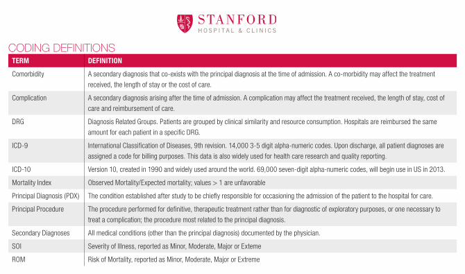
CODING DEFINITIONSTERM DEFINITION
Comorbidity A secondary diagnosis that co-exists with the principal diagnosis at the time of admission. A co-morbidity may affect the treatment
received, the length of stay or the cost of care.
Complication A secondary diagnosis arising after the time of admission. A complication may affect the treatment received, the length of stay, cost of
care and reimbursement of care.
DRG Diagnosis Related Groups. Patients are grouped by clinical similarity and resource consumption. Hospitals are reimbursed the same
amount for each patient in a specific DRG.
ICD-9 International Classification of Diseases, 9th revision. 14,000 3-5 digit alpha-numeric codes. Upon discharge, all patient diagnoses are
assigned a code for billing purposes. This data is also widely used for health care research and quality reporting.
ICD-10 Version 10, created in 1990 and widely used around the world. 69,000 seven-digit alpha-numeric codes, will begin use in US in 2013.
Mortality Index Observed Mortality/Expected mortality; values > 1 are unfavorable
Principal Diagnosis (PDX) The condition established after study to be chiefly responsible for occasioning the admission of the patient to the hospital for care.
Principal Procedure The procedure performed for definitive, therapeutic treatment rather than for diagnostic of exploratory purposes, or one necessary to
treat a complication; the procedure most related to the principal diagnosis.
Secondary Diagnoses All medical conditions (other than the principal diagnosis) documented by the physician.
SOI Severity of Illness, reported as Minor, Moderate, Major or Exteme
ROM Risk of Mortality, reported as Minor, Moderate, Major or Extreme

COMMON QUESTIONSQUESTION ANSWER
Why does provider
documentation matter?
Provider documentation is converted into medical codes that are used to:1. Compare actual mortality with expected mortality2. Compare actual length of stay to expected length of stay3. Compare physician and facility performance to peers- “benchmarking”4. Generate hospital bills5. Causes reimbursement adjustments
What documentation counts? All narrative documentation by treating providers is converted into codes. Coding staff cannot make clinical assumptions or code from reports written by non-treating MD (such as radiologist or pathologist). Accurate coding of the inpatient medical record is dependent upon documentation by the treating provider.
What is documentation used for? Documentation establishes the severity of illness and risk of mortality, medical necessity of admission, accurate data and statistics for hospital and provider, and regulatory compliance.
What if I can’t say a diagnosis
for certain?
If a provider writes “possible”, “probable” or “cannot rule out” before a diagnosis and this “possible”, “probable” or “cannot rule out” diagnosis is still documented at time of discharge, it will be coded as existing. [This only applies for inpatients.]
Can codes for chronic conditions
be taken from previous admission?
No. Every admission is looked at separately and coders cannot use documentation from previous admissions to assign codes.
Why does Present On Admission
(POA) matter?
Hospital acquired conditions are publically reported and used as an indicator of the quality of care given at that facility. Clearly stating if a condition is present on admission or acquired in the hospital helps avoid erroneous reporting and differentiates what the patient arrived with and what was acquired in the hospital, establishing an accurate expected mortality and severity of illness.

HOW TO ANSWER A QUERYIf a diagnoses does not contain specific language, a documentation reviewer may place a “Documentation Clarification” in the chart which needs to be cosigned and addended by the assigned provider. Follow the steps below to respond to a documentation clarification in EPIC.
1. Select note from “Cosign Notes” folder in EPIC In Basket
2. Choose “Addendum” option from EPIC toolbar
3. Enter appropriate diagnosis. Then sign note. Remember to use this diagnosis in subsequent notes.
2011 CLINICAL DOCUMENTATION PROGRAM2011 CLINICAL DOCUMENTATION PROGRAM
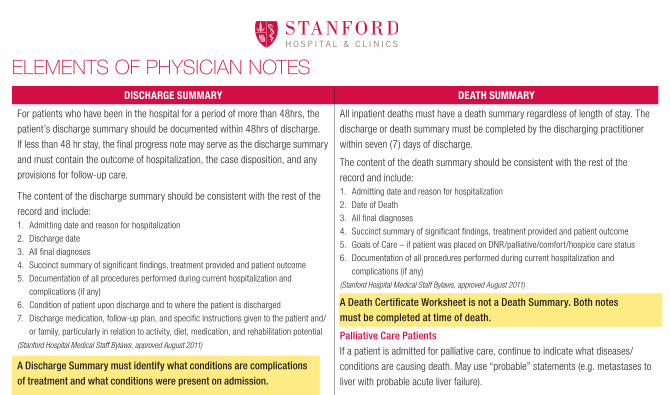
ELEMENTS OF PHYSICIAN NOTESDISCHARGE SUMMARY DEATH SUMMARY
For patients who have been in the hospital for a period of more than 48hrs, the
patient’s discharge summary should be documented within 48hrs of discharge.
If less than 48 hr stay, the final progress note may serve as the discharge summary
and must contain the outcome of hospitalization, the case disposition, and any
provisions for follow-up care.
The content of the discharge summary should be consistent with the rest of the
record and include:1. Admitting date and reason for hospitalization
2. Discharge date
3. All final diagnoses
4. Succinct summary of significant findings, treatment provided and patient outcome
5. Documentation of all procedures performed during current hospitalization and
complications (if any)
6. Condition of patient upon discharge and to where the patient is discharged
7. Discharge medication, follow-up plan, and specific instructions given to the patient and/
or family, particularly in relation to activity, diet, medication, and rehabilitation potential
(Stanford Hospital Medical Staff Bylaws, approved August 2011)
A Discharge Summary must identify what conditions are complications of treatment and what conditions were present on admission.
All inpatient deaths must have a death summary regardless of length of stay. The
discharge or death summary must be completed by the discharging practitioner
within seven (7) days of discharge.
The content of the death summary should be consistent with the rest of the
record and include:1. Admitting date and reason for hospitalization
2. Date of Death
3. All final diagnoses
4. Succinct summary of significant findings, treatment provided and patient outcome
5. Goals of Care – if patient was placed on DNR/palliative/comfort/hospice care status
6. Documentation of all procedures performed during current hospitalization and
complications (if any)
(Stanford Hospital Medical Staff Bylaws, approved August 2011)
A Death Certificate Worksheet is not a Death Summary. Both notes must be completed at time of death.
Palliative Care PatientsIf a patient is admitted for palliative care, continue to indicate what diseases/
conditions are causing death. May use “probable” statements (e.g. metastases to
liver with probable acute liver failure).
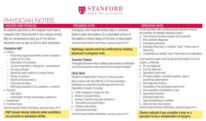
PHYSICIAN NOTESHISTORY AND PHYSICAL
All patients admitted to the hospital must have a complete H&P documented in the medical record. May be completed as early as 24 hrs before admission and as late as 24 hrs after admission.
Complete H&P a. History:
– Presenting diagnosis/condition (chief complaint/reason for the visit)
– Description of symptoms – Current medications, biologicals, nutraceuticals – Any drug allergies – Significant past medical & surgical history – Review of systems – Significant family history – Psychosocial status – Nutritional evaluation (if GI, pediatrics, or elderly)
b. Physical: – Vital Signs – examination of body areas/organ systems as
c. Assessmentd. Treatment plan(Stanford Hospital Medical Staff Bylaws, approved August 2011)
H&P should clearly indicate what conditions are present on admission (POA).
PROGRESS NOTEA progress note must be written daily in sufficient detail to allow formulation of a reasonable picture of the patient’s clinical status at the time of observation. (Stanford Hospital Medical Staff Bylaws, approved August 2011)
Radiology reports must be confirmed by treating physician in progress note.
Transfer PatientThoroughly document what conditions were present on admission and what procedures were performed at outside hospital.
Clinic NoteShould be created within 24 hours of the encounter.
Must conform with the CMS 95 or 97 Documentation Guidelines for evaluation and Management Services (regardless of payer), including:
a. Chief complaint or reason for visitb. History of present illnessc. Review of systems and pain evaluationd. Past family and social historye. Physical examinationf. Assessment and plan
(Stanford Hospital Medical Staff Bylaws, approved August 2011)
OPERATIVE NOTEA brief operative note containing these elements must be documented immediately following surgery:a. The name(s) of primary surgeon and assistantsb. Post-operative diagnosisc. Procedure performedd. Estimated blood loss, or indicate “none” if there was no
blood losse. Complications or indicate “none” if there were no complications
A full operative report must be documented within 24 hrs of surgery, containing:a. Pre-op diagnosisb. Post-op diagnosisc. Operations performedd. Principal surgeon, assistant surgeons, type of
anesthesia administerede. Intra-operative findingsf. Description of the procedures performedg. Intra-operative complications, if anyh. Specimens removedi. Estimated blood lossj. Type of anesthesia or sedationk. Date and time of procedure(Stanford Hospital Medical Staff Bylaws, approved August 2011)
Clearly indicate if you consider a tear or a puncture to be a complication of surgery.
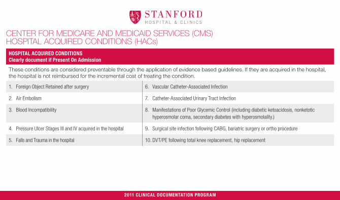
CENTER FOR MEDICARE AND MEDICAID SERVICES (CMS) HOSPITAL ACQUIRED CONDITIONS (HACs)
HOSPITAL ACQUIRED CONDITIONSClearly document if Present On Admission
These conditions are considered preventable through the application of evidence based guidelines. If they are acquired in the hospital, the hospital is not reimbursed for the incremental cost of treating the condition.
1. Foreign Object Retained after surgery 6. Vascular Catheter-Associated Infection
2. Air Embolism 7. Catheter-Associated Urinary Tract Infection
3. Blood Incompatibility 8. Manifestations of Poor Glycemic Control (including diabetic ketoacidosis, nonketotic hyperosmolar coma, secondary diabetes with hyperosmolality.)
4. Pressure Ulcer Stages III and IV acquired in the hospital 9. Surgical site infection following CABG, bariatric surgery or ortho procedure
5. Falls and Trauma in the hospital 10. DVT/PE following total knee replacement, hip replacement
2011 CLINICAL DOCUMENTATION PROGRAM
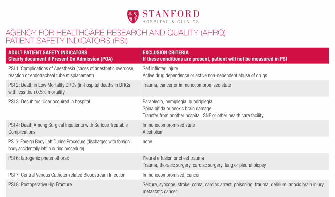
AGENCY FOR HEALTHCARE RESEARCH AND QUALITY (AHRQ) PATIENT SAFETY INDICATORS (PSI)
ADULT PATIENT SAFETY INDICATORS Clearly document if Present On Admission (POA)
EXCLUSION CRITERIA If these conditions are present, patient will not be measured in PSI
PSI 1: Complications of Anesthesia (cases of anesthetic overdose, reaction or endotracheal tube misplacement)
Self inflicted injuryActive drug dependence or active non-dependent abuse of drugs
PSI 2: Death in Low Mortality DRGs (in-hospital deaths in DRGs with less than 0.5% mortality
Trauma, cancer or immunocompromised state
PSI 3: Decubitus Ulcer acquired in hospital Paraplegia, hemiplegia, quadriplegiaSpina bifida or anoxic brain damageTransfer from another hospital, SNF or other health care facility
PSI 4: Death Among Surgical Inpatients with Serious Treatable Complications
Immunocompromised stateAlcoholism
PSI 5: Foreign Body Left During Procedure (discharges with foreign body accidentally left in during procedure)
none
PSI 6: Iatrogenic pneumothorax Pleural effusion or chest traumaTrauma, thoracic surgery, cardiac surgery, lung or pleural biopsy
PSI 7: Central Venous Catheter-related Bloodstream Infection Immunocompromised, cancer
PSI 8: Postoperative Hip Fracture Seizure, syncope, stroke, coma, cardiac arrest, poisoning, trauma, delirium, anoxic brain injury, metastatic cancer
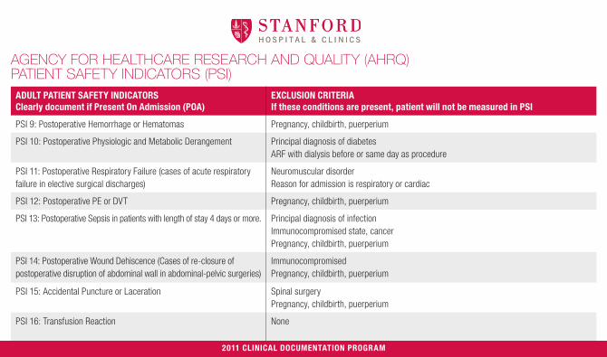
AGENCY FOR HEALTHCARE RESEARCH AND QUALITY (AHRQ) PATIENT SAFETY INDICATORS (PSI)
ADULT PATIENT SAFETY INDICATORS Clearly document if Present On Admission (POA)
EXCLUSION CRITERIA If these conditions are present, patient will not be measured in PSI
PSI 9: Postoperative Hemorrhage or Hematomas Pregnancy, childbirth, puerperium
PSI 10: Postoperative Physiologic and Metabolic Derangement Principal diagnosis of diabetesARF with dialysis before or same day as procedure
PSI 11: Postoperative Respiratory Failure (cases of acute respiratory failure in elective surgical discharges)
Neuromuscular disorderReason for admission is respiratory or cardiac
PSI 12: Postoperative PE or DVT Pregnancy, childbirth, puerperium
PSI 13: Postoperative Sepsis in patients with length of stay 4 days or more. Principal diagnosis of infectionImmunocompromised state, cancerPregnancy, childbirth, puerperium
PSI 14: Postoperative Wound Dehiscence (Cases of re-closure of postoperative disruption of abdominal wall in abdominal-pelvic surgeries)
ImmunocompromisedPregnancy, childbirth, puerperium
PSI 15: Accidental Puncture or Laceration Spinal surgeryPregnancy, childbirth, puerperium
PSI 16: Transfusion Reaction None
2011 CLINICAL DOCUMENTATION PROGRAM

AHRQ INPATIENT QUALITY INDICATORS RELATING TO NEUROSCIENCESCRANIOTOMY MORTALITY RATE ACUTE STROKE MORTALITY RATE CEA MORTALITY RATE
Relationship to Quality Better processes of care may reduce mortality for craniotomy, which represents better quality care.
Better processes of care may reduce short-term mortality, which represents better quality.
Better processes of care may reduce short-term mortality, which represents better quality.
Definition Number of deaths per 100 discharges with DRG code for craniotomy
Number of deaths per 100 discharges with principal diagnosis code of stroke
Number of deaths per 100 CEAs
Exclusion criteria (clearly note when present)
•Principal diagnosis of head trauma •Pregnancy, childbirth, puerperium•Newborns and other neonates
•Missing discharge disposition•Transferred to another short term hospital•Pregnancy, childbirth and puerperium•Newborns and other neonates
2011 CLINICAL DOCUMENTATION PROGRAM

THE JOINT COMMISSION PRIMARY STROKE CERTIFICATION MEASURES If MD documents valid reason why measure was not followed, that patient will not be measured.
MEASURE ISCHEMIC STROKE HEMORRHAGIC STROKE
Discharged on Antithrombotic Therapy
Patients with Atrial Fibrillation Receiving Anticoagulation Therapy
Thrombolytic Therapy Administered
Antithrombotic Therapy By End of Hospital Day Two
Discharged on Cholesterol Reducing Medication
Dysphagia Screening
Stroke Education
Smoking Cessation/Advice/Counseling
Assessed for Rehabilitation
2011 CLINICAL DOCUMENTATION PROGRAM