Embed Size (px)
Citation preview

8/10/2019 Infection strategies of enteric pathogenic Escherichia coli
http://slidepdf.com/reader/full/infection-strategies-of-enteric-pathogenic-escherichia-coli 1/17
Infection strategies of enteric pathogenicEscherichia coli
Abigail Clements,* Joanna C. Young, Nicholas Constantinou and Gad Frankel
Centre for Molecular Microbiology and Infection; Imperial College; London, UK
Keywords: enteric E. coli , host-pathogen interactions, molecular mechanisms of pathogenesis, gut microbes, T
Enteric Escherichia coli (E. coli) are both natural flora of humansand important pathogens causing significant morbidity andmortality worldwide. Traditionally enteric E. coli have beendivided into 6 pathotypes, with further pathotypes oftenproposed. In this review we suggest expansion of the entericE. coli into 8 pathotypes to include the emerging pathotypesof adherent invasive E. coli (AIEC) and Shiga-toxin producingenteroaggregative E. coli (STEAEC). The molecular mechanismsthat allow enteric E. coli to colonize and cause disease in thehuman host are examined and for two of the pathotypes thatexpress a type 3 secretion system (T3SS) we discuss thecomplex interplay between translocated effectors andmanipulation of host cell signaling pathways that occursduring infection.
Since its identification in 1885, Escherichia coli has become one of the most comprehensively studied bacterial species. E. coli strainsare comparatively easy to grow and manipulate in the laboratory,are amenable to genetic manipulation, and naturally acquiremobile genetic elements. While E. coli isolates form part of the
beneficial normal flora of the intestine, some strains have evolved pathogenic mechanisms to cause disease in humans and animals.E. coli strains can cause enteric/diarrhogenic or extraintestinal(ExPEC) infections in humans. ExPEC infections are primarily urinary tract (caused by uropathogenic E. coli , UPEC) and sepsis/meningitis (caused by neonatal meningitis E. coli , NMEC). Only the enteric E. coli will be covered in this review.
Enteric E. coli infections are traditionally divided into 6pathotypes based on their pathogenicity profiles (virulence factors,clinical disease and phylogenetic profile): Enteropathogenic E. coli (EPEC), Enterohamerrhagic E. coli (EHEC), Enteroinvasive E.coli (EIEC, including Shigella sp), Enteroaggregative E. coli (EAEC), Enterotoxigenic E. coli (ETEC) and Diffusely AdherentE. coli (DAEC).1 ,2 Characteristic features of these pathotypes areshown in Table 1. Two further pathotypes have recently emerged; Adherent Invasive E. coli (AIEC) which is thought to be associated with Crohn disease but does not cause diarrhogenic infection3
and the Shiga Toxin (Stx) producing Enteroaggregative E. coli (STEAEC) responsible for the 2011 Germany E. coli outbreak.E. coli strains can also be categorized by their serogroup, e.g.,
E. coli O157 where O refers to the LPS O-antigen or see.g., E. coli O157:H7 where H refers to the flagellar antHowever, as each pathotype contains many serotypes (11serotypes have been identified 4) and some serotypes can belonmore than one pathotype (e.g., O26:H11 can be either EPEHEC), serotyping strains may not provide definitive idtion of pathotypes.
Epidemiology
Enteric E. coli are part of the natural flora of many aniHuman infections occur through consumption of contamfood products (undercooked meat, or contaminated fresh such as salad leaves), drinking water contaminated with ahuman waste, or through direct person-to-person spreapoor hygiene.5 Accurate figures of the incidence of enteric E. coli infections worldwide are difficult to determine, as the cagents of diarrhogenic infections are often not identifiedeveloping world ETEC, EPEC and EAEC appear to bcauses of infantile diarrhea with potentially fatal conse
when untreated, while in the developed world these infecmild and self-limiting. EHEC and more recently EAESTEAEC are the main E. coli pathotypes associated with fpoisoning outbreaks in the developed world.
ETEC is reported to be the most commonly isolated benteropathogen in children under 5 y of age in devcountries, accounting for approximately 20% of cases, eto several hundred million cases of diarrhea and severathousands of deaths each year.6 ETEC is also the most commcause of travelers’ diarrhea accounting for 10– 60% of infectiondepending on the region visited.7 ,8 Extrapolation of these figusuggests there may be 10 million cases of travelers’ diarrhea causedby ETEC per year.9 ETEC also causes disease in ani
including cattle and neonatal and post-weaning pigs10
with hostspecificity occurring through acquisition of colonizatio(CF) rather than emergence of animal specific lineages.
EAEC is the second most common cause of travelers’ diarrhea after ETEC11 and its prevalence in endemic and epidemic dis becoming well recognized. It causes persistent diachildren in developing countries12 ,13 and has been implicated asimportant enteric pathogen affecting AIDS patients.14 No animalreservoir has been described for EAEC suggesting thpersisting in the human population. The 2011 German E. coli foodborne outbreak was caused by an EAEC strain (O1
*Correspondence to: Abigail Clements; Email: [email protected] Submitted: 09/30/11; Revised: 12/22/11; Accepted: 12/26/11http://dx.doi.org/10.4161/gmic.19182
REVIEWGut Microbes 3:2, 71 –87; March/April 2012; G 2012 Landes Bioscience
www.landesbioscience.com Gut Microbes 71
8/10/2019 Infection strategies of enteric pathogenic Escherichia coli
http://slidepdf.com/reader/full/infection-strategies-of-enteric-pathogenic-escherichia-coli 2/17
that had acquired typical EHEC phenotypes, most notably Stx production. Infection with STEAEC O104:H4 resulted in a highpercentage of patients developing hemolytic uremic syndrome(HUS) and a mortality rate of 1%;15 852 cases of HUS, resulting
in 32 deaths and 3469 cases of non-HUS STEAEC, resulting in18 deaths.16 Taking into account this large outbreak and theprevious outbreaks of Stx2-positive O104:H4,17 ,18 STEAECcould now be considered an emerging pathotype of enteric E.coli . Confirmation of STEAEC as an emerging pathotype willrequire continued detection of this distinct population of hybrid EAEC/EHEC strains.
The importance of DAEC to enteric disease remains uncertain.Some studies suggest DAEC may be an important contributor todiarrhogenic disease in children, however problems of cross-reactivity of one of the standard detection probes raises questions
about this.19 A correlation with disease may occur in specdemographics (children aged 18 mo– 5 y 20 or 13– 24 mo21 )although further epidemiological studies are required if Dto remain a distinct enteric E. coli pathotype.
It is still under debate whether the association of AIECrohn disease (CD) is causative or symptomatic. A comof the two is likely with a genetic predisposition to develoexacerbated by microbial infection (including AIEC) inCD. AIEC strains have been found associated with CD leileal and neo-terminal ileal and colonic specimens.22 An increasedimmune response to E. coli in CD patients also suggestsinvolvement of E. coli in the pathology of CD.23
In the developed world epidemiological data for enteri E. coli infections is generally collected based on toxin productithan pathotypes or serotypes and infections are th
Table1. Summary of enteric E. coli pathotypes
Pathotype Adhesin Toxin T3SS SPATE Disease
ETEC Colonization factors (CF)Porcine A/E associated
adhesin (Paa)
Heat-labile enterotoxin (LT)Heat-stable enterotoxin (ST)
Cytolysin A (ClyA)
- ETEC autotransporter A (EatA) Acute watery diarrhea(, 5yo)
Travelers ’ diarrhea
EAEC Aggregative adherencefimbriae (AAF) (I, II, III, Hda)
Toxigenic invasion loci A (Tia)
EAEC heat-stable enterotoxin 1(EAST1)
Shigella enterotoxin (ShET)1Hemolysin E (HlyE)
+/ 2 * Plasmid-encoded toxin (Pet)Protein involved in intestinal
colonization (Pic)Secreted autotransporter toxin
(Sat)Shigella IgA-like protease
homology (SigA)E. coli secreted protein (Esp)P
Travelers ’ diarrheaInfant diarrhea
STEAEC AAFIrgA homolog adhesin (Iha)
Shiga toxin (Stx) - PicPet
Food poisoning
DAEC afimbrial (Afa) or fimbrial (Dr)adhesins
- - Sat Acute diarrhea ( , 5yo)
AIEC Type 1 piliLong polar fimbriae (LPF)
- - - Crohn disease
EHEC IntiminPaa
Toxin B (ToxB)E. coli factor for adherence
(Efa)-1LPF
STEC autoagglutinating adhesin(Saa)
E. coli immunoglobulin-bindingprotein (EibG)
EHEC autotransporter encodinggene A (EhaA)
Outer membrane protein A(OmpA)
Iha
Stx LEE encoded EspP Food poisoning
EPEC IntiminBundle forming pili (BFP)
PaaLPFIha
EhaA
- LEE encoded EspC Infant diarrhea
EIEC(Shigella)
- ShET1/2 pINV encoded Shigella extracellularprotein (Sep)A
SigA
Shigellosis
*One potentially functional but as yet uncharacterized T3SS (ETT2) was found in the genome sequence of EAEC O42 (and remnants of a second).
72 Gut Microbes Volume 3 Issue 2

8/10/2019 Infection strategies of enteric pathogenic Escherichia coli
http://slidepdf.com/reader/full/infection-strategies-of-enteric-pathogenic-escherichia-coli 3/17
commonly referred to as Stx-producing E. coli (STEC) or Verotoxigenic E. coli (VTEC). These classifications can includeall of a pathotype (all EHEC strains are STECs) or part of a pathotype (STEAEC strains are also STECs) and may be further categorized as STEC/VTEC O157 referring to the most prevalentEHEC serogroup. 2011 estimates from the United States suggest9.4 million foodborne illnesses occur annually, resulting in55,961 hospitalizations and 1,351 deaths.24 While STEC O157infections accounted for only 4% of laboratory confirmed foodborne infections in the US from 1996– 2005, STEC O157had the highest case fatality rate across the population and thehighest annual population mortality rate in children 0– 4 y.25 Data from the European Union for 2009 suggests 1% of laboratory confirmed zoonotic infections were attributable to VTEC with7% of those developing HUS.26 Therefore while incidence rates of STEC/VTEC are relatively low compared with Campylobacter and Salmonella infection rates, the severity of the disease and highcase fatality rates means these infections are of major healthconcern.
While EPEC was the first strain of E. coli generally accepted tocause diarrhogenic outbreaks in the developed world,27,28 itsincidence has declined and EPEC outbreaks are now rare indeveloped countries. However it does remain an important causeof infant diarrhea in the developing world with recent estimates of EPEC prevalence among children with diarrhea ranging from 6–
54%, although high carriage rates among healthy controls makesthe contribution of EPEC to disease difficult to assess.29 AtypicalEPEC [i.e., those lacking the EAF plasmid that encodes bundle-forming pili (BFP)] appear to have a propensity to cause persistentdiarrhea.30 ,31
EIEC and Shigella can be distinguished by minor biochemicaltests but in general have the same virulence mechanisms and
disease symptoms. Strains of EIEC and Shigella appear to haveevolved independently to share many characteristics,32 and EIECstrains seen today may simply be intermediates between E. coli and Shigella. We therefore direct the reader to the excellent review on Shigella species contained in this issue for in depth analysis of this pathotype.
Molecular Mechanisms of Virulence
Three pathotypes of E. coli (EHEC, EPEC and EIEC) employ a T3SS to translocate bacterial proteins, known as effectors, directly into the eukaryotic host cell in order to subvert host cell processes.For these pathotypes the T3SS is a major, but not the only,
contributor to virulence. For convenience the pathotypes havebeen divided into non-T3SS dependent pathotypes (ETEC,EAEC, STEAEC, DAEC and AIEC) and T3SS dependentpathotypes (EHEC, EPEC and EIEC). The non-T3SS dependentpathotypes of enteric E. coli have comparatively simple and efficient molecular mechanisms of virulence requiring effectivecolonization factors followed by secretion of toxins that sub-sequently enter the host cell (for ETEC, EAEC and STEAEC).
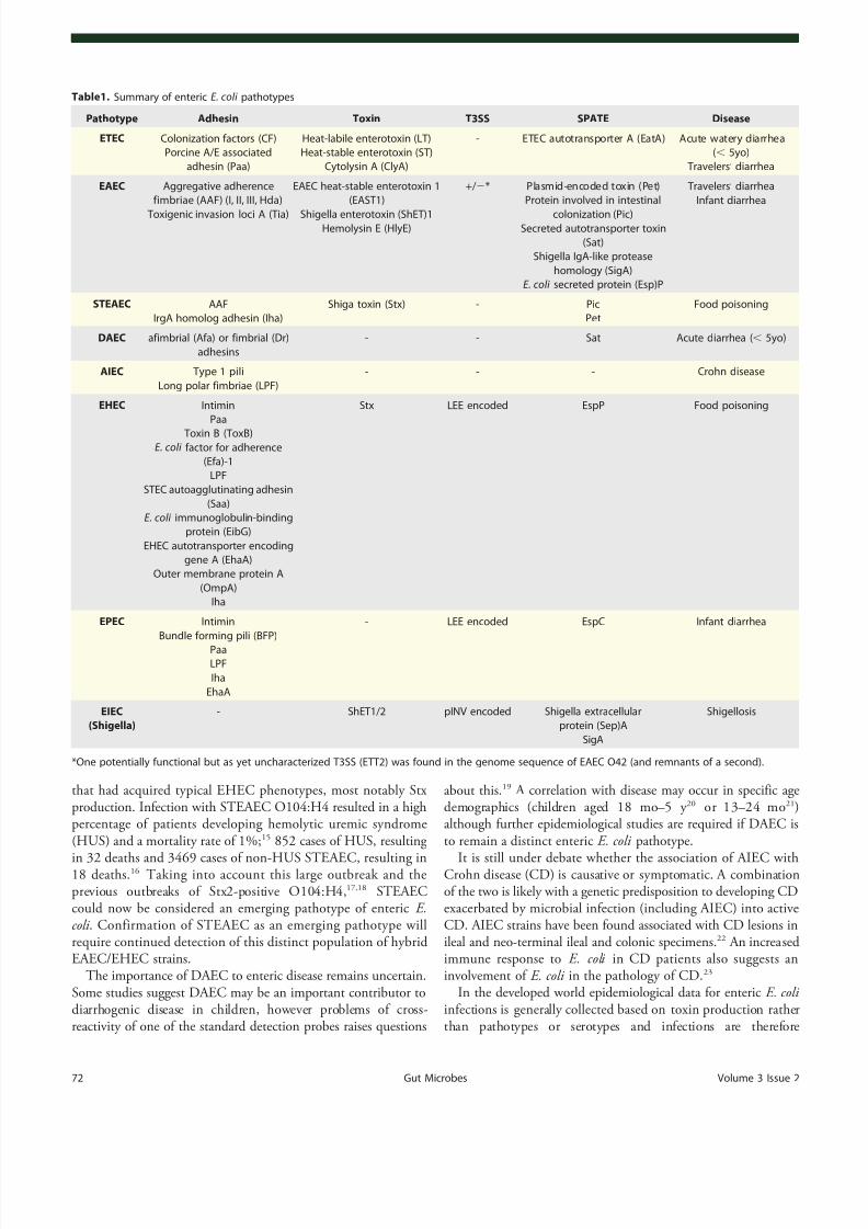
Non-T3SS dependent pathotypes. ETEC. At least 25 distinctproteinaceous colonization factors (CFs) have been identified inETEC strains33 which mediate adhesion to epithelial cells (Fig.1).
Although 30– 50% of ETEC isolates have no characterized phenotypic screening,6 novel CFs are constantly being identgenetically thus reducing the number of isolates with no CF.34 Two further proteins, the outer membrane protein Tthe glycosylated autotransporter TibA, have been repomediate intimate cell attachment and to induce ETEC ininto epithelial cells, for the prototype ETEC strain H135
While ETEC binds to leaf surfaces through the flagellum 36 a novel adhesin, EtpA, located on the tip of ETEC flagella attachment to mammalian host cells.37 EtpA is degraded by thserine protease autotransporter of Enterobacteriaceae (SEatA, thereby modulating bacterial adhesion and accdelivery of heat labile (LT) toxin into host cells.38 A model ofsequential attachment is proposed whereby the lonflagella-EtpA first anchors the bacterium to the host allows shorter CFs to interact. EatA then degrades Etfinally intimate attachment is mediated by Tia and TibB
The main pathology of ETEC occurs through secretionstable (ST) and/or heat labile (LT) toxins. Two small (2,0distinct heat-stable toxins, STa/STI and STb/STII, exist aonly the former is thought to contribute to human diseasSTI mimics the native intestinal hormone guanylin, binand activating the intestinal brush border guanylate-c(GC-C) receptor, increasing intracellular messenger cyc(cGMP). This activates cGMP-dependent protein kinleading to phosphorylation of the cystic fibrosis transmregulator (CFTR) and deregulated ion absorption/secrethence diarrhea.39-41
The LT toxins can be divided into Type I (LT-I), generhuman isolates and closely related to cholera toxin, and(LT-II), which are mainly from non-human isolates.6 LT toxinsare AB5 toxins (one A subunit linked to a pentameric B su
and are transported across the bacterial outer membranetype 2 secretion system.42 LT remains membrane-associatedbinding lipopolysaccharide (LPS)43 and is secreted in outmembrane vesicles (OMVs) that bind to ganglioside recethe mammalian cell (GM1a for LT-I or GD1a/b for LT-II) via theLT-B subunit. The OMVs are then actively endocytosed LT transported via the Golgi and endoplasmic reticulum the cytosol44 where the A1 subunit then ADP-ribosymammalian guanine nucleotide binding protein a-subunit(Gsa ). This inhibits the GTPase activity of Gsa and constitutivelyactivates adenylate cyclase leading to uncontrolled elevatintracellular cAMP concentration.45 This has pleotropic effecwithin the cell including phosphorylation of the CFTR c
channel by protein kinase A. The combination of LT aultimately leads to secretion of electrolytes and water reosmotic diarrhea.
Several other putative toxins have been described foincluding the pore-forming cytotoxin ClyA,46 however details otheir mechanism of action, frequency among isolatimportance during infection is unclear.
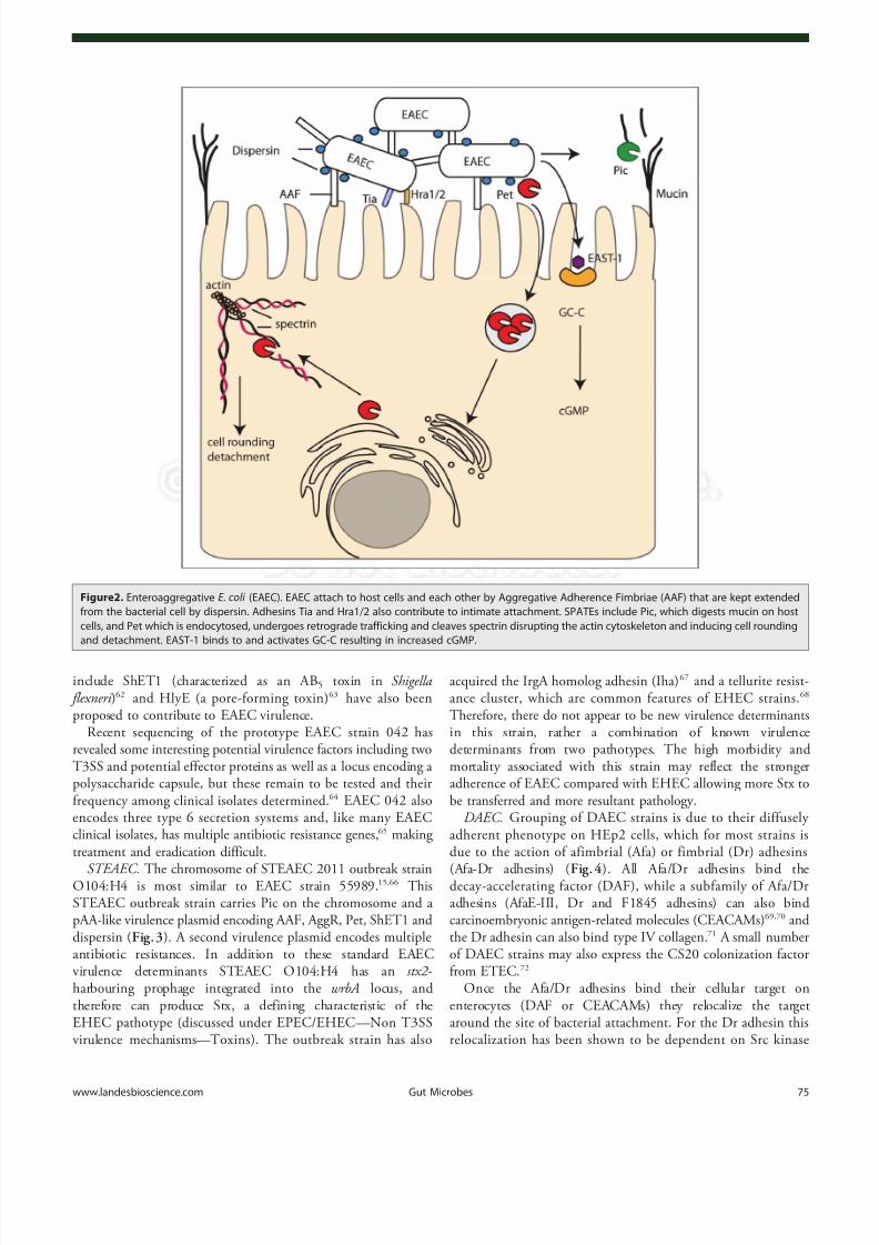
EAEC. EAEC strains are defined by their aggregativeence or “ stacked brick ” phenotype on HEp-2 cells while inintestinal mucosa EAEC forms a biofilm with bacteria ina thick mucus layer. Colonization requires the AAF (Fig.2) and
www.landesbioscience.com Gut Microbes 73

8/10/2019 Infection strategies of enteric pathogenic Escherichia coli
http://slidepdf.com/reader/full/infection-strategies-of-enteric-pathogenic-escherichia-coli 4/17
the regulator AggR both encoded on a large virulence plasmid pAA. AAF, of which four variants have been described, mediatesattachment of EAEC to salad leaves (in combination withflagella),47 host cells48 and human intestine ex vivo49 but were notshown to confer a colonization advantage in a mouse model.50
Other than AAF and AggR, there is a great deal of genomicdiversity among EAEC strains with corresponding heterogeneity in virulence and few conserved virulence factors. Further coloni-zation factors found in some EAEC isolates include Heat-resistantagglutinin (Hra) 1 and 2 and Tia (also found in ETEC).51 The
secreted small hydrophilic protein dispersin (encoded by aap) aidscolonization by attaching noncovalently to the bacterial cell surfacepotentially neutralizing the negative charge of the LPS and allowing the positively charged AAF to extend away from the cell.52
EAEC strains produce a variety of SPATEs of either class I(cytotoxic) or class II (non-cytotoxic). Pic (protease involved incolonization, also found in Shigella flexneri and UPEC) is a class IISPATE with hemagglutinin and mucinolytic activity which may help to penetrate the mucus layer in which EAEC resides onenterocytes.53 Conversely, Pic can induce mucus hypersecretionand an increase in the number of mucus-producing goblet cells.54
Pic has also been implicated in immunomodulation by leukocyte surface glycoproteins and inducing both activaapoptosis in T cells, but impaired migration, of polymnuclear leukocytes (PMNs).55 Pet is a well characterized claSPATE that is endocytosed by host cells, undergoes rettrafficking and utilizes the ER-associated degradation pathway to be released into the cytosol. Pet then cleaves binding protein spectrin in the host cytosol, disrupting thcytoskeleton and causing cell rounding and detachment.56 Recentevidence has also suggested a role for Pet in disrupti
adhesions.57
Pet is only present in a small minority of strain58
and alternative class I SPATES (Sat, SigA, EspP) may havroles. Sat in particular has 52% amino acid identity withis discussed further under DAEC.
Non-SPATE toxins include the EAEC heat-stable ente1 (EAST-1), which is encoded on pAA and is 50% idenbut antigenetically distinct from, the enterotoxic domainLike STa, EAST-1 activates guanylate cyclase leading to cGMP, although the toxigenic effect appears milder thSTa.59 The prevalence of EAST-1 among EAEC strains contribution to virulence remains unclear.60,61 Further toxins
Figure1. Enterotoxigenic E. coli (ETEC). EtpA located on the tip of flagella attaches to host cells but is then degraded by the SPATE EatA. Adherence ismaintained by colonization factors (CF) and intimate attachment achieved with Tia and the autotransporter TibB. Heat stable toxin (ST) is secreted byETEC and binds to guanylate cyclase-C receptor increasing cGMP and cGMP-dependent protein kinase II. Heat labile toxin (LT) is contained in outermembrane vesicles, which are endocytosed after interaction with ganglioside receptors (GM1). Retrograde transport through the Golgi and ER leads tothe A1 subunit being released in the cytosol where it can ADP ribosylate mammalian guanine nucleotide binding protein a -subunit (G s a ) inhibiting theGTPase activity of Gs a and activating adenylate cyclase resulting in uncontrolled cAMP levels. cAMP and cGMP both contribute to phosphorylation of the
cystic fibrosis transmembrane regulator (CFTR) chloride channel and modulation of other ion channels leading to osmotic diarrhea.
74 Gut Microbes Volume 3 Issue 2
8/10/2019 Infection strategies of enteric pathogenic Escherichia coli
http://slidepdf.com/reader/full/infection-strategies-of-enteric-pathogenic-escherichia-coli 5/17
include ShET1 (characterized as an AB5 toxin in Shigella flexneri )62 and HlyE (a pore-forming toxin)63 have also beenproposed to contribute to EAEC virulence.
Recent sequencing of the prototype EAEC strain 042 hasrevealed some interesting potential virulence factors including twoT3SS and potential effector proteins as well as a locus encoding a polysaccharide capsule, but these remain to be tested and their frequency among clinical isolates determined.64 EAEC 042 alsoencodes three type 6 secretion systems and, like many EAECclinical isolates, has multiple antibiotic resistance genes,65 making treatment and eradication difficult.
STEAEC. The chromosome of STEAEC 2011 outbreak strainO104:H4 is most similar to EAEC strain 55989.15 ,66 ThisSTEAEC outbreak strain carries Pic on the chromosome and a pAA-like virulence plasmid encoding AAF, AggR, Pet, ShET1 and dispersin (Fig.3). A second virulence plasmid encodes multipleantibiotic resistances. In addition to these standard EAECvirulence determinants STEAEC O104:H4 has an stx2 -harbouring prophage integrated into the wrbA locus, and therefore can produce Stx, a defining characteristic of theEHEC pathotype (discussed under EPEC/EHEC— Non T3SSvirulence mechanisms— Toxins). The outbreak strain has also
acquired the IrgA homolog adhesin (Iha)67 and a tellurite resisance cluster, which are common features of EHEC st68
Therefore, there do not appear to be new virulence deterin this strain, rather a combination of known virdeterminants from two pathotypes. The high morbidimortality associated with this strain may reflect the adherence of EAEC compared with EHEC allowing mobe transferred and more resultant pathology.
DAEC. Grouping of DAEC strains is due to their difadherent phenotype on HEp2 cells, which for most stdue to the action of afimbrial (Afa) or fimbrial (Dr) a
(Afa-Dr adhesins) (Fig.4). All Afa/Dr adhesins bind decay-accelerating factor (DAF), while a subfamily oadhesins (AfaE-III, Dr and F1845 adhesins) can alscarcinoembryonic antigen-related molecules (CEACAMs69,70 and the Dr adhesin can also bind type IV collagen.71 A small numbeof DAEC strains may also express the CS20 colonizatifrom ETEC.72
Once the Afa/Dr adhesins bind their cellular targenterocytes (DAF or CEACAMs) they relocalize tharound the site of bacterial attachment. For the Dr adherelocalization has been shown to be dependent on Src
Figure2. Enteroaggregative E. coli (EAEC). EAEC attach to host cells and each other by Aggregative Adherence Fimbriae (AAF) that are kept extendedfrom the bacterial cell by dispersin. Adhesins Tia and Hra1/2 also contribute to intimate attachment. SPATEs include Pic, which digests mucin on hostcells, and Pet which is endocytosed, undergoes retrograde trafficking and cleaves spectrin disrupting the actin cytoskeleton and inducing cell roundingand detachment. EAST-1 binds to and activates GC-C resulting in increased cGMP.
www.landesbioscience.com Gut Microbes 75

8/10/2019 Infection strategies of enteric pathogenic Escherichia coli
http://slidepdf.com/reader/full/infection-strategies-of-enteric-pathogenic-escherichia-coli 6/17
Figure3. For figure legend, see page 77.
76 Gut Microbes Volume 3 Issue 2

8/10/2019 Infection strategies of enteric pathogenic Escherichia coli
http://slidepdf.com/reader/full/infection-strategies-of-enteric-pathogenic-escherichia-coli 7/17
activation.73
Following target mobilization, enterocyte signaling pathways (e.g., MAPK and PI3K) are activated and IL-8 issynthesized (for the F1845 adhesin this requires HIF-1a 74 )inducing transepithelial migration of human polymorphonuclear neutrophils (PMN). This stimulates the enterocytes to synthesizeTNFa and IL-1β and upregulate DAF to strengthen bacterialadhesion.75 DAEC can interact with the transmigrating PMNsand induce type I pili-dependent IL-8 release.76 Transmigrated PMNs are also induced to undergo apoptosis after interactionwith DAEC and have a diminished phagocytic capacity,prolonging bacterial persistence in the gut.77
The only documented secreted factor associated with DAECinfection is the SPATE Sat. Sat can induce rearrangement of thetight junction proteins ZO-1, ZO-3 and occludin increasing paracellular permeability but not transepithelial resistance78 and can also bind spectrin,79 rearrange focal adhesion associated
proteins vinculin and paxillin, and cause cell detachmcaspase-independent cell death.80
AIEC. While AIEC strains are genetically related to Estrains they appear to have acquired novel virulencefeatures which can be characterized phenotypically (ainvasion and intramacrophage replication) but the geneof which is still largely undetermined.81 This has hamperedidentification and hence research into the prevalenimportance of AIEC. The majority of AIEC research hasingle strain, LF82, and extrapolation of these results AIEC strains is vital as the field progresses.
AIEC infection requires both a susceptible host as bacterial virulence determinants. The first step in AIEC is abnormal colonization of the intestinal epithelium via tbinding to the CEACAM6 receptor, which is overexpressileal mucosa of CD patients.82,83 In addition to type I pili, lo
Figure3. (See opposite page). Shiga Toxin producing Enteroaggregative E. coli (STEAEC). (A) STEAEC attach to each other and to enterocytes by AAFand dispersin, as for EAEC. STEAEC also encodes the Iha adhesin and SPATES Pet and Pic, although their contribution to infection is unknown. The actionof shiga toxin (Stx) on an endothelial, toxin sensitive cell is shown in (B). The B subunit of Stx interacts with Gb 3 on the host cell and Stx is endocytosedand undergoes retrograde trafficking through the Golgi and ER, the A subunit then cleaves an adenine residue from the 28S rRNA of eukaryoticribosomes inhibiting protein synthesis and leading to cell death. Stx can also cause cells to undergo a ribotoxic stress response, which leads to release of IL-8, or to undergo ER-dependent apoptosis.
Figure4. Diffusely Adherent E. coli (DAEC). AFA/Dr adhesins interact with the decay-accelerating factor (DAF) on host cells. Src kinase activation mobilizesDAF around the attachment site mediating stronger attachment and MAPK and PI3K pathway activation culminating in IL-8 synthesis, which inducestransmigration of PMNs. PMN transmigration stimulates upregulation of DAF and TNF a and Il-1b synthesis. DAEC Type 1 pili induces IL-8 release fromPMNs and apoptosis. Sat induces rearrangement of the tight junction proteins ZO-1, ZO-2 and occludin leading to paracellular permeability.
www.landesbioscience.com Gut Microbes 77

8/10/2019 Infection strategies of enteric pathogenic Escherichia coli
http://slidepdf.com/reader/full/infection-strategies-of-enteric-pathogenic-escherichia-coli 8/17
polar fimbriae were recently shown to also be required for AIECto bind to M cells in Peyers patches.84 This suggests that AIECmay utilize the transcytic characteristics of M cells to cross theintestinal barrier, as is the case for many other intestinal pathogens(e.g., Yersinia sp).
AIEC secrete OMVs that appear to be required for AIEC toinvade intestinal epithelial cells (IECs),85 potentially by delivering effector proteins into the host cell. The OMVs contain OmpA which interacts with the ER-localized stress response proteinGp96 which has been shown to be overexpressed on the apicalsurface of ileal epithelial cells in Crohn disease patients.86 Other than OmpF/C and OmpA the composition of these OMVs and potential effector proteins delivered by this mechanism areunknown. While AIEC invasion into epithelial cells requiresactin and microtubule involvement87 the molecular mechanismsremain unknown. AIEC can survive and replicate in phagoly-somes of infected macrophages in the lamina propria, resulting inincreased TNFa secretion88 which may lead to the inflammationassociated with Crohn disease.
T3SS-dependent pathotypes. Of the T3SS-dependent patho-types EHEC and EPEC primarily remain extracellular during infection while EIEC are found intracellular. Despite these very different lifestyles and the different T3SS origins (LEE encoded or pINV encoded, respectively) EPEC/EHEC and EIEC/Shigella actually share a number of T3SS translocated proteins, e.g., EspG/VirA, EspO/OspE, NleE/OspZ reflecting similar infectionstrategies.
EPEC/EHEC. The EPEC and EHEC T3SS is encoded on a pathogenicity island termed the locus of enterocyte effacement(LEE), a region highly conserved between the attaching/effacing (A/E) pathogens EPEC, EHEC, rabbit enteropathogenic E. coli (REPEC) and murine pathogen Citrobacter rodentium.89 The LEE
encodes gene regulators, structural components of the T3SS,chaperones, the bacterial surface protein intimin and a number of translocated proteins.90
EHEC O157:H7 appears to have evolved from EPEC O55:H791 and non-O157:H7 EHEC strains to have evolved by parallel evolution.92 Defining characteristics of EHEC are thepresence of stx genes, leading to more serious disease pathology and complications (HUS), and absence of BFP, leading todifferent adherence mechanisms. EPEC and EHEC are primarily human pathogens; although a range of ruminants carry EHEC itis generally asymptomatic in these animals. This host restrictionmakes modeling EPEC/EHEC infections difficult and so therelated A/E pathogens REPEC and C. rodentium are commonly
used as infection models.Non T3SS virulence mechanisms. Adherence. EPEC/EHECencode several well-characterized fimbrial (pili) adhesins. HumanEPEC strains are divided into typical or atypical strains according to the presence or absence of the EPEC adherence factor (EAF)plasmid 93 which encodes the BFP. BFP is responsible for theformation of microcolonies through bacterial-bacterial interac-tions and a binding pattern known as localized adherence.94 ,95
Long polar fimbria (LPF) play a significant role in EHECadherence to epithelial cells, although they are not present in allstrains.96
A variety of non-fimbrial adhesins have been identifiedEPEC and EHEC (Table1 ). EHEC factor for adherence (Efafound in some EHEC strains, contributes to in vitro adhe97
The major outer membrane protein OmpA has been repointeract with cultured human intestinal cells in O157 infections.98 Porcine attaching and effacing associated a(Paa), found in EHEC, EPEC and ETEC strains, contrib A/E lesions in pig ileal explants.99 Additionally, there is a growlist of autotransporters involved in adherence in someEHEC strains including STEC autoagglutinating adhesin 100
E. coli immunoglobulin-binding protein (EibG)101 and EhaA.102
Intimate attachment of the bacteria to the host cell is dependent and is described in detail below (Intimate attaand A/E lesion).
Toxins. The major toxin produced by EHEC is the pencoded Stx of which there are two subgroups, Stx1 and Svariants thereof), with Stx2 more common among human Antibiotic treatment of Stx-producing bacteria irecommended as it induces both expression of the toxallows release and dissemination (Stx does not appeaactively transported from the cell).103 For a review of Stx activand intracellular trafficking see Johannes et al.104
Stx are AB5 toxins with the B subunit mediating bindithe membrane glycolipid globotriaosylceramide (Gb3). The Bsubunit induces endocytic plasma membrane invags105 and vesicles traffic to early endosomes where, isensitive cells, Stx leaves the endocytic pathway 106 and travelsthrough the Golgi apparatus and ER by retrograde traThe catalytic A-subunit is translocated to the cytosol to molecular target, rRNA, where it cleaves an adeninefrom the 28S rRNA of eukaryotic ribosomes, inhibitingsynthesis and eventually leading to cell death.107 In toxin
resistant cells (e.g., Monocytes, macrophages) Stx does the endocytic pathway but is degraded by lysosomes.108 In thesecells Stx activates the MAPK pathway and produces ITNFa , which can in turn increase Gb3 expression onendothelial cells.
Once Stx is released into the gut lumen it is translocatethe intestinal epithelium into the underlying tissuebloodstream and then targets host cells expressing G3 . Inhumans, high concentrations of Gb3 are found in renal tubulcells and microvascular endothelial cells, particularly thokidney, gut and brain, explaining the clinical manifestaHUS. Stx has also been shown to induce apoptosis in vatypes which may contribute to disease pathology.109 ,110
Some EHEC and EPEC strains also produce SPATEs, best characterized being EspP from EHEC and EspC fromEspC shares 70% amino acid similarity with Pet (EAEC)cleaves spectrin, although at a different site than Pet. Unwhich is internalized by receptor-mediated endocytosinternalization requires EPEC contact with the host cproduction of a T3SS.111 Four subtypes of EspP have bdescribed with subtype a associated with highly pathogstrains.112 EspP has recently been described to cleave compfactors C3/C3b and C5 and impair complement functvitro.113
78 Gut Microbes Volume 3 Issue 2

8/10/2019 Infection strategies of enteric pathogenic Escherichia coli
http://slidepdf.com/reader/full/infection-strategies-of-enteric-pathogenic-escherichia-coli 9/17
The T3SS and delivered effectors. The general T3SS iscomprised of a cytosolic ATPase, inner and outer membranerings, a periplasmic shaft and an extracellular needle protein(reviewed in ref. 114). The EPEC and EHEC systems have anadditional filament of ~260 nm protruding from the T3SS needlewhich is generated by polymerization of EspA subunits.115 Thisfilament functions in initial attachment to the host cell beforesecretion of the translocators, EspB and EspD, which form thetranslocation pore in the host cell membrane.116 ,117 EHEC and EPEC use the T3SS to inject dozens of effector proteins into theeukaryotic cell cytoplasm. Once translocated, these effector proteins are targeted to different subcellular compartments and affect diverse signaling pathways and physiological processes.In addition to the seven effectors encoded in the LEE, there areother effectors encoded within prophages and other integrativeelements.118 Although EPEC and EHEC show high levels of conservation between the LEE encoded effectors, there issignificant diversity in their non-LEE effector (NLE) repertoire;EPEC E2348/69 has been shown to have 21 intact effector geneswhereas the EHEC O157 strain Sakai is estimated to have closer to 50 T3SS effectors.118,119 For a recent detailed review of EPEC/EHEC effectors see Wong et al.120
Intimate attachment and A/E lesion. EPEC/EHEC coloniza-tion results in the formation of A/E lesions on the apical surfacesof enterocytes (Fig. 5A ). These characteristic T3SS-dependentlesions describe the effacement of microvilli and intimate bacterialattachment with actin accumulation at the bacterial host cellinterface; in vitro the actin polymerization activity results information of raised pedestal-like structures underneath theattached bacteria.121 The outer membrane adhesin, intimin wasthe first bacterial gene product found to be essential for theintimate attachment of bacteria to epithelial cells.122 The intimin
encoding eae gene was later found on the LEE and its product wasshown to be secreted via the general secretory pathway and inserted into the bacterial outer membrane.123 ,124 Four distinctintimin types were originally reported (a , β, c and d),125 althoughmore than 20 types are now recognized.126 Intimin shareshomology (31% identity) with invasin from Yersinia pseudotu- berculosis and further studies on the intimin/invasin family haveshown that the C-terminal 280 amino acids (Int280/Inv280) arerequired for binding β1-chain integrins.127 ,128 The transmembraneintimin receptor (Tir) is a LEE-encoded effector translocated via the T3SS,129 which resides in the host cell plasma membrane and interacts with intimin to allow intimate attachment of the bacteria with the host cell. EPEC/EHEC therefore delivers its own adhesin
into the host to ensure intimate attachment is achieved. Intimincan also interact with the host protein nucleolin, which isupregulated by Stx.130
In addition to the role of Tir as a receptor for intimin, it isalso an important mediator of protein signaling within epithelialcells. In EPEC, Tir is phosphorylated at tyrosine 474 (Y474p) topromote its interaction with the adaptor protein Nck leading tothe recruitment of neural Wiskott-Aldrich syndrome protein (N- WASP).131 ,132 This initiates actin polymerization mediated by the actin-related protein 2/3 (Arp 2/3) complex.131 ,133 However,EHEC Tir lacks a tyrosine 474 equivalent and the process of
actin polymerization is mediated via the T3SS traneffector protein, TccP (Tir-cytoskeleton coupling protein134 alsoknown as EspFU (E. coli secreted protein F in prophage U135
TccP/EspFU interacts with the IRSp53/MIM proteins, IRand IRSp53, which also bind Tir at an Asn-Pro-Tyr (NPtripeptide in the Tir C-terminal domain thereby liTccP/EspFU indirectly to Tir.136 -138 TccP/EspFU interacts withand activates N-WASP preventing its autoinhibition fothus, initiating an Nck-independent actin polymerpathway.139 The NPY motif is conserved in EPEC(NPY454) but in EPEC belonging to lineage 1 this paccounts only for low levels of pedestal formation in theof TccP/EspFU .138 Previous studies have demonstratedconservation of the Nck and TccP/EspFU pathways in EPEClineage 1 and EHEC O157 respectively.140 Interestingly, bothpathways are utilized simultaneously in vitro for most noEHEC strains, EPEC O119:H6141 and EPEC lineage strains.140 Unexpectedly neither pathway appears to be nefor A/E lesion formation in vivo in C. rodentium infections, inEPEC and EHEC infection of human intestinal in vitrocultures (IVOC),142 ,143 or for EHEC colonization in the inrabbits and gnotobiotic piglets models,144 indicating themolecular mechanisms of A/E formation and their fuare far from understood.
Actin remodeling. Several EPEC T3SS effectors disruptGTPase signaling and hence subvert actin dynamics (Fig. 5B).145
The Rho family small G proteins are crucial in the regukey cellular functions and the best characterized are Cdcand RhoA, which trigger filopodia, lamellipodia/ruffles afibers respectively.146 T3SS effectors have been shown to regRho GTPases by functioning as guanine exchange factorswhich control the switch from GDP-bound (inactive)
GTP-bound (active) state,147
or GTPase-activating prote(GAPs), which increase the hydrolysis of GTP leadininactive state of the Rho GTPases.148
Alto et al. grouped 24 bacterial effector proteins in thefamily based on a conserved motif of two invariant amiTrp and Glu separated by three variable amino acids.149 Membersof the family include Map, EspM, EspT from EPEC/EIpgB1 and IpgB2 from Shigella spp, SopE and SifSalmonella spp Map and EspM function as GEFs on CdRhoA respectively, leading to filopodia 150 and stress fiberformation.151 In the case of EspT, Rac1 and Cdc42 are activated which results in the formation of membrane rulamellipodia and bacterial internalization.152 Map and SopE hav
been shown to have similar crystal structures when rescomplex with Cdc42.147 Despite the lack of sequence or strucsimilarity bacterial WxxxE effectors and eukaryotic GEFto form the same conformational complex with Cdc42. Theffector EspH was shown to subvert actin dynamics afilopodia and pedestal formation.153 EspH inactivates mammaliDbl-homology and pleckstrin-homology (DH-PH) Rho G154
but not the bacterial Rho GEFs. This suggests that EspHclear the cell of endogenous Rho GEFs allowing the bactGEFs (WxxxE effectors) to take over cell signaling to bacterial infection strategy.
www.landesbioscience.com Gut Microbes 79

8/10/2019 Infection strategies of enteric pathogenic Escherichia coli
http://slidepdf.com/reader/full/infection-strategies-of-enteric-pathogenic-escherichia-coli 10/17
Figure5. Enteropathogenic E. coli and Enterohamerrhagic E. coli (EPEC and EHEC). EPEC/EHEC inject an array of T3SS effector proteins to mediateintimate attachment and subvert host cell processes. In addition EHEC produces Stx, the action of which is described in Figure 3B . (A) Intimateadherence. The translocated intimin receptor (Tir) binds intimin on the bacterial surface to initiate intimate attachment, actin accumulation and pedestalformation. Tir EPEC is phosphorylated at Y474 resulting in Nck and N-WASP recruitment. Tir EHEC signaling proceeds independently of Nck via the T3SSeffector TccP/EspF U that interacts with IRTKS/IRSp53 and N-WASP. Both pathways lead to Arp2/3 mediated initiation of actin polymerization. (B) Actinremodeling. Map, EspM and EspT activate Rho GTPases leading to filopodia, stress fibers and ruffles/lamellipodia respectively. Additionally, EspT can
induce internalization of EPEC. EspH disrupts Rho GTPase signaling by targeting host DH-PH GEFs. (C) Disruption of gut integrity. Tir, Map, EspF and EspBcontribute to effacement of the normal absorptive microvilli. Map, EspF and EspI disrupt tight junction (TJ) integrity and epithelial barrier function. EspGdisrupts microtubules while EspI and EspG both modulate protein trafficking and affect TJs and the DRA Cl 2 /OH2 exchanger respectively. EspG and EspFalter aquaporin levels disrupting water and ion absorption. EspF reduces the activity of the NHE3 Na + /H+ exchanger and multiple effectors target theSGLT1 Na+ /glucose co-transporter. (D) Manipulating immune responses. NleB, NleC, NleD NleE and NleH inhibit inflammatory responses throughtargeting NF kB, JNK and p38 pathways. NleB and NleE inhibit I kB degradation and subsequent nuclear translocation of NF kB. NleH can also block NFkB nuclear translocation. NleC and NleD function as metalloproteases acting on NF kB and JNK/p38 respectively blocking transcription of pro-inflammatory genes initiated by NF kB and AP-1 transcription factors. EspT promotes expression of inflammatory genes through Erk, JNK and NF kBpathways. (E) Balancing apoptosis and survival. Pro-apoptotic EspF causes mitochondrial dysfunction leading to activation of apoptotic pathways whileCif causes cell cycle disruption. Anti-apoptotic NleH interacts with BI-1 at the ER and NleD inhibition of AP-1 dependent gene expression (shown in C)reduces pro-apoptotic gene expression. EspO and EspZ promote integrin mediated cell adhesion and survival through interacting with ILK and CD98respectively. (F) Inhibiting phagocytosis. EspF, EspB, EspH and EspJ inhibit phagocytosis by macrophages through disruption of PI3K signaling,myosin-actin interactions, Rho GTPase signaling and an unknown mechanism respectively.
80 Gut Microbes Volume 3 Issue 2

8/10/2019 Infection strategies of enteric pathogenic Escherichia coli
http://slidepdf.com/reader/full/infection-strategies-of-enteric-pathogenic-escherichia-coli 11/17
Another T3SS effector, EspV present in EPEC strains E110019and E22 is able to cause nuclear condensation, cell rounding and actin-rich dendrite-like projections upon overexpression inmammalian cells155 although the mechanisms involved in thisactin rearrangement are not yet known.
Disrupting gut integrity. EPEC induces diarrhea through a variety of mechanisms (Fig. 5C). First, the effacement of themicrovilli, a defining feature of the A/E lesion, results in a reduced surface area for normal absorptive processes. EspB, Map, EspF,Tir and intimin have all been shown to be involved in microvilluseffacement.156,157 EPEC also inhibits ion and water exchangethrough more targeted mechanisms. For example, Cl- absorptionis reduced by targeting the DRA Cl-/OH- exchanger throughreducing the exchanger cell surface levels in an EspG and EspG2dependent manner.158 While EspG was originally described todegrade microtubules it can also bind the Golgi matrix proteinGM130, p21-activated kinases (PAKs) and ADP-ribosylationfactors (ARFs)159,160 acting as a molecular scaffold to regulate hostsignaling cascades, leading to decreased protein secretion and receptor trafficking. Consequences of this manipulation of proteintrafficking within the host cell include altering the paracellular permeability and membrane channel expression in entero-cytes.161,162
In addition, the effector EspF is responsible for reducing theactivity of Na +/H+ exchanger 3163 and EPEC infection leads to a significant reduction in the activity of the SGLT1 Na +/glucosecotransporter.156 EspF and EspG have both been implicated inaffecting water transport by altering aquaporin levels on apical and lateral membranes.164 EPEC infections also affect the integrity of the epithelial monolayer and so disrupt barrier function. EspF,Map and intimin have all been shown to contribute to diarrhea through the disruption of tight junction integrity.165 EspI (NleA)
also disrupts intestinal tight junctions166
and like EspG is able todecrease protein transport. EspI does so by binding to SEC24through a PDZ binding motif and inhibiting COPII vesiclefusion167 ,168 indicating EPEC/EHEC have developed multiplemechanisms to modulate the intestinal barrier function and integrity during infection.
Manipulating the immune response. Manipulation of the hostimmune system is a common theme in bacterial infections and a requirement for successful colonization and dissemination.EPEC and EHEC infections lead to an inflammatory responsepredominantly initiated by recognition of the bacterial flagella 169 .The production of pro-inflammatory cytokines is subsequently dampened by the bacteria with increased bacterial loads leading to
reduced IL-8 production170
with several effector proteins acting inconcert to illicit this effect (Fig. 5D). The transcription factor NFkB is central to the initiation of inflammatory responses;NFkB dimers are held inactive in the cytoplasm by IkB until IkBis phosphorylated and degraded allowing NFkB to translocate tothe nucleus and initiate transcription. The effector proteins NleBand NleE inhibit IkB degradation and therefore the nuclear translocation of NFkB subunits.171 ,172 Additionally, the effectorsNleC and NleD have been shown to function as metalloproteasesdownregulating inflammatory responses by targeting NFkB(NleC) and the mitogen activated protein kinases c-Jun
N-terminal Kinase (JNK) and p38 (NleD).173 ,174 NleH effectorshave an uncertain role in immune modulation as they hashown to enhance inflammatory responses175 and converselyattenuate NFkB activation176 with NleH1 able to block nucltranslocation of the RPS3 subunit of NFkB while NleH2 caninduce expression of RPS3 dependent genes.177 ,178 Interestinglythe WxxxE effector EspT, triggers the expression inflammatory genes through Erk, JNK and NFkB pathways179
suggesting a careful balance between pro- and anti-inflaactions exists.
Balancing apoptosis and maintaining cell survival. Like manyenteric pathogens EPEC and EHEC are able to induce aupon infection.180 ,181 However, apoptosis induction needs tmodulated in order to maintain an infective niche withinby balancing pro and anti-apoptotic effectors (Fig. 5E ). In EPECand EHEC, two T3SS effectors are currently known tinducers of apoptosis, EspF182 and Cycle inhibitory factor (Cif183
EspF is targeted to the mitochondria via an N-temitochondrion-targeting sequence and interferes with thchondrial membrane potential.184 EspF has specifically beshown to induce the release of cytochrome c and cleavage ocaspases 9 and 3182 while Cif acts mainly as a bactcyclomodulin subverting the eukaryotic cell cycle by both the G1/S and G2/M transitions resulting in apoptosis.182 Theaction of these pro-apoptotic effectors is balanced translocation of anti-apoptotic effectors such as NleNleD. NleH inhibits intrinsic apoptotic pathways via itinteraction with the anti-apoptotic protein Bax inhibitor1)185 while NleD inactivates JNK leading to the suppressiodownstream transcription factor AP-1 which activates sevapoptotic proteins.186
Additionally, other T3SS effectors indirectly contribut
inhibition of apoptosis through their ability to promote hsurvival mechanisms. The LEE encoded effector EspZ with CD98, a host protein involved in integrin mediatadhesion and cell survival187 while the non-LEE encoded effeEspO increases cell adhesion and survival through itbinding with integrin-linked kinase (ILK).188 EPEC and EHECalso activate several other signaling pathways known tapoptosis including the protein kinase C, tyrosine kinaphosphatidylinositol 3-kinase189 -191 suggesting other effectomight also be involved in manipulation of apoptosis.
Inhibiting phagocytosis. During infection EPEC and EHEprimarily survive extracellularly and have developed meto inhibit internalization by phagocytic cells (Fig. 5F). Phago-
cytosis involves rearrangement of the actin cytoskeleadditional membrane recruitment and is initiated bengagement of surface receptors, which recognize a vligands including pathogen-associated lipids and sugars immunoglobulin (Ig) and components of the compcascade. EPEC and EHEC inhibit internalization of unop(cis-phagocytosis) and opsonized bacteria (trans-phagthrough the actions of a number of effector proteins. EspPI3K signaling, inhibiting its activation rather than recruithe site of attachment, preventing cis-phagocytosis annalization of IgG opsonized particles.192 EspB has been shown
www.landesbioscience.com Gut Microbes 81

8/10/2019 Infection strategies of enteric pathogenic Escherichia coli
http://slidepdf.com/reader/full/infection-strategies-of-enteric-pathogenic-escherichia-coli 12/17
bind several members of the myosin superfamily preventing theinteraction of Myosin -1c and actin, inhibiting cis-phagocyt-osis.193 Recently the effector EspH, which inactivates Rho GTPasesignaling, was also shown to inhibit both cis-phagocytosis and trans-phagocytosis,154 while, EspJ has been shown to inhibit trans-phagocytosis through an unknown mechanism. EspJ is capable of preventing phagocytosis of both IgG and complement opsonized particles despite significant differences between these FccR and CR3 mediated phagocytic pathways.194
The T3SS is obviously of fundamental importance to EPECand EHEC infection. Some effectors appear redundant and othersto have multiple functions. The challenge is now to move away from studying effector proteins in isolation and to apply moreholistic approaches to study the function of individual effectors inthe context of the whole effector repertoire and to determine thesignificance of the many different effector phenotypes in vivo withrespect to the temporal-spatial kinetics of infection.
Conclusions
E. coli are a remarkably versatile and diverse genus of bacteria,which includes both commensals and pathogens. Despite morethan 100 years of research we still do not understand all of
the pathogenic mechanisms utilized by the different E. coli pathotypes. Making research on E. coli more challenging is thfact that the species is constantly evolving, as is evidenrecent emergence of a new pathotype (STEAEC). Emernovel pathotypes is likely due to selective pressure, chhuman behavior and demographics and to the enviroallowing these strains to present within the human populatchanges in food growing, harvesting and consumption). Tgrowthand easy acquisition of newtraits by these pathotypresearchers and clinicians are forced to play ‘ catch-up’ in treatmentsand preventative methods. However better understandinemergence and changes in pathotypes may help us to prepotentially prevent the next round of adaptations. The role oE. coli and other bacteria in complex disease processes such adisease is only beginning to be studied and requires muchresearch to unravel the contributions and interplay betwhost, microbiota and pathogen.
Acknowledgments
We would like to thank Dr James Collins for critical rethe manuscript. A.C. is a Marie Curie International IncFellow. J.C.Y. and N.C. are MRC and BBSRC PhD canrespectively.
References1. Nataro JP, Kaper JB. Diarrheagenic Escherichia coli.
Clin Microbiol Rev 1998; 11:142-201; PMID:9457432
2. Kaper JB, Nataro JP, Mobley HL. PathogenicEscherichia coli. Nat Rev Microbiol 2004; 2:123-40;PMID:15040260;http://dx.doi.org/10.1038/nrmicro818
3. Croxen MA, Finlay BB. Molecular mechanisms of Escherichia coli pathogenicity. Nat Rev Microbiol2010; 8:26-38; PMID:19966814
4. Wolf MK. Occurrence, distribution, and associations
of O and H serogroups, colonization factor antigens,and toxins of enterotoxigenic Escherichia coli. ClinMicrobiol Rev 1997; 10:569-84; PMID:9336662
5. Berger CN, Sodha SV, Shaw RK, Griffin PM, Pink D,Hand P, et al. Fresh fruit and vegetables as vehicles for the transmission of human pathogens. EnvironMicrobiol 2010; 12:2385-97; PMID:20636374;http://dx.doi.org/10.1111/j.1462-2920.2010.02297.x
6. Qadri F, Svennerholm AM, Faruque AS, Sack RB.Enterotoxigenic Escherichia coli in developing coun-tries: epidemiology, microbiology, clinical features,treatment, and prevention. Clin Microbiol Rev 2005;18:465-83; PMID:16020685; http://dx.doi.org/10.1128/CMR.18.3.465-483.2005
7. Gascón J, Vargas M, Quintó L, Corachán M, Jimenezde Anta MT, Vila J. Enteroaggregative Escherichia colistrains as a cause of traveler ’ s diarrhea: a case-controlstudy. J Infect Dis 1998; 177:1409-12; PMID:
9593036; http://dx.doi.org/10.1086/5178268. Black RE. Epidemiology of travelers’ diarrhea and relative importance of various pathogens. Rev InfectDis 1990; 12(Suppl 1):S73-9; PMID:2406860; http://dx.doi.org/10.1093/clinids/12.Supplement_1.S73
9. Steffen R, Castelli F, Dieter Nothdurft H, Rombo L, Jane Zuckerman N. Vaccination against enterotoxi-genic Escherichia coli, a cause of travelers’ diarrhea. JTravel Med 2005; 12:102-7; PMID:15996455; http://dx.doi.org/10.2310/7060.2005.12207
10. Nagy B, Fekete PZ. Enterotoxigenic Escherichia coli inveterinary medicine. Int J Med Microbiol 2005;295:443-54; PMID:16238018; http://dx.doi.org/10.1016/j.ijmm.2005.07.003
11. Shah N, DuPont HL, Ramsey DJ. Global etiology of travelers’ diarrhea: systematic review from 1973 tothe present. Am J Trop Med Hyg 2009; 80:609-14;PMID:19346386
12. Sarantuya J, Nishi J, Wakimoto N, Erdene S, Nataro JP, Sheikh J, et al. Typical enteroaggregativeEscherichia coli is the most prevalent pathotype among E. coli strains causing diarrhea in Mongolian children. J Clin Microbiol 2004; 42:133-9; PMID:14715743;http://dx.doi.org/10.1128/JCM.42.1.133-139.2004
13. Dutta S, Pal S, Chakrabarti S, Dutta P, Manna B. Useof PCR to identify enteroaggregative Escherichia colias an important cause of acute diarrhoea among children living in Calcutta, India. J Med Microbiol1999; 48:1011-6; PMID:10535645; http://dx.doi.org/10.1099/00222615-48-11-1011
14. Wanke CA, Mayer H, Weber R, Zbinden R, WatsonDA, Acheson D. Enteroaggregative Escherichia coli asa potential cause of diarrheal disease in adults infected with human immunodeficiency virus. J Infect Dis1998; 178:185-90; PMID:9652439; http://dx.doi.org/10.1086/314443
15. Bielaszewska M, Mellmann A, Zhang W, Köck R,Fruth A, Bauwens A, et al. Characterisation of theEscherichia coli strain associated with an outbreak of haemolytic uraemic syndrome in Germany, 2011: a microbiological study. Lancet Infect Dis 2011; 11:671-6; PMID:21703928
16. RKI. EHEC/HUS O104:H4 – The outbreak isconsidered to be over. Robert Koch Institute, 2011.
17. Mellmann A, Bielaszewska M, Köck R, Friedrich AW,Fruth A, Middendorf B, et al. Analysis of collection of hemolytic uremic syndrome-associated enterohemor-rhagic Escherichia coli. Emerg Infect Dis 2008;14:1287-90; PMID:18680658; http://dx.doi.org/10.3201/eid1408.071082
18. Bae WK, Lee YK, Cho MS, Ma SK, Kim SW, KimNH, et al. A case of hemolytic uremic syndromecaused by Escherichia coli O104:H4. Yonsei Med J2006; 47:437-9; PMID:16807997; http://dx.doi.org/10.3349/ymj.2006.47.3.437
19. Snelling AM, Macfarlane-Smith LR, FletcheOkeke IN. The commonly-used DNA probediffusely-adherent Escherichia coli cross-reactssubset of enteroaggregative E. coli. BMC Mic2009; 9:269; PMID:20025771; http://dx.doi.org/10.1186/1471-2180-9-269
20. Gunzburg ST, Chang BJ, Elliott SJ, Burke V, GM. Diffuse and enteroaggregative patterns of adhof enteric Escherichia coli isolated from abochildren from the Kimberley region of We Australia. J Infect Dis 1993; 167:755-8; PMID:8440943; http://dx.doi.org/10.1093/infdis/167.3.7
21. Scaletsky IC, Fabbricotti SH, Carvalho RL, NCR, Maranhão HS, Morais MB, et al. Diffadherent Escherichia coli as a cause of acute diayoung children in Northeast Brazil: a case-cstudy. J Clin Microbiol 2002; 40:645-8; PMID:11825986; http://dx.doi.org/10.1128/JCM.40.2.64648.2002
22. Darfeuille-Michaud A, Boudeau J, Bulois P, NGlasser AL, Barnich N, et al. High prevalenadherent-invasive Escherichia coli associated wmucosa in Crohn’ s disease. Gastroenterology 20127:412-21; PMID:15300573; http://dx.doi.org/10.1053/j.gastro.2004.04.061
23. Rolhion N, Darfeuille-Michaud A. Adherent-inEscherichia coli in inflammatory bowel diInflamm Bowel Dis 2007; 13:1277-83; PMID:17476674; http://dx.doi.org/10.1002/ibd.20176
24. Scallan E, Hoekstra RM, Widdowson MA, HaGriffin PM. Foodborne illness acquired in the UStates. Emerg Infect Dis 2011; 17:1339-40; http://dx.doi.org/10.3201/eid1707.110572
25. Barton Behravesh C, Jones TF, Vugia DJ, LoMarcus R, Smith K, et al & FoodNet Working GDeaths associated with bacterial pathogens transcommonly through food: foodborne diseases surveillance network (FoodNet), 1996-2005. J Dis 2011; 204:263-7; PMID:21673037; http://dx.doi.org/10.1093/infdis/jir263
82 Gut Microbes Volume 3 Issue 2
8/10/2019 Infection strategies of enteric pathogenic Escherichia coli
http://slidepdf.com/reader/full/infection-strategies-of-enteric-pathogenic-escherichia-coli 13/17
26. European Food Safety Authority.. The EuropeanUnion Summary Report on Trends and Sources of Zoonoses, Zoonotic Agents and Food-borneOutbreaks in 2009. EFSA Journal 2011; 9:2090-468.
27. Neter E, Westphal O, Luderitz O, Gino RM,Gorzynski EA. Demonstration of antibodies againstenteropathogenic Escherichia coli in sera of children of various ages. Pediatrics 1955; 16:801-8; PMID:13273119
28. Bray J. Isolation of antigenically homogenous strains of Bact. coli neopolitanum from summer diarrhoea of infants. J Pathol Bacteriol 1945; 57:239-47; http://dx.doi.org/10.1002/path.1700570210
29. Ochoa TJ, Barletta F, Contreras C, Mercado E. New insights into the epidemiology of enteropathogenicEscherichia coli infection. Trans R Soc Trop Med Hyg 2008; 102:852-6; PMID:18455741; http://dx.doi.org/10.1016/j.trstmh.2008.03.017
30. Nguyen RN, Taylor LS, Tauschek M, Robins-BrowneRM. Atypical enteropathogenic Escherichia coli infec-tion and prolonged diarrhea in children. Emerg InfectDis 2006; 12:597-603; PMID:16704807
31. Afset JE, Bevanger L, Romundstad P, Bergh K. Asso-ciation of atypical enteropathogenic Escherichia coli(EPEC) with prolonged diarrhoea. J Med Microbiol2004; 53:1137-44; PMID:15496393; http://dx.doi.org/10.1099/jmm.0.45719-0
32. Yang J, Nie H, Chen L, Zhang X, Yang F, Xu X, et al.Revisiting the molecular evolutionary history of Shigella spp. J Mol Evol 2007; 64:71-9; PMID:17160643; http://dx.doi.org/10.1007/s00239-006-0052-8
33. Gaastra W, Svennerholm AM. Colonization factors of human enterotoxigenic Escherichia coli (ETEC).Trends Microbiol 1996; 4:444-52; PMID:8950814;http://dx.doi.org/10.1016/0966-842X(96)10068-8
34. Nada RA, Shaheen HI, Khalil SB, Mansour A, El-Sayed N, Touni I, et al. Discovery and phylogeneticanalysis of novel members of class b enterotoxigenicEscherichia coli adhesive fimbriae. J Clin Microbiol2011; 49:1403-10; PMID:21289147; http://dx.doi.org/10.1128/JCM.02006-10
35. Elsinghorst EA, Kopecko DJ. Molecular cloning of epithelial cell invasion determinants from enterotoxi-genic Escherichia coli. Infect Immun 1992; 60:2409-
17; PMID:158760836. Shaw RK, Berger CN, Pallen MJ, Sjöling Å, FrankelG. Flagella mediate attachment of enterotoxigenicEscherichia coli to fresh salad leaves. Env MicrobiolRep 2011; 3:112-7; http://dx.doi.org/10.1111/j.1758-2229.2010.00195.x
37. Roy K, Hilliard GM, Hamilton DJ, Luo J, OstmannMM, Fleckenstein JM. Enterotoxigenic Escherichia coli EtpA mediates adhesion between flagella and hostcells. Nature 2009; 457:594-8; PMID:19060885;http://dx.doi.org/10.1038/nature07568
38. Roy K, Kansal R, Bartels SR, Hamilton DJ, Shaaban S,Fleckenstein JM. Adhesin degradation acceleratesdelivery of heat-labile toxin by enterotoxigenicEscherichia coli. J Biol Chem 2011; 286:29771-9;PMID:21757737; http://dx.doi.org/10.1074/jbc.M111.251546
39. Schulz S, Green CK, Yuen PS, Garbers DL. Guanylylcyclase is a heat-stable enterotoxin receptor. Cell 1990;63:941-8; PMID:1701694; http://dx.doi.org/10.1016/0092-8674(90)90497-3
40. Vaandrager AB. Structure and function of the heat-stable enterotoxin receptor/guanylyl cyclase C. MolCell Biochem 2002; 230:73-83; PMID:11952098;http://dx.doi.org/10.1023/A:1014231722696
41. Chao AC, de Sauvage FJ, Dong YJ, Wagner JA,Goeddel DV, Gardner P. Activation of intestinalCFTR Cl- channel by heat-stable enterotoxin and guanylin via cAMP-dependent protein kinase. EMBO J 1994; 13:1065-72; PMID:7510634
42. Tauschek M, Gorrell RJ, Strugnell RA, Robins-Browne RM. Identification of a protein secretory pathway for the secretion of heat-labile enterotoxin by an enterotoxigenic strain of Escherichia coli. Proc Natl Acad Sci U S A 2002; 99:7066-71; PMID:12011463;http://dx.doi.org/10.1073/pnas.092152899
43. Horstman AL, Bauman SJ, Kuehn MJ. Lipopoly-saccharide 3-deoxy-D-manno-octulosonic acid (Kdo)core determines bacterial association of secreted toxins. J Biol Chem 2004; 279:8070-5; PMID:14660669;http://dx.doi.org/10.1074/jbc.M308633200
44. Kesty NC, Mason KM, Reedy M, Miller SE, KuehnMJ. Enterotoxigenic Escherichia coli vesicles targettoxin delivery into mammalian cells. EMBO J 2004;23:4538-49; PMID:15549136; http://dx.doi.org/10.1038/sj.emboj.7600471
45. Spangler BD. Structure and function of cholera toxinand the related Escherichia coli heat-labile enterotoxin.Microbiol Rev 1992; 56:622-47; PMID:1480112
46. Ludwig A, Bauer S, Benz R, Bergmann B, Goebel W. Analysis of the SlyA-controlled expression, subcellular localization and pore-forming activity of a 34 kDa haemolysin (ClyA) from Escherichia coli K-12. MolMicrobiol 1999; 31:557-67; PMID:10027972; http://dx.doi.org/10.1046/j.1365-2958.1999.01196.x
47. Berger CN, Shaw RK, Ruiz-Perez F, Nataro JP,Henderson IR, Pallen MJ, et al. Interaction of enteroaggregative Escherichia coli with salad leaves.Env Microbiol Rep 2009; 1:234-9; http://dx.doi.org/10.1111/j.1758-2229.2009.00037.x
48. Nataro JP, Deng Y, Maneval DR, German AL, Martin WC, Levine MM. Aggregative adherence fimbriae I of enteroaggregative Escherichia coli mediate adherenceto HEp-2 cells and hemagglutination of humanerythrocytes. Infect Immun 1992; 60:2297-304;PMID:1350273
49. Czeczulin JR, Balepur S, Hicks S, Phillips A, Hall R,Kothary MH, et al. Aggregative adherence fimbria II, a second fimbrial antigen mediating aggregative adher-ence in enteroaggregative Escherichia coli. InfectImmun 1997; 65:4135-45; PMID:9317019
50. Harrington SM, Sheikh J, Henderson IR, Ruiz-PerezF, Cohen PS, Nataro JP. The Pic protease of enteroaggregative Escherichia coli promotes intestinalcolonization and growth in the presence of mucin.
Infect Immun 2009; 77:2465-73; PMID:19349428;http://dx.doi.org/10.1128/IAI.01494-0851. Mancini J, Weckselblatt B, Chung YK, Durante JC,
Andelman S, Glaubman J, et al. The heat-resistantagglutinin family includes a novel adhesin fromenteroaggregative Escherichia coli strain 60A. JBacteriol 2011; 193:4813-20; PMID:21764925;http://dx.doi.org/10.1128/JB.05142-11
52. Sheikh J, Czeczulin JR, Harrington S, Hicks S,Henderson IR, Le Bouguénec C, et al. A noveldispersin protein in enteroaggregative Escherichia coli. J Clin Invest 2002; 110:1329-37; PMID:12417572
53. Henderson IR, Czeczulin J, Eslava C, Noriega F,Nataro JP. Characterization of pic, a secreted proteaseof Shigella flexneri and enteroaggregative Escherichia coli. Infect Immun 1999; 67:5587-96; PMID:10531204
54. Navarro-Garcia F, Gutierrez-Jimenez J, Garcia-Tovar C, Castro LA, Salazar-Gonzalez H, Cordova V. Pic, anautotransporter protein secreted by different pathogensin the Enterobacteriaceae family, is a potent mucussecretagogue. Infect Immun 2010; 78:4101-9; PMID:20696826; http://dx.doi.org/10.1128/IAI.00523-10
55. Ruiz-Perez F, Wahid R, Faherty CS, Kolappaswamy K,Rodriguez L, Santiago A, et al. Serine proteaseautotransporters from Shigella flexneri and pathogenicEscherichia coli target a broad range of leukocyteglycoproteins. Proc Natl Acad Sci U S A 2011;108:12881-6; PMID:21768350; h ttp://dx.doi.org/10.1073/pnas.1101006108
56. Eslava C, Navarro-García F, Czeczulin JR, HenIR, Cravioto A, Nataro JP. Pet, an autotranspenterotoxin from enteroaggregative EscherichiInfect Immun 1998; 66:3155-63; PMID:9632580
57. Cappello RE, Estrada-Gutierrez G, Irles C, GCerezo S, Bloch RJ, Nataro JP. Effects of the plencoded toxin of enteroaggregative Escherichon focal adhesion complexes. FEMS ImmunolMicrobiol 2011; 61:301-14; PMID:21205005; http://dx.doi.org/10.1111/j.1574-695X.2010.00776.x
58. Czeczulin JR, Whittam TS, Henderson IR, NaGarcia F, Nataro JP. Phylogenetic analysis of oaggregative and diffusely adherent EscherichInfect Immun 1999; 67:2692-9; PMID:10338471
59. Savarino SJ, Fasano A, Watson J, Martin BM, MM, Guandalini S, et al. EnteroaggregEscherichia coli heat-stable enterotoxin 1 repranother subfamily of E. coli heat-stable toxinNatl Acad Sci U S A 1993; 90:3093-7; PMID:8385356; http://dx.doi.org/10.1073/pnas.90.7.309
60. Rich C, Favre-Bonte S, Sapena F, Joly B, ForeCharacterization of enteroaggregative Escherichisolates. FEMS Microbiol Lett 1999; 173:5PMID:10220881; http://dx.doi.org/10.1111/j.15746968.1999.tb13484.x
61. Zamboni A, Fabbricotti SH, Fagundes-NetoScaletsky IC. Enteroaggregative Escherichia colence factors are found to be associated with indiarrhea in Brazil. J Clin Microbiol 2004; 42:105PMID:15004053; http://dx.doi.org/10.1128/JCM.43.1058-1063.2004
62. Fasano A, Noriega FR, Maneval DR, Chanasongcram S, Russell R, Guandalini S, Shigella enterotoxin 1: an enterotoxin of Shflexneri 2a active in rabbit small intestine in viin vitro. J Clin Invest 1995; 95:2853-61; PMID:7769126; http://dx.doi.org/10.1172/JCI117991
63. Mueller M, Grauschopf U, Maier T, GlockshuBan N. The structure of a cytolytic a-helical toxinpore reveals its assembly mechanism. Nature 459:726-30; PMID:19421192; http://dx.doi.org/10.1038/nature08026
64. Chaudhuri RR, Sebaihia M, Hobman JL, WeMA, Leyton DL, Goldberg MD, et al. Comgenome sequence and comparative metabolic pr
of the prototypical enteroaggregative Escherichstrain 042. PLoS One 2010; 5:e8801; PMID:20098708; http://dx.doi.org/10.1371/journal.pon0008801
65. Aslani MM, Alikhani MY, Zavari A, YousZamani AR. Characterization of enteroaggreEscherichia coli (EAEC) clinical isolates andantibiotic resistance pattern. Int J Infect Dis 201e136-9; PMID:21130676; http://dx.doi.org/10.1016 j.ijid.2010.10.002
66. Mellmann A, Harmsen D, Cummings CA, ZentLeopold SR, Rico A, et al. Prospective gecharacterization of the German enterohemorrEscherichia coli O104:H4 outbreak by rapid generation sequencing technology. PLoS One 20e22751; PMID:21799941; http://dx.doi.org/10.1371 journal.pone.0022751
67. Tarr PI, Bilge SS, Vary JC, Jr., Jelacic S, Habe Ward TR, et al. Iha: a novel Escherichia coli O1adherence-conferring molecule encoded on a reacquired chromosomal island of conserved strInfect Immun 2000; 68:1400-7; PMID:10678953;http://dx.doi.org/10.1128/IAI.68.3.1400-1407.20
68. Bielaszewska M, Middendorf B, Tarr PI, ZhaPrager R, Aldick T, et al. Chromosomal instabienterohaemorrhagic Escherichia coli O157:H7: ion adherence, tellurite resistance and colony ptype. Mol Microbiol 2011; 79:1024-44; PMID:21299654; http://dx.doi.org/10.1111/j.1365-2952010.07499.x
www.landesbioscience.com Gut Microbes 83

8/10/2019 Infection strategies of enteric pathogenic Escherichia coli
http://slidepdf.com/reader/full/infection-strategies-of-enteric-pathogenic-escherichia-coli 14/17
69. Guignot J, Peiffer I, Bernet-Camard MF, Lublin DM,Carnoy C, Moseley SL, et al. Recruitment of CD55and CD66e brush border-associated glycosylphospha-tidylinositol-anchored proteins by members of the Afa/Dr diffusely adhering family of Escherichia coli thatinfect the human polarized intestinal Caco-2/TC7cells. Infect Immun 2000; 68:3554-63; PMID:10816511; http://dx.doi.org/10.1128/IAI.68.6.3554-3563.2000
70. Berger CN, Billker O, Meyer TF, Servin AL, Kansau I.Differential recognition of members of the carcinoem-bryonic antigen family by Afa/Dr adhesins of diffusely adhering Escherichia coli (Afa/Dr DAEC). MolMicrobiol 2004; 52:963-83; PMID:15130118;http://dx.doi.org/10.1111/j.1365-2958.2004.04033.x
71. Nowicki B, Moulds J, Hull R, Hull S. A hemagglu-tinin of uropathogenic Escherichia coli recognizes theDr blood group antigen. Infect Immun 1988;56:1057-60; PMID:2895740
72. Ochoa TJ, Rivera FP, Bernal M, Meza R, Ecker L, Gil AI, et al. Detection of the CS20 colonization factor antigen in diffuse-adhering Escherichia coli strains.FEMS Immunol Med Microbiol 2010; 60:186-9;PMID:21064230; http://dx.doi.org/10.1111/j.1574-695X.2010.00730.x
73. Queval CJ, Nicolas V, Beau I. Role of Src kinases inmobilization of glycosylphosphatidylinositol-anchored decay-accelerating factor by Dr fimbria-positive adher-ing bacteria. Infect Immun 2011; 79:2519-34; PMID:21518786; http://dx.doi.org/10.1128/IAI.01052-10
74. Cane G, Ginouvès A, Marchetti S, Buscà R,Pouysségur J, Berra E, et al. HIF-1alpha mediatesthe induction of IL-8 and VEGF expression oninfection with Afa/Dr diffusely adhering E. coli and promotes EMT-like behaviour. Cell Microbiol 2010;12:640-53; PMID:20039880; http://dx.doi.org/10.1111/j.1462-5822.2009.01422.x
75. Bétis F, Brest P, Hofman V, Guignot J, Kansau I,Rossi B, et al. Afa/Dr diffusely adhering Escherichia coli infection in T84 cell monolayers induces increased neutrophil transepithelial migration, which in turnpromotes cytokine-dependent upregulation of decay-accelerating factor (CD55), the receptor for Afa/Dr adhesins. Infect Immun 2003; 71:1774-83; PMID:12654791; http://dx.doi.org/10.1128/IAI.71.4.1774-
1783.200376. Sémiramoth N, Gleizes A, Turbica I, Sandré C, GorgesR, Kansau I, et al. Escherichia coli type 1 pili trigger late IL-8 production by neutrophil-like differentiated PLB-985 cells through a Src family kinase- and MAPK-dependent mechanism. J Leukoc Biol 2009;85:310-21; PMID:19015376; http://dx.doi.org/10.1189/jlb.0608350
77. Brest P, Bétis F, Cuburu N, Selva E, Herrant M,Servin A, et al. Increased rate of apoptosis and diminished phagocytic ability of human neutrophilsinfected with Afa/Dr diffusely adhering Escherichia coli strains. Infect Immun 2004; 72:5741-9; PMID:15385473; http://dx.doi.org/10.1128/IAI.72.10.5741-5749.2004
78. Guignot J, Chaplais C, Coconnier-Polter MH, Servin AL. The secreted autotransporter toxin, Sat, functionsas a virulence factor in Afa/Dr diffusely adhering
Escherichia coli by promoting lesions in tight junctionof polarized epithelial cells. Cell Microbiol 2007;9:204-21; PMID:16903846; http://dx.doi.org/10.1111/j.1462-5822.2006.00782.x
79. Maroncle NM, Sivick KE, Brady R, Stokes FE,Mobley HL. Protease activity, secretion, cell entry,cytotoxicity, and cellular targets of secreted autotran-sporter toxin of uropathogenic Escherichia coli. InfectImmun 2006; 74:6124-34; PMID:16954394; http://dx.doi.org/10.1128/IAI.01086-06
80. Liévin-Le Moal V, Comenge Y, Ruby V, Amsellem R,Nicolas V, Servin AL. Secreted autotransporter toxin(Sat) triggers autophagy in epithelial cells that relies oncell detachment. Cell Microbiol 2011; 13:992-1013;PMID:21501364; http://dx.doi.org/10.1111/j.1462-5822.2011.01595.x
81. Martinez-Medina M, Mora A, Blanco M, López C, Alonso MP, Bonacorsi S, et al. Similarity and divergence among adherent-invasive Escherichia coliand extraintestinal pathogenic E. coli strains. J ClinMicrobiol 2009; 47:3968-79; PMID:19828750;http://dx.doi.org/10.1128/JCM.01484-09
82. Barnich N, Carvalho FA, Glasser AL, Darcha C, Jantscheff P, Allez M, et al. CEACAM6 acts as a receptor for adherent-invasive E. coli, supporting ilealmucosa colonization in Crohn disease. J Clin Invest2007; 117:1566-74; PMID:17525800; http://dx.doi.org/10.1172/JCI30504
83. Carvalho FA, Barnich N, Sivignon A, Darcha C, ChanCH, Stanners CP, et al. Crohn’ s disease adherent-invasive Escherichia coli colonize and induce strong gut inflammation in transgenic mice expressing humanCEACAM. J Exp Med 2009; 206:2179-89; PMID:19737864; http://dx.doi.org/10.1084/jem.20090741
84. Chassaing B, Rolhion N, de Vallée A, Salim SY,Prorok-Hamon M, Neut C, et al. Crohn disease–
associated adherent-invasive E. coli bacteria targetmouse and human Peyer ’ s patches via long polar fimbriae. J Clin Invest 2011; 121:966-75; PMID:21339647; http://dx.doi.org/10.1172/JCI44632
85. Rolhion N, Barnich N, Claret L, Darfeuille-Michaud A. Strong decrease in invasive ability and outer membrane vesicle release in Crohn’ s disease-associated adherent-invasive Escherichia coli strain LF82 with theyfgL gene deleted. J Bacteriol 2005; 187:2286-96;PMID:15774871; http://dx.doi.org/10.1128/JB.187.7.2286-2296.2005
86. Rolhion N, Barnich N, Bringer MA, Glasser AL, Ranc J, Hébuterne X, et al. Abnormally expressed ER stressresponse chaperone Gp96 in CD favours adherent-invasive Escherichia coli invasion. Gut 2010; 59:1355-62; PMID:20587550; http://dx.doi.org/10.1136/gut.2010.207456
87. Boudeau J, Glasser AL, Masseret E, Joly B, Darfeuille-Michaud A. Invasive ability of an Escherichia coli
strain isolated from the ileal mucosa of a patient withCrohn’ s disease. Infect Immun 1999; 67:4499-509;PMID:10456892
88. Glasser AL, Boudeau J, Barnich N, Perruchot MH,Colombel JF, Darfeuille-Michaud A. Adherent invas-ive Escherichia coli strains from patients with Crohn’ sdisease survive and replicate within macrophageswithout inducing host cell death. Infect Immun2001; 69:5529-37; PMID:11500426; http://dx.doi.org/10.1128/IAI.69.9.5529-5537.2001
89. McDaniel TK, Jarvis KG, Donnenberg MS, Kaper JB. A genetic locus of enterocyte effacement conserved among diverse enterobacterial pathogens. Proc Natl Acad Sci U S A 1995; 92:1664-8; PMID:7878036;http://dx.doi.org/10.1073/pnas.92.5.1664
90. Elliott SJ, Wainwright LA, McDaniel TK, Jarvis KG,Deng YK, Lai LC, et al. The complete sequence of thelocus of enterocyte effacement (LEE) from entero-pathogenic Escherichia coli E2348/69. Mol Microbiol1998; 28:1-4; PMID:9593291; http://dx.doi.org/10.1046/j.1365-2958.1998.00783.x
91. Feng P, Lampel KA, Karch H, Whittam TS.Genotypic and phenotypic changes in the emergenceof Escherichia coli O157:H7. J Infect Dis 1998;177:1750-3; PMID:9607864; http://dx.doi.org/10.1086/517438
92. Ogura Y, Ooka T, Iguchi A, Toh H, AsadulghaOshima K, et al. Comparative genomics revemechanism of the parallel evolution of O157 andO157 enterohemorrhagic Escherichia coli. Proc Acad Sci U S A 2009; 106:17939-44; PMID:19815525;http://dx.doi.org/10.1073/pnas.0903585106
93. Trabulsi LR, Keller R, Tardelli Gomes TA. Tand atypical enteropathogenic Escherichia coli. Infect Dis 2002; 8:508-13; PMID:11996687
94. Girón JA, Ho AS, Schoolnik GK. An indubundle-forming pilus of enteropathogenic Eschcoli. Science 1991; 254:710-3; PMID:1683004;http://dx.doi.org/10.1126/science.1683004
95. Scaletsky IC, Silva ML, Trabulsi LR. Distipatterns of adherence of enteropathogenic Eschcoli to HeLa cells. Infect Immun 1984; 45:53PMID:6146569
96. Fitzhenry R, Dahan S, Torres AG, ChongHeuschkel R, Murch SH, et al. Long polar fimand tissue tropism in Escherichia coli O157Microbes Infect 2006; 8:1741-9; PMID:16815722;http://dx.doi.org/10.1016/j.micinf.2006.02.012
97. Nicholls L, Grant TH, Robins-Browne RM. Idecation of a novel genetic locus that is requiredvitro adhesion of a clinical isolate of enteroharhagic Escherichia coli to epithelial cells. Mol biol 2000; 35:275-88; PMID:10652089; http://dx.doi.org/10.1046/j.1365-2958.2000.01690.x
98. Torres AG, Li Y, Tutt CB, Xin L, Eaves-PySoong L. Outer membrane protein A of Eschecoli O157:H7 stimulates dendritic cell activInfect Immun 2006; 74:2676-85; PMID:16622204;http://dx.doi.org/10.1128/IAI.74.5.2676-2685.20
99. Batisson I, Guimond MP, Girard F, An H, ZhOswald E, et al. Characterization of the novel paa involved in the early steps of the adhmechanism of attaching and effacing EscherichInfect Immun 2003; 71:4516-25; PMID:12874331;http://dx.doi.org/10.1128/IAI.71.8.4516-4525.20
100. Paton AW, Srimanote P, Woodrow MC, PatonCharacterization of Saa, a novel autoagglutiadhesin produced by locus of enterocyte effacnegative Shiga-toxigenic Escherichia coli straiare virulent for humans. Infect Immun 2001; 69:7009; PMID:11598075; http://dx.doi.org/10.1128/
IAI.69.11.6999-7009.2001101. Lu Y, Iyoda S, Satou H, Satou H, Itoh K, Saitet al. A new immunoglobulin-binding protein, Eis responsible for the chain-like adhesion phenotlocus of enterocyte effacement-negative, shigaproducing Escherichia coli. Infect Immun 274:5747-55; PMID:16988252; http://dx.doi.org/10.1128/IAI.00724-06
102. Wells TJ, Sherlock O, Rivas L, Mahajan A, BSA, Torpdahl M, et al. EhaA is a novel autotransprotein of enterohemorrhagic Escherichia coli OH7 that contributes to adhesion and biofilm fotion. Environ Microbiol 2008; 10:589-604; PMID:18237301; http://dx.doi.org/10.1111/j.1462-2922007.01479.x
103. Wong CS, Jelacic S, Habeeb RL, Watkins SL, TThe risk of the hemolytic-uremic syndrome antibiotic treatment of Escherichia coli O15infections. N Engl J Med 2000; 342:193PMID:10874060; http://dx.doi.org/10.1056/NEJM200006293422601
104. Johannes L, Römer W. Shiga toxins– from cell biologyto biomedical applications. Nat Rev Microbiol 8:105-16; PMID:20023663
84 Gut Microbes Volume 3 Issue 2
8/10/2019 Infection strategies of enteric pathogenic Escherichia coli
http://slidepdf.com/reader/full/infection-strategies-of-enteric-pathogenic-escherichia-coli 15/17

8/10/2019 Infection strategies of enteric pathogenic Escherichia coli
http://slidepdf.com/reader/full/infection-strategies-of-enteric-pathogenic-escherichia-coli 16/17
143. Schüller S, Chong Y, Lewin J, Kenny B, Frankel G,Phillips AD. Tir phosphorylation and Nck/N-WASPrecruitment by enteropathogenic and enterohaemor-rhagic Escherichia coli during ex vivo colonization of human intestinal mucosa is different to cell culturemodels. Cell Microbiol 2007; 9:1352-64; PMID:17474908; http://dx.doi.org/10.1111/j.1462-5822.2006.00879.x
144. Ritchie JM, Brady MJ, Riley KN, Ho TD,Campellone KG, Herman IM, et al. EspFU, a typeIII-translocated effector of actin assembly, fostersepithelial association and late-stage intestinal coloniza-tion by E. coli O157:H7. Cell Microbiol 2008;10:836-47; PMID:18067584; http://dx.doi.org/10.1111/j.1462-5822.2007.01087.x
145. Bulgin R, Raymond B, Garnett JA, Frankel G, CrepinVF, Berger CN, et al. Bacterial guanine nucleotideexchange factors SopE-like and WxxxE effectors. InfectImmun 2010; 78:1417-25; PMID:20123714; http://dx.doi.org/10.1128/IAI.01250-09
146. Hall A. Rho GTPases and the actin cytoskeleton.Science 1998; 279:509-14; PMID:9438836; http://dx.doi.org/10.1126/science.279.5350.509
147. Huang Z, Sutton SE, Wallenfang AJ, Orchard RC, Wu X, Feng Y, et al. Structural insights into hostGTPase isoform selection by a family of bacterial GEFmimics. Nat Struct Mol Biol 2009; 16:853-60; PMID:19620963; http://dx.doi.org/10.1038/nsmb.1647
148. Jaffe AB, Hall A. Rho GTPases: biochemistry and biology. Annu Rev Cell Dev Biol 2005; 21:247-69;PMID:16212495; http://dx.doi.org/10.1146/annurev.cellbio.21.020604.150721
149. Alto NM, Shao F, Lazar CS, Brost RL, Chua G,Mattoo S, et al. Identification of a bacterial type IIIeffector family with G protein mimicry functions. Cell2006; 124:133-45; PMID:16413487; http://dx.doi.org/10.1016/j.cell.2005.10.031
150. Kenny B, Ellis S, Leard AD, Warawa J, Mellor H, Jepson MA. Co-ordinate regulation of distinct host cellsignalling pathways by multifunctional enteropatho-genic Escherichia coli effector molecules. MolMicrobiol 2002; 44:1095-107; PMID:12046591;http://dx.doi.org/10.1046/j.1365-2958.2002.02952.x
151. Arbeloa A, Bulgin RR, MacKenzie G, Shaw RK, PallenMJ, Crepin VF, et al. Subversion of actin dynamics by
EspM effectors of attaching and effacing bacterialpathogens. Cell Microbiol 2008; 10:1429-41; PMID:18331467; http://dx.doi.org/10.1111/j.1462-5822.2008.01136.x
152. Bulgin R, Arbeloa A, Goulding D, Dougan G, CrepinVF, Raymond B, et al. The T3SS effector EspT definesa new category of invasive enteropathogenic E. coli(EPEC) which form intracellular actin pedestals. PLoSPathog 2009; 5:e1000683; PMID:20011125; http://dx.doi.org/10.1371/journal.ppat.1000683
153. Tu X, Nisan I, Yona C, Hanski E, Rosenshine I.EspH, a new cytoskeleton-modulating effector of enterohaemorrhagic and enteropathogenic Escherichia coli. Mol Microbiol 2003; 47:595-606; PMID:12535063; http://dx.doi.org/10.1046/j.1365-2958.2003.03329.x
154. Dong N, Liu L, Shao F. A bacterial effector targetshost DH-PH domain RhoGEFs and antagonizesmacrophage phagocytosis. EMBO J 2010; 29:1363-76; PMID:20300064; http://dx.doi.org/10.1038/emboj.2010.33
155. Arbeloa A, Oates CV, Marchès O, Hartland EL,Frankel G. Enteropathogenic and enterohemorrhagicEscherichia coli type III secretion effector EspV induces radical morphological changes in eukaryoticcells. Infect Immun 2011; 79:1067-76; PMID:21189318; http://dx.doi.org/10.1128/IAI.01003-10
156. Dean P, Maresca M, Schüller S, Phillips AD, Kenny B.Potent diarrheagenic mechanism mediated by thecooperative action of three enteropathogenicEscherichia coli-injected effector proteins. Proc Natl Acad Sci U S A 2006; 103:1876-81; PMID:16446436;http://dx.doi.org/10.1073/pnas.0509451103
157. Iizumi Y, Sagara H, Kabe Y, Azuma M, Kume K,Ogawa M, et al. The enteropathogenic E. coli effector EspB facilitates microvillus effacing and antiphagocy-tosis by inhibiting myosin function. Cell Host Microbe2007; 2:383-92; PMID:18078690; http://dx.doi.org/10.1016/j.chom.2007.09.012
158. Gill RK, Borthakur A, Hodges K, Turner JR,Clayburgh DR, Saksena S, et al. Mechanism under-lying inhibition of intestinal apical Cl/OH exchangefollowing infection with enteropathogenic E. coli. JClin Invest 2007; 117:428-37; PMID:17256057;http://dx.doi.org/10.1172/JCI29625
159. Clements A, Smollett K, Lee SF, Hartland EL, LoweM, Frankel G. EspG of enteropathogenic and enterohemorrhagic E. coli binds the Golgi matrix protein GM130 and disrupts the Golgi structure and function. Cell Microbiol 2011; 13:1429-39; PMID:21740499; http://dx.doi.org/10.1111/j.1462-5822.2011.01631.x
160. Selyunin AS, Sutton SE, Weigele BA, Reddick LE,Orchard RC, Bresson SM, et al. The assembly of a GTPase-kinase signalling complex by a bacterialcatalytic scaffold. Nature 2011; 469:107-11; PMID:21170023; http://dx.doi.org/10.1038/nature09593
161. Tomson FL, Viswanathan VK, Kanack KJ, KantetiRP, Straub KV, Menet M, et al. EnteropathogenicEscherichia coli EspG disrupts microtubules and inconjunction with Orf3 enhances perturbation of thetight junction barrier. Mol Microbiol 2005; 56:447-64; PMID:15813736; http://dx.doi.org/10.1111/j.1365-2958.2005.04571.x
162. Matsuzawa T, Kuwae A, Abe A. EnteropathogenicEscherichia coli type III effectors EspG and EspG2alter epithelial paracellular permeability. Infect Immun2005; 73:6283-9; PMID:16177299; http://dx.doi.org/10.1128/IAI.73.10.6283-6289.2005
163. Hodges K, Alto NM, Ramaswamy K, Dudeja PK,Hecht G. The enteropathogenic Escherichia colieffector protein EspF decreases sodium hydrogen
exchanger 3 activity. Cell Microbiol 2008; 10:1735-45; PMID:18433466; http://dx.doi.org/10.1111/j.1462-5822.2008.01163.x
164. Guttman JA, Samji FN, Li Y, Deng W, Lin A, Finlay BB. Aquaporins contribute to diarrhoea caused by attaching and effacing bacterial pathogens. CellMicrobiol 2007; 9:131-41; PMID:16889624; http://dx.doi.org/10.1111/j.1462-5822.2006.00773.x
165. Dean P, Kenny B. Intestinal barrier dysfunction by enteropathogenic Escherichia coli is mediated by twoeffector molecules and a bacterial surface protein. MolMicrobiol 2004; 54:665-75; PMID:15491358; http://dx.doi.org/10.1111/j.1365-2958.2004.04308.x
166. Thanabalasuriar A, Koutsouris A, Weflen A, MimeeM, Hecht G, Gruenheid S. The bacterial virulencefactor NleA is required for the disruption of intestinaltight junctions by enteropathogenic Escherichia coli.Cell Microbiol 2010; 12:31-41; PMID:19712078;http://dx.doi.org/10.1111/j.1462-5822.2009.01376.x
167. Kim J, Thanabalasuriar A, Chaworth-Musters T,Fromme JC, Frey EA, Lario PI, et al. The bacterialvirulence factor NleA inhibits cellular protein secretionby disrupting mammalian COPII function. Cell HostMicrobe 2007; 2:160-71; PMID:18005731; http://dx.doi.org/10.1016/j.chom.2007.07.010
168. Lee SF, Kelly M, McAlister A, Luck SN, GarHall RA, et al. A C-terminal class I PDZ bindingof EspI/NleA modulates the virulence of attachineffacing Escherichia coli and Citrobacter rodenCell Microbiol 2008; 10:499-513; PMID:17979986
169. Schüller S, Lucas M, Kaper JB, Girón JA, PhillThe ex vivo response of human intestinal mucenteropathogenic Escherichia coli infection.Microbiol 2009; 11:521-30; PMID:19134113;http://dx.doi.org/10.1111/j.1462-5822.2008.0127
170. Ruchaud-Sparagano MH, Maresca M, KennEnteropathogenic Escherichia coli (EPEC) inainnate immune responses prior to compromepithelial barrier function. Cell Microbiol 29:1909-21; PMID:17388785; http://dx.doi.org/10.1111/j.1462-5822.2007.00923.x
171. Nadler C, Baruch K, Kobi S, Mills E, Haviv G, M, et al. The type III secretion effector NleE inNF-kappaB activation. PLoS Pathog 2010e1000743; PMID:20126447; http://dx.doi.org/10.1371/journal.ppat.1000743
172. Newton HJ, Pearson JS, Badea L, Kelly M, LuHolloway G, et al. The type III effectors NleENleB from enteropathogenic E. coli and OspZ Shigella block nuclear translocation of NF-kp65. PLoS Pathog 2010; 6:e1000898; PMID:20485572; http://dx.doi.org/10.1371/journal.pp1000898
173. Pearson JS, Riedmaier P, Marchès O, FrankHartland EL. A type III effector protease NleCenteropathogenic Escherichia coli targets NF-kB for degradation. Mol Microbiol 2011; 80:219-30; PMID:21306441; http://dx.doi.org/10.1111/j.1365-2952011.07568.x
174. Baruch K, Gur-Arie L, Nadler C, Koby S, YeruG, Ben-Neriah Y, et al. Metalloprotease typeffectors that specifically cleave JNK and NkB.EMBO J 2011; 30:221-31; PMID:21113130; http://dx.doi.org/10.1038/emboj.2010.297
175. Hemrajani C, Marches O, Wiles S, Girard F, D A, Dziva F, et al. Role of NleH, a type III seeffector from attaching and effacing pathogecolonization of the bovine, ovine, and murineInfect Immun 2008; 76:4804-13; PMID:18725419;http://dx.doi.org/10.1128/IAI.00742-08
176. Royan SV, Jones RM, Koutsouris A, Roxas JL,K, Weflen AW, et al. Enteropathogenic E. coli LEE encoded effectors NleH1 and NleH2 attenNF-kB activation. Mol Microbiol 2010; 78:1232PMID:21091507; http://dx.doi.org/10.1111/j.13652958.2010.07400.x
177. Gao X, Wan F, Mateo K, Callegari E, Wang D, W, et al. Bacterial effector binding to ribosprotein s3 subverts NF-kappaB function. PLoS P2009; 5:e1000708; PMID:20041225; http://dx.doi.org/10.1371/journal.ppat.1000708
178. Wan F, Weaver A, Gao X, Bern M, HardwidgLenardo MJ. IKK β phosphorylation regulates RPnuclear translocation and NF-kB function duringinfection with Escherichia coli strain O157:H7Immunol 2011; 12:335-43; PMID:21399639; http://dx.doi.org/10.1038/ni.2007
179. Raymond B, Crepin VF, Collins JW, Frankel G WxxxE effector EspT triggers expression of immediators in an Erk/JNK and NF-kB-dependentmanner. Cell Microbiol 2011; 13:1881-93; PMID:21848814; http://dx.doi.org/10.1111/j.1462-5822011.01666.x
86 Gut Microbes Volume 3 Issue 2

8/10/2019 Infection strategies of enteric pathogenic Escherichia coli
http://slidepdf.com/reader/full/infection-strategies-of-enteric-pathogenic-escherichia-coli 17/17
180. Crane JK, Majumdar S, Pickhardt DF, 3rd. Host celldeath due to enteropathogenic Escherichia coli hasfeatures of apoptosis. Infect Immun 1999; 67:2575-84; PMID:10225923
181. Barnett Foster D, Abul-Milh M, Huesca M, Lingwood CA. Enterohemorrhagic Escherichia coli inducesapoptosis which augments bacterial binding and phosphatidylethanolamine exposure on the plasma membrane outer leaflet. Infect Immun 2000;68:3108-15; PMID:10816451; http://dx.doi.org/10.1128/IAI.68.6.3108-3115.2000
182. Nougayrède JP, Donnenberg MS. EnteropathogenicEscherichia coli EspF is targeted to mitochondria and is required to initiate the mitochondrial death pathway.Cell Microbiol 2004; 6:1097-111; PMID:15469437;http://dx.doi.org/10.1111/j.1462-5822.2004.00421.x
183. Samba-Louaka A, Nougayrède JP, Watrin C, Oswald E, Taieb F. The enteropathogenic Escherichia colieffector Cif induces delayed apoptosis in epithelialcells. Infect Immun 2009; 77:5471-7; PMID:19786559; http://dx.doi.org/10.1128/IAI.00860-09
184. Crane JK, McNamara BP, Donnenberg MS. Role of EspF in host cell death induced by enteropathogenicEscherichia coli. Cell Microbiol 2001; 3:197-211;PMID:11298644; http://dx.doi.org/10.1046/j.1462-5822.2001.00103.x
185. Hemrajani C, Berger CN, Robinson KS, Marchès O,Mousnier A, Frankel G. NleH effectors interact withBax inhibitor-1 to block apoptosis during enteropatho-genic Escherichia coli infection. Proc Natl Acad Sci US A 2010; 107:3129-34; PMID:20133763; http://dx.doi.org/10.1073/pnas.0911609106
186. Baruch K, Gur-Arie L, Nadler C, Koby S, YerushalmiG, Ben-Neriah Y, et al. Metalloprotease type IIIeffectors that specifically cleave JNK and NF-kB.EMBO J 2011; 30:221-31; PMID:21113130; http://dx.doi.org/10.1038/emboj.2010.297
187. Shames SR, Deng W, Guttman JA, de Hoog CL, Li Y,Hardwidge PR, et al. The pathogenic E. coli type IIIeffector EspZ interacts with host CD98 and facilitateshost cell prosurvival signalling. Cell Microbiol 2010;12:1322-39; PMID:20374249; http://dx.doi.org/10.1111/j.1462-5822.2010.01470.x
188. Kim M, Ogawa M, Fujita Y, Yoshikawa Y, Nagai T,Koyama T, et al. Bacteria hijack integrin-linked kinaseto stabilize focal adhesions and block cell detachment.Nature 2009; 459:578-82; PMID:19489119; http://dx.doi.org/10.1038/nature07952
189. Crane JK, Oh JS. Activation of host cell protein kinaseC by enteropathogenic Escherichia coli. Infect Immun1997; 65:3277-85; PMID:9234787
190. Rosenshine I, Donnenberg MS, Kaper JB, Finlay BB. Signal transduction between enteropathogenicEscherichia coli (EPEC) and epithelial cells: EPECinduces tyrosine phosphorylation of host cell proteinsto initiate cytoskeletal rearrangement and bacterialuptake. EMBO J 1992; 11:3551-60; PMID:1396556
191. Sason H, Milgrom M, Weiss AM, Melamed-BoBalla T, Grinstein S, et al. EnteropathogEscherichia coli subverts phosphatidylinositobisphosphate and phosphatidylinositol 3,4,5-trisphate upon epithelial cell infection. Mol Bio2009; 20:544-55; PMID:18987340; http://dx.doi.org/10.1091/mbc.E08-05-0516
192. Quitard S, Dean P, Maresca M, Kenny B. enteropathogenic Escherichia coli EspF effectorcule inhibits PI-3 kinase-mediated uptake inddently of mitochondrial targeting. Cell Micr2006; 8:972-81; PMID:16681838; http://dx.doi.org/10.1111/j.1462-5822.2005.00680.x
193. Iizumi Y, Sagara H, Kabe Y, Azuma M, KumOgawa M, et al. The enteropathogenic E. coli efEspB facilitates microvillus effacing and antiphtosis by inhibiting myosin function. Cell Host Mi2007; 2:383-92; PMID:18078690; http://dx.doi.org/10.1016/j.chom.2007.09.012
194. Marchès O, Covarelli V, Dahan S, Cougoule C, P, Frankel G, et al. EspJ of enteropathogenicenterohaemorrhagic Escherichia coli inhibits opphagocytosis. Cell Microbiol 2008; 10:110PMID:18201246; http://dx.doi.org/10.1111/j.14625822.2007.01112.x