Embed Size (px)
Citation preview
Graduate Theses and Dissertations Iowa State University Capstones, Theses andDissertations
2011
Infection and immunity in the Pacific white shrimp,Litopenaeus vannameiJohn Dustin LoyIowa State University
Follow this and additional works at: https://lib.dr.iastate.edu/etd
Part of the Veterinary Preventive Medicine, Epidemiology, and Public Health Commons
This Dissertation is brought to you for free and open access by the Iowa State University Capstones, Theses and Dissertations at Iowa State UniversityDigital Repository. It has been accepted for inclusion in Graduate Theses and Dissertations by an authorized administrator of Iowa State UniversityDigital Repository. For more information, please contact [email protected].
Recommended CitationLoy, John Dustin, "Infection and immunity in the Pacific white shrimp, Litopenaeus vannamei" (2011). Graduate Theses andDissertations. 10327.https://lib.dr.iastate.edu/etd/10327

Infection and immunity in the Pacific white shrimp, Litopenaeus vannamei
by
John Dustin Loy
A dissertation submitted to the graduate faculty
in partial fulfillment of the requirements for the degree of
DOCTOR OF PHILOSOPHY
Major: Veterinary Microbiology
Program of Study Committee: Bradley Blitvich, Major Professor
Lyric Bartholomay D.L. Hank Harris
Bruce Janke Joseph Morris
Charles O. Thoen
Iowa State University Ames, Iowa
2011
Copyright © John Dustin Loy, 2011. All rights reserved.
ii
TABLE OF CONTENTS
LIST OF FIGURES iv LIST OF TABLES viii
CHAPTER 1: GENERAL INTRODUCTION 1 Introduction 1 Dissertation Organization 4 Literature Review 5 References 19
CHAPTER 2: A METHOD FOR IN VIVO PROPAGATION FOR THE NECROTIZING HEPATOPANCREATITIS BACTERIUM (NHPB) IN LITOPENAEUS VANNAMEI 31
Abstract 31 Introduction 32 Materials and Methods 33 Results 35 Discussion 36 Acknowledgements 38 References 39
CHAPTER 3. DOUBLE STRANDED RNA PROVIDES SEQUENCE DEPENDENT PROTECTION AGAINST INFECTIOUS MYONECROSIS VIRUS IN LITOPENAEUS VANNAMEI 44
Abstract 44 Introduction 45 Materials and Methods 48 Results 54 Discussion 58 Acknowledgements 64 References 65
CHAPTER 4: SEQUENCE OPTIMIZED AND TARGETED DOUBLE STRANDED RNA AS A THERAPEUTIC ANTIVIRAL TREATMENT AGAINST INFECTIOUS MYONECROSIS VIRUS IN LITOPENAEUS VANNAMEI 78
Abstract 78 Introduction 79 Materials and Methods 80 Results 84 Discussion 87 Acknowledgements 89

iii
References 91 CHAPTER 5: GENERAL CONCLUSIONS 102
References 109 ACKNOWLEDGMENTS 110
iv
LIST OF FIGURES
42
43
72
73
CHAPTER 2 FIGURE 1. Kaplan–Meier survival curve generated with survival time calculated from the date of introduction into propagation tank until date of death by necrotizing hepatopancreatitis (NHP). Shrimp that were removed or died of causes other than NHP were censored at the time time of loss. FIGURE 2. Kaplan–Meier survival curve generated with survival time calculated from the date of introduction into propagation tank until date of death by necrotizing hepatopancreatitis (NHP) following addition of the control tank. Shrimp removed for experimental studies or that died of causes other than NHP were censored at the time of loss. CHAPTER 3 FIGURE 1. Shrimp survival post challenge with serial dilutions of a stock viral clarification (containing ~3.2 x 10⁵ IMNV copy numbers/µL RNA) of infectious myonecrosis virus. Shrimp weighing 8-10 grams were injected with 100 µL of clarification into the third abdominal segment. n=20 shrimp for each viral challenge dose administered. FIGURE 2. Diagram of the IMNV genome transcription and translation products (modified from Nibert (2007) Journal of General Virology) showing regions targeted for RNAi. Predicted protein products are indicated by dark gray lines (Proteins 1-3) or gray shading (major capsid protein and RNA-dependent RNA polymerase). Target regions for dsRNA production are indicated as thick black lines with corresponding nucleotide regions. Longer length dsRNAs that were generated to Protein 1 and 2 encoding sequence included two iterations truncated by ~100 bp on either side (193-475 and 95-376). Shorter dsRNAs generated within the Protein 1 encoding region are magnified (bottom left).
v
74
75
76
FIGURE 3. Shrimp survival post-administration of candidate antiviral dsRNAs corresponding to virus gene targets and post-infection with IMNV. Animals were inoculated with 2.0 µg dsRNA at day 0, then challenged with virus 2 days later. Positive control animals received an inoculation containing an equivalent volume of RNase free water at day 0. Negative control animals received an equivalent volume of 2% saline (viral diluent) on day 2. The dsRNA target position on the IMNV genome or heterologous dsRNA (eGFP) is indicated (see Figure 2). Animal survival (displayed as a percent of the total animals per study group, y-axis)) was monitored daily after dsRNA administration (x-axis). 3 replicates of 20 animals (n=60) weighing 5-7 grams were used for each treatment group. FIGURE 4. Quantitative Reverse Transcription Polymerase Chain Reaction (qRT-PCR) analysis of virus load in muscle tissue of animals subjected to candidate antiviral dsRNAs and IMNV infection, at 9-12 days post-challenge. dsRNA inoculated groups are underlined and numbers indicate the target position on the IMNV genome. The y-axis indicates log viral genome copy number calculated from an RNA standard curve generated per Andrade et al 2007. Bars represent standard error within the sample. FIGURE 5. Shrimp survival was monitored post-administration of serial dilutions of dsRNA 95-475 (A) or dsRNA 3764-4805 (B) and after infection with IMNV. Animals were inoculated with 2.0 µg, 0.2 µg, or 0.02 µg dsRNA at day 0 and were challenged at day 10. Control animals (C) were injected at day 0 with dsRNA to eGFP, or an equivalent volume of RNAse free water (positive control), or with an equivalent volume of 2% saline at day 10 (negative control) prior to IMNV infection. Animals were inoculated with 2.0 µg dsRNA at day 0 and were challenged at day 10. D) Survival of all treatment groups at termination of study (Day 40). Positive control animals received an inoculation containing an equivalent volume of RNase free water at day 0. Overall mortality at the termination of this experiment is shown in D) for which y-axis indicates the percent of survivors at day 40. 10 animals with weights of 3-5 grams were used for each treatment (n=10).
vi
77
96
97
98
FIGURE 6. Shrimp survival was monitored post-administration with successive truncations of the initial dsRNA targeted to the Protein 1 and 2 encoding regions of the genome (see Figure 2). Animals were inoculated with 0.02 µg dsRNA at day 0 and infected with IMNV at day 10. X-axis indicates days post dsRNA administration. A) Shrimp survival after administration of dsRNAs greater than 200 bp, including dsRNA 95-475, 193-475, and 95-376, followed by IMNV infection. B) Shrimp survival post-administration of dsRNAs less than 200 bp, including dsRNA 223-376, 194-275, or 219-273 and post-IMNV infection. C) Survival of positive or negative control animals and post-IMNV infection. Positive control animals were inoculated with an equivalent volume of RNase free water at day 0 and negative control animals received an equivalent volume of 2% saline (viral diluent) on day 10. D) Overall mortality (expressed as % survivors) at the termination of this experiment is shown for each of the dsRNAs (indicated according to fragment length). Three replicate tanks of 10 animals weighing 3-5 grams were used for each treatment (n=30) except the negative control for which there was a single tank (n=10). CHAPTER 4 FIGURE 1. Percent survivorship of animals that were infected with a lethal dose of IMNV (day 0), then injected with dsRNA194-275 at 48 h post-challenge. Animals were observed for 20 days post-infection. n=30 animals with 3 replicate tanks of 10 animals per treatment. FIGURE 2. Virus copy number, as determined by quantitative RT-PCR, in muscle tissue from IMNV-infected L. vannamei that were provided with dsRNA treatment or control conditions. Muscle issues were collected from dsRNA194-275 survivors (Survivors) that survived and were sacrificed at day 20 post challenge. For the dsRNA 194-275 Mortalities, Sham, and dseGFP groups, samples were collected from dead or moribund animals from day 9-14 post challenge. Y-axis indicates mean log viral copy number calculated on The y-axis indicates log viral genome copy number calculated from an RNA standard curve generated per (Andrade et al 2007). Bars represent standard error within the sample. n=6 animals per group FIGURE 3. Percent survivorship of animals following injection challenge with IMNV inoculum and treatment by injection of dsRNA after 24 hours. X-axis is days post challenge. Y-axis is percent of animals surviving. Sham animals received an equivalent dose of RNase free water and negative control animals were sham challenged with an equivalent dose of sterile 2% NaCl.
vii
99
100
101
FIGURE 4. Percent survivorship of animals following injection challenge with IMNV inoculum and treatment by injection of dsRNA after 48 hours. X-axis is days post challenge. Y-axis is percent of animals surviving. Sham animals received an equivalent dose of RNase free water and negative control animals were sham challenged with an equivalent dose of sterile 2% NaCl. FIGURE 5. Percent survivorship of animals following injection challenge with IMNV inoculum and treatment by injection of dsRNA after 72 hours. X-axis is days post challenge. Y-axis is percent of animals surviving. Sham animals received an equivalent dose of RNase free water and negative control animals were sham challenged with an equivalent dose of sterile 2% NaCl. FIGURE 6. Images of gross lesions of the same shrimp abdomen taken on day 4 post challenge (Left) and day 12 post challenge (Right). Focal to multifocal muscle opacities were observed 4 d post challenge and 2 days post dsRNA194-275 administration. Gross lesions were not apparent in animals administered dsRNA at 12 days post challenge and 10 days post dsRNA administration
viii
LIST OF TABLES
41
41
71
95
CHAPTER 2 TABLE 1. Number of individual animals added and removed prior to death from NHP propagation tanks.
TABLE 2. PCR surveillance testing results from NHP propagation tanks following initiation of control tank. CHAPTER 3 TABLE 1. Oligonucleotide primer sequences used for dsRNA generation, initial cloning, and quantitative reverse transcription polymerase chain reaction (qRT-PCR). T7 promoter sequences are underlined CHAPTER 4 TABLE 1. Oligonucleotide primer sequences used for dsRNA generation, initial cloning, and quantitative reverse transcription polymerase chain reaction (qRT-PCR). T7 promoter sequences are underlined.
1
CHAPTER 1: GENERAL INTRODUCTION
Introduction
Aquaculture has become one of the most rapidly growing sources of food animal
protein in the world. Decreasing stocks within wild fisheries and increasing demand for
seafood are driving this precipitous growth mostly from the United States, Europe, and
Japan. More recently demand in developing economies has grown at a rapid pace. One
of the fastest growing market segments within aquaculture is farm-raised shrimp.
According to data from the United Nations Food and Agriculture Organization (FAO),
the last twenty years has seen farmed shrimp production of Litopenaeus vannamei,
commonly known as the Pacific white shrimp or the whiteleg shrimp, rise from 8000
metric tons produced in 1980 to 1,380,000 metric tons produced in 2004.13 Most of this
rapid increase in production can be accounted for by growth in culture throughout
Southeast Asia, as these countries began to intensively culture imported L. vannamei in
lieu of the native Penaeus monodon, the black tiger shrimp, for export. Countries with
rapid growth of L. vannamei production include countries such as China, Thailand,
Indonesia, and Vietnam.13 Domesticated stocks of L. vannamei, a species native to the
west coast of South and Central America, were perfectly suited to culture conditions in
Asia. L. vannamei proved to be faster growing, more adaptable to pond conditions, had
less stringent dietary requirements, and most importantly, was amenable to much higher
stocking densities. The culture of farm-raised shrimp for export to the US and Europe
provide a very important source of foreign currencies for many of these developing
countries and thus, expansion of the industry was pursued quite rapidly.56

2
Following the introduction of exotic L. vannamei into Asia as the first
domesticated shrimp species, it rapidly became the dominant species worldwide, where
in 2004 it accounted for half of the farm raised shrimp produced globally.24
Subsequently, demand in the United States and the European Union rose as a more
affordable shrimp came onto the market. For example, shrimp imports rose from $1.6
billion to $3.7 billion from 1990-200452 representing 34% of total seafood imports and
25% of total seafood consumption in 2004. As of 2004, 70% of the United States
seafood was imported with 40% of it being farm-raised in origin.52 Rapidly expanding
production of L. vannamei outstripped demand and led to price depression in
international markets, mostly in the United States and European Union. Farm gate value
for 15–20 gram size L. vannamei has steadily decreased from $5 in 2000 to about $3 in
2005.13 Per capita consumption of seafood products by consumers has also seen a strong
increase over this time period. In 2006, the average per capita consumption of seafood in
the United States was 16.5 pounds, with 4.4 pounds of this consisting of shrimp.4 Current
consumer demand for seafood has remained high with per capita consumption of shrimp
being 4.1 pounds per year in 2009, all while per capita consumption of all other levels of
animal protein in the United States have been in decline, with total consumption dropping
in a six year period from 237 pounds in 2004 to 224 pounds in 2010.33
As farm-raised shrimp production of L. vannamei has increased in market share
and industry size, the impact of disease on production and profitability has also increased.
Producers have adopted practices such as higher stocking densities, smaller inland lined
pond culture, and higher feeding rates using genetically selected animals for higher
growth to increase competitiveness and increase production. This increase in intensity,

3
and thus higher stocking densitities has led to an increasing vulnerability to infectious
diseases, specifically viral pathogens and bacterial infections.55 In addition, such a new
and rapidly growing industry in developing economies often does not have the regulatory
apparatuses in place to prevent importation of new infectious diseases with stocks of live
animals. For example, pathogen translocation and disease outbreaks have ensued with
significant economic losses immediately following the emergence of each of the major
viral pathogens55 often crossing global hemispheres in a very short period of time.
Infectious diseases caused by viral pathogens can cause substantial mortality in L.
vannamei and result in devastating financial losses. It has been estimated that annual
global losses, mostly viral in etiology, have been as high as $3 billion a year.27,56 The
impact of disease as an impediment to growth in this new production system and species
cannot be understated and thus, novel research that may mitigate these disease losses or
assist in disease prevention is crucial.
The objectives of the research described within this dissertation are focused on
developing disease models and model systems for viral and bacterial pathogens in the
commercially important, farm-raised marine shrimp species, L. vannamei. Following the
establishment of these models is the development and evaluation of RNA interference
(RNAi) based technology for use as both a prophylactic and therapeutic intervention
against disease. In other words, could a scientific approach be used to model relevant
infections with different pathogenic organisms in this species? Can these models then be
used as a foundation to develop antiviral prophylaxis or therapeutics based upon a newly
described RNAi based specific immune response?
4
The first paper describes the development of a propagation system for use in
conjunction with a challenge model for a bacterial pathogen, the Necrotizing
Hepatopancreatitis Bacterium (NHPB). This paper provides the foundations for
conducting experimental challenge studies using an obligate intracellular bacterium that
cannot be cultured in vitro and therefore must be propagated in vivo.
The second paper describes the development of a viral disease challenge model
using a newly emerged viral pathogen, Infectious Myonecrosis Virus (IMNV), as well as
the use of RNAi based antiviral molecules to prevent mortality caused by this disease. It
also describes a novel method to optimize an RNAi antiviral molecule by down-selection
of sequences in order to elucidate the minimum requirement for a successful RNAi
antiviral. Additionally, this manuscript evaluates the dosage requirements and duration
of the protective response observed using a down-selected RNAi antiviral molecule.
Finally, the third paper examines the application of these aforementioned down-
selected RNAi antiviral molecules as a therapeutic treatment against preexisting IMNV
infection. The objective of this work was to ascertain if these triggers could not only
prevent disease, but also be used as a therapeutic treatment which may have application
in the field during an acute outbreak or latent infection within a pond system.
Dissertation Organization
This dissertation is organized in a journal paper format. Chapter 1 includes a brief
introduction to the topic followed by a review of the literature, with a focus on current
knowledge of the modeled disease organisms and disease intervention strategies being
evaluated and explored in shrimp infectious disease research. Chapters 2, 3, and 4 are the

5
author’s research projects prepared in manuscript format for publication. John Dustin Loy
was the primary researcher and author of all manuscripts with assistance from the co-
authors as listed. Chapter 5 contains the general conclusions and implications of the
conducted research followed by an acknowledgments section.
Literature Review
White Spot Syndrome Virus: The first shrimp disease pandemic White spot syndrome virus (WSSV) was one of the first economically significant
diseases in shrimp production to emerge and become pandemic, resulting in worldwide
losses in the billions of dollars. WSSV was first observed in 1992 after several outbreaks
of a high mortality disease of viral origin in cultured Penaeus japonicus occurred in
northern Taiwan. In 1993 the disease was seen in the black tiger prawn, P. monodon.6
This disease quickly spread across the Asian continent having a tremendous impact on
the shrimp industry. It is estimated that Asia alone has lost over $6 billion in production
since 1992. Following the introduction of WSSV into the Americas (Ecuador) in 1999,
it accounted for losses of $1-2 billion in a few years time.27 This disease translocation
was devastating to the shrimp industries in countries with high levels of shrimp culture.
For example, Ecuador alone experienced dramatic losses with a 65% loss in production
observed immediately after the introduction of WSSV into the country. This accounts
for, in lost exports alone, over $500 million. In addition, 130,000 jobs were lost and over
100,000 hectares of ponds were left abandoned as WSSV made it impossible to profitably
culture shrimp.32 Similarly, Peru experienced a precipitous drop in production to one
tenth of the production levels seen in 1998, just two years following the introduction of

6
WSSV. In Peru in 2000, 85% of shrimp ponds had been abandoned as producers had
accumulated $9 million in lost feed costs.32 In China, it was estimated that 80% of total
production losses annually were and continue to be attributed to WSSV outbreaks.64 The
vast majority of these losses occur because a pathogenic virus in a pond of growing
shrimp forces the grower to flush out a stocked pond, prematurely harvest (if the animals
are marketable), and increase production costs through decreased feed conversion.
Moreover, this can be a complete loss of animals from mortality if the disease is not
diagnosed rapidly.55
Necrotizing Hepatopancreatitis: and emerging bacterial pathogen In addition to viral diseases, diseases with bacterial etiology remain a serious
concern for shrimp farmers. However, often these disease problems exist as secondary
infections in healthy animals or are food safety issues, such as observed infections or
colonization with Vibrio sp. Few primary bacterial pathogens have been characterized or
described in shrimp. However, at least one primary pathogen has been found to be
associated with disease in cultured L. vannamei, called necrotizing hepatopancreatitis
bacterium (NHPB). The syndrome caused by NHPB, necrotizing hepatopancreatitis
(NHP) was first reported on a shrimp farm in Texas in 1985 and has since been
demonstrated in cultured shrimp in Peru, Ecuador, Venezuela, Brazil, Panama, Costa
Rica, and Mexico where it has caused significant mortalities in affected ponds.9,25,28
NHPB is a gram-negative, pleomorphic, obligate intracellular bacterium.15
Phylogenetic analysis of 16S rRNA has placed it in the α-subclass of Proteobacteria,
along with such pathogens as Rickettsia (~83.5% nucleotide identity).29 Clinical signs of
infection include a reduction in feed intake and empty guts, softened shells, flaccid

7
muscle tissue, epicommensal fouling, darkened gills and pleopods, and atrophy of the
hepatopancreas organ.25 The hepatopancreas organ is the primary site of infection for
NHPB, and as such it undergoes changes over the course of infection. In severe NHPB
infection, the hepatopancreas may develop gross black streaks indicating the
melanization of hepatopancreatic tubules. However, few, if any, of these gross lesions are
pathopneumonic for NHP, making confirmatory testing by more specific molecular or
immunologic tests such as Polymerase Chain Reaction (PCR) or immunohistochemistry
prudent. Histopathological characteristics of NHPB infection include hepatopancreatic
atrophy and multifocal granulomatous lesions. Lipids in resorptive (R) cells are reduced
and the number of blister-like (B) cells is drastically reduced or absent in the
hepatopancreatic tubules. Cells present in the granulomatous lesion may be
hypertrophied and contain masses of pale, basophilic non-membrane bound NHP-bacteria
free in the cytoplasm. Secondary Vibrio infections, have been observed in some severe
NHPB infections, complicating disease diagnosis and obscuring the primary cause of
disease.25
NHPB causes a disease in shrimp characterized by a 2-6 week incubation period
followed by high mortality that peaks at approximately 34 days post exposure.54 In
experimental infections using individual exposures of L. vannamei, Vincent and Lotz54
observed stage I at 6 to 23 d post-exposure, stage II at 16 to 37 d post-exposure, and stage
III at 16 to 51 d post-exposure. The timing of stage III of NHP disease corresponded to
observed mortalities from infection.54 Further work examining the time course of NHP
disease, modeling of pond conditions favoring the disease, and development of an in vivo
propagation method is further discussed in Chapter 2 of this dissertation.
8
Although this agent is currently restricted to the Americas, the opportunity for
worldwide spread of NHPB is high. Frozen whole shrimp may provide a means for
spread, as it is possible to propagate infections via frozen tissue7. In addition, concerns
over antibiotic residues have resulted in disuse of antibiotics in many areas, the only
available intervention for NHPB. Currently, little is understood about the life cycle or
etiology of NHPB outside the shrimp host and more research into this area could help
guide a prevention and control program in infected regions.
Disease Prevention and Control in farm systems The very high risk, or potential risk, for tremendous losses associated with a
disease outbreak has led to the development of various control strategies. The most
important of these has been the utilization of a concept called “stock control.” Stock
control in shrimp production has been defined by Dr. Donald Lightner as “the use of
captive or domesticated stocks, cultured under controlled conditions, and which have
been the subject of an active disease surveillance and control program.”24 Although this
practice is quite common among commercial operations in a variety of other farm-raised
species, stock control was not initially pursued aggressively in shrimp culture. This may
be because traditional shrimp culture methods, practiced in many countries, relied on
wild seed or post larvae to stock the ponds, and farmers felt little need to adopt these
practices without cause.
Once a controlled culture system with disease free stocks is in place, other
disease control practices focus largely on pathogen exclusion by stocking specific
pathogen free (SPF) larvae or postlarvae, decontamination and filtration of water to
prevent pathogen, pathogen vectors, or wild shrimp introduction, and strict biosecurity at
9
the hatchery and pond sites.24 These control strategies can be effective as long as virus
remains excluded from the culture system. However, this has become challenging due to
the prevalence of WSSV and other significant pathogens that have become endemic in
wild or extensively managed shrimp species in the estuarine waters surrounding shrimp
farms as well as the heavy use of live feeds in broodstock maturation diets.
Viral outbreaks also cause devastating financial losses due to such acute
mortality in these naïve susceptible SPF populations. Complicating matters, viruses can
also manifest as a low-level persistent infection without clinical signs in healthy animals,
but stress or lower temperatures can trigger disease and mass mortalities.56 Without
robust and vigilant diagnostics the disease can become widespread on pond sites and
farms resulting in the potential for massive losses of animals in a short period of time.
Currently, there are no truly effective interventions or therapeutic treatments for viral
disease in shrimp farms, though some have shown promise experimentally10 and are
described in detail later in this review.
Following on the WSSV pandemic of the early 1990s in Asia and late 1990s in
the Americas, the theme of a newly emerging disease causing massive untreatable losses
and being spread across the globe continues to repeat itself with several viral pathogens,
including most recently, infectious myonecrosis virus (IMNV).
Infectious Myonecrosis Virus In 2002, an outbreak of a novel, severe, and unknown disease causing significant
mortality and “white muscle” was reported in the northeastern parts of Brazil. This
disease was eventually named infectious myonecrosis (IMN) and it caused millions of
dollars in losses in Brazil in 2003.26 Through some unknown mechanism, IMN spread
10
across the globe to Southeast Asia, to the island of Java, resulting in significant financial
losses within Indonesia in 2006.44 Tremendous losses continue in both of these countries
at very high levels, as interventions that worked to exclude WSSV, a large enveloped
DNA virus, and the vectors of WSSV, appear to be ineffective at preventing and
excluding IMN from shrimp farms.
IMNV disease and pathology The causative agent for IMN was eventually isolated and named infectious
myonecrosis virus (IMNV). IMNV is a non-enveloped, small (40 nm), icosahedral, non-
segmented, dsRNA virus, and is a member of the family Totiviridae.37 IMN disease was
subsequently reproduced in SPF animals by injection of sucrose density gradient purified
IMNV virions, thus fulfilling Rivers’ postulates.38 Mortality attributed to IMNV can
range from 40% to 70% over a growout period, with large losses in production that can
continue even following a reduction in stocking density. Feed conversion ratio (FCR)
can vary from a normal 1.5 to upwards of 4.41, causing increases in feed input costs.
IMN disease itself is characterized by skeletal muscle necrosis, often grossly described as
“white muscle,” in the distal abdominal segments followed by mortality, especially
following periods of acute stress such as during cast netting or harvesting.
Histopathologically, animals demonstrate a characteristic coagulative necrosis of skeletal
muscle with fluid accumulation in between muscle fibers, along with pronounced
hypertrophy of the lymphoid organ due to accumulation of spheroids.37 Specific
diagnostic tests for IMNV have been developed that include the use of in situ
hybridization on histopathology slides37 and a quantitative Real-Time RT-PCR from
tissues.1 Outbreaks in Brazilian farms have been observed in association with the dry
11
season. An epidemiological survey conducted in Brazil among four farms in an endemic
area over the course of a year, found long rearing periods and high stocking densities as
two factors highly associated with significant increases in IMN occurrence.2
IMNV genome and related viruses Several Totiviruses have recently been discovered in other species including,
Armigeres subalbatus, a mosquito vector for the parasite Wuchereria bancrofti. This
virus shares some sequence homology with IMNV with a 29% amino acid identity within
the capsid protein and a 44% amino acid identity with the viral RNA dependent RNA
polymerase.63 In addition, deep sequencing of Drosophila cell lines revealed 5 additional
previously unknown viruses, including a Totivirus with sequence similarity to IMNV,
named Drosophila totivirus (DTV).60 Recently, Leishmania RNA virus 1 (LRV1), a
totivirus of the Trypanosomatid protozoan parasite Leishmania, has also been shown to
be associated with an increase in the pathogenicity and severity of Leishmania parasitic
infections in vivo18, indicating that a Totivirus infection within a parasite may modulate
host pathogenesis in some manner.
IMNV and other members of the family Totiviridae contain two open reading
frames (ORFs) in a single genome segment. For IMNV, ORF1 (nucleotides 136-4953)
encodes a 1606 amino acid major capsid protein (MCP) and ORF2 (nucleotides 5241-
7451) encodes a 736 amino acid RNA dependent RNA polymerase (RdRp)37. The IMNV
ORF1 sequence encodes a 1605 amino acid polypeptide including the N-terminal
sequence of the major capsid protein. The IMNV capsid is isometric with a diameter of
approximately 400 angstroms. In addition to these two ORFs, recent studies of the
IMNV genome have discovered a “2A-like” cleavage and “shifty heptamer” that may

12
contribute to formation of a capsid protein-RdRp fusion protein and three putative
cleavage protein products of ORF 1.35 These putative protein cleavage products have
been described as Protein 1, Protein 2, and Protein 3 by Nibert (2007). There remains
some speculation as to the precise role of these proteins. Protein 1 is a 60 amino acid
protein at the N-terminal region of ORF 1, and shares sequence similarities with known
dsRNA binding proteins, thus it may be involved in host immune suppression.48 This is a
host evasion strategy seen in RNA viruses in other arthropods. For example, Drosophila
C Virus (DCV) and Flock House Virus (FHV) are pathogenic single stranded RNA
viruses of other invertebrate species that encode proteins that have dsRNA binding
activity with the capacity to modify or inhibit the host RNAi machinery 23,53 and thus
suppress the antiviral immune response of the host. Protein 1 may be fulfilling a similar
immunomodulatory role for IMNV as to the dsRNA binding proteins characterized in
these other invertebrate viruses.
Protein 2 a 32 kDa protein spanning bases 415-1266 and Protein 3 a 38 kDa
protein spanning 1267-2247, together representing the first 704 amino acids of ORF1,
have been speculated to be candidate minor proteins visualized on viral protein
denaturing gels, however this remains speculative in nature.48 Protrusions of fiber-like
densities on the fivefold axis of symmetry were observed in transmission electron
microscopic images and further investigated by cryoTEM using 3D reconstruction.48
These protrusion proteins may be involved in the pathogenesis and transmission of
IMNV, which is a fairly uncharacteristic member of the Totiviridae, as many members of
this family are associated with latent or avirulent infections in the host. Close inspection
of these fiber complexes estimated them to be approximately 90 kDa, leading some

13
authors to speculate that a Protein 2 and Protein 3 heterotrimer may be likely candidates
for forming these 5f fiber complexes, but the exact protein structure of these fibers and
their role in pathogenesis remain elusive.48
Analysis of all the sequence data available identifies a replication strategy similar
to one of closest relatives to IMNV, Giardia lamblia virus (GLV). In addition to a
similar replication strategy, these viruses appear to possess cleavage elements similar to
invertebrate infecting members of the segmented dsRNA Reoviridae. Experiments
conducted using Giardia lamblia virus, demonstrated that a specific cell receptor is
utilized for virus entry into cells, and that protection was conferred to mutants lacking
this receptor.45 These experiments suggest that a specific cell membrane receptor likely
exists within shrimp cells, and a ligand present on the 5- fold fiber complexes of IMNV
could provide an avenue for entry into shrimp these cells. Recent work using yeast two-
hybrid screens identified shrimp laminin receptor (LamR) as interacting with capsid
proteins of IMNV, as well as other shrimp RNA viruses, providing some evidence that
this may be a putative cellular receptor for IMNV.5 Using this data, which underscores
the importance of these regions and cleavage products of ORF1, a series of sequence
candidates from this region were chosen as RNAi targets in studies described within this
dissertation.
Shrimp Vaccination With the severe impact viral disease has on shrimp farming, there has been a keen
interest in developing antiviral prophylaxis or therapies to mitigate disease. Strategies for
developing these for shrimp viral diseases have taken many forms. The first has
exploited viral envelope proteins in order to interfere with virus/host cell and cell
14
receptor interactions. This mechanism was hypothesized to be responsible for protection
from WSSV observed following the oral administration of formalin inactivated virus.34
This has since been followed by several other strategies using recombinant protein
administration, mammalian derived antisera, DNA vaccination, or delivery via bacterial
expression systems.
Protein subunit vaccines to WSSV envelope proteins have been shown to confer
protection against WSSV infection in several species of marine shrimp and freshwater
crayfish. WSSV contains at least 4 major envelope proteins with no known homology to
other virus proteins; these are VP28, VP26, VP24, and VP19. VP28 is present on the
outer membrane and is likely involved in cellular entry.32 VP28 antisera raised in rabbits
has been shown to neutralize virus in vivo.32 Recent studies have demonstrated that these
four major envelope proteins bind to form a complex, via several pairwise protein
interactions and one self-association (via VP28).66 This provides some evidence that
these proteins interact and are likely involved in viral entry into target cells, making them
ideal targets as subunit vaccine candidates or for antiviral prophylaxis.
Subunit protein vaccines consisting of both VP28 and VP19 recombinant proteins
conferred protection to WSSV infection and protection was seen up to 25 days post
administration.59 More recent approaches have tested co-inoculating shrimp with
recombinant cellular receptor proteins and virus, such as using Laminin receptor (Lamr),
the protein proposed to be the cellular receptor for Yellowhead virus (YHV) and Taura
syndrome virus (TSV), two other highly pathogenic RNA viruses in shrimp.5
Experiments demonstrated that recombinant laminin receptor (rLamr) produced in yeast
protected shrimp from laboratory challenge with YHV when virus and recombinant

15
protein were co-inoculated.5 This observation is thought to due to a “blocking” effect
mediated by interference caused by rLamr between viral attachment and cellular entry.
DNA vaccines encoding various WSSV envelope proteins have also demonstrated
some efficacy in preventing or reducing infection. Naked DNA vaccines for VP28 and
VP281 were injected into Penaeus monodon, and demonstrable protection was observed
for up to 7 weeks following injection.42 However, injection of naked DNA or plasmid
DNA into individual animals is not ideal in a commercial setting due to cost of DNA
production and feasibility of individual animal injections in the field. Additionally, dose
requirements to induce protection may be prohibitively expensive.
Other groups have tried recombinant protein expression via bacterial organisms.
Ning et al. demonstrated that Salmonella typhimurium expressing VP28 conferred
protection in crayfish against WSSV infection for up to 25 days following oral
administration and that the bacteria could be isolated from the animals up to seven days
post treatment.36 However, several problems with using attenuated or genetically
modified bacterial expression systems exist. Current methods still require the
introduction of live organisms that may have the ability to revert to virulence and may be
pathogenic to humans. Additionally, there is a large amount of consumer anxiety over
the use of genetically modified organisms and genetically modified bacteria and DNA.
Many strategies to select for genetic modification involve the use of antibiotic resistance
markers, a growing concern in food borne illness pathogens. This combination of
regulatory, safety, and cost hurdles impedes a viable strategy for introducing recombinant
protein or protein subunits expressed via bacterial organisms as a prophylaxis into
commercial shrimp farms.
16
RNAi Exploiting the RNA interference or RNAi pathway has been developed as a novel
strategy to mitigate viral disease that requires no protein or protein expression.46 Instead
of administration of a recombinant subunit protein or DNA vaccines expressing a protein,
modulation of RNA transcription has been highly successful in preventing mortality
caused by a variety of pathogenic shrimp viruses. The RNAi pathway was first
characterized in the model nematode C. elegans by Fire et al.12, who was subsequently
awarded a Nobel Prize for this significant discovery. RNAi or RNA interference is a term
describing a sub-cellular process that results in messenger RNA degradation and
subsequent suppression of gene expression in a gene-specific and highly sequence-
dependent manner. It is likely that RNAi evolved naturally as an antiviral mechanism in
plants and invertebrate species. To modulate gene expression, RNAi triggers, including
small interfering RNAs (siRNA) and double stranded RNAs (dsRNA), can be provided
exogenously to shrimp to induce this pathway. RNAi has become an invaluable tool for
studying invertebrate physiology and host-pathogen interactions. For example, RNAi has
been observed as an antiviral response in mosquitoes that transmit human viral
pathogens.19 This has been proposed as a possible strategy to inhibit viral transmission
in transgenic mosquitoes expressing an RNAi trigger in the form of an inverted-repeat of
the Dengue virus (DENV) genome.14 This tool would provide special utility in species
such as penaeid shrimp, which lack an in vitro model or cell culture system for study.
There is evidence that the requisite RNAi machinery is present and functioning in
penaeid shrimp in a similar manner to other model invertebrates.8,47,51 Exploiting this
RNAi machinery using dsRNA has been demonstrated to prevent infection or mortality
17
caused by several different shrimp viral diseases, in both a sequence dependent and
independent manner.21,31,39,50,57 Based on these studies, RNAi is a promising approach to
shrimp disease control.16,20,39,40,46 Specific RNAi triggers have been demonstrated to
prevent diseases caused by WSSV39,61, Yellow head virus (YHV)49,50,62, Taura Syndrome
Virus (TSV)41 Penaeus stylirostris densovirus (PstDNV) (formerly called infectious
hypodermal and hematopoietic necrosis virus (IHHNV))17, and IMNV30. Other research
examining the ability of dsRNA to not only prevent, but treat both natural and simulated
viral exposures has also been reported.3,17,50,61
In addition to a being a useful mechanism to exploit for treatment and prevention
of viral disease, RNAi has proven extremely useful in elucidating the functions of shrimp
gene function. Previous studies have demonstrated a global effect throughout shrimp
tissues following inoculation, however, the mechanism facilitating this spread and
distribution remains unknown.21 In C. elegans, it occurs via a transmembrane protein
called SID-1, a mediator for dsRNA uptake11 and necessary requirement for global
signaling.58 Recently, the roles of SID-1, Argonaute-1 (Ago-1), and Argonaute-2 (Ago-
2) homologues in uptake and processing of dsRNA within shrimp cells have begun to be
studied.22 Ago-1 and Ago-2 are thought to be a critical catalytic component of the RNA
induced silencing complex (RISC), the protein that facilitates degradation of target RNA.
These genes have been characterized as core components of the RNAi system in some
shrimp species, and are thought to be critical in the RNA binding activity of RISC.22
Additionally, L. vannamei Sid-1 (Lv SID-1) was shown to be significantly upregulated
along with members of the Argonaute gene family, specifically Ago-2, when shrimp were
administered dsRNA of various sequences.22 This effect was only observed when
18
duplexes are greater than 50bp in length were administered, and not with siRNAs (20-
30bp), likely indicating a necessary length requirement for efficient RNA transport into
the cell or assembly into the RISC complex. Double-stranded RNA specific to Lv Sid-1
reportedly causes significant mortality, indicating that it likely has other necessary
functions in addition to RNAi facilitation.22
Nanoparticle delivery of RNAi Specific RNAi signaling induces strong antiviral responses, however no feasible
delivery vehicle has been developed to exploit this mechanism at a population scale such
as in a pond culture system. One preliminary delivery system that showed some efficacy
used nanoparticles made of chitosan, a natural product made from deacytlation of chitin
extracted from shrimp exoskeleton or shells. These nanoparticles were manufactured
using chitosan that had been mixed with VP28 dsRNA, a sequence corresponding to
coding regions of subunit envelope glycoproteins of WSSV.43 Nanoparticles were then
top-coated onto feed along with an ovalbumin binder and fed to juvenile P. monodon.
This preparation conferred 37% increased survival over controls following administration
and subsequent challenge with WSSV. This same study also described protection
conferred administering feed coated with inactivated DE3 E. coli that had been induced
to express VP28 dsRNA. Animals administered this feed demonstrated 67% survival
over controls following challenge with WSSV, the best protection observed in the
experiment.
Chitosan nanoparticles have also shown preliminary efficacy in other arthropod
species such as mosquitoes.65 In a recent study, the authors demonstrate that chitosan
nanoparticles can be used to silence endogenous genes in mosquito larvae, including
19
chitin synthase genes in Anopheles gambiae (the African malaria mosquito). Results
from this work provide a proof of concept for an oral delivery route for RNAi that could
be used to silence both viral and endogenous genes in multiple arthropod species. This
paves the way for an entire new generation of RNAi based antiviral therapies for
commercially important arthropods such as shrimp. In addition, due to the versatility of
RNAi, it could provide the foundation for novel and highly targetable pesticides targeting
agricultural pests or disease vectors. To further these goals, additional research into
dosage, production of commercial scale quantities of dsRNA, and refinement of
administration and delivery protocols needs to be conducted.
References
1. Andrade TPD, Srisuvan Thinnarat, Tang KFJ, Lightner DV. (2007). Real-time
reverse transcription polymerase chain reaction assay using TaqMan probe
for detection and quantification of Infectious myonecrosis virus (IMNV).
Aquaculture. 264:9-15
2. Arns da Silva V, dos Santos FL, Bezarro SS, Pedrosa VF, Mendes P, Mendes E.S.
(2010). A multi-season survey for infectious myonecrosis in farmed
shrimp, Litopenaeus vannamei, in Pernambuco, Brazil. Journal of
Invertebrate Pathology. 104:161-165
3. Attasart P, Kaewkhaw R, Chimwai C, Kongphom U, Panyim S. (2011). Clearance
of Penaeus monodon densovirus in naturally pre-infected shrimp by
combined ns1 and vp dsRNAs. Virus Research. 159:79-82

20
4. Buchanan S. (2007). Seafood Consumption Increases in 2006. NOAA Fisheries.
2007-R123 Accessed 05 May 2011
http://www.publicaffairs.noaa.gov/releases2007/jul07/noaa07-r123.html
5. Busayarat N, Senapin S, Tonganunt M, Phiwsaiya K, Meemetta W, Unajak S,
Jitrapakdee S, Lo CF, Phongdara A. (2011). Shrimp laminin receptor
binds with capsid proteins of two additional shrimp RNA viruses YHV
and IMNV. Fish & Shellfish Immunology. 31:66-72
6. Chou H-Y, Huang C-Y, Wang C-H, Chiang H-C, Lo C-F. (1995).
Pathogenicity of a baculovirus infection causing white spot syndrome in
cultured penaeid shrimp in Taiwan. Diseases of Aquatic Organisms.
23:165-173
7. Crabtree BG, Erdman M, Harris D, Harris I. (2006). Preservation of neocrotizing
hepatopancreatitis bacterium (NHPB) by freezing tissue collected from
experimentally infected Litopeanaeus vannamei. Diseases of Aquatic
Organisms. 70:175-179
8. Dechlar M, Udomkit A, Panyim S. (2008). Characterization of argonaute cDNA
from Penaeus monodon and implication of its role in RNA interference.
Biochemical and Biophysical Research Communications. 367:768-774
9. del Río-Rodríguez RE, S.Soto-Rodríguez, M.Lara-Flores, A.D.Cu-Escamilla,
M.I.Gomez-Solano. (2005). A necrotizing hepatopancreatitis (NHP)

21
outbreak in a shrimp farm in Campeche, Mexico. Aquaculture. 255:606-
609
10. Escobedo-Bonilla CM, Alday-Sanz V, Wille M, Sorgeloos P, Pensaert M,
Nauwyck HJ. (2009). A review on the morphology, molecular
characterization, morphogenesis and pathogenesis of white spot syndrome
virus. Journal of Fish Diseases. 31:1-18
11. Feinberg EH and Hunter CP. (2003). Transport of dsRNA into cells by the
transmembrane protein SID-1. Science. 301:1545-1547
12. Fire A, Xu S, Montgomery MK, Kostas SA, Driver SE, Mello CC. (1998). Potent
and specific genetic interference by double-stranded RNA in
Caenorhabditis elegans. Nature. 391:806-811
13. Food and Agricultural Organization of the United Nations Fisheries and
Aquaculture Department. (2009). FishStat Plus - Fishery Statistical
software. http://www.fao.org/fishery/statistics/software/fishstat/en
14. Franz AWE, Sanchez-Vargas I, Adelman ZN, Blair CD, Beaty BJ, James AA,
Olson KE. (2006). Engineering RNA interference-based resistance to
dengue virus type 2 in genetically modified Aedes aegypti. Proceedings of
the National Academy of Sciences USA. 103:4198-4203
22
15. Frelier PF, Sis RF, Bell TA, Lewis DH. (1992). Microscopic and Ultrastructural
Studies of Necrotizing Hepatopancreatitis in Pacific White Shrimp
(Penaeus vannamei) Cultured in Texas. Veterinary Pathology. 29:269-277
16. Hirono I, Fagutao F, Kondo H, Aoki T. (2011). Uncovering the Mechanisms of
Shrimp Innate Immune Response by RNA Interference. Marine
Biotechnology. 13(4):622-8
17. Ho T, Yasri P, Panyim S, Udomkit A. (2011). Double-stranded RNA confers both
preventive and therapeutic effects against Penaeus stylirostris densovirus
(PstDNV) in Litopenaeus vannamei. Virus Research. 155:131-136
18. Ives A, Ronet C, Prevel F, Ruzzante G, Fuertes-Marraco S, Schutz F, Zangger H,
Revaz-Breton M, Lye L, Hickerson SM, Beverley SM, Acha-Orbea H,
Launois P, Fasel N, Masina S. (2011). Leishmania RNA Virus Controls
the Severity of Mucocutaneous Leishmaniasis. Science. 331:775-778
19. Keene KM, Foy BD, Sanchez-Vargas I, Beaty BJ, Blair CD, Olson KE. (RNA
interference acts as a natural antiviral response to O'nyong-nyong virus
(Alphavirus; Togaviridae) infection of Anopheles gambiae. Proceedings of
the National Academy of Sciences USA. 101(49):17240-17245.
20. Krishnan P, Gireesh-Babu P, Rajendran KV, Chaudhari A. (2009). RNA
interference-based therapeutics for shrimp viral diseases. Diseases of
Aquatic Organisms. 86:263-272
23
21. Kronke J, Kittler R, Buchholz F, Windisch MP, Pietschmann T, Bartenschlager R,
Frese M. (2004). Alternative Approaches for Efficient Inhibition of
Hepatitis C Virus RNA Replication by Small Interfering RNAs. Journal of
Virology. 78:3436-3446
22. Labreuche Y, Veloso A, de l, V, Gross PS, Chapman RW, Browdy CL, Warr
GW. (2010). Non-specific activation of antiviral immunity and induction
of RNA interference may engage the same pathway in the Pacific white
leg shrimp Litopenaeus vannamei. Developmental & Comparative
Immunology. 4(11):1209-1218.
23. Li H, Li WX, Ding SW. (2002). Induction and suppression of RNA silencing by
an animal virus. Science. 296:1319-1321
24. Lightner DV. (2005). Biosecurity in shrimp farming: pathogen exclusion through
use of SPF stock and routine surveillance. Journal of the World
Aquaculture Society. 36:229-248
25. Lightner DV. (2003). A Handbook of Shrimp Pathology and Diagnostic
Procedures for Diseases of Cultured Penaeid Shrimp. The World
Aquaculture Society Press, Baton Rouge, LA.
26. Lightner DV. (1999). The Penaeid Shrimp Viruses TSV, IHHNV, WSSV, and
YHV: Current Status in the Americas, Available Diagnostic Methods, and
Management Stragegies. Journal of Applied Aquaculture. 9:27-52
24
27. Lightner DV. (2003). The Penaeid Shrimp Viral Pandemics due to IHHNV,
WSSV, TSV and YHV: History in the Americas and Current Status.
Proceedings of the 32nd UJNR
28. Lightner DV and Redman RM. (1994). An epizootic of necrotizing
hepatopancreatitis in cultured penaeid shrimp (Crustacea:Decapoda) in
northwestern Peru. Aquaculture. 122:9-18
29. Loy JK, Dewhirst FE, Weber W, Frelier PF, Garbar TL, Tasca SI, Templeton JW.
(1996). Molecular Phylogeny and In Situ Detection of the Etiologic Agent
of Necrotizing Hepatopancreatitis in Shrimp. Applied and Environmental
Microbiology. 62:3439-3445
30. Loy JD, Mogler MA, Loy DS, Janke BH, Kamrud KI, Harris DL, Bartholomay
LC. (2011). Double Stranded RNA Provides Sequence Dependent
Protection Against Infectious Myonecrosis Virus in Litopenaeus
vannamei. Ref Type: Submitted
31. Martinez J, Patkaniowska A, Urlaub H, Luhrmann R, Tuschl T. (2002). Single-
stranded antisense siRNAs guide target RNA cleavage in RNAi. Cell.
110:563-574
32. McClennen C. (2004). White Spot Syndrome Virus, The Economic,
Environmental and Technical Implications on the Developement of Latin
American Shrimp Farming. Master of Arts in Law and Diplomacy Thesis.
The Fletcher School. Tufts University. Medford, MA

25
33. Meyer S and Steiner L. (2011). Per Capita Meat, Poultry, and Fish Consumption.
Daily Livestock Report. Chicago Mercantile Exchange. Chicago, IL 9(22)
34. Namikoshi A, Wu JL, Yamashita T, Nishizawa T, Nishioka T, Arimoto M,
Muroga K. (2004). Vaccination trials with Penaeus japonicus to induce
resistance to white spot syndrome virus. Aquaculture. 229:25-35
35. Nibert ML. (2007). '2A-like' and 'shifty heptamer' motifs in penaeid shrimp
infectious myonecrosis virus, a monosegmented double-stranded RNA
virus. Journal of General Virology. 88:1315-1318
36. Ning J-F, Zhu W, Xu J-P, Zheng Y, Meng X-L. (2009). Oral delivery of DNA
vaccine encoding VP28 against white spot syndrome virus in crayfish by
attenuated Salmonella typhimurium. Vaccine. 27:1127-1135
37. Poulos BT, Tang KFJ, Pantoja CR, Bonami JR, Lightner DV. (2006). Purification
and characterization of infectious myonecrosis virus of penaeid shrimp.
Journal of General Virology. 87:987-996
38. Rivers TM. (1937). Viruses and Koch's Postulates. The Journal of Bacteriology.
33:1-12
39. Robalino J, Bartlett T, Shepard E, Prior S, Jaramillo G, Scura ED, Chapman RW,
Gross PS, Browdy CL, Warr GW. (2005). Double-Stranded RNA Induces
Sequence-Specific Antiviral Silencing in Addition to Nonspecific
Immunity in a Marine Shrimp: Convergence of RNA Interference and
26
Innate Immunity in the Invertebrate Antiviral Response? Journal of
Virology. 79:13561-13571
40. Robalino J, Bartlett TC, Chapman RW, Gross PS, Browdy CL, Warr GW. (2007).
Double-stranded RNA and antiviral immunity in marine shrimp: Inducible
host mechanisms and evidence for the evolution of viral counter-
responses. Developmental & Comparative Immunology. 31:539-547
41. Robalino J, Browdy CL, Prior S, Metz A, Parnell P, Gross P, Warr G. (2004).
Induction of Antiviral Immunity by Double-Stranded RNA in a Marine
Invertebrate. Journal of Virology. 78:10442-10448
42. Rout N, Kumar S, Jaganmohan S, Murugan V. (2007). DNA vaccines encoding
viral envelope proteins confer protective immunity against WSSV in black
tiger shrimp. Vaccine. 25:2778-2786
43. Sarathi M, Simon MC, Venkatesan C, Hameed ASS. (2008). Oral Administration
of Bacterially Expressed VP28dsRNA to Protect Penaeus monodon from
White Spot Syndrome Virus. Marine Biotechnology10:242-249
44. Senapin S, Phewsaiya K, Briggs M, Flegel TW. (2006). Outbreaks of infectious
myonecrosis virus (IMNV) in Indonesia confirmed by genome sequencing
and use of an alternative RT-PCR detection method. Aquaculture. 266:32-
38
27
45. Sepp T, Wang AL, Wang CC. (1994). Giardiavirus-Resistant Giardia lamblia
Lacks a Virus Receptor on the Cell Membrane Surface. Journal of
Virology. 68:1426-1431
46. Shekhar MS and Lu Y. (2009). Application of Nucleic-acid-based Theraputics for
Viral Infections in Shrimp Aquaculture. Marine Biotechnology 11:1-9
47. Su J, Oanh DT, Lyons RE, Leeton L, van Hulten MC, Tan SH, Song L, Rajendran
KV, Walker PJ. (2008). A key gene of the RNA interference pathway in
the black tiger shrimp, Penaeus monodon: identification and functional
characterisation of Dicer-1. Fish & Shellfish Immunology. 24:223-233
48. Tang J, Ochoa WF, Sinkovits RS, Poulos BT, Ghabrial SA, Lightner DV, Baker
TS, Nibert ML. (2008). Infectious myonecrosis virus has a totivirus-like
120-subunit capsid, but with fiber complexes at the fivefold axes.
Proceedings of the National Academy of Sciences. 105:17526-17531
49. Tirasophon W, Roshorm Y, Panyim. (2005). Silencing of yellow head virus
replication in penaeid shrimp cells by dsRNA. Biochemical and
Biophysical Research Communications. 334:102-107
50. Tirasophon W, Yodmuang S, Chinnirunvong W, Plongthongkum N, Panyim S.
(2007). Therapeutic inhibition of yellow head virus multiplication in
infected shrimps by YHV-protease dsRNA. Antiviral Research. 74:150-
155

28
51. Unajak S, Boonsaeng V, Jitrapakdee S. (2006). Isolation and characterization of
cDNA encoding Argonaute, a component of RNA silencing in shrimp
(Penaeus monodon). Comparative Biochemistry and Physiology Part B:
Biochemistry and Molecular Biology. 145:179-187
52. USDA Foreign Agriculture Service. (2005). US Seafood Imports Continue to
Soar. International Trade Reports. Accessed 02 May 2011
www.fas.usda.gov/ffpd/Fish-
Circular/Market_News/IATR_Seafood_Imports.pdf
53. van Rij RP, Saleh MC, Berry B, Foo C, Houk A, Antoniewski C, Andino R.
(2006). The RNA silencing endonuclease Argonaute 2 mediates specific
antiviral immunity in Drosophila melanogaster. Genes Dev. 20:2985-
2995
54. Vincent AG and Lotz JM. (2005). Time course of necrotizing hepatopancreatitis
(NHP) in experimentally infected Litopenaeus vannamei and
quantification of NHP-bacterium using real-time PCR. Disease of Aquatic
Organisms. 67:163-169
55. Walker PJ and Winton JR. (2010). Emerging viral diseases of fish and shrimp.
Veterinary Research 41:51
56. Walker PJ and Mohan CV. (2009). Viral disease emergence in shrimp
aquaculture: origins, impact and the effectiveness of health management
strategies. Reviews in Aquaculture. 1:125-154
29
57. Westenberg M, Heinhuis B, Zuidema D, Vlak JM. (2005). siRNA injection
induces sequence-independent protection in Penaeus monodon against
white spot syndrome virus. Virus Research. 114:133-139
58. Winston WM, Molodowitch C, Hunter CP. (2002). Systemic RNAi in C. elegans
requires the putative transmembrane protein SID-1. Science. 295:2456-
2459
59. Witteveldt J, Vlak JM, Van Hulten MCW. (2004). Protection of Penaeus
monodon against white spot syndrome virus using a WSSV subunit
vaccine. Fish & Shellfish Immunology. 16:571-579
60. Wu Q, Luo Y, Lu R, Lau N, Lai EC, Li WX, Ding SW. (2010). Virus discovery
by deep sequencing and assembly of virus-derived small silencing RNAs.
Proceedings of the National Academy of Sciences. 107:1606-1611
61. Xu J, Han F, Zhang X. (2007). Silencing shrimp white spot syndrome virus
(WSSV) genes by siRNA. Antiviral Research. 73:126-131
62. Yodmuang S, Tirasophon W, Roshorm Y, Chinnirunvong W, Panyim S. (2006).
YHV-protease dsRNA inhibits YHV replication in Penaeus monodon and
prevents mortality. Biochemical and Biophysics Research
Communications. 341:351-356
63. Zhai Y, Attouti H, Mohd Jaafar F, Wang H, Chao Y, Fan S, Liu L, Mertens PC,
Meng W, Wang D, Liang G. (2010). Isolation and full-length sequence
30
analysis of 'Armigeres subalbatus totivirus' the first totivirus isolate from
mosquitoes representing a proposed new genus (Artivirus) of the family
Totiviridae. Journal of General Virology. 91:2836-2845
64. Zhan W-B and Wang Y-H. (1998). White Spot Syndrome Virus Infection of
Cultured Shrimp in China. Journal of Aquatic Animal Health. 10:405-410
65. Zhang X, Zhang J, Zhu KY. (2010). Chitosan/double-stranded RNA nanoparticle-
mediated RNA interference to silence chitin synthase genes through larval
feeding in the African malarial mosquito (Anopheles gambiae). Insect
Molecular Biology 19(5); 683-693.
66. Zhou Q, Xu L, Li H, Qi Y-P, Yang F. (2009). Four Major Envelope Proteins of
White Spot Syndrome Virus Bind To Form A Complex. Journal of
Virology. 83:4709-4712
31
CHAPTER 2: A METHOD FOR IN VIVO PROPAGATION FOR THE
NECROTIZING HEPATOPANCREATITIS BACTERIUM (NHPB) IN
LITOPENAEUS VANNAMEI
J. Dustin Loy1, D.L. Hank Harris1,2, Isabel Turney Harris1, and Bruce H. Janke2
A manuscript published in the Journal of the World Aquaculture Society
1Department of Animal Science, College of Agriculture and Life Sciences, Iowa State University, 11 Kildee Hall, Ames, Iowa, 50011, USA
2Department of Veterinary Diagnostic and Production Animal Medicine, College of Veterinary Medicine, Iowa State University, 1600 S. 16th Street, Ames, Iowa, 50011,
USA.
Abstract
The necrotizing hepatopancreatitis bacterium (NHPB) is difficult to preserve and
has not been propagated in vitro. NHPB can be maintained continuously by simply
adding specific pathogen free (SPF) shrimp to tanks containing NHP infected shrimp.
However, obtaining large amounts of highly concentrated infectious material free of
contaminating bacterial flora for repeating challenge experiments was found to be
exceedingly difficult using the current published methods. Therefore, a system was
implemented using visible implant elastomer (VIE) tags to identify animals within a
propagation tank by introduction group. Utilizing this method allowed for continuous
reproduction of NHP within propagation tanks and provided researchers with access to
infectious material allowing for consistent replication of challenge experiments and
concentrated material for preservation.

32
Introduction
The necrotizing hepatopancreatitis bacterium (NHPB) is a gram-negative,
pleomorphic, obligate intracellular bacterium.3 Phylogenetic analysis of 16S rRNA has
placed it in the α-subclass of Proteobacteria, along with such pathogens as Rickettsia
(~83.5% similarity).5 Clinical signs of infection include a reduction in feed intake and
empty guts, softened shells, flaccid muscle tissue, epicommensal fouling, darkened gills
and pleopods, and atrophy of the hepatopancreas organ.4 NHPB causes a disease in
shrimp characterized by a 2-6 week incubation period followed by high mortality that
peaks at 34 days post exposure.8
In vitro propagation has not been successful, however, NHPB can be maintained
continuously by adding SPF shrimp to tanks containing NHP affected shrimp.7 However,
consistently reproducing viable infections from propagation tanks maintained as per
Vincent was found to be exceedingly difficult and highly variable. Our observations
replicating this method corresponded with previously published data, where only 25% of
challenged individuals became infected. Therefore, the purpose of these studies was to
establish a target period post infection where individuals with high amounts of infectious
material could be identified and used for consistent replication of challenge experiments
or preservation.
33
Materials and Methods
Post larval specific pathogen free (SPF) pacific white shrimp were obtained from
the Oceanic Institute, Kailua-Kona, Hawaii. Animals were maintained at 27 + 2ºC,
salinity 30 + 2 parts per thousand (ppt) in synthetic seawater with constant airstone
aeration. Each tank was equipped with a carbon filter and an oystershell airlift biofilter.
Shrimp were fed a maintenance diet twice daily (Shrimp Production Formula 45/10,
Rangen Inc., Buhl, Idaho). Water quality was monitored weekly by measuring ammonia
and nitrite levels (Nitriver 3, Hach Company, Loveland, CO).
Origination and maintenance of NHPB infected shrimp NHPB infected shrimp were obtained from Amanda Vincent, Gulf Coast
Research Laboratory, University of Southern Mississippi, Ocean Springs, MS. Infected
shrimp were maintained in 160 gallon tanks containing approximately 80 gallons of
artificial salt water, temperature 30 + 2ºC, salinity 30 + 2 ppt. Each tank was equipped
with a carbon filter, oystershell biofilter, supplemental tank heater, and constant aeration.
As animals in the infected tanks died, carcasses and moribund animals were left in the
tanks and SPF shrimp were added to replace them as reported by Vincent et al 8 to
facilitate transmission by per os exposure to infectious animal remains.
In vivo propagation of NHPB infected shrimp Initially, 3 propagation tanks (E, F, and G) containing 25 animals were challenged
per os with infected hepatopancreas tissue. Each time a new introduction group of SPF
animals were added to a tank, each shrimp was injected with a visible implant elastomer
(VIE) tag (Northwest Marine Technology, Shaw Island, Washington) in the dorsal
skeletal muscle of the last abdominal segment. The day each group was added to the tank
34
was recorded along with the corresponding tag color. The number of shrimp in each tank
was maintained at approximately 25 (+/- 5) shrimp. A total inventory of each tank was
conducted daily and the color of shrimp which were dead, moribund, or missing was
recorded. When dead or moribund shrimp were found with an intact HP, approximately
0.1 g of tissue was collected in a 1.7 mL microcentrifuge tube and stored at -20 ºC for
testing by PCR analysis. These data were used as a basis for selection of shrimp likely
infected with high levels of NHPB for challenge experiments. Kaplan-Meier survival
curves were generated with survival time calculated from the date of introduction until
date of death by NHP (FIGURE 1). After 120 days of the experiment a negative control
tank was initiated in a room separate and in isolation from the room containing the tanks
with NHPB infected shrimp (FIGURE 2). The parameters for maintenance of the
negative control tank were identical to those for the tanks containing NHPB. At this
time, the inventory level of each tank was increased to 40 shrimp per tank (TABLE 1).
PCR extraction procedure Total genomic DNA was extracted from each HP sample according to the
instructions found in the High Pure PCR Template Preparation Kit (Roche Applied
Science, Indianapolis, Indiana). Template DNA was maintained at 4ºC to be run in a
PCR within 48 hours and stored at -20ºC for long-term use.
PCR protocol NHP primer sequences, positive control DNA, and PCR protocol were obtained
from Dr. Donald Lightner, University of Arizona, Tucson, AZ. NHP PCR was conducted
using puReTaq Ready-To-Go PCR Beads (GE Life Sciences, Piscataway, NJ, USA).
Primers sequences used were NHPF2: 5’- CGT TGG AGG TTC GTC CTT CAG T-3’
35
and NHPR2: 5’- GCC ATG AGG ACC TGA CAT CAT C-3’. PCR reactions were
performed in a GeneAmp PCR System 9700 (PE Applied Biosystems, Carlsbad, CA,
USA) with the following cycling conditions: Step 1: 95ºC for 2 minutes, 1 cycle; Step 2:
95ºC for 30 seconds, 60ºC for 30 seconds, 72ºC for 30 seconds, 25 cycles; Step 3: 60ºC
for 1 min, 72ºC for 2 minutes, 1 cycle; 4ºC infinite hold. PCR products were run on a
pre-cast 2% Invitrogen E-gel (Invitrogen, Carlsbad, California) in order to confirm
product size via comparison to a pGEM DNA ladder (Promega, Madison, WI). DNA
was visualized using Gel Doc imaging software (Bio-Rad Laboratories, Hercules, CA).
Histopathology In order to confirm the presence of NHPB within the tank system, fecal PCR
positive animals were fixed whole in Davidson’s fixative for 24 hours and then stored in
70% ethanol before being examined by histopathology. Presence of NHPB within the
system was confirmed by immunohistochemistry at the Iowa State Veterinary Diagnostic
Lab, Ames, IA utilizing techniques described by.6
Results
The propagation tanks were established and continually maintained for over 7
months in which SPF shrimp were naturally exposed to shrimp infected with NHPB.
Over 1000 individuals in over 120 introduction groups were tracked through the tanks.
The mortality rate of shrimp remaining in the propagation tanks was 100% with a
maximum time until death of 134 days (FIGURE 1). Antemortem study samples as well
as postmortem and moribund tissues were consistently positive for the presence of NHPB
as determined by PCR analysis of hepatopancreas tissue. In addition, clinical signs, gross
36
and histopathologic lesions consistent with NHP were observed in shrimp examined from
the propagation tanks. Negative control SPF shrimp held in an isolation room separate
from the NHPB propagation tanks had a mortality rate of 30 % with a range until death of
1 day until termination at 173 days. Shrimp removed periodically from the negative
control tank were all negative for NHPB by PCR analysis. No evidence of NHP was
observed by clinical signs, gross lesions or histopathologic examination of shrimp from
the negative control tank.
NHPB was consistently detected in the hepatopancreas tissue from infected tanks
by PCR (TABLE 2). In addition, tissues which were removed from infected shrimp for
various experiments not reported here1 were consistently found to be positive for NHPB
by PCR and by histopathologic examination. Hepatopancreas tissues collected from
shrimp found dead or removed alive from the negative control tank were consistently
found negative for NHPB by PCR (TABLE 2). Shrimp removed from the control tank
on day 313 and which had been in the tank for 165 days were PCR negative for NHPB
and were found to not contain lesions of NHP by histopathologic examination.
Discussion
The purpose of this study was to establish an in vivo propagation system for the
production of concentrated infectious material. This was done by tracking individual
animals using an elastomer tag, coded to an introduction group, thus allowing the
duration of the exposure period to quickly be assessed. It involved a per os natural
exposure providing a closer model of pond conditions within the laboratory. This

37
infectious material was used consistently for a wide array of studies including
development of preservation techniques 2 and challenge models.
During the study period over 1000 individual shrimp in over 40 introduction
groups were followed through the tanks and were used to consistently reproduce
infections in other experiments by selecting infected individuals in the acute stages of
infection that were positive by PCR analysis.2 Previous long-term maintenance models
developed by Vincent et al. utilized addition of SPF shrimp to maintenance tanks every
21-28 days containing populations of approximately 50 individuals. Due to difficulties in
reproducing experimental infections in this method, the described modifications were
made. In order to increase the likelihood that tissue removed contained high levels of
NHPB, individual shrimp were followed throughout the exposure period. The
implementation of this method appears to remedy some of the difficulty described by
Vincent in obtaining large amounts of infectious material for replicating challenge
experiments, where only a quarter of individuals exposed were diagnosed as positive for
NHPB following exposure.7 In order to effectively select individuals during the acute
stages of infection, and thus the highest concentration of bacteria within the challenge
tissue, animals were sacrificed immediately prior to the period with the highest mortality
rate. This period was found to begin at approximately day 18 after introduction and end
at approximately day 25 (FIGURE 1 and 2). Outside of this period reproduction of
disease in challenge experiments was found to be quite variable. Vincent and Lotz
reported a peak in mortality of 34 days which was similar our results of 27 and 34. 8 The
time course observed differed somewhat from studies done by Vincent et al 8 when
characterizing time course of disease. A longer duration of the chronic disease state was
38
observed as many PCR positive individuals never succumbed to the disease until well
after 100 days of exposure; however this was not confirmed by histopathology. In
conclusion, these methods allow for repeatable reproduction of NHP disease within L.
vannamei from ongoing propagation tanks and enable researchers to have access to a
consistent supply of infectious material for challenge models and experiments. This
provides for consistent access to inoculum from specific animals with known exposure
periods, thus reducing the number of low titer animals in the challenge pool.
Acknowledgements
The authors would like to recognize Mark Mogler, Brenda Crabtree, Zachary Loy,
Heather Williams, Adrienne Adams, Supaporn Sriyotee, and Kristin Baumgartner for
their technical assistance. Thanks to Amanda Vincent for kindly supplying the NHPB
organism and to Dr. Donald Lightner’s laboratory for assistance with diagnostic tests.
Support for this work was provided by the USDA/CSREES National Research Support
Project-7 Minor Use Animal Drug Program and SyAqua, Franklin, KY.
39
References
1. Crabtree B, Erdman MM, Harris DL, Harris IT. (2005). Preservation of
necrotizing hepatopancreatitis bacterium (NHPB) by freezing tissue
collected from experimentally infected Litopenaeus vannamei. Diseases of
Aquatic Organisms
2. Crabtree BG, Erdman M, Harris D, Harris I. (2006). Preservation of neocrotizing
hepatopancreatitis bacterium (NHPB) by freezing tissue collected from
experimentally infected Litopeanaeus vannamei. Diseases of Aquatic
Organisms. 70:175-179
3. Frelier PF, Sis RF, Bell TA, Lewis DH. (1992). Microscopic and Ultrastructural
Studies of Necrotizing Hepatopancreatitis in Pacific White Shrimp
(Penaeus vannamei) Cultured in Texas. Veterinary Pathology. 29:269-277
4. Lightner DV. (2003). A Handbook of Shrimp Pathology and Diagnostic
Procedures for Diseases of Cultured Penaeid Shrimp. Baton Rouge. USA.
308-309.
5. Loy JK, Dewhirst FE, Weber W, Frelier PF, Garbar TL, Tasca SI, Templeton JW.
(1996). Molecular Phylogeny and In Situ Detection of the Etiologic Agent
of Necrotizing Hepatopancreatitis in Shrimp. Applied and Environmental
Microbiology. 62:3439-3445
40
6. Poulos BT, Pantoja CR, Bradley-Dunlop D, Aguilar J, Lightner DV. (2001).
Development and application of monoclonal antibodies for the detection
of white spot syndrome virus of penaeid shrimp. Diseases of Aquatic
Organisms. 47:13-23
7. Vincent AG, Breland VM, Lotz JM. (2004). Experimental infection of Pacific
white shrimp Litopenaeus vannamei with Necrotizing Heptopancreatitis
(NHP) bacterium by per os exposure. Diseases of Aquatic Organisms
8. Vincent AG and Lotz JM. (2005). Time course of necrotizing hepatopancreatitis
(NHP) in experimentally infected Litopenaeus vannamei and
quantification of NHP-bacterium using real-time PCR. Diseases of
Aquatic Organisms. 67:163-169
41
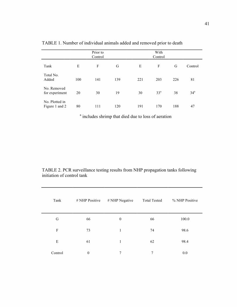
TABLE 1. Number of individual animals added and removed prior to death
Prior to Control
With Control
Tank E F G E F G Control Total No. Added 100 141 139 221 203 226 81 No. Removed for experiment 20 30 19 30 33a 38 34a
No. Plotted in Figure 1 and 2 80 111 120 191 170 188 47
a includes shrimp that died due to loss of aeration
TABLE 2. PCR surveillance testing results from NHP propagation tanks following initiation of control tank
Tank # NHP Positive # NHP Negative Total Tested % NHP Positive
G 66 0 66 100.0
F 73 1 74 98.6
E 61 1 62 98.4
Control 0 7 7 0.0

42
FIGURE 1. Kaplan–Meier survival curve generated with survival time calculated from the date of introduction into propagation tank until date of death by necrotizing hepatopancreatitis (NHP). Shrimp that were removed or died of causes other than NHP were censored at the time time of loss.
43
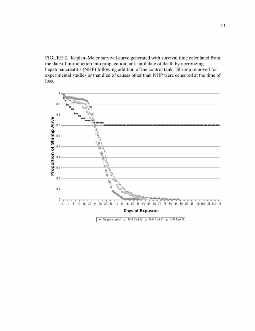
FIGURE 2. Kaplan–Meier survival curve generated with survival time calculated from the date of introduction into propagation tank until date of death by necrotizing hepatopancreatitis (NHP) following addition of the control tank. Shrimp removed for experimental studies or that died of causes other than NHP were censored at the time of loss.
44
CHAPTER 3. DOUBLE STRANDED RNA PROVIDES SEQUENCE
DEPENDENT PROTECTION AGAINST INFECTIOUS MYONECROSIS VIRUS
IN LITOPENAEUS VANNAMEI
J. Dustin Loy,1,2,4 Mark A. Mogler, 2,4 Duan S. Loy,1 Bruce Janke,3 Kurt Kamrud,1,4 Edward D. Scura5, DL Hank Harris1,3,4 Lyric Bartholomay6
A manuscript submitted for publication in the Journal of General Virology
1 Department of Animal Science, Iowa State University, Ames, IA, USA
2 Department of Veterinary Microbiology and Preventive Medicine, Ames, IA USA 3 Department of Veterinary Diagnostic and Production Animal Medicine, Ames, IA, USA
4 Harrisvaccines, Inc. Ames IA, USA 5 ART Biologics, LLC Ames, IA, USA
6 Department of Entomology, Iowa State University, Ames, IA, USA
Abstract
Viral diseases are significant impediments to the growth and sustainability of
commercial shrimp aquaculture. New viral diseases continue to emerge and, along with
endemic disease, cause significant economic losses to shrimp production. Disease caused
by Infectious myonecrosis virus (IMNV) has caused tremendous losses in farmed Pacific
white shrimp (Litopenaeus vannamei), the predominant species used in shrimp
aquaculture, since IMNV emerged in Brazil and subsequently spread to Indonesia. There
are no existing antiviral treatments or interventions, outside of pathogen exclusion, to
mitigate disease in commercial shrimp operations. Novel RNA interference (RNAi)
strategies that employ exogenous double-stranded RNA (dsRNA) are a promising means
to approach disease control. Here we describe an iterative process of panning the viral
genome of IMNV to discover RNAi trigger sequences that initiate a robust and long-
45
lasting protective response against viral disease in L. vannamei. Using this process, a
single, low dose (0.02 µg) of an 81 bp and 153 bp fragment targeting the 5’ end of the
viral genome was discovered which protected 100% of animals from disease and
mortality caused by IMNV. Furthermore, animals that were treated with highly
efficacious dsRNA survived an initial infection and were resistant to subsequent
infections 50 days later with a hundred-fold greater dose of virus. This protection is
highly sequence dependent, because dsRNAs specific to IMNV, but targeting the coding
regions for the polymerase and structural protein regions of the genome, conferred lesser
or no protection to animals. Interestingly, non-sequence specific or heterologous dsRNA
did not provide any degree of protection to animals as had been described for other
shrimp viruses. Our data indicate that the targeted region for dsRNA is a crucial factor
in maximizing the degree of protection and lowering the dose required to induce a
protective effect against IMNV infection in shrimp.
Introduction
Viral diseases that impact cultured shellfish are considerable impediments to
successful, sustainable aquaculture practices and trade, and have the most profound
economic impact in developing, tropical countries that provide optimal climate and
habitat for shrimp aquaculture. The Pacific white shrimp, Litopenaeus vannamei
(Decapoda: Penaeidae), is uniquely amenable to aquaculture and has rapidly become the
dominant farmed shrimp species globally; indeed, production of L. vannamei increased
from 8,000 metric tons in 1980 to 1.38 million tons in 2004 worldwide.6 Pathogen
translocation and disease outbreaks have ensued with increasing production, and
46
significant economic losses have resulted with the emergence of each of the major viral
pathogens.29 Epizootics of viral diseases in particular cause substantial mortality in L.
vannamei and result in devastating financial losses; it has been estimated that annual
losses globally as a result of diseases (mostly viral in etiology) have been as high as 3
billion USD.13,30 The occurrence of a pathogenic virus in a pond of growing shrimp
exerts a range of consequences, including forcing the grower to prematurely harvest,
increasing production costs through decreased feed conversion, or causing a complete
loss of animals.29 Beyond deliberate pathogen exclusion or selective breeding, which is
not effective against all viral pathogens, there are no means to control or treat viral
disease and prevent perpetual transmission in farmed shrimp. A practical and efficacious
means of protecting shrimp against viral disease would offset these losses and facilitate
more profitable production of farmed shrimp in this multibillion dollar per year industry.
RNA interference (RNAi) is promising and emerging approach to shrimp disease
control.9,10,17,18,21 RNAi is a sub-cellular process that results in gene-specific RNA
degradation. RNAi triggers, including small interfering RNAs (siRNA) and double
stranded RNAs (dsRNA), can be provided exogenously to arthropod cells or whole
organisms to study gene function. In recent years, RNAi has become an invaluable tool
for studying arthropod physiology and arthropod-pathogen interactions. It is likely that
RNAi evolved, in part, as an antiviral response. Indeed, the RNAi phenomenon also has
been observed as an antiviral response in mosquitoes that transmit human viral pathogens
and has been proposed as a transmission-blocking strategy in which transgenic
mosquitoes, expressing an RNAi trigger in the form of an inverted-repeat of the Dengue
virus (DENV) genome, no longer support amplification and transmission of the virus.7
47
There is evidence that the requisite RNAi machinery is present and functioning in a
similar manner in penaeid shrimp.4,22,27 Therefore, RNAi is an attractive means to
specifically disable virus infection in shrimp cells and thereby protect shrimp from
associated pathology and morbidity. Indeed, specific RNAi triggers mitigate disease
caused by White Spot Syndrome Virus (WSSV)17,32, Yellowhead Virus (YHV)25,26,33 and
Taura Syndrome Virus (TSV).19
Infectious Myonecrosis Virus (IMNV) is a non-enveloped, double stranded RNA
(dsRNA) virus that is a member of the Totiviridae family. IMNV was first discovered in
2003 after a severe disease outbreak in 2002 in Brazil that was characterized by high
mortality and animals exhibiting necrosis in the tail muscle.16 In previous outbreaks,
mortality has ranged from 40% to 70% prior to harvest.1 Moribund animals have ever-
increasing opaque to white muscle tissue visible through the cuticle, starting in the
ventral portion of the muscle and expanding dorsally in each abdominal segment.
Histopathologically, animals demonstrate a characteristic coagulative necrosis of skeletal
muscle with fluid accumulation between muscle fibers and pronounced hypertrophy of
the lymphoid organ due to formation of spheroids, well-delineated non-tubular
aggregates of lightly basophilic cells, a feature common to several virus infections that
affect shrimp.16
IMNV reached Indonesia in 200620; this translocation of the pathogen is evidence
for a very real risk for spread throughout Asia and the world. Due to the current and
tremendous future potential impact this disease has on the shrimp industry, development
of a vaccine or mitigation strategy is prudent. The aim of this work was to discover and
optimize RNAi trigger sequences that would elucidate a protective response to IMNV.
48
We describe an iterative process of panning the IMNV genome for antiviral sequences
through which we arrived upon a targeted RNAi trigger for a short, precise portion of the
5’ end of the IMNV genome that confers long-lasting and robust protection against
IMNV when administered in a single, low dose.
Materials and Methods
Animal Rearing Specific pathogen free (SPF) postlarvae were received from Shrimp Improvement
Systems (Plantation Key, Florida) and reared in a biosecure animal holding facility at
Iowa State University. Animals were placed into 1000L Poly tanks containing artificial
seawater (Crystal Sea Marine Mix), an oystershell airlift biofilter, and an activated carbon
filter. Animals were fed a commercial growout diet until ~5 grams in weight.
Preparation of Virus Inoculum Based on a previously described methodology8, tissue macerate from infected
animals was diluted 1:3 in sterile TN buffer (0.02 M Tris-HC1, 0.4 M NaC1, pH 7.4) and
clarified through three centrifugation steps after which supernatant was removed by
subsequent centrifugation: 1) 4000 x g for 30 minutes, 2) 15,000 x g for 15 minutes 3)
25,000 x g for 60 minutes. The final supernatant was diluted 1:10 in 2% saline and
passed through a 0.2 micron filter. This stock viral clarification was aliquoted and
frozen at -80C and aliquots of this stock were thawed for each viral challenge.
Quantitative polymerase chain reaction (qPCR) analysis of RNA extracted following a
freeze-thaw cycle indicated 105 viral copy numbers/µL were present in the stock.

49
Determination of Challenge Dose The optimum dose by injection resulting in infection and disease was tested in 20
SPF animals weighing 8-10 grams. Each animal was injected with 100 µL of stock, 1:10
or 1:100 dilutions thereof, or 2% NaCl (sham) into the third abdominal segment.
Animals were observed twice daily for mortality. All virus inoculums resulted in 100%
mortality at varying time points. Using these infectious doses, optimum survivorship was
observed with the 1:100 dilution, after which 100% of shrimp died by 18 days post
infection (dpi) as compared to the stock dilution that resulted in 100% mortality between
1 and 2 dpi, the 1:10 dilution which resulted in 100% mortality at 11 days, or the 1:100
dilution which resulted in 100% mortality at 18 days. The 100-fold dilution of the stock
virus was used as the viral challenge dose for the described challenge experiments.
Challenged shrimp show characteristic opaque tissue and hypoxic body posturing
(FIGURE 1).
Quantitative Reverse-Transcription PCR Quantitative Reverse-Transcription PCR (qRT-PCR) was performed by using
methods adapted from Andrade et al.1 RNA template was purified using an RNeasy
Minikit (Qiagen). Briefly, 30 mg of muscle tissue was dissected from the tail and placed
in 600 µL of Buffer RLT. This was then transferred to and mascerated in a ground glass
tissue grinder for approximately 100 turns. The tissue mascerate was then homogenized
on a QIAShredder column (Qiagen) and purified using RNeasy centrifuge columns
according to manufacturer’s instructions. 50 µL of RNA template was eluted into RNase
free water and stored at -80 °C until the RT-PCR was performed. For the qRT-PCR
templates were thawed and boiled at 100 °C for 5 minutes prior to reverse transcription.
50
One-Step RT-PCR Kit (Qiagen) was used. 2 µL of RNA template was mixed with 5 µL
of 5x One Step RT-PCR Master Mix (Qiagen), 1 µL Enzyme Mix, 1 µL DNTP, 0.3 µL
IMNV412 (20 uM) (5′-GGA CCT ATC ATA CAT AGC GTT TGC A-3′) , 0.3 µL
IMNV545R (20 uM) (5′-AAC CCA TAT CTA TTG TCGCTGGAT-3′), 0.3 µL IMNV
p1 (10 uM) (5′-6FAM CCA CCT TTA CTT TCA ATA CTA CAT CAT CCC CGG
TAMRA-3′) and 15 uL RNase free water for a total reaction volume of 25 µL. This
reaction was then run in duplicate on a BioRad CFX96 Real Time PCR Detection System
under the following conditions: RT for 30:00 at 48 °C and 10:00 at 95 °C, followed by
PCR for 35 cycles of 15s at 95 °C and 1:00 min at 60 °C. Viral copy number was
calculated using CFX Manager Software (BioRad) using an in vitro transcribed RNA
standard generated using methods similar to Andrade et al1.
Statistical Analysis Statistical significance was conducted on mean log viral RNA copies/mL RNA
and mean survival at termination of the experiment using One-way ANOVA followed by
Tukey’s multiple comparison test using JMP 8 Software (SAS Institute Inc. Cary, NC,
USA)
dsRNA Preparation dsRNA was prepared using Ambion Megascript RNAi Kit following the
manufacturer’s directions. Template DNA for in vitro transcription was created by
extracting RNA from virus infected animals using a commercial nucleic acid purification
kit (Qiagen RNeasy Minikit). cDNAs to IMNV genome were created using specific
oligonucleotide primers (TABLE 1) designed from sequences available (GenBank
accession no.EF061744)20 and reverse transcription (Thermoscript RT, Invitrogen). Five
51
mL of RNA extract was added to the reaction mix and incubated at 50°C for 60 minutes
per manufacturer’s instructions. Following reverse transcription, template cDNA (~50
ng) was added to a PCR master mix (PuReTaq Ready-To-Go PCR Beads, GE
Biosciences) and thermal-cycling was performed using oligonucleotide primers (Table 1)
designed to specific regions of the IMNV genome (GenBank accession EF061744.1).
Products were then cloned into pCR4.0 vectors (Zero Blunt TOPO PCR cloning kit) and
transformed into E. coli (TOP10 Invitrogen). Plasmids preparations from these
transformants were used as a template source for further PCRs to create DNA clones for
RNA transcription. Clones were sequenced and 100% identity with the IMNV genome
was confirmed. For synthesis of template for transcription, primers containing a T7
promoter sequence (TABLE 1) on the forward and reverse primer were used to amplify
the segments of cDNA with flanking T7 promoter sites. PCR products were subjected to
QIAquick (Qiagen) PCR cleanup to remove unincorporated nucleotides, primers, and
enzyme. T7 DNA templates were transcribed overnight following the manufacturer’s
directions. Following transcription, dsRNA products were incubated for 60 minutes with
DNase I and RNase, and purified using the provided columns. Transcription of dsRNA
was confirmed by gel electrophoresis in comparison with a molecular weight ladder
(pGEM ladder, Promega) and the product was quantified by using a spectrophotometer.
Histopathology Moribund animals that were found prior to death were fixed in whole Davidson’s
fixative3 for 24 hours before being transferred to 70% ethanol, In addition, two animals
from each tank were anesthetized in an ice slurry and fixed in Davidson’s fixative3,

52
embedded in paraffin, cut into slides, and stained with hematoxylin and eosin and
evaluated for the presence of IMNV lesions.
Shrimp Bioassays Two hundred liter tanks containing synthetic seawater and an oystershell airlift
biofilter were stocked with 10 or 20 SPF juveniles weighing 3-7 grams and allowed to
acclimate for 72 hours (h). Tanks were maintained at 28 ppt salinity and 28 °C.
Following acclimation, animals were injected intramuscularly at the desired dose and
volume of either dsRNA corresponding to regions of the IMNV genome, a heterologous
dsRNA control, or a sham vaccination (RNase free water diluent). Following dsRNA
administration, animals were challenged with IMNV intramuscularly (~3.2 x 10³ IMNV
copy numbers/µL RNA) into the third abdominal segment at the desired time point (2, 10,
or 52 days) or a sham virus challenge (2% NaCl viral diluent) as a strict negative control.
Animals were counted daily for mortality. Moribund animals were fixed for
histopathology in some experiments. At the termination of experiments animals were
euthanized by immersion in an ice water slurry and tissues from experiments were
immediately frozen at -80°C. Representative samples at termination were also fixed in
Davidson’s fixative for histopathology.
Target Specificity Tanks containing 20 SPF juveniles weighing 5-7 grams were divided into 5 virus
challenge groups with three replicates per group. Shrimp were injected with dsRNA
constructs corresponding to three different segments of the IMNV genome, a
heterologous dsRNA control (eGFP), or a sham vaccination. A total of 2.0 µg of in vitro
53
synthesized dsRNA was inoculated into animals into randomized tanks. Animals were
challenged with IMNV 48 hours after dsRNA administration.
Dose Response Tanks containing 10 animals weighing 3-5 grams were divided into 6
experimental groups that received a dose (2.0, 0.2 or 0.02 µg) of a dsRNA construct
(dsRNA 95-475 or dsRNA3764-4805), Three control groups received a heterologous
eGFP dsRNA at a single dosage of 2 µg of eGFP dsRNA. Following dsRNA
administration, animals were challenged 2 days (data not shown) or 10 days later with
IMNV.
Length Dependency Tanks containing 10 animals weighing 3-5 grams were divided into treatment
groups with 3 replicate tanks per treatment. Animals were subjected to injection with one
of six dsRNA constructs corresponding to or different truncations of the 95-475 dsRNA
(dsRNA95-475, 193-475, 95-376, 194-275, 219-273, or 223-376). A total of 0.02 µg of
in vitro synthesized dsRNA was inoculated into animals that were challenged 10 days
later with IMNV.
Long Term Protection Shrimp administered 2.0 µg of dsRNA95-475 that survived virus challenge from a
prior experiment (n=17) were subjected to a second infection at 100-fold higher dose of
virus inoculum (~3.2 x 10⁵ IMNV copy numbers/mL RNA), 52 days after the initial virus
challenge and 54 days after administration of dsRNA. A naïve challenge control group
was injected with the same challenge dosage. Animals were followed for an additional
54
40 days after secondary challenge. At that time the experiment was terminated and
samples taken.
Results
A disease challenge model was established that uses an intramuscular injection of
a clarified virus preparation; this protocol yields consistent morbidity, mortality and
pathology in infected animals. IMNV challenge resulted in 100% mortality, with
variation in duration to mortality according to dose (FIGURE. 1). A 100-fold dilution of
the tissue homogenate (~3.2 x 10³ IMNV copy numbers/µL RNA, data not shown)
resulted in 100% mortality over an 18 day period and was used as the virus challenge
dose for all subsequent experiments. Quantitative RT-PCR (hereafter referred to as
qPCR) provided evidence of virus replication because virus genome copy numbers
ranged from 2.5 x 10⁵ to 3.5 x 10⁷ IMNV copy numbers/µL in muscle tissues sampled at
9 day post-infection when compared with the inoculum dose (3.2 x 10³ IMNV copy
numbers/µL). These qPCR results support histopathological observations, because
moribund animals collected from this same experiment showed characteristic IMNV
pathology on histopathology sections. Negative control animals that were injected with
sterile 2% NaCl (virus diluent) showed no signs of histopathological muscle lesions or
trauma at the injection site and the presence of IMNV was not detected in this group by
qPCR. The development of this model allowed for a consistent and controlled exposure
of virus to facilitate comparison of protection provided by various RNAi triggers.
Three regions that encompass portions of the 5’, center and 3’ regions of the
IMNV genome were initially chosen as targets for dsRNA design (FIGURE 2); these are

55
designated as dsRNA95-475, 3764-4805, and 5518-6391 according to the range of
nucleotides represented from the genome (GenBank accession EF061744). Double
stranded RNAs were injected into shrimp in a 2.0 µg dose; animals were infected with
IMNV, using the established challenge model, 2 days later. Control shrimp were
subjected to heterologous dsRNA or sham-inoculation and subsequently challenged with
IMNV, and showed significant mortality with 1.67% and 3.33% survival, respectively
after 30 days. Of the animals subjected to IMNV-specific dsRNAs, 81.67% and 61.67%
in the dsRNA95-475 and dsRNA3764-4805 groups survived IMNV infection for the
duration of the experiment (30 days) (FIGURE 3), respectively; this is a statistically
significant difference (P<0.001) in survivorship as compared to controls, according to
Oneway ANOVA followed by Tukey’s multiple comparison test. By contrast, animals
injected with dsRNA5518-6391 had mortality similar to the sham-inoculated controls
with 5% survival post-challenge. No significant difference was evident between survival
in animals injected with dsRNA5518-6391, dsRNA eGFP, or the 2% saline control
inoculation at day 30. All of the non-infected controls survived for the duration of the
experiment (FIGURE 3). Further, qPCR on muscle tissues sampled from dead individuals
at 9-14 days post-infection demonstrated a significant reduction in genome copy number
between animals administered dsRNA95-475 or dsRNA3764-4805 (mean 2.44 x 10⁵ and
2.39 x 10⁴ IMNV copy numbers/µL RNA, respectively) when compared with animals
administered dsRNA5518-6391, dsRNA eGFP, or sham inoculation (3.44 x 10⁸, 2.1 x
108, and 2.72 x 10⁷ IMNV copy numbers/µL RNA, respectively) (FIGURE 4).
To test the longevity of the protective effect, a group of dsRNA95-475-injected
shrimp (n=17) from the initial target specificity assay was subjected to a second infection
56
at a 100-fold higher dose, 52 days after the initial virus challenge and 54 days after the
administration of dsRNA. Animals were followed for an additional 40 days after
secondary challenge (94 days from the start of the experiment) and showed 94% survival
(16/17), as compared to 10 challenge control animals that showed 100% mortality
(10/10) within 8 days.
Double stranded RNAs 95-475 and 3764-4805, which provided the highest
demonstrated levels of protection were used in a dose titration study to determine the
minimal dose to achieve protection from IMNV challenge. Animals were injected with
2.0. 0.2, or 0.02 µg of dsRNA95-475 or dsRNA3764-4805, then IMNV challenged at 2 d
or 10 d post-dsRNA inoculation. As shown in FIGURE 5, 80% of animals receiving
dsRNA95-475 were protected from IMNV challenge even at the lowest dose (0.02 µg)
and at 10 days post-dsRNA injection. dsRNA3764-4805 elicited this level of protection
but only when animals were injected with 10 times the amount of dsRNA (0.2 µg)
(FIGURE 5).
Moribund animals were sampled for histopathological analysis from sham-
vaccinated controls, eGFP dsRNA administered controls, and the low dose (0.02 µg) of
dsRNA3764-4805 at both 2 and 10 d post-dsRNA administration. Evidence of IMNV
infection and disease included extensive necrosis of muscle fibers, distributed in small to
large irregular foci, not limited to particular muscles but scattered throughout the tail as
well as affecting smaller muscle bundles in the cephalothorax and in the legs. The lesions
were consistent with histopathological changes previously described as characteristic of
IMNV infection.16 Both acute and chronic lesions were evident, often in adjacent
locations. Acute lesions were characterized by coagulative necrosis and fragmentation of
57
muscle bundles with edema prominent within the muscle bundle sheaths and between
affected muscle bundles. Infiltration of hemocytes was mostly limited to small clusters
scattered along the connective tissue septa that separated groups of muscles. Chronic
lesions were characterized by partial or total loss of myofibers with
condensation/contraction of the fibrous connective tissue components. The hypercellular
appearance of these bands of fibrous tissue appeared to be due primarily to collapse of
the muscle sheaths rather than active fibroplasia. Hemocyte infiltrates in chronic lesions
were minimal in contrast to a previous report.16 Lymphoid organs were evaluated if
present in the sections examined. Spheroid formation was evident and usually prominent
in all lymphoid organs examined, with multiple spheroids affecting most of the organ. In
contrast to histopathological observations in IMNV-infected control animals, muscle
lesions in the dsRNA-injected shrimp that were sacrificed at termination of the trial were
chronic in appearance. Acute muscle necrosis was not evident in most of these animals,
but was limited to a few very small foci in an occasional animal. In addition, two animals
that were re-challenged with additional virus and examined by histopathology showed
mature fibrous scar tissue but did not demonstrate necrotic lesions.
Because dsRNA95-475 provided significant (P=.0003) in comparison with
controls), long-term protection from IMNV-induced disease, and was highly protective
even at a low dose, this region was further scrutinized for development of shorter
dsRNAs as depicted in FIGURE 2. Two dsRNAs were generated as ~100 bp truncations
from the 5’ (193-475) or 3’ (95-376) ends of the original dsRNA95-475 dsRNA. When
administered 10 d prior to challenge, dsRNA95-376 conferred 100% protection, whereas
the 5’ truncation (193-475) conferred 93.33% protection in animals 30 d post-dsRNA
58
administration. This survival was statistically significant when compared with the control
groups (P<0.0001). Shorter dsRNAs were then designed within this truncated region to
the 194-275, 219-273, and 223-376 regions of the IMNV genome (Fig. 2). Of these,
dsRNA194-275 and 223-376 injected animals showed 100% survival (P<0.0001) and
dsRNA219-273 provided 73.33% protection (P=.0005) following IMNV challenge
(FIGURE 6).
Discussion
These studies describe the first use of RNAi to protect shrimp from IMNV, a
significant emerging shrimp pathogen; moreover, our data show that very specific, short
dsRNAs confer robust and long-term resistance to IMNV-induced disease and mortality.
This effect is highly dependent upon sequence selection and the virus gene that is
targeted.
Three target regions, spanning the length of the viral genome, were selected as
initial targets for dsRNA generation. The first target sequence (dsRNA95-475)
corresponds to the N-terminal region of ORF1 frame 1, which is predicted to contain two
co-translationally cleaved products, termed Proteins 1 and 2.22 Protein 1 (93 aa) and 2
(284 aa) are encoded by nucleotides 136-415 and 416-1266 of the IMNV genome within
ORF 1 frame 1, respectively, and their functions remain uncharacterized14. Protein 1
shares 35% sequence identity with previously described dsRNA binding proteins.16 In
addition, portions of the coding regions for the major capsid protein (MCP)
(dsRNA3764-4805) and RNA dependent RNA polymerase (RdRp) (dsRNA5518-6391)14
also were selected for dsRNA generation. The major capsid protein (909 amino acids) of
59
IMNV is encoded by nucleotides 2227-4953 within ORF 1, frame 1. The RNA
dependent RNA polymerase (736 amino acids) is encoded by nucleotides 5241-7451
within ORF2, frame 316. A non-specific control dsRNA was designed to an exogenous
sequence corresponding to enhanced green fluorescent protein (eGFP).5
Two of the three dsRNA constructs (dsRNA95-475 and dsRNA3764-4805)
conferred significant protection from IMNV-induced disease (FIGURE 3). The
dsRNA95-475 that targets the 5’-most end of the IMNV genome provided the greatest
protection from disease in terms of dose and duration. dsRNA3764-4805 that targets the
sequence that corresponds to the major capsid protein, provides a significant, but
intermediate, protective effect (61.67% survival) when dsRNA is administered 48 h prior
to virus challenge (FIGURE 3). An intermediate level of protection also was
demonstrated with this dsRNA in a dose titration experiment, wherein a ten-fold higher
dose was required for protection in comparison to dsRNA95-475 (FIGURE 5). This is an
interesting finding because dsRNA corresponding to structural proteins (VP19, VP28) of
a large DNA virus (WSSV), are very effective in reducing mortality when administered
before challenge.19
Specific targeting of the non-structural RdRP protein using dsRNA5518-6391
provided no measurable protection in comparison to the sham-injection used for positive
control animals (FIGURE 3). This is in contrast to previous work that demonstrated
effective protection from infection when non-structural proteins were the targets of
silencing in other RNA viruses such as Yellowhead virus.25,33
60
Dose Response In order to determine threshold protective doses of dsRNA and simultaneously
characterize a dose/response to confirm biological activity of the RNAi response, animals
were administered doses ranging from 0.02 - 2.0 µg. A reduction in survival was
apparent at the lowest levels for both sequences tested, but even at the lowest doses one
sequence (dsRNA95-475), was more protective (80%) (FIGURE 5). This very small
dose (20 ng) makes it an ideal candidate antiviral molecule for delivery to shrimp for the
purpose of preventing disease caused by IMNV. Previous reports in which dsRNA was
used to control disease associated with WSSV infection showed protection with much
larger quantities of dsRNA (~ 5 µg/gram of animal or higher) injected per animal18 and
that quantities less than 1 µg of specific WSSV dsRNA were insufficient to protect from
mortality.7 Our studies show that protection is long-lasting for IMNV, even at these lower
doses, because dsRNA was effective at disease prevention for at least 10 days as
compared to 5 days for YHV when a 25 µg dose was given.17 This disparity in dose
range may be a result of dose-response titration studies that did not extend to the lower
limit of what we tested because those authors used 100 µg as a high dose and 7-15 µg as
the intermediate dose19 or extrapolated a dosage based on 5.0 µg of dsRNA being
sufficient to suppress endogenous genes17. This work demonstrates that vastly smaller
amounts of specific dsRNA provide sufficient robust and long-term protection, against
IMNV disease (0.02 µg for 10 days). This provides new insight into the capacity of the
shrimp RNAi machinery and demonstrates the power of developing RNAi antivirals
through target optimization in devising a more cost-effective approach to shrimp disease
control.
61
Target Specificity The most significant protection against IMNV-induced disease was conferred by
dsRNA designed to target a protein-encoding region of the 5’-most end of the IMNV
genome, dsRNA95-475. This region encompasses Protein 1 and the N-terminal portion
of Protein 2 (FIGURE 2). The precise function of these peptides is unknown, other than
that Protein 1 contains a dsRNA binding motif that is postulated to play a role in host
immune suppression.24 Protein 1 may be a crucial gene involved in viral pathogenesis
within the host, a factor necessary for early viral replication, or a protein that modifies the
host RNAi antiviral response. By analogy, Drosophila C virus and Flock House Virus
are pathogenic RNA viruses of other invertebrate species that encode proteins that have
dsRNA binding activity and the capacity to modify or inhibit the host RNAi response12,28.
Protein 1 of IMNV may be fulfilling a similar role in facilitating virus replication by
suppressing host RNAi machinery.
Additionally, the accessibility of the targeted portion of the genome to RNAi may
be enabling this RNA to more efficaciously inhibit viral genome replication. Significant
factors associated with impeding target RNA accessibility include RNA secondary
structure and protein occupancy.15 A study examining small hairpin RNA vectors
(shRNAs) targeting conserved portions of the hepatitis B virus genome, found several
highly effective targets in a 5’ portion of the genome. These 5’ targets are much more
effective than was predicted because this region contains a highly structured stem-loop,
perhaps owing to availability according to genome location.23
62
Length Dependency These data demonstrate that by optimizing RNAi targets, beginning with larger
sequences and moving through gradual truncations, the protective effect of dsRNA, on a
per molecular weight basis, increases. However, in the context of the RNAi capacity of a
shrimp, a sequence length of greater than 50 base pairs (bp) may be a minimum length
requirement, because reduction in survival was observed when the 194-275 (81 bp)
dsRNA was truncated to 219-275 (56 bp). This optimum threshold size of the dsRNA
corresponds well to a recently published study that suggests that an RNAi trigger in L.
vannamei has to be greater than 50 bp in length to stimulate transcription of the LvSid-1
gene, which encodes the membrane-forming protein gene involved in dsRNA transport
into cells, and that 50 bp is the lower threshold for a dsRNA to stimulate transcript
suppression11. In addition, a dsRNA shorter than 50 bp that corresponded to an
endogenous shrimp mRNA CDP (shrimp transcript of unknown function) did not
stimulate a reduction in transcript in other tissues.11 Further, dsRNA corresponding to
WSSV genes that were shorter than 50 bp did not elucidate protection from virus
challenge.11
Long Term Protection The overall data strongly suggest that exposure to the optimized dsRNA produces
long-term, specific immunity to subsequent challenge with a single dose as compared to
siRNAs that require repeat injections to effect virus clearance.32 For example, animals
subjected to a low dose of dsRNA, and challenged 10 days later demonstrated significant
protection against IMNV disease. To further study this phenomenon, animals were re-
challenged with IMNV following the initial dsRNA administration and challenge.
63
Animals that were subjected to a second virus challenge demonstrated high survivorship,
even when given much higher doses of virus and after extended periods of time following
initial dsRNA administration. This observation suggests that RNAi may serve as a
pseudo memory-type response that provides long-term protection from IMNV infection
in shrimp. This may also be an indication that once animals are able to initially overcome
an IMNV challenge, they are resistant to subsequent challenges. RNAi may be a
mechanism that is utilized by the animal to adapt to new viral pathogens and establish
resistance to viral replication. This may also help to explain why in field observations,
gross lesions of IMN appear to occur concurrently with periods of high environmental
stress on animals, such as occurs with high stocking densities or longer grow out
periods.2
Sequence-Independent Effects Non-specific dsRNA that was generated to a heterologous target (eGFP) did not
have a positive impact on survival in animals challenged with IMNV, in contrast to what
has been described with other shrimp viruses including TSV, YHV and WSSV 17,19,31,33.
Heterologous dsRNAs for avian and mammalian immunoglobulin, fish non-coding
dsRNA, and bacterial plasmid provided moderate protection against subsequent infection
(2 pr 3 days) with TSV or WSSV.19 Limited protection also was conferred in animals
injected with dsRNA corresponding to GFP then infected with YHV. However, this
effect was not observed in these studies because no degree of protection from virus
challenge was observed in animals pre-injected with dsGFP, even when virus challenge
occurred 2 days post dsRNA administration. This may be due to differences in
pathogenesis or viral replication mechanisms that IMNV utilizes. Additionally, this

64
phenomenon may also be explained by differences in genome size; IMNV has a small
genome relative to WSSV and TSV, and thus it may have fewer off-target sequences with
significant homology to the control dsRNA sequences that are capable of providing this
apparent non-specific protection.
Here we describe a process of defining IMNV genome dsRNA targets that
provide shrimp protection from virus challenge. Optimization of target specificity,
nucleotide length and dsRNA dosage level are key elements that support the potential
economical use of RNAi triggers to protect shrimp from viral pathogens. Targets
identified in this manner could be utilized in downstream efforts to develop therapeutics
or vaccines for shrimp viral pathogens, either newly emerged or pre-existing.
Acknowledgements
The authors would like to thank Kay Kimpston-Burkgren, Cara Loy, Kara
Burrack, Kristen Summerfelt, and Pamela Whitson for animal care. Additionally they
would like to thank Jill Gander, Joe Brodie, Ryan Vander Veen, Patrick Jennings, and
Brandon Russell for laboratory assistance and reagents.

65
References
1. Andrade TPD, Srisuvan T, Tang KFJ, Lightner DV. (2007). Real-time reverse
transcription polymerase chain reaction assay using TaqMan probe for
detection and quantification of Infectious myonecrosis virus (IMNV).
Aquaculture. 264:9-15
2. Arns da Silva V, dos Santos FL, Bezarro SS, Pedrosa VF, Mendes P, Mendes E.S.
(2010). A multi-season survey for infectious myonecrosis in farmed
shrimp, Litopenaeus vannamei, in Pernambuco, Brazil. Journal of
Invertebrate Pathology. 104:161-165
3. Bell TA, Lightner DV (1988) A Handbook of Normal Shrimp Histology. Baton
Rouge; World Aquaculture Society. 114 p
4. Dechlar M, Udomkit A, Panyim S. (2008). Characterization of argonaute cDNA
from Penaeus monodon and implication of its role in RNA interference.
Biochemical and Biophysical Research Communications. 367:768-774
5. Dong Y, Aguilar R, Xi Z, Warr E, Mongin E, Dimopoulos G. (2006). Anopheles
gambiae immune responses to human and rodent Plasmodium parasite
species. PLoS Pathogens. 2(6): 52
6. Food and Agricultural Organization of the United Nations Fisheries and
Aquaculture Department. (2009). FishStat Plus - Fishery Statistical
software. http://www.fao.org/fishery/statistics/software/fishstat/en
66
7. Franz AWE, Sanchez-Vargas I, Adelman ZN, Blair CD, Beaty BJ, James AA,
Olson KE. (2006). Engineering RNA interference-based resistance to
dengue virus type 2 in genetically modified Aedes aegypti. Proceedings of
the National Academy of Sciences. 103:4198-4203
8. Hasson KW, Lightner DV, Poulos BT, Redman RM, White BL, Brock JA,
Bonami JR. (1995). Taura syndrome in Penaeus vannamei: demonstration
of a viral etiology. Diseases of Aquatic Organisms. 23:115-126
9. Hirono I, Fagutao F, Kondo H, Aoki T. (2011). Uncovering the Mechanisms of
Shrimp Innate Immune Response by RNA Interference. Marine
Biotechnology. 13(4):622-8
10. Krishnan P, Gireesh-Babu P, Rajendran KV, Chaudhari A. (2009). RNA
interference-based therapeutics for shrimp viral diseases. Diseases of
Aquatic Organisms. 86:263-272
11. Labreuche Y, Veloso A, de l, V, Gross PS, Chapman RW, Browdy CL, Warr
GW. (2010). Non-specific activation of antiviral immunity and induction
of RNA interference may engage the same pathway in the Pacific white
leg shrimp Litopenaeus vannamei. Developmental & Comparative
Immunology. 4(11):1209-1218.
12. Li H, Li WX, Ding SW. (2002). Induction and suppression of RNA silencing by
an animal virus. Science. 296:1319-1321
67
13. Lightner DV. (2003). The Penaeid Shrimp Viral Pandemics due to IHHNV,
WSSV, TSV and YHV: History in the Americas and Current Status.
Proceedings of the 32nd UJNR
14. Nibert ML. (2007). '2A-like' and 'shifty heptamer' motifs in penaeid shrimp
infectious myonecrosis virus, a monosegmented double-stranded RNA
virus. Journal of General Virology. 88:1315-1318
15. Pei Y and Tuschl T. (2006). On the art of identifying effective and specific
siRNAs. Nature Methods. 3:670-676
16. Poulos BT, Tang KFJ, Pantoja CR, Bonami JR, Lightner DV. (2006). Purification
and characterization of infectious myonecrosis virus of penaeid shrimp.
Journal of General Virology. 87:987-996
17. Robalino J, Bartlett T, Shepard E, Prior S, Jaramillo G, Scura ED, Chapman RW,
Gross PS, Browdy CL, Warr GW. (2005). Double-Stranded RNA Induces
Sequence-Specific Antiviral Silencing in Addition to Nonspecific
Immunity in a Marine Shrimp: Convergence of RNA Interference and
Innate Immunity in the Invertebrate Antiviral Response? Journal of
Virology. 79:13561-13571
18. Robalino J, Bartlett TC, Chapman RW, Gross PS, Browdy CL, Warr GW. (2007).
Double-stranded RNA and antiviral immunity in marine shrimp: Inducible
host mechanisms and evidence for the evolution of viral counter-
responses. Developmental & Comparative Immunology. 31:539-54

68
19. Robalino J, Browdy CL, Prior S, Metz A, Parnell P, Gross P, Warr G. (2004).
Induction of Antiviral Immunity by Double-Stranded RNA in a Marine
Invertebrate. Journal of Virology. 78:10442-10448
20. Senapin S, Phewsaiya K, Briggs M, Flegel TW. (2006). Outbreaks of infectious
myonecrosis virus (IMNV) in Indonesia confirmed by genome sequencing
and use of an alternative RT-PCR detection method. Aquaculture. 266:32-
38
21. Shekhar MS and Lu Y. (2009). Application of Nucleic-acid-based Theraputics for
Viral Infections in Shrimp Aquaculture. Marine Biotechnology 11:1-9
22. Su J, Oanh DT, Lyons RE, Leeton L, van Hulten MC, Tan SH, Song L, Rajendran
KV, Walker PJ. (2008). A key gene of the RNA interference pathway in
the black tiger shrimp, Penaeus monodon: identification and functional
characterisation of Dicer-1. Fish & Shellfish Immunology. 24:223-233
23. Sun D, Rissler C, Kidd-Ljunggren K, Nassal M. (2010). Quantitative assessment
of the antiviral potencies of 21 shRNA vectors targeting conserved,
including structured, hepatitis B virus sites. Journal of Hepatology.
52:817-826
24. Tang J, Ochoa WF, Sinkovits RS, Poulos BT, Ghabrial SA, Lightner DV, Baker
TS, Nibert ML. (2008). Infectious myonecrosis virus has a totivirus-like
120-subunit capsid, but with fiber complexes at the fivefold axes.
Proceedings of the National Academy of Sciences. 105:17526-17531
69
25. Tirasophon W, Roshorm Y, Panyim. (2005). Silencing of yellow head virus
replication in penaeid shrimp cells by dsRNA. Biochemical and
Biophysical Research Communications. 334:102-107
26. Tirasophon W, Yodmuang S, Chinnirunvong W, Plongthongkum N, Panyim S.
(2007). Therapeutic inhibition of yellow head virus multiplication in
infected shrimps by YHV-protease dsRNA. Antiviral Research. 74:150-
155
27. Unajak S, Boonsaeng V, Jitrapakdee S. (2006). Isolation and characterization of
cDNA encoding Argonaute, a component of RNA silencing in shrimp
(Penaeus monodon). Comparative Biochemistry and Physiology Part B:
Biochemistry and Molecular Biology. 145:179-187
28. van Rij RP, Saleh MC, Berry B, Foo C, Houk A, Antoniewski C, Andino R.
(2006). The RNA silencing endonuclease Argonaute 2 mediates specific
antiviral immunity in Drosophila melanogaster. Genes Dev. 20:2985-
2995
29. Walker PJ and Winton JR. (2010). Emerging viral diseases of fish and shrimp.
Veterinary Research 41:51
30. Walker PJ and Mohan CV. (2009). Viral disease emergence in shrimp
aquaculture: origins, impact and the effectiveness of health management
strategies. Reviews in Aquaculture. 1:125-154
70
31. Westenberg M, Heinhuis B, Zuidema D, Vlak JM. (2005). siRNA injection
induces sequence-independent protection in Penaeus monodon against
white spot syndrome virus. Virus Research. 114:133-139
32. Xu J, Han F, Zhang X. (2007). Silencing shrimp white spot syndrome virus
(WSSV) genes by siRNA. Antiviral Research. 73:126-131
33. Yodmuang S, Tirasophon W, Roshorm Y, Chinnirunvong W, Panyim S. (2006).
YHV-protease dsRNA inhibits YHV replication in Penaeus monodon and
prevents mortality. Biochemical and Biophysics Research
Communications. 341:351-356
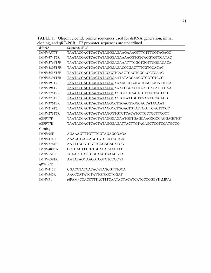
71
TABLE 1. Oligonucleotide primer sequences used for dsRNA generation, initial cloning, and qRT-PCR. T7 promoter sequences are underlined.
dsRNA Sequence 5’-3’ IMNV95T7F TAATACGACTCACTATAGGGAGAAGAAAGTTTGTTTCGTAGAGC IMNV474T7R TAATACGACTCACTATAGGGAGAAAAGGTGGCAGGTGTCCATAC IMNV3764T7F TAATACGACTCACTATAGGGAGAAATTTGGGTGGTTGGGACACA IMNV4805T7R TAATACGACTCACTATAGGGAGACCCGACTTTCGTGCACAC IMNV5518T7F TAATACGACTCACTATAGGGTCAACTCACTCGCAGCTGAAG IMNV6391T7R TAATACGACTCACTATAGGGAATATAGCAACGTCGTCTCCG IMNV193T7F TAATACGACTCACTATAGGGAAAACCGGAGCTGACCACATTCCA IMNV194T7F TAATACGACTCACTATAGGGAAACCGGAGCTGACCACATTCCAA IMNV275T7R TAATACGACTCACTATAGGGACTGTGTCACATGTTGCTGCTTCG IMNV223T7F TAATACGACTCACTATAGGGACTGTATTGGTTGAGTTCGCAGG IMNV376T7R TAATACGACTCACTATAGGGCTGGAGGTGGCAGCATACAAT IMNV219T7F TAATACGACTCACTATAGGGCTGGACTGTATTGGTTGAGTTCGC IMNV273T7R TAATACGACTCACTATAGGGTGTGTCACATGTTGCTGCTTCGCT eGFPT7F TAATACGACTCACTATAGGGAGAATGGTGAGCAAGGGCGAGGAGCTGT eGFPT7R TAATACGACTCACTATAGGGAGATTACTTGTACAGCTCGTCCATGCCG Cloning IMNV95F AGAAAGTTTGTTTCGTAGAGCGAGA IMNV474R AAAGGTGGCAGGTGTCCATACTGA IMNV3764F AATTTGGGTGGTTGGGACACATGG IMNV4805 R CCCGACTTTCGTGCACACAACTTT IMNV5518F TCAACTCACTCGCAGCTGAAGGTA IMNV6391R AATATAGCAACGTCGTCTCCGCGT qRT-PCR IMNV412F GGACCTATCATACATAGCGTTTGCA IMNV545R AACCCATATCTATTGTCGCTGGAT IMNVP1 (6FAM) CCACCTTTACTTTCAATACTACATCATCCCCGG (TAMRA)
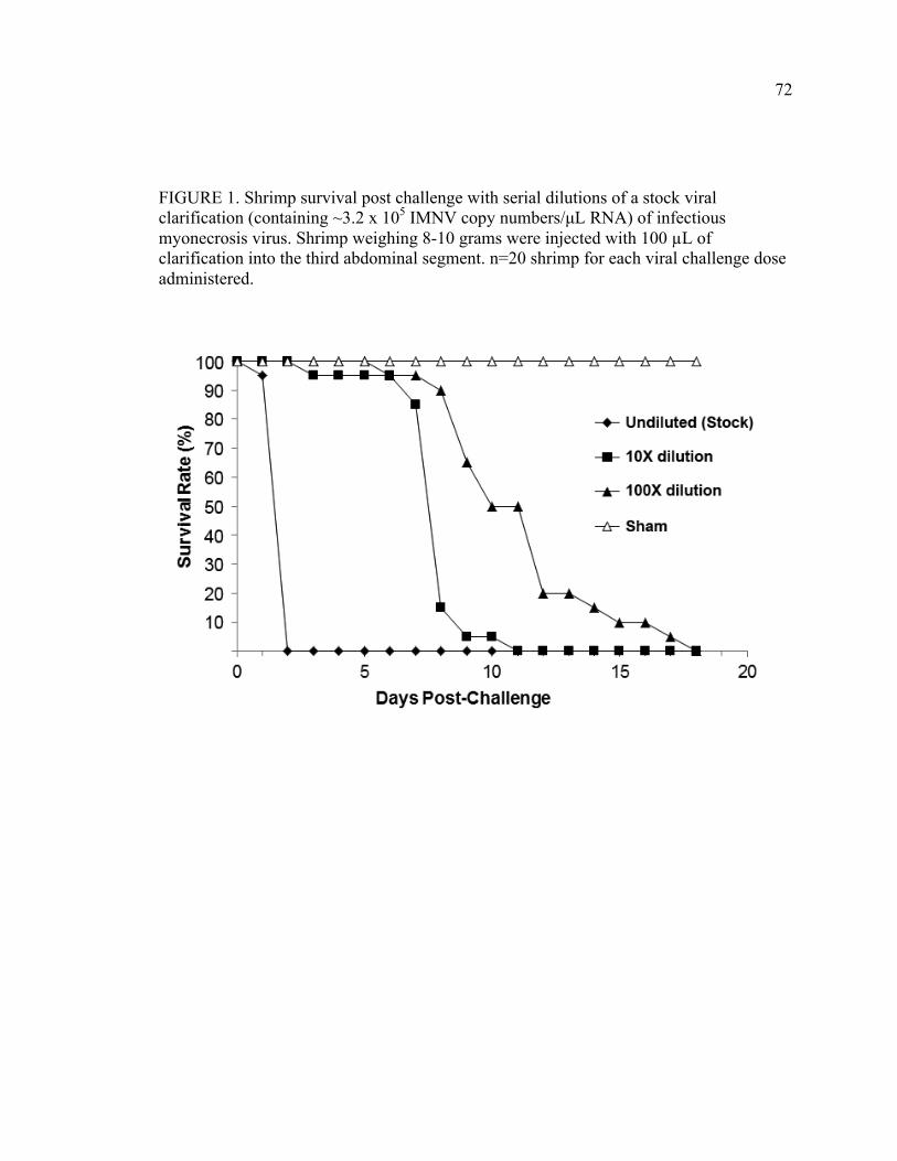
72
FIGURE 1. Shrimp survival post challenge with serial dilutions of a stock viral clarification (containing ~3.2 x 10⁵ IMNV copy numbers/µL RNA) of infectious myonecrosis virus. Shrimp weighing 8-10 grams were injected with 100 µL of clarification into the third abdominal segment. n=20 shrimp for each viral challenge dose administered.
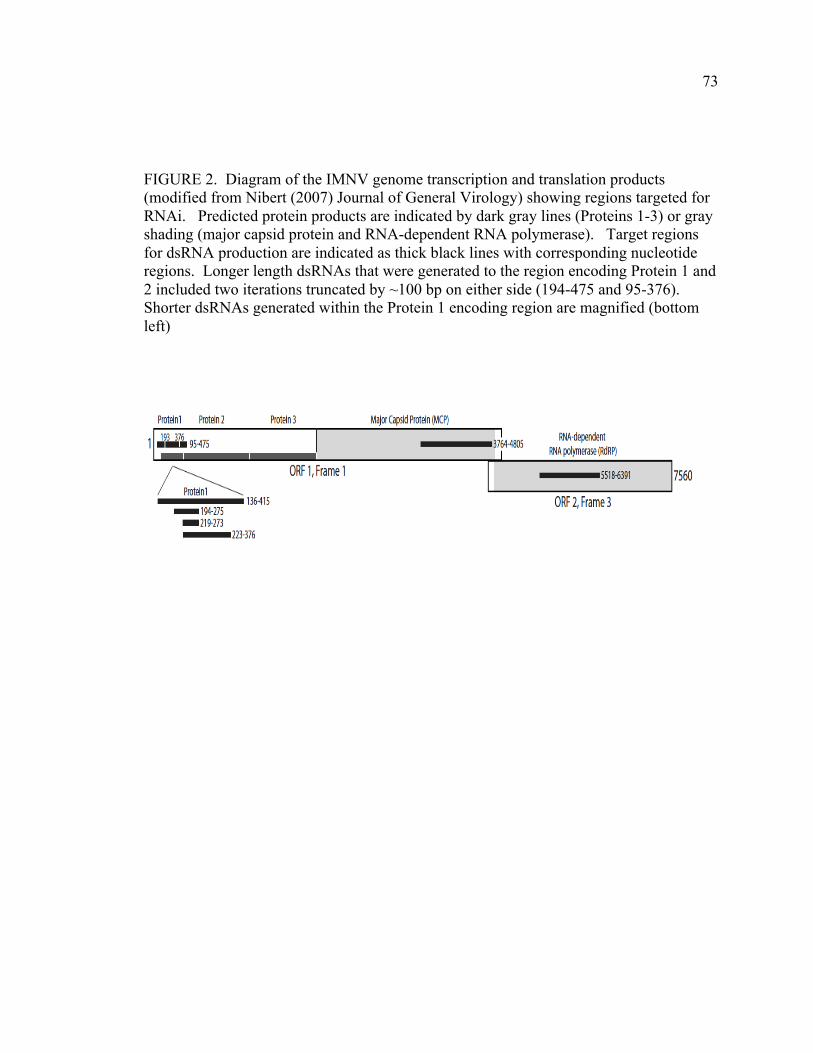
73
FIGURE 2. Diagram of the IMNV genome transcription and translation products (modified from Nibert (2007) Journal of General Virology) showing regions targeted for RNAi. Predicted protein products are indicated by dark gray lines (Proteins 1-3) or gray shading (major capsid protein and RNA-dependent RNA polymerase). Target regions for dsRNA production are indicated as thick black lines with corresponding nucleotide regions. Longer length dsRNAs that were generated to the region encoding Protein 1 and 2 included two iterations truncated by ~100 bp on either side (194-475 and 95-376). Shorter dsRNAs generated within the Protein 1 encoding region are magnified (bottom left)
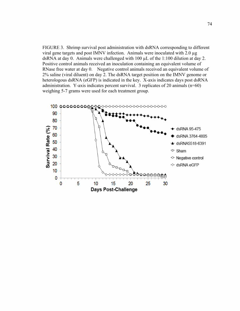
74
FIGURE 3. Shrimp survival post administration with dsRNA corresponding to different viral gene targets and post IMNV infection. Animals were inoculated with 2.0 µg dsRNA at day 0. Animals were challenged with 100 µL of the 1:100 dilution at day 2. Positive control animals received an inoculation containing an equivalent volume of RNase free water at day 0. Negative control animals received an equivalent volume of 2% saline (viral diluent) on day 2. The dsRNA target position on the IMNV genome or heterologous dsRNA (eGFP) is indicated in the key. X-axis indicates days post dsRNA administration. Y-axis indicates percent survival. 3 replicates of 20 animals (n=60) weighing 5-7 grams were used for each treatment group.
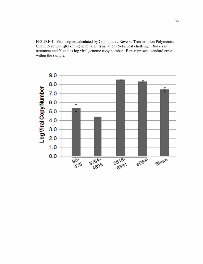
75
FIGURE 4. Viral copies calculated by Quantitative Reverse Transcription Polymerase Chain Reaction (qRT-PCR) in muscle tissue at day 9-12 post challenge. X-axis is treatment and Y-axis is log viral genome copy number. Bars represent standard error within the sample.
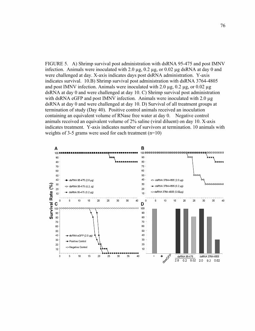
76
FIGURE 5. A) Shrimp survival post administration with dsRNA 95-475 and post IMNV infection. Animals were inoculated with 2.0 µg, 0.2 µg, or 0.02 µg dsRNA at day 0 and were challenged at day. X-axis indicates days post dsRNA administration. Y-axis indicates survival. 10.B) Shrimp survival post administration with dsRNA 3764-4805 and post IMNV infection. Animals were inoculated with 2.0 µg, 0.2 µg, or 0.02 µg dsRNA at day 0 and were challenged at day 10. C) Shrimp survival post administration with dsRNA eGFP and post IMNV infection. Animals were inoculated with 2.0 µg dsRNA at day 0 and were challenged at day 10. D) Survival of all treatment groups at termination of study (Day 40). Positive control animals received an inoculation containing an equivalent volume of RNase free water at day 0. Negative control animals received an equivalent volume of 2% saline (viral diluent) on day 10. X-axis indicates treatment. Y-axis indicates number of survivors at termination. 10 animals with weights of 3-5 grams were used for each treatment (n=10)

77
FIGURE 6. A) Shrimp survival post administration with initial dsRNA truncations of 95-475, 193-475, and 95-376 and post IMNV infection. Animals were inoculated with 0.02 µg dsRNA at day 0 and were challenged at day 10. X-axis indicates days post dsRNA administration. Y-axis indicates percent survival. B) Shrimp survival post administration with further truncations of dsRNA 223-376, 194-275, or 219-273 and post IMNV infection. Animals were inoculated 0.02 µg dsRNA at day 0 and were challenged at day 10. C) Shrimp survival of positive or negative controls and post IMNV infection. Positive control animals received an inoculation containing an equivalent volume of RNase free water at day 0 and viral challenge on day 10. Negative control animals received an equivalent volume of 2% saline (viral diluent) on day 10. D) Survival of all treatment groups at termination of study (Day 30). X-axis indicates treatment by dsRNA fragment length. Y-axis indicates number of survivors at termination. 3 replicate tanks of 10 animals weighing 3-5 grams were used for each treatment (n=30) except the negative control which had a single tank (n=10).
78
CHAPTER 4: SEQUENCE OPTIMIZED AND TARGETED DOUBLE
STRANDED RNA AS A THERAPEUTIC ANTIVIRAL TREATMENT AGAINST
INFECTIOUS MYONECROSIS VIRUS IN LITOPENAEUS VANNAMEI
J. Dustin Loy,1,2,4 Duan S. Loy,1 Mark A. Mogler, 2,4 Bruce Janke,3 Kurt Kamrud,1,4 DL Hank Harris1,3,4 Lyric Bartholomay5
A manuscript to be submitted for publication in Diseases of Aquatic Organisms
1 Department of Animal Sciences, Iowa State University, Ames, IA, USA,2 Department of Veterinary Microbiology and Preventive Medicine, Ames, IA USA,3 Department of
Veterinary Diagnostic and Production Animal Medicine, Ames, IA, USA 4
Harrisvaccines, Ames IA, USA , 5 Department of Entomology, Iowa State University, Ames, IA, USA
Abstract
Infectious myonecrosis virus (IMNV) is a significant and emerging pathogen that
has a tremendous impact on the culture of the farmed Pacific white shrimp (Litopenaeus
vannamei). IMNV first emerged in Brazil in 2002 and subsequently spread to Indonesia
causing large economic losses in both countries. No existing therapeutic treatments or
effective interventions currently exist for IMNV. RNA interference (RNAi) has been one
effective strategy developed to prevent viral disease in shrimp. Here we describe the
efficacy of a dsRNA applied as an antiviral therapeutic following viral challenge. The
antiviral therapeutic used was a sequence-optimized dsRNA construct targeting a viral
sequence encoding dsRNA binding motifs, that had previously showed outstanding
antiviral protection when administered prior to infection. Our data indicates that at least
50% survival can be attained with low doses (0.5 µg) of dsRNA if administered 48 hours
79
following an infection with an otherwise lethal dose of IMNV, and that this degree of
protection was not observed when dsRNA was administered 72 hours post infection.
Additionally, the administration of this antiviral demonstrated a significant reduction in
viral loads in the muscle of animals that died from disease or survived until termination
of the study as assessed by quantitative RT-PCR. This data indicates that a sequence and
length optimized RNAi antiviral molecule holds promise for use as an antiviral
therapeutic against IMNV.
Introduction
Infectious Myonecrosis (IMN) is a severe and emerging shrimp disease that was
first described in 2003 after a severe outbreak in northeastern Brazil characterized by
high mortality and animals exhibiting necrosis in the tail muscle.12 The etiologic agent is
a non-enveloped, double-stranded RNA (dsRNA) virus that is a member of the
Totiviridae family, subsequently named Infectious Myonecrosis Virus (IMNV). IMNV
was discovered in Indonesia in 2006;16 and is causing tremendous impact on the shrimp
industry within the country. During outbreaks of IMNV, mortality has ranged from 40%
to 70% prior to harvest and causes a dramatic increase in feed conversion.1 IMNV
outbreaks have been found to be associated with longer growout periods and higher
stocking densities following an epidemiological survey of risk factors.2 The insidious
presentation of this disease, resulting in a gradual loss of animals over duration of the
growout period, makes IMNV an excellent target for a therapeutic agent, as treatments
could be initiated immediately following a diagnosis of IMNV within a pond.
80
RNA interference (RNAi) is one possible and promising method to mitigate viral
disease in shrimp.7,9,13,14,17 RNAi has been used to prevent shrimp viral diseases caused
by White Spot Syndrome Virus (WSSV)13,22 Yellowhead Virus (YHV),19,20,23 Taura
Syndrome Virus (TSV),15 Penaeus stylirostris densovirus (PstDNV) (formerly called
infectious hypodermal and hematopoietic necrosis virus (IHHNV)),8 and IMNV.11 In
addition to administration prior to viral exposure, the therapeutic effect of RNAi has been
tested against many different RNA and DNA viruses of animals and humans, several of
which are being evaluated in Phase 1 and 2 human clinical trials as antiviral therapeutics.5
In shrimp, a therapeutic effect has been described against WSSV22 and YHV20 post
dsRNA inoculation. Some evidence indicates that dsRNA administration and induction of
RNAi pathways may allow for clearance of virus in shrimp naturally-infected with
Penaeus monodon densovirus (PmDV).3 Recently described methods allow for a
sequence and length optimized construct to be developed against a specific shrimp
pathogen.11 The objective of these experiments was to test the therapeutic efficacy of a
sequence-optimized dsRNA construct that showed outstanding protection when
administered prior to infection (Loy et al. 2011), in the face of an active IMNV infection.
Materials and Methods
Animal Rearing Specific pathogen free (SPF) L. vannamei postlarvae were received from Shrimp
Improvement Systems (Plantation Key, Florida) and reared in a biosecure animal holding
facility at Iowa State University. Animals were placed into 1000 L tanks containing
artificial seawater (Crystal Sea Marine Mix) at 25 ppt salinity, an oystershell airlift
81
biofilter, and an activated carbon filter. Temperature was maintained at 30º C. Animals
were fed a commercial growout diet (Rangen 35/10, Buhl, Idaho) until 3-5 grams in
weight.
Preparation of Viral Inoculum Based on a previously described methodology6, tissue macerate from infected
animals (that tested negative for other shrimp viruses by PCR) was diluted 1:3 in TN
buffer (0.02 M Tris-HCL, 0.4 M NaCL, pH 7.4) and clarified through three centrifugation
steps after each of which supernatant was removed: 1) 4000 x g for 30 min, 2)15,000 x g
for 15 min 3) 25,000 x g for 60 min. The final supernatant was diluted 1:10 in sterile 2%
NaCl and passed through a 0.2 micron filter. This stock viral clarification was aliquoted
and frozen at -80º C. Quantitative RT-PCR analysis of RNA extracted from a freeze-
thaw indicated 105 viral copy numbers/µl were present in the stock.
Determination of Challenge Dose The optimum lethal dose by injection resulting in infection and disease was tested
in SPF L. vannamei weighing 8-10 g and described in detail by Loy et al.11 Briefly, each
animal was injected with 100 µl of a 1:100 dilution of viral clarification into the third
abdominal segment, a dose which resulted in 100% mortality at 18 days post infection
(dpi).
Quantitative Reverse-Transcription PCR Quantitative Reverse-Transcription PCR (qRT-PCR) was performed using
methods adapted from Andrade et al.1 Briefly, 30 mg of macerated muscle tissue and
homogenized using a QIAShedder column (Qiagen) followed RNA extraction with an
RNeasy Minikit (Qiagen). RNA template (50 µl) was eluted from the column into RNase
82
free water and stored at -80º C until the RT-PCR was performed. qRT-PCR templates
were thawed and boiled at 100ºC for 5 min prior to reverse transcription. RNA template
(2 µl) was mixed with 5 µl of 5x One Step RT-PCR Master Mix (Qiagen), 1 µl Enzyme
Mix, 1 µl DNTP, 0.3 µl IMNV412 (20 uM) (5′-GGA CCT ATC ATA CAT AGC GTT
TGC A-3′), 0.3 µl IMNV312R (20 uM) (5′-AAC CCA TAT CTA TTG TCGCTGGAT-
3′), 0.3 µl IMNV p1 (10 uM) (5′-6FAM CCA CCT TTA CTT TCA ATA CTA CAT
CAT CCC CGG TAMRA-3′) and 15 µl RNase free water for a total reaction volume of
25 µl. This reaction was then run in duplicate or triplicate on a BioRad CFX96 Real
Time PCR Detection System under the following conditions: RT for 30 min at 48°C and
10 min at 95°C, followed by PCR for 35 cycles of 15 s at 95°C and 1 min at 60°C. Viral
copy number was calculated using CFX Manager Software (BioRad) using an in vitro
transcribed RNA standard generated using methods from Andrade et al.1
dsRNA Preparation dsRNA was prepared using Ambion Megascript RNAi Kit following the
manufacturer’s directions. To create template for dsRNA synthesis, RNA was extracted
from virally infected animals using a commercial nucleic acid purification kit (Qiagen
RNeasy Mini). Oligonucleotide primers for cDNA synthesis were then designed
sequence available (GenBank accession no.EF061744)16 (TABLE 1). Reverse
transcription of RNA was performed using Thermoscript RT per manufacturer’s
instructions (Invitrogen). Following reverse transcription, template cDNA (~50 ng, not
calculated) was added to a PCR master mix (PuReTaq Ready-To-Go PCR Beads) and
thermal-cycling was performed using oligonucleotide primers (TABLE 1) designed to
specific regions of the IMNV genome (GenBank accession EF061744.1). Products were
83
then cloned into pCR4.0 vectors (Zero Blunt TOPO PCR cloning kit) and transformed
into E. coli (TOP10 Invitrogen). Plasmids preparations from these transformants were
used as a template source for further PCR reactions to create DNA clones for RNA
transcription For synthesis of template for transcription, primers containing a T7
promoter sequence (TABLE 1) on the forward and reverse primer were used to amplify
the segments of cDNA with flanking T7 promoter sites. T7 DNA templates were then
transcribed overnight (16 hours) following the manufacturer’s directions. Following
transcription, dsRNA products were incubated for 60 minutes with DNase I and RNase,
and purified using the provided columns. Transcription of dsRNA was confirmed by gel
electrophoresis in comparison with a molecular weight ladder (pGEM ladder, Promega)
and product was quantified by spectrophotometer.
Histopathology Moribund animals that were found prior to death were fixed in whole Davidson’s
fixative4 for 24 hours before being transferred to 70% EtOH before embedding in
paraffin. Two animals from each tank were anesthetized in an ice slurry and fixed in
Davidson’s fixative4 embedded in paraffin, cut into slides and stained with hematoxylin
and eosin and evaluated for the presence of IMNV lesions.
Statistical Analysis End points of mean survival was analyzed using One-way ANOVA followed by
Tukey’s multiple comparison test using JMP 8 software. Mean log viral copy numbers
ran in triplicate were normalized and then compared following using One-way ANOVA
followed by Tukey’s multiple comparison test using JMP 8 software
84
Shrimp Bioassays Tanks containing 200 L synthetic seawater and an oystershell airlift biofilter tanks
were stocked with 10 SPF juveniles weighing 3-5 g and allowed to acclimate for 72 h.
Tanks were randomized and three replicates were assigned for each treatment. Following
acclimation animals were challenged by injection into the third abdominal segment with
lethal dose of IMNV. Forty eight hours later a total of 5.0 µg of in vitro synthesized
dsRNA was inoculated into animals that had been challenged with IMNV. Sham
treatment groups received an equivalent volume of RNase free water. Sham challenge
(negative) groups received an equivalent volume of 2% NaCl (viral diluent). Animals
were counted daily for mortality and assessed for clinical signs. Moribund animals that
were found were fixed for histopathology. At termination 2 animals from each tank were
fixed and submitted for histopathological examination. Dead animals removed
throughout the experimental period were frozen at -80ºC.
Dose/Duration Bioassay Tanks were challenged with IMNV virus simultaneously with groups receiving
treatments at 24, 48, or 72 h. Treatment groups consisted of either 5.0 or 0.5 µg of in
vitro synthesized dsRNA 194-475, 5.0 µg of eGFP dsRNA, or a sham treatment of an
equivalent volume of RNase free water at an equivalent volume and time interval. Sham
challenge (strict negative) groups received an equivalent volume of 2% NaCl (viral
diluent).
Results
A disease challenge model was established that uses an intramuscular injection of
a clarified virus preparation that resulted in 100% mortality within 18 d, using a 100-fold
85
dilution of the tissue homogenate (~3.2 x 10³ IMNV copy numbers/µl RNA Specific
pathogen free (SPF) L. vannamei (3-5 g in weight) were subjected to a lethal
intramuscular challenge and 48 h later to a single dose containing 5.0 µg of dsRNA194-
275 demonstrated 50% survival. By comparison animals that received the same dose of
eGFP dsRNA (dseGFP) or the equivalent volume of RNase free water showed 100%
mortality following challenge (FIGURE 1). Significant differences (P=0.007) in
survivorship were evident between the experimental and control groups.
Viral Copy Number Quantification Differences in virus genome copy number were assessed via qRT-PCR on muscle
tissues sampled from dead animals at 8-12 d post-infection or at the termination of the
study. A significant reduction (P=0.01) in viral copy number was evident between
animals administered 5.0 µg dsRNA194-275 that either succumbed to disease or survived
until termination (P<.0001) (mean 5.84 x 10⁵ and 4.87 x 10⁴ IMNV copy numbers/µl
RNA, respectively), when compared with animals that received 5.0 µg of eGFP dsRNA
or RNAse free water.
Dosage and Temporal Effects To further characterize the observed therapeutic response, the temporal effects
and dose response factors that influence a successful IMNV therapeutic treatment were
tested. Animals treated 24 h after viral challenge demonstrated 60% survival after
treatment with 5.0 µg dsRNA (high dose) as compared to 50% survival in the animals
treated with 0.5 µg dsRNA (low dose) (FIGURE 3). Control animals that received 5.0 µg
heterologous dsRNA eGFP (dseGFP) demonstrated 30% survival and the RNase free
86
water injected animals showed 0% survival. Animals treated 48 hours post-challenge
demonstrated 90% survival in the high dose treatment group and 50% survival in the low
dose group with 0% survival in groups receiving dseGFP and 20% survival in the RNase
free water injected group (FIGURE 4). Experimental and control groups showed 0%
survival when dsRNA was provide 72 h post-infection (FIGURE 5).
Gross Lesions Animals in each treatment group were examined for gross lesions daily. Presence
of gross lesions was evident as single or multiple foci of opacity within the tail muscle.
All individuals in all challenged groups developed a focal or multi-focal muscle tissue
opacity by day 5 post challenge. Several individuals treated with dsRNA194-275 in the
48 h post challenge had opacities resolve by 12 days post challenge (FIGURE 6).
Lesions in untreated animals did not resolve, and all of these animals succumbed to
death.
Histopathology Acute histopathologic lesions were characterized by coagulative necrosis and
fragmentation of muscle bundles with edema prominent within the muscle bundle sheaths
and between affected muscle bundles. Infiltration of hemocytes was mostly limited to
small clusters scattered along the connective tissue septa that separated groups of
muscles. Chronic lesions were characterized by partial or total loss of myofibers with
condensation/contraction of the fibrous connective tissue components. These lesions
correspond with characteristic histopathological changes typically seen with IMNV
infection.11,12
87
Moribund animals that were found in tanks were sampled from sham-vaccinated
controls (7 animals), eGFP dsRNA administered controls (3 animals), and animals treated
with dsRNA 194-275 (4 animals). Additionally, 2 animals from each tank were sampled
from surviving animals at the termination of the study (9 animals). Histopathological
lesions in moribund animals in animals from all groups were characterized predominantly
by extensive fibrosis occasionally with concurrent acute coagulative necrosis. Acute
necrosis without chronic lesions was evident in only one animal. Animals treated with
dsRNA 194-275 that survived until termination of the study had chronic resolving lesions
that varied from extensive fibrosis (2 animals) to scattered small foci of fibrosis (3
animals) or no significant lesions present (3 animals). No acute lesions were seen in any
of these longer-surviving treated animals.
Discussion
These studies describe the first use of RNAi to treat disease in shrimp infected
with IMNV, a significant emerging shrimp pathogen. Previous data demonstrated that a
very specific short dsRNA confers robust and long-term resistance to IMNV-induced
disease and mortality when administered prior to viral infection.11 These experiments
demonstrate that the same short dsRNA can also therapeutically treat animals with pre-
existing viral infections. Additionally, it was noted that non-sequence specific dsRNA
administered at high dosage levels (5.0 µg) does not induce a protective response when
administered therapeutically at 48 hours post-infection. This is similar to previous
findings that showed no positive effect on survival when dseGFP was administered
prophylactically.11 Demonstrating therapeutic efficacy is a significant step towards

88
developing an antiviral therapeutic for infected animals as part of a disease control
program. Such a protocol could help to eliminate or reduce IMNV loads in infected
ponds.
In order to determine threshold therapeutic doses of dsRNA and simultaneously
characterize the temporal activity of the RNAi response, animals were administered two
doses levels of dsRNA (0.5 or 5.0 µg). Significant differences in survival were apparent
at both doses, when given either 24 or 48 h post-virus exposure. This is similar to
findings using IMNV specific dsRNA delivered prior to infection, where doses as low as
20 ng protected animals from succumbing to IMNV disease11 and such a protocol may
help eliminate or reduce IMNV loads in infected ponds.
Dose and Temporal Response Low dosage levels appear to provide protection in animals early in the disease
process. By contrast, when dsRNA 194-275 was administered 72 h post-challenge, no
survival-enhancing effect was observed. This may be due to a significant amount of viral
replication taking place between 48 h and 72 h, where the amount of virus replication that
has occurred likely exceeds the capacity of an RNAi antiviral effect. Prior work
examining the therapeutic effect of dsRNA on survival of shrimp from YHV or PstDNV
challenge has been demonstrated only at higher dosages and shorter intervals (3 or 12 h
post infection at a 25.0 µg dose dsRNA20 or 24 hours post infection with a 5.0-12.5 µg
dose dsRNA)8. No experimental studies have examined the replication dynamics of
IMNV in shrimp, but these data suggest that a large amount of virus replication is
occurring between 48 and 72 hours post infection, and after which time animals are
refractory to treatment with specific dsRNA.

89
This temporal phenomenon may also be explained by the target sequence of this
dsRNA, a cleavage product called “Protein 1” that contains a dsRNA binding motif.18
This protein may be involved in suppression of host cellular RNAi responses or innate
immunity during early viral replication. Viral proteins with dsRNA binding activity have
been described and characterized as inhibiting the RNAi machinery such as Drosophila C
virus (DCV) and Flock House virus (FHV).21,10 If this putative dsRNA binding protein,
indeed suppresses an element of the RNAi machinery and it were expressed to a high
degree in a large number of infected cells, the ability for the host to elicit an appropriate
antiviral RNAi response would be impeded. Further studies into the role of this dsRNA
binding protein and the role it may play in immune suppression of the host and
pathogenesis during IMNV infection are warranted.
The use of a small and optimized dsRNA sequence and the ability of this
sequence to be used as an antiviral therapeutic for IMNV in L. vannamei was
demonstrated. Small amounts (0.5 µg) of IMNV specific dsRNA can prevent mortality
in animals with active viral infection. RT-PCR diagnostic methods have been developed
that would allow for a rapid turnaround for a diagnosis.1 If feasible delivery methods for
dsRNA in shrimp ponds become available, a therapeutic treatment of small, targeted
dsRNA could provide mitigation of viral disease losses in virus-infected animals.
Acknowledgements
The authors would like to thank Kay Kimpston-Burkgren, Cara Loy, Kara
Burrack, Kristen Summerfelt, and Pamela Whitson for animal care.

90
Additionally, they would like to thank Jill Gander, Ryan Vander Veen, and Patrick
Jennings for reagents and laboratory assistance.

91
References
1. Andrade TPD, Srisuvan Thinnarat, Tang KFJ, Lightner DV. (2007). Real-time
reverse transcription polymerase chain reaction assay using TaqMan probe
for detection and quantification of Infectious myonecrosis virus (IMNV).
Aquaculture. 264:9-15
2. Arns da Silva V, dos Santos FL, Bezarro SS, Pedrosa VF, Mendes P, Mendes E.S.
(2010). A multi-season survey for infectious myonecrosis in farmed
shrimp, Litopenaeus vannamei, in Pernambuco, Brazil. Journal of
Invertebrate Pathology. 104:161-165
3. Attasart P, Kaewkhaw R, Chimwai C, Kongphom U, Panyim S. (2011). Clearance
of Penaeus monodon densovirus in naturally pre-infected shrimp by
combined ns1 and vp dsRNAs. Virus Research. 159:79-82
4. Bell TA and Lightner DV. (1988). A Handbook of Normal Shrimp Histology.
Baton Rouge, USA, 114p
5. Haasnoot J, Westerhout EM, Berkhout B. (2007). RNA interference against
viruses: strike and counterstrike. Nature Biotechnology. 25:1435-1443
6. Hasson KW, Lightner DV, Poulos BT, Redman RM, White BL, Brock JA,
Bonami JR. (1995). Taura syndrome in Penaeus vannamei: demonstration
of a viral etiology. Disease of Aquatic Organisms. 23:115-126

92
7. Hirono I, Fagutao F, Kondo H, Aoki T. (2010). Uncovering the Mechanisms of
Shrimp Innate Immune Response by RNA Interference. Marine
Biotechnology 13: 622-628
8. Ho T, Yasri P, Panyim S, Udomkit A. (2011). Double-stranded RNA confers both
preventive and therapeutic effects against Penaeus stylirostris densovirus
(PstDNV) in Litopenaeus vannamei. Virus Research. 155:131-136
9. Krishnan P, Gireesh-Babu P, Rajendran KV, Chaudhari A. (2009). RNA
interference-based therapeutics for shrimp viral diseases. Diseases of
Aquatic Organisms 86:263-272
10. Li H, Li WX, Ding SW. (2002). Induction and suppression of RNA silencing by
an animal virus. Science. 296:1319-1321
11. Loy J, Mogler M, Loy D, Janke B, Kamrud K, Harris D, Bartholomay L. (2011).
Double Stranded RNA Provides Sequence Dependent Protection Against
Infectious Myonecrosis Virus in Litopenaeus vannamei. Journal of
General Virology. Ref Type: Submitted
12. Poulos BT, Tang KFJ, Pantoja CR, Bonami JR, Lightner DV. (2006). Purification
and characterization of infectious myonecrosis virus of penaeid shrimp.
Journal of General Virology. 87:987-996
13. Robalino J, Bartlett T, Shepard E, Prior S, Jaramillo G, Scura ED, Chapman RW,
Gross PS, Browdy CL, Warr GW. (2005). Double-Stranded RNA Induces
93
Sequence-Specific Antiviral Silencing in Addition to Nonspecific
Immunity in a Marine Shrimp: Convergence of RNA Interference and
Innate Immunity in the Invertebrate Antiviral Response? Journal of
Virology. 79:13561-13571
14. Robalino J, Bartlett TC, Chapman RW, Gross PS, Browdy CL, Warr GW. (2007).
Double-stranded RNA and antiviral immunity in marine shrimp: Inducible
host mechanisms and evidence for the evolution of viral counter-
responses. Developmental & Comparative Immunology. 31:539-547
15. Robalino J, Browdy CL, Prior S, Metz A, Parnell P, Gross P, Warr G. (2004).
Induction of Antiviral Immunity by Double-Stranded RNA in a Marine
Invertebrate. Journal of Virology. 78:10442-10448
16. Senapin S, Phewsaiya K, Briggs M, Flegel TW. (2006). Outbreaks of infectious
myonecrosis virus (IMNV) in Indonesia confirmed by genome sequencing
and use of an alternative RT-PCR detection method. Aquaculture. 266:32-
38
17. Shekhar MS and Lu Y. (2009). Application of Nucleic-acid-based Theraputics for
Viral Infections in Shrimp Aquaculture. Marine Biotechnology. 11:1-9
18. Tang J, Ochoa WF, Sinkovits RS, Poulos BT, Ghabrial SA, Lightner DV, Baker
TS, Nibert ML. (2008). Infectious myonecrosis virus has a totivirus-like,
120-subunit capsid, but with fiber complexes at the fivefold axes.
Proceedings National Academy of Sciences USA. 105
94
19. Tirasophon W, Roshorm Y, Panyim. (2005). Silencing of yellow head virus
replication in penaeid shrimp cells by dsRNA. Biochemical and
Biophysical Research Communications. 334:102-107
20. Tirasophon W, Yodmuang S, Chinnirunvong W, Plongthongkum N, Panyim S.
(2007). Therapeutic inhibition of yellow head virus multiplication in
infected shrimps by YHV-protease dsRNA. Antiviral Research. 74:150-
155
21. van Rij RP, Saleh MC, Berry B, Foo C, Houk A, Antoniewski C, Andino R.
(2006). The RNA silencing endonuclease Argonaute 2 mediates specific
antiviral immunity in Drosophila melanogaster. Genes Development.
20:2985-2995
22. Xu J, Han F, Zhang X. (2007). Silencing shrimp white spot syndrome virus
(WSSV) genes by siRNA. Antiviral Research. 73:126-131
23. Yodmuang S, Tirasophon W, Roshorm Y, Chinnirunvong W, Panyim S. (2006).
YHV-protease dsRNA inhibits YHV replication in Penaeus monodon and
prevents mortality. Biochemical and Biophysics Research
Communications. 341:351-356

95
TABLE 1. Oligonucleotide primer sequences for dsRNA synthesis, cloning and qPCR
Primer Sequence 5’-3’ Source
IMNV194T7F
TAATACGACTCACTATAGGGAAACCGGAGCTGACCACATTCCAA
Loy et al (2011)
IMNV275T7R
TAATACGACTCACTATAGGGACTGTGTCACATGTTGCTGCTTCG
Loy et al (2011)
eGFPT7F TAATACGACTCACTATAGGGAGAATGGTGAGCAAGGGCGAGGAGCTGT
Loy et al (2011)
eGFPT7R TAATACGACTCACTATAGGGAGATTACTTGTACAGCTCGTCCATGCCG
Loy et al (2011)
IMNV95F AGAAAGTTTGTTTCGTAGAGCGAGA Loy et al (2011)
IMNV474R AAAGGTGGCAGGTGTCCATACTGA Loy et al (2011)
IMNV412F GGACCTATCATACATAGCGTTTGCA Andrade et al (2007)
IMNV545R AACCCATATCTATTGTCGCTGGAT Andrade et al (2007)
IMNVP1 (6FAM) CCACCTTTACTTTCAATACTACATCATCCCCGG (TAMRA)
Andrade et al (2007)
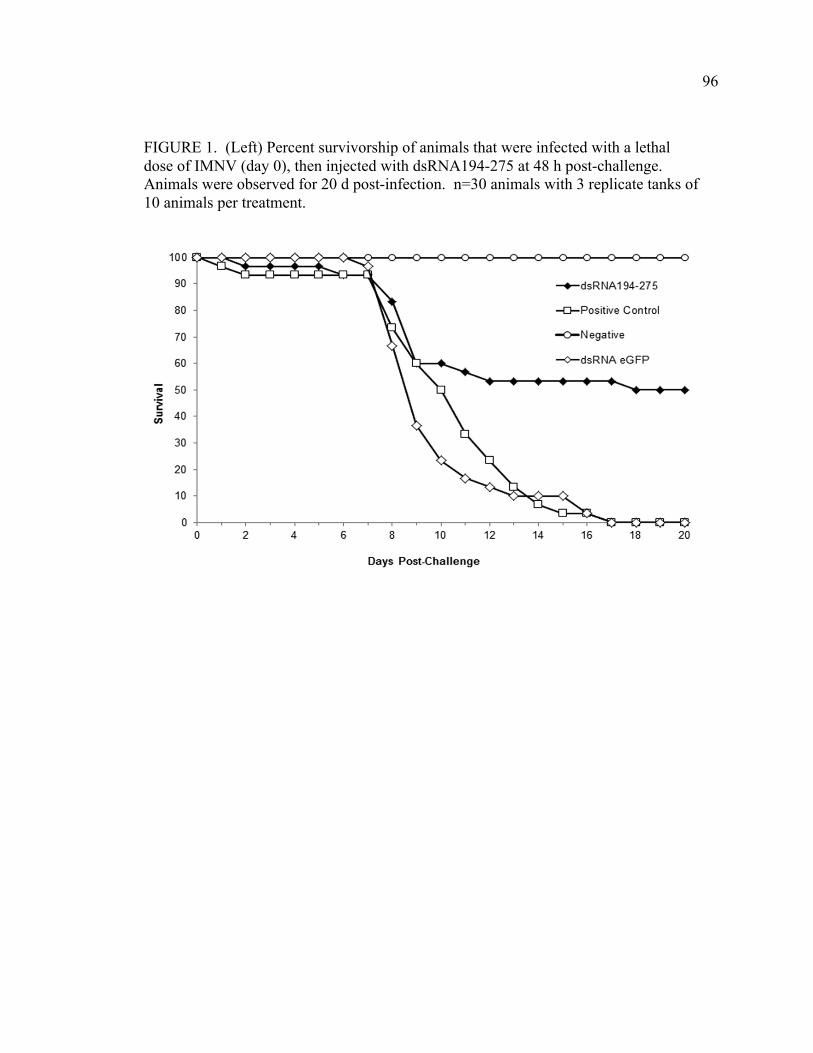
96
FIGURE 1. (Left) Percent survivorship of animals that were infected with a lethal dose of IMNV (day 0), then injected with dsRNA194-275 at 48 h post-challenge. Animals were observed for 20 d post-infection. n=30 animals with 3 replicate tanks of 10 animals per treatment.
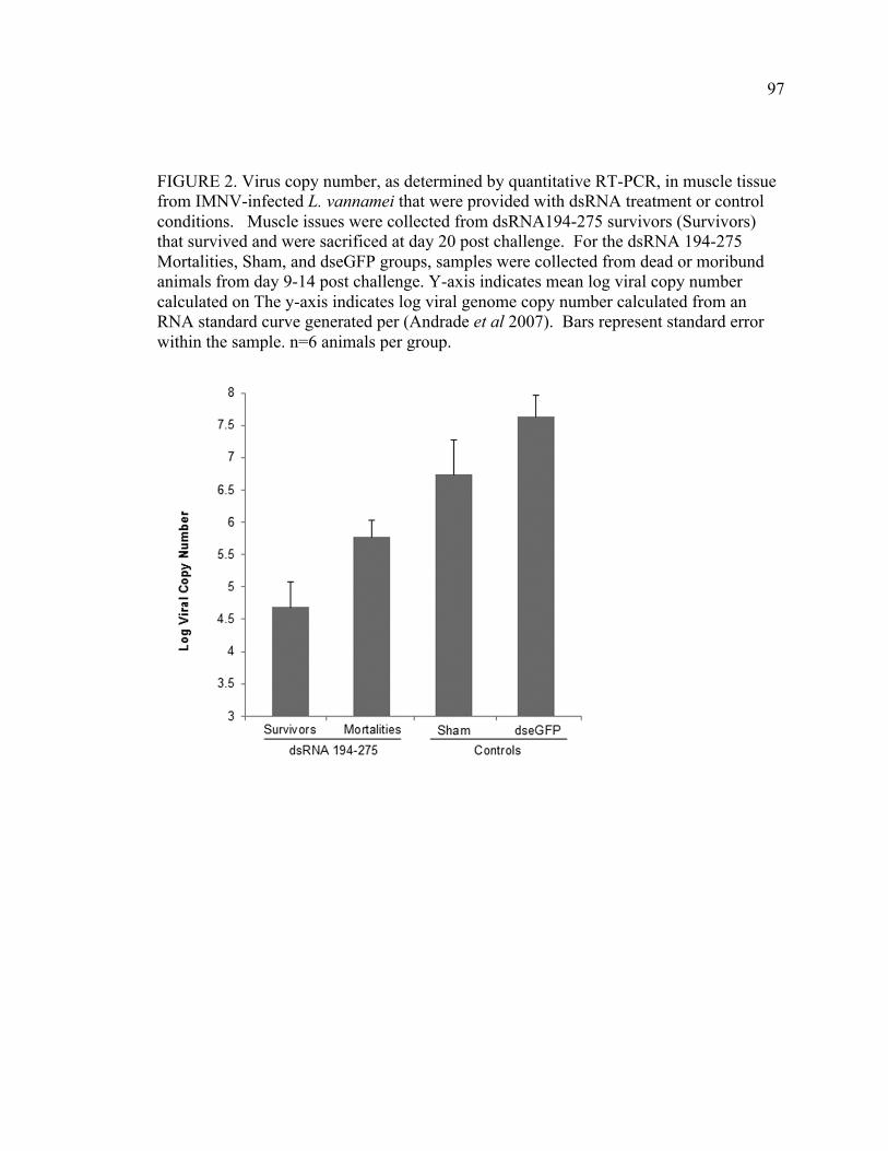
97
FIGURE 2. Virus copy number, as determined by quantitative RT-PCR, in muscle tissue from IMNV-infected L. vannamei that were provided with dsRNA treatment or control conditions. Muscle issues were collected from dsRNA194-275 survivors (Survivors) that survived and were sacrificed at day 20 post challenge. For the dsRNA 194-275 Mortalities, Sham, and dseGFP groups, samples were collected from dead or moribund animals from day 9-14 post challenge. Y-axis indicates mean log viral copy number calculated on The y-axis indicates log viral genome copy number calculated from an RNA standard curve generated per (Andrade et al 2007). Bars represent standard error within the sample. n=6 animals per group.
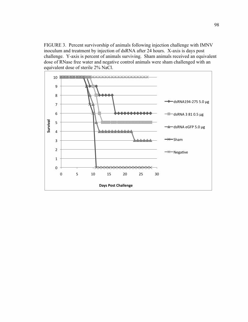
98
FIGURE 3. Percent survivorship of animals following injection challenge with IMNV inoculum and treatment by injection of dsRNA after 24 hours. X-axis is days post challenge. Y-axis is percent of animals surviving. Sham animals received an equivalent dose of RNase free water and negative control animals were sham challenged with an equivalent dose of sterile 2% NaCl.
0
1
2
3
4
5
6
7
8
9
10
0 5 10 15 20 25 30
Survival
Days Post Challenge
dsRNA194-‐275 5.0 μg
dsRNA 3 81 0.5 μg
dsRNA eGFP 5.0 μg
Sham
Nega=ve
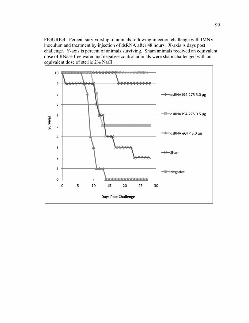
99
FIGURE 4. Percent survivorship of animals following injection challenge with IMNV inoculum and treatment by injection of dsRNA after 48 hours. X-axis is days post challenge. Y-axis is percent of animals surviving. Sham animals received an equivalent dose of RNase free water and negative control animals were sham challenged with an equivalent dose of sterile 2% NaCl.
0
1
2
3
4
5
6
7
8
9
10
0 5 10 15 20 25 30
Survival
Days Post Challenge
dsRNA194-‐275 5.0 μg
dsRNA194-‐275 0.5 μg
dsRNA eGFP 5.0 μg
Sham
Nega=ve
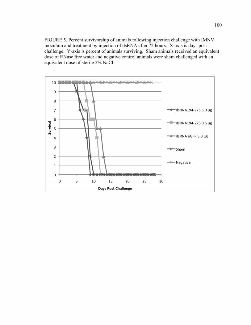
100
FIGURE 5. Percent survivorship of animals following injection challenge with IMNV inoculum and treatment by injection of dsRNA after 72 hours. X-axis is days post challenge. Y-axis is percent of animals surviving. Sham animals received an equivalent dose of RNase free water and negative control animals were sham challenged with an equivalent dose of sterile 2% NaCl.
0
1
2
3
4
5
6
7
8
9
10
0 5 10 15 20 25 30
Survival
Days Post Challenge
dsRNA194-‐275 5.0 μg
dsRNA194-‐275 0.5 μg
dsRNA eGFP 5.0 μg
Sham
Nega=ve
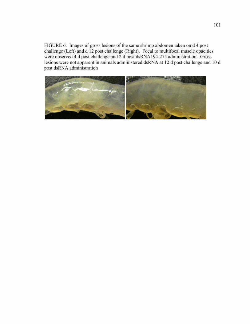
101
FIGURE 6. Images of gross lesions of the same shrimp abdomen taken on d 4 post challenge (Left) and d 12 post challenge (Right). Focal to multifocal muscle opacities were observed 4 d post challenge and 2 d post dsRNA194-275 administration. Gross lesions were not apparent in animals administered dsRNA at 12 d post challenge and 10 d post dsRNA administration
102
CHAPTER 5: GENERAL CONCLUSIONS
The results from this work substantiate the idea that the white leg or Pacific white
shrimp, Litopenaeus vannamei, possess a complex immune system capable of specifically
recognizing and responding to insults from viral pathogens. Moreover, that this immune
system can be primed or modulated using very small quantities of exogenous double
stranded RNA to mount a highly specific, long-lasting, and effective response. Although
the mechanisms underlying this response are not well understood, they are likely to be
evolutionarily and fundamentally distinct from the specific and adaptive humoral
responses observed in vertebrate immune systems. Akin to the adaptive response, the
shrimp immune response is functionally capable of eliciting a specific and robust
response to viral disease that is functional for extended periods of time, and often that
amount to a significant portion of the organism’s lifespan. These observations are
suggestive of much more complex immune system, one that is distinct from general non-
specific innate immunity that is responsive in a generic manner and short-lived. The data
presented herein point to RNA interference (RNAi) as a primary antiviral response
underlying this long-term immunity. The RNAi antiviral response diverges from the
common understanding of proteins as long-term immune response elicitors, the hallmark
of “specific” immunity, in that it has instead diverged to recognize nucleic acids, and
specific sequence components of those nucleic acids that are produced during a viral
infection. In this sense, marine shrimp antiviral immunity is, at many levels, challenging
the current paradigms of immunity. This dissertation further expands that body of
research.
103
Specifically, it was observed for the first time in shrimp that very small
amounts (20 nanograms) of a precisely targeted dsRNA that elicits an RNAi response can
protect animals (100% survival vs. 0% survival) for at least ten days, and likely longer.
This is a 100% increase over previously described periods of protection (5 days vs. 10
days) and a 1000-fold lower dose that had been used to demonstrate protection in
previous studies.3 Additionally, shrimp that were subjected to this dsRNA construct then
exposed to IMNV, were resistant to subsequent challenges (of 100x higher dosage of
virus) for at least 90 days. This same molecule could not only prevent, but also
therapeutically treat infections existing infections up to 2 days after receiving a lethal
dose of virus. These results indicate that the RNAi antiviral immune response in a
shrimp is significantly more potent and longer lasting than had previously been thought.
Several new tools and methodologies were also developed and explored in this
dissertation that will enable future work to develop disease control technologies. We
developed a concept viral genome panning to develop the most effective antiviral effector
molecules is also a novel concept to fight shrimp diseases. Previous work had targeted
only a handful of sequences or gene targets against each virus (1 or 2 sequences per
virus). This work demonstrates that even within a very small virus such as IMNV (~7500
bp) there are extreme differences observed in survival between the viral sequences
targeted (0% vs 100%) and that panning the genome to discover the most protective
sequences is a prudent means to develop an antiviral prophylactic. From a production
standpoint it was also determined that smaller molecules can also be as effective or more
effective than longer ones (80 bp vs. 500 bp) thereby facilitating more efficient
production. Additionally, this work also has large implications to developing therapeutic
104
drugs for shrimp and other invertebrates, because the sequences used to develop antiviral
molecules also demonstrated efficacy as a therapy against a pre-exsisting infection.
A lack of in vitro tools to explore and mechanistically define the shrimp immune
system impedes research into the pathways responsible for the immune phenomena. Not
only are marine shrimp a model for a fundamental understanding of invertebrate
immunity, but they are of great commercial importance as well. Shrimp culture is rapidly
growing, is of tremendous economic importance to many developing countries, and
shrimp products are in high demand by consumers. Shrimp production is plagued by
viral disease, and limited research and knowledge into the immune response has
generated a tremendous deficit in knowledge about the shrimp response to pathogens.
Unfortunately, the creation of immortal cell culture lines from marine invertebrates has
continued to elude researchers. Therefore, studies on pathogen-shrimp interaction must
be done in vivo using reliable and repeatable disease challenge models. The work
outlined in this dissertation establishes repeatable disease models for a bacterial pathogen
as well as a newly emerged viral disease of marine shrimp, to enable such research in
vivo until such in vitro tools are developed. Having a repeatable challenge model and
propagation system available for NHP, it is now possible to propagate enough organisms
to study these primitive bacteria at a more fundamental level. Currently, only one
sequence is available for researchers in GenBank, and it would be prudent to sequence
the genome or add to the available sequences of this organism. The analysis of the
available sequences suggest that it is a unique and early form of proteobacteria, and
sequencing of the NHPB genome may prove not only useful for shrimp farming, but to
further a more fundamental understanding of the evolution of early bacterial organisms.
105
Future Directions With the models and strategies developed for the study of pathogens in L.
vannamei established, the way is paved for a fundamental study of genes involved in both
viral and bacterial pathogenesis and the host response to these pathogens. Current
models of the RNAi machinery and immune pathways in penaeid shrimp are based on an
extrapolation from model systems of other invertebrate species including Caenorhabditis
elegans or Drosophila melanogaster. The research presented herein underscores the
possibility that a comparatively larger and longer-lived marine arthropod has evolved
alternative or enhanced specialized pathways to protect itself in the face of pathogens
during the lifetime of the animal, which is significantly longer than most insects and
nematodes. Data presented in this dissertation show that the duration of protection
against viral agents exceeds that previously described in other animals by weeks or more,
and that shrimp have a robust capacity to protect themselves from virus infection for a
significant part of its lifespan. It may be that only during severe stress, such as what
occurs at high stocking densities and feeding rates, that viruses overcome this immune
response and manifest in disease, similar to what occurs to some degree in concentrated
vertebrate animal feeding operations. The ability to specifically augment this natural
response to specific viral infections would prove a great boon to shrimp farmers
throughout the world, who culture their animals in an environment that facilitates and
enhances transmission of viruses and leads to devastating disease outbreaks. Research
should be undertaken to explore the underlying mechanisms responsible for this duration
of specific antiviral protection, and if RNAi machinery is responsible for this “memory.”
106
The discovery process described in this dissertation for a potent and effective anti-
viral molecules that can prevent or therapeutically treat shrimp disease caused by IMNV
is a critical step in the development of efficacious interventions that can mitigate
significant losses producers face from this and other viruses in the field. Lack of
knowledge and understanding of the fundamental principles of epidemiology,
immunology, and pathology of shrimp viral infection is evident, in that it remains
unknown how these signals are initiated, propagated, and maintained during the lifetime
of shrimp. A basic understanding of these mechanisms and processes would provide a
much-needed background into developing efficient delivery methods. In addition, the
varying anatomic and physiologic conditions across the multiple life stages and ages in
this species should also be taken into consideration in future mechanistic studies, because
fundamental differences in observed responses may occur in this species with that has a
broad range of diets and environments.
There is an enormous amount of research needed to help mitigate the impact these
viruses have on shrimp production. For example, challenge models that replicate IMN
disease dynamics within pond systems and specific triggers that induce acute mortalities
or “breaks” in the field need to be established, in the same vein as first chapter of this
dissertation as it relates to NHPB disease. This would allow for interventions to be tested
in a laboratory under simulated field conditions. Such a model would allow study of the
etiology and disease transmission that occurs under pond conditions and would allow
researchers to study interventions that may reduce transmission or break disease cycles
within culture ponds.
107
Much of the shrimp culture employs animals derived from a small number of
founder stocks. Selectively bred animals do demonstrate antiviral resistance after
multiple generations.1 The genetic basis for viral resistance should be explored because it
could reveal new innate immune capacities to tap into for different disease control
strategies. Work should continue on evaluating wild stocks for genetic diversity.
Genetic resistances to additional viruses using selection pressures on captive animals
should be applied to develop animals that are increasingly resistant to viral pathogens and
more amenable to intensive rearing conditions. Furthermore, existing disease resistant
stocks should be assessed using molecular genetics, to try and establish a genetic basis
for immunity and/or resistance to virus induced disease. This knowledge would certainly
be applicable to other viruses and diseases in shrimp.
In addition to exploiting the genetic capacity of the shrimp to mitigate disease,
interventions must be developed that allow for efficient delivery of molecules to the
shrimp. Delivery systems using microparticle or nanoparticle encapsidation of nucleic
acids to enhance or optimize delivery across the GI epithelia should be evaluated. Proof
of concept work described in the literature review has demonstrated that these RNA
antiviral molecules have some degree of efficacy when delivered orally. The next step
should be taken to evaluate all available vectors or delivery systems to optimize and
facilitate efficient delivery of these RNA molecules through some vehicle. Great strides
have been made developing complex and targeted delivery systems for RNA based
human drug candidates; such applications of these technologies to shrimp and arthropods
would be prudent.
108
For example, the usage of viral-based delivery systems should be evaluated.
Invertebrates are naturally infected with a wide variety of viruses, many of which, such as
baculoviruses or alphaviruses are routinely used as laboratory expression vectors. These
systems should be evaluated for use in delivering nucleic acid or protein based antivirals
to shrimp.
One promising concept utilizes the idea of paratransgenesis, a technique that
involves transforming symbionts or colonizing organisms to express a gene that enables
disease reduction or disease transmission in the host. One example of this in shrimp used
Artemia, a common live food source in shrimp hatcheries, as a paratransgenic vector.
Artemia has shown to bioconcentrate transgenic algae and E. coli, and thus may be a
vehicle to deliver those transgenic products to shrimp upon ingestion.3 The usage of a
paratransgenic delivery system may be one method of overcoming the difficulties of
delivering drugs in marine environments.
In summary, the models for many infectious pathogens in vivo have been
developed and the molecules needed to induce robust and potent antiviral responses in
shrimp have been characterized. Further work needs to focus on methods of delivering
these efficiently to the animal in a cost-effective manner. Currently, it is unclear what
exact guise or form these delivery systems might take. However, the scientific tools to
forge such a delivery system exist and a focused and comprehensive research program
will likely be able to overcome this problem in the future.
109
References
1. Argue BJ, Arce SM, Lotz JM, Moss SM. (2002) Selective breeding of Pacific
white shrimp (Litopenaeus vannamei) for growth and resistance to Taura
Syndrome Virus. Aquaculture. 204: 447-460
2. Bartholomay LC, Loy DS, Loy JD, and Harris DL (2011). Nucleic-acid based
antivirals: augmenting RNA interference to ‘vaccinate’ Litopenaeus vannamei.
Journal of Invertebrate Pathology. In press
3. Subhadra B, Hurwitz I, Fleck A, Rao DVS, Rao GS, Durvasula R. (2010).
Development of paratransgenic Artemia as a platform for control of infectious
diseases in shrimp mariculture. Journal of Applied Microbiology. 108: 831-840
110
ACKNOWLEDGMENTS
“The more we learn about the world, and the deeper our learning, the more conscious,
specific, and articulate will be our knowledge of what we do not know, our knowledge of
our ignorance. For this, indeed, is the main source of our ignorance — the fact that our
knowledge can be only finite, while our ignorance must necessarily be infinite.”
Sir Karl Popper
Conjectures and Refutations: The Growth of Scientific Knowledge
Firstly, I would like to thank my current and past graduate committee members
for continued support, guidance, and mentorship. Navigating the course through a
graduate program is not something undertaken alone, and the graduate committee
members have my gratitude for all of their assistance in this journey.
A special thanks to Dr. Bradley Blitvich for serving as my major professor and
for all his work in helping me at such a critical time complete my graduate study. I am
sincerely grateful for all his assistance.
Thank you to Dr. Bruce Janke for his eternal willingness to help me with shrimp
histopathology, and for his assistance in development and implementation of new
techniques for multiple organisms. Thanks to Dr. Charles Thoen, whose passion for
microbiology and zoonoses was infectious, and your classes were some of my favorite
and most beneficial in veterinary and graduate school. Thank you to Dr. Joe Morris for
sparking my interest not only in aquaculture in natural resources, but for encouraging me
to contemplate the philosophy of science.
111
I would also like to specifically thank Dr. Hank Harris for his continued support
from undergraduate, through a very challenging Veterinary Medicine curriculum and
concurrent PhD study, and beyond. It was through him that I was able to grasp that a
veterinary career was not limited to practice, and that a path existed to prepare for a
career in research. Dr. Harris was not only willing to take on and mentor a concurrent
student, but actively supported and encouraged me in every step of the process. The
opportunities he has given me and my family have been enormous and the challenges
great, and Dr. Harris has truly been a great influence and mentor in my life.
Also I would like to include a special thank you to Dr. Lyric Bartholomay, whose
continued mentorship has been invaluable. I have truly become an improved writer,
presenter, and scientist because of her countless hours of guidance. Her collaborative
spirit, work ethic, humor, and enthusiasm for science have inspired me, as well as
hundreds of other students of science, greatly.
I would like to thank both Dr. Alan Loynachan and Dr. Matthew Erdman, two
individuals who provided much guidance, encouragement, and inspiration to complete a
concurrent program when it seemed impossible. Thank you to Mark Mogler and Ryan
Vander Veen for assistance in countless ways scientifically, professionally, and
personally. You and your families have both been and continue to be great friends and
colleagues and I cannot thank you enough in the space here. Thanks to other current and
former members of Harris Labs including the first shrimp engineer I had the pleasure to
work with, Zachary Loy, Cara Loy, Sam Robinson, Andy Robinson, Brenda Crabtree,
Kristen Baumgartner, and Stephen Gaul and all the other undergraduates who assisted me
over the years.
112
I would also like to thank Grishma Parikh, Jonathon Oliver, and Patrick Jennings
from the Bartholomay Lab for not only opening their laboratory and office doors, but for
putting together some great collaborative efforts.
I would like to thank Kurt Kamrud for his continued guidance and input both
professionally and personally. I am truly fortunate to have him as a mentor.
Thanks to Dr. Edward Scura whose life and cultural lessons, networking
connections, and boundless knowledge of shrimp farming have been invaluable and have
shaped my views to a global perspective.
Additionally, I would like to thank the following individuals from Harrisvaccines
for helping with so much laboratory work and shrimp care. Pamela Whitson, Ashley
Baert, Kristen Summerfelt, Ryan Olive, Katie Schubert, and Katie Stueck for shrimp care
and animal assistance. I cannot thank my research assistant Jill Gander and laboratory
assistants Kay Kimpston-Burkgren, and Kara Burrack enough for all their contributions
to this work.
Thank you to Dr. Max Rothschild and Dr. Benny Mote for introducing me to the
world of research (and PCR!) and giving me a fantastic opportunity as an undergraduate
student to do great things. You both inspire me greatly, and the skills I learned in your
lab gave me the ability to answer the new students when they inevitably ask, “How did
they extract DNA before Qiagen kits?”
I would like to thank all my classmates, friends and family for their continued
support. Thank you to the CVM class of 2009 for a great four years and a network of
extraordinary individuals. Thank you to my parents Dan and Laurie Loy and all four of
my wonderful grandparents, for their continued support, guidance, assistance, and
113
patience for over a decade of higher education. I could not have gotten this far without
your support and guidance.
Lastly, and most importantly, I would like to thank my wife, Duan Loy for her
endless support, assistance, and encouragement. There is not enough room in the
dissertation to describe her many contributions to this work, but this would have been an
impossible task without her, and I am exceedingly lucky and blessed to have such a
person in my life.


![r e Rese A D Journal of Aquaculture Research & Development€¦ · white shrimp . Litopenaeus vannamei, Macrobrachiumrosenbergii, Farfantepenaeus aztecus [3] and . Farfantepenaeus](https://img.pdfslide.us/doc/110x75/5e97db758217677a5f29d3fe/r-e-rese-a-d-journal-of-aquaculture-research-development-white-shrimp-litopenaeus.jpg)