Embed Size (px)
Citation preview

Synthesis of copper sulfide nanoparticles and evaluation of in vitro antibacterial activity and in vivo therapeutic effect in bacteria
infected zebrafishKhan Behlol Ayaz Ahmed and Veerappan Anbazhagan*
Department of Chemistry, School of Chemical & Biotechnology, SASTRA University, Thanjavur – 613401, Tamil Nadu, India.
Supporting Information
Zebrafish studyAdult zebrafish (Danio rerio) irrespective of sex, measuring 4 to 5 cm in length, weighing approx. 300 mg, were purchased from a local aquarium and used in the following experiments. They were maintained in aerated glass tanks containing tap water at 25 ± 2oC. Fish were fed ad libitum with commercial fish diet and the tanks were cleaned, sterilized and water replaced periodically. Water quality was monitored regularly and water from the same source after filtering was always allowed to stand for 24 h prior to use. The fish were allowed to acclimatize for a week before initiation of nanoparticle exposure. Water parameters were: pH - 7.45; Total Hardness (as CaCO3) – 160±4.6 mg/L; chlorides - 72±3 mg/L; Total dissolved solid - 284±5 mg/L. Water temperature was 25±2oC. All fish experiments were conducted as per institutional protocol.
The optimal bacterial dosage for the study was determined by intramuscular infection of zebrafish with A. hydrophila. Typically, zebrafish were injected intramuscularly using a 3/10-cc U-100 insulin syringe with a 0.5-in.-long, 29-gauge needle. The anesthetized fish were made to lie down prostrate on a wet sponge in a tank containing water and the needle of the insulin syringe was inserted cephalad at a 45º angle to the spine into a position immediately lateral to the dorsal fin. The needle was inserted up to bevel, and 10l of bacterial suspension (OD660nm – 0.01, 0.05, 0.1, 0.3, and 0.5) was slowly injected, and the needle was held in place for a few seconds to make sure the material stayed in before being slowly withdrawn. Similarly, control fish were injected with equal volume of sterile PBS buffer. Five fish were used for this study and the mortality was followed for 24 h. The fishes exposed to 0.3 and 0.5 OD660nm died in in less than 8 h, whereas the fish injected with 0.1 OD660nm culture could survive for 8 – 10 h and then succumbed to bacterial infection. In the case of 0.05 and 0.01 OD660nm, the survival of fish was better but the fish were unable to survive beyond 24 h. Based on the mortality study, 0.1 OD cells were used in this work.
1
Electronic Supplementary Material (ESI) for RSC Advances.This journal is © The Royal Society of Chemistry 2017

Fig. S1: EDAX spectrum of NLTA-CuS NPs. The presence of elemental Cu and S in the preparation confirms the formation of CuS NPs.
2

Fig. S2: Particle size analysis of CuS NPs. PSA was measured in Malvern Zetasizer version 6.20. The sample was analyzed in quartz cuvettes, and the refractive index and the viscosity values used were that of pure water. Experiments were performed in triplicate. For each measurement data was acquired for 40 s at wavelength = 633 nm. Temperature = 25oC.
3

Fig. S3: Zeta potential analysis of CuS NPs
4
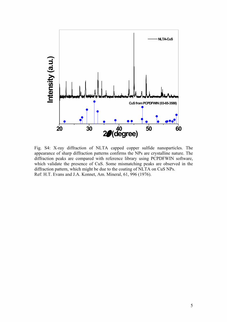
Fig. S4: X-ray diffraction of NLTA capped copper sulfide nanoparticles. The appearance of sharp diffraction patterns confirms the NPs are crystalline nature. The diffraction peaks are compared with reference library using PCPDFWIN software, which validate the presence of CuS. Some mismatching peaks are observed in the diffraction pattern, which might be due to the coating of NLTA on CuS NPs. Ref: H.T. Evans and J.A. Konnet, Am. Mineral, 61, 996 (1976).
5
20 30 40 50 60
CuS from PCPDFWIN (03-65-3588)
NLTA-CuS
Inte
nsity
(a.u
.)
B
2(degree)
100 50 25 12.5
6.25
3.13
1.56
0.78
0.39 0.2
0.0
0.2
0.4
0.6
0.8
1.0
1.2
1.4
Abso
rban
ce a
t 660
nm
CuS NPs (M)
E.coli A.hydrophilia S.aureus B.subtilis
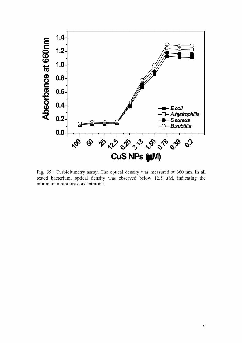
Fig. S5: Turbiditimetry assay. The optical density was measured at 660 nm. In all tested bacterium, optical density was observed below 12.5 M, indicating the minimum inhibitory concentration.
6
a
cd
bI
d
ba
c
II
d
b
c
aIII
d
b
c
aIV
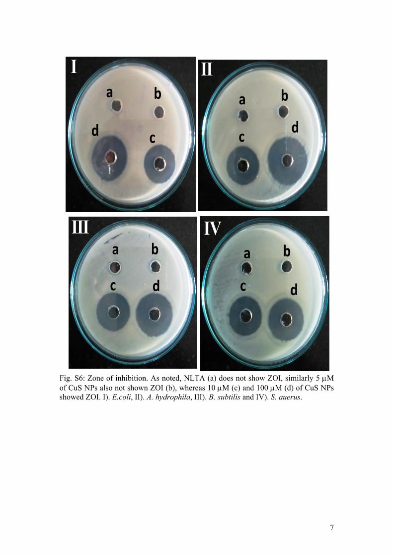
Fig. S6: Zone of inhibition. As noted, NLTA (a) does not show ZOI, similarly 5 M of CuS NPs also not shown ZOI (b), whereas 10 M (c) and 100 M (d) of CuS NPs showed ZOI. I). E.coli, II). A. hydrophila, III). B. subtilis and IV). S. auerus.
7

Fish not alive Fish not alive
Time (h) 3 6 12 24(A)
(B)
Fig. S7: Injection Method. Bacterial colonies are formed on LB-agar plate. Muscle tissues of the fish, (A) infected with E. coli and (B) treated with injection of CuS NPs. Muscle tissues obtained by sacrificing fish at definite time point was homogenized, diluted 10-4 times and plated (50 L) on LB-agar plate. After 24 h, fish treated with CuS NPs are nearly free from bacterial colonies, as a result fish survived.
8

Time (h) 3 6 12 24
untre
ated Fish not
aliveFish not
alive
E. c
oli
treat
edun
treat
ed
Fish not alive
Fish not alive
A. h
ydro
phili
a
treat
edun
treat
ed
Fish not alive
Fish not alive
S. a
ureu
s
treat
edun
treat
ed
Fish not alive
Fish not alive
B. su
btili
s
treat
ed
Fig. S8: Bacterial colony count assay. Muscle tissues of the fish, infected with bacteria and treated with injection of CuS NPs. Muscle tissues obtained by sacrificing fish at definite time point was homogenized, diluted 10-4 times and plated (50 L) on LB-agar plate. After 24 h, fishes treated with CuS NPs are nearly free from bacterial colonies, as a result fishes survived.
9
(B)(A)
NC 1 3 5 7 10 PC0
20
40
60
80
100
% M
DA p
rodu
ctio
n
CuS NPs (M)NC 1 3 5 7 10 PC
0
20
40
60
80
100
CuS NPs (M)
E. coli A. hydrophila B. Subtilis S. aureus
E. coli A. hydrophila B. Subtilis S. aureus
GSH
(% o
f -ve
con
trol)
(B)(A)
NC 1 3 5 7 10 PC0
20
40
60
80
100
% M
DA p
rodu
ctio
n
CuS NPs (M)NC 1 3 5 7 10 PC
0
20
40
60
80
100
CuS NPs (M)
E. coli A. hydrophila B. Subtilis S. aureus
E. coli A. hydrophila B. Subtilis S. aureus
GSH
(% o
f -ve
con
trol)
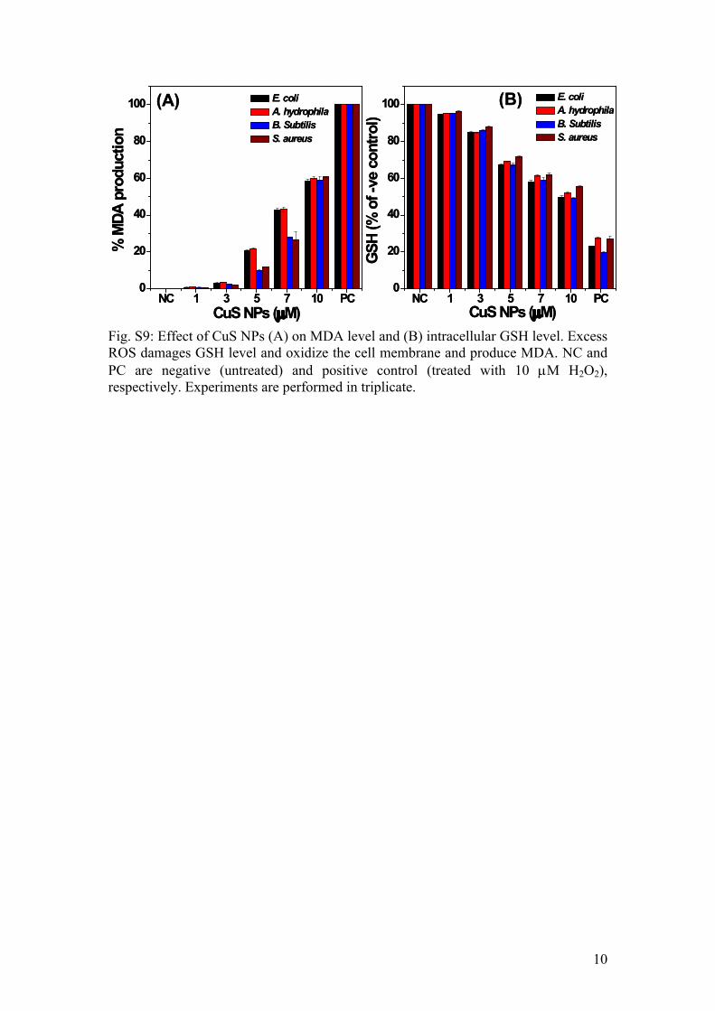
Fig. S9: Effect of CuS NPs (A) on MDA level and (B) intracellular GSH level. Excess ROS damages GSH level and oxidize the cell membrane and produce MDA. NC and PC are negative (untreated) and positive control (treated with 10 M H2O2), respectively. Experiments are performed in triplicate.
10

Time (h) 3 6 12 24(A)
(B)
Fig. S10: Viable bacterial colonies present in water (A) untreated and (B) treated with 10 M CuS NPs. Diluted water (10-4 times) was used for LB-agar plating.
11

Control 10 1000
100
200
300
-na
ptho
l rel
ease
(µ
M/m
g pr
otei
n/m
in)
[NPs] (M)
CuNPs CuS NPs
Control 10 1000
50
100
150
[NPs] (M)
-na
ptho
l rel
ease
(µ
M/m
g pr
otei
n/m
in)
(A) (B)
Fig. S11: Liver carboxylesterase activity in zebrafish. (A) -Carboxylesterase and (B) -carboxylesterase. For toxicity assessment, fish were intramuscularly injected with 10 µL CuS NPs/CuNPs. Fish exposed to CuNPs died in 120 min, therefore fish were scarified in 90 min and liver were dissected and tested for carboxylesterase activity. Fish exposed to CuS NPs was living like control, therefore fish scarified after 48 h and liver were dissected and tested for carboxylesterase activity. Fish exposed with 10 µL of PBS buffer severed as a control. Values are expressed as mean±SD of 5 determinations using liver pooled from two fish of the same treatment group. Black bar – NLTA-CuS NPs, Gray bar – NLTA-CuNPs.As noted from Fig. S11, -Carboxylesterase activity was significantly reduced when exposed to CuNPs, indicating the damage of liver. Whereas CuS NPs does not influence the activity of -Carboxylesterase and -carboxylesterase, indicating that sulfidation of Cu NPs prevents xenobiotic stress.
12
Control 10 1000
10
20
30
40
Acet
ylch
olin
e hy
drol
yzed
(µ
M /m
g pr
otei
n/m
in)
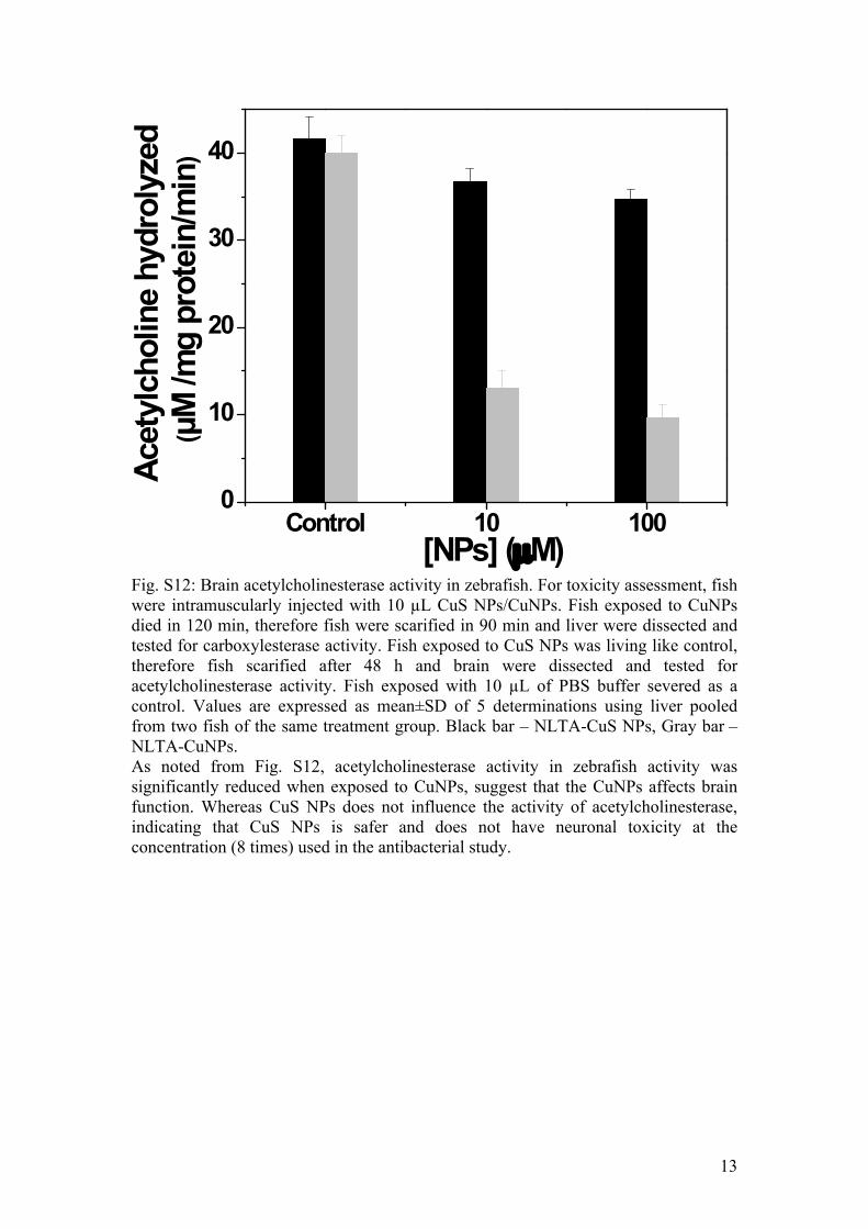
[NPs] (M)Fig. S12: Brain acetylcholinesterase activity in zebrafish. For toxicity assessment, fish were intramuscularly injected with 10 µL CuS NPs/CuNPs. Fish exposed to CuNPs died in 120 min, therefore fish were scarified in 90 min and liver were dissected and tested for carboxylesterase activity. Fish exposed to CuS NPs was living like control, therefore fish scarified after 48 h and brain were dissected and tested for acetylcholinesterase activity. Fish exposed with 10 µL of PBS buffer severed as a control. Values are expressed as mean±SD of 5 determinations using liver pooled from two fish of the same treatment group. Black bar – NLTA-CuS NPs, Gray bar – NLTA-CuNPs.As noted from Fig. S12, acetylcholinesterase activity in zebrafish activity was significantly reduced when exposed to CuNPs, suggest that the CuNPs affects brain function. Whereas CuS NPs does not influence the activity of acetylcholinesterase, indicating that CuS NPs is safer and does not have neuronal toxicity at the concentration (8 times) used in the antibacterial study.
13

400 600 8000.0
0.5
1.0
1.5
2.0
Abso
rban
ce (a
.u)
Wavelength (nm)
0 M CuS 12.5 M CuS 25 M CuS 50 M CuS 100 M CuS 100 M NH4Cl
Fig. S13: Hemolytic study. Addition of CuS NPs to RBC does not induce hemolysis, whereas NH4Cl damages the RBC as evidenced from the strong hemoglobin peaks. Peaks corresponds to hemoglobin was observed at 412 nm, 540 nm and 575 nm. RBC were treated with the materials and kept in shaker incubator (200 rpm) at 37oC. After 30 min, the reaction mixture was centrifuged and the supernatant absorbance was measured.
14