Embed Size (px)
Citation preview
Research Collection
Doctoral Thesis
Induction of phytochelatins by lead in the alga Chlamydomonasreinhardtii
Author(s): Scheidegger, Christian
Publication Date: 2010
Permanent Link: https://doi.org/10.3929/ethz-a-006376831
Rights / License: In Copyright - Non-Commercial Use Permitted
This page was generated automatically upon download from the ETH Zurich Research Collection. For moreinformation please consult the Terms of use.
ETH Library
DISS. ETH NO. 19243
Induction of phytochelatins by lead in the alga
Chlamydomonas reinhardtii
A dissertation submitted to
ETH ZURICH
for the degree of
Doctor of Sciences
Presented by
Christian Scheidegger
Master of Science in Biochemistry, University of Bern
born on March 30, 1982
Citizen of Trub (BE)
Accepted on the recommendation of
Prof. Dr. Laura Sigg, examiner
Prof. Dr. Bernhard Wehrli, co-examiner
Prof. Dr. Claude Fortin, co-examiner
Dr. Renata Behra, co-examiner
2010

II
III
Table of content
Summary ...................................................................................................................... V
Zusammenfassung ..................................................................................................... IX
1 Introduction .............................................................................................................. 1
1.1 Metals in the aquatic environment .............................................................. 3 1.1.1 Metal speciation ................................................................................................. 3 1.1.2 Metal speciation and bioavailability to algae ..................................................... 4 1.1.3 Intracellular metal homeostasis and detoxification ............................................ 7
1.2 Phytochelatins ............................................................................................. 8 1.2.1 Structure and biosynthesis ................................................................................. 8 1.2.2 Induction of phytochelatin synthesis in algae .................................................... 9 1.2.3 Role of phytochelatins in metal detoxification and formation of
metal-phytochelatin complexes ....................................................................... 11 1.3 Scope of the thesis .................................................................................... 12 1.4 References ................................................................................................. 15
2 Phytochelatin formation kinetics and toxic effects in the freshwater alga
Chlamydomonas reinhardtii upon short- and long-term exposure to lead(II) .. 23
2.1 Abstract ..................................................................................................... 25 2.2 Introduction ............................................................................................... 25 2.3 Material and Methods ............................................................................... 27
2.3.1 Chemicals ......................................................................................................... 27 2.3.2 Test organism and growth conditions .............................................................. 27 2.3.3 Experimental setup to determine short-term phytochelatin synthesis kinetics . 28 2.3.4 Experimental setup for chronic lead exposures ................................................ 28 2.3.5 Growth rate determination and photosynthetic yield measurements ................ 29 2.3.6 Cell homogenization and non protein thiol analysis ........................................ 29 2.3.7 Total and intracellular metal measurements ..................................................... 30
2.4 Results ....................................................................................................... 31 2.4.1 Total and intracellular lead in Chlamydomonas reinhardtii under lead
exposure ........................................................................................................... 31 2.4.2 Kinetics of phytochelatin synthesis in the green alga Chlamydomonas
reinhardtii as a function of free lead ions ........................................................ 32 2.4.3 Effects of lead on growth, photosynthetic yield and phytochelatin
production upon long-term exposure ............................................................... 35 2.5 Discussion ................................................................................................. 37 2.6 References ................................................................................................. 40
3 Characterization of lead induced metal-phytochelatin complexes in
Chlamydomonas reinhardtii ................................................................................... 43
3.1 Abstract ..................................................................................................... 45 3.2 Introduction ............................................................................................... 45
IV
3.3 Material and Methods ............................................................................... 47 3.3.1 Chemicals and materials .................................................................................. 47 3.3.2 Test organism, culture and experimental conditions ........................................ 47 3.3.3 Preparation of algal cell extracts ...................................................................... 48 3.3.4 Size-exclusion chromatography ....................................................................... 49 3.3.5 Metal recovery experiments ............................................................................. 50 3.3.6 Metal and thiol analysis in SEC fractions by ICP-MS and HPLC ................... 50
3.4 Results ....................................................................................................... 51 3.4.1 Calibration and recovery of size-exclusion chromatography ........................... 51 3.4.2 Peptide fractionation by size-exclusion chromatography and
fraction analysis ............................................................................................... 52 3.5 Discussion ................................................................................................. 57 3.6 References ................................................................................................. 60
4 Characterization of standard lead-phytochelatin complexes by
nano-electrospray ionization mass spectrometry ................................................ 63
4.1 Abstract ..................................................................................................... 65 4.2 Introduction ............................................................................................... 65 4.3 Material and Methods ............................................................................... 67
4.3.1 Chemicals ......................................................................................................... 67 0B4.3.2 Sample preparation .......................................................................................... 67 4.3.3 Nano-electrospray ionization mass spectrometry (nano-ESI-MS) analysis of
metal-phytochelatin complexes ....................................................................... 68 4.4 Results ....................................................................................................... 69
4.4.1 Analysis of standard Pb-PCn complexes .......................................................... 69 4.4.2 Competition between Cu or Zn and Pb for PC2 binding .................................. 76
4.5 Discussion ................................................................................................. 77 4.6 References ................................................................................................. 80
5 Outlook .................................................................................................................... 83
5.1 Outlook ..................................................................................................... 85 5.2 References ................................................................................................. 86
Acknowledgements ................................................................................................... 87 Curriculum vitae ....................................................................................................... 91

V
Summary

VI
Summary
VII
Lead is a non-essential trace metal, occurring in elevated concentrations in many
aquatic systems. Uptake of non-essential trace metals in algae leads to interactions
with macromolecules, potentially inducing toxic effects. Algae possess protective
mechanisms as a response to metal uptake and toxicity. One of these mechanisms is
the control of intracellular metal speciation by synthesis of metal-binding ligands,
such as phytochelatins. Phytochelatins (PCs) are metal-binding oligopeptides with the
general structure (γ-Glu-Cys)n-Gly (n = 2 – 11), which are enzymatically synthesized
from glutathione. Metal detoxification by PC is assumed to result through
immobilization of metals, preventing non-specific binding of metals to important
biomolecules, followed by the transport of these complexes into vacuoles. The aim of
this project was to examine induction of PC synthesis by Pb in the unicellular
freshwater alga Chlamydomonas reinhardtii as a function of the Pb speciation and to
explore the role of phytochelatins in Pb detoxification. Furthermore, Pb induced
metal-phytochelatin complexes and standard Pb-PC complexes were analyzed to
examine whether Pb is actually bound to phytochelatins.
Phytochelatin formation kinetics as function of the free Pb concentration was
examined at free Pb ion concentrations ranging from 10-11 – 10-7 M. Pb accumulation
after 6 h showed an increase of intracellular Pb with increasing [Pb2+]. PC2 – PC4
were present at low concentrations in C. reinhardtii, grown under optimal growth
conditions. Upon short-term exposure, PC2 and PC3 synthesis was induced within
minutes at [Pb2+] ≥ 10-8 M, and PC4 after a lag phase at 10-7 M. The PC2, PC3 and PC4
concentrations increased with increasing [Pb2+] and continuously over 6 h. In contrast,
upon long-term exposure, induction of PC synthesis was also detected at 10-9 M and
the production of PC with a higher degree of polymerization was observed (PC5).
Comparisons of PC concentrations and intracellular Pb content showed that PC is not
present at sufficiently high concentration to immobilize accumulated Pb. Inhibition of
photosynthesis and growth up to 100 % was observed upon long-term exposure;
whereas no Pb toxicity was observed in short-term experiments.
To examine whether Pb is bound by PCs in C. reinhardtii, metal-phytochelatin
complexes induced by Pb were isolated by size-exclusion chromatography and the
collected fractions were analyzed for their PC and metal content by HPLC and ICP-
MS. PC2 and PC3 were detected in a molecular weight range from 500 – 5300 Da,
indicating the formation of complexes with various stoichiometries. Cu, Zn and Pb
were observed in PC containing fractions, suggesting the formation of complexes with
Summary
VIII
these metals. Molecular weight considerations allowed the prediction of putative
metal-phytochelatin complex stoichiometries. The prevalent stoichiometries Me-
(PC2)2 and Me-(PC3)2 were assumed. This study suggests that PCs play a minor role
in Pb detoxification, yet from the obtained results, phytochelatins are assumed to be
involved in Cu and Zn homeostasis. As unambiguous identification of complex
stoichiometry and composition requires higher mass resolution than size-exclusion
chromatography, a nano-electrospray ionization mass spectrometry method was
developed to further characterize Me-PC complexes. Analysis of standard Pb-PC2
indicated the formation of four different complexes at m/z 746.10, 952.06, 1285.24
and 1491.20, corresponding to singly charged Pb-PC2, Pb2-PC2, Pb-(PC2)2 and Pb2-
(PC2)2. Their m/z indicated coordination of Pb through thiol groups of PC cysteine as
well as through the carboxyl groups of PC glutamic acid. For both standard PC3 and
PC4, the singly charged complexes Pb-PC3, Pb2-PC3, Pb-PC4, and Pb2-PC4 and the
twicly charged Pb-PC4 were detected. The isotopic patterns of all complexes were
identical to the theoretical patterns. Competition experiments between Zn and Pb
indicated the appearance of a singly charged Zn-PC2 peak as well as the reduction of
the Pb-PC2 peak.
The results of this work indicate that phytochelatins are involved in Zn and Cu
homeostasis rather than in Pb detoxification in C. reinhardtii at environmentally
relevant Pb concentrations. Furthermore, the study shows that toxic effects of Pb to C.
reinhardtii mainly appear upon long-term exposure, which suggests the importance of
long-term exposures to predict Pb toxicity, as C. reinhardtii is an often used model
organism.
IX
Zusammenfassung

X
Zusammenfassung
XI
Blei ist ein nicht essentielles Spurenmetall, dessen Konzentration in vielen
aquatischen Systemen erhöht ist. Die Aufnahme von nicht essentiellen
Spurenmetallen in Algen führt zu Interaktionen mit Makromolekülen und dadurch zu
Toxizität. Algen verfügen über Schutzmechanismen als Antwort auf Metallaufnahme
und -toxizität. Ein spezifischer Mechanismus ist die Kontrolle der intrazellulären
Metallspeziierung durch metallbindende Liganden, zum Beispiel Phytochelatine.
Phytochelatine (PCs) sind metallbindende Oligopeptide mit der Struktur (γ-Glu-
Cys)n-Gly (n = 2 – 11), die enzymatisch aus Glutathion synthetisiert werden. Die
vermutete Rolle von PC in der Metalldetoxifizierung erfolgt durch Immobilisierung
von Metallen, die das nicht-spezifische Binden an Biomoleküle verhindern, gefolgt
vom Transport des Komplexes in die Vakuole. Das Ziel dieses Projekts war es die
Induktion der PC Synthese durch Pb in der einzelligen Süsswasseralge
Chlamydomonas reinhardtii zu erforschen sowie die Rolle von Phytochelatinen in der
Pb-Detoxifizierung zu untersuchen. Zusätzlich wurden Pb induzierte Metall-
Phytochelatin (Me-PC) Komplexe analysiert, um festzustellen, ob Pb in diese
Komplexe gebunden ist.
Die Kinetik der Phytochelatin Synthese wurde in Funktion der freien Pb-
Konzentration ([Pb2+]) zwischen 10-11 – 10-7 M untersucht. Die Pb Akkumulation
nach 6 h Exposition zeigte einen Anstieg in der intrazellulären Konzentration mit
ansteigender [Pb2+]. PC2 – PC4 waren in C. reinhardtii, gewachsen unter optimalen
Wachstumsbedinungen, in tiefen Konzentrationen vorhanden. Unter kurzzeitiger Pb-
Exposition wurde die PC2 und PC3 Synthese innerhalb von Minuten induziert bei
[Pb2+] ≥ 10-8 M und PC4 nach einer Lagphase bei 10-7 M. Die PC2 – PC4
Konzentrationen stiegen mit steigender [Pb2+] und kontinuierlich über 6 h. In
Langzeitexperimenten hingegen wurde die Induktion der PC Synthese auch bei
10-9 M festgestellt und PC Oligomere mit höherem Polymerisierungsgrad wurden
beobachtet (PC5). Der Vergleich zwischen PC Konzentrationen und intrazellulärem
Pb zeigte, dass PC nicht in genügender Menge vorhanden ist um alles akkumulierte
Pb zu immobilisieren. Die Inhibition von Photosynthese und Wachstum bis 100 %
wurde in Langzeitexperimenten beobachtet, während in Kurzzeitexperimenten keine
toxischen Effekte von Pb auftraten.
Um herauszufinden ob Pb in C. reinhardtii von PCs gebunden wird, wurden Pb-
induzierte Me-PC Komplexe mit size-exclusion Chromatographie isoliert und die
gesammelten Fraktionen wurden mit HPLC und ICP-MS auf ihren PC- und
Zusammenfassung ______________________________________________________
XII
Metallgehalt untersucht. PC2 und PC3 wurden in einem Molekulargewichtsbereich
zwischen 500 – 5300 Da nachgewiesen, was auf die Bildung von Komplexen mit
verschiedenen Stöchiometrien hinweist. Cu, Zn und Pb wurden in denselben
Fraktionen wie PC nachgewiesen was auf Komplexbildung mit diesen Metallen
hinweist. Unter Berücksichtigung des Molekulargewichtes wurden mögliche
Komplexstöchiometrien vorhergesagt. Die am wahrscheinlichsten vorhandenen
Komplexe sind Me-(PC2)2 und Me-(PC3)2. Diese Studie deutet auf eine geringe Rolle
der PCs in der Pb-Detoxifizierung hin, aber aufgrund der erhaltenen Resultate wird
angenommen, dass PCs in der Cu und Zn Homöostase beteiligt sind. Da die
eindeutige Identifizierung der Komplexstöchiometrie und -zusammensetzung höhere
Massenauflösung als size-exclusion Chromatographie benötigt, wurde eine Nano-
Elektrospray Ionisierung Massenspektrometrie Methode zur weiteren
Charakterisierung der Me-PC Komplexes entwickelt. Die Analyse von Standard Pb-
PC2 zeigte die Bildung von vier verschiedenen Komplexen mit m/z 746.10, 952.06,
1285.24 und 1491.20, die den einfach geladenen Komplexen Pb-PC2, Pb2-PC2, Pb-
(PC2)2 und Pb2-(PC2)2 entsprechen. Die detektierten m/z wiesen auf die Koordination
von Pb durch die Thiolgruppen des PC Cystein und die Carboxylgruppen der PC
Glutaminsäure hin. Für Standard PC3 und PC4 wurden die einfach geladenen
Komplexe Pb-PC3, Pb2-PC3, Pb-PC4, und Pb2-PC4 und der zweifach geladene Pb-PC4
Komplex detektiert. Die Isotopenverteilung von allen Komplexen war identisch zu
der theoretischen Verteilung. Kompetitionsexperimente zwischen Zn und Pb zeigte
das Auftreten des einfach Zn-PC2 Peaks und die Reduktion des Pb-PC2 Peaks.
Die Resultate dieser Arbeit zeigen, dass PCs in C. reinhardtii unter umweltrelevanten
Pb-Konzentrationen eher an der Homöostase von Cu und Zn als an der Pb-
Detoxifizierung beteiligt sind. Zusätzlich wurde in dieser Studie gezeigt, dass toxische
Effekte von Pb hauptsächlich unter langzeitiger Exposition auftreten, was auf die
Wichtigkeit von Langzeitexpositionen zur Abschätzung der Pb-Toxizität hinweist da
C. reinhardtii ein oft benutzter Modelorganismus ist.

1
UChapter 1
Introduction

2
Chapter 1
3
1.1 Metals in the aquatic environment
Metals are typically released into the aquatic environment from either natural
processes (e.g. erosion of rocks, volcanic activity or dissolution from soil) or due to
anthropogenic activities, such as industrial discharge. Trace metals are divided into
two major classes: essential (e.g. Cu, Zn, Mn, Fe) and non-essential metals (e.g. Cd,
Pb, Hg) (Rand, 1995). Essential metals are important to sustain the biological
functions for aquatic organisms. However, these metals become toxic at elevated
concentrations, whereas non-essential metals may be toxic already at low
concentrations (Tessier and Turner, 1995).
Pb is a non-essential heavy metal and considered to be a major pollutant in aquatic
systems. It is introduced into the aquatic system by natural weathering processes and
from direct or indirect anthropogenic sources. In the last century, the concentrations
of Pb in natural waters increased drastically until mid 1960s or early 1970s
concomitant with the increased use of leaded gasoline. The limitation of the Pb
concentrations in gasoline led to a continuous decrease of environmental
concentrations in the last decades (Nriagu, 1990). The main sources besides the use
leaded gasoline are fertilizers, dissolution from soils, mining and smelting of metallic
ores (Sharma and Dubey, 2005). In pristine mountain streams with only atmospheric
deposition of Pb, background concentrations as low as 10-12 M were reported (Erel et
al., 1991). Surface waters in more populated areas show Pb concentrations up to 10-7
M (Warnken et al., 2009). The bioavailability, and hence uptake and effects of
elevated Pb concentrations, is mainly dependent on the speciation of Pb in the water
(Morel and Hering, 1993; Tessier and Turner, 1995).
1.1.1 Metal speciation
In natural waters, metals occur in a variety of chemical forms or species, primarily
complexed by ligands, in particulate form, or as free metal ions (Buffle, 1988; Sigg
and Xue, 1994). Both inorganic and organic ligands bind metals, resulting in
dissolved metal-ligand complexes. Inorganic ligands include water, hydroxide,
carbonate and chloride (Sigg and Stumm, 1989; Sigg and Behra, 2005). Natural
organic ligands are present in a wide range of low and high molecular weight
compounds. The small organic ligands include carboxylic acids (citrate, acetate,
oxalate and malonate), amino acids, phenols and catechols, which result from
Chapter 1
4
decomposition of organic matter or are excreted by organisms. The most important
larger ligands are humic and fulvic acids, which have binding sites for metals such as
phenolic, carboxylic, nitrogen and sulfur containing groups. Other ligands include
synthetic organic ligands like EDTA and NTA that are introduced into the aquatic
environment by anthropogenic activities. The bulk particulate matter consists of
oxides and hydroxides of iron, manganese, aluminum, silicates, clay minerals,
carbonates and organic matter (bacteria, algae, organic debris) (Sigg and Behra,
2005). Trace metals may be bound by adsorption to functional groups on the surface
of these particulate phases.
As a consequence in natural waters metal species with different complexing
properties, with respect to kinetic lability and thermodynamic stability, are present.
Kinetic lability allows the distinction between labile and “inert” complexes based on
the dissociation rate of metals from the complexes. Compared to “inert” complexes,
which have high stability constants and low dissociation rates, labile complexes have
lower stability constants and a higher dissociation rates. These complexes are
operationally defined and therefore their categorization depends on the method used
to determine stability constants and dissociation rates. Metal speciation can either be
measured with various speciation techniques or it can be estimated with chemical
equilibrium models.
1.1.2 Metal speciation and bioavailability to algae
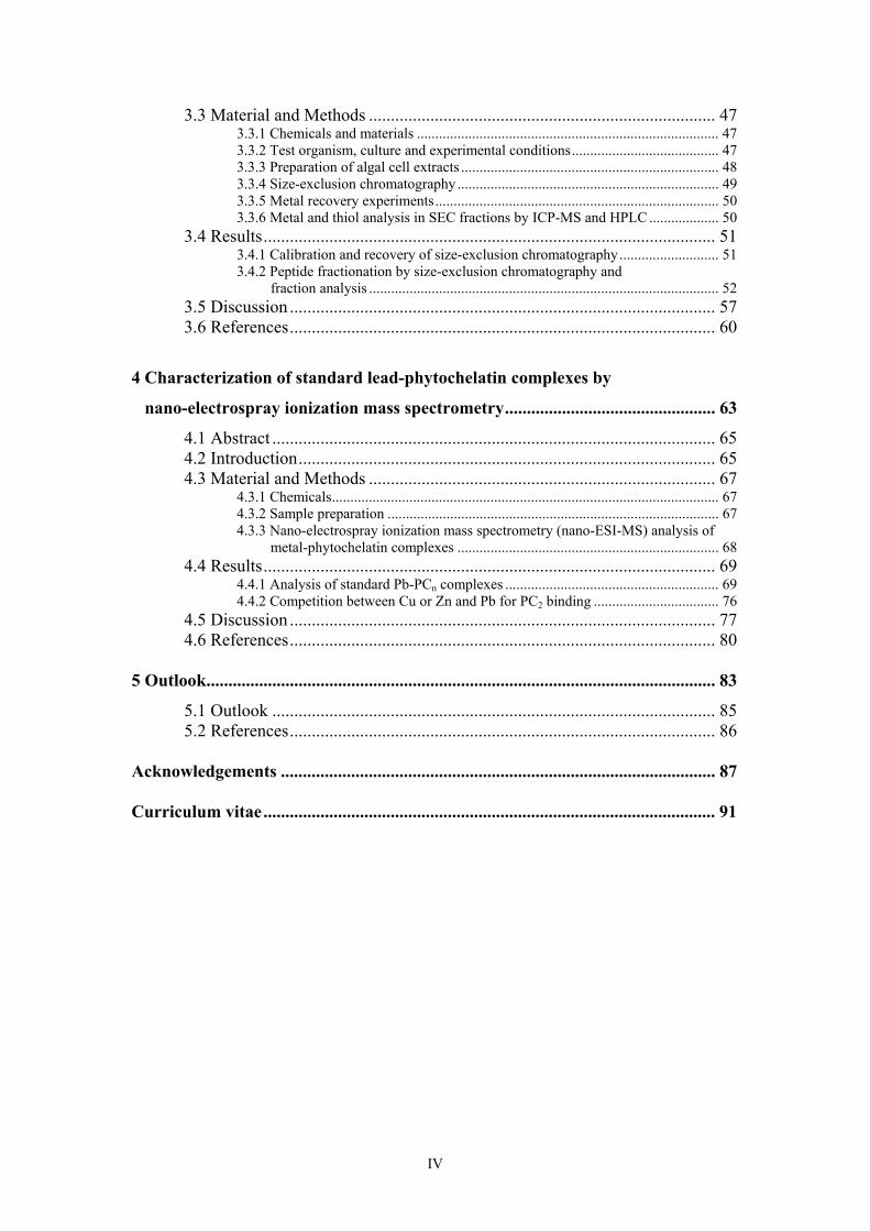
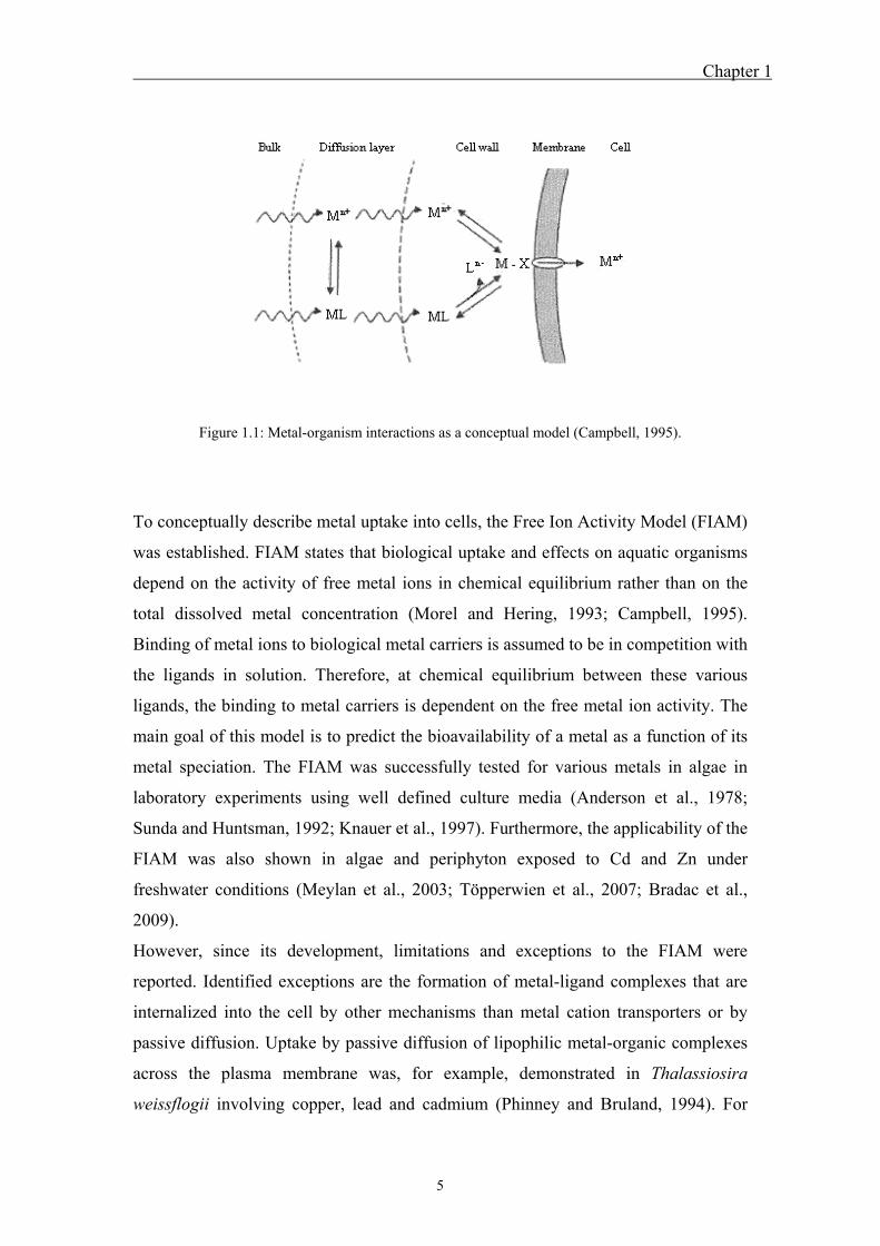
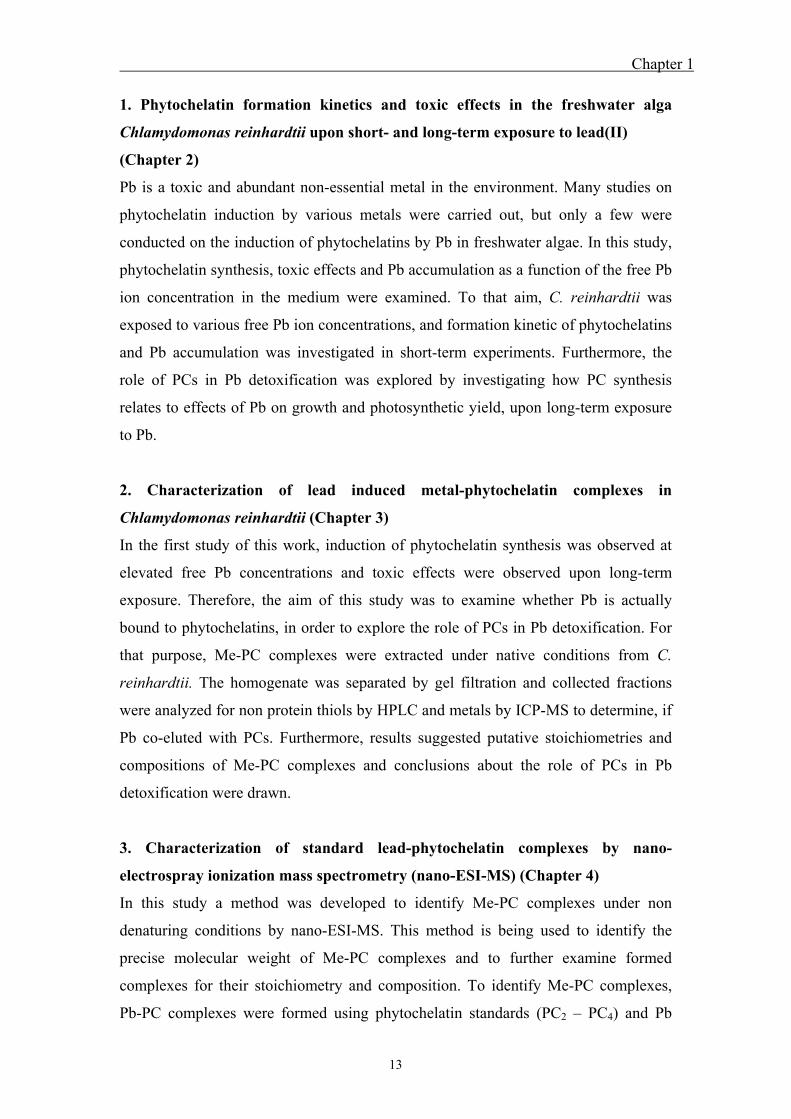
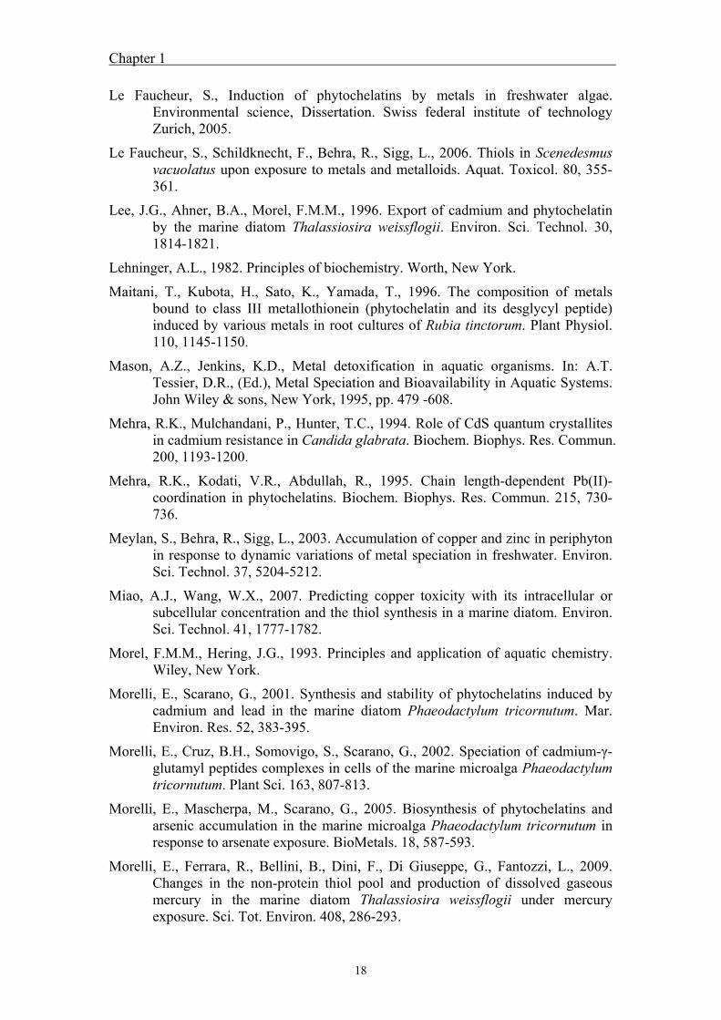
Metal uptake into algae generally includes three steps (Tessier and Turner, 1995;
Campbell et al., 2002). In the first step, the free metal ion or metal-ligand complex
diffuses through the diffusion layer and the cell wall to the plasma membrane. At the
plasma membrane free metal ions bind very fast to transport sites of the membrane,
following chemical equilibrium. The fast adsorption is followed by the slower, and
uptake limiting, transport through the plasma membrane (Fig. 1.1). The transport can
be mediated by transmembrane proteins, such as ion channels, which allow diffusion
along an electrochemical gradient, or ion transporters, which are energy dependent.
Once inside the cell, metals are either used in metabolism or accumulated in the algal
cell (Tessier and Turner, 1995).
Chapter 1
5
Figure 1.1: Metal-organism interactions as a conceptual model (Campbell, 1995).
To conceptually describe metal uptake into cells, the Free Ion Activity Model (FIAM)
was established. FIAM states that biological uptake and effects on aquatic organisms
depend on the activity of free metal ions in chemical equilibrium rather than on the
total dissolved metal concentration (Morel and Hering, 1993; Campbell, 1995).
Binding of metal ions to biological metal carriers is assumed to be in competition with
the ligands in solution. Therefore, at chemical equilibrium between these various
ligands, the binding to metal carriers is dependent on the free metal ion activity. The
main goal of this model is to predict the bioavailability of a metal as a function of its
metal speciation. The FIAM was successfully tested for various metals in algae in
laboratory experiments using well defined culture media (Anderson et al., 1978;
Sunda and Huntsman, 1992; Knauer et al., 1997). Furthermore, the applicability of the
FIAM was also shown in algae and periphyton exposed to Cd and Zn under
freshwater conditions (Meylan et al., 2003; Töpperwien et al., 2007; Bradac et al.,
2009).
However, since its development, limitations and exceptions to the FIAM were
reported. Identified exceptions are the formation of metal-ligand complexes that are
internalized into the cell by other mechanisms than metal cation transporters or by
passive diffusion. Uptake by passive diffusion of lipophilic metal-organic complexes
across the plasma membrane was, for example, demonstrated in Thalassiosira
weissflogii involving copper, lead and cadmium (Phinney and Bruland, 1994). For
Chapter 1
6
silver, increased accumulation was detected in presence of thiosulfate. It was
concluded that silver-thiosulfate complexes are transported across the plasma
membrane via anion transporters responsible for thiolsulfate / sulfate transport (Fortin
and Campbell, 2001). Similarly, enhanced uptake of cadmium and zinc as metal-
citrate complex were reported (Errécalde et al., 1998; Errécalde and Campbell, 2000).
Pb uptake into algae was observed to be mainly governed by the free ion
concentration, following the FIAM in the presence of citrate and malonate
(Slaveykova and Wilkinson, 2002). In presence of natural organic matter, such as
humic acid and fulvic acid, however, Pb uptake increased compared to the amount
predicted by FIAM. It was suggested that changes in the algal surface charge through
binding of fulvic acid (Slaveykova et al., 2003) or the formation of a ternary complex,
between the Pb-humic acid complex and the internalization sites on the algal surface,
might be responsible for the enhanced Pb uptake (Lamelas et al., 2005; Lamelas and
Slaveykova, 2007; Lamelas et al., 2009).
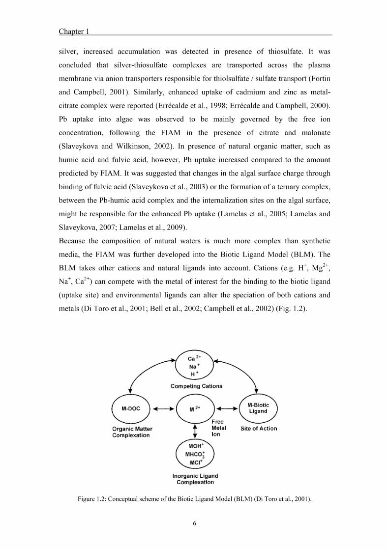
Because the composition of natural waters is much more complex than synthetic
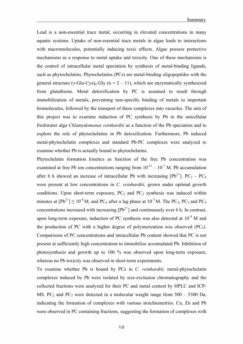
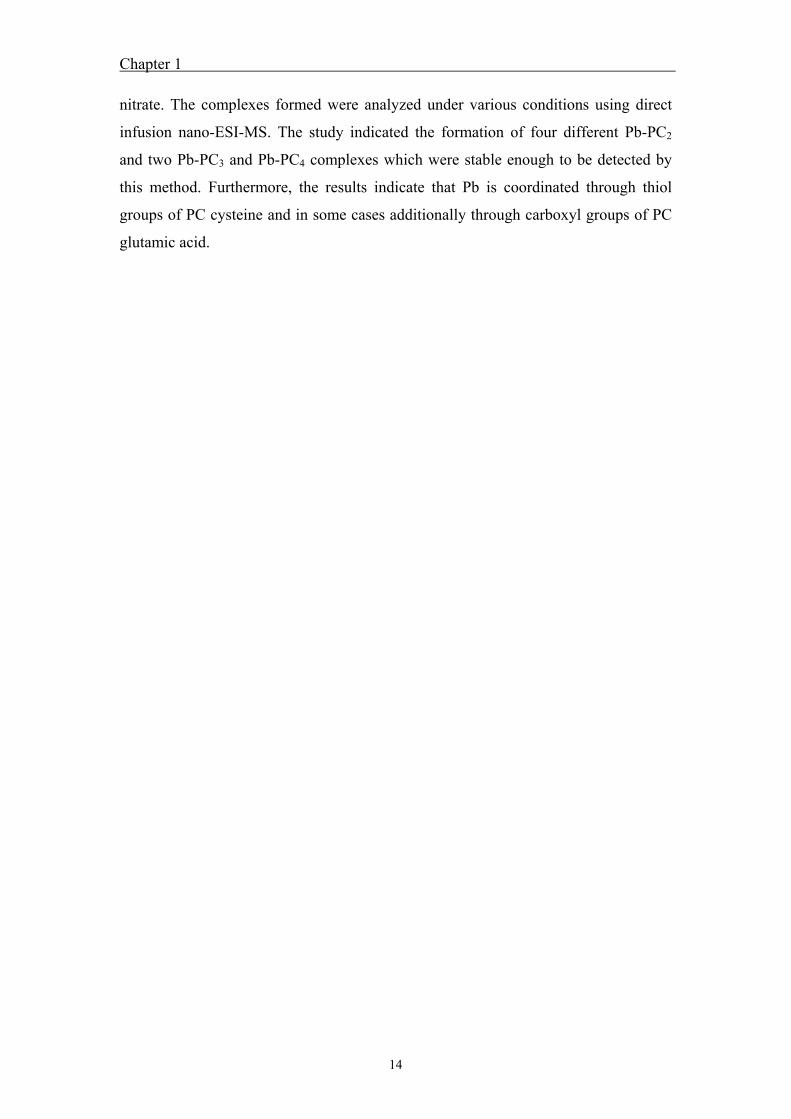
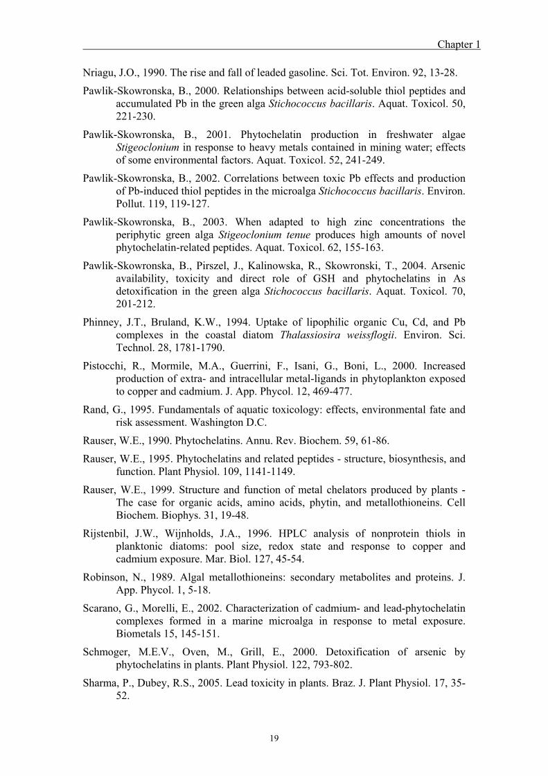
media, the FIAM was further developed into the Biotic Ligand Model (BLM). The
BLM takes other cations and natural ligands into account. Cations (e.g. H+, Mg2+,
Na+, Ca2+) can compete with the metal of interest for the binding to the biotic ligand
(uptake site) and environmental ligands can alter the speciation of both cations and
metals (Di Toro et al., 2001; Bell et al., 2002; Campbell et al., 2002) (Fig. 1.2).
Figure 1.2: Conceptual scheme of the Biotic Ligand Model (BLM) (Di Toro et al., 2001).
Chapter 1
7
Both FIAM and BLM are models based on the assumption of chemical equilibrium
and do not consider kinetic effects. They assume equilibrium between free metal ion
concentration in solution and metal adsorbed to the biotic ligand and further, that
diffusion of metal species to the cell membrane is faster than internalization. In
contrast, non-equilibrium based models consider diffusion and internalization fluxes
(van Leeuwen, 1999; Slaveykova and Wilkinson, 2005; van Leeuwen et al., 2005). If
diffusion to the cell membrane is rate limiting, metal uptake is controlled by the
concentration of chemically labile species, which have low stability and high
dissociation rate (Hudson, 1998; van Leeuwen, 1999; Slaveykova and Wilkinson,
2005). This was confirmed for silver uptake in Chlamydomonas reinhardtii (Fortin
and Campbell, 2000) and for copper and cadmium uptake in periphyton under
freshwater conditions (Meylan et al., 2003; Bradac et al., 2009).
1.1.3 Intracellular metal homeostasis and detoxification
The need for essential trace metals in organisms is linked to their function; they are
essential as cofactor of enzymes, for electron transport, for catalysis of redox
reactions and for the maintenance of conformations and tertiary structures of proteins
(Lehninger, 1982). Aquatic organisms have specific requirements in terms of trace
metal concentrations to sustain vital processes and optimal development.
Nevertheless, these trace metals become toxic for organisms at concentrations above
the requirement or deficiencies arise at too low concentrations. In contrast, non-
essential metals are not used in the metabolism and already toxic at low
concentrations.
Therefore, intracellular metal concentrations are tightly regulated in order to maintain
physiological functions and to prevent toxic effects. Once metals are inside the cell,
they are rapidly bound by ligands, such as chaperones and chelators (Mason and
Jenkins, 1995; Clemens, 2001). Essential metals are bound by chaperones that are
involved in metal trafficking and deliver them to specific organelles and metal-
requiring proteins. Uptake into organelles is catalyzed by metal transporters that
directly interact with specific chaperones. To store metals or to prevent toxic effects
of essential and non-essential metals, metal ions can be directly sequestered into the
vacuole or bound to chelators (metallothioneins, phytochelatins, glutathione, organic
acids, amino acids), which can be also transported into vacuoles or excreted from the
cell (Lee et al., 1996; Clemens, 2001).
Chapter 1
8
The Subcellular Partitioning Model (SPM) has been developed to predict metal
toxicity to aquatic organisms, such as algae (Wang and Rainbow, 2006). It assumes
that toxicity depends on the distribution of metal among different operationally
defined subcellular fractions. It is hypothesized in SPM that elevated metal
concentrations saturate detoxifying ligands, leading to a redistribution of metals to
sensitive ligands, resulting in toxicity (Wang and Wang, 2008). A few recent studies
have tried to relate subcellular metal distribution to toxicity in marine phytoplankton
(Miao and Wang, 2007; Wang and Wang, 2008) and freshwater species (Lavoie et al.,
2009).
The toxicity of Pb is assumed to arise from non-specific binding to important
biomolecules, due to the high affinity of Pb to oxygen, nitrogen and sulfur functional
groups (Sharma and Dubey, 2005). Therefore, it can substitute essential metal ions or
directly bind to functional groups. Binding to functional groups can have an effect on
enzyme activity, by reducing or suppressing its biological activity. In addition, Pb can
affect photosynthesis by inhibition of chlorophyll synthesis due to impaired uptake of
Mg and Fe and by inhibition of electron transport in photosystem II (Sharma and
Dubey, 2005). Pb can also cause toxicity by production of reactive oxygen species
(Szivak et al., 2009). Due to the high affinity of Pb to various functional groups, Pb is
bound to various ligands, including metal detoxifying ligands and metal sensitive
ligands. To explore the mechanisms involved in Pb detoxification, Pb distribution
among the different operationally defined subcellular fractions have to be examined.
1.2 Phytochelatins
1.2.1 Structure and biosynthesis
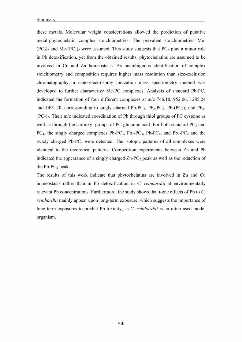
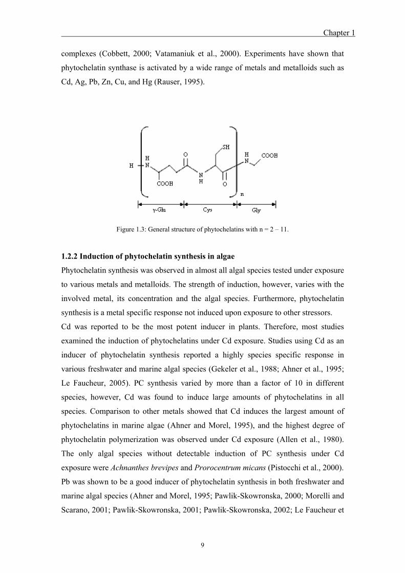
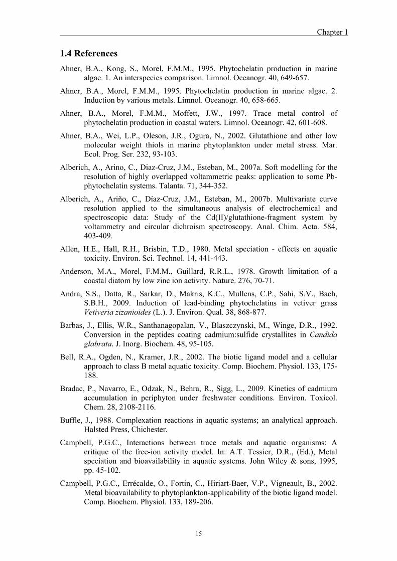
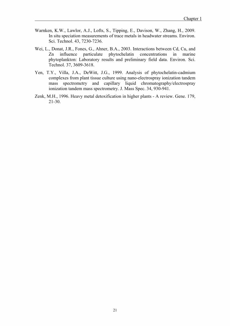
Phytochelatins (PCs) are heat stable, metal-binding polypeptides with the general
structure (γ-Glu-Cys)n-Gly where n = 2 – 11 (Robinson, 1989; Rauser, 1990; Steffens,
1990; Cobbett, 2000) (Fig. 1.3). On the basis of the number of -Glu-Cys units, PCs
have been classified as PC2, PC3, PC4 etc (Grill et al., 1985; Rauser, 1995). PCs are
synthesized upon elevated metal concentrations in a wide variety of plants (Rauser,
1995; Zenk, 1996) and algae (Gekeler et al., 1988; Ahner et al., 1995). PC is
synthesized enzymatically by the addition of glutathione (GSH) to (γ-Glu-Cys)n by
the phytochelatin synthase to produce a n + 1 oligomer. The activation of
phytochelatin synthase occurs by the binding of metal ions and metal-glutathione
Chapter 1
9
complexes (Cobbett, 2000; Vatamaniuk et al., 2000). Experiments have shown that
phytochelatin synthase is activated by a wide range of metals and metalloids such as
Cd, Ag, Pb, Zn, Cu, and Hg (Rauser, 1995).
Figure 1.3: General structure of phytochelatins with n = 2 – 11.
1.2.2 Induction of phytochelatin synthesis in algae
Phytochelatin synthesis was observed in almost all algal species tested under exposure
to various metals and metalloids. The strength of induction, however, varies with the
involved metal, its concentration and the algal species. Furthermore, phytochelatin
synthesis is a metal specific response not induced upon exposure to other stressors.
Cd was reported to be the most potent inducer in plants. Therefore, most studies
examined the induction of phytochelatins under Cd exposure. Studies using Cd as an
inducer of phytochelatin synthesis reported a highly species specific response in
various freshwater and marine algal species (Gekeler et al., 1988; Ahner et al., 1995;
Le Faucheur, 2005). PC synthesis varied by more than a factor of 10 in different
species, however, Cd was found to induce large amounts of phytochelatins in all
species. Comparison to other metals showed that Cd induces the largest amount of
phytochelatins in marine algae (Ahner and Morel, 1995), and the highest degree of
phytochelatin polymerization was observed under Cd exposure (Allen et al., 1980).
The only algal species without detectable induction of PC synthesis under Cd
exposure were Achnanthes brevipes and Prorocentrum micans (Pistocchi et al., 2000).
Pb was shown to be a good inducer of phytochelatin synthesis in both freshwater and
marine algal species (Ahner and Morel, 1995; Pawlik-Skowronska, 2000; Morelli and
Scarano, 2001; Pawlik-Skowronska, 2001; Pawlik-Skowronska, 2002; Le Faucheur et
Chapter 1
10
al., 2006). Under Pb exposure mainly short chain phytochelatins such as PC2 and PC3
were observed and the highest degree of polymerization detected was PC6 in
Phaeodactylum tricornutum (Morelli and Scarano, 2001). Furthermore, time
dependence of PC induction was examined and revealed that shorter chain oligomers
are induced first, followed by longer chain oligomers. In addition, fast degradation of
PC oligomers when transferred to metal free medium was observed (Morelli and
Scarano, 2001; Pawlik-Skowronska, 2001).
Examination of PC synthesis under Cu exposure was mainly examined in marine
algae and reported induction of PC2 – PC4 (Ahner and Morel, 1995; Rijstenbil and
Wijnholds, 1996; Ahner et al., 1997; Ahner et al., 2002; Wei et al., 2003). Only in one
freshwater alga phytochelatins were quantified under Cu exposure, showing an
increase of total PCn from 1 – 2 µmol / g chl a at pCu15 to a maximum 20 µmol / g
chl a at pCu9 (Knauer et al., 1997).
PC synthesis induced by Zn was extensively studied in various Stigeoclonium species
(Pawlik-Skowronska, 2001; Pawlik-Skowronska, 2003). For various strains, similar
amounts of PCs were quantified, but the tolerant species produced a novel peptide,
differing from phytochelatins by an additional cysteine. In T. weissfolgii, PC content
was found to decrease under Zn limiting conditions, whereas high Zn concentrations
did not lead to increased PC production (Ahner and Morel, 1995). In addition, other
species only showed induction of PC synthesis at very high Zn concentrations (pZn
7.2 in Skeletonema costatum) (Ahner and Morel, 1997) or no induction in Chlorella
kesslerii (Hassler et al., 2005).
Studies on induction of PC synthesis by other metals are scarce. Low PC2
concentrations were observed upon Ag exposure in Pseudokirchneriella Subcapitata
(Hiriart-Baer et al., 2006), but no induction of PCs was detected in Scenedesmus
vacuolatus, C. reinhardtii and T. weissflogii (Howe and Merchant, 1992; Ahner and
Morel, 1995; Le Faucheur et al., 2006). PCn formation was slightly induced in
Chlorella fusca by Co and Ni (Gekeler et al., 1988). Hg was found to induce PC
synthesis in T. weissflogii (Ahner and Morel, 1995; Morelli et al., 2009) but not in C.
reinhardtii (Howe and Merchant, 1992). Furthermore, induction of PC synthesis was
reported upon exposure to the metalloids As(V), As(III) and Sb(III) in the freshwater
species S. vacuolatus (Pawlik-Skowronska et al., 2004; Morelli et al., 2005; Le
Faucheur et al., 2006) and marine algae (Pawlik-Skowronska et al., 2004; Morelli et
al., 2005).
Chapter 1
11
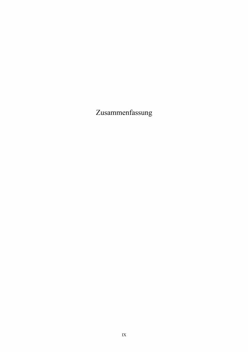
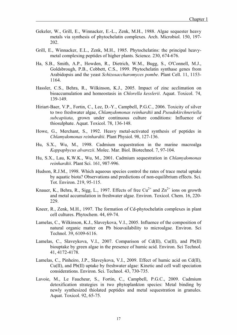
1.2.3 Role of phytochelatins in metal detoxification and formation of metal-
phytochelatin complexes
First evidence on the role of PC in metal detoxification were obtained by using yeast
(Schizosaccharomyces pombe and Saccharomyces cerevisiae) and plant mutants
(Arabidopsis spp.) that were deficient in PC synthase genes (Clemens et al., 1999; Ha
et al., 1999). Indeed, these species were more sensitive to Cd, As(V) and Cu than the
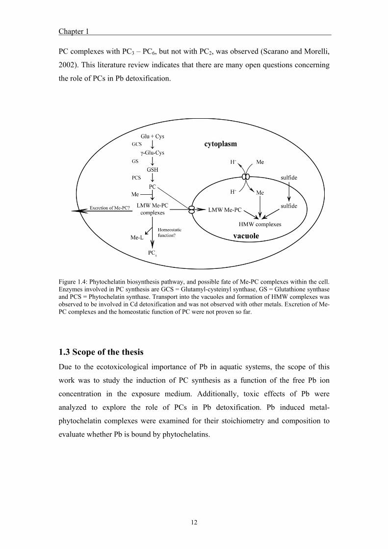
wild-type. It is assumed, that the role of PC in metal detoxification is the formation of
a metal-phytochelatin (Me-PC) complex, used to avoid metal binding to other target
sites, followed by its transport into vacuoles (Fig. 1.4). Formation of Me-PC
complexes was detected in vitro for Cd, Pb, Ag, Cu, As(V), As(III) and Hg (Mehra et
al., 1995; Maitani et al., 1996; Rauser, 1999; Schmoger et al., 2000). In more recent
studies in vitro Me-PC complexes formed with Pb, Cd, Zn and Hg were analyzed by
differential pulse polarography and voltammetry reporting the formation of Me-PC
complexes with various stoichiometries for the examined metals (Chekmeneva et al.,
2008; Cruz et al., 2001; Cruz et al., 2005; Alberich et al., 2007a; Alberich et al.,
2007b; Chekmeneva et al., 2007). It was found that PCs interact with metals through
the thiol (-SH) group of PC cysteine. It was reported that an increase in the degree of
polymerization of PCs led to increased binding stability of Me-PC complexes (Mehra
et al., 1995).
Most studies in plants (Kneer and Zenk, 1997; Yen et al., 1999; Chen et al., 2007) and
algae (Hu and Wu, 1998; Hu et al., 2001; Morelli et al., 2002; Scarano and Morelli,
2002) were carried out examining Cd-PC complexes. These studies revealed the
presence of two kinds of complexes, low molecular weight complexes (LMW) and
high molecular weight complexes (HMW). LMW complexes are defined as
complexes formed by Cd (or other metals) bound to a single phytochelatin oligomer.
The composition of HMW complexes was found to be from the formation of a CdS
core, generated by Cd and acid labile sulfur (S-), surrounded by PCs (mainly PC2 and
PC3). The role of PC coating is to stabilize the CdS core. The presence of HMW
complexes increases metal tolerance, as shown in the yeast Candida glabrata, where a
strain with enhanced CdS production revealed increased Cd resistance compared to
the wildtype (Barbas et al., 1992; Mehra et al., 1994).
Complexes identified in vivo were mainly Cd-PC complexes. Studies on Me-PC
complexes induced by Pb in vivo are very scarce in plants (Andra et al., 2009) and
algae (Scarano and Morelli, 2002). In the marine alga P. tricornutum formation of Pb-
Chapter 1
12
PC complexes with PC3 – PC6, but not with PC2, was observed (Scarano and Morelli,
2002). This literature review indicates that there are many open questions concerning
the role of PCs in Pb detoxification.
Figure 1.4: Phytochelatin biosynthesis pathway, and possible fate of Me-PC complexes within the cell. Enzymes involved in PC synthesis are GCS = Glutamyl-cysteinyl synthase, GS = Glutathione synthase and PCS = Phytochelatin synthase. Transport into the vacuoles and formation of HMW complexes was observed to be involved in Cd detoxification and was not observed with other metals. Excretion of Me-PC complexes and the homeostatic function of PC were not proven so far.
1.3 Scope of the thesis
Due to the ecotoxicological importance of Pb in aquatic systems, the scope of this
work was to study the induction of PC synthesis as a function of the free Pb ion
concentration in the exposure medium. Additionally, toxic effects of Pb were
analyzed to explore the role of PCs in Pb detoxification. Pb induced metal-
phytochelatin complexes were examined for their stoichiometry and composition to
evaluate whether Pb is bound by phytochelatins.
Chapter 1
13
1. Phytochelatin formation kinetics and toxic effects in the freshwater alga
Chlamydomonas reinhardtii upon short- and long-term exposure to lead(II)
(Chapter 2)
Pb is a toxic and abundant non-essential metal in the environment. Many studies on
phytochelatin induction by various metals were carried out, but only a few were
conducted on the induction of phytochelatins by Pb in freshwater algae. In this study,
phytochelatin synthesis, toxic effects and Pb accumulation as a function of the free Pb
ion concentration in the medium were examined. To that aim, C. reinhardtii was
exposed to various free Pb ion concentrations, and formation kinetic of phytochelatins
and Pb accumulation was investigated in short-term experiments. Furthermore, the
role of PCs in Pb detoxification was explored by investigating how PC synthesis
relates to effects of Pb on growth and photosynthetic yield, upon long-term exposure
to Pb.
2. Characterization of lead induced metal-phytochelatin complexes in
Chlamydomonas reinhardtii (Chapter 3)
In the first study of this work, induction of phytochelatin synthesis was observed at
elevated free Pb concentrations and toxic effects were observed upon long-term
exposure. Therefore, the aim of this study was to examine whether Pb is actually
bound to phytochelatins, in order to explore the role of PCs in Pb detoxification. For
that purpose, Me-PC complexes were extracted under native conditions from C.
reinhardtii. The homogenate was separated by gel filtration and collected fractions
were analyzed for non protein thiols by HPLC and metals by ICP-MS to determine, if
Pb co-eluted with PCs. Furthermore, results suggested putative stoichiometries and
compositions of Me-PC complexes and conclusions about the role of PCs in Pb
detoxification were drawn.
3. Characterization of standard lead-phytochelatin complexes by nano-
electrospray ionization mass spectrometry (nano-ESI-MS) (Chapter 4)
In this study a method was developed to identify Me-PC complexes under non
denaturing conditions by nano-ESI-MS. This method is being used to identify the
precise molecular weight of Me-PC complexes and to further examine formed
complexes for their stoichiometry and composition. To identify Me-PC complexes,
Pb-PC complexes were formed using phytochelatin standards (PC2 – PC4) and Pb
Chapter 1
14
nitrate. The complexes formed were analyzed under various conditions using direct
infusion nano-ESI-MS. The study indicated the formation of four different Pb-PC2
and two Pb-PC3 and Pb-PC4 complexes which were stable enough to be detected by
this method. Furthermore, the results indicate that Pb is coordinated through thiol
groups of PC cysteine and in some cases additionally through carboxyl groups of PC
glutamic acid.
Chapter 1
15
1.4 References
Ahner, B.A., Kong, S., Morel, F.M.M., 1995. Phytochelatin production in marine algae. 1. An interspecies comparison. Limnol. Oceanogr. 40, 649-657.
Ahner, B.A., Morel, F.M.M., 1995. Phytochelatin production in marine algae. 2. Induction by various metals. Limnol. Oceanogr. 40, 658-665.
Ahner, B.A., Morel, F.M.M., Moffett, J.W., 1997. Trace metal control of phytochelatin production in coastal waters. Limnol. Oceanogr. 42, 601-608.
Ahner, B.A., Wei, L.P., Oleson, J.R., Ogura, N., 2002. Glutathione and other low molecular weight thiols in marine phytoplankton under metal stress. Mar. Ecol. Prog. Ser. 232, 93-103.
Alberich, A., Arino, C., Diaz-Cruz, J.M., Esteban, M., 2007a. Soft modelling for the resolution of highly overlapped voltammetric peaks: application to some Pb-phytochelatin systems. Talanta. 71, 344-352.
Alberich, A., Ariño, C., Díaz-Cruz, J.M., Esteban, M., 2007b. Multivariate curve resolution applied to the simultaneous analysis of electrochemical and spectroscopic data: Study of the Cd(II)/glutathione-fragment system by voltammetry and circular dichroism spectroscopy. Anal. Chim. Acta. 584, 403-409.
Allen, H.E., Hall, R.H., Brisbin, T.D., 1980. Metal speciation - effects on aquatic toxicity. Environ. Sci. Technol. 14, 441-443.
Anderson, M.A., Morel, F.M.M., Guillard, R.R.L., 1978. Growth limitation of a coastal diatom by low zinc ion activity. Nature. 276, 70-71.
Andra, S.S., Datta, R., Sarkar, D., Makris, K.C., Mullens, C.P., Sahi, S.V., Bach, S.B.H., 2009. Induction of lead-binding phytochelatins in vetiver grass Vetiveria zizanioides (L.). J. Environ. Qual. 38, 868-877.
Barbas, J., Ellis, W.R., Santhanagopalan, V., Blaszczynski, M., Winge, D.R., 1992. Conversion in the peptides coating cadmium:sulfide crystallites in Candida glabrata. J. Inorg. Biochem. 48, 95-105.
Bell, R.A., Ogden, N., Kramer, J.R., 2002. The biotic ligand model and a cellular approach to class B metal aquatic toxicity. Comp. Biochem. Physiol. 133, 175-188.
Bradac, P., Navarro, E., Odzak, N., Behra, R., Sigg, L., 2009. Kinetics of cadmium accumulation in periphyton under freshwater conditions. Environ. Toxicol. Chem. 28, 2108-2116.
Buffle, J., 1988. Complexation reactions in aquatic systems; an analytical approach. Halsted Press, Chichester.
Campbell, P.G.C., Interactions between trace metals and aquatic organisms: A critique of the free-ion activity model. In: A.T. Tessier, D.R., (Ed.), Metal speciation and bioavailability in aquatic systems. John Wiley & sons, 1995, pp. 45-102.
Campbell, P.G.C., Errécalde, O., Fortin, C., Hiriart-Baer, V.P., Vigneault, B., 2002. Metal bioavailability to phytoplankton-applicability of the biotic ligand model. Comp. Biochem. Physiol. 133, 189-206.

Chapter 1
16
Chekmeneva, E., Prohens, R., Diaz-Cruz, J.M., Arino, C., Esteban, M., 2008, Thermodynamics of Cd2+ and Zn2+ binding by the phytochelatin (γ-Glu-Cys)4-Gly and its precursor glutathione. Anal. Biochem. 375, 82-89
Chekmeneva, E., Diaz-Cruz, J.M., Arino, C., Esteban, M., 2007. Binding of Cd2+ and Zn2+ with the phytochelatin (γ-Glu-Cys)4-Gly: a voltammetric study assisted by multivariate curve resolution and electrospray ionization mass spectrometry. Electroanal. 19, 310-317.
Chen, L., Guo, Y., Yang, L., Wang, Q., 2007. SEC-ICP-MS and ESI-MS/MS for analyzing in vitro and in vivo Cd-phytochelatin complexes in a Cd-hyperaccumulator Brassica chinensis. J. Anal. Atom. Spec. 22, 1403-1408.
Clemens, S., Kim, E.J., Neumann, D., Schroeder, J.I., 1999. Tolerance to toxic metals by a gene family of phytochelatin synthases from plants and yeast. EMBO J. 18, 3325-3333.
Clemens, S., 2001. Molecular mechanisms of plant metal tolerance and homeostasis. Planta. 212, 475-486.
Cobbett, C.S., 2000. Phytochelatins and their roles in heavy metal detoxification. Plant Physiol. 123, 825-832.
Cruz, B.H., Díaz-Cruz, J.M., Díaz-Cruz, M.S., Ariño, C., Esteban, M., Tauler, R., 2001. Differential pulse polarographic study of the Pb(II) complexation by glutathione. J. Electroanal. Chem. 516, 110-118.
Cruz, B.H., Diaz-Cruz, J.M., Arino, C., Esteban, M., 2005. Complexation of heavy metals by phytochelatins: voltammetric study of the binding of Cd2+ and Zn2+ ions by the phytochelatin (γ-Glu-Cys)3Gly assisted by multivariate curve resolution. Environ. Sci. Technol. 39, 778-786.
Di Toro, D.M., Allen, H.E., Bergman, H.L., Meyer, J.S., Paquin, P.R., Santore, R.C., 2001. Biotic ligand model of the acute toxicity of metals. 1. technical basis. Envrion. Toxicol. Chem. 20, 2383-2396.
Erel, Y., Morgan, J.J., Patterson, C.C., 1991. Natural levels of Pb and Cd in a remote mountain stream. Geochim. Cosmochim. Acta. 55, 707-719.
Errécalde, O., Seidl, M., Campbell, P.G.C., 1998. Influence of a low molecular weight metabolite (citrate) on the toxicity of cadmium and zinc to the unicellular green alga Selenastrum Capricornutum: An exception to the free-ion model. Water Res. 32, 419-429.
Errécalde, O., Campbell, P.G.C., 2000. Cadmium and zinc bioavailability to Selenastrum capricornutum (chlorophyceae): Accidental metal uptake and toxicity in the presence of citrate. J. Phycol. 36, 473-483.
Fortin, C., Campbell, P.G.C., 2000. Silver uptake by the green alga Chlamydomonas reinhardtii in relation to chemical speciation: Influence of chloride. Environ. Toxicol. Chem. 19, 2769-2778.
Fortin, C., Campbell, P.G.C., 2001. Thiosulfate enhances silver uptake by a green alga: Role of anion transporters in metal uptake. Environ. Sci. Technol. 35, 2214-2218.
Chapter 1
17
Gekeler, W., Grill, E., Winnacker, E.-L., Zenk, M.H., 1988. Algae sequester heavy metals via synthesis of phytochelatin complexes. Arch. Microbiol. 150, 197-202.
Grill, E., Winnacker, E.L., Zenk, M.H., 1985. Phytochelatins: the principal heavy-metal complexing peptides of higher plants. Science. 230, 674-676.
Ha, S.B., Smith, A.P., Howden, R., Dietrich, W.M., Bugg, S., O'Connell, M.J., Goldsbrough, P.B., Cobbett, C.S., 1999. Phytochelatin synthase genes from Arabidopsis and the yeast Schizosaccharomyces pombe. Plant Cell. 11, 1153-1164.
Hassler, C.S., Behra, R., Wilkinson, K.J., 2005. Impact of zinc acclimation on bioaccumulation and homeostasis in Chlorella kesslerii. Aquat. Toxicol. 74, 139-149.
Hiriart-Baer, V.P., Fortin, C., Lee, D.-Y., Campbell, P.G.C., 2006. Toxicity of silver to two freshwater algae, Chlamydomonas reinhardtii and Pseudokirchneriella subcapitata, grown under continuous culture conditions: Influence of thiosulphate. Aquat. Toxicol. 78, 136-148.
Howe, G., Merchant, S., 1992. Heavy metal-activated synthesis of peptides in Chlamydomonas reinhardtii. Plant Physiol. 98, 127-136.
Hu, S.X., Wu, M., 1998. Cadmium sequestration in the marine macroalga Kappaphycus alvarezii. Molec. Mar. Biol. Biotechnol. 7, 97-104.
Hu, S.X., Lau, K.W.K., Wu, M., 2001. Cadmium sequestration in Chlamydomonas reinhardtii. Plant Sci. 161, 987-996.
Hudson, R.J.M., 1998. Which aqueous species control the rates of trace metal uptake by aquatic biota? Observations and predictions of non-equilibrium effects. Sci. Tot. Environ. 219, 95-115.
Knauer, K., Behra, R., Sigg, L., 1997. Effects of free Cu2+ and Zn2+ ions on growth and metal accumulation in freshwater algae. Environ. Toxicol. Chem. 16, 220-229.
Kneer, R., Zenk, M.H., 1997. The formation of Cd-phytochelatin complexes in plant cell cultures. Phytochem. 44, 69-74.
Lamelas, C., Wilkinson, K.J., Slaveykova, V.I., 2005. Influence of the composition of natural organic matter on Pb bioavailability to microalgae. Environ. Sci Technol. 39, 6109-6116.
Lamelas, C., Slaveykova, V.I., 2007. Comparison of Cd(II), Cu(II), and Pb(II) biouptake by green algae in the presence of humic acid. Environ. Sci Technol. 41, 4172-4178.
Lamelas, C., Pinheiro, J.P., Slaveykova, V.I., 2009. Effect of humic acid on Cd(II), Cu(II), and Pb(II) uptake by freshwater algae: Kinetic and cell wall speciation considerations. Environ. Sci. Technol. 43, 730-735.
Lavoie, M., Le Faucheur, S., Fortin, C., Campbell, P.G.C., 2009. Cadmium detoxification strategies in two phytoplankton species: Metal binding by newly synthesized thiolated peptides and metal sequestration in granules. Aquat. Toxicol. 92, 65-75.
Chapter 1
18
Le Faucheur, S., Induction of phytochelatins by metals in freshwater algae. Environmental science, Dissertation. Swiss federal institute of technology Zurich, 2005.
Le Faucheur, S., Schildknecht, F., Behra, R., Sigg, L., 2006. Thiols in Scenedesmus vacuolatus upon exposure to metals and metalloids. Aquat. Toxicol. 80, 355-361.
Lee, J.G., Ahner, B.A., Morel, F.M.M., 1996. Export of cadmium and phytochelatin by the marine diatom Thalassiosira weissflogii. Environ. Sci. Technol. 30, 1814-1821.
Lehninger, A.L., 1982. Principles of biochemistry. Worth, New York.
Maitani, T., Kubota, H., Sato, K., Yamada, T., 1996. The composition of metals bound to class III metallothionein (phytochelatin and its desglycyl peptide) induced by various metals in root cultures of Rubia tinctorum. Plant Physiol. 110, 1145-1150.
Mason, A.Z., Jenkins, K.D., Metal detoxification in aquatic organisms. In: A.T. Tessier, D.R., (Ed.), Metal Speciation and Bioavailability in Aquatic Systems. John Wiley & sons, New York, 1995, pp. 479 -608.
Mehra, R.K., Mulchandani, P., Hunter, T.C., 1994. Role of CdS quantum crystallites in cadmium resistance in Candida glabrata. Biochem. Biophys. Res. Commun. 200, 1193-1200.
Mehra, R.K., Kodati, V.R., Abdullah, R., 1995. Chain length-dependent Pb(II)-coordination in phytochelatins. Biochem. Biophys. Res. Commun. 215, 730-736.
Meylan, S., Behra, R., Sigg, L., 2003. Accumulation of copper and zinc in periphyton in response to dynamic variations of metal speciation in freshwater. Environ. Sci. Technol. 37, 5204-5212.
Miao, A.J., Wang, W.X., 2007. Predicting copper toxicity with its intracellular or subcellular concentration and the thiol synthesis in a marine diatom. Environ. Sci. Technol. 41, 1777-1782.
Morel, F.M.M., Hering, J.G., 1993. Principles and application of aquatic chemistry. Wiley, New York.
Morelli, E., Scarano, G., 2001. Synthesis and stability of phytochelatins induced by cadmium and lead in the marine diatom Phaeodactylum tricornutum. Mar. Environ. Res. 52, 383-395.
Morelli, E., Cruz, B.H., Somovigo, S., Scarano, G., 2002. Speciation of cadmium-γ-glutamyl peptides complexes in cells of the marine microalga Phaeodactylum tricornutum. Plant Sci. 163, 807-813.
Morelli, E., Mascherpa, M., Scarano, G., 2005. Biosynthesis of phytochelatins and arsenic accumulation in the marine microalga Phaeodactylum tricornutum in response to arsenate exposure. BioMetals. 18, 587-593.
Morelli, E., Ferrara, R., Bellini, B., Dini, F., Di Giuseppe, G., Fantozzi, L., 2009. Changes in the non-protein thiol pool and production of dissolved gaseous mercury in the marine diatom Thalassiosira weissflogii under mercury exposure. Sci. Tot. Environ. 408, 286-293.
Chapter 1
19
Nriagu, J.O., 1990. The rise and fall of leaded gasoline. Sci. Tot. Environ. 92, 13-28.
Pawlik-Skowronska, B., 2000. Relationships between acid-soluble thiol peptides and accumulated Pb in the green alga Stichococcus bacillaris. Aquat. Toxicol. 50, 221-230.
Pawlik-Skowronska, B., 2001. Phytochelatin production in freshwater algae Stigeoclonium in response to heavy metals contained in mining water; effects of some environmental factors. Aquat. Toxicol. 52, 241-249.
Pawlik-Skowronska, B., 2002. Correlations between toxic Pb effects and production of Pb-induced thiol peptides in the microalga Stichococcus bacillaris. Environ. Pollut. 119, 119-127.
Pawlik-Skowronska, B., 2003. When adapted to high zinc concentrations the periphytic green alga Stigeoclonium tenue produces high amounts of novel phytochelatin-related peptides. Aquat. Toxicol. 62, 155-163.
Pawlik-Skowronska, B., Pirszel, J., Kalinowska, R., Skowronski, T., 2004. Arsenic availability, toxicity and direct role of GSH and phytochelatins in As detoxification in the green alga Stichococcus bacillaris. Aquat. Toxicol. 70, 201-212.
Phinney, J.T., Bruland, K.W., 1994. Uptake of lipophilic organic Cu, Cd, and Pb complexes in the coastal diatom Thalassiosira weissflogii. Environ. Sci. Technol. 28, 1781-1790.
Pistocchi, R., Mormile, M.A., Guerrini, F., Isani, G., Boni, L., 2000. Increased production of extra- and intracellular metal-ligands in phytoplankton exposed to copper and cadmium. J. App. Phycol. 12, 469-477.
Rand, G., 1995. Fundamentals of aquatic toxicology: effects, environmental fate and risk assessment. Washington D.C.
Rauser, W.E., 1990. Phytochelatins. Annu. Rev. Biochem. 59, 61-86.
Rauser, W.E., 1995. Phytochelatins and related peptides - structure, biosynthesis, and function. Plant Physiol. 109, 1141-1149.
Rauser, W.E., 1999. Structure and function of metal chelators produced by plants - The case for organic acids, amino acids, phytin, and metallothioneins. Cell Biochem. Biophys. 31, 19-48.
Rijstenbil, J.W., Wijnholds, J.A., 1996. HPLC analysis of nonprotein thiols in planktonic diatoms: pool size, redox state and response to copper and cadmium exposure. Mar. Biol. 127, 45-54.
Robinson, N., 1989. Algal metallothioneins: secondary metabolites and proteins. J. App. Phycol. 1, 5-18.
Scarano, G., Morelli, E., 2002. Characterization of cadmium- and lead-phytochelatin complexes formed in a marine microalga in response to metal exposure. Biometals 15, 145-151.
Schmoger, M.E.V., Oven, M., Grill, E., 2000. Detoxification of arsenic by phytochelatins in plants. Plant Physiol. 122, 793-802.
Sharma, P., Dubey, R.S., 2005. Lead toxicity in plants. Braz. J. Plant Physiol. 17, 35-52.

Chapter 1
20
Sigg, L., Stumm, W., 1989. Aquatische Chemie: Eine Einführung in die Chemie wässriger Lösungen und natürlicher Gewässer. Hochschulverlag ETH Zürich, Teubner, Zürich, Stuttgart.
Sigg, L., Xue, H., 1994. Chemistry of aquatic systems: Local and global perspectives. Kluwer Academic Publishers, Dordrecht.
Sigg, L., Behra, R., Speciation and bioavailability of trace metals in fresh water environments. In: A.S. Sigel, H.; Sigel, R., (Ed.), Metal ions in biological systems. Taylor & Francis Group, 2005, 44, pp. 47-73.
Slaveykova, V.I., Wilkinson, K.J., 2002. Physicochemical aspects of lead bioaccumulation by Chlorella vulgaris. Environ. Sci. Technol. 36, 969-975.
Slaveykova, V.I., Wilkinson, K.J., Ceresa, A., Pretsch, E., 2003. Role of fulvic acid on lead bioaccumulation by Chlorella kesslerii. Environ. Sci. Technol. 37, 1114-1121.
Slaveykova, V.I., Wilkinson, K.J., 2005. Predicting the bioavailability of metals and metal complexes: Critical review of the biotic ligand model. Environ. Chem. 2, 9-24.
Steffens, J.C., 1990. The heavy-metal binding peptides of plants. Annu. Rev. Plant. physiol. Plant. Mol. Biol. 41, 553-575.
Sunda, W.G., Huntsman, S.A., 1992. Feedback interactions between zinc and phytoplankton in seawater. Limnol. Oceanogr. 37, 25–40.
Szivak, I., Behra, R., Sigg, L., 2009. Metal-induced reactive oxygen species production in Chlamydomonas reinhardtii (chlorophyceae). J. Phycol. 45, 427-435.
Tessier, A., Turner, D.R., 1995. Metal speciation and bioavailability in aquatic systems. Chichester: John Wiley & Sons.
Töpperwien, S., Xue, H., Behra, R., Sigg, L., 2007. Cadmium accumulation in Scenedesmus vacuolatus under freshwater conditions. Environ. Sci. Technol. 41, 5383-5388.
van Leeuwen, H.P., 1999. Metal speciation dynamics and bioavailability: Inert and labile complexes. Environ. Sci. Technol. 33, 3743-3748.
van Leeuwen, H.P., Town, R.M., Buffle, J., Cleven, R.F.M.J., Davison, W., Puy, J., van Riemsdijk, W.H., Sigg, L., 2005. Dynamic speciation analysis and bioavailability of metals in aquatic systems. Environ. Sci. Technol. 39, 8545-8556.
Vatamaniuk, O.K., Mari, S., Lu, Y.P., Rea, P.A., 2000. Mechanism of heavy metal ion activation of phytochelatin (PC) synthase. J. Biol. Chem. 275, 31451-31459.
Wang, M., Wang, W.X., 2008. Cadmium toxicity in a marine diatom as predicted by the cellular metal sensitive fraction. Environ. Sci. Technol. 42, 940-946.
Wang, W., Rainbow, P.S., 2006. Subcellular partitioning and the prediction of cadmium toxicity to aquatic organisms. Environ. Chem. 3, 395-399.
Chapter 1
21
Warnken, K.W., Lawlor, A.J., Lofts, S., Tipping, E., Davison, W., Zhang, H., 2009. In situ speciation measurements of trace metals in headwater streams. Environ. Sci. Technol. 43, 7230-7236.
Wei, L., Donat, J.R., Fones, G., Ahner, B.A., 2003. Interactions between Cd, Cu, and Zn influence particulate phytochelatin concentrations in marine phytoplankton: Laboratory results and preliminary field data. Environ. Sci. Technol. 37, 3609-3618.
Yen, T.Y., Villa, J.A., DeWitt, J.G., 1999. Analysis of phytochelatin-cadmium complexes from plant tissue culture using nano-electrospray ionization tandem mass spectrometry and capillary liquid chromatography/electrospray ionization tandem mass spectrometry. J. Mass Spec. 34, 930-941.
Zenk, M.H., 1996. Heavy metal detoxification in higher plants - A review. Gene. 179, 21-30.

22
23
UChapter 2
Phytochelatin formation kinetics and toxic effects in the freshwater alga Chlamydomonas reinhardtii upon short-
and long-term exposure to lead(II)
This chapter will be published in Aquatic toxicology

24
Chapter 2
25
2.1 Abstract
Phytochelatins (PC) are metal-binding ligands synthesized by algae in response to
elevated concentrations of various metals, such as Pb. Kinetics of PC synthesis and Pb
accumulation in Chlamydomonas reinhardtii were investigated as a function of [Pb2+]
= 10-11 – 10-7 M (pPb11 – pPb7.1) in the exposure medium for up to 6 h. The role of
PC in Pb detoxification was explored by relating PC synthesis to the effects of Pb on
growth and photosynthetic yield upon exposure to pPb9 and pPb8.3 for up to 72 h. Pb
accumulation increased with increasing [Pb2+], reaching a maximum concentration of
596 ± 77 amol / cell (intracellular concentration 2.98 mM) at pPb7.1. Low
concentrations of PC2 – PC4 were present in C. reinhardtii grown in control media
without Pb addition. Upon short-term exposure, PC2 and PC3 synthesis was induced
within minutes at [Pb2+] ≥ pPb8 and PC4 synthesis after a lag phase at pPb7.1.
Cellular PC2 – PC4 concentrations increased with time over 6 h and with increasing
[Pb2+]. PC concentrations after 6 h exposure to pPb7.1 were 28.5 ± 0.2 amol / cell
(142 µM) PC2, 2.8 ± 0.05 amol / cell (14 µM) PC3 and 0.30 ± 0.01 amol / cell (1.5
µM) PC4. Upon long-term exposure, induction of PC synthesis was detected at pPb9
and synthesis of PCs with a higher degree of polymerization was observed (PC5). PC
concentrations were lower than intracellular Pb and were thus not present at
sufficiently high concentrations to immobilize accumulated Pb. Inhibition of
photosynthesis and growth up to 100 % was observed upon long-term exposure;
whereas in short-term experiments no inhibitory effects were detected.
2.2 Introduction
Lead, a non essential and toxic heavy metal, enters the environment through various
sources like smelting of metallic ores, industrial fabrication and mainly the use of
leaded gasoline (Nriagu, 1988; Sharma and Dubey, 2005). Pb concentrations in the
range 1 – 8 x 10-12 M were reported in remote mountain streams, which are only
exposed to atmospheric Pb deposition (Erel et al., 1991). Rivers in more populated
areas contain Pb concentrations up to 10-7 M (Warnken et al., 2009). It has been
shown that Pb accumulates in algae (Crowder, 1991; Gupta et al., 1995; Debelius et
al., 2009). Pb uptake into animal cells might occur through Ca or Mg channels
(Kerper and Hinkle, 1997), which are also assumed to be involved in Pb uptake into
algal cells. Furthermore, concentration dependent effects of Cu on Pb internalization
Chapter 2
26
were reported (Chen et al.2010), indicating a role of Cu-transporters in Pb uptake.
Upon elevated concentrations of Pb, toxic effects on ultrastructure, metabolism and
the production of reactive oxygen species (ROS) were observed in plants (Sharma and
Dubey, 2005) and algae (Sharma and Dubey, 2005; Szivak et al., 2009). The
mechanisms leading to Pb toxicity in algae are unknown and assumed to be caused by
non-specific binding of Pb to physiologically important molecules and the production
of ROS species.
Algae possess extracellular and intracellular mechanisms to prevent metal toxicity.
Extracellularly, metal uptake into algal cells is limited by lowering the metal
bioavailability through excretion of non-specific ligands (Soldo et al., 2005) or by
altering concentration and affinity of metal carrier proteins. Furthermore, chemical
transformations such as oxidation, reduction and methylation of metals may play a
role in metal detoxification, because these transformation reactions may influence the
solubility and toxicity of a metal. In addition, intracellular metal detoxification can
occur through precipitation or immobilization of metals by ligands (Mason and
Jenkins, 1995).
Specific ligands synthesized to immobilize metals within the cell are phytochelatins
(PCs). PCs are metal-binding oligopeptides with a general structure (γ-Glu-Cys)n-Gly
(n = 2 – 11), which are synthesized upon elevated metal concentrations in plants
(Rauser, 1995; Zenk, 1996) and algae (Gekeler et al., 1988; Ahner et al., 1995a; Le
Faucheur et al., 2005). Cd was shown to be the strongest inducer of phytochelatin
synthesis, but induction of PC synthesis by other metals and metalloids (Pb, Cu, Zn,
Ag, As(V)) was also observed (Gekeler et al., 1988; Ahner and Morel, 1995b; Le
Faucheur et al., 2006). PC is synthesized post-translationally from glutathione (GSH)
by the enzyme phytochelatin synthase. The expression of phytochelatin synthase is
constitutive. Enzyme activation occurs by the binding of inducing metal ions and
metal-glutathione complexes (Cobbett, 2000; Vatamaniuk et al., 2000).
Immobilization of metals by PCs occurs through complex formation of PCs with
metals through thiol coordination of the PC cysteine.
Induction of PC synthesis is a metal specific response not induced by other chemicals,
but the role of PCs in metal detoxification is not fully understood. Few studies on
induction of PCs by Pb in algae were carried out (Ahner and Morel, 1995b; Scarano
and Morelli, 2002; Le Faucheur et al., 2006) and reported phytochelatin synthesis at
various Pb concentrations. In the alga Stichococcus bacillaris, PCs increased
Chapter 2
27
continuously with exposure time at nominal concentrations up to 20 µM (Pawlik-
Skowronska, 2000). The present study aimed to investigate the kinetics of PC
synthesis in the alga Chlamydomonas reinhardtii as a function of [Pb2+] by measuring
cellular PC concentrations and intracellular Pb concentrations during exposure over
several hours, using exposure media which are controlled and buffered with respect to
Pb speciation. A further aim was to explore the role of PCs in Pb detoxification by
investigating how PC synthesis relates to effects of Pb on growth and photosynthetic
yield, which were measured during exposure for several days.
2.3 Materials and methods
2.3.1 Chemicals
Metal salts for metal solutions, ethylenediaminetetraacetic acid (EDTA),
nitrilotriacetic acid (NTA) and most other chemicals used in this study were obtained
from Sigma-Aldrich (St. Louis, MO, USA). Trifluoroacetic acid (TFA) was from
Supelco (Bellefonte, PA, USA). Nitric acid (HNO3; 65 %), hydrochloric acid (HCl;
30 %) and hydrogen peroxide (H2O2; 30 %) were suprapure chemicals from Merck
(Darmstadt, Germany). Phytochelatin standards (PC2, PC3 and PC4) were obtained
from Invitrogen (San Diego, CA, USA).
Glassware and polycarbonate containers were presoaked in 0.01 M HNO3 and rinsed
with nanograde water (18 MΩ-cm, Q-H2O grade, Barnstead Nanopure, Alleschwil,
Switzerland) before use. All solutions and media were prepared in nanograde water in
autoclaved containers.
2.3.2 Test organism and growth conditions
Experiments were carried out with cultures of the unicellular green alga C. reinhardtii
(strain CC125). Stock cultures were obtained from the Chlamydomonas Genetics
Center (Durham, NC, USA).
Algae were grown in glass Erlenmeyer flasks in a HT Infors shaker (Infors,
Bottimingen, Switzerland) at 25 °C under continuous illumination of 120 µEm-2s-1
provided by cool white fluorescent lamps and were shaken at 90 rpm. For the
experiments and exposure to Pb, algae were acclimatized to the growth medium by
transferring an inoculum of exponentially growing algae to the growth medium in at
least two successive batch cultures.
Chapter 2
28
C. reinhardtii were cultured in Talaquil, a complete growth medium containing major
nutrients and trace metals buffered by EDTA (Knauer et al., 1997), in which pH is
buffered using MOPS (3-morpholinopropanesulfonic acid) as a non-metal complexing
buffer. The total concentrations in the growth medium are the following: 2 x 10⁻⁵ M
Na2EDTA; 5 x 10⁻⁴ M CaCl2 · 2H2O; 1.5 x 10⁻⁴ M MgSO4 · 7H2O; 1.2 x 10⁻³ M
NaHCO3; 5 x 10⁻⁵ M K2HPO4 · 3H2O; 1 x 10⁻³ M NH4Cl; 5 x 10⁻⁸ M CoCl2 · 6H2O; 5
x 10-5 M H3BO3; 8 x 10⁻⁸ M Na2MoO4 · 2H2O; 1.63 x 10⁻⁷ M CuSO4 · 5H2O; 1.22 x
10-6 M MnCl2 · 4H2O; 1.58 x 10⁻⁷ M ZnSO4 · 7H2O and 9 x 10⁻⁷ M FeCl3 · 6H20 in 1
x 10-2 M MOPS buffer (pH 7.5). All media were prepared at least 24 h before use to
allow equilibration. Free ion concentrations of Cu2+, Mn2+, Zn2+ and Fe3+ in Talaquil,
modeled using the speciation software VMINTEQ (Gustafsson, 2005), are 3 x 10-14,
1.7 x 10⁻⁸, 9 x 10-12 and 1 x 10-20 M.
2.3.3 Experimental setup to determine short-term phytochelatin synthesis
kinetics
For the kinetics experiments, exponentially growing algae were exposed for 0 – 6 h to
various pPb (pPb = -log [Pb2+]; pPb11, pPb10, pPb9, pPb8, pPb7.5 and pPb7.1). The
cell density was ~ 8.4 x 10⁵ cells / ml for all experiments. The experimental medium
used for Pb exposures was Talaquil without trace metal mix, containing Pb buffered
with 20 µM EDTA (pPb11 – pPb8) or NTA (> pPb8) to keep [Pb2+] constant. Trace
metals were omitted to avoid metal competitions and cumulative effects resulting
from the presence of various metal ions. Total metal concentrations in the
experimental medium were measured by ICP-MS and [Pb2+] was modeled using
VMINTEQ (Gustafsson, 2005).
A sample was taken at the start of exposure (time 0) and later at 30, 60, 120, 180, 240,
300 and 360 min after start of exposure. At every time point non protein thiols (GSH,
PCs), photosynthetic activity, cell number and cell volume were measured. Cellular
concentrations of PCs were calculated using the average algal cell volume of 200 fL
measured by the electronic particle counter (Orifice, 50µm; Multisizer II; Beckham
Coulter, Fullerton, CA, USA). All experiments were carried out in triplicate cultures.
2.3.4 Experimental setup for chronic lead exposures
To examine long-term effects of lead on growth, photosynthesis and non protein
thiols algae were exposed in a modified Talaquil (containing trace metal mix) at pPb9
Chapter 2
29
and pPb8.3. Free metal ion concentrations for all trace metals were maintained the
same as in the growth medium by adjusting their total concentration. Chronic
exposure was done over three subsequent cultures and was started by exposing
exponentially growing algae up to late exponential growth phase (24 – 27 h). Then, an
inoculum of this culture was transferred to fresh exposure medium (subculture 1) and
from subculture 1 to subculture 2, again until late exponential growth phase. The
initial cell density in each of the three replicate cultures was 8.4 x 10⁵ cells / ml in
order to allow exponential growth of C. reinhardtii. Growth, photosynthesis and PCs
were measured in each subculture.
2.3.5 Growth rate determination and photosynthetic yield measurements
Cell number was measured with an electronic particle counter (Orifice, 50µm;
Multisizer II; Beckham Coulter, Fullerton, CA, USA) at all time points in short-term
and chronic exposure experiments. For chronic exposures the growth rate of the
cultures was determined by measuring cell density in each culture every 2 h during
exponential phase and calculating the growth rate µ ( tot eNN ; Nt = cell number
at time t; N0 = initial cell number; µ = growth rate; t = time).
To measure the photosynthetic yield of C. reinhardtii, algal cultures were diluted with
the corresponding experimental medium to 105 cells / ml and the photosynthetic yield
was read on the Maxi-Imaging-PAM (IPAM, Walz GmbH, Germany) by applying
three saturation pulses each 20 s after acclimatization to actinic light. For each time
point and concentration triplicate measurements of the photosynthetic yield were
done.
2.3.6 Cell homogenization and non protein thiol analysis
For non protein thiol analysis 50 ml algal cells (8.4 x 10⁵ cells / ml) were harvested by
centrifugation for 10 min at 5000 x g. The cells were resuspended in 200 µl HCl /
DTPA (diethylenetriamine-pentaacetic acid) (0.12 M / 5 mM) to allow protein
precipitation and the suspension was homogenized for 2 x 45 s at 6.0 m / s by
mechanical breaking of the cells with silica beads using a Fast prep instrument
(FP120, Savant Instruments, Inc., Holbrook, NY, USA). Cell breakage was
determined microscopically after staining with methylene blue. The homogenate was
centrifuged for 10 min at 9900 x g and 4 °C to pellet precipitated proteins and cell
Chapter 2
30
fragments. The resulting supernatant (first extract) was removed and kept on ice prior
to derivatization and thiol analysis. A second extract was obtained through repetition
of the previously described extraction procedure. Both extracts were derivatized and
analyzed for non-protein thiols. The recovery of the second extract was 5 – 10 % of
the first extract. Additional extraction steps resulted in recoveries of < 1 % of the first
extract.
Thiols were measured by reversed phase high performance liquid chromatography
(HPLC) after derivatization of thiol groups with monobromobimane as fluorescent tag
following the procedure described in (Le Faucheur et al., 2005). For fluorescence
detection an Agilent 1200 series liquid chromatography system with a fluorescence
detector (Agilent Technologies, Santa Clara CA, United States) was used. The
separation of the derivatized sample was carried out on a reversed phase
octadecylsilica column (Nucleosil C18, 5 µm, 250 x 4 mm) obtained from Macherey
und Nagel and a flow rate of the mobile phase of 1 ml / min. Trifluoroacetic acid (0.1
%) and a gradient from 10 – 36 % acetonitrile over 65 min were used as the mobile
phase. Between each measurement the column was cleaned with 100 % acetonitrile
for 5 min, followed by 10 min at 10 % acetonitrile for equilibration. For sample
injection a 100 µl loop was used resulting in a detection limit of ~ 0.1 pmol thiol
groups (~ 0.02 amol / cell). Thiols were detected by fluorescence at an excitation
wavelength of 380 nm and an emission wavelength of 470 nm. The retention time of
the PC oligomers was verified by PC2 – PC4 standards. For quantification of GSH and
PCs a calibration curve relating GSH concentrations of standard solutions and
resulting peak area was used.
2.3.7 Total and intracellular metal measurements
The total (cell wall associated and intracellular Pb) and intracellular Pb concentration
in C. reinhardtii was quantified after 6 h exposure by acidic digestion. For
determination of total cellular Pb, 20 ml aliquots of algal cultures were filtered on
acid-washed (cellulose nitrate; pore size 0.45 µm; Sartorius, Goettingen, Germany)
and dried (15 h; 50 °C) filters. After filtration the filters were dried at 50 °C for one
day. The filters were placed in Teflon digestion flasks, 1 ml H2O2 (30 %) and 4 ml
HNO3 (65 %) were added and digestion was performed in a high performance
microwave digestion unit (mls1200 mega; Microwave Laboratory Systems, Oberwil,
Switzerland) for 30 min. The metal concentrations were measured after appropriate
Chapter 2
31
dilution by inductively coupled plasma mass spectrometry (ICP-MS; Perkin-Elmer;
Perkin Elmer Sciex, Ueberlingen, Germany), using rhodium as an internal standard.
The ICP-MS measurements were checked using SLRS-4 reference water (River
Water Reference Material for Trace Metals; National Research Council Canada,
Ottawa). The remaining algal culture was washed by addition of 4 mM EDTA (final
concentration) for 10 min to remove metals adsorbed to the cell walls. The remaining
metal content is considered to be intracellular (nonexchangeable with EDTA)
(Meylan et al., 2004). Cell wall adsorbed metal can be calculated as the difference
between the total and the intracellular Pb. For each Pb concentration three replicate
cultures were used. In addition, three filter replicates of each algal culture were done
for both total and intracellular Pb. The cellular Pb concentration was calculated using
the average algal cell volume of 200 fL.
2.4 Results
2.4.1 Total and intracellular lead in Chlamydomonas reinhardtii under lead
exposure
The total Pb content in C. reinhardtii after short-term exposure (6 h) increased with
increasing [Pb2+] from 7.79 ± 0.63 amol / cell (amol / cell = 10-18 mol / cell) in control
cultures (pPb15.8) to 476 ± 32 amol / cell at pPb9 and further to 684 ± 50 amol / cell
at pPb7.1 (Fig. 2.1). Intracellular Pb also increased in the same manner from 3.89 ±
1.49 amol / cell in control algae to 476 ± 10 amol / cell at pPb9, reaching a maximum
of 596 ± 77 amol / cell at pPb7.1 (Fig. 2.1). Cell wall adsorbed Pb increased from 0
amol / cell at pPb9 to 88 amol / cell (13 %) at pPb7.1.
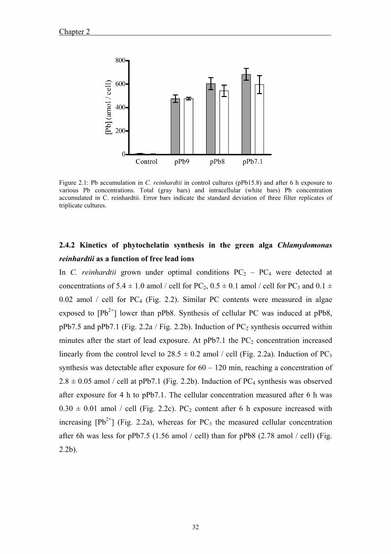
Chapter 2
32
Figure 2.1: Pb accumulation in C. reinhardtii in control cultures (pPb15.8) and after 6 h exposure to various Pb concentrations. Total (gray bars) and intracellular (white bars) Pb concentration accumulated in C. reinhardtii. Error bars indicate the standard deviation of three filter replicates of triplicate cultures.
2.4.2 Kinetics of phytochelatin synthesis in the green alga Chlamydomonas
reinhardtii as a function of free lead ions
In C. reinhardtii grown under optimal conditions PC2 – PC4 were detected at
concentrations of 5.4 ± 1.0 amol / cell for PC2, 0.5 ± 0.1 amol / cell for PC3 and 0.1 ±
0.02 amol / cell for PC4 (Fig. 2.2). Similar PC contents were measured in algae
exposed to [Pb2+] lower than pPb8. Synthesis of cellular PC was induced at pPb8,
pPb7.5 and pPb7.1 (Fig. 2.2a / Fig. 2.2b). Induction of PC2 synthesis occurred within
minutes after the start of lead exposure. At pPb7.1 the PC2 concentration increased
linearly from the control level to 28.5 ± 0.2 amol / cell (Fig. 2.2a). Induction of PC3
synthesis was detectable after exposure for 60 – 120 min, reaching a concentration of
2.8 ± 0.05 amol / cell at pPb7.1 (Fig. 2.2b). Induction of PC4 synthesis was observed
after exposure for 4 h to pPb7.1. The cellular concentration measured after 6 h was
0.30 ± 0.01 amol / cell (Fig. 2.2c). PC2 content after 6 h exposure increased with
increasing [Pb2+] (Fig. 2.2a), whereas for PC3 the measured cellular concentration
after 6h was less for pPb7.5 (1.56 amol / cell) than for pPb8 (2.78 amol / cell) (Fig.
2.2b).

Chapter 2
33
Figure 2.2: Kinetic of phytochelatin (PC2 – PC4) synthesis and GSH from 0 – 6 h exposure to various [Pb2+] (pPb7.1 = , pPb7.5 = , pPb8 = , pPb9 = , pPb10 = ◊, pPb11 = ×) and control cultures (Control = ). The cellular concentrations of PC2 – PC4 and GSH during the time of exposure are shown in Figure 2.2a – 2.2d (PC2, PC3, PC4, GSH). Error bars indicate the standard deviation of three replicate cultures.
The calculated rates of PC2 synthesis increased with increasing [Pb2+] from 0.05 amol
cell-1 min-1 at pPb8 to 0.09 amol cell-1 min-1 at pPb7.1. The rate of PC3 synthesis was
10 times lower and similar at pPb8 and at pPb7.1. The rate for PC4 synthesis at
pPb7.1 was 0.001 amol cell-1 min-1 (Table 2.1). GSH concentrations measured in
control algae were 837 ± 153 amol / cell and were similar at all [Pb2+] concentrations
and exposure times (Fig. 2.2d).
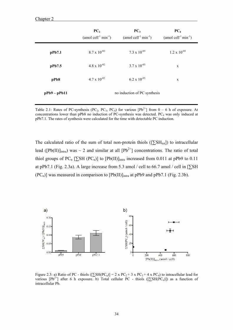
Chapter 2
34
PC2
(amol cell-1 min-1)
PC3
(amol cell-1 min-1)
PC4
(amol cell-1 min-1)
pPb7.1
pPb7.5
pPb8
pPb9 – pPb11
8.7 x 10-02
4.8 x 10-02
4.7 x 10-02
7.3 x 10-03
3.7 x 10-03
6.2 x 10-03
1.2 x 10-03
x
x
no induction of PC synthesis
Table 2.1: Rates of PC-synthesis (PC2, PC3, PC4) for various [Pb2+] from 0 – 6 h of exposure. At concentrations lower than pPb8 no induction of PC-synthesis was detected. PC4 was only induced at pPb7.1. The rates of synthesis were calculated for the time with detectable PC induction.
The calculated ratio of the sum of total non-protein thiols ([∑SHtot]) to intracellular
lead ([Pb(II)]intra) was ~ 2 and similar at all [Pb2+] concentrations. The ratio of total
thiol groups of PCn [∑SH (PCn)] to [Pb(II)]intra increased from 0.011 at pPb9 to 0.11
at pPb7.1 (Fig. 2.3a). A large increase from 5.3 amol / cell to 66.7 amol / cell in [∑SH
(PCn)] was measured in comparison to [Pb(II)]intra at pPb9 and pPb7.1 (Fig. 2.3b).
Figure 2.3: a) Ratio of PC - thiols ([∑SH(PCn)] = 2 x PC2 + 3 x PC3 + 4 x PC4) to intracellular lead for various [Pb2+] after 6 h exposure. b) Total cellular PC - thiols ([∑SH(PCn)]) as a function of intracellular Pb.
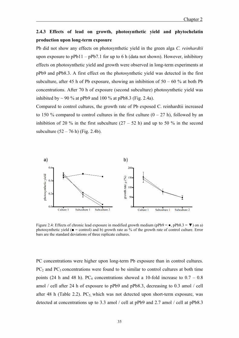
Chapter 2
35
2.4.3 Effects of lead on growth, photosynthetic yield and phytochelatin
production upon long-term exposure
Pb did not show any effects on photosynthetic yield in the green alga C. reinhardtii
upon exposure to pPb11 – pPb7.1 for up to 6 h (data not shown). However, inhibitory
effects on photosynthetic yield and growth were observed in long-term experiments at
pPb9 and pPb8.3. A first effect on the photosynthetic yield was detected in the first
subculture, after 45 h of Pb exposure, showing an inhibition of 50 – 60 % at both Pb
concentrations. After 70 h of exposure (second subculture) photosynthetic yield was
inhibited by ~ 90 % at pPb9 and 100 % at pPb8.3 (Fig. 2.4a).
Compared to control cultures, the growth rate of Pb exposed C. reinhardtii increased
to 150 % compared to control cultures in the first culture (0 – 27 h), followed by an
inhibition of 20 % in the first subculture (27 – 52 h) and up to 50 % in the second
subculture (52 – 76 h) (Fig. 2.4b).
Figure 2.4: Effects of chronic lead exposure in modified growth medium (pPb9 = , pPb8.3 = ) on a) photosynthetic yield ( = control) and b) growth rate as % of the growth rate of control culture. Error bars are the standard deviations of three replicate cultures.
PC concentrations were higher upon long-term Pb exposure than in control cultures.
PC2 and PC3 concentrations were found to be similar to control cultures at both time
points (24 h and 48 h). PC4 concentrations showed a 10-fold increase to 0.7 – 0.8
amol / cell after 24 h of exposure to pPb9 and pPb8.3, decreasing to 0.3 amol / cell
after 48 h (Table 2.2). PC5, which was not detected upon short-term exposure, was
detected at concentrations up to 3.3 amol / cell at pPb9 and 2.7 amol / cell at pPb8.3
Chapter 2
36
after 24 h and decreased to 1.3 amol / cell at pPb9 and 1.4 amol / cell at pPb8.3 after
48 h (Table 2.2). After 24 h and 48 h exposure to pPb8.3 and pPb9, [∑SH(PCn)] was
similar at both concentrations. Compared to control cultures [∑SH(PCn)] was
approximately twofold under lead exposure (Table 2.2). Comparison of [∑SH(PCn)]
of short- and long-term exposures shows a decrease by almost 50 % and a large
difference in PC oligomer distribution (Fig. 2.2, Table 2.2).
Exposure time Phytochelatins
(amol / cell)
Control
PC2 5.4 ± 1.0
PC3 0.5 ± 0.1
PC4 0.1 ± 0.02
PC5 0
∑SH(PCn) 13.0 ± 2.1
pPb9 pPb8.3
24 h
PC2 3.6 ± 1.3 3.1 ± 0.3
PC3 0.2 ± 0.07 0.1 ± 0.02
PC4 0.8 ± 0.03 0.7 ± 0.05
PC5 3.3 ± 0.15 2.7 ± 0.14
∑SH(PCn) 27.5 ± 2.7 22.8 ± 0.9
48 h
PC2 6.4 ± 0.1 7.1 ± 0.3
PC3 0.6 ± 0.04 0.6 ± 0.10
PC4 0.3 ± 0.03 0.3 ± 0.04
PC5 1.3 ± 0.06 1.4 ± 0.19
∑SH(PCn) 22.3 ± 0.4 24.2 ± 1.2
Table 2.2: PC2 - PC5 and ∑SH(PCn) (∑SH(PCn) = 2 x PC2 + 3 x PC3 + 4 x PC4 + 5 x PC5) detected in control cultures and after 24h and 48h exposure to pPb9 and pPb8.3 in modified Talaquil.
Chapter 2
37
2.5 Discussion
A more than 100-fold increase of [Pb(II)]intra was observed between control algae and
pPb9. However, only a slight increase of [Pb(II)]intra from pPb9 to pPb7.1 was
observed. This slight increase upon a 100-fold increase of [Pb2+] in the exposure
medium might indicate saturation of Pb accumulation or saturation of involved uptake
transporters at these concentrations (Fig. 2.1). Furthermore, the use of a simplified
medium for accumulation experiments, in order to reach a well defined Pb speciation,
might lead to an overestimation of lead uptake compared to more natural water
composition. The absence of certain essential nutrients in this medium could also
cause deficiency influencing Pb uptake. Similar results for saturation of Pb
accumulation at higher [Pb2+] (> pPb6) were obtained with the freshwater alga
Chlorella kesslerii after 35 min exposure (Lamelas and Slaveykova, 2007). The
obtained results suggest, that in addition to [Pb2+] in the exposure medium, excretion
and the subcellular distribution of Pb in C. reinhardtii might influence Pb uptake. The
measured [Pb(II)]intra were very high, reaching calculated cellular concentrations of
2.98 mM at pPb7.1. These high [Pb(II)]intra may indicate precipitation of Pb solid
phases inside the cell. Intracellular Pb precipitation might be involved in Pb
detoxification (Mason and Jenkins, 1995) and was observed in the prawn Penaeus
monodon (Vogt and Quinitio, 1994).
The detection of PC2 and the larger oligomers PC3 and PC4 in control algae indicate
that PCs appear to be constitutive in C. reinhardtii. Similarly, PCn have been found to
occur constitutively in marine algae (Ahner et al., 1995a; Ahner et al., 2002; Morelli
et al., 2009). Compared to C. reinhardtii (PC2 = 27 µM; PC3 = 2.7 µM; PC4 = ~ 0.5
µM), similar molar concentrations were detected in Thalassiosira Pseudonana (PC2 =
44 µM; PC3 = 5.3 µM; PC4 = 2.2 µM) (Ahner et al., 2002). For the other six algal
species examined in this study lower concentrations were detected. The presence of
constitutive PCs might indicate the role of PCs in metal homeostasis.
Under Pb exposure the induction of PC2 synthesis was very fast and detectable within
30 min of exposure to [Pb2+] ≥ pPb8 and increased continuously during 6 h (Fig.
2.2a). Comparable kinetics of PC2 induction were reported in Stichococcus bacillaris
(Pawlik-Skowronska, 2000) and Phaeodactylum tricornutum (Morelli and Scarano,
2001). The rate of PC2 synthesis increased with increasing [Pb2+] in the exposure
medium. From pPb8 to pPb7.1 a twofold increase of the PC2 synthesis rate was
Chapter 2
38
observed. PC3 synthesis rates were lower than those of PC2 and similar at various
[Pb2+]. These similar PC3 synthesis rates at various [Pb2+] may result from the limited
availability of PC2 for PC3 synthesis (Vatamaniuk et al., 2000). The lowest [Pb2+]
inducing PC synthesis in short-term experiments was pPb8. However, in long-term
experiments induction of PC synthesis was also observed at pPb9. Induction of PC
synthesis was observed at ≥ pPb8 in short-term experiments, even though [Pb(II)]intra
was similar at pPb9, suggesting that induction of PC synthesis is dependent on the
subcellular distribution of Pb. Induction of PC synthesis at pPb9 upon long-term
exposure might occur because of additional Pb uptake with increased exposure time
or because of changes in the subcellular distribution of Pb. Similarly, induction of PC2
synthesis was observed in Scenedesmus vacuolatus at pPb10 after 72 h exposure (Le
Faucheur et al., 2006).
While in short-term experiments PC2 and PC3 were detected, long-term exposure
showed induction of PC oligomers up to PC5 at even lower [Pb2+]. Consequently, the
degree of polymerization of PC oligomers increased with increasing [Pb2+] and time.
Furthermore, synthesis of PC oligomers, with a higher degree of polymerization than
PC2, are induced after a lag phase, which increases with the chain length (PC3 = 1 h;
PC4 = 4 h; PC5 = > 6 h). This lag phase may appear because PC2 is the substrate for
PC3 synthesis or, in more general terms, PCn is substrate for PCn+1 synthesis
(Vatamaniuk et al., 2000). The synthesis of PC oligomers with increasing chain length
is believed to occur because of the higher stability of metal complexes (Mehra et al.,
1995).
For [∑SH(PCn)] / [Pb(II)]intra a continuous increase with increasing [Pb2+] was
observed. This increase results from the enhanced induction of PC synthesis at higher
[Pb2+], with only a slight increase in [Pb(II)]intra. The ratio [∑SH(PCn)] / [Pb(II)]intra
increased to a maximum of 0.11 at pPb7.1 indicating that PCs synthesized in C.
reinhardtii were not present at a sufficiently high concentration to fully immobilize
the intracellular accumulated Pb. The ratio [∑SHtot] / [Pb(II)]intra was approximately
two and similar for all [Pb2+], because the GSH concentration was much higher than
the PC concentration. Although it is known that GSH can form complexes with Pb, it
remains unclear whether GSH is directly involved in Pb detoxification (Cruz et al.,
2001).
Despite the high [Pb]intra and the production of reactive oxygen species (ROS) upon
Pb exposure (Szivak et al., 2009), no effects on photosynthesis were observed in
Chapter 2
39
short-term experiments. However, upon long-term exposure to Pb, strong inhibition of
the photosynthetic yield was observed. Furthermore, damaging effects on growth
were detected in long-term experiments. The inhibition of photosynthetic yield and
growth increased with exposure time. These time dependent effects may result as a
consequence of the high affinity of Pb to sulfur and oxygen functional groups (Mason
and Jenkins, 1995). In other words, Pb binds non-specifically to functional groups of
proteins containing sulfur or oxygen, inducing various effects that may affect
photosynthesis and growth upon long-term exposure. In other studies using the
freshwater alga Chlorella vulgaris or Emiliania huxleyi, a marine alga, no significant
inhibition of growth under short-term and long-term Pb exposure was detected (Rosko
and Rachlin, 1977; Vasconcelos and Leal, 2001; Baumann et al., 2009). Debelius et
al. (2009) reported inhibition of growth up to 100 % in five marine algae after 72 h of
exposure at nominal Pb concentrations between 1.5 – 30 µM. Fujiwara et al. (2008)
reported growth inhibition of 50% for much higher [Pb2+] of 146 µM in Chlorella
kessleri. This variability indicates strong differences in sensitivity to Pb for different
algal species. In summary, this study showed no damaging effects of Pb under short-
term exposure, although [Pb(II)]intra were much higher than cellular PC
concentrations. This suggests a minor role of PC in Pb detoxification and that other
mechanisms must be involved to protect cells from short-term damaging effects.
However, these Pb detoxification mechanisms are not effective enough to avoid
damaging effects upon long-term exposure.
At concentrations of pPb9, an environmentally relevant concentration, Pb is
accumulated in algal cells, reaching a high concentration after short exposure times.
Because of delayed toxic effects induced by Pb, which are not detected under short-
term exposure, it is important to examine long-term exposures to assess the toxicity of
Pb on organisms. Further research needs to be done to show whether PC synthesis is
actually induced as a direct response to Pb exposure or because of indirect effects,
such as metal redistribution. Furthermore, it is unclear whether Pb is actually bound
by PCs or if other metals are bound due to metal redistribution and to which other
functional groups Pb may be bound.
Chapter 2
40
2.6 References
Ahner, B.A., Kong, S., Morel, F.M.M., 1995a. Phytochelatin production in marine algae. 1. An interspecies comparison. Limnol. Oceanogr. 40, 649-657.
Ahner, B.A., Morel, F.M.M., 1995b. Phytochelatin production in marine algae. 2. Induction by various metals. Limnol. Oceanogr. 40, 658-665.
Ahner, B.A., Wei, L.P., Oleson, J.R., Ogura, N., 2002. Glutathione and other low molecular weight thiols in marine phytoplankton under metal stress. Mar. Ecol. Prog. Ser. 232, 93-103.
Baumann, H.A., Morrison, L., Stengel, D.B., 2009. Metal accumulation and toxicity measured by PAM-Chlorophyll fluorescence in seven species of marine macroalgae. Ecotoxicol. and Environ. Safety. 72, 1063-1075.
Chen, Z., Zhu, L., Wilkinson, K.J., 2010. Validation of the biotic ligand model in metal mixtures: Bioaccumulation of lead and copper. Environ. Sci Technol. 44, 3580-3586.
Cobbett, C.S., 2000. Phytochelatins and their roles in heavy metal detoxification. Plant Physiol. 123, 825-832.
Crowder, A., 1991. Acidification, metals and macrophytes. Environ. Pollut. 71, 171-203.
Cruz, B.H., Díaz-Cruz, J.M., Díaz-Cruz, M.S., Ariño, C., Esteban, M., Tauler, R., 2001. Differential pulse polarographic study of the Pb(II) complexation by glutathione. J. Electroanal. Chem. 516, 110-118.
Debelius, B., Forja, J.M., DelValls, Á., Lubián, L.M., 2009. Toxicity and bioaccumulation of copper and lead in five marine microalgae. Ecotoxicol. and Environ. Safety. 72, 1503-1513.
Erel, Y., Morgan, J.J., Patterson, C.C., 1991. Natural levels of Pb and Cd in a remote mountain stream. Geochim. Cosmochim. Acta. 55, 707-719.
Fujiwara, K., Matsumoto, Y., Kawakami, H., Aoki, M., Tuzuki, M., 2008. Evaluation of metal toxicity in Chlorella kessleri from the perspective of the periodic table. Bull. Chem. Soc. Jpn. 81, 478-488.
Gekeler, W., Grill, E., Winnacker, E.L., Zenk, M.H., 1988. Algae sequester heavy metals via synthesis of phytochelatin complexes. Arch. Microbiol. 150, 197-202.
Gupta, M., Rai, U.N., Tripathi, R.D., Chandra, P., 1995. Lead induced changes in glutathione and phytochelatin in Hydrilla verticillata (l. f.) Royle. Chemosphere. 30, 2011-2020.
Gustafsson, J.P. Visual MINTEQ 2.15. 2005, Stockholm, Sweden. Hwww.lwr.kth.se/English/OurSoftware/vminteq/
Kerper, L.E., Hinkle, P.M., 1997. Cellular uptake of lead is activated by depletion of intracellular calcium stores. J. Biol. Chem. 272, 8346-8352.
Knauer, K., Behra, R., Sigg, L., 1997. Adsorption and uptake of copper by the green alga Scenedesmus subspicatus (Chlorophyta). J. Phycol. 33, 596-601.
Chapter 2
41
Lamelas, C., Slaveykova, V.I., 2007. Comparison of Cd(II), Cu(II), and Pb(II) biouptake by green algae in the presence of humic acid. Environ. Sci Technol. 41, 4172-4178.
Le Faucheur, S., Behra, R., Sigg, L., 2005. Phytochelatin induction, cadmium accumulation, and algal sensitivity to free cadmium ion in Scenedesmus vacuolatus. Environ. Toxicol. Chem. 24, 1731-1737.
Le Faucheur, S., Schildknecht, F., Behra, R., Sigg, L., 2006. Thiols in Scenedesmus vacuolatus upon exposure to metals and metalloids. Aquat. Toxicol. 80, 355-361.
Mason, A.Z., Jenkins, K.D., Metal detoxification in aquatic organisms. In: A.T. Tessier, D.R., (Ed.), Metal speciation and bioavailability in aquatic systems. John Wiley & sons, New York, 1995, pp. 479-608.
Mehra, R.K., Kodati, V.R., Abdullah, R., 1995. Chain length-dependent Pb(II)-coordination in phytochelatins. Biochem. Biophys. Res. Commun. 215, 730-736.
Meylan, S., Behra, R., Sigg, L., 2004. Influence of metal speciation in natural freshwater on bioaccumulation of copper and zinc in periphyton: A microcosm study. Environ. Sci Technol. 38, 3104-3111.
Morelli, E., Scarano, G., 2001. Synthesis and stability of phytochelatins induced by cadmium and lead in the marine diatom Phaeodactylum tricornutum. Mar. Environ. Res. 52, 383-395.
Morelli, E., Ferrara, R., Bellini, B., Dini, F., Di Giuseppe, G., Fantozzi, L., 2009. Changes in the non-protein thiol pool and production of dissolved gaseous mercury in the marine diatom Thalassiosira weissflogii under mercury exposure. Sci. Tot. Environ. 408, 286-293.
Nriagu, J.O., 1988. A silent epidemic of environmental metal poisoning? Environ. Pollut. 50, 139-161.
Pawlik-Skowronska, B., 2000. Relationships between acid-soluble thiol peptides and accumulated Pb in the green alga Stichococcus bacillaris. Aquat. Toxicol. 50, 221-230.
Rauser, W.E., 1995. Phytochelatins and related peptides-structure, biosynthesis, and function. Plant Physiol. 109, 1141-1149.
Rosko, J.J., Rachlin, J.W., 1977. The effect of cadmium, copper, mercury, zinc and lead on cell division, growth, and chlorophyll a content of the chlorophyte Chlorella vulgaris. Bull. Torr. Bot. Club. 104, 226-233.
Scarano, G., Morelli, E., 2002. Characterization of cadmium- and lead-phytochelatin complexes formed in a marine microalga in response to metal exposure. Biometals. 15, 145-151.
Sharma, P., Dubey, R.S., 2005. Lead toxicity in plants. Bra. J. Plant. Physiol. 17, 35-52.
Soldo, D., Hari, R., Sigg, L., Behra, R., 2005. Tolerance of Oocystis nephrocytioides to copper: intracellular distribution and extracellular complexation of copper. Aquat. Toxicol. 71, 307-317.
Chapter 2
42
Szivak, I., Behra, R., Sigg, L., 2009. Metal-induced reactive oxygen species production in Chlamydomonas reinhardtii (Chlorophyceae). J. Phycol. 45, 427-435.
Vasconcelos, M.T.S.D., Leal, M.F.C., 2001. Antagonistic interactions of Pb and Cd on Cu uptake, growth inhibition and chelator release in the marine algae Emiliania huxleyi. Mar. Chem. 75, 123-139.
Vatamaniuk, O.K., Mari, S., Lu, Y.P., Rea, P.A., 2000. Mechanism of heavy metal ion activation of phytochelatin (PC) synthase. J. Biol. Chem. 275, 31451-31459.
Vogt, G., Quinitio, E.T., 1994. Accumulation and excretion of metal granules in the prawn, Penaeus monodon, exposed to water-borne copper, lead, iron and calcium. Aquat. Toxicol. 28, 223-241.
Warnken, K.W., Lawlor, A.J., Lofts, S., Tipping, E., Davison, W., Zhang, H., 2009. In situ speciation measurements of trace metals in headwater streams. Environ. Sci Technol. 43, 7230-7236.
Zenk, M.H., 1996. Heavy metal detoxification in higher plants - A review. Gene. 179, 21-30.
43
UChapter 3
Characterization of lead induced metal-phytochelatin complexes in Chlamydomonas reinhardtii
This chapter will be submitted to Environmental Toxicology and Chemistry

44
Chapter 3
45
3.1 Abstract
Phytochelatins (PCs) are metal-binding oligopeptides with the general structure (γ-
Glu-Cys)n-Gly (n = 2 – 11), which are synthesized enzymatically from glutathione
upon exposure of algae to various metals.. Accumulation of Pb as well as induction of
phytochelatin synthesis was observed in Chlamydomonas reinhardtii upon Pb(II)
exposure. The aim of this study was to examine whether Pb(II) is bound by PCs in C.
reinhardtii and to examine formed complexes for their stoichiometry and
composition. Metal-phytochelatin (Me-PC) complexes induced by Pb were isolated
by size-exclusion chromatography in 13 collected fractions, which were analyzed for
their PC and metal content by HPLC and ICP-MS. A recovery
of over 90 % of Pb from standard Pb-PC2 complexes within the total volume of the
size-exclusion column indicated the adequacy of the method for Pb-PCn complex
separation and characterization. PCs were mainly detected in a molecular weight
range from 1000 – 5300 Da, indicating the formation of complexes with various
stoichiometries. Approximately 72 % of total PC2 eluted in the range 1000 – 1600 Da
and 80 % of total PC3 eluted in the molecular weight range 1600 – 2300 Da. The
distribution of Cu, Zn and Pb showed that over 70 % of these metals were associated
with the high molecular weight fractions. Cu, Zn and Pb were also observed in PC
containing fractions, suggesting the formation of various Me-PC complexes.
Molecular weight considerations suggested that complexes with one or two metals
bound by two PC2 or PC3 were formed. PCs present in higher molecular weight
fractions are assumed to be involved in the formation of high molecular weight
complexes. The results of this study indicate that the role of PCs in Pb detoxification
is minor as only 13 % of total Pb was associated with PCs. The observed formation of
Me-PC complexes with Cu and Zn suggests a homeostatic role of PC.
3.2 Introduction
Phytochelatins (PCs) are metal-binding oligopeptides with the general structure (γ-
Glu-Cys)n-Gly (n = 2 – 11). They are produced enzymatically from glutathione by the
enzyme phytochelatin synthase, which is activated by binding of metal ions and
metal-glutathione complexes (Grill et al., 1985; Grill et al., 1987; Vatamaniuk et al.,
2000). PC synthesis is induced in various plants (Rauser, 1995; Zenk, 1996) and algae
(Gekeler et al., 1988; Ahner et al., 1995) as a response to metal exposure. Besides Cd,
Chapter 3
46
which is the most potent inducer of phytochelatins, other metals and metalloids (e.g.
Pb, Cu, Zn, Hg, Ag, As(III), As(V)) were found to induce PC synthesis in algae
(Gekeler et al., 1988; Ahner and Morel, 1995; Le Faucheur et al., 2006). Pb was
observed to be a strong inducer of phytochelatin synthesis in marine and freshwater
algae (Ahner and Morel, 1995; Pawlik-Skowronska, 2000; Morelli and Scarano,
2001; Pawlik-Skowronska, 2002; Le Faucheur et al., 2006).
Because of their high cysteine content, PCs bind metals through thiolate coordination
and are assumed to be involved in metal homeostasis and detoxification (Grill et al.,
1985; Rauser, 1995; Zenk, 1996; Clemens, 2001). The first convincing evidence of
PC involvement in metal detoxification was obtained using yeast mutants
(Schizosaccharomyces pombe and Saccharomyces cerevisiae) and plants (Arabidopsis
spp.) that are deficient in PC synthase genes (Clemens et al., 1999; Ha et al., 1999).
These species were more sensitive to Cd, As(V) and Cu than the wild-type. Studies on
Cd-PC complexes formed in plants (Kneer and Zenk, 1997; Yen et al., 1999), yeast
(Reese and Winge, 1988; Mehra et al., 1994) and algae (Hu and Wu, 1998; Hu et al.,
2001; Morelli et al., 2002; Scarano and Morelli, 2002) under Cd exposure revealed
the presence of two types of complexes, defined as Low Molecular Weight (LMW)
and High Molecular Weight (HMW) complexes. LMW complexes consist of metals
bound to PCn, whereas HMW complexes are composed of Cd and acid labile sulfur
(S-) forming a CdS core surrounded by PCs. The role of the PC coating is to stabilize
the CdS core and to avoid its precipitation. Formation of HMW complexes increases
metal tolerance in the yeast Candida glabrata, where a strain with enhanced CdS
production revealed increased Cd resistance compared to the wildtype (Barbas et al.,
1992; Mehra et al., 1994). Therefore, the role of PC in Cd detoxification is assumed to
result from immobilization of metals, preventing non-specific binding of cadmium to
important biomolecules, followed by the transport of Cd-PC complexes into the
vacuole.
The role of PC in detoxification of metals, other than Cd, is unclear because, for most
metals that were found to induce PC synthesis (e.g. Pb, Zn, Ag, Cu, As(V), As(III)
and Hg), the formation of metal-phytochelatin complexes (Me-PC) was only
examined in vitro but not in vivo (Rauser, 1999; Schmoger et al., 2000; Alberich et
al., 2007; Chekmeneva et al., 2007). Studies on Me-PC complexes induced by Pb are
very scarce (Scarano and Morelli, 2002; Andra et al., 2009). In the marine alga
Phaeodactylum tricornutum formation of Pb-PC complexes including PC3 – PC6 but
Chapter 3
47
not PC2 was suggested (Scarano and Morelli, 2002). In vitro formation of Pb-PC
complexes and examination by differential pulse polarography indicated the
formation of Pb1-PC2 or Pb2-PC2 complexes (Alberich et al., 2007), and an increased
complex stability with increasing number of coordinating thiols was demonstrated.
In a previous study, PC synthesis was induced in short-term experiments at
concentrations of pPb8 (pPb = -log[Pb2+]) and higher (Chapter 2). The aim of the
present study is to examine whether Pb is bound by PCs in C. reinhardtii and to
characterize putative complexes for their stoichiometry and composition. In order to
examine Me-PC complexes, size-exclusion chromatography coupled off-line with
reversed phase high pressure liquid chromatography (RP HPLC) and inductively
coupled plasma mass spectrometry (ICP-MS) was used to separate native Me-PC
complexes and to determine thiol and metal content.
3.3 Materials and methods
3.3.1 Chemicals and materials
Tris(2-carboxyethyl)phosphine hydrochloride (TCEP), 3-morpholinopropanesulfonic
acid (MOPS), diethylenetriamine-pentaacetic acid (DTPA), standard peptides
(aprotinin, gastrin, (Glycine)6, (Glycine)3) and metal salts used in this study were
obtained from Sigma-Aldrich (St. Louis, MO, USA). Trifluoroacetic acid (TFA) was
from Supelco (Bellefonte, PA, USA). Nitric acid suprapure (HNO3; 65 %) and
protease inhibitor cocktail III (EDTA free), containing serine, aspartic and cysteine
proteases and aminopeptidases, was ordered from Merck (Darmstadt, Germany).
Phytochelatin standards (PC2, PC3 and PC4) were obtained from Invitrogen (San
Diego, CA; USA). Bradford protein assay and Chelex was from BioRad laboratories
(Muenchen, Germany). Glassware and polycarbonate containers were presoaked in
0.01 M HNO3 and rinsed with deionized water (18 MΩ-cm, Q-H2O grade, Barnstead
Nanopure, Allschwil, Switzerland) before use. All solutions and media were prepared
in deionized water in autoclaved containers.
3.3.2 Test organism, culture and experimental conditions
The unicellular green alga Chlamydomonas reinhardtii (CC125) was obtained from
the Chlamydomonas Genetics Center (Durham, NC, USA) and was grown in glass
Erlenmeyers in a HT Infors shaker (Infors, Bottmingen, Switzerland) at 25 °C with
Chapter 3
48
continuous illumination of 120 µEm-2s-1, provided by cool white fluorescent lamps,
and shaken at 90 rpm.
For experiment and exposure to Pb, algae were acclimatized to the growth medium by
transferring an inoculum of exponentially growing algae to the growth medium in at
least two successive batch cultures. C. reinhardtii were cultured in Talaquil, a
complete growth medium containing major nutrients and trace metals buffered by
EDTA (Knauer et al., 1997), in which pH is buffered using MOPS (3-
morpholinopropanesulfonic acid) as a non-metal complexing buffer. The total
concentrations in the growth medium are the following: 2 x 10⁻⁵ M Na2EDTA; 5 x 10-
4 M CaCl2 · 2H2O; 1.5 x 10⁻⁴ M MgSO4 · 7H2O; 1.2 x 10⁻³ M NaHCO3; 5 x 10⁻⁵ M
K2HPO4 · 3H2O; 1 x 10⁻³ M NH4Cl; 5 x 10⁻⁸ M CoCl2 · 6H2O; 5 x 10⁻⁵ M H3BO3; 8 x
10-8 M Na2MoO4 · 2H2O; 1.63 x 10⁻⁷ M CuSO4 · 5H2O; 1.22 x 10⁻⁶ M MnCl2 · 4H2O;
1.58 x 10⁻⁷ M ZnSO4 · 7H2O and 9 x 10⁻⁷ M FeCl3 · 6H2O in 1 x 10⁻² M MOPS buffer
(pH 7.5). All media were prepared at least 24 h before use to allow equilibration. Free
ion concentrations of Cu2+, Mn2+, Zn2+ and Fe3+ in Talaquil, modeled using the
speciation software VMINTEQ (Gustafsson, 2005), are 3 x 10-14, 1.7 x 10⁻⁸, 9 x 10-12
and 1 x 10-20 M.
As experimental medium, growth medium containing Pb buffered with 20 µM NTA
to keep [Pb2+] constant was used. Other trace metals were omitted from the
experimental medium in order to avoid metal competition and cumulative effects
resulting from the presence of various metal ions. Exponentially growing algae were
transferred to the experimental medium and exposed to pPb7.5 (pPb = -log [Pb2+]) for
14 h with an initial cell density of 8.4 x 10⁵ cells / ml. Free metal concentrations were
computed using the speciation software VMINTEQ (Gustafsson, 2005).
3.3.3 Preparation of algal cell extracts
Algal cells were harvested at the end of exposure by centrifugation (5000 x g for 10
min at 25 °C) and resuspended in 1 ml of 50 mM MOPS (pH 7.5) containing 1 mM
TCEP (antioxidant) and protease inhibitor cocktail III, all degassed with argon prior
to the experiment.
The algal suspension was then homogenized for 4 x 30 s at 100 % sonication intensity
on ice using an ultrahomogenizer (Labsonic® M, Sartorius Stedim Biotech, France)
with titanium tip to obtain native Me-PCn. Between each sonication step the samples
were cooled on ice for 3 min. The homogenate was centrifuged at 9900 x g for 10 min
Chapter 3
49
(4 °C). After centrifugation, the supernatant was transferred to a clean Eppendorf vial,
followed by a second centrifugation step under the same conditions. This procedure
was repeated up to five times to obtain a clear cytosolic fraction. Before analysis of
supernatant using size-exclusion chromatography (SEC), total protein concentration
was measured photometrically at 595 nm using a Bradford protein assay as dye
reagent after performing calibration curves with diluted BSA solutions. In addition,
metal concentrations and total PCs in the algal supernatant homogenate were
measured by inductively coupled plasma mass spectrometry (ICP-MS) and high
pressure liquid chromatography (HPLC).
3.3.4 Size-exclusion chromatography
For SEC, a pre-packed Superdex peptide HR 10/30 column (Amersham), designed for
separation of small peptides (100 – 10000 Da), was used. The SEC system and
solvents were stored and operated at 4 °C. The flow rate of the eluent, 150 mM
ammonium nitrate (NH4NO3) and 50 mM MOPS (pH = 7.5), was 0.15 ml / min. All
eluents were filtered on 0.22 µm, degassed with nitrogen and cleaned from metal
contamination by using a Chelex 100 Resin. The void volume (V0) of SEC was
determined using a solution of blue dextran and the total volume (Vt) using CuCl2.
The calibration curve for molecular weight determination were performed by
measuring the elution volumes (Ve) of standard peptides (aprotinin (6.5 kDa), gastrin
(2.1 kDa), PC4 (1004 Da), PC3 (772 Da), PC2 (540 Da), (Glycine)6 (360 Da),
(Glycine)3 (189 Da)), calculating their partition coefficient Kav (0
0
VV
VVK
t
eav
) and
plotting their Kav values versus the logarithm of their molecular weight (log Mr).
For separation of algal peptides and Me-PCn complexes 100 µl algal homogenate
(protein concentration: 6.21 mg / ml) was applied to the SEC. The eluate was
collected in fractions (Fraction 1 (F1) = 3600 µl, F2 = 1800 µl, F3 = 900 µl, F4 =
2700 µl, F5 = 1800 µl, F6 – F11 = 900 µl, F12 = 1800 µl and F13 = 3600 µl) and
absorbance at 254 nm was recorded. Between different samples, the column was
cleaned with 0.1 % HCl and then flushed with 10 column volumes eluent containing 1
mM EDTA, to remove remaining metals adsorbed to the column. After the EDTA
wash, the column was equilibrated with 10 column volumes eluent.
Chapter 3
50
3.3.5 Metal recovery experiments
Elution and possible adsorption of Pb and Pb-PC complexes to the column Superdex
peptide HR 10/30 column were tested in independent experiments. To investigate the
recovery of inorganic Pb(II), 100 µl of 15 µM Pb(NO3)2 in the elution buffer (150
mM ammonium nitrate (NH4NO3) and 50 mM MOPS; pH = 7.5) was applied to the
SEC column. During the elution of Vt, fractions were taken and analyzed for their
metal content by ICP-MS. After the elution of Vt, 100 µl elution buffer containing 2
mM EDTA was injected to remobilize Pb adsorbed to the column. Fractions taken
during the elution of EDTA were analyzed for their Pb content by ICP-MS.
To investigate the elution of Pb-PC2 complexes in SEC, a solution of 30 µM PC2
standard and 15 µM Pb(NO3)2 in elution buffer were mixed and incubated for 15 min
at 20 °C to allow complex formation, followed by the injection of 100 µl to the SEC
column. The Pb and PC2 concentration were determined in each of the collected
eluate fractions.
3.3.6 Metal and thiol analysis in SEC fractions by ICP-MS and HPLC
Metal and thiol concentrations of each fraction were determined off-line of SEC.
After appropriate dilution of a 250 µl fraction aliquot, metal concentrations were
analyzed by ICP-MS (Perkin-Elmer; Perkin Elmer Sciex, Ueberlingen, Germany)
using rhodium as an internal standard. ICP-MS measurements were checked using
SLRS-4 reference water (River Water Reference Material for Trace Metals; National
Research Council Canada, Ottawa).
For thiol determination, the remaining fraction was lyophilized for 15 h using a
Lyovac GT2 (Leybold, Switzerland). The lyophilized sample was resuspended in 50
µl HCl / DTPA (120 mM / 5 mM) before pre-column derivatization with
monobromobimane according to the procedure explained elsewhere (Le Faucheur et
al., 2005). Thiols were measured by reverse-phase high performance liquid
chromatography (HPLC) equipped with a fluorescence detector (Agilent 1200 series,
Agilent Technologies, Santa Clara CA, USA). The separation of the derivatized
sample was carried out on a reversed phase octadecylsilica column (Nucleosil C18, 5
µm, 250 x 4 mm) obtained from Macherey und Nagel (Düren, Germany) with a
mobile phase flow rate of 1 ml / min. The mobile phase was composed of 0.1 %
trifluoroacetic acid and a gradient of 10 – 36 % acetonitrile during 65 min. Between
each measurement the column was cleaned with 100 % acetonitrile for 5 min,
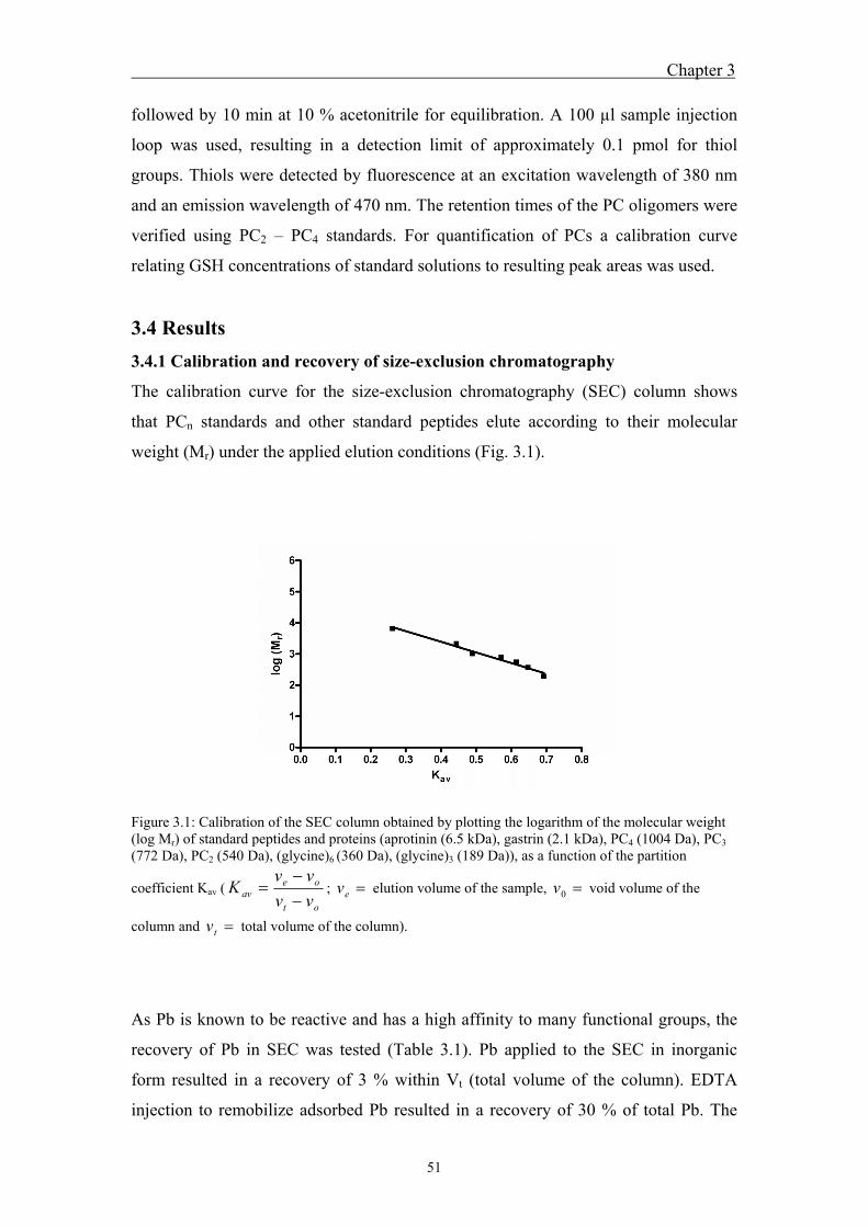
Chapter 3
51
followed by 10 min at 10 % acetonitrile for equilibration. A 100 µl sample injection
loop was used, resulting in a detection limit of approximately 0.1 pmol for thiol
groups. Thiols were detected by fluorescence at an excitation wavelength of 380 nm
and an emission wavelength of 470 nm. The retention times of the PC oligomers were
verified using PC2 – PC4 standards. For quantification of PCs a calibration curve
relating GSH concentrations of standard solutions to resulting peak areas was used.
3.4 Results
3.4.1 Calibration and recovery of size-exclusion chromatography
The calibration curve for the size-exclusion chromatography (SEC) column shows
that PCn standards and other standard peptides elute according to their molecular
weight (Mr) under the applied elution conditions (Fig. 3.1).
Figure 3.1: Calibration of the SEC column obtained by plotting the logarithm of the molecular weight (log Mr) of standard peptides and proteins (aprotinin (6.5 kDa), gastrin (2.1 kDa), PC4 (1004 Da), PC3 (772 Da), PC2 (540 Da), (glycine)6 (360 Da), (glycine)3 (189 Da)), as a function of the partition
coefficient Kav (ot
oeav vv
vvK
; ev elution volume of the sample, 0v void volume of the
column and tv total volume of the column).
As Pb is known to be reactive and has a high affinity to many functional groups, the
recovery of Pb in SEC was tested (Table 3.1). Pb applied to the SEC in inorganic
form resulted in a recovery of 3 % within Vt (total volume of the column). EDTA
injection to remobilize adsorbed Pb resulted in a recovery of 30 % of total Pb. The
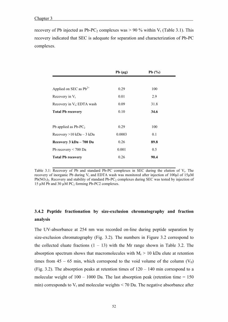
Chapter 3
52
recovery of Pb injected as Pb-PC2 complexes was > 90 % within Vt (Table 3.1). This
recovery indicated that SEC is adequate for separation and characterization of Pb-PC
complexes.
Table 3.1: Recovery of Pb and standard Pb-PC complexes in SEC during the elution of Vt. The recovery of inorganic Pb during Vt and EDTA wash was monitored after injection of 100µl of 15µM Pb(NO3)2. Recovery and stability of standard Pb-PC2 complexes during SEC was tested by injection of 15 µM Pb and 30 µM PC2 forming Pb-PC2 complexes.
3.4.2 Peptide fractionation by size-exclusion chromatography and fraction
analysis
The UV-absorbance at 254 nm was recorded on-line during peptide separation by
size-exclusion chromatography (Fig. 3.2). The numbers in Figure 3.2 correspond to
the collected eluate fractions (1 – 13) with the Mr range shown in Table 3.2. The
absorption spectrum shows that macromolecules with Mr > 10 kDa elute at retention
times from 45 – 65 min, which correspond to the void volume of the column (V0)
(Fig. 3.2). The absorption peaks at retention times of 120 – 140 min correspond to a
molecular weight of 100 – 1000 Da. The last absorption peak (retention time = 150
min) corresponds to Vt and molecular weights < 70 Da. The negative absorbance after
Pb (µg) Pb (%)
Applied on SEC as Pb2+ 0.29 100
Recovery in Vt 0.01 2.9
Recovery in Vt; EDTA wash 0.09 31.8
Total Pb recovery 0.10 34.6
Pb applied as Pb-PC2 0.29 100
Recovery >10 kDa – 3 kDa 0.0003 0.1
Recovery 3 kDa – 700 Da 0.26 89.8
Pb recovery < 700 Da 0.001 0.5
Total Pb recovery 0.26 90.4
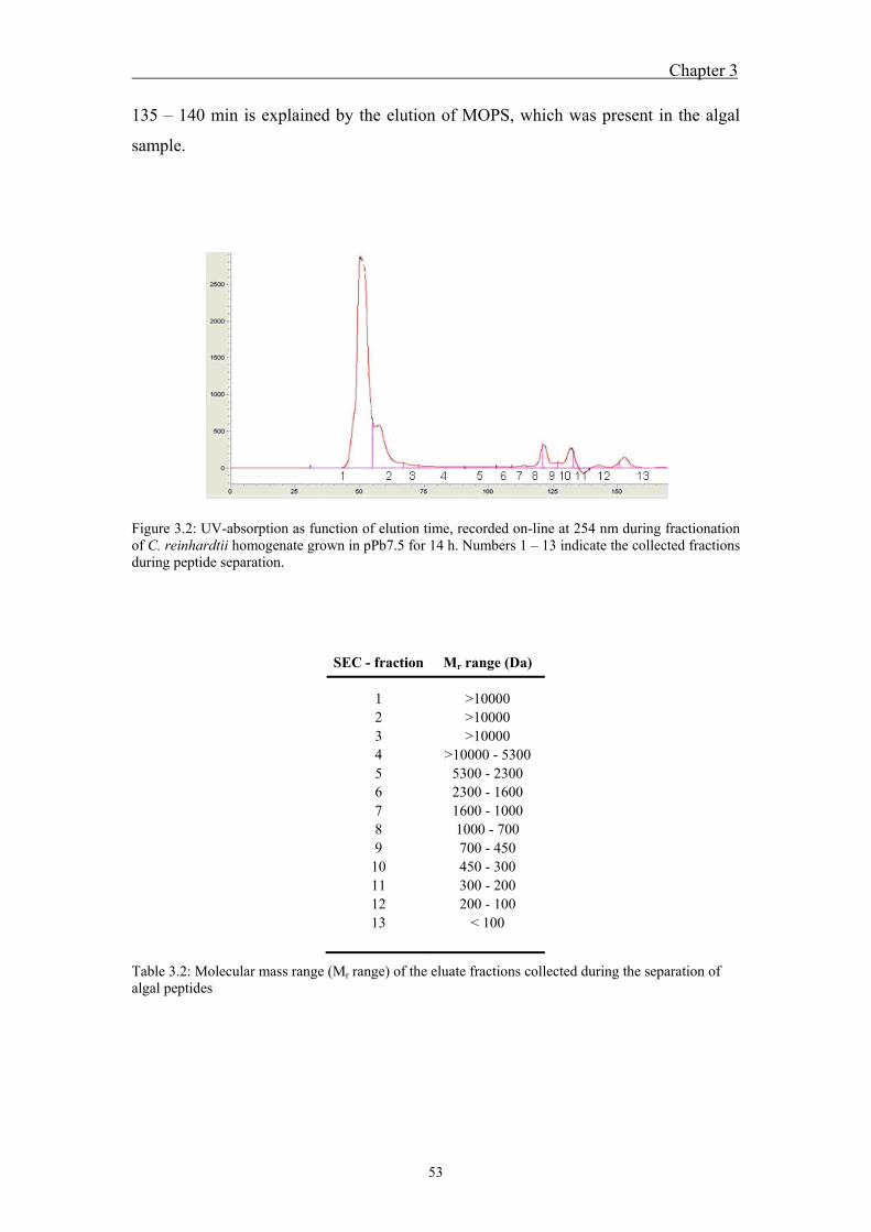
Chapter 3
53
135 – 140 min is explained by the elution of MOPS, which was present in the algal
sample.
Figure 3.2: UV-absorption as function of elution time, recorded on-line at 254 nm during fractionation of C. reinhardtii homogenate grown in pPb7.5 for 14 h. Numbers 1 – 13 indicate the collected fractions during peptide separation.
SEC - fraction Mr range (Da)
1 >10000 2 >10000 3 >10000 4 >10000 - 5300 5 5300 - 2300 6 2300 - 1600 7 1600 - 1000 8 1000 - 700 9 700 - 450
10 450 - 300 11 300 - 200 12 200 - 100 13 < 100
Table 3.2: Molecular mass range (Mr range) of the eluate fractions collected during the separation of algal peptides
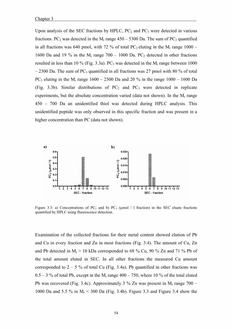
Chapter 3
54
Upon analysis of the SEC fractions by HPLC, PC2 and PC3 were detected in various
fractions. PC2 was detected in the Mr range 450 – 5300 Da. The sum of PC2 quantified
in all fractions was 640 pmol, with 72 % of total PC2 eluting in the Mr range 1000 –
1600 Da and 19 % in the Mr range 700 – 1000 Da. PC2 detected in other fractions
resulted in less than 10 % (Fig. 3.3a). PC3 was detected in the Mr range between 1000
– 2300 Da. The sum of PC3 quantified in all fractions was 27 pmol with 80 % of total
PC3 eluting in the Mr range 1600 – 2300 Da and 20 % in the range 1000 – 1600 Da
(Fig. 3.3b). Similar distributions of PC2 and PC3 were detected in replicate
experiments, but the absolute concentration varied (data not shown). In the Mr range
450 – 700 Da an unidentified thiol was detected during HPLC analysis. This
unidentified peptide was only observed in this specific fraction and was present in a
higher concentration than PC (data not shown).
Figure 3.3: a) Concentrations of PC2 and b) PC3 (µmol / l fraction) in the SEC eluate fractions quantified by HPLC using fluorescence detection.
Examination of the collected fractions for their metal content showed elution of Pb
and Cu in every fraction and Zn in most fractions (Fig. 3.4). The amount of Cu, Zn
and Pb detected in Mr > 10 kDa corresponded to 68 % Cu, 90 % Zn and 71 % Pb of
the total amount eluted in SEC. In all other fractions the measured Cu amount
corresponded to 2 – 5 % of total Cu (Fig. 3.4a). Pb quantified in other fractions was
0.5 – 3 % of total Pb, except in the Mr range 400 – 750, where 10 % of the total eluted
Pb was recovered (Fig. 3.4c). Approximately 3 % Zn was present in Mr range 700 –
1000 Da and 5.5 % in Mr < 300 Da (Fig. 3.4b). Figure 3.3 and Figure 3.4 show the
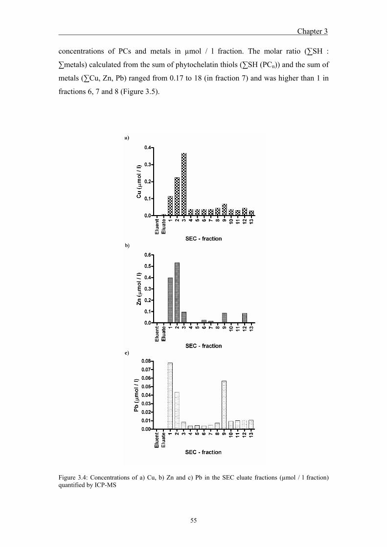
Chapter 3
55
concentrations of PCs and metals in µmol / l fraction. The molar ratio (∑SH :
∑metals) calculated from the sum of phytochelatin thiols (∑SH (PCn)) and the sum of
metals (∑Cu, Zn, Pb) ranged from 0.17 to 18 (in fraction 7) and was higher than 1 in
fractions 6, 7 and 8 (Figure 3.5).
Figure 3.4: Concentrations of a) Cu, b) Zn and c) Pb in the SEC eluate fractions (µmol / l fraction) quantified by ICP-MS
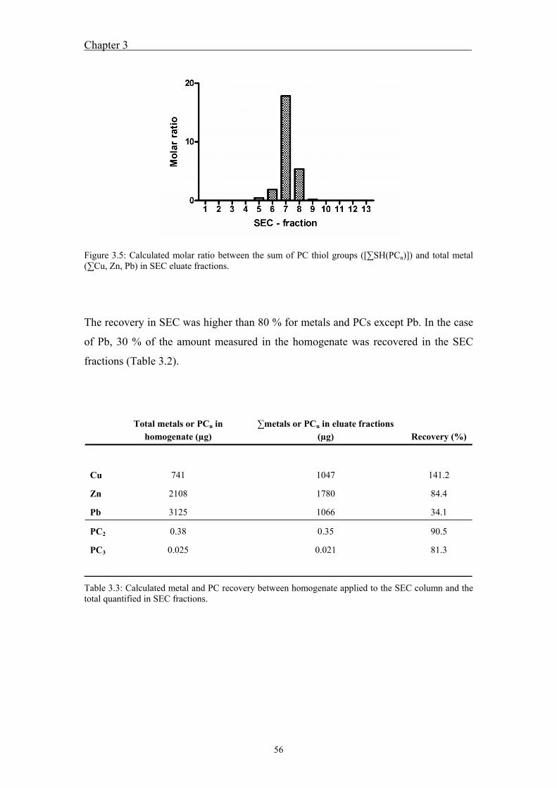
Chapter 3
56
Figure 3.5: Calculated molar ratio between the sum of PC thiol groups ([∑SH(PCn)]) and total metal (∑Cu, Zn, Pb) in SEC eluate fractions.
The recovery in SEC was higher than 80 % for metals and PCs except Pb. In the case
of Pb, 30 % of the amount measured in the homogenate was recovered in the SEC
fractions (Table 3.2).
Total metals or PCn in
homogenate (µg) ∑metals or PCn in eluate fractions
(µg) Recovery (%)
Cu 741 1047 141.2
Zn 2108 1780 84.4
Pb 3125 1066 34.1
PC2 0.38 0.35 90.5
PC3 0.025 0.021 81.3
Table 3.3: Calculated metal and PC recovery between homogenate applied to the SEC column and the total quantified in SEC fractions.
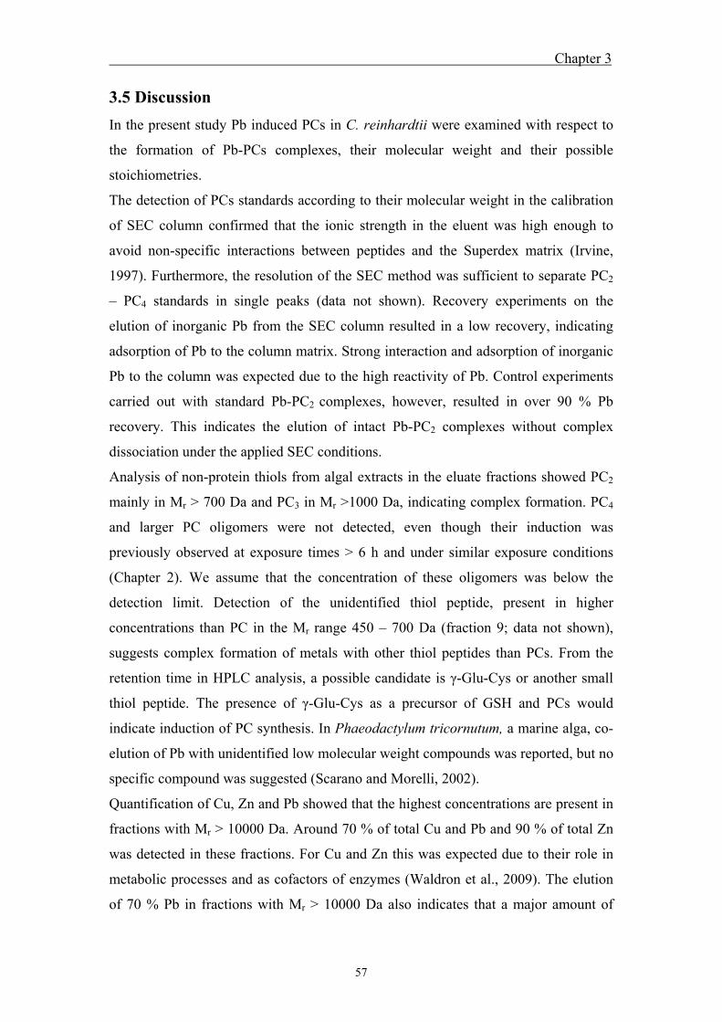
Chapter 3
57
3.5 Discussion
In the present study Pb induced PCs in C. reinhardtii were examined with respect to
the formation of Pb-PCs complexes, their molecular weight and their possible
stoichiometries.
The detection of PCs standards according to their molecular weight in the calibration
of SEC column confirmed that the ionic strength in the eluent was high enough to
avoid non-specific interactions between peptides and the Superdex matrix (Irvine,
1997). Furthermore, the resolution of the SEC method was sufficient to separate PC2
– PC4 standards in single peaks (data not shown). Recovery experiments on the
elution of inorganic Pb from the SEC column resulted in a low recovery, indicating
adsorption of Pb to the column matrix. Strong interaction and adsorption of inorganic
Pb to the column was expected due to the high reactivity of Pb. Control experiments
carried out with standard Pb-PC2 complexes, however, resulted in over 90 % Pb
recovery. This indicates the elution of intact Pb-PC2 complexes without complex
dissociation under the applied SEC conditions.
Analysis of non-protein thiols from algal extracts in the eluate fractions showed PC2
mainly in Mr > 700 Da and PC3 in Mr >1000 Da, indicating complex formation. PC4
and larger PC oligomers were not detected, even though their induction was
previously observed at exposure times > 6 h and under similar exposure conditions
(Chapter 2). We assume that the concentration of these oligomers was below the
detection limit. Detection of the unidentified thiol peptide, present in higher
concentrations than PC in the Mr range 450 – 700 Da (fraction 9; data not shown),
suggests complex formation of metals with other thiol peptides than PCs. From the
retention time in HPLC analysis, a possible candidate is γ-Glu-Cys or another small
thiol peptide. The presence of γ-Glu-Cys as a precursor of GSH and PCs would
indicate induction of PC synthesis. In Phaeodactylum tricornutum, a marine alga, co-
elution of Pb with unidentified low molecular weight compounds was reported, but no
specific compound was suggested (Scarano and Morelli, 2002).
Quantification of Cu, Zn and Pb showed that the highest concentrations are present in
fractions with Mr > 10000 Da. Around 70 % of total Cu and Pb and 90 % of total Zn
was detected in these fractions. For Cu and Zn this was expected due to their role in
metabolic processes and as cofactors of enzymes (Waldron et al., 2009). The elution
of 70 % Pb in fractions with Mr > 10000 Da also indicates that a major amount of
Chapter 3
58
cellular Pb is bound to macromolecules, most probably due to the high affinity of Pb
to oxygen, nitrogen and sulfur containing functional groups. The recovery of Zn and
Cu in the SEC was > 80 %, whereas only 30 % Pb was recovered. Considering the Pb
recovery of 3 % for inorganic Pb and > 90 % for standard Pb-PC2 complexes,
dissociation of non-specifically bound Pb from high Mr complexes and Pb adsorption
to the column matrix could have occurred, whereas the Pb-PC2 complexes were
probably recovered.
Cu, Zn and Pb were also observed in PC containing fractions, suggesting the
formation of Me-PC complexes. Cu and Zn are known as inducers of PC synthesis at
elevated concentrations, and complex formation was demonstrated in vitro (Rauser,
1995; Maitani et al., 1996; Chekmeneva et al., 2007). In the fraction with the highest
PC concentration, 1000 – 1600 Da, Cu and Zn concentrations were 10-fold and 3-fold
higher compared to Pb (Fig. 3.4). The high relative concentrations of Cu and Zn in
this fraction and the induction of PC synthesis observed under Pb exposure suggests
various possible functions of PCs. Firstly, PCs might be involved in Cu and Zn
homeostasis, which is supported by the results obtained in a previous study, showing
that PCs are also present in C. reinhardtii grown under optimal growth conditions
(Chapter 2). Secondly, induction of PC synthesis by Pb might cause a cellular
redistribution of these metals, leading to the formation of various Me-PC complexes,
or thirdly, Pb uptake leads to the displacement of Cu and Zn, which also induce PC
synthesis. Further research is needed to investigate the function of PCs concerning Cu
and Zn. To our knowledge, this is the first study that reports complex formation of PC
with other than the inducing metal.
Considering the elution of PC2 and PC3 in a wide molecular weight range, Me-PC
complexes of various stoichiometries may be assumed. In the Mr range 1000 – 1600
Da, molecular weight considerations indicate complexes with a general stoichiometry
of one or two metals forming a complex with two PC2 (Me1 – 2-(PC2)2). Depending on
the metal bound, the molecular weight of these complexes is 1287 Da or 1494 Da for
Pb1 – 2-(PC2)2, 1143 Da or 1206 Da for Cu1 – 2-(PC2)2 and 1145 Da or 1210 Da for Zn1
– 2-(PC2)2. Similar assumptions for PC3 suggest complex stoichiometries of Me1 – 3-
(PC3)2 in the Mr range 1600 – 2300 Da. Possible PC3 complexes detected in the Mr
range 1000 – 1600 Da are Pb2 – 3-PC3 complexes and the formation of complexes
involving PC2 and PC3 (e.g. Me1 – 3-PC2-PC3). In contrast to other studies (Scarano
and Morelli, 2002; Andra et al., 2009), PC2 was detected in molecular weights > 2000
Chapter 3
59
Da. In this Mr range formation of complexes involving PCs and cysteine (Andra et al.,
2009) or incorporation of sulfide, as observed in plants and yeast exposed to Cd
(Barbas et al., 1992; Mehra et al., 1994), is possible. The prevalent formation of Me-
(PC2)2 or Me-PC2-PC3 complexes is suggested by the high SH(PCn) / ∑Me ratios in
some of the fractions and by considering that complex stability increases with
increasing number of coordinating thiols (Alberich et al., 2007). Considering
formation of Me-(PC2)2 or Me-PC2-PC3 complexes involving Cu, Zn and Pb, PC-SH
is still in excess, which might indicate oxidation of PC-SH or metal dissociation from
PCs. The proposed stoichiometries of the Me-PC complexes are tentative, as
unambiguous identification of composition and stoichiometry of Me-PC complexes
requires higher mass resolution, such as mass spectrometry (Yen et al., 1999; Andra et
al., 2009).
This study indicates several roles of PCs in Pb exposed C. reinhardtii. Considering
that 13 % of total recovered Pb eluted in PC containing fractions, including also the
fraction containing the unidentified thiol and 10 % of total Pb, it seems that PCs have
a minor role in Pb detoxification. In a previous study, no toxic effects of Pb were
observed in short-term experiments but toxicity on photosynthesis and growth was
observed upon exposure for several days (Chapter 2). Considering that upon short-
term exposure no toxic effects were observed, although ROS formation in C.
reinhardtii upon Pb exposure was reported (Szivak et al., 2009), suggests that toxicity
may be prevented by antioxidant mechanisms. Toxicity observed upon long-term
exposure indicates that neither PCs nor other protective mechanisms are able to
prevent Pb toxicity. Additional research is needed to further examine Pb toxicity and
to identify the protective mechanisms involved in Pb detoxification in C. reinhardtii.
Chapter 3
60
3.6 References
Ahner, B.A., Kong, S., Morel, F.M.M., 1995. Phytochelatin production in marine algae. 1. An interspecies comparison. Limnol. Oceanogr. 40, 649-657.
Ahner, B.A., Morel, F.M.M., 1995. Phytochelatin production in marine algae. 2. Induction by various metals. Limnol. Oceanogr. 40, 658-665.
Alberich, A., Arino, C., Diaz-Cruz, J.M., Esteban, M., 2007. Soft modelling for the resolution of highly overlapped voltammetric peaks: application to some Pb-phytochelatin systems. Talanta. 71, 344-352.
Andra, S.S., Datta, R., Sarkar, D., Makris, K.C., Mullens, C.P., Sahi, S.V., Bach, S.B.H., 2009. Induction of lead-binding phytochelatins in vetiver grass Vetiveria zizanioides (L.). J. Environ. Qual. 38, 868-877.
Barbas, J., Ellis, W.R., Santhanagopalan, V., Blaszczynski, M., Winge, D.R., 1992. Conversion in the peptides coating cadmium:sulfide crystallites in Candida glabrata. J. Inorg. Biochem. 48, 95-105.
Chekmeneva, E., Diaz-Cruz, J.M., Arino, C., Esteban, M., 2007. Binding of Cd2+ and Zn2+ with the phytochelatin (γ-Glu-Cys)4-Gly: a voltammetric study assisted by multivariate curve resolution and electrospray ionization mass spectrometry. Electroanalysis. 19, 310-317.
Clemens, S., Kim, E.J., Neumann, D., Schroeder, J.I., 1999. Tolerance to toxic metals by a gene family of phytochelatin synthases from plants and yeast. EMBO. 18, 3325-3333.
Clemens, S., 2001. Molecular mechanisms of plant metal tolerance and homeostasis. Planta. 212, 475-486.
Gekeler, W., Grill, E., Winnacker, E.L., Zenk, M.H., 1988. Algae sequester heavy metals via synthesis of phytochelatin complexes. Arch. Microbiol. 150, 197-202.
Grill, E., Winnacker, E.L., Zenk, M.H., 1985. Phytochelatins: the principal heavy-metal complexing peptides of higher plants. Science. 230, 674-676.
Grill, E., Winnacker, E.L., Zenk, M.H., 1987. Phytochelatins, a class of heavy-metal-binding peptides from plants, are functionally analogous to metallothioneins. PNAS. 84, 439-443.
Gustafsson, J.P. Visual MINTEQ 2.15. 2005, Stockholm, Sweden. Hwww.lwr.kth.se/English/OurSoftware/vminteq/
Ha, S.B., Smith, A.P., Howden, R., Dietrich, W.M., Bugg, S., O'Connell, M.J., Goldsbrough, P.B., Cobbett, C.S., 1999. Phytochelatin synthase genes from Arabidopsis and the yeast Schizosaccharomyces pombe. Plant Cell. 11, 1153-1164.
Hu, S.X., Wu, M., 1998. Cadmium sequestration in the marine macroalga Kappaphycus alvarezii. Mol. Mar. Biol. Biotech. 7, 97-104.
Hu, S.X., Lau, K.W.K., Wu, M., 2001. Cadmium sequestration in Chlamydomonas reinhardtii. Plant Sci. 161, 987-996.
Irvine, G.B., 1997. Size-exclusion high-performance liquid chromatography of peptides: a review. Anal. Chim. Acta. 352, 387-397.
Chapter 3
61
Knauer, K., Behra, R., Sigg, L., 1997. Adsorption and uptake of copper by the green alga Scenedesmus subspicatus (Chlorophyta). J. Phycol. 33, 596-601.
Kneer, R., Zenk, M.H., 1997. The formation of Cd-phytochelatin complexes in plant cell cultures. Phytochemistry. 44, 69-74.
Le Faucheur, S., Behra, R., Sigg, L., 2005. Phytochelatin induction, cadmium accumulation, and algal sensitivity to free cadmium ion in Scenedesmus vacuolatus. Environ. Toxicol. Chem. 24, 1731-1737.
Le Faucheur, S., Schildknecht, F., Behra, R., Sigg, L., 2006. Thiols in Scenedesmus vacuolatus upon exposure to metals and metalloids. Aquat. Toxicol. 80, 355-361.
Maitani, T., Kubota, H., Sato, K., Yamada, T., 1996. The composition of metals bound to class III metallothionein (phytochelatin and its desglycyl peptide) induced by various metals in root cultures of Rubia tinctorum. Plant Physiol. 110, 1145-1150.
Mehra, R.K., Mulchandani, P., Hunter, T.C., 1994. Role of CdS quantum crystallites in cadmium resistance in Candida glabrata. Biochem. Biophys. Res. Commun. 200, 1193-1200.
Morelli, E., Scarano, G., 2001. Synthesis and stability of phytochelatins induced by cadmium and lead in the marine diatom Phaeodactylum tricornutum. Mar. Environ. Res. 52, 383-395.
Morelli, E., Cruz, B.H., Somovigo, S., Scarano, G., 2002. Speciation of cadmium-γ-glutamyl peptides complexes in cells of the marine microalga Phaeodactylum tricornutum. Plant Sci. 163, 807-813.
Pawlik-Skowronska, B., 2000. Relationships between acid-soluble thiol peptides and accumulated Pb in the green alga Stichococcus bacillaris. Aquat. Toxicol. 50, 221-230.
Pawlik-Skowronska, B., 2002. Correlations between toxic Pb effects and production of Pb-induced thiol peptides in the microalga Stichococcus bacillaris. Environ. Pollut.119, 119-127.
Rauser, W.E., 1995. Phytochelatins and related peptides - structure, biosynthesis, and function. Plant Physiol. 109, 1141-1149.
Rauser, W.E., 1999. Structure and function of metal chelators produced by plants - The case for organic acids, amino acids, phytin, and metallothioneins. Cell Biochem. Biophys. 31, 19-48.
Reese, R.N., Winge, D.R., 1988. Sulfide stabilization of the cadmium-γ-glutamyl peptide complex of Schizosaccharomyces pombe. J. Biol. Chem. 263, 12832-12835.
Scarano, G., Morelli, E., 2002. Characterization of cadmium- and lead-phytochelatin complexes formed in a marine microalga in response to metal exposure. Biometals. 15, 145-151.
Schmoger, M.E.V., Oven, M., Grill, E., 2000. Detoxification of arsenic by phytochelatins in plants. Plant Physiol. 122, 793-802.
Chapter 3
62
Szivak, I., Behra, R., Sigg, L., 2009. Metal-induced reactive oxygen species production in Chlamydomonas reinhardtii (chlorophyceae). Plant Physiol. 45, 427-435.
Vatamaniuk, O.K., Mari, S.P., Lu, Y.P., Rea, P.A., 2000. Mechanism of heavy metal ion activation of phytochelatin (PC) synthase. J. Biol. Chem. 275, 31451-31459.
Waldron, K.J., Rutherford, J.C., Ford, D., Robinson, N.J., 2009. Metalloproteins and metal sensing. Nature. 460, 823-830.
Yen, T. Y., Villa, J.A., DeWitt, J.G., 1999. Analysis of phytochelatin-cadmium complexes from plant tissue culture using nano-electrospray ionization tandem mass spectrometry and capillary liquid chromatography/electrospray ionization tandem mass spectrometry. J. Mass Spec. 34, 930-941.
Zenk, M.H., 1996. Heavy metal detoxification in higher plants - A review. Gene. 179, 21-30.
63
UChapter 4
Characterization of standard lead-phytochelatin complexes by nano-electrospray ionization mass
spectrometry

64
Chapter 4
65
4.1 Abstract
The role of phytochelatins (PCs) in metal detoxification is assumed to result from
immobilization of metals, preventing interaction with important biomolecules,
followed by the transport of the complexes into the vacuole of the algal cell.
Therefore the characterization of Me-PC complexes is important to predict the role of
PCs in the detoxification of a specific metal. Several analytical methods were used to
examine metal-phytochelatin complexes. Most of these methods do not provide exact
molecular weights, stoichiometries and compositions of the Me-PC complexes. The
aim of this study was to develop a nano-electrospray ionization mass spectrometry
method which allows unambiguous identification of Me-PC complexes. Standard Pb-
PCn complexes (n = 2 – 4) were used to develop a nano-ESI-MS method which allows
their identification and characterization. Pb-PCn mass spectra indicated the presence
of the [M+H]+ peak of PCn and complexes with various stoichiometries. A peak at
[M+H–2]+ in the zoom spectra of PCn indicated the formation of intramolecular
disulfide bonds. Standard Pb-PC2 analysis allowed the identification of four different
complexes observed at m/z 746.10, 952.06, 1285.24 and 1491.20, corresponding to
the singly charged Pb-PC2, Pb2-PC2, Pb-(PC2)2 and Pb2-(PC2)2 complexes. Their m/z
indicated coordination of Pb by PC2 through the thiol groups of PC cysteine and the
carboxyl groups of PC glutamic acid. For standard PC3 and PC4 two different
complexes were observed corresponding to Pb-PC3, Pb2-PC3, Pb-PC4 and Pb2-PC4.
The isotopic patterns for all complexes were identical to the theoretical isotopic
patterns. Addition of Zn to previously formed Pb-PC2 complexes showed the
appearance of the singly charged Zn-PC2 complex at m/z 602.06 and the decrease of
the Pb-PC2 peak. A general signal loss with increasing Zn concentration, most
probably due to the increase of oxidized PC2 was observed.
4.2 Introduction
Phytochelatins (PCs) are metal-binding oligopeptides with the general structure (γ-
Glu-Cys)n-Gly (n = 2 – 11) which are enzymatically produced from glutathione by the
enzyme phytochelatin synthase. PC synthesis is induced as a response to various
metals in plants (Rauser, 1995; Zenk, 1996) and algae (Gekeler et al., 1988; Ahner et
al., 1995). Because of their high cysteine content, PCs are assumed to bind metals
through thiolate coordination and might be involved in metal homeostasis and
Chapter 4
66
detoxification (Grill et al., 1985; Rauser, 1995; Zenk, 1996; Clemens, 2001). The role
of PCs in metal detoxification is assumed to result from immobilization of metals,
preventing non-specific binding to important biomolecules, followed by the transport
of the Me-PC complexes into the vacuoles. Therefore, Me-PC complexes need to be
analyzed to investigate whether PCs form complexes with a specific metal and might
be involved in its detoxification.
Several analytical methods such as chromatographic separation (gel filtration or
HPLC) coupled with UV detection, flame atomic absorption spectrometry (AAS),
radioactive labeling, differential pulse polarography and inductively coupled plasma
mass spectrometry (ICP-MS) have been used to analyze PCs and metal-phytochelatin
complexes (Me-PC) (Grill et al., 1985; Maitani et al., 1996; Leopold and Günther,
1997; Leopold et al., 1999; Vacchina et al., 1999; Schmoger et al., 2000; Vacchina et
al., 2000; Scarano and Morelli, 2002; Cruz et al., 2005; Kobayashi and Yoshimura,
2006). These methods, however, do not provide exact molecular weights,
stoichiometries or compositions of Me-PCn complexes. In most in vivo studies
focusing on Me-PC complex characterization, Me-PC complexes are isolated from the
cell homogenate by gel filtration and the collected eluate fractions are further
analyzed for PC and metal content. PC detection often involves acidification and
derivatization, which leads to dissociation of the Me-PC complexes, followed by
HPLC analysis. Based on the detected molecular weight range obtained from gel
filtration and the PC oligomers detected by HPLC, assumptions on stoichiometry and
composition of the Me-PC complexes can be made. However, unambiguous
characterization of Me-PC complexes regarding stoichiometry and composition is not
possible with these methods.
Considering that complex formation is assumed to be a key factor in metal tolerance
and detoxification, a technique to precisely detect and characterize these complexes is
required. Several studies reported identification of Cd-PC and As(III)-PC complexes
using electrospray ionization mass spectrometry (ESI-MS) (Yen et al., 1999; Raab et
al., 2005; Navaza et al., 2006; Chen et al., 2007; Bluemlein et al., 2008; Bluemlein et
al., 2009), but analysis of Me-PC complexes formed with other metals are not
reported.
In a previous study, induction of PC2 – PC4 synthesis by Pb was observed in the green
alga C. reinhardtii. Furthermore, PCn were shown to form complexes with Cu, Zn and
Pb upon Pb exposure. Therefore, the aim of this study was to develop an ESI-MS
Chapter 4
67
method to identify and characterize Me-PC complexes formed with Pb and to assess
the competition with Cu and Zn. In a first step, sample composition and ESI-MS
conditions to analyze standard Pb-PCn (n = 2 – 4) complexes were optimized. In a
second part of the study, the impact of Zn and Cu addition in various concentrations
to previously formed Pb-PC complexes was examined.
4.3 Material and Methods
4.3.1 Chemicals
Pb(NO3)2, CuSO4 and ZnSO4 salts, ammonium acetate (NH4CH3COO), ammonium
carbonate ((NH4)2CO3; pH 7), polylysine, and 3-morpholinopropanesulfonic acid
(MOPS) used in this study were analytical grade and obtained from Sigma-Aldrich
(St. Louis, MO, USA). Phytochelatin standards (PC2, PC3 and PC4) were obtained
from Invitrogen (San Diego, CA, USA). Formic acid was a suprapure chemical
obtained from Merck (Darmstadt, Germany). Ultrafree-MC centrifugal filters (0.45
µm cut-off) were ordered from Millipore AG (Zug, Switzerland).
1B4.3.2 Sample preparation
In preliminary experiments, the solvent mixture for the analysis of Pb-PC complexes
was optimized. The following sample composition resulted in the highest signal
intensities in nano-electrospray ionization mass spectrometry (nano-ESI-MS)
analysis. PCn and Pb were mixed, resulting in final concentrations of 0.5 mM PCn and
0.25 mM Pb(NO3)2 in 100 mM NH4CH3COO and 50 mM (NH4)2CO3. Complex
formation was allowed for 15 min at room temperature. Prior to sample analysis
formic acid was added to a final concentration of 0.1 % (pH 6), followed by sample
filtration using an Ultrafree-MC centrifugal filter device with a 0.45 µm cut-off.
2BFor competition experiments between Cu or Zn and Pb, standard Pb-PC2 complexes
were prepared as described and complex formation was allowed, followed by the
addition of the Cu or Zn solution. Final concentrations of Cu and Zn were 0.125 mM,
0.25 mM, 0.5 mM and 5 mM. Acidification was done by addition formic acid to a
final concentration of 0.1 %.

Chapter 4
68
4.3.3 Nano-electrospray ionization mass spectrometry (nano-ESI-MS) analysis of
metal-phytochelatin complexes
3BSolvent mixture optimization for nano-ESI-MS analysis of Pb-PCn (n = 2 – 4) was
carried out on a API4000 triple quadrupole mass spectrometer (Applied Biosystems,
Rotkreuz, Switzerland) with attached nanospray source (Sciex, NanoSpray® III
Source, Zug, Switzerland).
4BFinal analysis of Pb-PCn complexes was carried out on a LTQ Orbitrap XL mass
spectrometer (Thermo Fisher Scientific, San Jose, CA, USA). The nanospray capillary,
a coated fused silica tubing (TSP-FS; o.d. 375 µm; i.d. 100 µm; BGB, USA), was
pulled with a needle puller (Model P-2000, Sutter Instruments Co., Novato, CA,
USA). The resulting capillary was used for nanospray injection and was connected to
a 250 µl Hamilton gas tight syringe with a stainless steel needle (Hamilton Bonaduz
AG, Bonaduz, Switzerland). The sample was loaded into the syringe and injected into
the mass spectrometer by direct infusion. Between each measurement, the syringe was
cleaned with MeOH and H2O.
5BPrior to analysis of Pb-PCn complexes, the instrument performance and calibration
was checked with polylysine. The flow rate for standard Pb-PCn complex analysis
was 1 – 3 µl / min. The optimal settings of the mass spectrometer operated in positive
electrospray ionization mode were: ion spray voltage, 1.5 kV; capillary temperature,
200 °C; capillary voltage, 11 V; tube lens, 130 V; resolution, 30’000; max. injection
time, 100’000 ms; auto gain control FT, 1 x 10⁶. The spectra were acquired from m/z
300 – 1600 for Pb-PC2 and from m/z 300 – 2000 for Pb-PC3 and Pb-PC4. Mass
accuracy of the measurement was < 2 ppm for lower mass ions (< 1’000 Da).
6BMass spectra were acquired using LTQ Tune Plus V.2.5.5 (Thermo Fisher Scientific,
San Jose, CA, USA) and analyzed using the software Xcalibur V2.0.7 (Thermo Fisher
Scientific, San Jose, CA, USA). The isotopic distributions for the positively charged
molecular ions of Me-PC complexes were generated with the spectrum simulation
software integrated in Xcalibur.
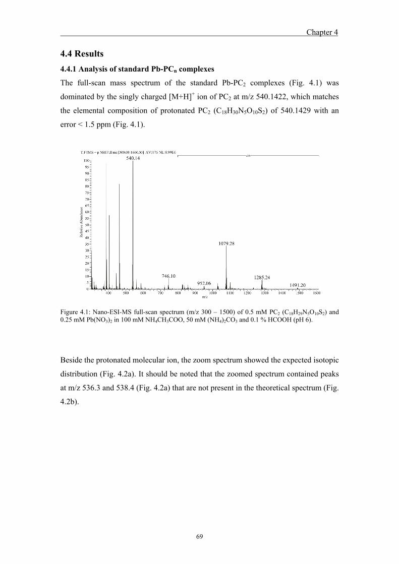
Chapter 4
69
4.4 Results
4.4.1 Analysis of standard Pb-PCn complexes
The full-scan mass spectrum of the standard Pb-PC2 complexes (Fig. 4.1) was
dominated by the singly charged [M+H]+ ion of PC2 at m/z 540.1422, which matches
the elemental composition of protonated PC2 (C18H30N5O10S2) of 540.1429 with an
error < 1.5 ppm (Fig. 4.1).
Figure 4.1: Nano-ESI-MS full-scan spectrum (m/z 300 – 1500) of 0.5 mM PC2 (C18H29N5O10S2) and 0.25 mM Pb(NO3)2 in 100 mM NH4CH3COO, 50 mM (NH4)2CO3 and 0.1 % HCOOH (pH 6).
Beside the protonated molecular ion, the zoom spectrum showed the expected isotopic
distribution (Fig. 4.2a). It should be noted that the zoomed spectrum contained peaks
at m/z 536.3 and 538.4 (Fig. 4.2a) that are not present in the theoretical spectrum (Fig.
4.2b).
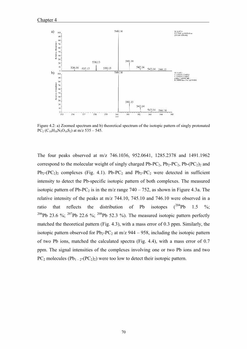
Chapter 4
70
Figure 4.2: a) Zoomed spectrum and b) theoretical spectrum of the isotopic pattern of singly protonated PC2 (C18H30N5O10S2) at m/z 535 – 545.
The four peaks observed at m/z 746.1036, 952.0641, 1285.2378 and 1491.1962
correspond to the molecular weight of singly charged Pb-PC2, Pb2-PC2, Pb-(PC2)2 and
Pb2-(PC2)2 complexes (Fig. 4.1). Pb-PC2 and Pb2-PC2 were detected in sufficient
intensity to detect the Pb-specific isotopic pattern of both complexes. The measured
isotopic pattern of Pb-PC2 is in the m/z range 740 – 752, as shown in Figure 4.3a. The
relative intensity of the peaks at m/z 744.10, 745.10 and 746.10 were observed in a
ratio that reflects the distribution of Pb isotopes (204Pb 1.5 %; 206Pb 23.6 %; 207Pb 22.6 %; 208Pb 52.3 %). The measured isotopic pattern perfectly
matched the theoretical pattern (Fig. 4.3), with a mass error of 0.3 ppm. Similarly, the
isotopic pattern observed for Pb2-PC2 at m/z 944 – 958, including the isotopic pattern
of two Pb ions, matched the calculated spectra (Fig. 4.4), with a mass error of 0.7
ppm. The signal intensities of the complexes involving one or two Pb ions and two
PC2 molecules (Pb1 – 2-(PC2)2) were too low to detect their isotopic pattern.
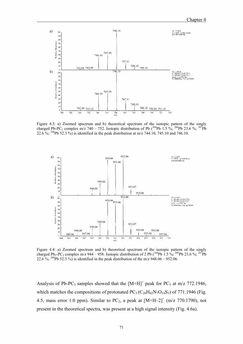
Chapter 4
71
Figure 4.3: a) Zoomed spectrum and b) theoretical spectrum of the isotopic pattern of the singly charged Pb-PC2 complex m/z 740 – 752. Isotopic distribution of Pb (204Pb 1.5 %; 206Pb 23.6 %; 207Pb 22.6 %; 208Pb 52.3 %) is identified in the peak distribution at m/z 744.10, 745.10 and 746.10.
Figure 4.4: a) Zoomed spectrum and b) theoretical spectrum of the isotopic pattern of the singly charged Pb2-PC2 complex m/z 944 – 958. Isotopic distribution of 2 Pb (204Pb 1.5 %; 206Pb 23.6 %; 207Pb 22.6 %; 208Pb 52.3 %) is identified in the peak distribution of the m/z 948.06 – 952.06.
Analysis of Pb-PC3 samples showed that the [M+H]+ peak for PC3 at m/z 772.1946,
which matches the compositions of protonated PC3 (C26H42N7O14S3) of 771.1946 (Fig.
4.5, mass error 1.0 ppm). Similar to PC2, a peak at [M+H–2]+ (m/z 770.1790), not
present in the theoretical spectra, was present at a high signal intensity (Fig. 4.6a).
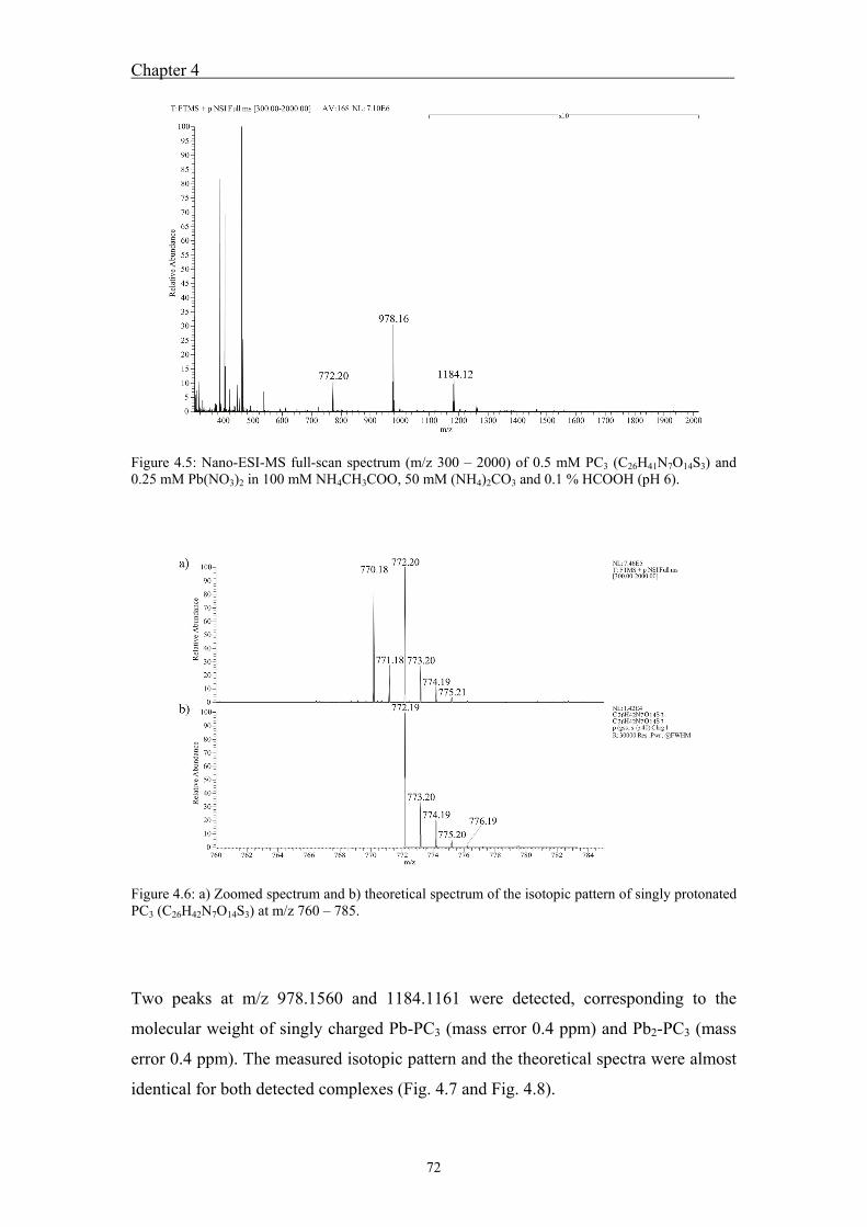
Chapter 4
72
Figure 4.5: Nano-ESI-MS full-scan spectrum (m/z 300 – 2000) of 0.5 mM PC3 (C26H41N7O14S3) and 0.25 mM Pb(NO3)2 in 100 mM NH4CH3COO, 50 mM (NH4)2CO3 and 0.1 % HCOOH (pH 6).
Figure 4.6: a) Zoomed spectrum and b) theoretical spectrum of the isotopic pattern of singly protonated PC3 (C26H42N7O14S3) at m/z 760 – 785.
Two peaks at m/z 978.1560 and 1184.1161 were detected, corresponding to the
molecular weight of singly charged Pb-PC3 (mass error 0.4 ppm) and Pb2-PC3 (mass
error 0.4 ppm). The measured isotopic pattern and the theoretical spectra were almost
identical for both detected complexes (Fig. 4.7 and Fig. 4.8).
Chapter 4
73
Figure 4.7: a) Zoomed spectrum and b) theoretical spectrum of the isotopic pattern of the singly charged Pb-PC3 complex m/z 970 – 990. Isotopic distribution of Pb (204Pb 1.5 %; 206Pb 23.6 %; 207Pb 22.6 %; 208Pb 52.3 %) is identified in the peak distribution at m/z 976.15, 977.15 and 978.16.
Figure 4.8: a) Zoomed spectrum and b) theoretical spectrum of the isotopic pattern of the singly charged Pb2-PC3 complex m/z 1175 – 1190. Isotopic distribution of 2 Pb (204Pb 1.5 %; 206Pb 23.6 %; 207Pb 22.6 %; 208Pb 52.3 %) is identified in the peak distribution of the m/z 1180.11 – 1184.12.
Analysis of the Pb-PC4 spectra showed the [M+H]+ peak for PC4 at m/z 1004.2406,
corresponding to the compositions of protonated PC4 (C34H54N9O18S4) of 1004.2464
(Fig. 4.9, mass error 5.7 ppm). Comparison of the [M+H]+ peak at 1004.2406 for PC4
to the theoretical spectra shows an excellent match of the isotopic patterns (Fig. 4.10),
Chapter 4
74
except the [M+H–2]+ peaks (m/z 1002.2238), as observed for PC2 and PC3, and the
[M+H–4]+ peak (m/z 1000.2084).
Figure 4.9: Nano-ESI-MS full-scan spectrum (m/z 300 – 2000) of 0.5 mM PC4 (C34H53N9O18S4) and 0.25 mM Pb(NO3)2 in 100 mM NH4CH3COO, 50 mM (NH4)2CO3 and 0.01 % HCOOH (pH 6).
Figure 4.10: a) Zoomed spectrum and b) theoretical spectrum of the isotopic pattern of singly protonated PC4 (C34H54N9O18S4) at m/z 990 – 1020.
Chapter 4
75
Two peaks corresponding to the molecular weight of the singly charged PC4
complexes, Pb-PC4 and Pb2-PC4, were detected at m/z 1210.1986 (mass error 7.3
ppm) and 1416.1564 (mass error 8.5 ppm). The measured and theoretical isotopic
patterns are shown in Figure 4.11 and Figure 4.12. In addition, a peak at m/z 605.6035
was observed, matching perfectly with the isotopic pattern of [Pb-PC4]2+ (Fig. 4.13).
Figure 4.11: a) Zoomed spectrum and b) theoretical spectrum of the isotopic pattern of the singly charged Pb-PC4 complex m/z 1200 – 1220. Isotopic distribution of Pb (204Pb 1.5 %; 206Pb 23.6 %; 207Pb 22.6 %; 208Pb 52.3 %) is identified in the peak distribution at m/z 1208.20, 1209.20 and 1210.20.
Figure 4.12: a) Zoomed spectrum and b) theoretical spectrum of the isotopic pattern of the singly charged Pb2-PC4 complex m/z 1405 – 1425. Isotopic distribution of 2 Pb (204Pb 1.5 %; 206Pb 23.6 %; 207Pb 22.6 %; 208Pb 52.3 %) is identified in the peak distribution of the m/z 1412.15 – 1416.16.
Chapter 4
76
Figure 4.13: a) Zoomed spectrum and b) theoretical spectrum of the isotopic pattern of the twicly charged Pb-PC4 complex m/z 480 – 500. Isotopic distribution of Pb (204Pb 1.5 %; 206Pb 23.6 %; 207Pb 22.6 %; 208Pb 52.3 %) is identified in the peak distribution of the m/z 604.60 – 605.60.
4.4.2 Competition between Cu or Zn and Pb for PC2 binding
Addition of Zn to Pb-PC2 complexes resulted in the appearance of the singly charged
Zn-PC2 peak at m/z 602.0547, already at the lowest Zn concentration (Fig. 4.14a). In
addition, the Pb-PC2 peak was observed to decrease with increasing Zn concentration
(Fig. 4.14).
Figure 4.14: Nano-ESI-MS full scan spectrum of competition experiments between Pb and Zn with increasing Zn concentrations from a) – c). The concentrations are a) 0.125 mM Zn, b) 0.25 mM Zn and c) 0.5 mM Zn.
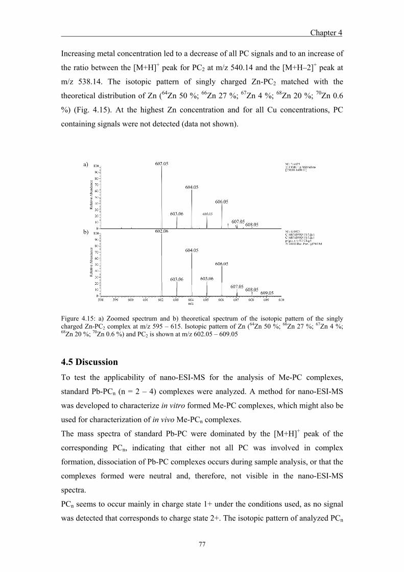
Chapter 4
77
Increasing metal concentration led to a decrease of all PC signals and to an increase of
the ratio between the [M+H]+ peak for PC2 at m/z 540.14 and the [M+H–2]+ peak at
m/z 538.14. The isotopic pattern of singly charged Zn-PC2 matched with the
theoretical distribution of Zn (64Zn 50 %; 66Zn 27 %; 67Zn 4 %; 68Zn 20 %; 70Zn 0.6
%) (Fig. 4.15). At the highest Zn concentration and for all Cu concentrations, PC
containing signals were not detected (data not shown).
Figure 4.15: a) Zoomed spectrum and b) theoretical spectrum of the isotopic pattern of the singly charged Zn-PC2 complex at m/z 595 – 615. Isotopic pattern of Zn (64Zn 50 %; 66Zn 27 %; 67Zn 4 %; 68Zn 20 %; 70Zn 0.6 %) and PC2 is shown at m/z 602.05 – 609.05
4.5 Discussion
To test the applicability of nano-ESI-MS for the analysis of Me-PC complexes,
standard Pb-PCn (n = 2 – 4) complexes were analyzed. A method for nano-ESI-MS
was developed to characterize in vitro formed Me-PC complexes, which might also be
used for characterization of in vivo Me-PCn complexes.
The mass spectra of standard Pb-PC were dominated by the [M+H]+ peak of the
corresponding PCn, indicating that either not all PC was involved in complex
formation, dissociation of Pb-PC complexes occurs during sample analysis, or that the
complexes formed were neutral and, therefore, not visible in the nano-ESI-MS
spectra.
PCn seems to occur mainly in charge state 1+ under the conditions used, as no signal
was detected that corresponds to charge state 2+. The isotopic pattern of analyzed PCn
Chapter 4
78
was completely resolved. For all PCn, the isotopic distribution matched the theoretical
spectra of the corresponding elemental composition (Fig. 4.2 / 4.6 / 4.10) and mass
accuracy was high (< 2 ppm for ions < 1’000 Da). The [M+H–2]+ peaks observed for
PC2 – PC4, as well as the [M+H–4]+ peak observed for PC4, indicate the formation of
one or two intramolecular disulfide bonds between cysteine thiol groups within PC.
The formation of a disulfide bond results in a loss of 2H+ and, therefore, a shift from
[M+H]+ to [M+H–2]+, which was also observed in other studies (Yen et al., 1999;
Navaza et al., 2006).
Analysis of Pb-PC complexes revealed the formation of Pb-PCn complexes with
various stoichiometries and compositions. The m/z detected for the Pb-PC2 complex
allows two complexes, assuming Pb coordination through thiol groups of PC cysteine.
Either Pb is coordinated by one thiol group whereas the other is present as reduced
thiol group, or the Pb ion is coordinated by both thiol groups present in PC2. In the
second case, additional protonation of the complex must occur to result in a singly
charged complex detectable by nano-ESI-MS. For the Pb2-PC2 complex, the detected
m/z corresponds to [PC2+Pb–3]+, indicating a loss of two H+. This observation
suggests that besides the thiols from cysteines, the carboxylic groups of the glutamic
acid must be involved in complex formation to obtain a singly charged Pb2-PC2
complex. Coordination of Pb by thiol and carboxyl functional groups is assumed to
result in increased complex stability. Further studies would be required to examine
whether the complex formation between one Pb and PC2 involves only thiol groups or
if Pb is coordinated by one thiol and one carboxylic group. Similarly, another study
observed the loss of 2H+ and 4H+ for the binding of two Cd ions to standard PC5.
Binding of a third Cd ion to PC5 was not accompanied with the loss of H+. The
authors suggested the formation of complexes that involve two thiol coordinated Cd
and a Cd that is bound electrostatically to the Cd2-PC5 complex (Yen et al., 1999). To
investigate whether the coordination of metals by PC is dependent on the metal and /
or the chain length of PC, more work needs to be conducted.
From m/z considerations, four different Pb-PC2, two Pb-PC3 and two Pb-PC4
complexes were identified. To prove that both, Pb and PCn, are present in detected
signals, the measured isotopic pattern was compared to the theoretical isotopic
pattern. The isotopic patterns of the Pb-PC complexes are complicated because of the
isotopic pattern of PC coupled to that of one or more Pb ions (204Pb 1.5 %; 206Pb 23.6 %; 207Pb 22.6 %; 208Pb 52.3 %). Comparison of the isotopic pattern of
Chapter 4
79
measured and theoretical Pb-PC2 at m/z 740 – 752 is indicative of the presence of Pb
in the complex, as its isotopic pattern can be identified. Furthermore, the zoomed
spectrum shows the PC2 specific isotopic distribution (Fig.4.3). The detection of the
various complexes also indicates that the formed complexes are stable enough to
detect them by nano-ESI-MS. Formation of Pb-PC2 and Pb2-PC2 complexes was also
observed using differential pulse polarography (Alberich et al., 2007). Similar
observations were made for Pb-PC3 and Pb-PC4 complexes, showing a good match
between measured and theoretical patterns and a loss of 2H+ for each additionally
bound Pb ion. Surprisingly, no complexes including three and four Pb ions were
observed for PC3 and PC4. In addition, accurate mass measurements confirmed the
proposed elemental compositions.
The singly charged Zn-PC2 peak appearing after Zn addition to Pb-PC2 complexes is
indicative for complex formation between PC and Zn, which was confirmed by the
isotopic pattern for Zn clearly visible in the zoomed spectrum (Fig. 4.15). The
expected increase of the Zn-PC2 peak with increasing Zn concentration was not
observed, perhaps due to an increase of oxidized PC2, indicated by the increase of the
ratio between m/z 538.14 and 540.14 and leading to a loss of potential metal binding
sites. This hypothesis could also explain the signal loss observed with increasing Zn
concentration. Similar observations were made in a study with Cd, where a signal loss
was observed at concentrations higher than 0.3 mM Cd (Yen et al., 1999).
A previous study using Pb exposed C. reinhardtii showed that formation of Me-PC
complexes includes Cu, Zn and Pb. PC2 and PC3 complexes were detected in a
molecular weight range between 700 – 5300 Da. PC2 was mainly observed between
1000 – 1600 Da and results suggested complexes with Me1 – 2-(PC2)2 (Chapter 3).
Most complexes detected in the present study have a stoichiometry of Pb-PC2 – 4 or
Pb2-PC2 – 4. The ratio of PC-SH to Pb was much higher in the gel filtration fractions
than in the present study, which possibly leads to the formation of different
complexes. Another possibility is that nano-ESI-MS, despite being a soft ionization
technique, results in the dissociation of complexes.
In vivo complexes were not analyzed by nano-ESI-MS because the concentration of
the complexes was not sufficient. Therefore, further research is needed to improve the
sensitivity for Me-PC complexes by nano-ESI-MS. In addition, both in vivo and in
vitro Me-PC complexes have to be further examined to investigate whether these
complexes differ.
Chapter 4
80
4.6 References
Ahner, B.A., Kong, S., Morel, F.M.M., 1995. Phytochelatin production in marine algae. 1. An interspecies comparison. Limnol. Oceanogr. 40, 649-657.
Alberich, A., Arino, C., Diaz-Cruz, J.M., Esteban, M., 2007. Soft modelling for the resolution of highly overlapped voltammetric peaks: application to some Pb-phytochelatin systems. Talanta. 71, 344-352.
Bluemlein, K., Raab, A., Meharg, A., Charnock, J., Feldmann, J., 2008. Can we trust mass spectrometry for determination of arsenic peptides in plants: comparison of LC–ICP–MS and LC–ES-MS/ICP–MS with XANES/EXAFS in analysis of Thunbergia alata. Anal. Bioanal. Chem. 390, 1739-1751.
Bluemlein, K., Raab, A., Feldmann, J., 2009. Stability of arsenic peptides in plant extracts: off-line versus on-line parallel elemental and molecular mass spectrometric detection for liquid chromatographic separation. Anal. Bioanal. Chem. 393, 357-366.
Chen, L., Guo, Y., Yang, L., Wang, Q., 2007. SEC-ICP-MS and ESI-MS/MS for analyzing in vitro and in vivo Cd-phytochelatin complexes in a Cd-hyperaccumulator Brassica chinensis. J. Anal. At. Spectrom. 22, 1403-1408.
Clemens, S., 2001. Molecular mechanisms of plant metal tolerance and homeostasis. Planta. 212, 475-486.
Cruz, B.H., Diaz-Cruz, J.M., Arino, C., Esteban, M., 2005. Complexation of heavy metals by phytochelatins: voltammetric study of the binding of Cd2+ and Zn2+ ions by the phytochelatin (γ-Glu-Cys)3Gly assisted by multivariate curve resolution. Environ. Sci. Technol. 39, 778-786.
Gekeler, W., Grill, E., Winnacker, E.-L., Zenk, M.H., 1988. Algae sequester heavy metals via synthesis of phytochelatin complexes. Arch. Microbiol. 150, 197-202.
Grill, E., Winnacker, E.L., Zenk, M.H., 1985. Phytochelatins: the principal heavy-metal complexing peptides of higher plants. Science. 230, 674-676.
Kobayashi, R., Yoshimura, E., 2006. Differences in the binding modes of phytochelatin to cadmium(II) and zinc(II) ions. Biol. Tra. Ele. Res. 114, 313-318.
Leopold, I., Günther, D., 1997. Investigation of the binding properties of heavy-metal-peptide complexes in plant cell cultures using HPLC-ICP-MS. Fre. J. Anal. Chem. 359, 364-370.
Leopold, I., Günther, D., Schmidt, J., Neumann, D., 1999. Phytochelatins and heavy metal tolerance. Phytochem. 50, 1323-1328.
Maitani, T., Kubota, H., Sato, K., Yamada, T., 1996. The composition of metals bound to class III metallothionein (phytochelatin and its desglycyl peptide) induced by various metals in root cultures of Rubia tinctorum. Plant Physiol. 110, 1145-1150.
Chapter 4
81
Navaza, A., Montes-Bayón, M., LeDuc, D.L., Terry, N., Sanz-Medel, A., 2006. Study of phytochelatins and other related thiols as complexing biomolecules of As and Cd in wild type and genetically modified Brassica juncea plants. J. Mass Spec. 41, 323-331.
Raab, A., Schat, H., Meharg, A.A., Feldmann, J., 2005. Uptake, translocation and transformation of arsenate and arsenite in sunflower Helianthus annuus: formation of arsenic;phytochelatin complexes during exposure to high arsenic concentrations. New Phytol. 168, 551-558.
Rauser, W.E., 1995. Phytochelatins and related peptides - structure, biosynthesis, and function. Plant Physiol. 109, 1141-1149.
Scarano, G., Morelli, E., 2002. Characterization of cadmium- and lead-phytochelatin complexes formed in a marine microalga in response to metal exposure. Biometals. 15, 145-151.
Schmoger, M.E.V., Oven, M., Grill, E., 2000. Detoxification of arsenic by phytochelatins in plants. Plant Physiol. 122, 793-802.
Vacchina, V., Chassaigne, H., Oven, M., Zenk, M.H., Lobinski, R., 1999. Characterisation and determination of phytochelatins in plant extracts by electrospray tandem mass spectrometry. Analyst. 124, 1425 - 1430.
Vacchina, V., Lobinski, R., Oven, M., Zenk, M.H., 2000. Signal identification in size-exclusion HPLC-ICP-MS chromatograms of plant extracts by electrospray tandem mass spectrometry (ES MS/MS). J. Anal. At. Spectrom. 15, 529 - 534.
Yen, T.Y., Villa, J.A., DeWitt, J.G., 1999. Analysis of phytochelatin-cadmium complexes from plant tissue culture using nano-electrospray ionization tandem mass spectrometry and capillary liquid chromatography/electrospray ionization tandem mass spectrometry. J. Mass Spec. 34, 930-941.
Zenk, M.H., 1996. Heavy metal detoxification in higher plants - A review. Gene. 179, 21-30.

82

83
UChapter 5
Outlook

84
Chapter 5
85
5.1 Outlook
This project showed that Pb is a potent inducer of phytochelatin synthesis in C.
reinhardtii, highlighting the importance of exposure time and concentration on PC
synthesis. The intracellular Pb concentration was observed to be similar after 6 h of
Pb exposure to the concentrations 10-8 M and 10-9 M, indicating saturation of Pb
accumulation. This saturation indicates regulation of Pb accumulation either by
limitation of uptake or by excretion of accumulated metal. Despite the detection of
similar intracellular concentrations, induction of PC synthesis was observable within
minutes at 10-8 M, but only upon long-term exposure to 10-9 M, indicating the
importance of the subcellular metal distribution for the induction of PC synthesis. We
assume that regulation of Pb accumulation leads to a redistribution of metals, essential
and non-essential, in the cell, which induces PC synthesis. Redistribution of all metals
may also explain complex formation between PCs and essential metals, such as Cu
and Zn. To explore whether metal redistribution is responsible for induction of PC
synthesis, the subcellular distribution of metals could be examined as a function of
time. A few experimental procedures for subcellular fractionation of algae were
reported in recent publications, which might be used for this purpose (Wang and
Rainbow, 2006; Wang and Wang, 2008; Lavoie et al., 2009a; Lavoie et al., 2009b).
Subcellular fractionation might also give indications why toxic effects on
photosynthesis and growth were only observed upon long-term exposure and not in
short-term experiments.
This work further indicated that nano-electrospray ionization mass spectrometry is
suitable for identification and characterization of lead-phytochelatin (Pb-PC)
complexes. However, additional research using this method is required to investigate
whether the stoichiometry of Pb-PC complexes is determined by the ratio of PC-SH to
metal or if the composition of the surrounding medium has an influence on complex
stoichiometry. Nano-ESI-MS also allows the investigation of whether coordination of
metals by PCs is dependent on the metal, the chain length of the involved PC
oligomers, or on both. Furthermore, improvement of the solvent mixture for metal
competition experiments might allow additional research on competition of metals for
PC binding, giving more information on the complex stability of Me-PC complexes
formed with various metals.
Chapter 5
86
At the environmentally relevant free Pb ion concentration, 10-9 M, Pb is accumulated
in algal cells, reaching a high concentration close to saturation, after short exposure
times. Furthermore, despite induction of PC synthesis, long-term exposure to this
concentration caused photosynthetic and growth inhibition. This finding indicates that
PCs are not protective against toxic effects of Pb upon long-term exposure. Because
of these delayed toxic effects observed in C. reinhardtii, it is important to examine
long-term exposures to assess the toxicity of Pb on organisms.
5.2 References
Lavoie, M., Bernier, J., Fortin, C., Campbell, P.G.C., 2009a. Cell homogenization and subcellular fractionation in two phytoplanktonic algae: implications for the assessment of metal subcellular distributions. Limnol. Oceanogr.: Methods. 7, 277-286.
Lavoie, M., Le Faucheur, S., Fortin, C., Campbell, P.G.C., 2009b. Cadmium detoxification strategies in two phytoplankton species: Metal binding by newly synthesized thiolated peptides and metal sequestration in granules. Aquat. Toxicol. 92, 65-75.
Wang, M., Wang, W.X., 2008. Cadmium toxicity in a marine diatom as predicted by the cellular metal sensitive fraction. Environ. Sci Technol. 42, 940-946.
Wang, W., Rainbow, P.S., 2006. Subcellular partitioning and the prediction of cadmium toxicity to aquatic organisms. Environ. Chem. 3, 395-399.

87
Acknowledgements

88
89
I would like to express my gratitude to my two advisors Laura Sigg and Renata Behra,
who gave me the opportunity for this thesis, which introduced me into the various
fields about metals, algae and detoxification. I would also like to gratefully
acknowledge Prof. C. Fortin, who came from Canada to assist at my PhD defense, and
Prof. B. Wehrli who accepted to be a member of my committee.
Special thanks go out to all my lab colleagues from whom I learned a lot during my
thesis. First of all I am very thankful to Bettina Wagner for her help in the laboratory,
for everything she taught me about algae and for the nice moments we had in the lab.
Further thanks go to David Kistler for his help with the ICP-MS measurements, René
Schönenberger and Marc Suter for their help with the nano-ESI-MS measurements
and data analysis and to Adrian Amman for the helpful discussions on SEC.
Thanks also go to Flavio, Danielle, Ilona, Irène, Thea, Miriam, Philippe and all other
colleagues in UTOX and at Eawag who supported me in my work and mentally in
moments of doubt.
I would also like to thank my family for their support and caring over these years. My
immense gratitude goes to my girlfriend, Franziska, who supported me with her love
and kindness. Her patience and help in the daily life gave me the spirit and power to
finish my PhD.
Finally I also thank the Swiss National Science Foundation for their financial support.