Embed Size (px)
Citation preview
ORIGINAL PAPER
Induction of death receptor 5 expression in tumor vasculatureby perifosine restores the vascular disruption activityof TRAIL-expressing CD34+ cells
Arianna Giacomini • Marco Righi • Loredana Cleris • Silvia L. Locatelli •
Stefania Mitola • Maria Grazia Daidone • Alessandro M. Gianni •
Carmelo Carlo-Stella
Received: 12 October 2012 / Accepted: 15 April 2013
� Springer Science+Business Media Dordrecht 2013
Abstract The proapoptotic death receptor 5 (DR5)
expressed by tumor associated endothelial cells (TECs)
mediates vascular disrupting effects of human CD34? cells
engineered to express membrane-bound tumor necrosis
factor-related apoptosis-inducing ligand (CD34-TRAIL1
cells) in mice. Indeed, lack of DR5 on TECs causes
resistance to CD34-TRAIL1 cells. By xenografting in
nonobese diabetic/severe combined immunodeficient mice
the TRAIL-resistant lymphoma cell line SU-DHL-4V,
which generates tumors lacking endothelial DR5 expres-
sion, here we demonstrate for the first time that the Akt
inhibitor perifosine induces in vivo DR5 expression on
TECs, thereby overcoming tumor resistance to the vascular
disruption activity of CD34-TRAIL1 cells. In fact,
CD34-TRAIL1 cells combined with perifosine, but not
CD34-TRAIL1 cells alone, exerted marked antivascular
effects and caused a threefold increase of hemorrhagic
necrosis in SU-DHL-4V tumors. Consistent with lack of
DR5 expression, CD34-TRAIL1 cells failed to affect the
growth of SU-DHL-4V tumors, but CD34-TRAIL1 cells
plus perifosine reduced tumor volumes by 60 % compared
with controls. In view of future clinical studies using
membrane-bound TRAIL, our results highlight a strategy
to rescue patients with primary or acquired resistance due
to the lack of DR5 expression in tumor vasculature.
Keywords Tumor endothelial cells � Death receptor 5 �Perifosine � TRAIL-expressing CD34? cells � Vascular
disruption activity
Introduction
PI3K/Akt signaling pathway represents an attractive target
for anticancer therapy [1–5]. Many new agents targeting
this pathway are currently being developed [6]. Perifosine
Electronic supplementary material The online version of thisarticle (doi:10.1007/s10456-013-9348-7) contains supplementarymaterial, which is available to authorized users.
A. Giacomini � S. L. Locatelli � C. Carlo-Stella (&)
Department of Oncology and Hematology, Humanitas Cancer
Center, Humanitas Clinical and Research Center, Via Manzoni,
56, 20089 Rozzano, Milan, Italy
e-mail: [email protected]
A. Giacomini � M. Righi � S. L. Locatelli � C. Carlo-Stella
Department of Medical Biotechnology and Translational
Medicine, University of Milan, Milan, Italy
M. Righi
National Research Council, Institute of Neuroscience, Milan,
Italy
L. Cleris � M. G. Daidone
Experimental Oncology, Fondazione IRCCS Istituto Nazionale
Tumori, Milan, Italy
S. Mitola
Department of Molecular and Translational Medicine,
University of Brescia, Brescia, Italy
A. M. Gianni (&)
Medical Oncology, Fondazione IRCCS Istituto Nazionale
Tumori, Via Venezian 1, 20133 Milan, Italy
e-mail: [email protected]
A. M. Gianni
Department of Medical Physiopathology and Transplants,
University of Milan, Milan, Italy
123
Angiogenesis
DOI 10.1007/s10456-013-9348-7
is an oral Akt inhibitor that is being tested in phase I/II
clinical trials [7–11]; this drug is a synthetic alkyl-
phospholipid (APL) that targets the pleckstrin homology
domain of Akt, thereby preventing its translocation to the
plasma membrane [12–15]. Recent reports have shown that
the in vitro sensitivity of cancer cell lines to tumor necrosis
factor (TNF)-related apoptosis-inducing ligand (TRAIL)
can be considerably increased by cotreatment with perifo-
sine due to significant upregulation of death receptors 4/5
(DR4/5) expression as well as downregulation of the
antiapoptotic protein cFLIP [16–18].
TRAIL, a proapoptotic member of the TNF superfamily
of death receptor ligands, is an attractive cancer cell-specific
cytotoxic agent that exerts remarkable antitumor activity
in vitro and in vivo in athymic nude mice as well as in
non-obese diabetic/severe combined immunodeficient
(NOD-SCID) mice [19–29]. The therapeutic activity of
recombinant soluble (s)TRAIL and of monoclonal antibod-
ies targeting TRAIL receptors has been extensively explored
in phase I/II clinical trials [30–38]. Despite their low toxicity
profiles, these molecules have so far shown limited, if any,
clinical activity, likely due to the short half-life, the pattern of
receptor expression or tumor cell resistance. We have pre-
viously demonstrated that adenovirus-transduced CD34?
cells expressing full-length membrane-bound (m)TRAIL
(CD34-TRAIL1 cells) efficiently vehiculate TRAIL within
tumors and exhibit tumor-specific targeting, overcoming
limitations inherent to the pharmacokinetic profile of the
soluble molecule [39, 40]. While the intratumor homing of
CD34-TRAIL1 cells involves interactions of SDF-1 and
VCAM-1 expressed by tumor endothelial cells with their
counter receptors (CXCR4 and VLA-4) expressed by
transduced cells, the antitumor activity of mTRAIL requires
DR5 expression by tumor vasculature [40, 41]. Engagement
of DR5 by CD34-TRAIL1 cells seems to be a critical step
that results in potent vascular disruption activity, which is a
distinct property of the membrane-bound TRAIL not shared
by its soluble counterpart [40, 41]. Because vascular tar-
geting appears to be the first step in the complex cascade of
events leading to mTRAIL-mediated tumor cell death, lack
of DR5 expression in tumor vasculature represents a mech-
anism of resistance to CD34-TRAIL1 cells. Thus, modu-
lation of DR5 expression on TECs might not only enhance
the antitumor efficacy of CD34-TRAIL1 cells but also
eventually overcome CD34-TRAIL1 cells resistance
caused by the lack of DR5 expression in tumor vasculature.
Since in vivo modulation of TRAIL receptors on TECs
by perifosine has never been reported, we analyzed endo-
thelial DR5 levels in perifosine-treated tumors and inves-
tigated the antitumor effects of CD34-TRAIL1 cells when
combined with perifosine. At this aim, two human tumor
xenografts growing in NOD/SCID mice were used: the
TRAIL-resistant diffuse large B cell lymphoma cell line
SU-DHL-4V, which generates in vivo nodules lacking DR5
expression on TECs, and the TRAIL-sensitive multiple
myeloma cell line KMS-11, which generates in vivo nod-
ules expressing DR5 on TECs. Here we demonstrate that
perifosine induces expression of DR5 in tumor vasculature
and highlight a strategy for overcoming resistance to
CD34-TRAIL1 cells vascular disruption activity. These
results appear to be relevant in view of future clinical
studies using CD34-TRAIL1 cells as well as other
membrane-bound or cross-linked forms of TRAIL.
Materials and methods
Reagents
Perifosine was purchased from Aeterna Zentaris
(Frankfurt, Germany, EU); sulfosuccinimidyl-6-(biotinamido)
hexanoate (sulfo-NHS-LC-biotin) was purchased from
Thermo Fisher Scientific (Rockford, IL, USA) and the specific
JNK inhibitor SP600125 was purchased from Calbiochem
(Billerica, MA, USA).
Cell lines and CD34? cells
The tumor-derived endothelial cell line 2H-11 was
obtained from the American Type Culture Collection
(ATCC, Teddington, UK, EU). The multiple myeloma cell
line KMS-11 has been previously described [42]; the dif-
fuse large B cell lymphoma cell line SU-DHL-4 were
obtained from the German Collection of Microorganisms
and Cell Cultures (DSMZ, Braunschweig, Germany, EU).
The SU-DHL-4V cell line was generated in our laboratory
after in vivo passaging of the SU-DHL-4 cell line in
NOD/SCID mice. SU-DHL-4V cells are TRAIL-resistant
and generate in vivo nodules in which TECs lack DR5
expression. KMS-11 cells are TRAIL-sensitive and gen-
erate in vivo nodules in which the TECs express DR5.
2H-11 cell line was cultured in DMEM supplemented with
10 % fetal bovine serum (FBS), whereas the SU-DHL-4V
and KMS-11 cell lines were cultured in RPMI-1640 sup-
plemented with 10 % FBS. All cell lines were tested for
mycoplasma contamination. CD34? cells were positively
selected from the peripheral blood of consenting cancer
patients undergoing peripheral blood stem cell mobiliza-
tion using an AutoMACS device (Miltenyi Biotec,
Bergisch-Gladbach, Germany, EU).
Adenoviruses and adenoviral transduction of CD34?
cells
Adenoviral transduction was performed as previously
described [39, 43].
Angiogenesis
123
siRNA gene silencing
2H-11 cells were transfected with 100 nM of either
Silencer� Select DR5 small interference RNA (siRNA) or
Silencer� Select Negative Control siRNA (Ambion,
Carlsbad, CA, USA), according to the instructions of the
manufacturer. 24 h after siRNA transfection, 2H-11 cells
were treated with either CD34-TRAIL1 cells or perifosine
5 lM or CD34-TRAIL1 cells/perifosine in combination.
Gene silencing effects were evaluated by western blot and
viable cell counting 48 h after treatments.
Viable cell counting
Annexin-V-fluorescein isothiocyanate/propidium iodide
(PI) double staining (Immunostep, Salamanca, SP, EU)
was used to detect Annexin-V-/PI- viable cells by flow
cytometry. Absolute cell counts were obtained by the true
volumetric absolute counting function of the CyFlow�
Space flow cytometer (Partec, Munster, Germany, EU).
In vivo experiments in nonobese diabetic/severe
combined immunodeficient (NOD/SCID) mice
Six- to eight-week-old female NOD/SCID mice with body
weights of 20–25 g were purchased from Charles River
(Milano, Italy, EU). The mice were housed under standard
laboratory conditions according to our institutional guide-
lines. Animal experiments were performed according to the
Italian laws (D.L. 116/92 and following additions) that
enforce the EU 86/109 Directive and were approved by the
institutional Ethical Committee for Animal Experimenta-
tion. KMS-11 and SU-DHL-4V cells (5 9 106 cells/
mouse) were inoculated subcutaneously (SC) in the left
flank of each mouse. When tumors were palpable, mice
were randomly assigned to receive either short- or long-
term treatments with either CD34-TRAIL1 cells, perifo-
sine or CD34-TRAIL1 cells plus perifosine. Short-term
treatments consisted of either a single intravenous (IV)
injection of CD34-TRAIL1 cells (3 9 106 cells/mouse,
on day 6 after SU-DHL-4V inoculation and day 12 after
KMS-11 inoculation), one 5-days course of oral (PO)
perifosine (15 mg/Kg/day, days 5–9 for SU-DHL-4V and
days 11–15 for KMS-11), or a combination of both. In vivo
biotinylation of tumor vasculature was performed where
appropriate 72 h after the injection of CD34-TRAIL1
cells and 3 h after the last perifosine administration. Tumor
nodules were then excised and processed for either histo-
logical analysis or ex vivo cytofluorimetric analysis. Long-
term treatments consisted of either four daily injections of
CD34-TRAIL1 cells (1 9 106 cells/mouse/day, days 6–9
for SU-DHL-4V and days 12–15 for KMS-11), or two
5-day courses of perifosine (10 mg/Kg/day, days 5–9 and
11–15 for SU-DHL-4V; 5 mg/Kg/day, days 11–15 and
18–22 for KMS-11), or a combination of both. Control
mice received normal saline. The mice were checked twice
weekly for the appearance of tumors, tumor dimensions,
body weight, and toxicity. The endpoint of the long-term
treatment was tumor weight. Tumor volumes were mea-
sured with calipers, and their weights were calculated using
the formula (a 9 b2)/2, where a and b represented the
longest and shortest diameters, respectively. The mice were
followed for 2 weeks after the end of the treatments. Each
experiment was performed on at least three separate
occasions using 5 mice per treatment group. Doses of
perifosine were determined as reported in Online resource 1,
Fig. S1.
In vivo biotinylation of tumor vasculature
In order to analyze functional tumor vasculature, in vivo
biotinylation was performed as previously described [40].
TECs separation from tumor xenografts
SU-DHL-4V and KMS-11 tumor nodules from mice
receiving the vehicle control or one 5-days course of
perifosine (15 mg/kg/day) were processed for ex vivo cyto-
fluorimetric analysis. Briefly, tumor nodules were excised,
immersed in 5–10 ml of DMEM medium with 10 % FBS
(Fetal Bovine Serum) and fragmented with a GentleMacs
machine (Miltenyi Biotech) to obtain a cell suspension. When
the nodules were completely homogenized, the suspensions
were incubated for 45 min at 37 �C under constant rotation
and in the presence of Collagenase D (at a final concentration
of 2 mg/ml) and DNase (at a final concentration of 25 lg/ml).
The cell suspensions were then washed twice at 300 g for
15 min to inactivate Collagenase D and eliminate platelets
and were finally passed through a 30 lm filter and labeled
with magnetic bead-conjugated anti-mouse CD146 antibody
(Miltenyi Biotech) for endothelial cell immunomagnetic
sorting by an AutoMACS device (Miltenyi Biotech). Labeling
was performed according to the manufacturer’s instructions.
Flow cytometric analysis
2H-11 cells co-cultured or not with tumor cells were har-
vested after 72 h of exposure to perifosine 5 lM and labeled
with PE-conjugated anti-DR5 antibody (Biolegend, San
Diego, CA, USA). For ex vivo flow cytometric analysis,
CD146? subpopulations were collected and labeled with a
FITC-conjugated anti-mouse CD146 antibody (Miltenyi
Biotech), PE-conjugated anti-mouse DR5 antibody (Bio-
legend) and 7-AAD to exclude dead cells. Labeling was
carried out according to the manufacturer’s instructions.
Angiogenesis
123
Samples were analyzed with a FACSCalibur flow cytometry
system (BD Biosciences) equipped with a Macintosh Pow-
erMac G4 personal computer (Apple Computer, Cupertino,
CA, USA) using CellQuest software (BD Biosciences). The
percentages of positive cells were measured according to
gates set on the basis of isotype control staining. The inten-
sity of receptor expression was measured as the mean fluo-
rescence intensity (MFI) ratio, which was calculated as the
MFI of anti- DR5-stained samples divided by the MFI of
isotype control-stained samples.
Western blot analysis
Cell samples were washed in cold PBS (Phosphate Buf-
fered Saline) and homogenized in NP-40 lysis buffer (1 %
NP-40, 20 mM Tris–HCl pH 8, 137 mM NaCl, 10 %
glycerol, 2 mM EDTA, 1 mM sodium orthovanadate,
10 lg/mL aprotinin, 10 lg/mL leupeptin). Protein con-
centrations were determined using the Bradford protein
assay (Bio-Rad Laboratories, Milano, Italy, EU). Blotting
analysis was performed using anti-phospho Akt, anti-total
Akt, anti-phospho JNK, anti-total JNK and anti-phospho
c-Jun antibodies from Cell Signaling (Danvers, MA, USA),
and anti-CD31 and anti-DR5 antibodies from Santa Cruz
Biotechnology (Santa Cruz, CA, USA). To normalize the
amount of loaded protein, all blots were stripped and
reprobed with rabbit polyclonal antibody anti–ß-actin (GE
Healthcare Life Sciences, Milano, Italy, EU).
Reverse transcription-PCR
Total RNA was isolated with TRIzol reagent (Invitrogen,
Carlsbad, CA, USA). Conventional reverse transcription-
PCR and real-time PCR were performed as described
previously [39]. The following primers were used to carry
out real time PCR:
murine DR5: 50-TGCTGCTCAAGTGGCGC-30 and
50-GGCATCCAGCAGATGGTTG-30; murine TBP (inter-
nal control): 50-GGTGTGCACAGGAGCCAAGAGTG-30
and 50-AGCTACTGAACTGCTGGTGGGTC-30
Immunofluorescence and confocal microscopy
Sections (4 lm thick) of frozen tumors and healthy organs
(including lungs, livers and spleens) from in vivo biotin-
ylated mice were fixed with acetone (for 5 min at 4 �C),
rinsed with PBS, and blocked with 2 % BSA (Bovine
Serum Albumine) in PBS. Sections were then incubated
with the appropriate primary antibody (for 1 h at room
temperature), consisting in rabbit anti-human/mouse
phospho-Akt (Cell Signaling), rat anti-mouse CD31 (BD
Pharmingen) or rabbit anti-mouse CD146 (Novus Biolog-
icals, Littleton, CO, USA). After washing with PBS,
sections were incubated with the appropriate Alexa Fluor
488 or 568-conjugated secondary antibody (Invitrogen).
Biotinylated tumor vessels were revealed with Alexa Fluor
488-conjugated streptavidin (Invitrogen). TdT-mediated
dUTP nick end labeling (TUNEL) staining (Roche, Milano,
Italy, EU) was performed according to the manufacturer’s
instructions in sections from biotinylated, formalin-fixed,
paraffin-embedded tumor nodules, and dead cells were
detected in the green channel. TUNEL-stained sections
were washed with PBS containing 0.05 % Tween 20,
blocked with 2 % BSA (for 10 min at room temperature)
and incubated with Alexa Fluor 568-conjugated streptavi-
din (Invitrogen) to detect apoptotic tumor vessels or with a
rat anti-mouse glycophorin A antibody (BD Pharmingen)
followed by the appropriate Alexa Fluor 568-conjugated
secondary antibody (Invitrogen) to detect hemorrhagic
necrosis areas. Double-stained sections were finally incu-
bated with TO-PRO-3 nuclear dye (Invitrogen). After
mounting in a drop of anti-bleaching mounting medium
(Vectashield, Vector Laboratories, Burlingame, CA, USA),
stained sections were examined under an epifluores-
cent microscope equipped with a laser confocal system
(MRC-1024, Bio-Rad Laboratories, Milano, Italy, EU).
Image processing was carried out with LaserSharp com-
puter software (Bio-Rad Laboratories).
Histological analysis and immunohistochemistry
Formalin-fixed, paraffin-embedded tumor nodules and
healthy organs (including lungs, livers, spleens and femurs)
were sectioned at a thickness of 2 lm, dewaxed, hydrated,
and stained with hematoxylin and eosin or processed for
immunohistochemistry. Tumor cell death was detected
using TUNEL staining (Roche) according to the manu-
facturer’s instructions. Cryostat sections (4 lm thick) of
in vivo biotinylated tumor nodules were dried on positively
charged glass slides and incubated for 1 h at room tem-
perature with 1 lg/ml horseradish peroxidase (HRP)-con-
jugated streptavidin diluted in PBS. Biotinylated tumor
endothelia were revealed by 3,30-diaminobenzidine stain-
ing, and tumor sections were then counterstained with
Carazzi’s hematoxylin, rapidly dehydrated using graded
ethanols to xylene and mounted in a drop of fast-drying
mounting medium. Sections were examined under a light
microscope (BX51; Olympus, Tokyo, Japan) and acquired
with an automatic high-resolution scanner (dotSlide Sys-
tem; Olympus). Image processing was carried out using
dotSlide software (Olympus).
Analysis of stained sections
Image analysis was performed using the open source
ImageJ software (http://rsb.info.nih.gov/ij/). Routines for
Angiogenesis
123
image analysis were coded in ImageJ macro language and
executed on RGB images without further treatment. For
each experimental condition, at least three sections from
different tumor nodules were analyzed. Analysis to calcu-
late the endothelial area (i.e., the percentage of the tissue
section occupied by endothelium) and the percent of tumor
necrosis expressed as (necrotic area)/(total tissue
area) 9 100 were performed as previously described [40].
Statistical analyses
Statistical analyses were performed using the statistical
package Prism 5 (GraphPad Software) run on a Macintosh
Pro personal computer (Apple Computers). Student’s t test
for unpaired data (2-tailed) was used to test the probability
of significant differences between untreated and treated
samples. TUNEL staining and tumor vasculature data were
statistically analyzed with a 1-way analysis of variance,
and individual group comparisons were evaluated by the
Bonferroni multiple comparison test. Tumor volume data
were statistically analyzed with a 2-way analysis of vari-
ance, and individual group comparisons were evaluated by
the Bonferroni correction. Differences were considered
significant if P was B0.05.
Results
Perifosine inhibits Akt phosphorylation and increases
DR5 expression in tumor-derived endothelial cells
in vitro
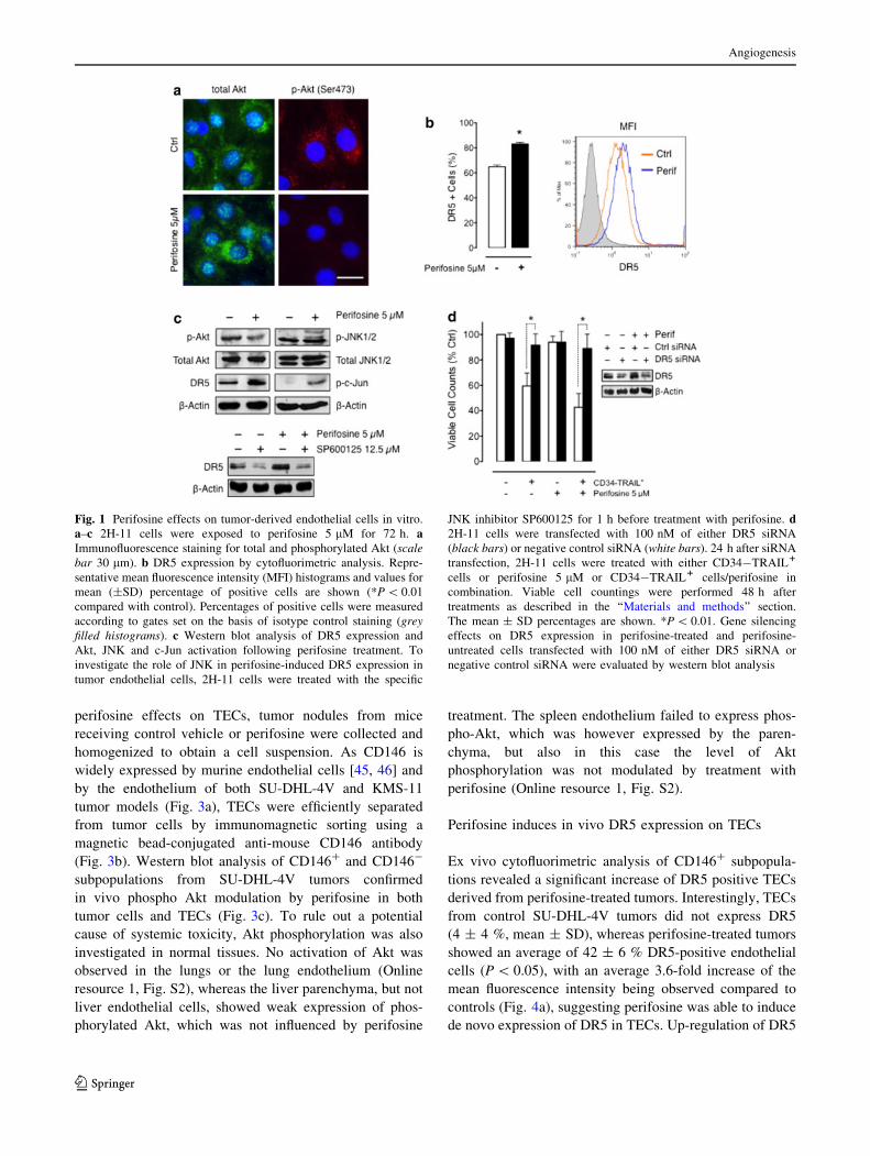
In order to investigate the effects of perifosine on TECs,
we first performed in vitro experiments using the 2H-11
tumor endothelial cell line [44]. 2H-11 cells were exposed
to perifosine 5 lM for 72 h and analyzed for phospho Akt
and DR5 expression. As assessed by immunofluorescence
and western blot analysis, perifosine caused a strong
decrease in Akt phosphorylation levels without affecting
total Akt expression (Fig. 1a, c). Interestingly, reduced
levels of activated Akt were paralleled by a significant
increase in DR5 expression as shown by flow cytometric
(64.8 ± 1.4 vs. 83.0 ± 1.2 %, mean ± SD; *P \ 0.01)
and western blot analysis (Fig. 1b, c). Considering that it
has been reported the c-Jun NH2-terminal kinase-depen-
dent upregulation of DR5 by perifosine in leukemia cells
[17] and head and neck squamous cell carcinomas [18], we
examined the possibility that perifosine upmodulates DR5
levels via JNK also in tumor-derived endothelial cells.
Thus, we investigated by western blot analysis the modu-
lation of JNK and c-Jun activation in 2H-11 cells following
treatment with perifosine. As shown in Fig. 1c, perifosine
increased the phosphorylation levels of both JNK and
c-Jun. Interestingly, inhibition of JNK by the specific
inhibitor SP600125 prevented DR5 perifosine-induced
upregulation (Fig. 1c), supporting the hypothesis that
perifosine upmodulates DR5 levels via JNK activation.
DR5 expression on TEC is needed for CD34-TRAIL1
cells antivascular activity
In order to investigate whether the DR5 expression on tumor
endothelial cells is required for CD34-TRAIL1 cells anti-
vascular activity, we silenced the expression of DR5 in 2H-
11 cells using small interfering RNA (siRNA) and then
examined cell sensitivity to CD34-TRAIL1 cells alone or in
combination with perifosine. As shown in Fig. 1d, 2H-11
cell viability accounted for 59.5 ± 10.2 % (mean ± SD) of
controls following treatment with CD34-TRAIL1 cells and
addition of perifosine caused a 28 % further reduction in cell
viability compared with CD34-TRAIL1 cells alone
(42.8 ± 10.8 vs. 59.5 ± 10.2 %, mean ± SD, P \ 0.05),
whereas perifosine alone was not able to significantly affect
2H-11 cell viability. By western blotting, we detected sub-
stantially reduced levels of DR5 in both perifosine-treated
and perifosine-untreated 2H-11 cells transfected with DR5
siRNA compared with control siRNA-transfected cells
(Fig. 1d). Interestingly, silencing of DR5 expression pre-
vented CD34-TRAIL1 cells-induced 2H-11 cell death. In
fact, a significant increase in cell viability was observed in
DR5 siRNA-transfected cells after treatment with both
CD34-TRAIL1 cells alone and in combination with pe-
rifosine compared to control siRNA-transfected cells
(91.7 ± 8.9 vs. 59.5 ± 10.2 %, mean ± SD, P \ 0.01, and
89.0 ± 11.3 vs. 42.8 ± 10.8 %, mean ± SD, P \ 0.01,
respectively). These findings suggest that DR5 expression is
required for CD34-TRAIL1 cells-induced tumor endothe-
lial cell death and confirm that DR5 up-regulation is
the mechanism involved in perifosine-enhancement of
CD34-TRAIL1 cells antivascular activity.
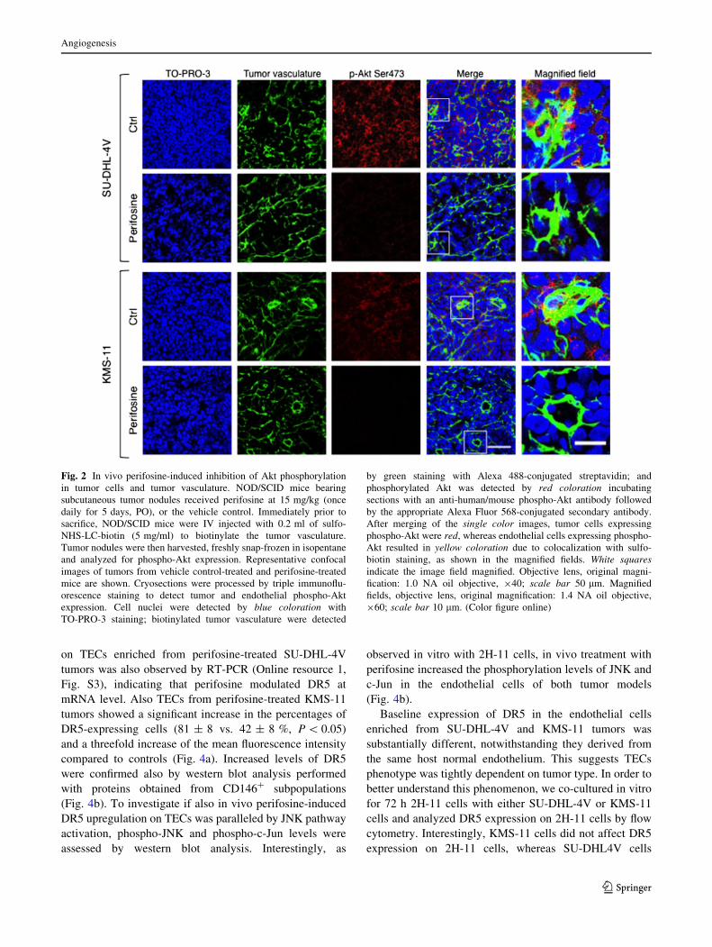
Perifosine inhibits in vivo Akt phosphorylation both in
tumor cells and tumor vasculature
To determine whether treatment with perifosine modulated
Akt phosphorylation in vivo, tissue sections from control
and perifosine-treated tumors were analyzed by confocal
microscopy. Both SU-DHL-4V and KMS-11 tumor cells
showed high levels of phosphorylated Akt (Fig. 2) and, in
both instances, perifosine markedly decreased Akt phos-
phorylation (Fig. 2). Interestingly, in keeping with in vitro
observations, in vivo biotinylated endothelial cells (see
‘‘Materials and methods’’ section) of both SU-DHL-4V and
KMS-11 tumors also showed intense expression of phos-
phorylated Akt, which was strongly inhibited by perifosine
(Fig. 2, magnified fields). In order to better evaluate in vivo
Angiogenesis
123
perifosine effects on TECs, tumor nodules from mice
receiving control vehicle or perifosine were collected and
homogenized to obtain a cell suspension. As CD146 is
widely expressed by murine endothelial cells [45, 46] and
by the endothelium of both SU-DHL-4V and KMS-11
tumor models (Fig. 3a), TECs were efficiently separated
from tumor cells by immunomagnetic sorting using a
magnetic bead-conjugated anti-mouse CD146 antibody
(Fig. 3b). Western blot analysis of CD146? and CD146-
subpopulations from SU-DHL-4V tumors confirmed
in vivo phospho Akt modulation by perifosine in both
tumor cells and TECs (Fig. 3c). To rule out a potential
cause of systemic toxicity, Akt phosphorylation was also
investigated in normal tissues. No activation of Akt was
observed in the lungs or the lung endothelium (Online
resource 1, Fig. S2), whereas the liver parenchyma, but not
liver endothelial cells, showed weak expression of phos-
phorylated Akt, which was not influenced by perifosine
treatment. The spleen endothelium failed to express phos-
pho-Akt, which was however expressed by the paren-
chyma, but also in this case the level of Akt
phosphorylation was not modulated by treatment with
perifosine (Online resource 1, Fig. S2).
Perifosine induces in vivo DR5 expression on TECs
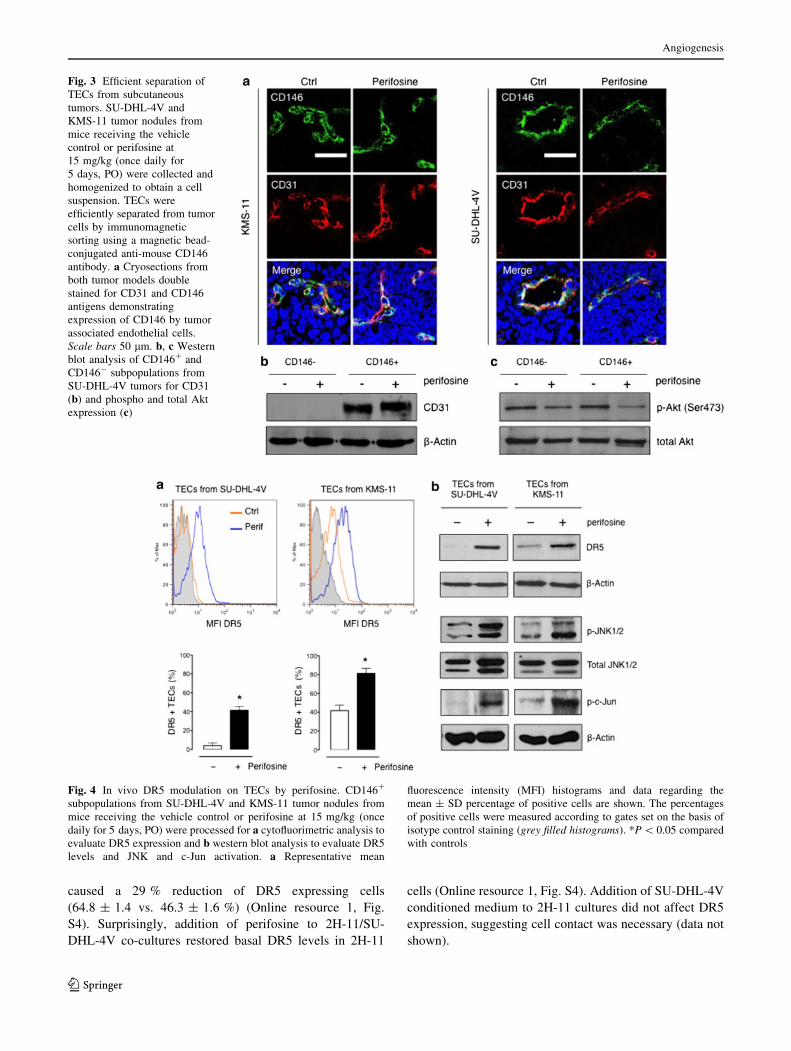
Ex vivo cytofluorimetric analysis of CD146? subpopula-
tions revealed a significant increase of DR5 positive TECs
derived from perifosine-treated tumors. Interestingly, TECs
from control SU-DHL-4V tumors did not express DR5
(4 ± 4 %, mean ± SD), whereas perifosine-treated tumors
showed an average of 42 ± 6 % DR5-positive endothelial
cells (P \ 0.05), with an average 3.6-fold increase of the
mean fluorescence intensity being observed compared to
controls (Fig. 4a), suggesting perifosine was able to induce
de novo expression of DR5 in TECs. Up-regulation of DR5
Fig. 1 Perifosine effects on tumor-derived endothelial cells in vitro.
a–c 2H-11 cells were exposed to perifosine 5 lM for 72 h. aImmunofluorescence staining for total and phosphorylated Akt (scalebar 30 lm). b DR5 expression by cytofluorimetric analysis. Repre-
sentative mean fluorescence intensity (MFI) histograms and values for
mean (±SD) percentage of positive cells are shown (*P \ 0.01
compared with control). Percentages of positive cells were measured
according to gates set on the basis of isotype control staining (greyfilled histograms). c Western blot analysis of DR5 expression and
Akt, JNK and c-Jun activation following perifosine treatment. To
investigate the role of JNK in perifosine-induced DR5 expression in
tumor endothelial cells, 2H-11 cells were treated with the specific
JNK inhibitor SP600125 for 1 h before treatment with perifosine. d2H-11 cells were transfected with 100 nM of either DR5 siRNA
(black bars) or negative control siRNA (white bars). 24 h after siRNA
transfection, 2H-11 cells were treated with either CD34-TRAIL1
cells or perifosine 5 lM or CD34-TRAIL1 cells/perifosine in
combination. Viable cell countings were performed 48 h after
treatments as described in the ‘‘Materials and methods’’ section.
The mean ± SD percentages are shown. *P \ 0.01. Gene silencing
effects on DR5 expression in perifosine-treated and perifosine-
untreated cells transfected with 100 nM of either DR5 siRNA or
negative control siRNA were evaluated by western blot analysis
Angiogenesis
123
on TECs enriched from perifosine-treated SU-DHL-4V
tumors was also observed by RT-PCR (Online resource 1,
Fig. S3), indicating that perifosine modulated DR5 at
mRNA level. Also TECs from perifosine-treated KMS-11
tumors showed a significant increase in the percentages of
DR5-expressing cells (81 ± 8 vs. 42 ± 8 %, P \ 0.05)
and a threefold increase of the mean fluorescence intensity
compared to controls (Fig. 4a). Increased levels of DR5
were confirmed also by western blot analysis performed
with proteins obtained from CD146? subpopulations
(Fig. 4b). To investigate if also in vivo perifosine-induced
DR5 upregulation on TECs was paralleled by JNK pathway
activation, phospho-JNK and phospho-c-Jun levels were
assessed by western blot analysis. Interestingly, as
observed in vitro with 2H-11 cells, in vivo treatment with
perifosine increased the phosphorylation levels of JNK and
c-Jun in the endothelial cells of both tumor models
(Fig. 4b).
Baseline expression of DR5 in the endothelial cells
enriched from SU-DHL-4V and KMS-11 tumors was
substantially different, notwithstanding they derived from
the same host normal endothelium. This suggests TECs
phenotype was tightly dependent on tumor type. In order to
better understand this phenomenon, we co-cultured in vitro
for 72 h 2H-11 cells with either SU-DHL-4V or KMS-11
cells and analyzed DR5 expression on 2H-11 cells by flow
cytometry. Interestingly, KMS-11 cells did not affect DR5
expression on 2H-11 cells, whereas SU-DHL4V cells
Fig. 2 In vivo perifosine-induced inhibition of Akt phosphorylation
in tumor cells and tumor vasculature. NOD/SCID mice bearing
subcutaneous tumor nodules received perifosine at 15 mg/kg (once
daily for 5 days, PO), or the vehicle control. Immediately prior to
sacrifice, NOD/SCID mice were IV injected with 0.2 ml of sulfo-
NHS-LC-biotin (5 mg/ml) to biotinylate the tumor vasculature.
Tumor nodules were then harvested, freshly snap-frozen in isopentane
and analyzed for phospho-Akt expression. Representative confocal
images of tumors from vehicle control-treated and perifosine-treated
mice are shown. Cryosections were processed by triple immunoflu-
orescence staining to detect tumor and endothelial phospho-Akt
expression. Cell nuclei were detected by blue coloration with
TO-PRO-3 staining; biotinylated tumor vasculature were detected
by green staining with Alexa 488-conjugated streptavidin; and
phosphorylated Akt was detected by red coloration incubating
sections with an anti-human/mouse phospho-Akt antibody followed
by the appropriate Alexa Fluor 568-conjugated secondary antibody.
After merging of the single color images, tumor cells expressing
phospho-Akt were red, whereas endothelial cells expressing phospho-
Akt resulted in yellow coloration due to colocalization with sulfo-
biotin staining, as shown in the magnified fields. White squaresindicate the image field magnified. Objective lens, original magni-
fication: 1.0 NA oil objective, 940; scale bar 50 lm. Magnified
fields, objective lens, original magnification: 1.4 NA oil objective,
960; scale bar 10 lm. (Color figure online)
Angiogenesis
123
caused a 29 % reduction of DR5 expressing cells
(64.8 ± 1.4 vs. 46.3 ± 1.6 %) (Online resource 1, Fig.
S4). Surprisingly, addition of perifosine to 2H-11/SU-
DHL-4V co-cultures restored basal DR5 levels in 2H-11
cells (Online resource 1, Fig. S4). Addition of SU-DHL-4V
conditioned medium to 2H-11 cultures did not affect DR5
expression, suggesting cell contact was necessary (data not
shown).
Fig. 3 Efficient separation of
TECs from subcutaneous
tumors. SU-DHL-4V and
KMS-11 tumor nodules from
mice receiving the vehicle
control or perifosine at
15 mg/kg (once daily for
5 days, PO) were collected and
homogenized to obtain a cell
suspension. TECs were
efficiently separated from tumor
cells by immunomagnetic
sorting using a magnetic bead-
conjugated anti-mouse CD146
antibody. a Cryosections from
both tumor models double
stained for CD31 and CD146
antigens demonstrating
expression of CD146 by tumor
associated endothelial cells.
Scale bars 50 lm. b, c Western
blot analysis of CD146? and
CD146- subpopulations from
SU-DHL-4V tumors for CD31
(b) and phospho and total Akt
expression (c)
Fig. 4 In vivo DR5 modulation on TECs by perifosine. CD146?
subpopulations from SU-DHL-4V and KMS-11 tumor nodules from
mice receiving the vehicle control or perifosine at 15 mg/kg (once
daily for 5 days, PO) were processed for a cytofluorimetric analysis to
evaluate DR5 expression and b western blot analysis to evaluate DR5
levels and JNK and c-Jun activation. a Representative mean
fluorescence intensity (MFI) histograms and data regarding the
mean ± SD percentage of positive cells are shown. The percentages
of positive cells were measured according to gates set on the basis of
isotype control staining (grey filled histograms). *P \ 0.05 compared
with controls
Angiogenesis
123
Perifosine triggers the vascular disruption activity of
CD34-TRAIL1 cells in SU-DHL-4V tumors lacking
vascular DR5
Expression of DR5 on TECs is a pre-requisite for the
vascular disruption activity of CD34-TRAIL1 cells
[40, 41]. To investigate the impact of perifosine-induced
DR5 modulation on the vascular disrupting effects of
CD34-TRAIL1 cells, tumor vasculature was extensively
analyzed following in vivo biotinylation for the presence of
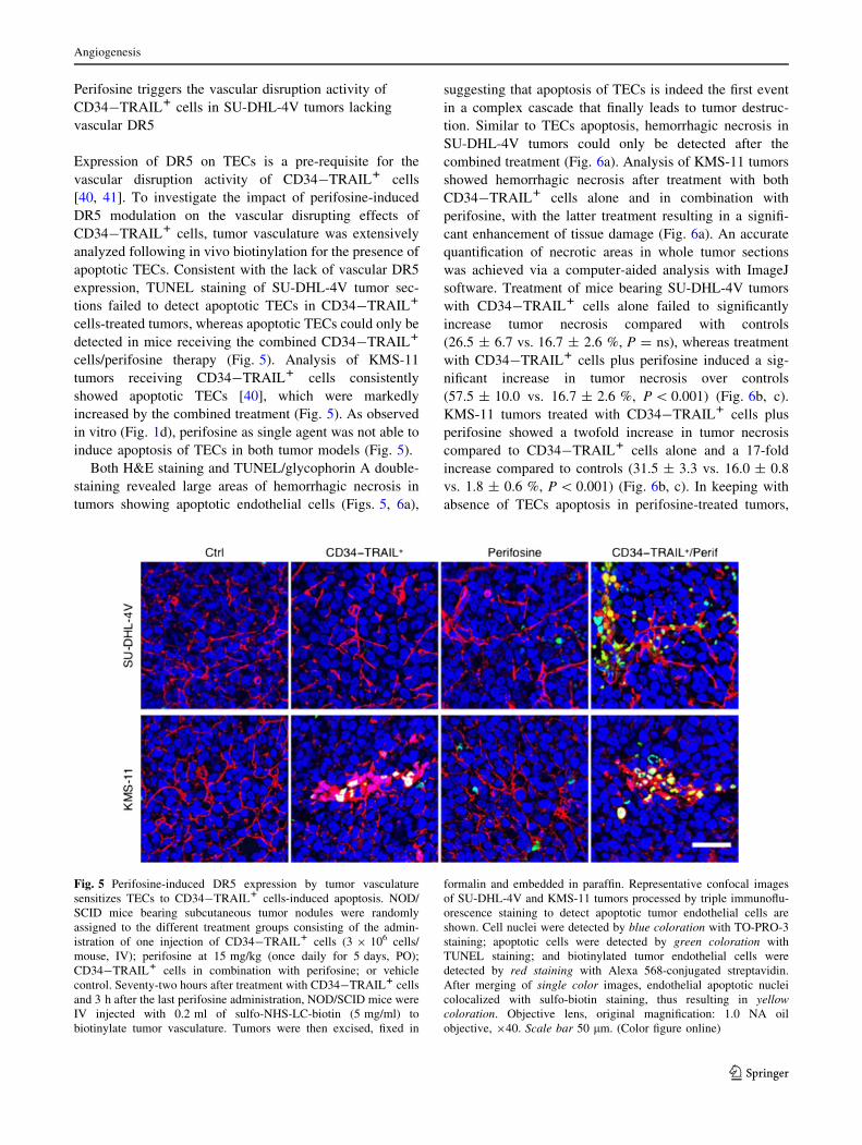
apoptotic TECs. Consistent with the lack of vascular DR5
expression, TUNEL staining of SU-DHL-4V tumor sec-
tions failed to detect apoptotic TECs in CD34-TRAIL1
cells-treated tumors, whereas apoptotic TECs could only be
detected in mice receiving the combined CD34-TRAIL1
cells/perifosine therapy (Fig. 5). Analysis of KMS-11
tumors receiving CD34-TRAIL1 cells consistently
showed apoptotic TECs [40], which were markedly
increased by the combined treatment (Fig. 5). As observed
in vitro (Fig. 1d), perifosine as single agent was not able to
induce apoptosis of TECs in both tumor models (Fig. 5).
Both H&E staining and TUNEL/glycophorin A double-
staining revealed large areas of hemorrhagic necrosis in
tumors showing apoptotic endothelial cells (Figs. 5, 6a),
suggesting that apoptosis of TECs is indeed the first event
in a complex cascade that finally leads to tumor destruc-
tion. Similar to TECs apoptosis, hemorrhagic necrosis in
SU-DHL-4V tumors could only be detected after the
combined treatment (Fig. 6a). Analysis of KMS-11 tumors
showed hemorrhagic necrosis after treatment with both
CD34-TRAIL1 cells alone and in combination with
perifosine, with the latter treatment resulting in a signifi-
cant enhancement of tissue damage (Fig. 6a). An accurate
quantification of necrotic areas in whole tumor sections
was achieved via a computer-aided analysis with ImageJ
software. Treatment of mice bearing SU-DHL-4V tumors
with CD34-TRAIL1 cells alone failed to significantly
increase tumor necrosis compared with controls
(26.5 ± 6.7 vs. 16.7 ± 2.6 %, P = ns), whereas treatment
with CD34-TRAIL1 cells plus perifosine induced a sig-
nificant increase in tumor necrosis over controls
(57.5 ± 10.0 vs. 16.7 ± 2.6 %, P \ 0.001) (Fig. 6b, c).
KMS-11 tumors treated with CD34-TRAIL1 cells plus
perifosine showed a twofold increase in tumor necrosis
compared to CD34-TRAIL1 cells alone and a 17-fold
increase compared to controls (31.5 ± 3.3 vs. 16.0 ± 0.8
vs. 1.8 ± 0.6 %, P \ 0.001) (Fig. 6b, c). In keeping with
absence of TECs apoptosis in perifosine-treated tumors,
Fig. 5 Perifosine-induced DR5 expression by tumor vasculature
sensitizes TECs to CD34-TRAIL1 cells-induced apoptosis. NOD/
SCID mice bearing subcutaneous tumor nodules were randomly
assigned to the different treatment groups consisting of the admin-
istration of one injection of CD34-TRAIL1 cells (3 9 106 cells/
mouse, IV); perifosine at 15 mg/kg (once daily for 5 days, PO);
CD34-TRAIL1 cells in combination with perifosine; or vehicle
control. Seventy-two hours after treatment with CD34-TRAIL1 cells
and 3 h after the last perifosine administration, NOD/SCID mice were
IV injected with 0.2 ml of sulfo-NHS-LC-biotin (5 mg/ml) to
biotinylate tumor vasculature. Tumors were then excised, fixed in
formalin and embedded in paraffin. Representative confocal images
of SU-DHL-4V and KMS-11 tumors processed by triple immunoflu-
orescence staining to detect apoptotic tumor endothelial cells are
shown. Cell nuclei were detected by blue coloration with TO-PRO-3
staining; apoptotic cells were detected by green coloration with
TUNEL staining; and biotinylated tumor endothelial cells were
detected by red staining with Alexa 568-conjugated streptavidin.
After merging of single color images, endothelial apoptotic nuclei
colocalized with sulfo-biotin staining, thus resulting in yellowcoloration. Objective lens, original magnification: 1.0 NA oil
objective, 940. Scale bar 50 lm. (Color figure online)
Angiogenesis
123
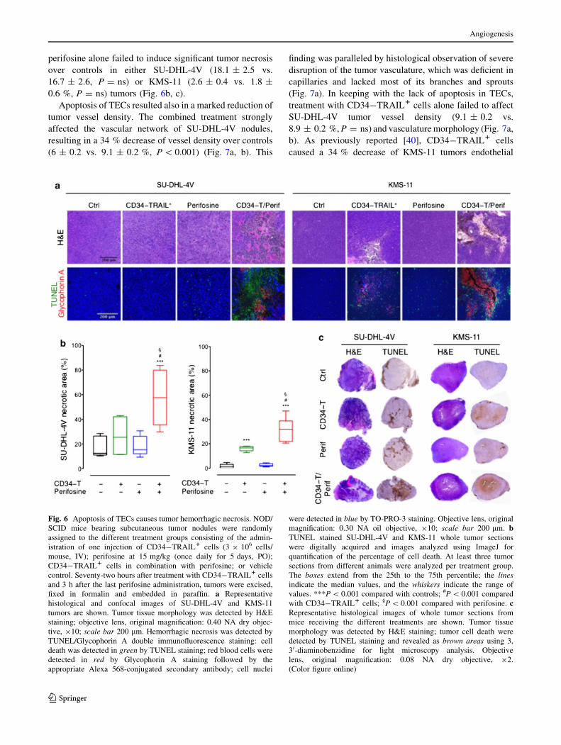
perifosine alone failed to induce significant tumor necrosis
over controls in either SU-DHL-4V (18.1 ± 2.5 vs.
16.7 ± 2.6, P = ns) or KMS-11 (2.6 ± 0.4 vs. 1.8 ±
0.6 %, P = ns) tumors (Fig. 6b, c).
Apoptosis of TECs resulted also in a marked reduction of
tumor vessel density. The combined treatment strongly
affected the vascular network of SU-DHL-4V nodules,
resulting in a 34 % decrease of vessel density over controls
(6 ± 0.2 vs. 9.1 ± 0.2 %, P \ 0.001) (Fig. 7a, b). This
finding was paralleled by histological observation of severe
disruption of the tumor vasculature, which was deficient in
capillaries and lacked most of its branches and sprouts
(Fig. 7a). In keeping with the lack of apoptosis in TECs,
treatment with CD34-TRAIL1 cells alone failed to affect
SU-DHL-4V tumor vessel density (9.1 ± 0.2 vs.
8.9 ± 0.2 %, P = ns) and vasculature morphology (Fig. 7a,
b). As previously reported [40], CD34-TRAIL1 cells
caused a 34 % decrease of KMS-11 tumors endothelial
Fig. 6 Apoptosis of TECs causes tumor hemorrhagic necrosis. NOD/
SCID mice bearing subcutaneous tumor nodules were randomly
assigned to the different treatment groups consisting of the admin-
istration of one injection of CD34-TRAIL1 cells (3 9 106 cells/
mouse, IV); perifosine at 15 mg/kg (once daily for 5 days, PO);
CD34-TRAIL1 cells in combination with perifosine; or vehicle
control. Seventy-two hours after treatment with CD34-TRAIL1 cells
and 3 h after the last perifosine administration, tumors were excised,
fixed in formalin and embedded in paraffin. a Representative
histological and confocal images of SU-DHL-4V and KMS-11
tumors are shown. Tumor tissue morphology was detected by H&E
staining; objective lens, original magnification: 0.40 NA dry objec-
tive, 910; scale bar 200 lm. Hemorrhagic necrosis was detected by
TUNEL/Glycophorin A double immunofluorescence staining: cell
death was detected in green by TUNEL staining; red blood cells were
detected in red by Glycophorin A staining followed by the
appropriate Alexa 568-conjugated secondary antibody; cell nuclei
were detected in blue by TO-PRO-3 staining. Objective lens, original
magnification: 0.30 NA oil objective, 910; scale bar 200 lm. bTUNEL stained SU-DHL-4V and KMS-11 whole tumor sections
were digitally acquired and images analyzed using ImageJ for
quantification of the percentage of cell death. At least three tumor
sections from different animals were analyzed per treatment group.
The boxes extend from the 25th to the 75th percentile; the linesindicate the median values, and the whiskers indicate the range of
values. ***P \ 0.001 compared with controls; #P \ 0.001 compared
with CD34-TRAIL1 cells; §P \ 0.001 compared with perifosine. cRepresentative histological images of whole tumor sections from
mice receiving the different treatments are shown. Tumor tissue
morphology was detected by H&E staining; tumor cell death were
detected by TUNEL staining and revealed as brown areas using 3,
30-diaminobenzidine for light microscopy analysis. Objective
lens, original magnification: 0.08 NA dry objective, 92.
(Color figure online)
Angiogenesis
123
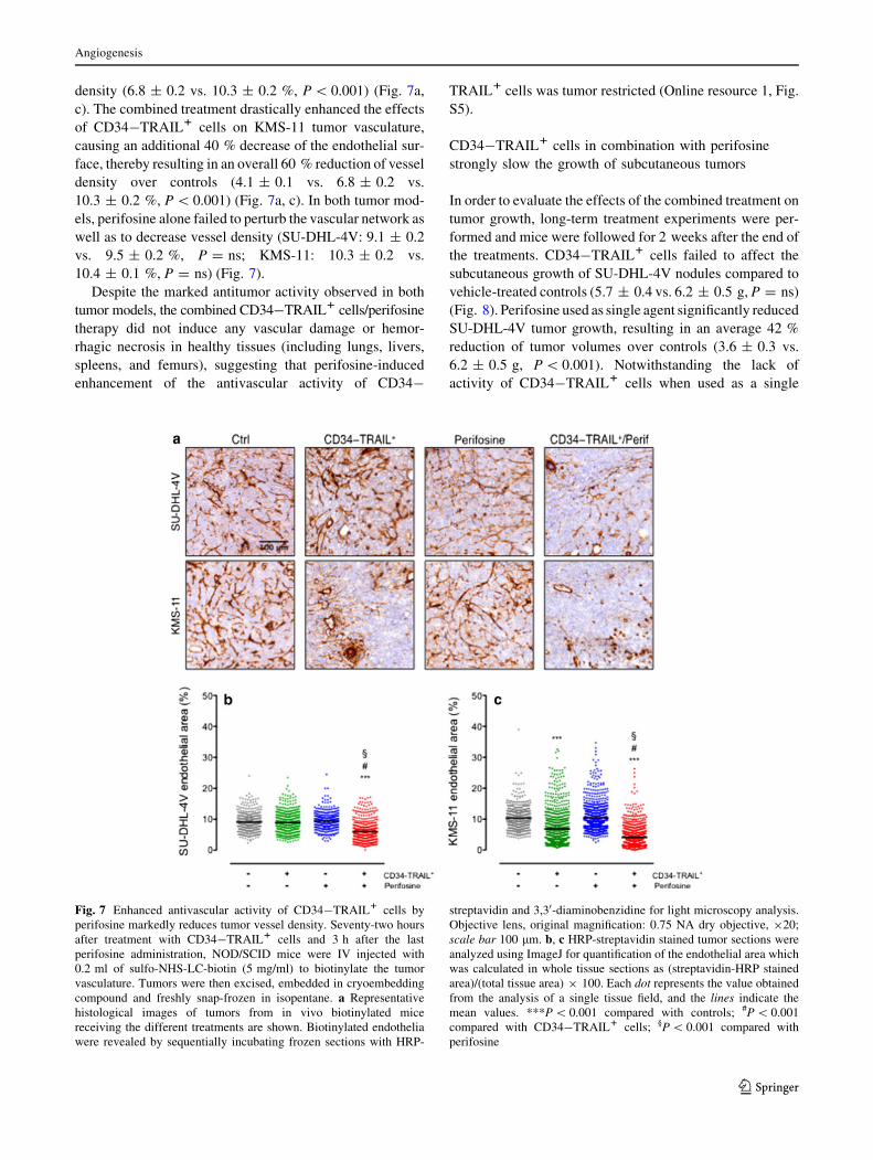
density (6.8 ± 0.2 vs. 10.3 ± 0.2 %, P \ 0.001) (Fig. 7a,
c). The combined treatment drastically enhanced the effects
of CD34-TRAIL1 cells on KMS-11 tumor vasculature,
causing an additional 40 % decrease of the endothelial sur-
face, thereby resulting in an overall 60 % reduction of vessel
density over controls (4.1 ± 0.1 vs. 6.8 ± 0.2 vs.
10.3 ± 0.2 %, P \ 0.001) (Fig. 7a, c). In both tumor mod-
els, perifosine alone failed to perturb the vascular network as
well as to decrease vessel density (SU-DHL-4V: 9.1 ± 0.2
vs. 9.5 ± 0.2 %, P = ns; KMS-11: 10.3 ± 0.2 vs.
10.4 ± 0.1 %, P = ns) (Fig. 7).
Despite the marked antitumor activity observed in both
tumor models, the combined CD34-TRAIL1 cells/perifosine
therapy did not induce any vascular damage or hemor-
rhagic necrosis in healthy tissues (including lungs, livers,
spleens, and femurs), suggesting that perifosine-induced
enhancement of the antivascular activity of CD34-
TRAIL1 cells was tumor restricted (Online resource 1, Fig.
S5).
CD34-TRAIL1 cells in combination with perifosine
strongly slow the growth of subcutaneous tumors
In order to evaluate the effects of the combined treatment on
tumor growth, long-term treatment experiments were per-
formed and mice were followed for 2 weeks after the end of
the treatments. CD34-TRAIL1 cells failed to affect the
subcutaneous growth of SU-DHL-4V nodules compared to
vehicle-treated controls (5.7 ± 0.4 vs. 6.2 ± 0.5 g, P = ns)
(Fig. 8). Perifosine used as single agent significantly reduced
SU-DHL-4V tumor growth, resulting in an average 42 %
reduction of tumor volumes over controls (3.6 ± 0.3 vs.
6.2 ± 0.5 g, P \ 0.001). Notwithstanding the lack of
activity of CD34-TRAIL1 cells when used as a single
Fig. 7 Enhanced antivascular activity of CD34-TRAIL1 cells by
perifosine markedly reduces tumor vessel density. Seventy-two hours
after treatment with CD34-TRAIL1 cells and 3 h after the last
perifosine administration, NOD/SCID mice were IV injected with
0.2 ml of sulfo-NHS-LC-biotin (5 mg/ml) to biotinylate the tumor
vasculature. Tumors were then excised, embedded in cryoembedding
compound and freshly snap-frozen in isopentane. a Representative
histological images of tumors from in vivo biotinylated mice
receiving the different treatments are shown. Biotinylated endothelia
were revealed by sequentially incubating frozen sections with HRP-
streptavidin and 3,30-diaminobenzidine for light microscopy analysis.
Objective lens, original magnification: 0.75 NA dry objective, 920;
scale bar 100 lm. b, c HRP-streptavidin stained tumor sections were
analyzed using ImageJ for quantification of the endothelial area which
was calculated in whole tissue sections as (streptavidin-HRP stained
area)/(total tissue area) 9 100. Each dot represents the value obtained
from the analysis of a single tissue field, and the lines indicate the
mean values. ***P \ 0.001 compared with controls; #P \ 0.001
compared with CD34-TRAIL1 cells; §P \ 0.001 compared with
perifosine
Angiogenesis
123
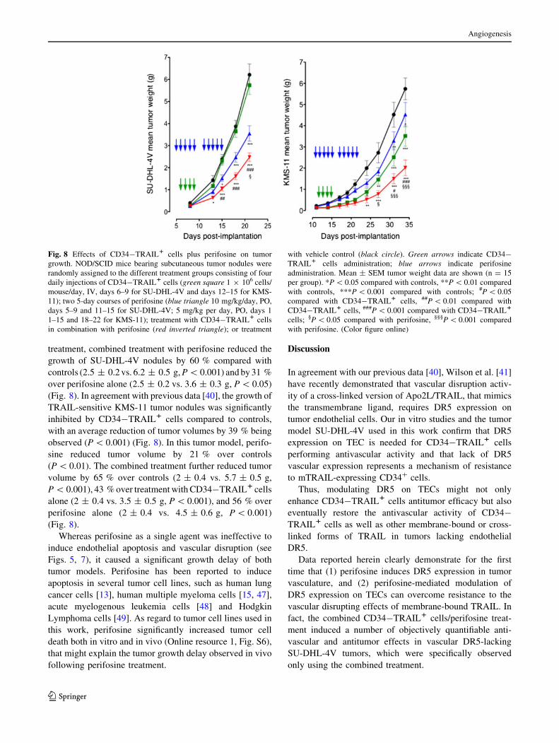
treatment, combined treatment with perifosine reduced the
growth of SU-DHL-4V nodules by 60 % compared with
controls (2.5 ± 0.2 vs. 6.2 ± 0.5 g, P \ 0.001) and by 31 %
over perifosine alone (2.5 ± 0.2 vs. 3.6 ± 0.3 g, P \ 0.05)
(Fig. 8). In agreement with previous data [40], the growth of
TRAIL-sensitive KMS-11 tumor nodules was significantly
inhibited by CD34-TRAIL1 cells compared to controls,
with an average reduction of tumor volumes by 39 % being
observed (P \ 0.001) (Fig. 8). In this tumor model, perifo-
sine reduced tumor volume by 21 % over controls
(P \ 0.01). The combined treatment further reduced tumor
volume by 65 % over controls (2 ± 0.4 vs. 5.7 ± 0.5 g,
P \ 0.001), 43 % over treatment with CD34-TRAIL1 cells
alone (2 ± 0.4 vs. 3.5 ± 0.5 g, P \ 0.001), and 56 % over
perifosine alone (2 ± 0.4 vs. 4.5 ± 0.6 g, P \ 0.001)
(Fig. 8).
Whereas perifosine as a single agent was ineffective to
induce endothelial apoptosis and vascular disruption (see
Figs. 5, 7), it caused a significant growth delay of both
tumor models. Perifosine has been reported to induce
apoptosis in several tumor cell lines, such as human lung
cancer cells [13], human multiple myeloma cells [15, 47],
acute myelogenous leukemia cells [48] and Hodgkin
Lymphoma cells [49]. As regard to tumor cell lines used in
this work, perifosine significantly increased tumor cell
death both in vitro and in vivo (Online resource 1, Fig. S6),
that might explain the tumor growth delay observed in vivo
following perifosine treatment.
Discussion
In agreement with our previous data [40], Wilson et al. [41]
have recently demonstrated that vascular disruption activ-
ity of a cross-linked version of Apo2L/TRAIL, that mimics
the transmembrane ligand, requires DR5 expression on
tumor endothelial cells. Our in vitro studies and the tumor
model SU-DHL-4V used in this work confirm that DR5
expression on TEC is needed for CD34-TRAIL1 cells
performing antivascular activity and that lack of DR5
vascular expression represents a mechanism of resistance
to mTRAIL-expressing CD34? cells.
Thus, modulating DR5 on TECs might not only
enhance CD34-TRAIL1 cells antitumor efficacy but also
eventually restore the antivascular activity of CD34-
TRAIL1 cells as well as other membrane-bound or cross-
linked forms of TRAIL in tumors lacking endothelial
DR5.
Data reported herein clearly demonstrate for the first
time that (1) perifosine induces DR5 expression in tumor
vasculature, and (2) perifosine-mediated modulation of
DR5 expression on TECs can overcome resistance to the
vascular disrupting effects of membrane-bound TRAIL. In
fact, the combined CD34-TRAIL1 cells/perifosine treat-
ment induced a number of objectively quantifiable anti-
vascular and antitumor effects in vascular DR5-lacking
SU-DHL-4V tumors, which were specifically observed
only using the combined treatment.
Fig. 8 Effects of CD34-TRAIL1 cells plus perifosine on tumor
growth. NOD/SCID mice bearing subcutaneous tumor nodules were
randomly assigned to the different treatment groups consisting of four
daily injections of CD34-TRAIL1 cells (green square 1 9 106 cells/
mouse/day, IV, days 6–9 for SU-DHL-4V and days 12–15 for KMS-
11); two 5-day courses of perifosine (blue triangle 10 mg/kg/day, PO,
days 5–9 and 11–15 for SU-DHL-4V; 5 mg/kg per day, PO, days 1
1–15 and 18–22 for KMS-11); treatment with CD34-TRAIL1 cells
in combination with perifosine (red inverted triangle); or treatment
with vehicle control (black circle). Green arrows indicate CD34-
TRAIL1 cells administration; blue arrows indicate perifosine
administration. Mean ± SEM tumor weight data are shown (n = 15
per group). *P \ 0.05 compared with controls, **P \ 0.01 compared
with controls, ***P \ 0.001 compared with controls; #P \ 0.05
compared with CD34-TRAIL1 cells, ##P \ 0.01 compared with
CD34-TRAIL1 cells, ###P \ 0.001 compared with CD34-TRAIL1
cells; §P \ 0.05 compared with perifosine, §§§P \ 0.001 compared
with perifosine. (Color figure online)
Angiogenesis
123
It has been established that the JNK/c-Jun axis could
positively regulate DR5 transcription [50]. A functional
activator protein-1 (AP-1) binding site has been shown in
the promoter region of DR5. JNK, by increasing c-Jun
phosphorylation, leads to an increase in AP-1 activity [51].
Interestingly, Akt appears to negatively regulate JNK sig-
naling by directly interacting with the JNK activators
Mixed Lineage Kinase 3 (MLK3) and Apoptosis Signal-
regulating Kinase 1 (ASK1), thus causing their inhibition
[52, 53]. Several in vitro studies have investigated the
mechanism(s) by which perifosine modulates TRAIL
receptors expression on tumor cells. Tazzari et al. [17]
suggested that perifosine induces JNK-dependent DR5
overexpression in leukemia cells. Fu et al. [18] have
recently reported that perifosine increases the levels of
p-JNK and p–c-Jun in head and neck squamous cell car-
cinomas and that activation of JNK signaling parallels the
upregulation of DR4 and DR5. However studies investi-
gating these effects in tumor endothelial cells are steel
lacking. Here we show that in vivo administration of pe-
rifosine strongly inhibits tumor endothelial Akt phosphor-
ylation and upregulates (see KMS-11 tumor model) or even
induces de novo expression (see SU-DHL-4V tumor
model) of DR5 in TECs in a JNK-dependent manner. In
fact, perifosine increased phosphorylation of JNK and
c-Jun in tumor endothelial cells both in vitro and in vivo
and inhibition of JNK by the specific inhibitor SP600125
prevented DR5 perifosine-induced upregulation, support-
ing the hypothesis perifosine might upmodulate DR5 levels
in TECs via JNK activation.
Interesting is to note as the phenotype of tumor endo-
thelial cells, which derived from the same host normal
endothelium, was tightly dependent on tumor type. Indeed,
TECs from SU-DHL-4V tumors did not express DR5 on
their surface, whereas about 40 % of TECs from KMS-11
tumors expressed the receptor. This evidence suggests that
different tumor cells can even modulate tumor microen-
vironment influencing therapeutic outcome [54]. It will be
interesting to determine what specific features of the tumor
microenvironment drive endothelial DR5 expression.
Akt activation plays an important role in regulating the
survival and proliferation of both tumor cells and TECs.
Although Akt phosphorylation in tumor cells as well as
tumor vasculature was strongly inhibited by perifosine,
analysis of the tumor vasculature of our xenografts clearly
showed that perifosine as a single agent was ineffective to
induce endothelial cell apoptosis and vascular disruption or
to inhibit tumor angiogenesis, whereas it caused tumor cell
apoptosis and a significant growth delay of both tumor
models. In 2008 Zerp et al. [55] studied the antiangiogenic
properties of the alkylphospholopids (APLs) perifosine,
miltefosine and edelfosine in vitro. They demonstrated that
APLs inhibit capillary-like endothelial tube formation and
that the sensitivity of normal vascular endothelial cells (ECs)
to APL-induced apoptosis is dependent on the proliferative
status of these cells: confluent, quiescent ECs were relatively
resistant, whereas proliferating ECs were highly sensitive to
APL-induced apoptosis. In agreement with these data, we
observed in vitro induction of apoptosis in HUVEC prolif-
erating cells treated with 5 lM perifosine (data not shown),
but no apoptosis was observed in perifosine-treated tumor-
derived 2H-11 endothelial cells. It is widely appreciated that
tumor-associated endothelial cells have a unique activated
phenotype and a different structure compared to those in
normal quiescent tissues beacause of the exposure to a dis-
tinct set of stimuli in their local environment [56]. Further-
more, although tumor-associated angiogenesis has
traditionally been defined as the sprouting of new vessels
from preexisting vessels, it is becoming clear that the blood
vessels that support tumor growth can also originate from
cells recruited from the bone marrow or can even differen-
tiate from tumor stem cells (vascular mimicry) [57, 58]. All
toghether these factors can determine different responses in
normal cultured endothelial cells and tumor-associated
endothelial cells exposed to the same drug.
As mentioned above, although perifosine as a single
agent was ineffective to induce endothelial cells apoptosis,
it sensitized TECs to CD34-TRAIL1 cells-induced vas-
cular disruption by upmodulating DR5 expression and
caused tumor cell apoptosis. In addition to targeting Akt
and modulating JNK activation and DR5 expression,
perifosine possesses other biologic activities (reviewed in
[59]) that may contribute to the effects observed in this
work. These include, accumulation into lipid rafts of the
plasma membrane [60] and downregulation of ERK 1/2
phosphorylation [48]. Analysis of tumor xenografts
revealed a strong reduction in ERK phosphorylation levels
following perifosine treatment (data not shown), indicating
that inhibition of MAPK signaling by perifosine toghether
with Akt inhibition might also be involved in inducing
tumor cells apoptosis and tumor growth delay. In 2007 van
der Luit et al. [60] reported perifosine incorporation into
lipid rafts followed by inhibition of phosphatidylcholine
synthesis and induction of apoptosis in lymphoma cells; in
the same year Gajate and Mollinedo [61] demonstrated that
perifosine induces apoptosis in multiple myeloma (MM) by
recruitment of death receptors into lipid rafts and observed
that the concentration of death receptors in lipid rafts fol-
lowing perifosine treatment rendered MM cells more sen-
sitive to the action of death receptor ligands, such as
TRAIL. Considering these previous observations, we can
not exclude that perifosine sensitized TECs to CD34-
TRAIL1 cells-induced apoptosis not only by upregulating
DR5 expression but also by acting on plasma membrane
reorganization and causing DR5 accumulation into lipid
rafts.
Angiogenesis
123
A major concern associated with the use of vascular dis-
rupting agents in the treatment of cancer is the possibility of
undesirable organ toxicities due to damaging healthy tissue
vasculature. We have previously demonstrated that repeated
injections of CD34-TRAIL1 cells used as a single agent fail
to induce normal endothelial cell toxicity [40]. Data reported
herein clearly show that perifosine-mediated enhancement
of the antivascular activity of CD34-TRAIL1 cells is
tumor-restricted not involving normal endothelial cells. In
fact, histological analysis of healthy organs, including lungs,
livers, spleens and femurs, failed to reveal any vascular
damage or necrotic events following treatment with CD34-
TRAIL1 cells plus perifosine. This finding rules out any
concern regarding the systemic toxicity of the combined
CD34-TRAIL1 cells/perifosine treatment in the clinical
setting. Furthermore, confocal microscopy analysis actually
revealed intense tumor-restricted expression of phosphory-
lated Akt in tumor cells as well as tumor vasculature, while
no Akt activation could be observed when parenchyma or
endothelial cells from healthy organs were extensively
analyzed, in agreement with data reported by others [62–64].
To the best of our knowledge, the work reported herein
represents the first demonstration of the capacity of perifo-
sine to upregulate in vivo DR5 expression in tumor vascu-
lature. These findings are relevant in view of future clinical
studies using membrane-bound TRAIL since perifosine
might be used to rescue patients with primary or acquired
resistance due to the lack of DR5 expression in tumor
vasculature.
Acknowledgments We are indebted to Prof. Marco Presta
(Department of Molecular and Translational Medicine, University of
Brescia, Brescia, Italy) for continuous support and critical revision of
the manuscript. This work was supported in part by grants from the
Ministry of Education, University and Research (Rome, Italy), the
Ministry of Health (Ricerca Finalizzata 2010 to C.C.-S.), and the
Italian Association for Cancer Research (MCO - 9998 to C.C.-S. and
A.M.G.).
Conflict of interest The authors declare no competing financial
interests.
Ethical standards Animal experiments were performed according
to the Italian laws (D.L. 116/92 and following additions) that enforce
the EU 86/109 Directive and were approved by the institutional
Ethical Committee for Animal Experimentation.
References
1. Altomare DA, Testa JR (2005) Perturbations of the AKT sig-
naling pathway in human cancer. Oncogene 24(50):7455–7464.
doi:10.1038/sj.onc.1209085
2. Kim D, Dan HC, Park S, Yang L, Liu Q, Kaneko S, Ning J, He L,
Yang H, Sun M, Nicosia SV, Cheng JQ (2005) AKT/PKB
signaling mechanisms in cancer and chemoresistance. Front
Biosci 10:975–987
3. Jiang BH, Liu LZ (2008) PI3K/PTEN signaling in tumorigenesis
and angiogenesis. Biochim Biophys Acta 1784(1):150–158. doi:
10.1016/j.bbapap.2007.09.008
4. Tokunaga E, Oki E, Egashira A, Sadanaga N, Morita M, Kakeji
Y, Maehara Y (2008) Deregulation of the Akt pathway in human
cancer. Curr Cancer Drug Targets 8(1):27–36
5. Cantley LC (2002) The phosphoinositide 3-kinase pathway.
Science 296(5573):1655–1657. doi:10.1126/science.296.5573.
1655296/5573/1655
6. LoPiccolo J, Blumenthal GM, Bernstein WB, Dennis PA (2008)
Targeting the PI3K/Akt/mTOR pathway: effective combinations
and clinical considerations. Drug Resist Updat 11(1–2):32–50.
doi:10.1016/j.drup.2007.11.003
7. Van Ummersen L, Binger K, Volkman J, Marnocha R, Tutsch K,
Kolesar J, Arzoomanian R, Alberti D, Wilding G (2004) A phase
I trial of perifosine (NSC 639966) on a loading dose/maintenance
dose schedule in patients with advanced cancer. Clin Cancer Res
10(22):7450–7456. doi:10.1158/1078-0432.CCR-03-0406
8. Posadas EM, Gulley J, Arlen PM, Trout A, Parnes HL, Wright J,
Lee MJ, Chung EJ, Trepel JB, Sparreboom A, Chen C, Jones E,
Steinberg SM, Daniels A, Figg WD, Dahut WL (2005) A phase II
study of perifosine in androgen independent prostate cancer.
Cancer Biol Ther 4(10):1133–1137
9. Bailey HH, Mahoney MR, Ettinger DS, Maples WJ, Fracasso
PM, Traynor AM, Erlichman C, Okuno SH (2006) Phase II study
of daily oral perifosine in patients with advanced soft tissue
sarcoma. Cancer 107(10):2462–2467. doi:10.1002/cncr.22308
10. Leighl NB, Dent S, Clemons M, Vandenberg TA, Tozer R, Warr
DG, Crump RM, Hedley D, Pond GR, Dancey JE, Moore MJ
(2008) A Phase 2 study of perifosine in advanced or metastatic
breast cancer. Breast Cancer Res Treat 108(1):87–92. doi:
10.1007/s10549-007-9584-x
11. Crul M, Rosing H, de Klerk GJ, Dubbelman R, Traiser M, Re-
ichert S, Knebel NG, Schellens JH, Beijnen JH, ten Bokkel Hu-
inink WW (2002) Phase I and pharmacological study of daily oral
administration of perifosine (D-21266) in patients with advanced
solid tumours. Eur J Cancer 38(12):1615–1621
12. Kondapaka SB, Singh SS, Dasmahapatra GP, Sausville EA, Roy
KK (2003) Perifosine, a novel alkylphospholipid, inhibits protein
kinase B activation. Mol Cancer Ther 2(11):1093–1103
13. Elrod HA, Lin YD, Yue P, Wang X, Lonial S, Khuri FR, Sun SY
(2007) The alkylphospholipid perifosine induces apoptosis of
human lung cancer cells requiring inhibition of Akt and activa-
tion of the extrinsic apoptotic pathway. Mol Cancer Ther
6(7):2029–2038. doi:10.1158/1535-7163.MCT-07-0004
14. Hennessy BT, Lu Y, Poradosu E, Yu Q, Yu S, Hall H, Carey MS,
Ravoori M, Gonzalez-Angulo AM, Birch R, Henderson IC,
Kundra V, Mills GB (2007) Pharmacodynamic markers of pe-
rifosine efficacy. Clin Cancer Res 13(24):7421–7431. doi:10.
1158/1078-0432.CCR-07-0760
15. Hideshima T, Catley L, Yasui H, Ishitsuka K, Raje N, Mitsiades
C, Podar K, Munshi NC, Chauhan D, Richardson PG, Anderson
KC (2006) Perifosine, an oral bioactive novel alkylphospholipid,
inhibits Akt and induces in vitro and in vivo cytotoxicity in
human multiple myeloma cells. Blood 107(10):4053–4062. doi:
10.1182/blood-2005-08-3434
16. David E, Sinha R, Chen J, Sun SY, Kaufman JL, Lonial S (2008)
Perifosine synergistically enhances TRAIL-induced myeloma
cell apoptosis via up-regulation of death receptors. Clin Cancer
Res 14(16):5090–5098. doi:10.1158/1078-0432.CCR-08-0016
17. Tazzari PL, Tabellini G, Ricci F, Papa V, Bortul R, Chiarini F,
Evangelisti C, Martinelli G, Bontadini A, Cocco L, McCubrey
JA, Martelli AM (2008) Synergistic proapoptotic activity of
recombinant TRAIL plus the Akt inhibitor Perifosine in acute
Angiogenesis
123
myelogenous leukemia cells. Cancer Res 68(22):9394–9403. doi:
10.1158/0008-5472.CAN-08-2815
18. Fu L, Lin YD, Elrod HA, Yue P, Oh Y, Li B, Tao H, Chen GZ,
Shin DM, Khuri FR, Sun SY (2010) c-Jun NH2-terminal kinase-
dependent upregulation of DR5 mediates cooperative induction
of apoptosis by perifosine and TRAIL. Mol Cancer 9:315. doi:
10.1186/1476-4598-9-315
19. Rieger J, Naumann U, Glaser T, Ashkenazi A, Weller M (1998)
APO2 ligand: a novel lethal weapon against malignant glioma?
FEBS Lett 427(1):124–128
20. Ashkenazi A, Pai RC, Fong S, Leung S, Lawrence DA, Marsters
SA, Blackie C, Chang L, McMurtrey AE, Hebert A, DeForge L,
Koumenis IL, Lewis D, Harris L, Bussiere J, Koeppen H,
Shahrokh Z, Schwall RH (1999) Safety and antitumor activity of
recombinant soluble Apo2 ligand. J Clin Invest 104(2):155–162.
doi:10.1172/JCI6926
21. Gazitt Y (1999) TRAIL is a potent inducer of apoptosis in
myeloma cells derived from multiple myeloma patients and is
not cytotoxic to hematopoietic stem cells. Leukemia 13(11):
1817–1824
22. Kelley SK, Harris LA, Xie D, Deforge L, Totpal K, Bussiere J,
Fox JA (2001) Preclinical studies to predict the disposition of
Apo2L/tumor necrosis factor-related apoptosis-inducing ligand in
humans: characterization of in vivo efficacy, pharmacokinetics,
and safety. J Pharmacol Exp Ther 299(1):31–38
23. Mitsiades CS, Treon SP, Mitsiades N, Shima Y, Richardson P,
Schlossman R, Hideshima T, Anderson KC (2001) TRAIL/
Apo2L ligand selectively induces apoptosis and overcomes drug
resistance in multiple myeloma: therapeutic applications. Blood
98(3):795–804
24. Pollack IF, Erff M, Ashkenazi A (2001) Direct stimulation of
apoptotic signaling by soluble Apo2 l/tumor necrosis factor-
related apoptosis-inducing ligand leads to selective killing of
glioma cells. Clin Cancer Res 7(5):1362–1369
25. Almasan A, Ashkenazi A (2003) Apo2L/TRAIL: apoptosis sig-
naling, biology, and potential for cancer therapy. Cytokine
Growth Factor Rev 14(3–4):337–348
26. Jin H, Yang R, Fong S, Totpal K, Lawrence D, Zheng Z, Ross J,
Koeppen H, Schwall R, Ashkenazi A (2004) Apo2 ligand/tumor
necrosis factor-related apoptosis-inducing ligand cooperates with
chemotherapy to inhibit orthotopic lung tumor growth and
improve survival. Cancer Res 64(14):4900–4905. doi:
10.1158/0008-5472.CAN-04-040864/14/4900
27. Daniel D, Yang B, Lawrence DA, Totpal K, Balter I, Lee WP,
Gogineni A, Cole MJ, Yee SF, Ross S, Ashkenazi A (2007)
Cooperation of the proapoptotic receptor agonist rhApo2L/
TRAIL with the CD20 antibody rituximab against non-Hodgkin
lymphoma xenografts. Blood 110(12):4037–4046. doi:10.1182/
blood-2007-02-076075
28. Koschny R, Walczak H, Ganten TM (2007) The promise of
TRAIL—potential and risks of a novel anticancer therapy. J Mol
Med 85(9):923–935. doi:10.1007/s00109-007-0194-1
29. Ashkenazi A, Holland P, Eckhardt SG (2008) Ligand-based tar-
geting of apoptosis in cancer: the potential of recombinant human
apoptosis ligand 2/tumor necrosis factor-related apoptosis-
inducing ligand (rhApo2L/TRAIL). J Clin Oncol 26(21):3621–
3630. doi:10.1200/JCO.2007.15.7198
30. Duiker EW, Mom CH, de Jong S, Willemse PH, Gietema JA, van
der Zee AG, de Vries EG (2006) The clinical trail of TRAIL. Eur
J Cancer 42(14):2233–2240. doi:10.1016/j.ejca.2006.03.018
31. Bellail AC, Qi L, Mulligan P, Chhabra V, Hao C (2009) TRAIL
agonists on clinical trials for cancer therapy: the promises and the
challenges. Rev Recent Clin Trials 4(1):34–41
32. Herbst RS, Eckhardt SG, Kurzrock R, Ebbinghaus S, O’Dwyer
PJ, Gordon MS, Novotny W, Goldwasser MA, Tohnya TM, Lum
BL, Ashkenazi A, Jubb AM, Mendelson DS (2010) Phase I dose-
escalation study of recombinant human Apo2L/TRAIL, a dual
proapoptotic receptor agonist, in patients with advanced cancer. J
Clin Oncol 28(17):2839–2846. doi:10.1200/JCO.2009.25.1991
33. Tolcher AW, Mita M, Meropol NJ, von Mehren M, Patnaik A,
Padavic K, Hill M, Mays T, McCoy T, Fox NL, Halpern W,
Corey A, Cohen RB (2007) Phase I pharmacokinetic and biologic
correlative study of mapatumumab, a fully human monoclonal
antibody with agonist activity to tumor necrosis factor-related
apoptosis-inducing ligand receptor-1. J Clin Oncol 25(11):1390–
1395. doi:10.1200/JCO.2006.08.8898
34. Leong S, Cohen RB, Gustafson DL, Langer CJ, Camidge DR,
Padavic K, Gore L, Smith M, Chow LQ, von Mehren M,
O’Bryant C, Hariharan S, Diab S, Fox NL, Miceli R, Eckhardt
SG (2009) Mapatumumab, an antibody targeting TRAIL-R1, in
combination with paclitaxel and carboplatin in patients with
advanced solid malignancies: results of a phase I and pharma-
cokinetic study. J Clin Oncol 27(26):4413–4421. doi:10.1200/
JCO.2008.21.7422
35. Trarbach T, Moehler M, Heinemann V, Kohne CH, Przyborek M,
Schulz C, Sneller V, Gallant G, Kanzler S (2010) Phase II trial of
mapatumumab, a fully human agonistic monoclonal antibody that
targets and activates the tumour necrosis factor apoptosis-
inducing ligand receptor-1 (TRAIL-R1), in patients with refrac-
tory colorectal cancer. Br J Cancer 102(3):506–512. doi:10.
1038/sj.bjc.6605507
36. Wakelee HA, Patnaik A, Sikic BI, Mita M, Fox NL, Miceli R,
Ullrich SJ, Fisher GA, Tolcher AW (2010) Phase I and phar-
macokinetic study of lexatumumab (HGS-ETR2) given every
2 weeks in patients with advanced solid tumors. Ann Oncol
21(2):376–381. doi:10.1093/annonc/mdp292
37. Younes A, Vose JM, Zelenetz AD, Smith MR, Burris HA, Ansell
SM, Klein J, Halpern W, Miceli R, Kumm E, Fox NL, Czuczman
MS (2010) A Phase 1b/2 trial of mapatumumab in patients with
relapsed/refractory non-Hodgkin’s lymphoma. Br J Cancer 103
(12):1783–1787. doi:10.1038/sj.bjc.6605987
38. Wiezorek J, Holland P, Graves J (2010) Death receptor agonists
as a targeted therapy for cancer. Clin Cancer Res 16(6):1701–
1708. doi:10.1158/1078-0432.CCR-09-1692
39. Carlo-Stella C, Lavazza C, Di Nicola M, Cleris L, Longoni P,
Milanesi M, Magni M, Morelli D, Gloghini A, Carbone A, Gianni
AM (2006) Antitumor activity of human CD34? cells expressing
membrane-bound tumor necrosis factor-related apoptosis-induc-
ing ligand. Hum Gene Ther 17(12):1225–1240. doi:10.1089/hum.
2006.17.1225
40. Lavazza C, Carlo-Stella C, Giacomini A, Cleris L, Righi M, Sia
D, Di Nicola M, Magni M, Longoni P, Milanesi M, Francolini M,
Gloghini A, Carbone A, Formelli F, Gianni AM (2010) Human
CD34? cells engineered to express membrane-bound tumor
necrosis factor-related apoptosis-inducing ligand target both
tumor cells and tumor vasculature. Blood 115(11):2231–2240.
doi:10.1182/blood-2009-08-239632
41. Wilson NS, Yang A, Yang B, Couto S, Stern H, Gogineni A, Pitti
R, Marsters S, Weimer RM, Singh M, Ashkenazi A (2012)
Proapoptotic activation of death receptor 5 on tumor endothelial
cells disrupts the vasculature and reduces tumor growth. Cancer
Cell 22(1):80–90. doi:10.1016/j.ccr.2012.05.014
42. Ronchetti D, Greco A, Compasso S, Colombo G, Dell’Era P,
Otsuki T, Lombardi L, Neri A (2001) Deregulated FGFR3
mutants in multiple myeloma cell lines with t(4;14): comparative
analysis of Y373C, K650E and the novel G384D mutations.
Oncogene 20(27):3553–3562. doi:10.1038/sj.onc.1204465
43. Lavazza C, Carlo-Stella C, Di Nicola M, Longoni P, Milanesi M,
Magni M, Gianni AM (2007) Highly efficient gene transfer into
mobilized CD34? hematopoietic cells using serotype-5 adeno-
viral vectors and BoosterExpress Reagent. Exp Hematol
35(6):888–897. doi:10.1016/j.exphem.2007.02.010
Angiogenesis
123
44. O’Connell KA, Rudmann AA (1993) Cloned spindle and epi-
thelioid cells from murine Kaposi’s sarcoma-like tumors are of
endothelial origin. J Invest Dermatol 100(6):742–745
45. Bardin N, Anfosso F, Masse JM, Cramer E, Sabatier F, Le Bivic
A, Sampol J, Dignat-George F (2001) Identification of CD146 as
a component of the endothelial junction involved in the control of
cell–cell cohesion. Blood 98(13):3677–3684
46. Schrage A, Loddenkemper C, Erben U, Lauer U, Hausdorf G,
Jungblut PR, Johnson J, Knolle PA, Zeitz M, Hamann A,
Klugewitz K (2008) Murine CD146 is widely expressed on
endothelial cells and is recognized by the monoclonal antibody
ME-9F1. Histochem Cell Biol 129(4):441–451. doi:
10.1007/s00418-008-0379-x
47. Catley L, Hideshima T, Chauhan D, Neri P, Tassone P, Bronson
R, Song W, Tai YT, Munshi NC, Anderson KC (2007) Alkyl
phospholipid perifosine induces myeloid hyperplasia in a murine
myeloma model. Exp Hematol 35(7):1038–1046. doi:
10.1016/j.exphem.2007.03.020
48. Papa V, Tazzari PL, Chiarini F, Cappellini A, Ricci F, Billi AM,
Evangelisti C, Ottaviani E, Martinelli G, Testoni N, McCubrey
JA, Martelli AM (2008) Proapoptotic activity and chemosensi-
tizing effect of the novel Akt inhibitor perifosine in acute mye-
logenous leukemia cells. Leukemia 22(1):147–160. doi:
10.1038/sj.leu.2404980
49. Locatelli SL, Giacomini A, Guidetti A, Cleris L, Mortarini R,
Anichini A, Gianni AM, Carlo-Stella C (2013) Perifosine and
sorafenib combination induces mitochondrial cell death and
antitumor effects in NOD/SCID mice with Hodgkin lymphoma
cell line xenografts. Leukemia. doi:10.1038/leu.2013.28
50. Zou W, Liu X, Yue P, Zhou Z, Sporn MB, Lotan R, Khuri FR,
Sun SY (2004) c-Jun NH2-terminal kinase-mediated up-regula-
tion of death receptor 5 contributes to induction of apoptosis by
the novel synthetic triterpenoid methyl-2-cyano-3,12-dioxoole-
ana-1, 9-dien-28-oate in human lung cancer cells. Cancer Res
64(20):7570–7578. doi:10.1158/0008-5472.CAN-04-1238
51. Verde P, Casalino L, Talotta F, Yaniv M, Weitzman JB (2007)
Deciphering AP-1 function in tumorigenesis: fra-ternizing on
target promoters. Cell Cycle 6(21):2633–2639
52. Barthwal MK, Sathyanarayana P, Kundu CN, Rana B, Pradeep A,
Sharma C, Woodgett JR, Rana A (2003) Negative regulation of
mixed lineage kinase 3 by protein kinase B/AKT leads to cell
survival. J Biol Chem 278(6):3897–3902. doi:10.1074/jbc.M211
598200
53. Kim AH, Khursigara G, Sun X, Franke TF, Chao MV (2001) Akt
phosphorylates and negatively regulates apoptosis signal-regu-
lating kinase 1. Mol Cell Biol 21(3):893–901. doi:10.1128/
MCB.21.3.893-901.2001
54. Khodarev NN, Yu J, Labay E, Darga T, Brown CK, Mauceri HJ,
Yassari R, Gupta N, Weichselbaum RR (2003) Tumour-
endothelium interactions in co-culture: coordinated changes of
gene expression profiles and phenotypic properties of endothelial
cells. J Cell Sci 116(Pt 6):1013–1022
55. Zerp SF, Vink SR, Ruiter GA, Koolwijk P, Peters E, van der Luit
AH, de Jong D, Budde M, Bartelink H, van Blitterswijk WJ, Verheij
M (2008) Alkylphospholipids inhibit capillary-like endothelial
tube formation in vitro: antiangiogenic properties of a new class of
antitumor agents. Anticancer Drugs 19(1):65–75. doi:10.1097/
CAD.0b013e3282f16d3600001813-200801000-00008
56. Baluk P, Hashizume H, McDonald DM (2005) Cellular abnor-
malities of blood vessels as targets in cancer. Curr Opin Genet
Dev 15(1):102–111. doi:10.1016/j.gde.2004.12.005
57. Bussolati B, Grange C, Camussi G (2011) Tumor exploits alter-
native strategies to achieve vascularization. FASEB J
25(9):2874–2882. doi:10.1096/fj.10-180323
58. Lyden D, Hattori K, Dias S, Costa C, Blaikie P, Butros L,
Chadburn A, Heissig B, Marks W, Witte L, Wu Y, Hicklin D,
Zhu Z, Hackett NR, Crystal RG, Moore MA, Hajjar KA, Manova
K, Benezra R, Rafii S (2001) Impaired recruitment of bone-
marrow-derived endothelial and hematopoietic precursor cells
blocks tumor angiogenesis and growth. Nat Med 7(11):1194–
1201. doi:10.1038/nm1101-1194
59. van Blitterswijk WJ, Verheij M (2008) Anticancer alkylphosp-
holipids: mechanisms of action, cellular sensitivity and resis-
tance, and clinical prospects. Curr Pharm Des 14(21):2061–2074
60. van der Luit AH, Vink SR, Klarenbeek JB, Perrissoud D, Solary
E, Verheij M, van Blitterswijk WJ (2007) A new class of anti-
cancer alkylphospholipids uses lipid rafts as membrane gateways
to induce apoptosis in lymphoma cells. Mol Cancer Ther
6(8):2337–2345. doi:10.1158/1535-7163.MCT-07-0202
61. Gajate C, Mollinedo F (2007) Edelfosine and perifosine induce
selective apoptosis in multiple myeloma by recruitment of death
receptors and downstream signaling molecules into lipid rafts.
Blood 109(2):711–719. doi:10.1182/blood-2006-04-016824
62. Bussolati B, Deambrosis I, Russo S, Deregibus MC, Camussi G
(2003) Altered angiogenesis and survival in human tumor-
derived endothelial cells. FASEB J 17(9):1159–1161. doi:
10.1096/fj.02-0557fje02-0557fje
63. Sun JF, Phung T, Shiojima I, Felske T, Upalakalin JN, Feng D,
Kornaga T, Dor T, Dvorak AM, Walsh K, Benjamin LE (2005)
Microvascular patterning is controlled by fine-tuning the Akt
signal. Proc Natl Acad Sci USA 102(1):128–133. doi:10.1073/
pnas.0403198102
64. Bussolati B, Assenzio B, Deregibus MC, Camussi G (2006) The
proangiogenic phenotype of human tumor-derived endothelial
cells depends on thrombospondin-1 downregulation via phos-
phatidylinositol 3-kinase/Akt pathway. J Mol Med (Berl)
84(10):852–863. doi:10.1007/s00109-006-0075-z
Angiogenesis
123