Embed Size (px)
Citation preview

Plant Physiol. (1989) 91, 119-1230032-0889/89/91/011 9/05/$01 .00/0
Received for publication December 29, 1988and in revised form April 6, 1989
Immunogold Localization of the Citrus Exocortis Viroid-Induced Pathogenesis-Related Proteinase P69 in
Tomato Leaves1
Pablo Vera, Jose Hernandez Yago, and Vicente Conejero*Departamento de Biotecnologia, Universidad PoIitecnica de Valencia, 46022-Valencia, Spain (P.V., J.H.Y., V.C.),
and Instituto de Investigaciones Citol6gicas, 46010-Valencia, Spain (J.H.Y.)
ABSTRACT
Citrus exocortis viroid induces in tomato plants (Lycopersiconesculentum) synthesis and accumulation of a pathogenesis-re-lated protein (P69) previously reported to be a proteinase (VeraP, Conejero V [1988] Plant Physiol 87: 58-63). By immunogold/transmission electron microscopy, we have studied the distnbu-tion of this protein in thin sections of parenchymatous leaf tissue.The enzyme was present intra- and extracellularly. The intracel-lular location was limited to the vacuole and was always associ-ated with engulfed cell material. When extracellularly located, theenzyme was associated with a dispersed, electron-dense mate-rial in the intercellular spaces. This latter location was confirmedafter analysis of intercellular washing fluids obtained by vacuuminfiltration of leaves. These observations provide new data forthe understanding of viroid pathogenesis and the biological roleof the pathogenesis-related proteinase P69.
stress responses. These enzymes may participate in biochem-ical defenses against pathogens.Here we present results of an immunological approach to
determine the cellular site of accumulation of proteinase P69in tomato leaf tissues infected with CEV. We have used amonospecific antiserum against P69 proteinase (25) to localizethis protein by immunogold staining. We have found P69 inthe vacuole and intercellular spaces of leaf parenchyma cells.In the vacuole, P69 appears specifically associated with de-fined electron dense material, which is similar to the "inclu-sion bodies" previously described in tobacco cell vacuolesafter saline stress (18), and in tomato leaves after physicalinjury (17). The intercellular space localization was confirmedby the recovery of P69 in IWF after vacuum infiltration ofleaf tissue. In view of this dual location of P69, we have alsoconsidered a mechanism that might explain the possible bio-logical roles of the viroid-induced proteinase P69.
Since the discovery of the PR2 proteins in tobacco (5, 23),it has been demonstrated that this response occurs in a greatvariety of plants. That is, pathogenic attack induces a smallnumber of specific genes to produce mRNAs to permit syn-thesis of a similar number of specific proteins. Furthermore,synthesis of some or all of these proteins is induced by manyother forms of stress. As reviewed by Van Loon (22), it appearsthat one or more of the proteins may have a physiologicalrole in resistance to these stressful situations.As a consequence ofviroid infection, tomato plants respond
with the development of a pathological syndrome that isaccompanied by de novo synthesis of several PR proteins (6).In this biological system, one of the newly synthesized PRproteins is an alkaline proteinase, that we have named pro-teinase P69 (24). This protein, as with other PR proteins, isalso induced by ethylene (25). Ethylene may be acting as amediator in the plant response to viroid infection as in otherplant-pathogens interactions (6, 16).Other PR proteins are endowed with 1,3-fl-glucanase and
chitinase activities (7-9, 12 ,13). These properties suggest thatplants possess a large arsenal of lytic enzymes inducible in
'This research was supported in part by grants from CAICYT(2509/84) and from Diputaci6n de Valencia, Spain.
2 Abbreviations: PR, pathogenesis-related; CEV, citrus exocortisviroid; IWF, intercellular washing fluids; FTC, fluorescein thiocar-bamoyl derivative.
MATERIALS AND METHODS
Plant Material
Tomato plants (Lycopersicon esculentum) were grown fromseeds in a greenhouse at 25 to 30C. Inoculation with purifiedpreparations ofCEV was performed at an early stage ofgrowthas previously described (24).
Isolation of CEV-Induced P69 Proteinase and Preparationof Antisera
PR proteinase P69 was purified from CEV-infected tomatoplants by affinity chromatography on casein-Sepharose aspreviously described (24). Antisera were produced in NewZealand rabbits, and their specificity has been described else-where (25).
Electrophoresis, Zymography, and Immunoblotting
Electrophoresis and immunoblotting were performed asdescribed elsewhere (24, 26). A 1:200 dilution of anti-P69antiserum was used (25). The immobilized conjugates weredetected with horseradish peroxidase-conjugated goat-anti-rabbit IgG (Bio-Rad) and color development was achievedwith 4-chloro-l-naphthol (Sigma) as substrate. Zymography
119
Dow
nloaded from https://academ
ic.oup.com/plphys/article/91/1/119/6085422 by guest on 13 January 2022

Plant Physiol. Vol. 91, 1989
was performed in 14% SDS-gels containing 2 mg/ml offibrinogen (Sigma) as described (25).
Quantification of Tomato P69 Protein Content
P69 protein was quantified by immunoradial diffusion (25),with purified tomato proteinase P69 as a standard. Proteinaseactivity was measured with FTC-casein as substrate (24).
Extraction of Intercellular Fluids from Tomato Leaves
IWF from either CEV-infected or healthy tomato leaveswere obtained by cutting leaf pieces (2 x 2 cm) in water andblotting dry on Whatman 3MM paper. The leaf pieces wereinfiltrated in vacuo with gentle shaking, either in distilledwater or in the presence of different salts and buffers for 30 sat prefixed pressures by means of a calibrated vacuum pump
EDM
v
vacuolar inclusion bodies (B) and electron-dense material (EDM)
clearly defined in the central cell vacuole. Different organelles and the
remainder of the cytoplasm are labeled at the level of background
(see Table and control sections in Fig. 2). The insert in the figure
corresponds to lower magnification micrographs in order to show the
cellular context that surrounds the inclusion bodies (indicated by
arrows). Key: chloroplast (CHL), vacuole (V), inclusion body (IB),
electron-dense material (EDM). Bar: 1 /im.
-.
*:s7,'f'
N
V _zr IB
Figure 2. Control sections of parenchymatous cells from CEV-in-fected leaf tissue. Sections were incubated with preimmune serumprior to incubation with protein A-gold. Abbreviations as described inFigure 1. Bar: 1 um.
Table 1. Density of Gold Labeling on Palisade Parenchyma Cells ofCEV-lnfected Tomato Leaves Using Anti-P69 and Control Serum
For each cell compartment and for each antiserum, the goldlabeling was examined in a number of samples accounting for a totalsurface of at least 90 IAm2 for each organelle. Values are means (n =3) ± SD.
Labeling DensityCell Compartment Control
Anti-P69 Correctedaserum
gold particles/Mm2Cell Wall 3.7 2.6Chloroplast 16.6 ± 3.0 10 ± 2.8 6 ± 3Ground cytosol 9.8 ± 5.3 7.1 ± 3.6 3 ± 1.5Intercellular space 125 ± 9.3 4 ± 0.3 120 ± 4Mitochondrion 2 ± 0.5 3.4 ± 1.1Nucleus 14 ± 4.8 7.7 ± 1.8 6 ± 2.5Vacuole (inclusion body) 116 ± 5.2 5.7 ± 1.9 110 ± 5
a Corrected = (labeling density for anti-P69) - (labeling density forcontrol antiserum).
(Millipore) at 40C. IWF were recovered by centrifugation ofthe infiltrated leaves at 2000g for 5 min in specially preparedcentrifuge tubes perforated at the bottom (15). IWF weredirectly analyzed for P69 content or stored frozen at -70'C.
Immunocytochemical Methods
Leaves were harvested 20 d after inoculation of tomatoplants with CEV, when they had expanded, were showingsymptoms, and possessed the highest amount of the CEV-induced proteinase P69 (25). Leaves of the same growth stagefrom noninoculated plants were taken as control. The leaveswere cut into 2 x 1 mm strips under water and fixed with 1%formaldehyde-0.7% glutaraldehyde as previously describedelsewhere (26). Tissue blocks were dehydrated, infiltrated, andpolymerized in Lowicryl K4M resin (Polaron EquipmentLimited, Bio-Rad) as described previously (26). Ultrathinsections were cut and mounted on 200-mesh nickel grids,covered with carbon film, and floated for 15 min at 20'C on1% ovalbumin (Sigma) prepared in PBS (20 mm PBS [pH7.3], 0.9% [w/v] NaCl). Sections were then transferred to P69antiserum diluted 1:100 in PBS and processed for protein A-
120 VERA ET AL.
1w:":.i l:i.
,s,
-W
.AM6-dMh
26m,"kB
Al....":1*
Dow
nloaded from https://academ
ic.oup.com/plphys/article/91/1/119/6085422 by guest on 13 January 2022
IMMUNOGOLD LOCALIZATION IN TOMATO LEAVES
1 2kOa
n_ -66
1 2
v
-30
v
jo
. i.
V
I1
Ls,.* t,
,r
4
v
B
-_ -20
-. -14
A B
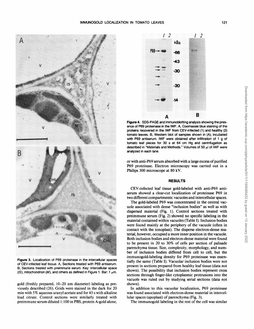
Figure 4. SDS-PAGE and immunoblotting analysis showing the pres-ence of P69 proteinase in the IWF. A, Coomassie blue staining of theproteins recovered in the IWF from CEV-infected (1) and healthy (2)tomato leaves. B, Western blot of samples shown in (A), incubatedwith P69 antiserum. IWF were obtained after infiltration of 1 g oftomato leaf pieces for 30 s at 64 cm Hg and centrifugation asdescribed in "Materials and Methods." Volumes of 50 Al of IWF wereanalyzed in each lane.
or with anti-P69 serum absorbed with a large excess ofpurifiedP69 proteinase. Electron microscopy was carried out in a
Philips 300 microscope at 80 kV.
v
is
,; ...... :A_
qDlw,
r '
Figure 3. Localization of P69 proteinase in the intercellular spacesof CEV-infected leaf tissue. A, Sections treated with P69 antiserum.B, Sections treated with preimmune serum. Key: intercellular space(IS), mitochondrion (M), and others as defined in Figure 1. Bar: 1 Jim.
gold (freshly prepared, 10-20 nm diameter) labeling as pre-viously described (26). Grids were stained in the dark for 20min with 5% aqueous uranyl acetate and for 45 s with alkalinelead citrate. Control sections were similarly treated withpreimmune serum diluted 1: 100 in PBS, protein A-gold alone,
RESULTS
CEV-infected leaf tissue gold-labeled with anti-P69 anti-serum showed a clear-cut localization of proteinase P69 intwo different compartments: vacuoles and intercellular spaces.The gold-labeled P69 was concentrated in the central vac-
uole associated with dense "inclusion bodies" as well as withdispersed material (Fig. 1). Control sections treated withpreimmune serum (Fig. 2) showed no specific labeling in thematerial contained within vacuoles (Table I). Inclusion bodieswere found mainly at the periphery of the vacuole (often incontact with the tonoplast). The disperse electron-dense ma-
terial, however, occupied a more inner position in the vacuole.Both inclusion bodies and electron-dense material were foundto be present in 20 to 30% of cells per section of palisadeparenchyma tissue. Size, complexity, morphology, and num-
ber of inclusion bodies differed from cell to cell, but theimmunogold-labeling density for P69 proteinase was essen-
tially the same (Table I). Vacuolar inclusion bodies were notpresent in sections prepared from healthy leaf tissue (data notshown). The possibility that inclusion bodies represent crosssections through finger-like cytoplasmic protrusions into thevacuole was ruled out by studying serial sections (data notshown).
In addition to this vacuolar localization, P69 proteinasewas found associated with electron-dense material in intercel-lular spaces (apoplast) of parenchyma (Fig. 3).The immunogold labeling in the rest of the cell was similar
A
121
Dow
nloaded from https://academ
ic.oup.com/plphys/article/91/1/119/6085422 by guest on 13 January 2022

Plant Physiol. Vol. 91, 1989
A
30
...
'. I
.4 ._
......:
.
B
t6
Figure 5. Recovery of P69 proteinase from intercellular spaces ofhealthy and CEV-infected tomato leaves. IWF were recovered bycentrifugation after infiltration of leaf segments for 30 s at the indi-cated vacuum pressures. A, After concentration by freeze-drying,equivalent amounts of IWF from CEV-infected and healthy plantswere assayed for P69 content by radial immunodiffusion (O = in-fected, V = healthy) and for P69 proteinase activity in Tris-HCI (pH9.0), 1 mm DTT, and 1 mm CaCI, with FTC-casein as substrate(0 = infected, V = healthy). Values are means of three differentextractions. B, Equal volumes (25 AL) of IWF from CEV-infected were
assayed for P69 proteinase activity by zymography on fibrinogen-containing SDS-gels after infiltration of leaf segments (1 g) for 30 swith different vacuum pressures. Numbers 1 through 5 correspondto IWF obtained after infiltration at 12, 25, 38, 50, and 64 cm Hg,respectively. C, control experiment with 3 jg of purified P69proteinase.
to the background observed in the control sections treatedwith preimmune serum or protein A-gold alone (Table I, Figs.2, 3B).An extracellular location of P69 protein was also demon-
strated by SDS-PAGE of the IWF. A majority of the proteinscorresponded to PR proteins previously reported in tomato(6) (Fig. 4). These proteins included a protein which wasdetermined by immunoblotting to be the P69 proteinase (Fig.4B). Zymography and hydrolysis ofFTC-casein demonstratedthe protein in the IWF to possess proteolytic activity (Fig. 5,A and B). Recovery of the protein, as quantified by radialdiffusion assay (Fig. 5A), after infiltration with different pres-sures, increased up to a maximum of 25 to 30 ,ug in IWF pergram of leaf tissue. By comparing the amount of P69 in IWFand whole tissue extracts (25), it appeared that at least 30 to40% of the total P69 proteinase was located outside the cell.The addition of different salts (e.g., NaCl, KCl, MgCl2, orCaCl2) to the infiltrating solution did not significantly increasethe recovery ofP69 from intercellular spaces (data not shown).The possibility that P69 was in IWF as a consequence of celldisruption during manipulation and infiltration of the tissuewas ruled out, because no traces of glucose-6-phosphate de-hydrogenase, Chl, or immunoreactive ribulose- 1 ,5-bisphos-phate carboxylase were detected (data not shown).P69 was practically undetectable in healthy plants as shown
in Figures 4 and 5 (24, 25).
DISCUSSION
We have provided evidence that proteinase P69 is locatedin vacuoles and in intercellular spaces. In the vacuole, P69 isassociated with engulfed 'inclusion bodies' and with dispersedmaterial, which could be an advanced stage ofdisorganizationof the inclusion bodies. In the intercellular spaces, the pro-teinase was found associated with an electron-dense materialresembling the residual stage of cytosol disorganization pre-viously described in the study of the localization of tomatoPR P1-p14 protein (26). The extracellular localization ofproteinase P69 is, therefore, consistent with the general con-sideration that PR proteins accumulate in the apoplast (4, 15,22, 26).The vacuolar localization of P69 proteinase is consistent
with the concept that the central vacuole might be a 'defensearsenal' (1, 10). Other lytic or defense proteins (e.g. chitinases[3], proteinase inhibitors [27], and proteinases [14, 19, 29])as well as many preformed secondary compounds are alsocompartmentalized within the vacuole. Besides the consider-ation of P69 proteinase as a potential defense weapon againstattacking pathogens (24, 25), it should be noted that P69, perse, as many other vacuolar components, is also deleterious tothe plant cell itself. The P69 proteinase causes rapid degra-dation of many cellular proteins, of which RuBPCase degra-dation is the most conspicuous (24). Thus, it seems logicalthat confinement of P69 within a separate cellular compart-ment (the vacuole) controls its hydrolytic function. Its actionwould be effective after vacuoles display autophagic activityby engulfing cytoplasm. This concept agrees with the propos-als of Matile and Winkenbach ( 11) and Wittenbach et al. (29)and also with the idea proposed by Van der Wilden et al.(20), which consider protein bodies as autophagic vacuoles.
122 VERA ET AL.
0 'I.
I I
i,
Dow
nloaded from https://academ
ic.oup.com/plphys/article/91/1/119/6085422 by guest on 13 January 2022

IMMUNOGOLD LOCALIZATION IN TOMATO LEAVES
Decompartmentalization of plant cells has been argued to bea response to pathogen attack (2, 22, 28). Accordingly, we
envisage that the last steps in viroid pathogenesis would bethe disruption of the vacuole with liberation of its hydrolyticconstituents, including P69 proteinase, to the intercellularspaces. The way in which the collapse of critical cells leads tothe appearance of pathological symptoms is a matter of dis-cussion. Clearly, such a complex process must involve thecooperative interaction of multiple factors and, as with othersimilarly regulated metabolic events, may be initiated by morethan one mechanism. Questions regarding further involve-ment of P69 proteinase and probably other relevant hydro-lases in viroid pathogenesis remain to be answered.
Targeting ofP69 proteinase to the apoplast could be a directprocess rather than a result of vacuolar disruption. The studyof this transport, as well as the isolation of vacuoles andcharacterization of 'inclusion bodies,' may further our under-standing of the biological role of PR proteins.
LITERATURE CITED
1. Boller T (1982) Enzymatic equipment of plant vacuoles. PhysiolVeget 20: 247-257
2. Boller T (1987) Hydrolytic enzymes in plant disease resistance.In T Kosuge, EW Nester, eds, Plant-Microbe Interactions, Vol2. Macmillan, New York, pp 385-413
3. Boller T, Vogeli U (1984) Vacuolar localization of ethylene-induced chitinase in bean leaves. Plant Physiol 74: 442-444
4. Caff JP, Dixon DC, Nikolau BT, Voelkerding KV, Klessing DF(1987) Synthesis and localization of pathogenesis-related pro-teins in tobacco. Mol Cell Biol 7: 1580-1583
5. Gianinazzi S, Martin C, Vallee JC (1970) Hypersensibilite auxvirus, temperature et proteines solubles chez le Nicotiana Xan-thi n.c. Apparition de nouvelles macromolecules lors de larepression de la synthese virale. C R Acad Sci Ser D 270:2383-2386
6. Granell A, Belles JM, Conejero V (1987) Induction ofpathogen-esis-related proteins in tomato by citrus exocortis viroid, silverion and Ethephon. Physiol Mol Plant Pathol 31: 83-89
7. Kauffmann S, Legrand M, Geoffroy P, Fritig B (1987) Biologicalfunction of pathogenesis-related proteins: four proteins of to-bacco have 1,3-fl-glucanase activity. EMBO J 6: 3209-3212
8. Kombrink E, Schoeder M, Hahlbrock K (1988) Several patho-genesis-related proteins in potato are 1,3-fl-glucanases andchitinases. Proc Natl Acad Sci USA 85: 782-786
9. Legrand M, Kauffmann S, Geoffroy P, Fritig B (1987) Biologicalfunction of pathogenesis-related proteins: four tobacco patho-genesis-related are chitinases. Proc Natl Acad Sci USA 84:6750-6754
10. Matile P (1975) The Lytic Compartment of Plant Cells. Springer-Verlag. Vienna
11. Matile P, Winkenbach F (1971) Functions of lysosomes andlysosomal enzymes in senescing corolla of the morning glory(Ipomea purpurea). J Exp Bot 22: 759-771
12. Metraux JP, Streit L, Stanb TH (1988) A pathogenesis-relatedprotein in cucumber is a chitinase. Physiol Mol Plant Pathol33: 1-9
13. Nasser N, De Tapia M, Kauffmann A, Montasser-Kouhsari S,Burkard D (1988) Identification and characterization of maizepathogenesis-related proteins. Four maize PR proteins arechitinases. Plant Mol Biol 11: 529-538
14. Nishimura M, Beevers H (1979) Hydrolysis of proteins in vacu-oles isolated from higher plant tissue. Nature 277: 412-413
15. Parent JG, Asselin A (1984) Detection of pathogenesis-relatedproteins (PR or b) and other proteins in the intercellular fluidof hypersensitive plants infected with tobacco mosaic virus.Can J Bot 62: 564-569
16. Semancik JS, Conejero V (1987) Viroid pathogenesis and expres-sion of biological activity. In JS Semancik, ed, Viroids andViroid-Like Pathogens. CRC Press, Boca Raton, FL, pp 71-127
17. Shumway LK, Rancour J, Ryan CA (1970) Vacuolar proteinbodies in tomato leaf cells and their relationship to storage ofchymotrypsin inhibitor I protein. Planta 93: 1-14
18. Singh NK, Bracker CA, Hasegawa PM, Handa AK, Buckel S,Herdmonson SA, Pfaukoch E, Regnier FE, Bressan RA (1987)Characterization of osmotin. Plant Physiol 85: 529-536
19. Thayer SS, Huffaker RC (1984) Vacuolar localization of endo-proteinases EPI and EP2 in barley mesophyll cells. PlantPhysiol 75: 70-73
20. Van der Wilden W, Herman EH, Chrispeels MJ (1980) Proteinbodies ofmung bean cotyledons as autophagic organelles. ProcNatl Acad Sci USA 77: 428-432
21. Van Loon LC (1982) Regulation of changes in proteins andenzymes associated with a defense against virus infection. InRKS Wood, ed, Active Defense Mechanisms in Plants. PlenumPress, New York, pp 247-273
22. Van Loon LC (1985) Pathogenesis-related proteins. Plant MolBiol4: 111-116
23. Van Loon LC, Van Kammen A (1970) Polyacrylamide discelectrophoresis of the soluble leaf proteins from Nicotianatabacum var. "Samsun" and "Samsun NN." II. Changes inprotein constitution after infection with tobacco mosaic virus.Virology 40: 199-211
24. Vera P, Conejero V (1988) Pathogenesis-related proteins of to-mato. P69 as an alkaline endoproteinase. Plant Physiol 87: 58-63
25. Vera P, Conejero V (1989) The induction and accumulation ofthe pathogenesis-related P69 proteinase in tomato during citrusexocortis viroid infection and after chemical treatments. Phys-iol Mol Plant Pathol (in press)
26. Vera P, Hernandez-Yago J, Conejero V (1988) Immunocyto-chemical localization of the major pathogenesis-related (PR)protein of tomato plants. Plant Sci 55: 223-230
27. Walker-Simmons M, Ryan CA (1977) Immunological identifi-cation of proteinase inhibitors I and II in isolated tomato leafvacuoles. Plant Physiol 60: 61-63
28. Wilson CL (1973) A lysosomal concept for plant pathology.Annu Rev Phytopathol 11: 247-272
29. Wittenbach VA, Lin W, Hebert R (1982) Vacuolar localizationof proteases and degradation of chloroplast in mesophyll pro-toplast from senescing primary wheat leaves. Plant Physiol 69:82-102
123
Dow
nloaded from https://academ
ic.oup.com/plphys/article/91/1/119/6085422 by guest on 13 January 2022