Embed Size (px)
Citation preview

RESEARCH Open Access
Indole-3-carbinol regulates microgliahomeostasis and protects the retina fromdegenerationAmir Saeed Khan1 and Thomas Langmann1,2*
Abstract
Background: Retinal degenerative diseases significantly contribute to visual impairment and blindness. Microgliareactivity is a hallmark of neurodegenerative diseases including retinal cell death and immunomodulation emergesas a therapeutic option. Indole-3-carbinol (I3C) is a natural ligand of aryl hydrocarbon receptor (AhR), with potentimmunomodulatory properties. Here, we hypothesized that I3C may inhibit microglia reactivity and exertneuroprotective effects in the light-damaged murine retina mimicking important immunological aspects of retinaldegeneration.
Methods: BV-2 microglia were treated in vitro with I3C followed by lipopolysaccharide (LPS) stimulation to analyzepro-inflammatory and anti-oxidant responses by quantitative real-time PCR (qRT-PCR) and Western blots. Nitricoxide (NO) secretion, caspase 3/7 levels, phagocytosis rates, migration, and morphology were analyzed in controland AhR knockdown cells. I3C or vehicle was systemically applied to light-treated BALB/cJ mice as an experimentalmodel of retinal degeneration. Pro-inflammatory and anti-oxidant responses in the retina were examined by qRT-PCR, ELISA, and Western blots. Immunohistochemical staining of retinal flat mounts and cryosections wereperformed. The retinal thickness and structure were evaluated by in vivo imaging using spectral domain-opticalcoherence tomography (SD-OCT).
Results: The in vitro data showed that I3C potently diminished LPS-induced pro-inflammatory gene expression of I-NOS, IL-1ß, NLRP3, IL-6, and CCL2 and induced anti-oxidants gene levels of NQO1, HMOX1, and CAT1 in BV-2 cells. I3Calso reduced LPS-induced NO secretion, phagocytosis, and migration as important functional microglia parameters.siRNA-mediated knockdown of AhR partially prevented the previously observed gene regulatory events. The in vivoexperiments revealed that I3C treatment diminished light-damage induced I-NOS, IL-1ß, NLRP3, IL-6, and CCL2transcripts and also reduced CCL2, I-NOS, IL-1ß, p-NFkBp65 protein levels in mice. Moreover, I3C increased anti-oxidant NQO1 and HMOX1 protein levels in light-exposed retinas. Finally, I3C therapy prevented the accumulationof amoeboid microglia in the subretinal space and protected from retinal degeneration.
Conclusions: The AhR ligand I3C potently counter-acts microgliosis and light-induced retinal damage, highlightinga potential treatment concept for retinal degeneration.
Keywords: Aryl hydrocarbon receptor, Indole-3-carbinol, Microglia, Retinal degeneration, Light damage
© The Author(s). 2020 Open Access This article is licensed under a Creative Commons Attribution 4.0 International License,which permits use, sharing, adaptation, distribution and reproduction in any medium or format, as long as you giveappropriate credit to the original author(s) and the source, provide a link to the Creative Commons licence, and indicate ifchanges were made. The images or other third party material in this article are included in the article's Creative Commonslicence, unless indicated otherwise in a credit line to the material. If material is not included in the article's Creative Commonslicence and your intended use is not permitted by statutory regulation or exceeds the permitted use, you will need to obtainpermission directly from the copyright holder. To view a copy of this licence, visit http://creativecommons.org/licenses/by/4.0/.The Creative Commons Public Domain Dedication waiver (http://creativecommons.org/publicdomain/zero/1.0/) applies to thedata made available in this article, unless otherwise stated in a credit line to the data.
* Correspondence: [email protected] for Experimental Immunology of the Eye, Department ofOphthalmology, Faculty of Medicine and University Hospital Cologne,University of Cologne, Joseph-Stelzmann-Str. 9, D-50931 Cologne, Germany2Center for Molecular Medicine Cologne, Cologne, Germany
Khan and Langmann Journal of Neuroinflammation (2020) 17:327 https://doi.org/10.1186/s12974-020-01999-8
BackgroundPhagocytes of the retina, microglia and macrophages, havebeen recognized as important factors in retinal degenera-tive diseases. In the healthy retina, ramified microglialocalize in the inner and outer plexiform layers (IPL, OPL)and scan their microenvironment with their surface recep-tors for cytokines, chemokines, and complement factor [1,2]. In the diseased retina, these cells become amoeboid andmigrate to subretinal (SR) space with upregulation ofdifferent pro-inflammatory molecules [2, 3]. Initially,microglia enhance their phagocytosis capacity to resolvetissue damage. However, persistent disease leads to chron-ically activate microglia, which may ingest not only cellulardebris but also healthy photoreceptors [4]. Therefore,microglia-targeted pharmacotherapy to mitigate chronicneuro-inflammation may be a promising treatment ap-proach in retinal degenerative diseases.Aryl hydrocarbon receptor (AhR), a ligand-dependent
transcriptional factor, is activated by various ligands [5].Initially, AhR was identified as key regulator of xeno-biotic metabolism and detoxification [6]. Inactive AhRlocalizes in the cytoplasm and upon binding of a ligand,the protein dissociates from chaperones and translocatesinto the nucleus where it binds to nuclear responseelement and changes gene transcription required forcellular homeostasis [6]. AhR deficiency accelerates agingin mice and triggers inflammation [7–9], and AhR ligandsplay an important role in immune modulation [9, 10].In the ocular compartment, AhR knockout triggers
apoptosis and inflammation in experimental autoimmuneuveitis [11]. AhR deficiency also disturbs extracellularmatrix biology, leading to an AMD-like pathology in mice[12]. Moreover, AhR−/− mice showed a reduction in visualfunction, displayed an accumulation of microglia in theSR space, and had retinal pigment epithelium (RPE) ab-normalities [13]. AhR−/− mice also showed larger lesionsin laser-induced choroidal neovascularization that mimicsthe wet form of AMD [14]. Furthermore, 2,2′-aminophe-nyl indole (2AI), a synthetic ligand of AhR could protectthe RPE and retina from environmental stress [15].Here, we hypothesized that AhR agonists may regulate
microglia homeostasis and dampen experimental neurode-generation in the retina. To test this hypothesis, we used anaturally occurring AhR agonist, indole-3-carbinol (I3C),which is abundant in green vegetables. We addressed theeffects of I3C in vitro using the microglial BV-2 cell line[16] and systemically applied I3C in BALB/cJ mice exposedto an acute white-light-damage paradigm.
MethodsReagentsI3C (I7256-5G) and Escherichia coli 0111: B4 lipopoly-saccharide (LPS) were purchased from Sigma-Aldrich(St. Louis, MO, USA).
Cell cultureBV-2 microglia were seeded in RPMI640 supplementedwith 5% fetal calf serum (FCS), 1% penicillin/streptomycin,2mM L-glutamine and 195 nM β-mercaptoethanol at 37 °Cin a humidified atmosphere of 5% CO2. The cells werepre-treated with 50 μM I3C for 4 h and then stimulatedwith 50 ng/ml LPS for 4 h. After treatments, cells wereharvested for RNA and protein extraction. Cell superna-tants were also collected for further analysis. The 661Wphotoreceptor-like cells were cultured in Dulbecco’s modi-fied Eagle’s medium (DMEM) supplemented with 10%FCS, L-glutamine, and 1% penicillin/streptomycin.
RNA isolation, reverse transcription, and quantitative RT-PCRTotal RNA was extracted from the harvested BV-2 cellsusing the NucleoSpin® RNA Mini Kit (Macherey-Nagel;Dueren, Germany). Total RNA was also extracted fromthe retinas of BALB/cJ mice using the Qiagen RNeasyMicro Kit according to the manufacturer. RNA wasquantified with a NanoDrop 2000 photometer (ThermoScientific). First-strand complementary DNA synthesiswas performed using the Thermo RevertAid RT Kit(ThermoFisher Scientific; Waltham, USA). qRT-PCRanalysis was performed with the Takyon™ qPCR Kit(Eurogentec Deutschland GmbH; Köln, Germany) andthe Roche Probe library using the LightCycler® 480 IImachine (Roche; Basel, Switzerland). Primer sequencesand Roche library probe numbers were as follows: i-NOS, forward primer 5′-ctttgccacggacgagac-3′, reverseprimer 5′-tcattgtactctgagggctga-3′, probe #13; IL-1ß,forward primer 5′-tcttctttgggtattgcttgg-3′, reverse pri-mer 5′-tgtaatgaaagacggcacacc-3′, probe #38; NLRP3,forward primer 5′-ttcccagacactcatgttgc-3′, reverse pri-mer 5′-agaagagaccacggcagaag-3′, probe #74; CCL2,forward primer 5′-catccacgtgttggctca-3′, reverse primer5′-gatcatcttgctggtgaatgagt-3′, probe #62; IL6, forwardprimer 5′-gatggatgctaccaaactggat-3′, reverse primer 5′-ccaggtagctatggtactccaga-3′, probe #6; NQO1, forwardprimer 5′-agcgttcggtattacgatcc-3′, reverse primer 5′-agtacaatcagggctcttctcg-3′, probe #50; HMOX1, forwardprimer 5′-agggtcaggtgtccagagaa-3′, reverse primer 5′-cttccagggccgtgtagata-3′, probe #9; CAT1, forward primer5′-ccttcaagttggttaatgcaga-3′, 5′-caagtttttgatgccctggt-3′,probe #34; ATPase, forward primer 5′-ggcacaatgcag-gaaagg-3′, reverse primer 5′-tcagcaggcacatagatagcc-3′,probe #77. Measurements were performed in triplicates.ATP5b expression was used as a reference gene. Forrelative quantification, the ΔΔC method was used as im-plemented in the LightCycler® 480 software.
Protein extraction, ELISA, and Western blotRIPA buffer (150 mM sodium chloride (NaCl), 1% NP-40, 0.5% sodium deoxycholate, 0.1% sodium dodecylsulfate (SDS), 50 mM Tris-HCl pH 7.4, supplemented
Khan and Langmann Journal of Neuroinflammation (2020) 17:327 Page 2 of 14
with protease inhibitor cocktail (Roche)) was used toextract cell lysates. Mouse retinal tissue was homogenizedin PBS using sonication. Insoluble debris was removed bycentrifugation for 15min at 16,000×g. Protein concentra-tion was determined using the PierceTM BicinchoninicAcid (BCA) Protein Assay Kit (Thermo. Scientific, Cat#23225). Twenty micrograms of cell or tissue lysates wereseparated by SDS-PAGE on 10% gels with PageRuler pre-stained protein ladder (Thermo Scientific; Waltham, MA,USA). Proteins were then transferred to nitrocellulosemembranes (Biorad; Munich, Germany). After incubationin blocking buffer (TBS-T containing 5% nonfat dry milk)for 1 h, membranes were incubated with primaryantibodies against I-NOS (dilution 1:2000 in PBS, Cat#610600, BD Transduction Laboratories™), IL-1ß (dilution1: 200 in PBS, Cat #B122, Santa Cruz Biotechnology), p-NFκB p65 (dilution 1:500 in PBS, Cat #sc136548, Santa-Cruz Biotechnology), COX2 (dilution 1:500 in PBS, Cat#Ab1519, Abcam), NQO1 (dilution 1:500 in PBS, Cat#sc32793, Santa Cruz Biotechnology), HMOX1 (dilution1:1000 in PBS, Cat #Ab137749, Abcam), and ß-ACTIN(dilution 1:200 in PBS, Cat #sc47778, Santa-Cruz Biotech-nology). After washing steps, blots were incubated withsecondary antibodies (dilution 1:4000 in PBS, Cat #P0448,Dako polyclonal goat anti-rabbit, immunoglobulins/HRP, and dilution 1:4000 in PBS, Cat #P0447, Dakopolyclonal goat anti-mouse, immunoglobulins/HRP).Enhanced chemiluminescence signals were then visual-ized and imaged with the MultiImage II system (AlphaInnotech; Santa Clara, CA, USA). Densitometry ofbands was measured using the Image J software (NIH).The concentration of CCL2 in total retinal lysates wasmeasured by ELISA according to the manufacturer’sinstructions (Mouse CCL2/JE/MCP-1 DuoSet ELISA,Cat #DY479-05, R&D Systems).
Nitrite measurementNitric oxide concentrations were determined using theGriess reagent system (Promega). Briefly, 50 μl cell cul-ture was incubated with 100 μl Griess reagent in 96-wellplates. After incubation for 30 min at room temperature,absorbance was measured at 540 nm on an Infinite F200Pro plate reader (Tecan). Nitrite concentrations werecalculated as described before [17].
Caspase 3/7 assayTo determine microglia neurotoxicity, a culture systemof 661W photoreceptors cells with the microglia-conditioned medium was established. The 661W photo-receptor cells were incubated for 48 h either in theirown medium or with culture supernatants from treatedBV-2 cells. The 661W cells morphology was assessed byphase-contrast microscopy and cell death was determined
with the Caspase-Glo® 3/7 Assay (Promega GmbH; Mann-heim, Germany) as previously described [17].
Phagocytosis assayBV-2 cells were pre-treated with DMSO as vehicle,50 μM I3C, vehicle +50 ng/ml LPS, and 50 ng/ml LPS +50 μM I3C for 4 h. After treatments, 2 μl latex bead solu-tion (polystyrene microparticles, Sigma Aldrich; St.Louis, MO, USA) was added to the wells for 4 h todetermine the influence of I3C on phagocytosis. Five mi-crographs per well were taken using an AxioVert.A1inverted microscope (Carl Zeiss; Germany). The phago-cytic activity was determined by calculating the numberof cells, which phagocytosed 10 or more latex beads.
Scratch wound healing assayBV-2 cells were seeded in six-well plates as 80% conflu-ent monolayers and were wounded with a sterile 200 μlpipette tip. Thereafter, the cells were treated withvehicle, 50 μM I3C, vehicle +50 ng/ml LPS, and 50 ng/mlLPS + 50 μM I3C. Migration into the open scar wasdocumented with microphotographs taken after 8 h ofwounding using an AxioVert.A1 inverted microscope(Carl Zeiss; Germany).
Phalloidin stainingBV-2 cells were seeded on coverslips in six-well plates.Cells were treated with vehicle, 50 μM I3C, vehicle+50 μM I3C, and 50 μM I3C + 50 ng/ml LPS. Thereafter,the cells were fixed, permeabilized with 0.1% Triton X-100and F-actin was labeled using 0.1 μg/ml Phalloidin-TRITC(Sigma). The nuclei were stained using 4′,6-diamidino-2-phenylindole (DAPI), and coverslips were mounted ontoslides using Dako fluorescent mounting medium (DakoDeutschland GmbH; Hamburg, Germany). Photos weretaken with a Zeiss Imager M.2 equipped with Apotome.2(Carl Zeiss; Germany).
siRNA-mediated AhR gene silencingBV-2 cells were transfected with AhR siRNA (FlexiTubesiRNA containing 4 preselected siRNAs for the AhRgene, Cat #1027416, Qiagen; Hilden Germany) and thesiRNA non-targeting negative control (Cat #1022076,Qiagen; Hilden Germany). All transfections were per-formed using lipofectamine 3000 (Invitrogen) for 6 hfollowed by medium refreshment and cells were furtherincubated for 48 h. During this incubation time, cellswere treated with 50 μM I3C or 50 ng/ml LPS for 4 h.
Animal experimentsAll in vivo experiments were performed with 8-10-week-old BALB/cJ mice of both sexes. The animals were keptin an air-conditioned environment with 12-h light anddark cycle and had full access to water and food ad
Khan and Langmann Journal of Neuroinflammation (2020) 17:327 Page 3 of 14

libitum. All experimental procedures complied with theARVO Statement for the Use of Animals in Ophthalmicand Vision Research. The animal protocols were ap-proved by the government office of animal welfare inNorth Rhine-Westphalia (Germany) (reference number81-02.04.2019.A092). The mice received intraperitonealinjections of I3C at a dose of 15 mg/kg body weight, dis-solved in DMSO or DMSO alone as vehicle control,starting 1 day before the light exposure and then oncedaily for the remaining days. BALB/cJ mice were dark-adapted for 16 h before light exposure. Pupil dilationwas performed with 2.5% phenylephrine and 0.5% tropi-camide under dim red light before the mice wereexposed to bright white light (15,000 lux) for 1 h. Afterlight exposure, the animals were kept in dark-rearedconditions overnight and then maintained under normallight conditions (12-h light and dark cycle) for theremaining experimental period.
ImmunohistochemistryEyes were enucleated and fixed with 4% paraformalde-hyde (ROTI®Histofix, Carl-Roth; Germany) for 2 h atroom temperature. Retinal flat mounts were preparedand permeabilized overnight (5% Triton X-100, 5%Tween-20 in PBS). The flat mounts were incubated withBLOTTO (1% milk powder, 0.01% Triton X-100 in PBS)for 1 h to block nonspecific antigen binding. Subse-quently, retinal flat mounts were incubated with primaryantibody (rabbit anti-Iba1, dilution 1:1000, FUJIFILMWako Chemicals; Neuss, Germany) overnight at 4 °C.After washing steps, the retinal flat mounts were incu-bated with secondary antibody (goat anti-rabbit Alexa-Flour 488 (green) A11008, Life Technologies) for 1 h atroom temperature. After washing steps, retinal flatmounts were mounted on a microscopic slide and em-bedded with fluorescence mounting medium (VECTASHIELD® HardSet™ Antifade Mounting Medium H-1400,Vector Laboratories; Burlingame, USA). Images weretaken with a Zeiss Imager M.2 equipped with Apotome.2(Carl Zeiss; Germany).For immunohistochemical analyses of cryosections,
the eyes were enucleated and fixed with 4% paraformal-dehyde (ROTI®Histofix, Carl-Roth; Germany) for 2 h atroom temperature. Whole eyes were then incubatedwith 30% sucrose overnight and embedded in optimalcutting temperature (OCT) compound, shock frozen ondry ice and then stored at −20 °C. Ten-micrometersections were cut with a cryostat (Leica CM 3050 S,Leica biosystem; Wetzlar, Germany). Sections were rehy-drated with phosphate-buffered saline (PBS) and blockedwith BLOTTO (1% milk powder and 0.3% Triton X-100in PBS) followed by overnight incubation with primaryantibody (rabbit anti-Iba1, dilution is 1:500 in BLOTTO,
FUJIFILM Wako Chemicals; Neuss, Germany) at 4 °C.After washing, sections were incubated with secondaryantibody (goat anti-rabbit AlexaFlour 488 (green), dilu-tion is 1:1000 in PBS, A11008, Life Technologies) for 1 hat room temperature. After washing steps, the sectionswere mounted with Flouromount and counterstainedwith Dapi (ThermoFisher Scientific; Waltham, USA).Images were taken with a Zeiss Imager M.2 equippedwith Apotome.2 (Carl Zeiss; Germany).
Optical coherence tomography (OCT)For OCT, mice were anesthetized by intraperitoneal injec-tion of ketamine hydrochloride (100mg/kg body weight,Ketavet; Zoetis) and xylazine hydrochloride (10mg/kgbody weight, Rompun; Bayer HealthCare) diluted in 0.9%sodium chloride. The pupils of the mice were dilatedusing 2.5% phenylephrine and 0.5% tropicamide beforeOCT. Spectral-domain optical coherence tomography(SD-OCT) was performed on both eyes with a Spectralis™HRA/OCT device (Heidelberg Engineering) to quantifythe retinal thickness using the Heidelberg Eye ExplorerSoftware with circular ring scans (circle diameter 3 and 6mm), centered around the optic nerve head.
Statistical analysisData were analyzed using GraphPad Prism version 7(GraphPad Software Inc., San Diego, CA). All data wereanalyzed using analysis of variance (one-way ANOVA)and Dunnett’s multiple comparison test. p < 0.05 wasconsidered as statistically significant.
ResultsI3C reduces pro-inflammatory and enhances anti-oxidantgene expression in BV-2 cellsWe first examined whether treatment with the AhRligand I3C has an effect on a selected set of pro-inflammatory markers in microglia-like BV-2 cells.The cells were pre-treated with 50 μM I3C for 4 hand then further stimulated with 50 ng/ml LPS foradditional 4 h. LPS stimulation-induced mRNA levelsfor inducible NO-synthase (i-NOS) (Fig. 1a), interleu-kin-1 beta (IL-1ß) (Fig. 1b), NOD-, LRR-, and pyrindomain-containing protein 3 (NLRP3) (Fig. 1c), inter-leukin-6 (IL-6) (Fig. 1d), and C–C motif chemokineligand 2 (CCL2) (Fig. 1e) and co-treatment with I3Csignificantly dampened expression of these markers.Since AhR ligands are known to exert anti-oxidant ef-fects, we also measured mRNAs for NADPH dehydro-genase quinone 1 (NQO1) (Fig. 1f), heme oxygenase 1HMOX1 (Fig. 1g), and catalase 1 (CAT1) (Fig. 1h).For each of these three genes, I3C enhanced mRNAexpression levels (Fig. 1f-h). We then aimed to con-firm these data using Western blots. These
Khan and Langmann Journal of Neuroinflammation (2020) 17:327 Page 4 of 14

Fig. 1 Effects of I3C on pro-inflammatory and anti-oxidant mRNA and protein levels. a-h BV-2 microglia cells were treated with 50 μM I3C orDMSO as a vehicle for 4 h followed by 50 ng/ml LPS for 4 h. After 8 h, mRNA expression levels of i-NOS a, IL-1ß b, NLRP3 c, IL-6 d, CCL2 e, NQO1 f,HMOX1 g, and CAT1 h were analyzed by real-time PCR. i Western blots show i-NOS, IL-1ß, COX2, NQO1, HMOX1, and ß-ACTIN protein levels as aloading control. Mean relative protein levels are shown for i-NOS j, IL-1ß k, and HMOX1 l. Data show mean ± SEM out of three independentexperiments for mRNA expression levels (n = 3/group, measured in triplicates). For Western blot, three independent experiments were performed.*p < 0.05, **p < 0.01, ***p < 0.001, ****p < 0.0001
Khan and Langmann Journal of Neuroinflammation (2020) 17:327 Page 5 of 14
experiments revealed that the LPS-induced expressionof i-NOS, IL 1ß, and cyclooxygenase 2 (COX2) wasalso reduced by I3C on the protein level, whereasHMOX1 was increased and NQO1 was unchanged(Fig. 1i-l). Taken together, the treatments with I3Creduced LPS-mediated pro-inflammatory markers andenhances antioxidant gene expression in BV-2 cells.
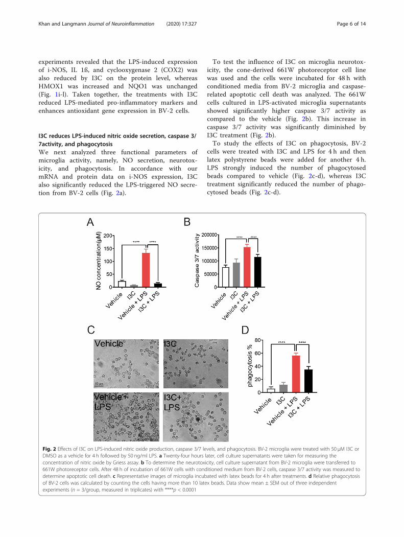
I3C reduces LPS-induced nitric oxide secretion, caspase 3/7activity, and phagocytosisWe next analyzed three functional parameters ofmicroglia activity, namely, NO secretion, neurotox-icity, and phagocytosis. In accordance with ourmRNA and protein data on i-NOS expression, I3Calso significantly reduced the LPS-triggered NO secre-tion from BV-2 cells (Fig. 2a).
To test the influence of I3C on microglia neurotox-icity, the cone-derived 661W photoreceptor cell linewas used and the cells were incubated for 48 h withconditioned media from BV-2 microglia and caspase-related apoptotic cell death was analyzed. The 661Wcells cultured in LPS-activated microglia supernatantsshowed significantly higher caspase 3/7 activity ascompared to the vehicle (Fig. 2b). This increase incaspase 3/7 activity was significantly diminished byI3C treatment (Fig. 2b).To study the effects of I3C on phagocytosis, BV-2
cells were treated with I3C and LPS for 4 h and thenlatex polystyrene beads were added for another 4 h.LPS strongly induced the number of phagocytosedbeads compared to vehicle (Fig. 2c-d), whereas I3Ctreatment significantly reduced the number of phago-cytosed beads (Fig. 2c-d).
Fig. 2 Effects of I3C on LPS-induced nitric oxide production, caspase 3/7 levels, and phagocytosis. BV-2 microglia were treated with 50 μM I3C orDMSO as a vehicle for 4 h followed by 50 ng/ml LPS. a Twenty-four hours later, cell culture supernatants were taken for measuring theconcentration of nitric oxide by Griess assay. b To determine the neurotoxicity, cell culture supernatant from BV-2 microglia were transferred to661W photoreceptor cells. After 48 h of incubation of 661W cells with conditioned medium from BV-2 cells, caspase 3/7 activity was measured todetermine apoptotic cell death. c Representative images of microglia incubated with latex beads for 4 h after treatments. d Relative phagocytosisof BV-2 cells was calculated by counting the cells having more than 10 latex beads. Data show mean ± SEM out of three independentexperiments (n = 3/group, measured in triplicates) with ****p < 0.0001
Khan and Langmann Journal of Neuroinflammation (2020) 17:327 Page 6 of 14

I3C reduces LPS-induced migration and induces filopodiaformation in BV-2 cellsTo study the influence of I3C on microglia migration,wound-healing scratch assays were conducted in BV-2cells. LPS induced the migration of BV-2 microglia cellsinto the scratch (Fig. 3a), and I3C effectively blocked thismigration response (Fig. 3a). The morphology of micro-glia is another hallmark of activation and hence westained the F-actin cytoskeleton with phalloidin-TRITCin the different culture conditions. LPS clearly triggereda change in morphology from ramified to an amoeboidshape (Fig. 3b). Incubations in the presence of I3C
clearly transformed the BV-2 cells to a highly ramifiedphenotype with most cells containing long filopodia(Fig. 3b). Taken together, I3C reduces LPS-induced mi-gration and induces filopodia formation in BV-2 micro-glia cells.
The AhR pathway is involved in the in vitro effects of I3Con pro-inflammatory gene expressionTo further investigate the AhR pathway in modulation ofpro-inflammatory gene expression, siRNA-mediated knock-down of AhR was performed in BV-2 microglia. The cellswere transfected with AhR siRNA or non-targeting negative
Fig. 3 Effects of I3C on LPS-induced migration and morphology of BV-2 microglia. BV-2 microglia cells were treated with 50 μM I3C or DMSO as avehicle for 4 h followed by 50 ng/ml LPS after scratch with a pipette tip. a Representative images of microglia migration toward the scratch areaafter 8 h. b Representative images of the BV-2 cell morphology stained with phalloidin and DAPI. (n = 3/group, measured in triplicates)
Khan and Langmann Journal of Neuroinflammation (2020) 17:327 Page 7 of 14

control siRNA. We detected that the mRNA level of AhR it-self was significantly downregulated by AhR siRNA (Fig. 4a).Treatment of AhR siRNA-transfected cells with I3C plusLPS caused a relative increase in i-NOS (Fig. 4b), IL-1ß(Fig. 4c), and NLRP3 (Fig. 4d) transcript levels com-pared to the siRNA negative control. In contrast, IL-6mRNA expression levels were not changed in the presenceof AhR siRNA (Fig. 4e). Thus, AhR knockdown partiallyprevented the previously detected anti-inflammatory ef-fects of I3C on LPS-activated BV-2 cells.
I3C treatment regulates pro-inflammatory andantioxidant genes in the light-damage model of murineretinal degenerationWe next aimed at analyzing the effects of I3C in themurine retina subjected to an established light damageparadigm of retinal degeneration. Light-sensitive BALB/cJ mice were dark-adapted for 16 h before the mice wereexposed to white light with an intensity of 15,000 lux for1 h. The animals received intraperitoneal injections of15 mg/kg I3C or DMSO as a vehicle, the day before thelight exposure, and then once daily for the remaining 3days (Fig. 5a).Four days after light exposure, retinal mRNA expression
levels of the pro-inflammatory markers i-NOS (Fig. 5b),IL-1ß (Fig. 5c), NLRP3 (Fig. 5d), IL-6 (Fig. 5e), and CCL2(Fig. 5f). Whereas relative expression of these genes chan-ged in light exposed and vehicle-treated retinas, I3C treat-ment significantly reversed these effects (Fig. 5b-f).We next verified these mRNA expression differences on
the protein level and further included two antioxidantproteins, NQO1 and HMOX1, that are mainly regulatedon protein level. The effects on retinal CCL2 proteinexpression were verified using ELISA, which showed thatI3C reduced light-induced CCL2 secretion (Fig. 5g).Western blots further revealed that I3C also reducedlight-damage induced i-NOS, IL-1ß, and p-NFKBp65protein levels in the retina (Fig. 5h-j), whereas proteinlevels for the antioxidant genes NQO1 and HMOX1 wereupregulated in I3C-treated animals (Fig. 5h, k).
I3C prevents microglia reactivity in the light-damagedretinaTo assess the effects on retinal microglia in vivo, ret-inal flat mounts of light-challenged and I3C-treatedmice were stained with the marker IBA1. We foundthat retinas of untreated mice show no microglia inthe subretinal (SR) space (Fig. 6a), whereas vehicle +light-treated animals showed abundant amoeboidmicroglia in this area (Fig. 6b). Thus, accumulation ofamoeboid microglia in the SR space was prevented byI3C treatment (Fig. 6c, d). The outer plexiform layer(OPL) of untreated control animals showed mainly
ramified microglia (Fig. 6e). In contrast, vehicle-treated and light-exposed mice had less ramificationsand showed a more amoeboid morphology (Fig. 6f).Again, these signs of microglia reactivity were pre-vented by I3C therapy (Fig. 6g).We also analyzed the localization of IBA1-positive
cells in retinal layers using cryosections. In untreated an-imals, mainly ramified microglia were detected in bothplexiform layers (Fig. 6h). In contrast, amoeboid micro-glia were present in the ONL of light-damage andvehicle-treated mice (Fig. 6i). Notably, I3C nearly fullyprevented this light-induced amoeboid microglia accu-mulation (Fig. 6j). Furthermore, the DAPI staining ofcryosections of light-damage and vehicle-treated miceindicated a significant cell loss in the ONL, which wasprevented by I3C (Fig. 6h-k).
I3C preserves retinal thickness after light exposureFinally, we analyzed the effects of I3C on retinal degen-eration 4 days after acute light exposure. In vivo imagingwith optical coherence tomography (OCT) showedstrong retinal thinning and nearly complete loss of theONL in light-exposed mice that received vehicle treat-ment compared to animals not subjected to light dam-age (Fig. 7a, b). Treatment with I3C clearly preventedthis retinal degeneration (Fig. 7c), as confirmed by re-peated quantitative analyses of the central retinal thick-ness within 3 mm and 6mm ring scan areas (Fig. 7d, e).
DiscussionRecent studies showed that AhR is important for themaintenance of photoreceptor and RPE homeostasis [13,15]. To our knowledge, we here show for the first timethat AhR also regulates microglia homeostasis in the de-generating retina and that its natural ligand I3C protectsfrom acute light damage, a model system that mimicscertain aspects of human retinal degeneration.We started our study with in vitro experiments using
BV-2 microglia treated with LPS and I3C. BV-2 is anestablished murine microglia cell line [16]. Despite itslimitation as an immortalized cell type, BV-2 cells repre-sent a good alternative for primary microglia as gene ex-pression profiling showed that LPS-treated BV-2 cellsshared 90% of genes with primary microglia [18–20].We found here that I3C potently reduced LPS-triggeredgene and protein expression of well-established pro-inflammatory transcripts [17, 21, 22]. Conversely, I3C in-duced anti-oxidant gene expression, which is consistentwith previous findings that I3C has antioxidant activity[23]. Our data are in line with previous findings that I3Ccan block LPS-induced IL-1β, IL-6, and NO inRAW264.7 macrophage cells [24]. Previous findings alsoshowed that I3C mediates apoptosis in different cancercells [25–27]. However, our results indicate that I3C can
Khan and Langmann Journal of Neuroinflammation (2020) 17:327 Page 8 of 14

reduce neurotoxic effects of activated microglia on661W photoreceptor cells. Despite the limitation that661W cells are derived from a retinal tumor and expressmainly cone-specific markers [28], the cells have been
documented as reliable in vitro model to study molecu-lar pathways of neuronal apoptosis [29, 30].In this study, LPS induced the phagocytosis of latex
beads in BV-2 cells, consistent with previous findings
Fig. 4 Effects of AhR knockdown on pro-inflammatory markers in BV-2 microglia. Cells were transfected with AhR-siRNA for 6 h followed bytreatment with I3C and LPS. a-e mRNA expression levels of AhR a, i-NOS b, IL-1ß c, NLRP3 d, and IL-6 e were analyzed by real-time PCR. Datashow mean ± SEM out of three independent experiments (n = 3/group, measured in triplicates) with ****p < 0.0001
Khan and Langmann Journal of Neuroinflammation (2020) 17:327 Page 9 of 14

[20, 21, 31]. We also found that I3C significantly re-duced this phagocytic capacity. Phagocytosis assays withlatex beads have clear limitations and may only serve asa proxy for the real situation in neuronal tissue. Clearly,in vivo phagocytosis requires multiple cellular interactionsand complex molecular signaling events in microglia that
are absent in cell culture phagocytosis assays [32, 33].Nevertheless, our previous data showed a good correlationof microglial phagocytosis rates measured with latex beadsand apoptotic photoreceptor debris [21]. Our study fur-ther revealed that I3C reduced LPS-induced migrationand reversed the typical LPS-triggered amoeboid
Fig. 5 Effects of I3C on mRNA and protein levels in light-challenged BALB/cJ mice. a Schematic overview of experimental design. Eight to ten-week-old BALB/cJ mice of both sexes were used. Mice were dark-adapted for 16 h before the mice were exposed to white light with an intensity 15,000 luxfor 1 h. The animals received intraperitoneal injections of 15mg/kg I3C or vehicle (DMSO), 1 day before the light exposure and once daily for theremaining 3 days. Four days after the light exposure, b-f mRNA expression levels of i-NOS b, IL-1ß c, NLRP3 d, IL-6 e, and CCL2 f, were analyzed by real-time PCR. Four days after the light exposure, retinal lysates were extracted to perform ELISA of CCL2 g and Western blots h detecting i-NOS, IL- 1ß, p-NFKB65, NQO1, and HMOX1 protein levels. Beta-actin served as loading control. Relative protein levels for i-NOS i, p-NFkBp65 j, and NQO1 k weredetermined by densitometry. Data show mean ± SEM out of three independent experiments for mRNA expression levels (untreated n = 8 retinas,vehicle + light n = 14 retinas, light + I3C treatment n = 14 retinas, where each dot represents one retina). For Western blots and ELISA threeindependent experiments were performed (one retina per group) with *p < 0.05, **p < 0.01, ***p < 0.001, and ****p < 0.0001
Khan and Langmann Journal of Neuroinflammation (2020) 17:327 Page 10 of 14

phenotype of BV-2 microglia [21, 34]. These data supportthe finding that AhR is a master regulator of cellularphagocytosis, migration, and morphology [35–37].We also investigated the specificity of I3C using
siRNA-mediated knockdown of AhR in BV-2 microglia.These data revealed that knockdown of AhR preventsthe I3C-mediated decrease of i-NOS, IL-1ß, and NLRP3.However, we found that AhR knockdown did not alterIL-6 mRNA. This suggests that AhR is not directly in-volved in LPS-induced IL-6 gene regulation. In accord-ance with our results, AhR activation downregulatedNLRP3 and IL-1ß mRNA levels in peritoneal macro-phages and siRNA-mediated knockdown of AhR re-versed this effect [38].The in vitro data of I3C effects lead us to the ana-
lysis of microglia in a white-light-damage paradigm ofretinal degeneration. Light damage is a convenientand reproducible method for inducing synchronized
photoreceptor damage [39]. However, differences inthe effects of neuroprotective agents between light dam-age and genetic mouse models have been documented [40].Therefore, effects seen in this model cannot be transferredto retinal degenerative diseases in general. Our resultsclearly showed that I3C inhibited light-induced pro-inflammatory gene expression in vivo as i-NOS, IL-1ß,NLRP3, and IL-6 transcripts were significantly diminishedin I3C-treated animals. These data are in agreement withother neuroinflammatory and ocular disorders. Thus, I3Cprevented ischemic reperfusion-induced inflammation andimproved neurobehavioral symptoms in a cerebral is-chemic stroke model [41, 42]. AhR agonists also reversedpro-inflammatory gene expression in experimental auto-immune uveitis [11]. Here, we found that I3C preventedlight-induced CCL2 expression in the retina. Upregulationof CCL2 in light-exposed animals has been documented byothers before [43, 44]. We further showed that I3C reduced
Fig. 6 Effects of I3C in retinal microglia reactivity elicited by light in BALB/cJ mice. a-g Representative photomicrographs of IBA 1 stainedmicroglia (green) of retinal flat mounts. Microglia in the subretinal space (SR) and outer plexiform layer (OPL) of control a, e, vehicle + light b, f,and I3C + light c, g treated mice. d Total IBA 1+ cells were counted in the subretinal space (untreated n = 20 retinas, vehicle + light = 20 retinas,and I3C + light n = 20 retinas). h-j Representative photomicrographs show retinal sections of control h, vehicle + light i, and I3C + light j treatedmice stained with IBA 1 (green) and DAPI (blue). k For quantification of ONL thickness in cryosections, rows of photoreceptor nuclei werecounted (untreated n = 10 retinas, vehicle + light n = 10 retinas, I3C + light n = 10 retinas). Data show mean ± SEM with ****p < 0.0001. ONLouter nuclear layer, OPL outer plexiform layer, INL inner nuclear layer, IPL inner plexiform layer, GCL ganglion cell layer
Khan and Langmann Journal of Neuroinflammation (2020) 17:327 Page 11 of 14

light-induced IL-1ß and p-NFkBp65 levels in the retina.Likewise, it has been shown that AhR activation limits IL-1ß and NFkB in microglia [38, 45, 46].We also noticed that I3C upregulated NQO1 and
HMOX1 protein levels in the retina of light-treated animals.Consistent with our data, 2AI, a synthetic AhR ligand en-hanced the antioxidant battery of genes in the retina [15].AhR is obviously necessary for normal immune physiologyas AhR-deficient mice display a highly inflammatory pheno-type with high levels of oxidative stress in the retina [47, 48].Here, we used the AhR ligand I3C in a retinal light
damage model and showed previously unknown effectson microglia reactivity. We have selected I3C as it is aknown natural anti-inflammatory agent that can act onneuronal tissues. For example, it reduced clonidine-induced neurotoxicity in rats, and enhanced antioxidantlevels in experimental models of Parkinson’s disease
[49, 50]. I3C is rapidly absorbed and distributed inthe blood, liver, kidney, lung, heart, and brain [51].AhR is expressed by various retinal cell types includingmicroglia, RPE, photoreceptors, and retinal ganglion cells[15, 52, 53]. Previous studies highlighted the potential roleof AhR in retinal neuroprotection without focusing onmicroglia [12, 13, 15]. Together with these published data,our findings suggest that AhR activation in the damagedretina may have a dual beneficial effect on RPE as well asmicroglia homeostasis.
ConclusionsOur results highlight an important function of AhR andits ligand I3C in regulating retinal microglia homeostasis.The observed anti-inflammatory and neuroprotective ef-fects suggest that AhR may be a potential therapeutic tar-get for retinal degeneration.
Fig. 7 Effects of I3C on retinal thickness in light-damaged BALB/cJ mice. a-c Four days after light damage, SD-OCT was performed to analyze thechanges in retinal thickness displaying heat maps of a untreated, b vehicle + light, and c I3C + light-treated mice. d-e Relative retinal thickness(μm) in 3 mm and 6mm areas were calculated using the SD-OCT software, where one data point represents the average thickness of the centralretina. Data show mean ± SEM (untreated n = 30 eyes, vehicle + light n = 32 eyes, light + I3C treatment n = 70 eyes). ****p < 0.0001
Khan and Langmann Journal of Neuroinflammation (2020) 17:327 Page 12 of 14

Abbreviations2AI: 2,2′-aminophenyl indole; AhR: Aryl hydrocarbon receptor; AMD: Age-related macular degeneration; CAT1: Catalase 1; CCL2: C–C motif chemokineligand 2; COX2: Cyclooxygenase 2; FCS: Fetal calf serum; HMOX1: Hemeoxygenase 1; i-NOS: Inducible NO synthase; I3C: Indole-3-carbinol; IL-6: Interleukin-6; IPL: Inner plexiform layer; LPS: Lipopolysaccharide;NLRP3: NOD-, LRR-, and pyrin domain-containing protein 3; NO: Nitric oxide;NQO1: NADPH dehydrogenase quinone 1; OPL: Outer plexiform layer; qRT-PCR: Quantitative real-time PCR; SD-OCT: Spectral domain-optical coherencetomography; siRNA: Small interfering RNA; SR: Subretinal space
AcknowledgementsWe thank Eva Scheiffert for her technical assistance in animal experiments.
Authors’ contributionsAK participated in the design of experiments, conducted the experiments,analyzed and interpreted the data, wrote the first manuscript draft, andassembled the figures. TL developed the study concept and all experimentaldesigns, supervised the project, and wrote and revised the manuscript. Theauthors read and approved the final manuscript.
FundingThis work was supported by the Hans and Marlies Stock foundation and thePro Retina Foundation. Open Access funding enabled and organized byProjekt DEAL.
Availability of data and materialsThe data supporting the findings of this study are available from thecorresponding author upon request.
Ethics approval and consent to participateAll animal experiments were approved by the government office of animalwelfare in North Rhine-Westphalia (Germany) (reference number 81-02.04.2019.A092).
Consent for publicationNot applicable.
Competing interestsThe authors declare no competing interests.
Received: 28 July 2020 Accepted: 14 October 2020
References1. Akhtar-Schäfer I, Wang L, Krohne TU, Xu H, Langmann T. Modulation of
three key innate immune pathways for the most common retinaldegenerative diseases. EMBO Mol Med. 2018;10:e8259.
2. Rashid K, Akhtar-Schaefer I, Langmann T. Microglia in retinal degeneration.Front Immunol. 2019;10:1975.
3. Madore C, Yin Z, Leibowitz J, Butovsky O. Microglia, lifestyle stress, andneurodegeneration. Immunity. 2020;52:222–40.
4. Karlstetter M, Scholz R, Rutar M, Wong WT, Provis JM, Langmann T. Retinalmicroglia: just bystander or target for therapy? Progress in Retinal and EyeResearch. 2015;45:30–57.
5. Hao N, Whitelaw ML. The emerging roles of AhR in physiology andimmunity. Biochem Pharmacol. 2013;86:561–70.
6. Larigot L, Juricek L, Dairou J, Coumoul X. AhR signaling pathways andregulatory functions. Biochimie Open. 2018;7:1–9.
7. Bravo-Ferrer I, Cuartero MI, Medina V, Ahedo-Quero D, Peña-Martinez C,Pérez-Ruíz A, et al. Lack of the aryl hydrocarbon receptor accelerates agingin mice. FASEB J. 2019;33:12644–54.
8. Brinkmann V, Ale-Agha N, Haendeler J, Ventura N. The aryl hydrocarbonreceptor (AhR) in the aging process: another puzzling role for this highlyconserved transcription factor. Front Physiol. 2020;10:1561.
9. Stockinger B, Di Meglio P, Gialitakis M, Duarte JH. The aryl hydrocarbonreceptor: multitasking in the immune system. Ann Rev Immunol. 2014;32:403–32.
10. Shinde R, McGaha TL. The aryl hydrocarbon receptor: connecting immunityto the microenvironment. Trends Immunol. 2018;39:1005–20.
11. Huang Y, He J, Liang H, Hu K, Jiang S, Yang L, et al. Aryl hydrocarbonreceptor regulates apoptosis and inflammation in a murine model ofexperimental autoimmune uveitis. Front Immunol. 2018;9:1713.
12. Hu P, Herrmann R, Bednar A, Saloupis P, Dwyer MA, Yang P, et al. Arylhydrocarbon receptor deficiency causes dysregulated cellular matrixmetabolism and age-related macular degeneration-like pathology. Proc NatlAcad Sci U S A. 2013;110:E4069–78.
13. Kim SY, Chang YS, Chang YS, Kim JW, Brooks M, Chew EY, et al.Deletion of aryl hydrocarbon receptor AHR in mice leads to subretinalaccumulation of microglia and RPE atrophy. Investig Ophthalmol Vis Sci.2014;55:6031–40.
14. Choudhary M, Kazmin D, Hu P, Thomas RS, McDonnell DP, Malek G. Arylhydrocarbon receptor knock-out exacerbates choroidal neovascularizationvia multiple pathogenic pathways. J Pathol. 2015;235:101–12.
15. Gutierrez MA, Davis SS, Rosko A, Nguyen SM, Mitchell KP, Mateen S, et al. Anovel AhR ligand, 2AI, protects the retina from environmental stress. SciRep. 2016;6:29025.
16. Blasi E, Barluzzi R, Bocchini V, Mazzolla R, Bistoni F. Immortalization ofmurine microglial cells by a v-raf/v-myc carrying retrovirus. JNeuroimmunol. 1990;27:229–237.
17. Scholz R, Sobotka M, Caramoy A, Stempfl T, Moehle C, Langmann T.Minocycline counter-regulates pro-inflammatory microglia responses in theretina and protects from degeneration. J Neuroinflammation. 2015;12:209.
18. Stansley B, Post J, Hensley K. A comparative review of cell culture systemsfor the study of microglial biology in Alzheimer’s disease. JNeuroinflammation. 2012;9:115.
19. Henn A, Lund S, Hedtjärn M, Schrattenholz A, Pörzgen P, Leist M. The suitabilityof BV2 cells as alternative model system for primary microglia cultures or foranimal experiments examining brain inflammation. Altex. 2009;26:83–94.
20. Majerova P, Zilkova M, Kazmerova Z, Kovac A, Paholikova K, Kovacech B,et al. Microglia display modest phagocytic capacity for extracellular tauoligomers. J Neuroinflammation. 2014;11:161.
21. Karlstetter M, Nothdurfter C, Aslanidis A, Moeller K, Horn F, Scholz R, et al.Translocator protein (18 kDa) (TSPO) is expressed in reactive retinalmicroglia and modulates microglial inflammation and phagocytosis. JNeuroinflammation. 2014;11:3.
22. Orihuela R, McPherson CA, Harry GJ. Microglial M1/M2 polarization andmetabolic states. Br J Pharmacol. 2016;173:649–65.
23. Lin H, Gao X, Chen G, Sun J, Chu J, Jing K, et al. Indole-3-carbinol asinhibitors of glucocorticoid-induced apoptosis in osteoblastic cells throughblocking ROS-mediated Nrf2 pathway. Biochem Biophys Res Commun.2015;460:422–7.
24. Jiang J, Kang TB, Shim DW, Oh NH, Kim TJ, Lee KH. Indole-3-carbinol inhibitsLPS-induced inflammatory response by blocking TRIF-dependent signalingpathway in macrophages. Food Chem Toxicol. 2013;57:256–61.
25. Vorontsova JE, Cherezov RO, Kuzin BA, Simonova OB. Aryl-hydrocarbonreceptor as a potential target for anticancer therapy. Biochem Suppl Ser BBiomed Chem. 2019;13:36–54.
26. Karimabad MN, Mahmoodi M, Jafarzadeh A, Darekordi A, Hajizadeh MR,Hassanshahi G. Molecular targets, anti-cancer properties and potency ofsynthetic indole-3-carbinol derivatives. Mini-Rev Med Chem. 2018;19:540–54.
27. Wang ML, Shih CK, Chang HP, Chen YH. Antiangiogenic activity of indole-3-carbinol in endothelial cells stimulated with activated macrophages. FoodChem. 2012;134:811–20.
28. Tan E, Ding XQ, Saadi A, Agarwal N, Naash MI, Al-Ubaidi MR. Expression ofcone-photoreceptor-specific antigens in a cell line derived from retinaltumors in transgenic mice. Investig Ophthalmol Vis Sci. 2004;45:764–8.
29. Wheway G, Nazlamova L, Turner D, Cross S. 661W photoreceptor cell line asa cell model for studying retinal ciliopathies. Front Genet. 2019;10:308.
30. Sayyad Z, Sirohi K, Radha V, Swarup G. 661W is a retinal ganglion precursor-like cell line in which glaucoma-associated optineurin mutants induce celldeath selectively. Sci Rep. 2017;7:1–13.
31. Wiedemann J, Rashid K, Langmann T. Resveratrol induces dynamic changesto the microglia transcriptome, inhibiting inflammatory pathways andprotecting against microglia-mediated photoreceptor apoptosis. BiochemBiophys Res Commun. 2018;501:239–45.
32. Nomura K, Vilalta A, Allendorf DH, Hornik TC, Brown GC. Activated microgliadesialylate and phagocytose cells via neuraminidase, galectin-3, and mertyrosine kinase. J Immunol. 2017;198:4792–801.
33. Galloway DA, Phillips AEM, Owen DRJ, Moore CS. Phagocytosis in the brain:homeostasis and disease. Front Immunol. 2019;10:790.
Khan and Langmann Journal of Neuroinflammation (2020) 17:327 Page 13 of 14
34. Aslanidis A, Karlstetter M, Scholz R, Fauser S, Neumann H, Fried C, et al.Activated microglia/macrophage whey acidic protein (AMWAP) inhibitsNFΚB signaling and induces a neuroprotective phenotype in microglia. JNeuroinflammation. 2015;12:77.
35. Lu HF, Tung WL, Yang JS, Huang FM, Lee CS, Huang YP, et al. In vitrosuppression of growth of murine WEHI-3 leukemia cells and in vivopromotion of phagocytosis in a leukemia mice model by indole-3-carbinol.J Agric Food Chem. 2012;60:7634–43.
36. Rico-Leo EM, Alvarez-Barrientos A, Fernandez-Salguero PM. Dioxin receptorexpression inhibits basal and transforming growth factor β-inducedepithelial-to-mesenchymal transition. J Biol Chem. 2013;288:7841–56.
37. Josyula N, Andersen ME, Kaminski NE, Dere E, Zacharewski TR, BhattacharyaS. Gene co-regulation and co-expression in the aryl hydrocarbon receptor-mediated transcriptional regulatory network in the mouse liver. ArchToxicol. 2020;94:113–26.
38. Huai W, Zhao R, Song H, Zhao J, Zhang L, Zhang L, et al. Aryl hydrocarbonreceptor negatively regulates NLRP3 inflammasome activity by inhibitingNLRP3 transcription. Nat Commun. 2014;5:4738.
39. Wenzel A, Grimm C, Samardzija M, Remé CE. Molecular mechanisms oflight-induced photoreceptor apoptosis and neuroprotection for retinaldegeneration. Progress in Retinal and Eye Research. 2005;24:275–306.
40. Grimm C, Wenzel A, Stanescu D, Samardzija M, Hotop S, Groszer M, et al.Constitutive overexpression of human erythropoietin protects the mouseretina against induced but not inherited retinal degeneration. J Neurosci.2004;24:5651–8.
41. Ampofo E, Lachnitt N, Rudzitis-Auth J, Schmitt BM, Menger MD, LaschkeMW. Indole-3-carbinol is a potent inhibitor of ischemia–reperfusion–inducedinflammation. J Surg Res. 2017;215:34–46.
42. Paliwal P, Chauhan G, Gautam D, Dash D, Patne SCU, Krishnamurthy S. Indole-3-carbinol improves neurobehavioral symptoms in a cerebral ischemic strokemodel. Naunyn Schmiedebergs Arch Pharmacol. 2018;391:613–25.
43. Rutar M, Natoli R, Valter K, Provis JM. Early focal expression of thechemokine Ccl2 by Müller cells during exposure to damage-inducing brightcontinuous light. Investig Ophthalmol Vis Sci. 2011;52:2379–88.
44. Rutar M, Natoli R, Provis JM. Small interfering RNA-mediated suppression ofCcl2 in Müller cells attenuates microglial recruitment and photoreceptordeath following retinal degeneration. J Neuroinflammation. 2012;9:221.
45. Rothhammer V, Borucki DM, Tjon EC, Takenaka MC, Chao CC, Ardura-Fabregat A, et al. Microglial control of astrocytes in response to microbialmetabolites. Nature. 2018;557:724–8.
46. Crunkhorn S. Autoimmune disease: aryl hydrocarbon receptor suppressesinflammation. Nat Rev Drug Discov. 2018;17:470.
47. Higgins PJ. Balancing AhR-dependent pro-oxidant and Nrf2-responsive anti-oxidant pathways in age-related retinopathy: is SERPINE1 expression atherapeutic target in disease onset and progression? J Mol Genet Med.2015;8:101.
48. Perepechaeva ML, Grishanova AY, Rudnitskaya EA, Kolosova NG. Themitochondria-targeted antioxidant SkQ1 downregulates aryl hydrocarbonreceptor-dependent genes in the retina of OXYS rats with AMD-likeretinopathy. J Ophthalmol. 2014;2014:530943.
49. El-Naga RN, Ahmed HI, Abd Al Haleem EN. Effects of indole-3-carbinol onclonidine-induced neurotoxicity in rats: impact on oxidative stress,inflammation, apoptosis and monoamine levels. Neurotoxicology. 2014;44:48–57.
50. Saini N, Akhtar A, Chauhan M, Dhingra N, Pilkhwal Sah S. Protective effect ofindole-3-carbinol, an NF-κB inhibitor in experimental paradigm ofParkinson’s disease: In silico and in vivo studies. Brain Behav Immun. 2020;90:108–37.
51. Anderton MJ, Manson MM, Verschoyle RD, Gescher A, Lamb JH, Farmer PB,et al. Pharmacokinetics and tissue disposition of indole-3-carbinol and itsacid condensation products after oral administration to mice. Clin CancerRes. 2004;10:5233–41.
52. Juricek L, Carcaud J, Pelhaitre A, Riday TT, Chevallier A, Lanzini J, et al. AhR-deficiency as a cause of demyelinating disease and inflammation. Sci Rep.2017;7:9794.
53. Rothhammer V, Quintana FJ. The aryl hydrocarbon receptor: anenvironmental sensor integrating immune responses in health and disease.Nature Reviews Immunology. 2019;19:184–97.
Publisher’s NoteSpringer Nature remains neutral with regard to jurisdictional claims inpublished maps and institutional affiliations.
Khan and Langmann Journal of Neuroinflammation (2020) 17:327 Page 14 of 14