Embed Size (px)
Citation preview

Hindawi Publishing CorporationClinical and Developmental ImmunologyVolume 2012, Article ID 293625, 11 pagesdoi:10.1155/2012/293625
Research Article
Indirect Effects of Oral Tolerance Inhibit PulmonaryGranulomas to Schistosoma mansoni Eggs
Geraldo Magela Azevedo Jr.,1 Raquel Alves Costa,1 Mariana Araujo Resende,1
Claudiney Melquiades Rodrigues,1 Nelson Monteiro Vaz,2 and Claudia Rocha Carvalho1
1 Departamento de Morfologia, ICB-UFMG, Avenue Antonio Carlos 6627, Pampulha, Belo Horizonte, MG,CEP 31270-901, Brazil
2 Departamentos de Bioquımica e Imunologia, Instituto de Ciencias Biologicas, Universidade Federal de Minas Gerais,Belo Horizonte, MG, CEP 31270-901, Brazil
Correspondence should be addressed to Claudia Rocha Carvalho, [email protected]
Received 15 May 2011; Accepted 26 July 2011
Academic Editor: Daniel Mucida
Copyright © 2012 Geraldo Magela Azevedo Jr et al. This is an open access article distributed under the Creative CommonsAttribution License, which permits unrestricted use, distribution, and reproduction in any medium, provided the original work isproperly cited.
Parenteral injection of tolerated proteins into orally tolerant mice inhibits the initiation of immunological responses to unrelatedproteins and blocks severe chronic inflammatory reactions of immunological origin, such as autoimmune reactions. Thisinhibitory effect which we have called “indirect effects of oral tolerance” is also known as “bystander suppression.” Herein, weshow that i.p. injection of OVA + Al(OH)3 minutes before i.v. injection of Schistosoma mansoni eggs into OVA tolerant miceblocked the increase of pulmonary granulomas. In addition, the expression of ICAM-1 in lung parenchyma in areas outside thegranulomas of OVA-orally tolerant mice was significantly reduced. However, at day 18 after granuloma induction there was nodifference in immunofluorescency intensity to CD3, CD4, F4/80, and α-SMA per granuloma area of tolerant and control groups.Reduction of granulomas by reexposure to orally tolerated proteins was not correlated with a shift in Th-1/Th-2 cytokines in serumor lung tissue extract.
1. Introduction
Oral tolerance is a T-cell-mediated phenomenon describedas the inhibition of immune responsiveness to a protein pre-viously contacted by the oral route [1, 2]. Oral tolerance mayprevent autoimmune and allergic diseases by mechanismsthat are still controversial [3–5]. Two aspects of oral toleranceare of special interest to us because they may reflect its broadand systemic character, suggesting that more insights intothese issues may improve our knowledge of the mechanismsof oral tolerance. First, oral tolerance is never absolute (com-plete), that is, parenteral immunization of tolerant animalswith the tolerated antigen may induce antibody formationat levels inversely proportional to the ingested (tolerizing)dose of the antigen, but, this antibody formation can nolonger be boostered by further parenteral immunizations[6]. Second, parenteral reexposure to a tolerated antigen
blocks the initiation of immune responses to a secondunrelated antigen—the effect we have named “indirect effectof oral tolerance” [7, 8] and is also known as “bystandersuppression” [3, 9]. We have shown that such inhibitoryeffect occurs with different orally tolerated antigens and evenwhen the tolerated antigen and the second unrelated antigenare injected into separated sites [7, 8]. The inhibitory indirecteffects of oral tolerance does not require the simultaneousinjection of the tolerated protein and the second antigen: itis still present 72 h after an injection of the tolerated antigen,but does not occur if the tolerated protein is injected after thesecond antigen [10]. Furthermore, parenteral re-exposureto a tolerated antigen has systemic effects on the migrationof leucocytes and bone-marrow eosinopoiesis [11], blocksdelayed-type hypersensitivity (DTH) reactions triggeredby keyhole limpet haemocyanin (KLH) and paw oedematriggered by carragenan [12]. Amazingly, the indirect effects

2 Clinical and Developmental Immunology
of oral tolerance to OVA also hinder the inflammation afteran incisional skin lesion and improve wound healing in skinreducing fibrosis [13].
Granulomatous inflammation is involved in a num-ber of diseases, and chronic granulomatous inflammationcan cause damage and fibrosis to surrounding tissue [14,15]. In schistosomiasis mansoni, the chronic egg-inducedgranulomatous response in the liver and intestines mayeventually cause extensive tissue scarring and developmentof portal hypertension [16]. Immune responses to productssecreted by the eggs (soluble egg antigens, SEA) result in theformation of granulomas that are composed of macrophages,eosinophils, lymphocytes, and fibroblasts [17]. Similarlyto other inflammatory reactions, one critical aspect ofgranuloma formation is leukocyte migration dependent onthe expression of adhesion molecules and cytokines [14, 18,19]. Granulomatous inflammation triggered by S. mansonieggs and the subsequent fibrosis has been considered aTh2-cytokine-driven inflammation [20]. However, differentcytokines including IL-4, TNF-α, IL-10, and IFN-γ areproduced during the course of granuloma formation [21].Schistosoma mansoni eggs injected into the tail vein of miceare transported into the lung tissue via the pulmonary arter-ies where they become trapped within the lung parenchyma[22, 23]. The injection of S. mansoni eggs into normalmice allows the study of granulomatous reaction to the eggswithout interference of additional factors triggered by thepresence of the worms and reduces the variability in the sizeof granulomas otherwise produced by natural oviposition[21, 22]. Using the pulmonary granuloma model we havepreviously shown that indirect effects of oral tolerancetriggered by i.p. injection of dinitrophenylated conjugates ofOVA (DNP-OVA) emulsified in complete Freund’s adjuvant(CFA) inhibit the formation of pulmonary granulomas [24].
To further characterize the indirect effects of oral toler-ance upon inflammatory reactions, we tested if re-exposureof OVA orally tolerant mice to OVA + Al(OH)3 blockthe concomitant formation of pulmonary granuloma. Miceorally tolerant to OVA and controls not tolerant were i.p.injected with OVA concomitant with i.v. injection of S.mansoni live eggs. We compared granulomas size from day1 to day 18 after i.v. eggs, granuloma cellular composition,spleen, lung and serum cytokines levels, and the expressionof intercellular adhesion molecule-1 (ICAM-1) in the lung.
2. Materials and Methods
2.1. Animals. 8-week-old female C57BL/6 mice were bredand maintained in the animal breeding unit at the Instituteof Biological Sciences, Universidade Federal de Minas Gerais(UFMG), Brazil. The animals were fed, housed, and treatedaccording to the guidelines of the Ethics Committee of Ani-mal Experimentation (CETEA) of the UFMG. Experimentalgroups contained at least five mice per each time point.
2.2. Feeding Regimens for Oral Tolerance Induction. Oraltolerance to ovalbumin (OVA) was induced by requiringmice to drink, ad libitum, a 1 : 5 solution of chicken egg
white in drinking water for 3 consecutive days. The eggwhite solution was prepared in our laboratory from com-mercially available eggs and contained an average of 4 mgOVA/mL. Daily estimated average consumption was 20 mgOVA/mouse, and this resulted in significant levels of tol-erance [25]. Bottles were changed every day to avoidcontamination. Control groups received filtered tap water.Oral treatment was discontinued 7 days before granulomainduction.
2.3. Pulmonary Granuloma. 7 days after oral toleranceinduction, control and experimental animals were injectedi.v. with 2,000 eggs from S. mansoni through a tail vein. LiveS. mansoni eggs were purified from the livers of S. mansonicercariae-infected Swiss mice, which were kindly providedby Dr. Debora Negrao Correa, from Universidade Federal deMinas Gerais, Brasil.
2.4. Parenteral Immunizations. Purified OVA was obtainedcommercially (grade V, Sigma, St. Louis, MO). Mice whichhad been pretreated orally with egg white (tolerant group)and control mice (immune group) received one intraperi-toneal (i.p.) injection of 0.25 mL of a suspension containing10 μg OVA plus 1.6 mg Al(OH)3 immediately before the i.v.egg injection. The other control group (granuloma group)was not i.p. immunized.
2.5. Bleeding. Blood samples were collected in the absenceof anticoagulant, and serum samples were obtained andstored at −20◦C until used in a serum antibody assay to testfor tolerance induction or cytometric bead array (CBA) forquantitative analysis of cytokines.
2.6. Sacrifice. Mice were sacrificed by cervical dislocation1, 5, 11, 14, and 18 days after inoculation of S. mansonieggs; lungs were collected and fixed for either histology orimmunostaining. In one experiment the spleens were alsocollected.
2.7. Histology. For histology lungs were fixed immediatelyin Carson’s modified Millonig’s phosphate buffered formalin(pH = 7,0 for 24 h) and embedded in paraffin. Serial sectionsof 4 μm were stained with hematoxylin and eosin (HE)or Gomori’s trichrome for bright field microscopy. Digitalimages of tissues were obtained using a BX50 Olympusmicroscope (Olympus, Japan) and an Olympus Q Colour 3Camera, which was connected to a computer running the Q-Capture Pro software program (Q Imaging, Canada).
2.8. Morphometry. The areas of the granulomas weremeasured in a blinded fashion on digitalized photomi-crographs of HE-stained sections with Image Tool 3.0software (UTHSCSA, San Antonio, Tex, USA http://ddsdx.uthscsaedu/dig/itdesc.html).
2.9. Immunostaining and Confocal Microscopy. Immunoflu-orescence labeling and quantitative confocal microscopy
Clinical and Developmental Immunology 3
0
500
1000
1500
NormalGranuloma
ImmuneTolerant
†
∗
ELI
SA∗
anti
-OV
A
(a)
500
1000
1500
NormalGranuloma
ImmuneTolerant
†
2000
0
ELI
SA∗
anti
-SE
A
(b)
NormalGranuloma
ImmuneTolerant
∗
0
10
20
30
40
ND
Gra
nu
lom
aar
eaμ
m2
(×10
3)
(c)
Granuloma
(d)
Immune
(e)
Tolerant
25 μm
(f)
(g) (h)
25 μm
(i)
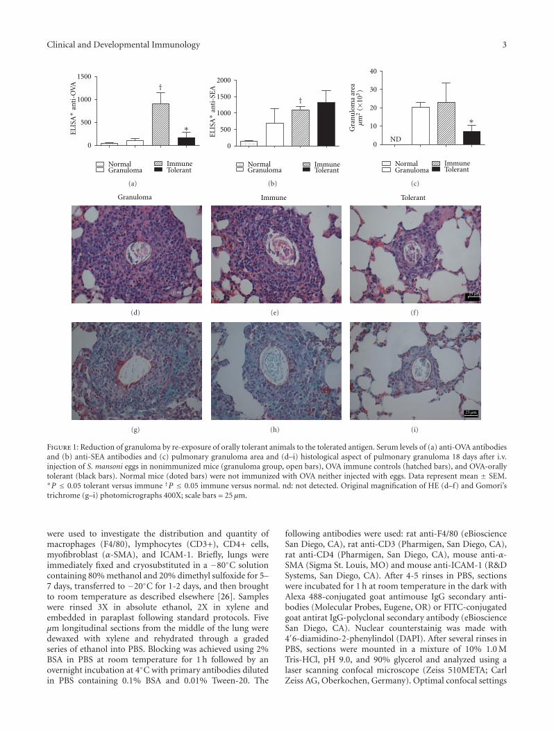
Figure 1: Reduction of granuloma by re-exposure of orally tolerant animals to the tolerated antigen. Serum levels of (a) anti-OVA antibodiesand (b) anti-SEA antibodies and (c) pulmonary granuloma area and (d–i) histological aspect of pulmonary granuloma 18 days after i.v.injection of S. mansoni eggs in nonimmunized mice (granuloma group, open bars), OVA immune controls (hatched bars), and OVA-orallytolerant (black bars). Normal mice (doted bars) were not immunized with OVA neither injected with eggs. Data represent mean ± SEM.∗P ≤ 0.05 tolerant versus immune †P ≤ 0.05 immune versus normal. nd: not detected. Original magnification of HE (d–f) and Gomori’strichrome (g–i) photomicrographs 400X; scale bars = 25 μm.
were used to investigate the distribution and quantity ofmacrophages (F4/80), lymphocytes (CD3+), CD4+ cells,myofibroblast (α-SMA), and ICAM-1. Briefly, lungs wereimmediately fixed and cryosubstituted in a −80◦C solutioncontaining 80% methanol and 20% dimethyl sulfoxide for 5–7 days, transferred to −20◦C for 1-2 days, and then broughtto room temperature as described elsewhere [26]. Sampleswere rinsed 3X in absolute ethanol, 2X in xylene andembedded in paraplast following standard protocols. Fiveμm longitudinal sections from the middle of the lung weredewaxed with xylene and rehydrated through a gradedseries of ethanol into PBS. Blocking was achieved using 2%BSA in PBS at room temperature for 1 h followed by anovernight incubation at 4◦C with primary antibodies dilutedin PBS containing 0.1% BSA and 0.01% Tween-20. The
following antibodies were used: rat anti-F4/80 (eBioscienceSan Diego, CA), rat anti-CD3 (Pharmigen, San Diego, CA),rat anti-CD4 (Pharmigen, San Diego, CA), mouse anti-α-SMA (Sigma St. Louis, MO) and mouse anti-ICAM-1 (R&DSystems, San Diego, CA). After 4-5 rinses in PBS, sectionswere incubated for 1 h at room temperature in the dark withAlexa 488-conjugated goat antimouse IgG secondary anti-bodies (Molecular Probes, Eugene, OR) or FITC-conjugatedgoat antirat IgG-polyclonal secondary antibody (eBioscienceSan Diego, CA). Nuclear counterstainig was made with4′6-diamidino-2-phenylindol (DAPI). After several rinses inPBS, sections were mounted in a mixture of 10% 1.0 MTris-HCl, pH 9.0, and 90% glycerol and analyzed using alaser scanning confocal microscope (Zeiss 510META; CarlZeiss AG, Oberkochen, Germany). Optimal confocal settings

4 Clinical and Developmental Immunology
Granuloma
Day
1
(a)
Immune
(b)
Tolerant
(c)
Day
5
(d) (e) (f)
Day
11
(g) (h) (i)
Day
14
(j) (k) (l)
Figure 2: Granuloma at different times after egg injection. Lung HE staining 1, 5, 11, and 14 days after i.v. injection of S. mansoni eggs. (a–c)At day 1, an inflammatory infiltrate with predominance of neutrophils and macrophages can be detected around eggs in all groups, but it isless intense in the tolerant group. (d–f) At day 5, macrophages, eosinophils, and some lymphocytes can be detected. (g–l) At days 11 and 14granulomas are more organized and some fibroblasts can be detected. Granulomas in tolerant mice follow the same pattern of organizationbut do not reach the same size of granulomas in controls group. Scale bars = 25 μm.
(aperture, gain, and laser power) for each antibody used weredetermined at the beginning of each imaging session andthen held constant during the analysis of all the samples.
The distribution patterns and levels of expression ofF4/80, CD3, CD4, α-SMA, and ICAM-1 were analyzed on di-gitalized photomicrographs with Image Tool 3.0 software(UTHSCSA, San Antonio, Tex, USA, http://ddsdx.uthscsa.edu/dig/itdesc.html). Images were captured at 12 bit andanalyzed in the gray scale range of 0 to 255. Green fluores-cence intensity was recorded as the sum of gray values of allpixels divided by the area (in μm2) ×10−3. Background flu-orescence was measured in each sample and subtracted fromthe values obtained for the fluorescence intensity.
2.10. Spleen Cell Cultures and Cytokine Assay. Spleen cellswere counted and adjusted to concentrations of 1 × 10⁷
cells/mL in RPMI 1640 supplemented with 2% heat inac-tivated FCS, 2 mM L-glutamine (Sigma-Aldrich, Inc.), andantibiotics (100 U/mL penicillin, 100 lg/mL streptomycin)(Sigma-Aldrich, Inc.). Cells were cultured in 96-well flat-bottom plates at 125 μL/well in a humidified atmospherewith 5% CO2 with or without soluble schistosome eggantigens at 50 μg/mL culture fluid, ovalbumin at 1 mg/mL,or concanavalin A at 2 μg/mL. After 72 h, supernatant fluidswere harvested and frozen −20◦C for subsequent cytokineanalysis. The production of IL-10 and IFN-γ by spleen cellswas measured by cytokine capture ELISA.

Clinical and Developmental Immunology 5
∗∗
0
1 5 11 14
10
20
30
40
GranulomaImmuneTolerant
Gra
nu
lom
aar
ea×1
03(μ
m2)
††
†
Days after i.v. egg injection
Figure 3: Re-exposure of orally tolerant animals to the toler-ated antigen block enlargement of granuloma area. The area ofgranulomas at days 1, 5, 11 and 14 after i.v. injection of eggs innonimmunized mice (granuloma group, open bars), OVA immunecontrols (hatched bars), and OVA-orally tolerant (black bars). Datarepresent mean± SEM (five mice/group). ∗P ≤ 0.05 tolerant versusimmune and †P ≤ 0.05 tolerant versus granuloma.
2.11. Quantitative Analysis of Serum and Lung Cytokines.Serum samples were collected as previously described andstored at −20◦C until used. One hundred milligrams oflung tissue samples from animals of each experimentalgroups were homogenized in 1 mL of PBS (0.4 M NaCland 10 mM de NaPO4) containing proteases inhibitors(0.1 mM phenylmethylsulfonyl fluoride, 0.1 mM benzetho-nium chloride, 10 mM EDTA, and 20 KI aprotinin A) and0.05% Tween 20. The samples were then centrifuged for 10minutes at 3,000×g and the supernatant immediately usedfor quantitative analysis of cytokines. The cytokines (IL-2,IL-4, IL-5, IFN-γ, and TNF-α) in serum and lung sampleswere measured with Cytometric Bead Array (CBA) MouseTh1/Th2 kit according to the manufacturer’s specifications(BD Biosciences, CA, USA).
2.12. Antibody Assay. Anti-OVA and antisoluble egg antigen(SEA) antibody titres were determined by standard enzyme-linked immunosorbent assay (ELISA) using an automaticELISA reader (BioRad, Hercules, CA). ELISA scores werecomputed by calculating the sums of the optical densitiesobtained from the six serum dilutions between 1 : 50 and1 : 1600 of individual mice. The details of the assay methodhave been described previously [11, 24, 27]. Each scoreshown represents the mean± SEM of the 5 animals in thegroup.
Statistical Analysis was performed using GraphPad Prism4 (GraphPad Software, CA, USA), and the statistical signifi-cance of differences between groups was determined usingone-way ANOVA followed by Student-Newman-Keuls test.Values of P ≤ 0.05 were considered significant. The resultsare expressed as the mean± SEM.
3. Results
3.1. Reexposure of OVA-Orally Tolerant Animals to the Tol-erated Antigen in Al(OH)3 Blocks Granuloma but Not Anti-SEA Antibody Formation. To induce oral tolerance to OVAC57BL/6 mice were offered an egg white solution for threedays as their only liquid source (called “tolerant”), andcontrol mice (called “immune”) drank tap water. Seven daysafter interrupting the oral treatment, mice were immunizedi.p. with OVA in Al(OH)3 immediately before the i.v. injec-tion of live S. mansoni eggs. Another control group (called“granuloma”) received i.v. injection of eggs without anyother previous treatment. Eighteen days thereafter, mice weresacrificed and blood and lung were removed for serum anti-bodies and pulmonary granuloma evaluation. Figure 1(a)shows that the oral pretreatment with egg white resultedin tolerance to OVA, that is, anti-OVA antibodies weresignificantly inhibited as compared with immune mice notorally pretreated. In contrast, anti-SEA antibodies were aug-mented in all groups injected with live eggs, irrespective ofother treatments (Figure 1(b)). Noteworthy, granuloma areawas significantly smaller in OVA-tolerant mice (Figure 1(c)).
We also performed histological analyses of Gomori’strichrome (Figures 1(g)–1(i)) and HE-stained lung sections(Figures 1(d)–1(f)). Eighteen days after i.v. injection of eggs,pulmonary granulomas were well organized with concentricarrangement and composed of macrophages, eosinophils,lymphocytes and some fibroblasts and epithelioid cells (Fig-ures 1(d)–1(f)). Granulomas were observed around smallbranches of pulmonary arteries. Initial collagen depositioncould be better observed after staining with Gomori’strichrome in all groups (Figures 1(g)–1(i)). Inflammatoryinfiltrates in lung parenchyma and alveolar macrophageswere characteristic of all groups, but more prominentlyin immune group. Of note, in OVA-tolerant mice themajority of eggs were surrounded by typical, althoughsmall, granulomas (Figure 1(f)) and their lung parenchymapresented less inflammatory infiltrates (not shown).
3.2. Re-Exposure of OVA Orally Tolerant Animals to the Toler-ated Antigen Blocks Initial Phases of Pulmonary GranulomaFormation. As described in the literature, the granulomaformed around S. mansoni eggs has a defined maturationalstage followed by a stage of involution, and, from amorphological point of view, these stages may be classi-fied as pregranulomatous and granulomatous stages [28].The pregranulomatous, exudative stage is characterized byaccumulation of neutrophils, eosinophils, and macrophagesaround the egg. The granulomatous stage can be dividedinto three phases: exudative-productive, productive, andinvolutional. In order to compare the kinetics of granulomaformation in OVA tolerant and not tolerant mice weperformed histological analyses of Gomori’s trichrome (notshown) and HE-stained lung sections. Figure 2 shows HE-stained sections of the pregranulomatous stage at days 1 and5 after egg inoculation (Figures 2(a)–2(f)) and granulomas inthe exudative-productive phase of the granulomatous stageat days 11 and 14 (Figures 2(g)–2(l)). At day 14, scarcedeposition of collagen fibres could be observed in Gomori’s

6 Clinical and Developmental Immunology
20 μm20 μm 20 μm 20 μm
Gra
nu
lom
aIm
mu
ne
Tole
ran
t
F4/80 CD3 CD4 α-SMA
F4/80
2
6
8
10
0
4
CD3
5
10
15
0
CD4
0
1
2
3
4
5α-SMA
0
1
2
3
4
Granuloma Immune Tolerant
Flu
ores
cen
cein
ten
sity
area
/μm×
(10−
3)
Figure 4: Cell subsets in pulmonary granulomas 18 days after i.v. egg injection. Immunolocalization using specific antibodies followed bysecondary antibodies coupled with fluorescein (green) and nuclear counterstainig with 4′6-diamidino-2-phenylindol (blue), 18 days afteri.v. eggs injection. Confocal microscope images were captured with a 63X objective, and the graphs represent the green fluorescence intensity(the sum of gray values of all pixels divided by the area (in μm2) × 10−3) of expression of F4/80 (macrophages), CD3 (T-lymphocytes),CD4+ cells, and α-SMA (myofibroblasts) in nonimmunized mice (granuloma group, open bars), OVA immune controls (hatched bars), andOVA-orally tolerant (black bars). Data represent mean ± SEM of fluorescence intensity of duplicate slides (n = 5 mice/group). The greenautofluorescency of eggs was excluded from all analyses. No significant difference was found between groups.

Clinical and Developmental Immunology 7
GranulomaG
ran
ulo
ma
Granuloma
Lung parenchymaIm
mu
ne
Tole
ran
t
Immune Tolerant
0
50
100
150
0
2
4
6
8
10
Flu
ores
cen
cein
ten
sity
area
/μm×
(10−
3)
∗
20 μm20 μm
(a) (b)
(c) (d)
(e) (f)
(g) (h)
Figure 5: Re-exposure of orally tolerant animals to the tol-erated antigen block the rise of ICAM-1 expression in lungparenchyma. Immunolocalization of ICAM-1 in granulomas andlung parenchyma using specific antibody coupled with fluores-cein (green) and nuclear counterstainig with 4′6-diamidino-2-phenylindol (blue), 18 days after i.v. eggs injection. Confocalmicroscope images were captured with a 63X objective, and thegraphs represent the green fluorescence intensity (the sum of grayvalues of all pixels divided by the area (in μm2)×10−3) of expressionof ICAM-1 in the granuloma area (a–d) and in lung parenchyma(e–h) in nonimmunized mice (granuloma group, open bars), OVAimmune controls (hatched bars), and OVA-orally tolerant mice(black bars). Data represent mean ± SEM of fluorescence intensityof duplicate slides (n = 5 mice/group).
trichrome-stained sections (not shown). Granulomas in thetolerant group followed similar kinetics as that from controlsgroup, but less intense.
Morphometric analysis (Figure 3) showed that at day 1the area of granulomas from OVA-tolerant mice is reducedas compared to control mice, but this difference disappears atday 5. However, after day 5, the area of granulomas increasedin controls group and became significantly higher than thearea of granulomas in OVA-tolerant mice at days 11 and 14(Figure 3).
3.3. Re-Exposure of Orally Tolerant Mice to the Tolerated Anti-gen Do Not Change Granuloma Cell Composition. To furthercharacterize granuloma cell composition macrophages, Tlymphocytes and myofibroblasts were identified and quan-tified by immunostaining followed by confocal microscopy.Despite their smaller area, granulomas from tolerant micepresent the same cell subsets as the large granulomas ofcontrols groups (Figure 4). Even myofibroblasts (α-SMA+)were present in the smaller granulomas of the Ova orally-tolerant mice (Figure 4). For technical reasons we couldnot perform double immunostaining with anti-CD3 andanti-CD4 antibodies. To quantify fluorescency, images werecaptured at 12 bit and analyzed in the gray scale range of 0 to255. Green fluorescence intensity was recorded as the sum ofgray values of all pixels divided by the area (in μm2)×10−3 asdescribed in Section 2. The green auto-fluorescency of eggswas excluded from all analyses. No significant difference influorescency intensity was found between groups. Then wecan conclude that the reduction in the area of granulomas isdue to proportional reduction of the inflammatory cells.
3.4. Re-Exposure of Orally Tolerant Mice to the ToleratedAntigen Reduces ICAM-1 Expression. Adhesion moleculesenable circulating leukocytes to accumulate in areas oflung inflammation, and adhesion is the initial phase of aprocess whereby activated endothelial cells induce leukocytemigration into tissues. As ICAM-1 has been described as apredominant adhesion molecule after egg deposition in theliver of S. mansoni infected mice [19] we compared its expres-sion in lungs after i.v. injection of eggs. Our results show thatthe majority of S. mansoni egg-induced ICAM-1 expression18 days after pulmonary granuloma induction was restrictedto the lung parenchyma outside the granulomas (Figure 5).The intensity of ICAM-1 expression in the granuloma oftolerant and controls group was not different. However, inthe lung parenchyma outside granulomas, the expressionof ICAM-1 was significantly inhibited in the tolerant mice(Figure 5).
3.5. Reduction of Granulomas by Re-Exposure to OrallyTolerant Proteins Was Not Correlated with a Shift in Th-1/Th-2 Cytokines. Using a commercial kit to detect typicalTh1/Th2 cytokines, we compared the levels of IFN-γ, TNF-α,IL-2, IL-4, and IL-5 in lung homogenates 14 days aftergranuloma induction (Figure 6) and in serum samples(Figure 7) collected 1, 5, 14 and 18 days after granulomainduction. IFN-γ could be detected in lung homogenates

8 Clinical and Developmental Immunology
0
5
10
15IF
N-γ
(pg/
mL
)
ND
(a)
5
10
15
0
IL-4
(pg/
mL
)
ND
(b)
5
15
0
10
TN
F-α
(pg/
mL
)
ND ND
(c)
5
15
0
10
IL-5
(pg/
mL
)
ND ND
(d)
0
10
20
30
40
IL-2
(pg/
mL
)
NormalGranuloma
ImmuneTolerant
(e)
Figure 6: Cytokines production of lungs. Fourteen days after i.v. injection of S. mansoni eggs in nonimmunized mice (granuloma group,open bars), OVA immune controls (hatched bars), and OVA-orally tolerant mice (black bars), lungs were removed and homogenized inextract buffer. Normal mice (doted bars) were not immunized with OVA and neither injected with eggs. Extract supernatant was collectedfor cytokine assay. IFN-γ, TNF-α, IL-2, IL-4, and IL-5 were measured using a Cytometric Bead Array (CBA) kit. The results are shown asmean concentrations ± SEM. nd: not detected.
(Figure 6) and serum at day 1 (Figure 7) in some miceinjected with S. mansoni eggs and not in normal (naıve)mice, but no difference was found between the experimentalgroups. IL-2 and IL-4 were not detected in serum samplesfrom any group. IL-2 was detected in the same level in lunghomogenates of normal and experimental groups, and thelow levels of IL-4 detected in lung homogenates of immuneand tolerant mice did not correlate with the size of their
granulomas. TNF-α was detected in the serum at the samelevel in all groups, 14 days after egg injection. At day 1, TNF-α, and IL-5 were detected in the serum only in the tolerantgroup, but not in all mice from this group. In conclusion,reduction of granulomas in tolerant mice does not correlatewith a shift in Th-1/Th-2 cytokines.
We also compared the production of IFN-γ and IL-10by spleen cell restimulated “in vitro” with OVA or SEA. The

Clinical and Developmental Immunology 9
14 18
0
50
100
150
200
5 10
Days after i.v. egg injection
IFN
-γ(p
g/m
L)
NDNDNDND NDNDND NDND ND ND
(a)
1 5 14 18
0
5
10
15
20
Days after i.v. egg injection
TN
F-α
(pg/
mL
)
NDNDNDNDNDNDNDNDND ND
(b)
1 5 14 18
0
10
20
30
40
Days after i.v. egg injection
NormalGranuloma
Immune
Tolerant
IL-5
(pg/
mL
)
ND NDNDND ND NDND NDND ND ND
(c)
Figure 7: Time course of serum cytokines after granulomainduction. IFN-γ, TNF-α, IL-2, IL-4, and IL-5 were measured usinga Cytometric Bead Array (CBA) kit in serum samples collectedfrom nonimmunized mice (granuloma group, open bars), OVAimmune controls (hatched bars), and OVA-orally tolerant mice(black bars). Normal mice (doted bars) were not immunized withOVA and neither injected with eggs. The results are shown as meanconcentrations ± SEM. nd: not detected. IL-2 and IL-4 were notdetected.
20
15
10
5
0
Medium ConA SEA OVA
NDNDNDNDNDNDND
IFN
-γ(p
g/m
L)
(a)
0
IL-1
0(p
g/m
L)
Normal
Granuloma
ImmuneTolerant
Medium ConA SEA OVA
0.14
0.21
0.28
0.35
0.7
ND
(b)
Figure 8: IFN-γ and IL-10 production of spleen cells stimulatedwith SEA or OVA. Eighteen days after i.v. injection of S. mansonieggs in nonimmunized mice (granuloma group, open bars), OVAimmune controls (hatched bars), and OVA-orally tolerant mice(black bars) spleen cells were cultured with medium, ConA, SEAor OVA for 3 days. Normal mice (doted bars) were not immunizedwith OVA neither injected with eggs. The culture supernatant fluidswere harvested and IFN-γ and IL-10 measured by sandwich ELISA.The results are shown as mean concentrations ± SEM. nd: notdetected.
results in Figure 8 do not make us confident to attributethe reduction of granulomas in tolerant mice to systemicalteration in the production of these cytokines.
4. Discussion
The standard protocols used to demonstrate tolerance inorally pre-treated animals involve challenge with the antigenin adjuvant, and there is evidence that adjuvants play a sig-nificant role in tolerogenesis during the triggering/parenteralphase affecting the kind of Ig isotype that is suppressedor maintained for long periods after oral feeding [29, 30].Tobagus et al. [30] suggested that when a Th1-selectiveadjuvant (such as CFA) is used the resulting response dis-played selective inhibition of the Th1 component (IFN-γ)of the immune response while orally pre-treated animals

10 Clinical and Developmental Immunology
challenged with the antigen in a Th2-selective adjuvant(alum) displayed a selective inhibition of Th2 responses.While oral tolerance is specific to the antigen contacted bythe oral route, it is noteworthy that the parenteral injectionof small doses (e.g., 10 μg) of proteins to which the animalis orally tolerant triggers a strong inhibition of primaryresponses to unrelated antigens [3, 8, 10].
In previous work we have shown that in mice orally-tolerant to ovalbumin (OVA), anti-SEA and pulmonarygranulomas triggered by i.v. injection of eggs from S.mansoni were inhibited by i.p. injection of dinitrophenylatedconjugates of OVA (DNP-OVA) emulsified in completeFreund’s adjuvant (CFA) [24]. In that work we analysedgranulomas only at day 18 after i.v. egg injection andfound that the more prominent granulomas occurred innontolerant mice concomitantly injected with DNP-OVA +CFA and small granulomas were found in orally tolerantmice injected with DNP-OVA + CFA. In the tolerant groupeggs were predominant in intravascular locations with initialperiovular reactions containing monocytes, eosinophils, andcollagen fibers derived from the vascular wall [24]. Herein,we have shown that i.p. injection of OVA plus Al(OH)3 intoOVA-tolerant mice also inhibited pulmonary granuloma butnot anti-SEA antibodies production (Figure 1). Our previousreport and the present one as well show that reduction ofgranuloma in orally-tolerant mice is independent of the kindof adjuvant used. On the other hand, inhibition of anti-SEA antibody formation only occurred when the toleratedantigens were injected with CFA [24]. Nevertheless, we haveshown that inhibition of antibodies to other proteins suchas KLH and haemoglobin occurs with injection of OVA +Al(OH)3 in OVA orally-tolerant mice [10, 12]. So, unknownfactors associated with the eggs make it more difficult toinhibit the anti-SEA antibody response.
This and already published work [3, 9, 11–13] show thatthe re-exposure of orally tolerant animals to the toleratedantigen blocks inflammatory reactions. One hallmark ofinflammatory processes is the migration of leukocytes tolocal areas. Herein we have shown that the injection oftolerated antigen into orally tolerant mice weakens the influxof leucocytes into the lung and reduces the size of granuloma(Figures 1, 2, and 3). However, the inhibitory effect of oraltolerance hindered the intensity of migration of cells intothe lung, but not its kinetics, since granulomas followedthe same pattern of formation in tolerant and not tolerantmice (Figure 2). Furthermore granulomas in tolerant micehave the same cell composition although in low numbers ascompared to not tolerant mice (Figure 4).
Changes in the expression of cell adhesion molecules ini-tiate leukocyte trafficking, and ICAM-1 is the predominantadhesion molecule in schistosome egg granuloma formation[19]. The reduction in the expression of ICAM-1 in tolerantmice, as shown herein (Figure 5), is certainly involved in thedemonstrated inhibitory effect. We could not find significantchanges in cytokine secretion, neither in the blood, nor inlung extracts (Figures 6 and 7). IL-10 was detected afterspleen cell cultures with SEA, but no difference was foundbetween tolerant and not tolerant group, and IL-10 concen-trations in supernatants of spleen cells cultured with OVA
were not different from basal production (Figure 8). Thisdetection may require proper timing, but the present resultsargue against major changes in the Th1/Th2 axis.
It is important to pursue these findings with additionalexperiments. Antibody formation may be involved in thereduction of granulomas in orally-tolerant mice, since B cellsand anti-idyotipic antibodies are involved in the regulationof granulomas [31, 32] and oral tolerance also affects Bcell and antibody production [29]. In searching for possiblemechanisms involved in inhibitory indirect effects triggeredby parenteral injection of tolerated antigens we must keep inmind that they affect the initial phases of the inflammatoryresponse which are thought to be primarily innate, as shownherein and in previous work [13]. This may be takenas indication that, in addition to specific immunological(clonal) events, the exposure to tolerated antigens triggersother phenomena, for example, of neuroendocrine nature.
5. Conclusion
Parenteral injection of tolerated proteins into orally tolerantmice blocked the increase of pulmonary granulomas and theexpression of ICAM-1 in lung parenchyma in areas outsidethe granulomas. The reduction in the area of granulomasin tolerant mice is due to proportional reduction of theinflammatory cells and was not correlated with a shift in Th-1/Th-2 cytokines in serum or lung tissue extract.
Conflict of Interests
The authors have no conflict of interests to be disclosed.
Acknowledgments
This work was financially supported by grants from Con-selho Nacional de Desenvolvimento Cientıfico e Tecnologico(CNPq, Brazil) and Fundacao de Amparo a Pesquisa deMinas Gerais (FAPEMIG). C. R. Carvalho is recipientof fellowship from CNPq, M. A. Resende is recipient ofscholarship from FAPEMIG and C. M. Rodrigues fromCoordenacao de Aperfeicoamento de Pessoal de Nıvel Supe-rior (CAPES). The confocal microscopic data shown in thiswork was obtained using the Zeiss 510 Meta confocal systemin the Center of Electron Microscopy at the UniversidadeFederal de Minas Gerais, Brazil. This work is dedicated to thememory of professor Henrique Leonel Lenzi.
References
[1] A. M. Faria and H. L. Weiner, “Oral tolerance: therapeuticimplications for autoimmune diseases,” Clinical and Develop-mental Immunology, vol. 13, no. 2–4, pp. 143–157, 2006.
[2] N. Vaz, A. M. Faria, B. A. Verdolin, and C. R. Carvalho,“Immaturity, ageing and oral tolerance,” Scandinavian Journalof Immunology, vol. 46, no. 3, pp. 225–229, 1997.
[3] A. Miller, O. Lider, and H. Weiner, “Antigen-driven bystandersuppression after oral administration of antigens,” Journal ofExperimental Medicine, vol. 174, no. 4, pp. 791–798, 1991.
Clinical and Developmental Immunology 11
[4] M. Russo, S. Jancar, A. L. Pereira de Siqueira et al., “Pre-vention of lung eosinophilic inflammation by oral tolerance,”Immunology Letters, vol. 61, no. 1, pp. 15–23, 1998.
[5] S. Yoshino, E. Quattrocchi, and H. L. Weiner, “Suppression ofantigen-induced arthritis in Lewis rats by oral administrationof type II collagen,” Arthritis and Rheumatism, vol. 38, no. 8,pp. 1092–1096, 1995.
[6] B. A. Verdolin, S. M. Ficker, A. M. Faria, N. M. Vaz, andC. R. Carvalho, “Stabilization of serum antibody responsestriggered by initial mucosal contact with the antigen inde-pendently of oral tolerance induction,” Brazilian Journal ofMedical and Biological Research, vol. 34, no. 2, pp. 211–219,2001.
[7] C. R. Carvalho and N. M. Vaz, “Indirect effects are indepen-dent of the way of tolerance induction,” Scandinavian Journalof Immunology, vol. 43, no. 6, pp. 613–618, 1996.
[8] C. R. Carvalho, B. A. Verdolin, A. V. de Souza, and N. M.Vaz, “Indirect effects of oral tolerance in mice,” ScandinavianJournal of Immunology, vol. 39, no. 6, pp. 533–538, 1994.
[9] N. F. Backstrom and U. I. Dahlgren, “Bystander suppressionof collagen-induced arthritis in mice fed ovalbumin,” ArthritisResearch & Therapy, vol. 6, no. 2, pp. R151–160, 2004.
[10] C. R. Carvalho, B. A. Verdolin, and N. M. Vaz, “Indirect effectsof oral tolerance cannot be ascribed to bystander suppression,”Scandinavian Journal of Immunology, vol. 45, no. 3, pp. 276–281, 1997.
[11] C. M. Rodrigues, O. A. Martins-Filho, N. M. Vaz, and C. R.Carvalho, “Systemic effects of oral tolerance on inflammation:mobilization of lymphocytes and bone marrow eosinopoiesis,”Immunology, vol. 117, no. 4, pp. 517–525, 2006.
[12] G. Ramos, C. M. Rodrigues, G. M. Azevedo Jr., V. Pinho, C. R.Carvalho, and N. M. Vaz, “Cell-mediated immune responseto unrelated proteins and unspecific inflammation blockedby orally tolerated proteins,” Immunology, vol. 126, no. 3, pp.354–362, 2009.
[13] R. A. Costa, V. Ruiz-de-Souza, G. M. Azevedo Jr. et al.,“Indirect effects of oral tolerance improve wound healing inskin,” Wound Repair and Regeneration, vol. 19, no. 4, pp. 487–497, 2011.
[14] A. Gonzalez, H. L. Lenzi, E. M. Motta et al., “Expression ofadhesion molecules in lungs of mice infected with Paracoccid-ioides brasiliensis conidia,” Microbes and Infection, vol. 7, no.4, pp. 666–673, 2005.
[15] M. S. Wilson, M. M. Mentink-Kane, J. T. Pesce, T. R. Rama-lingam, R. Thompson, and T. A. Wynn, “Immunopathologyof schistosomiasis,” Immunology and Cell Biology, vol. 85, no.2, pp. 148–154, 2007.
[16] F. G. Abath, C. N. Morais, C. E. Montenegro, T. A. Wynn,and S. M. Montenegro, “Immunopathogenic mechanisms inschistosomiasis: what can be learnt from human studies?”Trends in Parasitology, vol. 22, no. 2, pp. 85–91, 2006.
[17] D. O. Co, L. H. Hogan, S. Il-Kim, and M. Sandor, “T cellcontributions to the different phases of granuloma formation,”Immunology Letters, vol. 92, no. 1-2, pp. 135–142, 2004.
[18] J. T. Pesce, T. R. Ramalingam, M. S. Wilson et al., “Retnla(relmα/Fizz1) suppresses helminth-induced Th2- type immu-nity,” Plos Pathogens, vol. 5, no. 4, Article ID e1000393, 2009.
[19] D. M. Ritter and J. H. McKerrow, “Intercellular adhesionmolecule 1 is the major adhesion molecule expressed duringschistosome granuloma formation,” Infection and Immunity,vol. 64, no. 11, pp. 4706–4713, 1996.
[20] G. Schramm and H. Haas, “Th2 immune response againstSchistosoma mansoni infection,” Microbes and Infection, vol.12, no. 12-13, pp. 881–888, 2010.
[21] S. W. Chensue, P. D. Terebuh, K. S. Warmington et al., “Roleof IL-4 and IFN-γ in Schistosoma mansoni egg-inducedhypersensitivity granuloma formation: orchestration, relativecontribution, and relationship to macrophage function,”Journal of Immunology, vol. 148, no. 3, pp. 900–906, 1992.
[22] A. W. Cheever, J. A. Lenzi, H. L. Lenzi, and Z. A. Andrade,“Experimental models of Schistosoma mansoni infection,”Memorias do Instituto Oswaldo Cruz, vol. 97, no. 7, pp. 917–940, 2002.
[23] M. G. Nair, Y. Du, J. G. Perrigoue et al., “Alternatively activatedmacrophage-derived RELM-α is a negative regulator of type 2inflammation in the lung,” Journal of Experimental Medicine,vol. 206, no. 4, pp. 937–952, 2009.
[24] C. R. Carvalho, H. L. Lenzi, R. Correa-Oliveira, and N. M.Vaz, “Indirect effects of oral tolerance to ovalbumin interferewith the immune responses triggered by Schistosoma mansonieggs,” Brazilian Journal of Medical and Biological Research, vol.35, no. 10, pp. 1195–1199, 2002.
[25] A. M. Faria, G. Garcia, M. J. C. Rios, C. L. Michalaros,and N. M. Vaz, “Decrease in susceptibility to oral toleranceinduction and occurrence of oral immunization to ovalbuminin 20-38-week-old mice. The effect of interval between oralexposure and rate of antigen intake in the oral immunization,”Immunology, vol. 78, no. 1, pp. 147–151, 1993.
[26] L. S. Carvalhaes, O. L. Gervasio, C. Guatimosim et al., “Col-lagen XVIII/endostatin is associated with the epithelial-mes-enchymal transformation in the atrioventricular valves duringcardiac development,” Developmental Dynamics, vol. 235, no.1, pp. 132–142, 2006.
[27] A. P. Cunha, R. P. Oliveira, A. B. Junior, N. M. Vaz, and C.R. Carvalho, “Different requirements for the adoptive transferof oral tolerance and its indirect effects assessed by DTH andantibody responses in mice,” Cellular Immunology, vol. 258,no. 2, pp. 152–160, 2009.
[28] H. L. Lenzi, E. Kimmel, H. Schechtman et al., “Histoarchitec-ture of schistosomal granuloma development and involution:morphogenetic and biomechanical approaches,” Memorias doInstituto Oswaldo Cruz, vol. 93, supplement 1, pp. 141–151,1998.
[29] A. M. de Faria, S. M. Ficker, E. Speziali et al., “Aging affectsoral tolerance induction but not its maintenance in mice,”Mechanisms of Ageing and Development, vol. 102, no. 1, pp.67–80, 1998.
[30] I. T. Tobagus, W. R. Thomas, and P. G. Holt, “Adjuvant costim-ulation during secondary antigen challenge directs qualitativeaspects of oral tolerance Induction, particularly during theneonatal period,” Journal of Immunology, vol. 172, no. 4, pp.2274–2285, 2004.
[31] D. Jankovic, A. W. Cheever, M. C. Kullberg et al., “CD4+ Tcell-mediated granulomatous pathology in schistosomiasis isdownregulated by a B cell-dependent mechanism requiring Fcreceptor signaling,” Journal of Experimental Medicine, vol. 187,no. 4, pp. 619–629, 1998.
[32] M. A. Montesano, D. G. Colley, M. T. Willard, G. L. FreemanJr., and W. E. Secor, “Idiotypes expressed early in experimentalSchistosoma mansoni infections predict clinical outcomes ofchronic disease,” Journal of Experimental Medicine, vol. 195,no. 9, pp. 1223–1228, 2002.

Submit your manuscripts athttp://www.hindawi.com
Stem CellsInternational
Hindawi Publishing Corporationhttp://www.hindawi.com Volume 2014
Hindawi Publishing Corporationhttp://www.hindawi.com Volume 2014
MEDIATORSINFLAMMATION
of
Hindawi Publishing Corporationhttp://www.hindawi.com Volume 2014
Behavioural Neurology
EndocrinologyInternational Journal of
Hindawi Publishing Corporationhttp://www.hindawi.com Volume 2014
Hindawi Publishing Corporationhttp://www.hindawi.com Volume 2014
Disease Markers
Hindawi Publishing Corporationhttp://www.hindawi.com Volume 2014
BioMed Research International
OncologyJournal of
Hindawi Publishing Corporationhttp://www.hindawi.com Volume 2014
Hindawi Publishing Corporationhttp://www.hindawi.com Volume 2014
Oxidative Medicine and Cellular Longevity
Hindawi Publishing Corporationhttp://www.hindawi.com Volume 2014
PPAR Research
The Scientific World JournalHindawi Publishing Corporation http://www.hindawi.com Volume 2014
Immunology ResearchHindawi Publishing Corporationhttp://www.hindawi.com Volume 2014
Journal of
ObesityJournal of
Hindawi Publishing Corporationhttp://www.hindawi.com Volume 2014
Hindawi Publishing Corporationhttp://www.hindawi.com Volume 2014
Computational and Mathematical Methods in Medicine
OphthalmologyJournal of
Hindawi Publishing Corporationhttp://www.hindawi.com Volume 2014
Diabetes ResearchJournal of
Hindawi Publishing Corporationhttp://www.hindawi.com Volume 2014
Hindawi Publishing Corporationhttp://www.hindawi.com Volume 2014
Research and TreatmentAIDS
Hindawi Publishing Corporationhttp://www.hindawi.com Volume 2014
Gastroenterology Research and Practice
Hindawi Publishing Corporationhttp://www.hindawi.com Volume 2014
Parkinson’s Disease
Evidence-Based Complementary and Alternative Medicine
Volume 2014Hindawi Publishing Corporationhttp://www.hindawi.com


![arXiv:2003.10405v2 [physics.med-ph] 31 Mar 202031CIEMAT, Centro de Investigaciones Energ eticas, Medioambientales y Tecnol ogicas, Madrid 28040, Spain 32Policlinico San Pietro, Ponte](https://img.pdfslide.us/doc/110x75/5ea69e020d76d63b1b7a8c59/arxiv200310405v2-31-mar-2020-31ciemat-centro-de-investigaciones-energ-eticas.jpg)


![arXiv:2005.12972v1 [cond-mat.mes-hall] 26 May 2020 · Departamento de Ciˆencias Naturais, Universidade Federal do Esp ´ırito Santo, S˜ao Mateus, ES, 29932-540, Brazil. ... [cond-mat.mes-hall]](https://img.pdfslide.us/doc/110x75/5fd4014f28d04f6a2215c3dc/arxiv200512972v1-cond-matmes-hall-26-may-2020-departamento-de-ciencias-naturais.jpg)
![Stochastic Electrochemical Kinetics · PDF filearXiv:1608.07507v2 [ ] 18 Sep 2016 Stochastic Electrochemical Kinetics Ot´avio Beruski Instituto de Qu´ımica de](https://img.pdfslide.us/doc/110x75/5abbc4fa7f8b9a76038d1bff/stochastic-electrochemical-kinetics-160807507v2-18-sep-2016-stochastic-electrochemical.jpg)