Embed Size (px)
Citation preview
_______________________________________________________________________________________________________________________________________________________________
Research Article _______________________________________________________________________________________________________________________________________________________________
Indirect pulp treatment in primary teeth: 4-year results LUCIANO CASAGRANDE, DDS,MS, PHD, LETÍCIA WESTPHALEN BENTO, DDS, MS, DÉBORA MARTINI DALPIAN,DDS, MSFRANKLIN GARCÍA-GODOY, DDS, MS & FERNANDO BORBA DE ARAUJO, DDS, MS, PHD
ABSTRACT: Purpose: To evaluate clinical and radiographic outcomes of indirect pulp treatment (IPT) in primary molars after long-term function (up to 60 months). Methods: Teeth with deep carious lesions without signs and symptoms of irreversible pulpitis were divided by random allocation into two groups, according to the capping material utilized over demineralized dentin: experimental group (1): self-etching adhesive system (Clearfil SE Bond); and control group (2): calcium hydroxide liner (Dycal). Both groups were filled with resin composite (Z250) and submitted to a clinical and radiographic monitoring period until exfoliation. Results: After the follow-up period (up to 60 months), no statistical difference was found between groups (P= 0.514). The overall success rate reached 78%. The failures occurred after the first year period recall. (Am J Dent 2010;23:34-38).
CLINICAL SIGNIFICANCE: The IPT provides an alternative treatment of primary teeth with deep carious lesions representing a simple and effective technique to maintain the pulp vitality.
: Dr. Luciano Casagrande, School of Dentistry, Franciscan University Center (UNIFRA), Andradas 1614, Santa Maria, RS 97010 032, Brazil. E- : [email protected]
Introduction According to the guidelines of the American Academy of Pediatric Dentistry (AAPD),1 the indirect pulp treatment (IPT) is a procedure in which the caries closest to the pulp tissue is left in place and covered with a biocompatible material, and the tooth is restored to prevent microleakage. The main goal of this minimal intervention technique is to preserve dental substrate while maintaining pulp vitality, avoiding direct intervention on the pulp tissue.1 Besides, IPT showed higher rates of clinical and radiographic success when compared with other conservative pulp treatments, such as pulpotomy.2,3
The IPT is clearly not indicated when the pulp tissue is exposed by caries or when the teeth present irreversible pulp pathology, based on a thorough clinical and radiographic examination.3,4
Despite the indication of using a biocompatible material over the remaining demineralized tissue, clinical, radiographic, and bacteriologic studies5-7 have shown the inactivation of active carious lesion, even when inert materials (i.e. wax or gutta-percha) were used as capping materials. Other studies8,9
have shown that an acid-resistant tissue resulting from the interdiffusion of adhesive resin within the area of carious dentin does not affect the clinical performance of the restoration. The restorative material should completely seal the involved dentin from the oral environment avoiding nutrient influx and compromising the bacteria metabolism.3,9,10 Glass-ionomer cement and resin composite have shown to provide adequate marginal sealing in primary teeth submitted to IPT.8,11-13
The present study evaluated clinically and radiographically the long-term function of primary teeth submitted to IPT when a self-etching primer or a calcium hydroxide was used under resin-based composite restorations.
Materials and Methods
This clinical study was conducted at the Federal University of Rio Grande do Sul, Pediatric Dentistry Unit (Porto Alegre, Brazil), using a protocol that was reviewed and approved by the
institutional review board. The parents/legal guardians read and signed an informed consent form for this study (No. 08/04 according to the resolution 196/96 of the National Health Council). Forty primary molars were selected from 21 healthy children (4-8 years old). The inclusion criteria were: (1) active carious lesion in deep dentin (occlusal or occluso-proximal surface); (2) absence of clinical diagnosis of pulp exposure, fistula, swelling of periodontal tissues, and abnormal tooth mobility; (3) absence of clinical symptoms of irreversible pulpitis, such as spontaneous pain or sensitivity to pressure; (4) the extension of the carious lesion should be such that complete caries removal would risk pulp exposure, as determined by clinical and radiographic assessment; (5) absence of radiolu-cencies at the interradicular (furcation) or periapical regions, or thickening of the periodontal spaces, that would indicate the presence of the irreversible pulp pathologies or necrosis; (6) absence of internal or external root resorption; and (7) cases were selected based on the color and consistency of the caries in the lesion by one trained examiner, whose reliability was tested by the kappa test with an index of 0.80 and 0.75 respectively. All the subjects received treatment for caries activity arrest, including extractions, pulp treatments, and restoration of carious lesions, fluoride therapy, oral hygiene and dietary instructions. The subjects were anesthetized and under rubber dam isola-tion, received occlusal or occluso-proximal cavity preparations. The carious tissue was removed completely by the peripheral excavation of the cavity followed by a central excavation removing the outermost necrotic and infected demineralized dentin. Teeth were then randomly assigned into experimental (N =19, Clearfil SE Bonda), or control (N=21, Dycalb calcium hydroxide) groups. In the experimental group, the self-etching primer was applied for 20 seconds to the entire cavity and then the adhesive was placed and polymerized by light-curing with 500 mw/cm2 for 20 seconds (Elipar Highlightc). In the control group, a calcium hydroxide cement liner was applied over the carious dentin, before the self-etching system application. The cavities were filled with resin composite (Filtek Z250c) by incremental technique. To restore the Class II cavities, a metal
American Journal of Dentistry, Vol. 23, No. 1, February. 2010
Table 1. Results of clinical and radiographic evaluation of IPT after long-term follow-up. ________________________________________________________________________________________________________
Groups No. teeth Drop-out Success Failure rate ________________________________________________________________________________________________________
I CSE 17 (19) 2a 14b (82.4%) 3 (17.6%) II Dycal 15 (21) 6a 11b (73.3%) 4 (26.7%) Total 32 (40) 8 25 (78.1%) 7 (21.8%) ________________________________________________________________________________________________________
Numbers followed by superscript in columns show the statistical differences (a P= 0.514; b P= 0.678).
Table 2. Distribution of IPT success/failures according to surface (P= 0.083). ________________________________________________________________________________________________________
Final evaluation _____________________________
No. teeth Success Failure Total ________________________________________________________________________________________________________
Surface Occlusal 22 10 6 16 Occluso-proximal 18 15 1 16 Total 40 25 7 32 ________________________________________________________________________________________________________
Table 3. Results of IPT according to tooth type (P= 0.212). ________________________________________________________________________________________________________
Final evaluation _____________________________
No. teeth Success Failure Total ________________________________________________________________________________________________________
Tooth 1st primary molar 14 11 1 12 2nd primary molar 26 14 6 20 Total 40 25 7 32 ________________________________________________________________________________________________________
matrix (AutoMatrixd) was adapted to the cervical margin with an interproximal wedge. The teeth were clinically re-examined after 1, 6, and 12 months and radiographically after 6 and 12 months. After that, an evaluation was performed each year until the teeth exfoliated. The IPT was recorded as a successful outcome if: absence of spontaneous pain and/or sensitivity to pressure, absence of fistula, edema, abnormal mobility, absence of radiolucencies at interradicular and/or periapical regions, absence of internal or external root resorption that was not compatible with biological resorption due to the exfoliation process. Any tooth that pre-sented with irreversible pulpitis or symptoms at clinical or/and radiographic evaluation was recorded as a treatment failure and either pulpectomized or extracted. One blind examiner per-formed the clinical and radiographic follow-up examinations. The groups were statistically analyzed using Kaplan-Meier survival followed by Log Rank and tooth type and cavity configurations were analyzed by Fisher’s exact test. All results were considered significant if P< 0.05. The software SPSS 13.0e was used for these analyses.
Results
After a long-term follow-up (up to 60 months), 15 children from a total of 21 were reexamined for the final evaluation. Six subjects dropped from the study: three quit the program and the other three moved to another city; these occurred in 24 months and represented eight teeth out of the initial sample (two from the Clearfil SE Bond group and six from the Dycal group). Thirty-two teeth, from a total baseline of 40, were evaluated. Twenty-five cases met the criteria for clinical and radiographic success, reaching an overall success rate of 78% with no statistical difference between the groups (Table 1, Figs. 1-5).
Indirect pulp treatment in primary teeth 35
Fig. 1.Test of equality of survival distributions for the different levels of group (P= 0.514).
Failures occurred after the first year of follow-up, and were detected by radiographic evaluation. Second molars and the occlusal restorations presented higher frequency of failures, when compared to first molars and occluso-proximal resto-rations, but it was not statistically significant (Tables 2,3).
Discussion
According to the AAPD,1 the indications for IPT and pulpotomy are very similar for primary teeth with deep caries and reversible pulpitis, when the pulp is judged to be vital from clinical and radiographic criteria. The difference is that if during caries excavation a pulp exposure occurs, a pulpotomy is performed. IPT purposely avoids an exposure by leaving in place the deepest decay in place, adjacent to the pulp.14
Studies15,16 have shown that the pulpotomy success signi-ficantly decreases over time. Another concern is the early exfoliation of pulpotomized teeth, compared to IPT.2,15 Further, even when dental students with different abilities perform ITP, the success rate did not decrease significantly.17
At a meeting sponsored by the American Association of Endodontists (AAE) and the American Academy of Pediatric Dentistry (AAPD) in 2007, pre-symposium and post-symposium tests were given to endodontists and pediatric dentists to compare the level of agreement in terms of conservative pulp treatment of primary and young permanent teeth. The results indicated that the pediatric dentistry and endodontic communities agree that formocresol will be replaced as a primary tooth pulpotomy agent, that IPT in primary teeth holds hope as a replacement for pulpotomy, and that IPT is an acceptable technique for cariously involved young permanent teeth.18
Interestingly, a survey conducted among pediatric program directors in U.S. dental schools and pediatric dentists, regarding deep caries removal in primary molars, reported the pulpotomy as treatment of choice, for 70% and 80%, respectively.19
Follow-up studies6,8,9,12,13 have demonstrated the effective- ness of treating deep carious lesions by using a less invasive
36 Casagrande et al American Journal of Dentistry, Vol. 23, No. 1, February. 2010
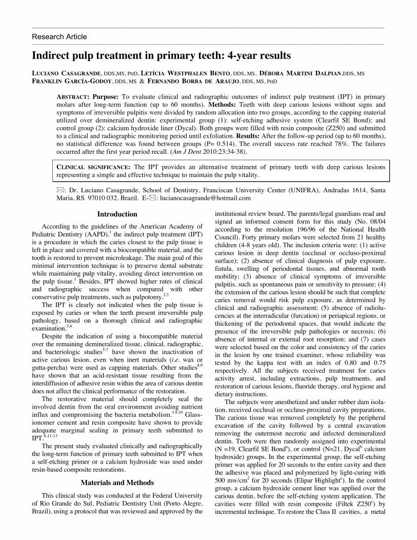
Fig. 2. Radiographic evaluation of a lower second primary molar that received indirect pulp treatment with self-etching primer over the remaining carious dentin. Pre-operative (A), 36 months (B), 60 months (C). The procedure was considered successful.
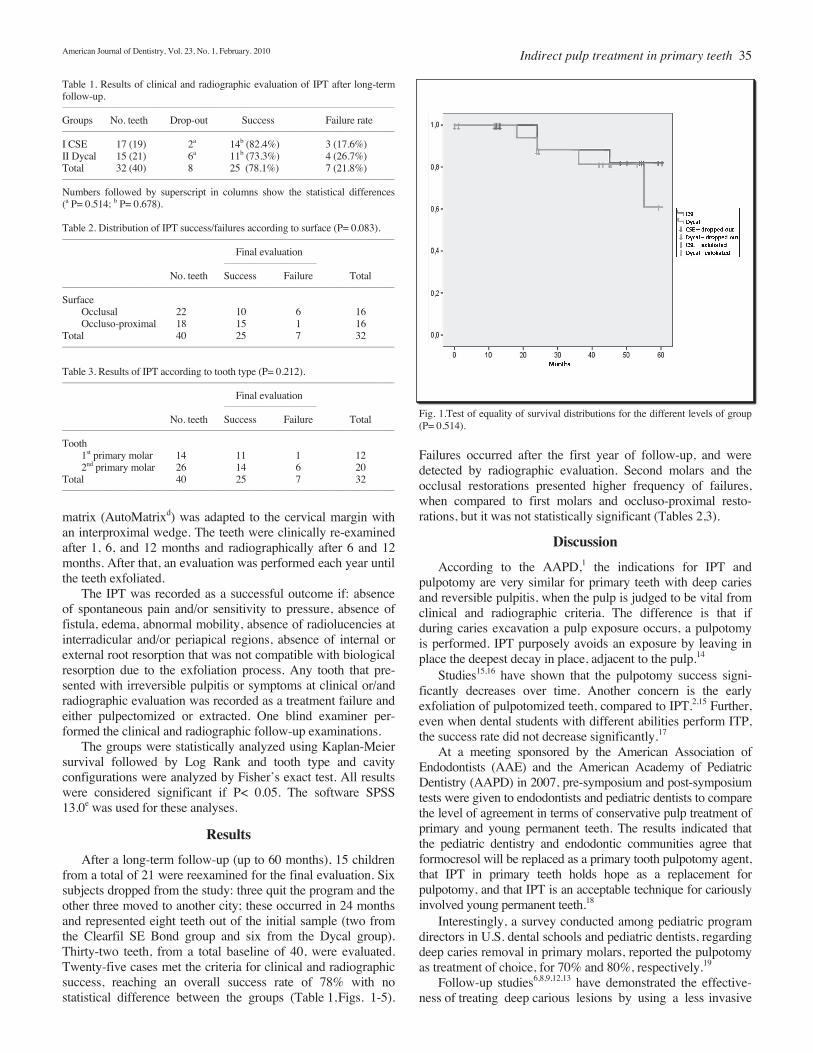
Fig. 3. Radiographic evaluation of a lower second primary molar that received indirect pulp treatment with calcium hydroxide over the remaining carious dentin. Pre-operative (A), 36 months (B), 65 months (C). The procedure was considered successful.
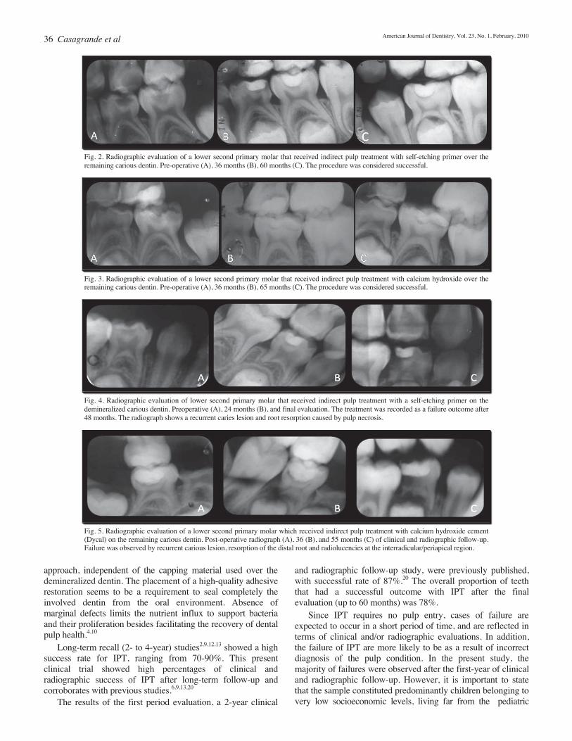
Fig. 4. Radiographic evaluation of lower second primary molar that received indirect pulp treatment with a self-etching primer on the demineralized carious dentin. Preoperative (A), 24 months (B), and final evaluation. The treatment was recorded as a failure outcome after 48 months. The radiograph shows a recurrent caries lesion and root resorption caused by pulp necrosis.
Fig. 5. Radiographic evaluation of a lower second primary molar which received indirect pulp treatment with calcium hydroxide cement (Dycal) on the remaining carious dentin. Post-operative radiograph (A), 36 (B), and 55 months (C) of clinical and radiographic follow-up. Failure was observed by recurrent carious lesion, resorption of the distal root and radiolucencies at the interradicular/periapical region.
approach, independent of the capping material used over the demineralized dentin. The placement of a high-quality adhesive restoration seems to be a requirement to seal completely the involved dentin from the oral environment. Absence of marginal defects limits the nutrient influx to support bacteria and their proliferation besides facilitating the recovery of dental pulp health.4,10
Long-term recall (2- to 4-year) studies2,9,12,13 showed a high success rate for IPT, ranging from 70-90%. This present clinical trial showed high percentages of clinical and radiographic success of IPT after long-term follow-up and corroborates with previous studies.6,9,13,20
The results of the first period evaluation, a 2-year clinical
and radiographic follow-up study, were previously published, with successful rate of 87%.20 The overall proportion of teeth that had a successful outcome with IPT after the final evaluation (up to 60 months) was 78%. Since IPT requires no pulp entry, cases of failure are expected to occur in a short period of time, and are reflected in terms of clinical and/or radiographic evaluations. In addition, the failure of IPT are more likely to be as a result of incorrect diagnosis of the pulp condition. In the present study, the majority of failures were observed after the first-year of clinical and radiographic follow-up. However, it is important to state that the sample constituted predominantly children belonging to very low socioeconomic levels, living far from the pediatric
American Journal of Dentistry, Vol. 23, No. 1, February. 2010
dentistry clinic. Once treated, achieving an adequate oral health condition, some children did not return for the scheduled recalls. Toothache and other invasive treatments were the reasons for returning after missing the scheduled research appointments. These subjects usually showed up with high caries activity and restorations with considerable marginal defects, which allowed for plaque accumulation and the recurrence of the carious lesion. Under these conditions, the restorations were replaced and the treatments recorded as failures. According to the literature, IPT therapy has a higher success rate in second primary molars compared to first primary molars.15 Al-Zayer et al17 assessed retrospectively the clinical and radiographic success of IPT on primary posterior teeth. The results revealed that IPT performed on primary first molars failed more frequently than on second primary molars (P= 0.045), however there was no significant difference between maxillary and mandibular primary molars. Surprisingly, in the present study more cases of IPT failures were recorded in second primary molars, and in occlusal restorations compared to the first primary molars and occluso-proximal restorations, but without statistical differences. These were probably due to various events acting over the restorations after long-term function in the oral environment. During the restorative procedures, it was observed that the cavities’ sizes of second molars were larger compared to the first molars, which needed considerably more resin composite to restore. Considering the cavity configuration (C-factor), especially for the occlusal cavity, it represents the greatest challenge for the composite adhesion, since a great number of adhesive surfaces were competing during the resin polymerization. The incre-mental technique was introduced to overcome the destructive influence of resin composite polymerization shrinkage and to achieve better marginal adaptation and seal, although another important factor that must be considered is the self-etching primer used in the present study. The literature shows satisfactory results of bond strength in dentin of primary teeth,21 even after aging in the oral environ-ment.22 However, when bonded to enamel, studies23,24 have shown contradictory results. The differences of mineral content between dentin and enamel suggest a lack of stable adhesion when a self-etching primer, with limited power of decalcifica-tion, is used in enamel. Considering the strategic position in the dental arch during occlusion development, the second primary teeth undergo considerably higher mastication loads, which could negatively contribute to the restoration failures, once the fatigue associated with biofilm accumulation could accelerate the interfacial degradation formed by a self-etching primer and enamel. In summary, the association of these conditions, such as the tooth position in the arch, the restoration size with a high C-factor and a self-etching primer bonded to enamel, after a long-term function and perhaps, under high caries activity challenge, may possibly explain the failure rate of occlusal restorations of second primary molars observed in the present study. Further knowledge in relation to stability of adhesion and quality of the restorations used in indirect pulp treatment is necessary to determine the clinical performance after long-term function in the oral environment. The results suggest the possibility of arresting the caries pro-
Indirect pulp treatment in primary teeth 37
cess, independent of the capping material used over the demineralized dentin. The subjects’ compliance with the recall visits was decisive for caries control and IPT success. This less invasive approach provides an alternative treatment for primary teeth with deep carious lesions, representing a simple and effective technique to maintain pulp vitality. a. Kuraray, Tokyo, Japan. b. Caulk Dentsply, Milford, DE, USA. c. 3M ESPE, St. Paul, MN, USA. d. Caulk Dentsply, York, PA, USA. e. SPSS, Chicago, IL, USA.
Disclosure statement: All authors have no conflict of interest.
Dr. Casagrande is Associate Professor, School of Dentistry, Franciscan University Center (UNIFRA), Santa Maria, Rio Grande do Sul, Brazil. Dr. Bento is a PhD student, Dr. Dalpian was a MS student, and is now in private practice, Dr. Araujo is Associate Professor, School of Dentistry, Department of Pediatric Dentistry, Federal University of Rio Grande do Sul (UFRGS), Porto Alegre, Rio Grande do Sul, Brazil. Dr. García-Godoy is Professor and Senior Executive Dean for Research, Director, Bioscience Research Center, College of Dentistry, University of Tennessee Health Science Center, Memphis, Tennessee, USA.
References 1. American Academy of Pediatric Dentistry. Clinical guidelines on pulp
therapy for primary and permanent teeth: Reference manual 2006-07. Pediatr Dent 2006;28:144-148.
2. Farooq NS, Coll JA, Kuwabara A, Shelton P. Success rates of formocresol pulpotomy and indirect pulp therapy in the treatment of deep dentinal caries in primary teeth. Pediatr Dent 2000;22:278-286.
3. Ricketts D. Management of the deep carious lesion and the vital pulp dentine complex. Br Dent J 2001;191:606-610.
4. Straffon LH, Loos P. The indirect pulp capping: A review and commentary. J Israel Dent Assoc 2000;17:7-14.
5. Bressani AEL. Evaluation of the color, consistency and contamination of the dentin of primary teeth subjected to indirect pulp treatment with partial removal of carious tissue (Master’s thesis). Faculty of Dentistry, Federal University of Rio Grande do Sul, Porto Alegre, 2003. (In Portuguese).
6. Pinto AS, de Araujo FB, Franzon R, Figueiredo MC, Henz S, García-Godoy F, Maltz M. Clinical and microbiological effect of calcium hydroxide protection in indirect pulp capping in primary teeth. Am J Dent2006;19:382-386.
7. Oliveira EF, Carminatti G, Fontanella V, Maltz M. The monitoring of deep caries lesions after incomplete dentine caries removal: Results after 14-18 months. Clin Oral Investig 2006;10:134-139.
8. Ribeiro CCC, Baratieri LN, Perdigão J, Baratieri NMM, Ritter AV. A clinical, radiographic, and scanning electron microscope evaluation of adhesive restorations on carious dentin in primary teeth. Quintessence Int1999;30:591–599.
9. Falster CA, Araujo FB, Straffon LH, Nör JE. Indirect pulp treatment: In vivo outcomes of an adhesive resin system vs calcium hydroxide for protection of the dentin-pulp complex. Pediatr Dent 2002;24:241-248.
10. Bergenholtz G. Evidence for bacterial causation of adverse pulpal responses in resin-based dental restorations. Crit Rev Oral Biol Med2000;11:467-480.
11. Massara ML, Alves JB, Brandao PR. Atraumatic restorative treatment: Clinical, ultrastructural and chemical analysis. Caries Res 2002;36:430-436.
12. Marchi JJ, de Araujo FB, Froner AM, Straffon LH, Nör JE. Indirect pulp capping in the primary dentition: A 4-year follow-up study. J Clin Pediatr Dent 2006;31:68-71.
13. Franzon R, Casagrande L, Pinto AS, García-Godoy F, Maltz M, de Araujo FB. Clinical and radiographic evaluation of indirect pulp treatment in primary molars: 36 months follow-up. Am J Dent 2007;20:189-192.
14. Coll JA. Indirect pulp capping and primary teeth: Is the primary tooth pulpotomy out of date? Pediatr Dent 2008;30:230-236.
15. Vij R, Coll JA, Shelton P, Farooq NS. Caries control and other variables associated with success of primary molar vital pulp therapy. Pediatr Dent2004;26:214-220.
16. Holan G, Eidelman E, Fuks AB. Long-term evaluation of pulpotomy in primary molars using mineral trioxide aggregate or formocresol. Pediatr Dent 2005;27:129-136.
17. Al-Zayer MA, Straffon LH, Feigal RJ, Welch KB. Indirect pulp treatment of posterior teeth: A retrospective study. Pediatr Dent 2003;25:29-36.
38 Casagrande et al
18. Seale NS, Glickman GN. Contemporary perspectives on vital pulp therapy: Views from the endodontists and pediatric dentists. Pediatr Dent2008;30:261-267.
19. Dunston B, Coll JA. A survey of primary tooth pulp therapy as taught in US dental schools and practiced by diplomates of the American Board of Pediatric Dentistry. Pediatr Dent 2008;30:42-48.
20. Casagrande L, Bento LW, Rerin SO, Lucas ER, Dalpian DM, Araujo FB. In vivo outcomes of indirect pulp treatment using a self-etching primer versus calcium hydroxide over the demineralized dentin in primary molars.J Clin Pediatr Dent 2008;33:45-49.
21. Casagrande L, De Hipólito V, De Góes MF, Araujo FB. Bond strength and
American Journal of Dentistry, Vol. 23, No. 1, February. 2010
interfacial morphology of two adhesive systems to deciduous dentin. In vitro study. J Clin Pediatr Dent 2005;29:317-322.
22. Casagrande L, De Hipolito V, De Góes MF, Barata JS, García-Godoy F, Araujo FB. Bond strength and failure patterns of adhesive restorations in primary teeth aged in the oral environment. Am J Dent 2006;19:279-282.
23. Telles PDS, Machado MA, Nör JE. SEM study of a self-etching primer adhesive system used for dentin bonding in primary and permanent teeth. J Clin Pediatr Dent 2001;23:315-320.
24. Hashimoto M, Ohno H, Kaga M, Sano H, Tay FR, Oguchi H, Araki Y, Kubota M. Over-etching effects on micro-tensile bond strength and failure patterns for two dentin bonding systems. J Dent 2002;30:99-105.
_________________________________________________________________________________________________________________
Subscribe now to the
American Journal of Dentistry!
The Journal publishes quality, reviewed research and review articles that are immediately applicable to the private practitioner’s clinical practice. Please enter my subscription to the AMERICAN JOURNAL OF DENTISTRY:
__________________________________________________________________ *Canada/ *All other
USA Mexico countries One year subscription, beginning with next issue. $90.00 $125.00 $145.00 *Includes airmail shipping Full remittance must accompany order.(Please check one) Check enclosed VISA MasterCard
Card no. ______________________________________________
Expiration date ________________________________________
Signature______________________________________________ Make check payable and send to:
AMERICAN JOURNAL OF DENTISTRY 1138 N. Germantown Pkwy, #360 Cordova, TN 38016, USA.
(Please print all information)
Name _________________________________________________ Address________________________________________________ ______________________________________________________ City__________________________State_____________________ Zipcode_________________ Country________________________ Telephone_____________________ Year of graduation _________ Type of practice _________________________________________
_____________________________________________________________________________