Embed Size (px)
Citation preview
1
Rare Earth Element (REE) Lanthanum doped zinc oxide (La: ZnO) nanomaterials:
Synthesis Structural Optical and Antibacterial Studies
A. Manikandan1*
, E. Manikandan1,6-7≠
, B. Meenatchi2, S. Vadivel
3, S. K. Jaganathan
4-5, R.
Ladchumananandasivam8, M. Henini
6-7,9, M. Maaza
6-7, Jagathrakshakan Sundeep Aanand
1
1Dept. of Chemistry & Central Research Laboratory, Bharath Institute of Higher education and
Research (BIHER), Bharath University, Chennai–600073, Tamil Nadu (TN), India 2PG & Research Dept. of Chemistry, Bishop Heber College, Tiruchirappalli-620017, TN, India
3 Dept. of Chemistry, PSG College of Technology, Coimbatore - 641004, India
4 Faculty of Applied Sciences, Ton Duc Thang University, Ho Chi Minh City 70000, Vietnam
5IJNUTM Cardiovascular Engineering Centre, Dept. of Clinical Sciences, Faculty of Biosciences
and Medical Engineering, Universiti Teknologi Malaysia, Johor Bahru, Malaysia 6UNESCO UNISA Africa Chair in Nanosciences & Nanotechnology, College of Graduate
Studies, University of South Africa, Muckleneuk Ridge, PO Box 392, Pretoria, South Africa 7Nanosciences African Network (NANO-AFNET), iThemba LABS-National Research
Foundation, 1 Old Faure Road, Somerset West, PO Box: 722, Cape Town, 7129, South Africa 8Dept. of Textile & Chemical Engineering & P. G. Programme in Mechanical Engineering,
Centre of Technology, Federal University of Rio Grande do Norte, Natal, Brazil 9School of Physics and Astronomy, Nottingham Nanotechnology and Nanoscience Center,
University of Nottingham, Nottingham, NG7 2RD, United Kingdom
Abstract
Lanthanum (La) doped zinc oxide (ZnO) nanomaterials (LaxZn1-xO, x = 0.0, 0.03, 0.05, 0.07 M)
were synthesized via co-precipitation method using zinc acetate, lanthanum nitrate as precursors,
octylamine as capping and reducing agent. The structures, morphologies, optical activity and
antibacterial properties of LaxZn1-xO were investigated by powder X-ray diffraction (XRD),
Fourier transform infrared (FT-IR) spectroscopy, High resolution scanning electron microscopy
(HR-SEM), Energy dispersive X-ray (EDX), UV-Visible, Photoluminescence (PL) spectroscopy.
The antibacterial activities of LaxZn1-xO were tested by modified disc diffusion method. The
XRD results showed that the La3+
ions were successfully incorporated into the ZnO host, and the
1*,≠
Corresponding author: [email protected] ; [email protected] (Prof. Manikandan)
*ManuscriptClick here to view linked References
2
products were well-crystalline. The average size of undoped and doped La-doped ZnO was
found to be in the ranges from 15.64 to 10.18 nm. In addition, the sphere-like nanoparticles
morphology of LaxZn1-xO was confirmed by HR-SEM images. The band gap of La-doped ZnO
nanoparticles were varied with the La3+
ions doping concentration. In addition, increasing the
doping concentration of La3+
ions in ZnO increases the defects in ZnO lattice and hence resulting
red-shift in UV emission, which indicate the presence of narrow band-gap in doped
nanoparticles.
Keywords: Chemical route; La-doped ZnO; Nanomaterials; Optical; Structural; Antibacterial
activity.
1. Introduction
The nanostructured metal oxides show unique properties like semiconducting, insulating
behavior etc., over their same bulk materials [1-3]. In recent years, zinc oxide (ZnO)
nanomaterials have attracted much consciousness within the scientific researchers owing to its
low-cost, easy fabrication, photocatalytic activity, wide band-gap semiconductor [4-9], unique
optical, magnetic and electronic properties etc. [10-12] ZnO materials are a translucent piezo
electric and electro conductive materials. In addition, ZnO materials act as an admirable
ultraviolet absorber and antibacterial agent. ZnO materials possess band gap energy of 3.37eV
and great excitation binding energy of 60 meV at room temperature (RT), which provides more
efficient excitonic emission even at high temperature. ZnO materials have been synthesized
through numerous methods, which include chemical precipitation, sol-gel, microwave radiation
and hydrothermal methods, etc.
3
The doping of ZnO materials with different types of metallic ions [12-15] enhances its
optical, magnetic and conducting properties. Such modified ZnO materials may be used as a base
material for magnetic semiconductors, solar cells, field-effect transistors, gas sensors, light-
emitting materials, photo-catalysts and biological systems (bio-imaging, drug delivery, etc.) [16-
17]. Furthermore, doping with rare earth elements (e.g., La, Tb, Er, Eu, Dy and Sm) provides
many interesting properties of ZnO materials, which includes the efficient modulation of the
emission in the visible range owing to their unique optical properties. Above all, Lanthanum
(La)-doped ZnO materials shows excellent gas sensitivity and photocatalytic activity.
Present research is focused on investigating the result of La doping concentration on the structure
and optical properties of undoped and La doped ZnO materials prepared by a facile chemical
precipitation method using zinc acetate as source of Zn2+
ions and lanthanum nitrate as source of
La3+
ions. The chemical precipitation route to synthesis of La doped ZnO materials has several
advantages like high quality, low-processing cost, quite low temperature and higher yield etc. in
comparison with other methods.
2. Materials and Methods
2.1. Synthesis of LaxZn1-xO (x= 0.0, 0.03, 0.05, 0.07 M) nanomaterials
Undoped and La-doped zinc oxide (LaxZn1-xO) nanomaterials were prepared by co-
precipitation method using zinc acetate and lanthanum nitrate as metal precursors (Zn, La
respectively) and octylamine as reducing and capping agent. To the solution of 0.1M of zinc
acetate in 100 mL methanol, 1 mL of octylamine was added then stirred continuously for 24 h at
room temperature to obtain homogenous precursor solution. Later, different moles (0.03, 0.05,
0.07 M) of lanthanum nitrate (LaNO3) were added into the above precursor solutions and stirred
4
for 3 h. Finally, 3 M NaOH was added drop wise into the obtained solution until pH attains 12.
The resulting solution was aged about 1h and the precipitates were collected and washed using
distilled water to remove the unreacted reagents. The slurry was dried in an oven at 80 ˚C for
about 10 h and annealed at 400 ˚C for 2 h. The pure ZnO sample was prepared by adopting the
same procedure without the addition of LaNO3.
2.2. Characterization
X-ray diffraction (XRD) pattern was recorded at room temperature using
PAN analytical X′Pert PRO equipment using CuKα irradiation as found the wavelength
(λ ). The morphology and elemental composition analysis of the samples were
investigated by High resolution scanning electron microscope using (JEOL, JSM-67001).
The optical absorption spectra were recorded by UV-Vis absorption spectrometer (Perkin Elmer
T90 Spectrophotometer). Room-temperature photoluminescence spectral measurements were
carried out using JY Fluorolog 3-11 spectrometer. The solid phase FT-IR spectrum in KBr pellet
technique was recorded with (FT-IR; JASCO, Model 6300).
2.3. Antibacterial activity
Antimicrobial activity of the prepared samples was tested in both gram-negative and
gram-positive bacteria namely Staphylococcus aureus, Proteus mirabilis, Salmonella typhii and
Bacillus subtilis by disc diffusion method with small modifications. The 24 h bacterial cultures
were swabbed in a Muller Hinton agar amended plates. Whatmann filter paper discs of 3 mm
diameter were impregnated with 00 μL of the solution containing samples (LaxZn1-xO; x = 0.0,
0.03, 0.05, 0.07 M) and these discs could evaporation for 1 h. Reference standard discs were
prepared with ampicillin ( 0 μg/mL) to compare the antibacterial activity of the samples After
5
drying, the discs were placed in swabbed bacterial plates and incubated at 28 °C for 24 h. After
incubation, plates were examined for clear zone around the discs. A clear zone more than 2 mm
in diameter was taken for antibacterial activity.
3. Results and Discussion
3.1. Powder X-ray diffraction (XRD) analysis
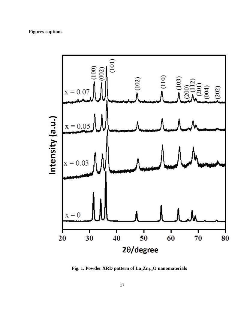
Figure 1 showed the powder XRD patterns of LaxZn1-xO (x = 0.0, 0.03, 0.05, 0.07 M)
nanoparticles with 2θ range from 30o to 75
o at room temperature. The diffraction peaks and their
relative intensities of both undoped and La doped ZnO samples were in good agreement with the
standard JCPDS card no. 36-1451. Hence, the observed patterns can be clearly endorsed to the
presence of hexagonal wurtzite structure. Furthermore, in Figure 1, no additional XRD peaks
were found which clearly indicates the absence of La oxide or Zn-La alloys formation. Hence, it
was clear that the introduction of dopant could not alter the crystal structure, whereas it dispersed
uniformly in the ZnO matrix [18].
In addition, from Figure 1, it was found that the most intense peak of doped ZnO
materials shifted towards higher θ value This shift is due to the internal strain developed by the
substitution of Zn2+
(host ions) by La3+
dopant ions [19-22]. The higher intensity of all peaks
suggested that the material was in highly crystalline nature.
The lattice parameter was calculated using the formula given in Eq. (1):
2
2
2
2222
3
4
4sin
c
l
a
khkh ---- (1)
where θ is the diffraction angle, λ, the incident wavelength (λ = 0.1540 nm), h, k, and l are
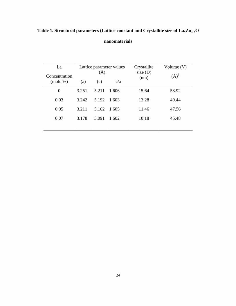
Miller’s indices The obtained lattice parameter values of both undoped and La-doped ZnO were
6
listed in Table 1. From Table 1, it was clear that the lattice parameter values of La-doped ZnO
materials, are less in comparison with those for pure ZnO materials, due to the shift of XRD
peaks towards higher 2θ values for doped ZnO nanoparticles. Similar lattice parameter values (a
= 3.251001 ± 0.000064 Å c = 5.208817 ± 0.000161 Å for undoped ZnO materials and a =
3.249680 ± 0.000054 Å, c = 5.205810 ± 0.000140 Å) has been reported by Goel et al. [23].
Moreover, it was clear that La-doping changed the lattice parameters values, but the crystal
system (hexagonal) and space group (P63mc) remain unchanged.
The average crystallite size was calculated using Scherer formula given in Eq. (2):
cos
89.0L
----(2)
where L is the crystallite size, λ, the X-ray wavelength, θ, the Bragg diffraction angle and β, the
full width at half maximum (FWHM). The average crystallite sizes of undoped and doped ZnO
were listed in Table 1. The average crystallite sizes of undoped and La-doped ZnO materials
were calculated to be 15.64 nm (x = 0M), 13.28 nm (x = 0.03M), 11.46 nm (x = 0.05M), 10.18
nm (x = 0.07 M) respectively. Thus, the particle size decreases because of La doping in ZnO
nanostructures. This reduction in the crystallite size is due to distortion in the ZnO matrix by
La3+
dopant ions, which decreases the rate of growth of ZnO.
3.2. Fourier transform infrared (FT-IR) spectral Analysis
The FT-IR spectra of LaxZn1-xO (x = 0.0, 0.03, 0.05, 0.07 M) nanoparticles were shown
in Figure 2. The strong intensity band at around 505 cm-1
clearly indicates the Zn-O stretching
vibration and the band at 607 cm-1
confirms the presence of ZnO and La. The 880 cm-1
corresponds to N-O deformation vibration. It was noted from the FT-IR data that the Zn-O
7
vibrational mode was more prominently observed and this clearly concludes a strong doping
exist in LaxZn1-xO.
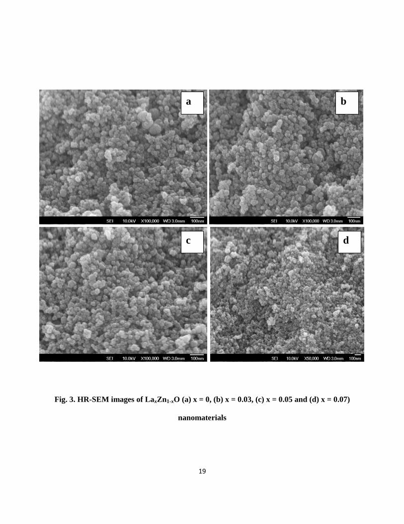
3.3 High Resolution-Scanning electron microscopy (HR-SEM) studies
The surface morphology of LaxZn1-xO (x = 0.0, 0.03, 0.05, 0.07 M) materials was
examined by HR-SEM analysis and showed in Figure 3. HR-SEM images clearly indicated the
sphere-like morphology of nanoparticles. Furthermore, from the Figure 3 it was noted that as La
concentration increases, particle size of the samples decreased, which was consistent with the
XRD results.
3.4. Energy dispersive X-ray (EDX) analysis
The chemical purity and elemental composition of the LaxZn1-xO materials were
investigated by Energy Dispersive X-ray analysis (EDX) as shown in Figure 4. The EDX results
showed the presence of Zn, O and La by the appearance of their corresponding peaks without
any other characteristic peaks and suggested that the prepared samples do not contain any other
element impurities.
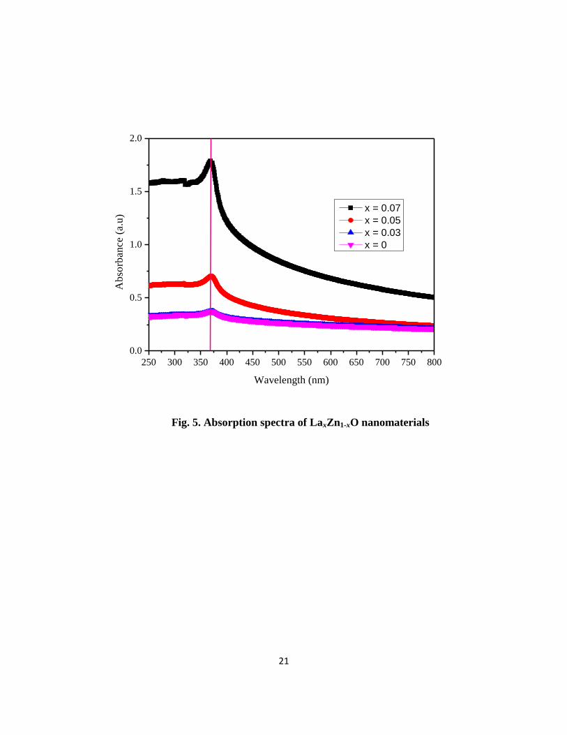
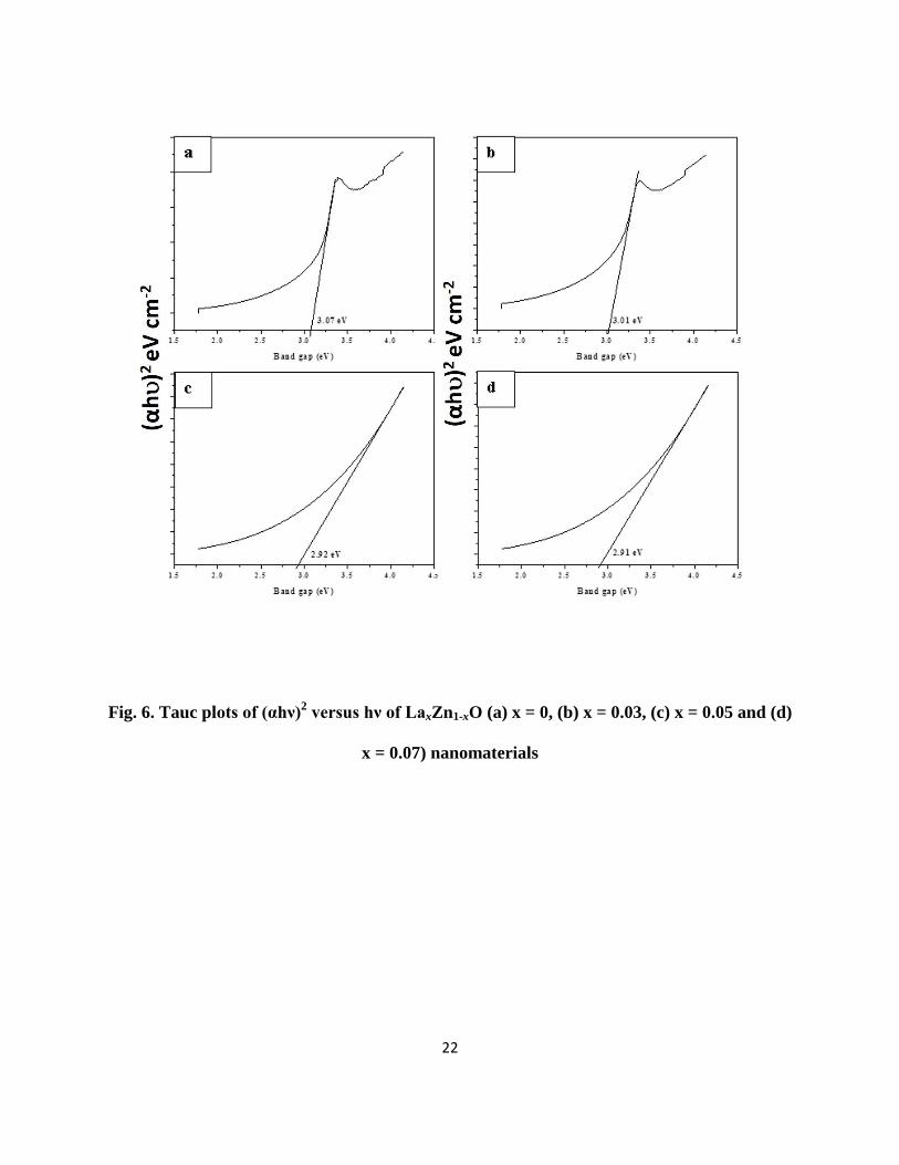
3.5. UV–Vis absorption spectroscopy
The optical properties of LaxZn1-xO materials with various doping concentrations were
investigated by UV–Vis absorption spectra which are shown in Figure 5. From Figure 5, it was
noted that the band-edge absorption of the synthesized LaxZn1-xO materials located at around
366-373 nm (in near UV region). The optical band gap of Eg is calculated using the following
Eq. 3 [23, 24].

8
α A(hν-Eg)n/hν ---------------- (3)
where A and n is a constant, equal to 1/2 for the direct band gap semiconductor. The calculated
band gaps of the synthesized LaxZn1-xO materials were shown in Figure 6. It was noted that the
lanthanum doping concentration had significant effects on the band gaps of synthesized
LaxZn1-xO materials. When the doping concentration of lanthanum changes from 0 to 0.07 M, the
value of Eg is decreased from 3.07 to 2.91 eV. When a Zn site in ZnO was occupied by a La
atom, there were two main effects were observed: (1) The impurity bands closer to the lower
edge of the conduction band, which was created by substituted La and (2) The obtained band-gap
exhibit narrow, due to the strong orbital coupling between La and O. The results illustrated that
La doping concentration plays vital role in tuning the band gap of the synthesized LaxZn1-xO
materials. Furthermore, the existence of extended tail band (from 450 to 800 nm) in the UV
spectra of La-doped ZnO nanoparticles (Figure 5) showed its optical capability almost in the
whole range of visible light spectra. The sp–d exchange interactions between the conduction
band electrons and the localized d electrons of the La3+
ions, which substitute Zn2+
ions lead to
the broad absorption in visible light range [25]. In addition, the s–d and p–d exchange
interactions cause a negative and a positive correction to the conduction band and the valence
band edges individually, resulting in strong visible light absorption of the La-doped ZnO
materials [26, 27]. Hence, La- doping in ZnO host can introduce the impurity energy levels in
band gap and expands its visible light response, which is favorable for several potential
applications. These results indicate that the La doped ZnO nanoparticles can absorb in UV as
well as in the visible region of the solar light suggesting that the La doped ZnO nanoparticles
could be applied as a visible light photocatalyst [28].
9
3.6. Photoluminescence spectral analysis
The PL spectra of LaxZn1-xO materials (x = 0, 0.03, 0.05 and 0.07) was showed in Figure
7. The emission bands around at 410 nm (UV emission) were observed due to the recombination
of the free excitons in ZnO [29-31]. Small variations (409.95, 410.15, 410.14, 410.25 nm) in the
positions of UV emission bands in PL spectra of LaxZn1-xO materials could be attributed to the
impact of lanthanum doping concentration. The position of UV emission bands slightly increases
as lanthanum doping concentration increases, but the intensity of the peaks decreased. Hence, the
UV emission had a red shift, since the doping concentration of lanthanum increases. In addition,
the shift in the emission spectra for undoped and doped ZnO may be attributed, due to the strain
created in the crystal lattice to accommodate larger La atoms in ZnO. Furthermore, the deep-
level emission in region from 490 nm to 540 nm was due to the intrinsic deep-level defects and
extrinsic impurities in ZnO [32-40].
3.7. Antibacterial Activities
The antibacterial activities of synthesized LaxZn1-xO materials were tested against the
human pathogens like P.mirabilis, S.typhi, S.aureus, B.subtilis with reference to Ampicillin. The
antibacterial activity values are listed in Table 2. From the results, it was observed that the
synthesized LaxZn1-xO materials showed desired activity against P. mirabilis and S. typhi.
Furthermore, when La concentration increases, activity of LaxZn1-xO materials against P.
mirabilis and S. typhi increases to higher extent, while LaxZn1-xO materials showed no activity
against S. aureus, B. subtilis, P. mirabilis and S. typhi causes kidney stone and typhoid fever
respectively. Hence, the synthesized LaxZn1-xO can be used in the treatment of kidney stone and
Typhoid Fever.
10
4. Conclusions
In summary, La-doped ZnO (LaxZn1-xO: x = 0.0, 0.03, 0.05, 0.07 M) nanoparticles have
been synthesized by chemical precipitation route. From XRD measurements, it was confirmed
that the particle size of the synthesized LaxZn1-xO materials decreases with increasing La
concentrations and possess hexagonal wurtzite structure. In addition, the sphere-like morphology
was revealed by HR-SEM and HR-TEM analysis. The elemental composition was confirmed by
EDX analysis. Furthermore, it was found that La3+
ions were successfully incorporated into the
ZnO lattice, and the red shift was appeared in PL spectra for doped nanoparticles compared with
undoped one. The band gap energies of doped nanoparticles were decreased from 3.07 eV to
2.91 eV with increasing the lanthanum doping concentration. Hence, these results indicated that
the lanthanum doping concentration plays an important role in tuning the size, band gap and
photoluminescence property of the ZnO nanoparticles. In addition the synthesized LaxZn1-xO
materials possess significant antibacterial activity against P.mirabilis, S.typhi and used for
treatment of kidney stone and typhoid fever.
ACKNOWLEDGMENT
The author’s A. M & E. M would like to thank for the institute founder Dr. S. Jagathrakshakan &
esteemed advisor Shri. V. Murthy, IAS, Bharath University, BIHER (Bharath Institute of Higher
Education & Research) for their inestimable support & constant encouragement, laboratories
chemicals, scientific computers facilities and future financial support for the upcoming work. In
addition, Dr. S. Bhuminathan, Registrar are acknowledged.
11
References
[1]. B. Meenatchi, V. Sathiya Lakshmi, A. Manikandan, V. Renuga, A. Sharmila, K. R.
Nandhine Deve & Saravana Kumar Jaganathan, Protic ionic liquid assisted synthesis and
characterization of ferromagnetic cobalt oxide nanocatalyst, J. Inorg. Organomet. Polym. 27
(2017) 446–454.
[2] A. Manikandan, M. Durka, K. Seevakan, S. Arul Antony, A novel one-pot combustion
synthesis and opto-magnetic properties of magnetically separable spinel MnxMg1-xFe2O4 (0 0≤
x≤ 0 ) Nanophotocatalysts, J. Supercond. Nov. Magn. 28 (2015) 1405-1416.
[3]. A. Manikandan, M. Durka, S. Arul Antony, A novel synthesis, structural, morphological,
and opto-magnetic characterizations of magnetically separable spinel CoxMn1-xFe2O4 (0≤ x≤ )
nano-catalysts, J. Supercond. Nov. Magn. 27 (2014) 2841-2857.
[4] E. Manikandan, M. K. Moodley, S. S. Ray, B. K. Panigrahi, R. Krishnan, K. G. M. Nair, A.
K. Tyagi, Zinc oxide epitaxial thin-film deposited over carbon on various substrates by PLD
technique, J. Nanosci. Nanotech. 10 (2010) 5601-5611.
[5] J. F. Zhu and Y. J. Zhu, Microwave-assisted one-step synthesis of polyacrylamide-metal
(M = Ag, Pt, Cu) nanocomposites in ethylene glycol, J. Phys. Chem. B 110 (2006) 8593-8597.
[6] N. C. S. Selvam, A. Manikandan, L. John Kennedy, and J. Judith Vijaya, Comparative
investigation of zirconium oxide (ZrO2) nano and microstructures for structural, optical and
photocatalytic properties, J. Colloid Interf. Sci. 389 (2013) 91-98.
[7] A. H. Shah, E. Manikandan, M B. Ahmed. Enhanced bioactivity of Ag/ZnO nanorods-a
comparative antibacterial study. 2013. J. Nanomed. Nanotech. 4 (2013) 6p.

12
[8] J Kennedy, PP Murmu, J Leveneur, A Markwitz, J Futter. Controlling preferred orientation
and electrical conductivity of zinc oxide thin films by post growth annealing treatment, Applied
Surface Science 367 (2016) 52-58.
[9] E. Manikandan, J. Kennedy, G. Kavitha, K. Kaviyarasu, M. Maaza, B.K. Panigrahi, U.
Kamachi Mudali “Hybrid Nanostructured Thin-Films by PLD for Enhanced Field Emission
Performance for Radiation Micro-Nano Dosimetry Applications. 2015. J. Alloys & Comps 647
(2015) 141-145.
[10]. J Kennedy, B Sundrakannan, RS Katiyar, A Markwitz, Z Li, W Gao, Raman scattering
investigation of hydrogen and nitrogen ion implanted ZnO thin films, Current Applied Physics 8
(2008) 291-294
[11]. PP Murmu, J Kennedy, GVM Williams, BJ Ruck, S Granville, SV Chong, Observation of
magnetism, low resistivity, and magnetoresistance in the near-surface region of Gd implanted
ZnO, Applied Physics Letters 101 (2012) 082408
[12]. J Kennedy, GVM Williams, PP Murmu, BJ Ruck, Intrinsic magnetic order and
inhomogeneous transport in Gd-implanted zinc oxide, Physical Review B 88 (2013) 214423
[13] B. Sathyaseelan, E. Manikandan, K. Sivakumar, J. Kennedy, M. Maaza. Enhanced visible
photoluminescent and structural properties of ZnO/KIT-6 nanoporous materials for white light
emitting diode (w-LED) application, J. Alloys Compds 651 (2015) 479-482.
13
[14] E. Hema, A. Manikandan, P. Karthika, S. Arul Antony, B. R. Venkatraman, A novel
synthesis of Zn2+
-doped CoFe2O4 spinel nanoparticles: Structural, morphological, opto-magnetic
and catalytic properties, J. Supercond. Nov. Magn. 28 (2015) 2539–2552.
[15] B. Meenatchi, K. R. Nandhine Deve, A. Manikandan, V. Renuga, and V. Sathiyalakshmi,
Protic ionic liquid assisted synthesis, structural, optical and magnetic properties of Mn-doped
ZnO nanoparticles, Adv. Sci. Eng. Med. 8 (2016) 653-659.
[16]. J Kennedy, PP Murmu, E Manikandan, SY Lee, Investigation of structural and
photoluminescence properties of gas and metal ions doped zinc oxide single crystals, Journal of
Alloys and Compounds 616 (2014) 614-617
[17]. F Fang, J Kennedy, DA Carder, J Futter, P Murmu, A Markwitz, Modulation of field
emission properties of ZnO nanorods during arc discharge, Journal of nanoscience and
nanotechnology 10 (2010) 8239-8243
[18] J. T. Chen, J. Wang, F. Zhang, G. A. Zhang, Z.G. Wu, P.X. Yan, The effect of La doping
concentration on the properties of zinc oxide films prepared by the sol–gel method, J. Crys.
Growth 310 (2008) 2627–2632.
[19] S. Goel, N.Sinha, H. Yadav, A.J. Joseph, B.Kumar, Experimental investigation on the
structural, dielectric, ferroelectric and piezoelectric properties of La doped ZnO nanoparticles
and their application in dye-sensitized solar cells, Physica E 91 (2017) 72–81.
[20] M. Poloju, N Jayababu, E. Manikandan, MVR Reddy. Enhancing the Isopropanol gas
sensing performance of SnO2/ZnO core/shell nanocomposite gas sensor. Journal of Materials
Chemistry C. 5 (2017) 2662-2668. DOI: 10.1039/C6TC05095F.
14
[21] A.S.H. Hameed, C. Karthikeyan, A.P. Ahamed, N. Thajuddin, N.S. Alharbi, S.A. Alharbi,
G. Ravi, In vitro antibacterial activity of ZnO and Nd doped ZnO nanoparticles against ESBL
producing Escherichia coli and Klebsiella pneumoniae, Sci. Rep. 6 (2016) 24312.
[22] E. Manikandan, V. Murugan, G. Kavitha, P. Babu, M. Maaza. Nanoflower rod wire-like
structures of dual metal (Al and Cr) doped ZnO thin films: Structural, optical and electronic
properties. Materials Letters, 131 (2014) pp.225-228.
[23] J. Cao, J. Yang, Y. Zhang, L. Yang, D. Wang, M. Wei, Y. Wang, Y. Liu, M. Gao, X. Liu,
Growth mechanism and blue shift of Mn2+
luminescence for wurtzite ZnS: Mn2+
nanowires, J.
Phys. D Appl. Phys. 43 (2010) 075403.
[24] M. Saif, H. Hafez, A.I. Nabeel, Photo-induced self-cleaning and sterilizing activity of Sm3+
doped ZnO nanomaterials, Chemosphere, 90 (2013) 840-847.
[25] E. Manikandan, L Krishnakumar, K Gnanasekaran, G Mani. et.al. Effective Ammonia
Detection Using n-ZnO/p-NiO Heterostructured Nanofibers. IEEE Sensors Journal 16 (2016),
2477 – 2483.
[26] A. H. Shah, E. Manikandan, MB Ahamed, D Ahmad Mir, S. Ahmad Mir, Antibacterial and
Blue shift investigations in sol-gel synthesized CrxZn1-xO Nanostructures. J Lumin.145 (2014)
944-948.
[27] C. Xu, L. Cao, G. Su, W. Liu, X. Qu, Y. Yu, Preparation, characterization and
photocatalytic activity of Co-doped ZnO powders, J. Alloys Compd. 497 (2010) 373-376.
[28] M. Shakir, M. Faraz, Asif Sherwani, Saud I. Al- Resayes, Photocatalytic degradation of the
paracetamol drug using lanthanum doped ZnO nanoparticles and their in-vitro cytotoxicity assay,
J. Lumin. 176 (2016) 159–167.

15
[29] D. Weissenberger, M. Durrschnabel, D. Gerthsen, F. Perez-Willard, A. Reiser, G. M. Prinz,
M. Feneberg, K. Thonke, R. Sauer, Conductivity of single ZnO nanorods after Ga implantation
in a focused-ion-beam system, Appl. Phys. Lett. 91 (2007) 132110.
[30] C Sasikala, N Durairaj, I Baskaran, B Sathyaseelan, M Henini, E. Manikandan, Transition
metal titanium (Ti) doped LaFeO3 nanoparticles for enhanced optical structural and magnetic
properties. J. Alloys & Comps 712 (2017) 870-877.
[31] L. L. Yang, Q. X. Zhao, M. Willander, X. J. Liu, M. Fahlman, J. H. Yang, Effective
suppression of surface recombination in ZnO nanorods arrays during the growth process, Cryst.
Growth Des. 10 (2010) 1904 -1910.
[32] D. Wang, G. Xing, M. Gao, L. Yang, J. Yang, W. Tom, Defects-mediated energy transfer in
red-light-emitting Eu-doped ZnO nanowire arrays, J. Phys. Chem. C 115 (2011) 22729-22735.
[33] J. Lang, J. Wang, Q. Zhang, X. Songsong, Q. Han, Y. Zhang, Hongju. Zhai, J. Cao, Y. Yan,
J. Yang, Rapid synthesis and photoluminescence properties of Eu-doped ZnO nano
needles via facile hydrothermal method, Chem. Res. Chin. Univ. 30 (2014) 538-542.
[34] L. L. Yang, Q. X. Zhao, M. Willander, X. J. Liu, M. Fahlman, J. H. Yang, Origin of the
surface recombination centers in ZnO nanorods arrays by XPS, Appl. Surf. Sci. 256 (2010)
3592-3597.
[35]. E. Manikandan, G. Kavitha, J. Kennedy. Epitaxial zinc oxide, graphene oxide composite
thin-films by laser technique for micro-Raman and enhanced field emission study. Ceramic
International. 40 (2014) 16065-16070.
[36]. RJ Mendelsberg, J Kennedy, SM Durbin, RJ Reeves. Raman scattering investigation of
hydrogen and nitrogen ion implanted ZnO thin films. Current Applied Physics. 6 (2006) 495-
498.
16
[37]. RJ Mendelsberg, J Kennedy, SM Durbin, RJ Reeves. Carbon enhanced blue–violet
luminescence in ZnO films grown by pulsed laser deposition. Current Applied Physics. 8 (2008)
283-286
[38]. F Fang, J Kennedy, J Futter, T Hopf, A Markwitz, E Manikandan, Size-controlled synthesis
and gas sensing application of tungsten oxide nanostructures produced by arc discharge.
Nanotechnology. 22 (2011), 335702.
[39]. J Kennedy, A Markwitz, Z Li, W Gao, C Kendrick, SM Durbin, R Reeves. Modification of
electrical conductivity in RF magnetron sputtered ZnO films by low-energy hydrogen ion
implantation. Current Applied Physics 6 (2006) 495-498.
[40]. AH Shah, MB Ahamed, E Manikandan, R Chandramohan, M Iydroose. Magnetic, optical
and structural studies on Ag doped ZnO nanoparticles. Journal of Materials Science: Materials in
Electronics 24 (2013) 2302-2308.
17
Figures captions
Fig. 1. Powder XRD pattern of LaxZn1-xO nanomaterials

18
Fig. 2. FT-IR spectra of LaxZn1-xO nanomaterials
19
Fig. 3. HR-SEM images of LaxZn1-xO (a) x = 0, (b) x = 0.03, (c) x = 0.05 and (d) x = 0.07)
nanomaterials
a b
c d

20
Fig. 4. EDX spectra of LaxZn1-xO (a) x = 0, (b) x = 0.03, (c) x = 0.05 and (d) x = 0.07)
nanomaterials
a
c d
b
21
250 300 350 400 450 500 550 600 650 700 750 800
0.0
0.5
1.0
1.5
2.0
Ab
sorb
an
ce (
a.u
)
Wavelength (nm)
x = 0.07
x = 0.05
x = 0.03
x = 0
Fig. 5. Absorption spectra of LaxZn1-xO nanomaterials
22
Fig. 6. Tauc plots of (αhν)2 versus hν of LaxZn1-xO (a) x = 0, (b) x = 0.03, (c) x = 0.05 and (d)
x = 0.07) nanomaterials

23
390 420 450 480 510 540 570 600
0
100
200
300
400
500
PL
In
ten
sity
(a.u
)
Wavelength (nm)
x = 0.07
x = 0.05
x = 0.03
x = 0
Fig. 7. PL spectra of LaxZn1-xO nanomaterials
24
Table 1. Structural parameters (Lattice constant and Crystallite size of LaxZn1-xO
nanomaterials
La
Concentration
(mole %)
Lattice parameter values
(Å)
Crystallite
size (D)
(nm)
Volume (V)
(Å)3
(a) (c) c/a
0
0.03
0.05
0.07
3.251
3.242
3.211
3.178
5.211
5.192
5.162
5.091
1.606
1.603
1.605
1.602
15.64
13.28
11.46
10.18
53.92
49.44
47.56
45.48

25
Table: 2 Antibacterial activities of LaxZn1-xO nanomaterials for against human pathogens
Antibacterial activities of samples were determined as zone of inhibition (in mm)
Samples P. mirabilis S .typhi S. aureus B. subtilis
Ampicillin (C) 24 19 12 11
x = 0 7 5 0 0
x = 0.03 9 9 0 0
x = 0.05 20 15 0 0
x = 0.07 22 7 0 0

26
Graphical Abstract
27
Highlights
Enhanced optical properties La doped ZnO nanoparticles
La-doped ZnO nanomaterials were synthesized by chemical method
XRD, FT-IR, UV-Visible, HR-SEM, EDX, PL spectroscopy techniques were used
The antibacterial activities of LaxZn1-xO were tested by modified disc diffusion method
Average particle size of undoped and doped La-doped ZnO was found 15 to 10 nm

Graphical Abstract (for review)
Highlights
Enhanced optical properties La doped ZnO nanoparticles
La-doped ZnO nanomaterials were synthesized by chemical method
XRD, FT-IR, UV-Visible, HR-SEM, EDX, PL spectroscopy techniques were used
The antibacterial activities of LaxZn1-xO were tested by modified disc diffusion method
Average particle size of undoped and doped La-doped ZnO was found 15 to 10 nm
*Highlights (for review)