Embed Size (px)
Citation preview
DEVELO
PMENT
3603RESEARCH ARTICLE
INTRODUCTIONLower metazoans meet nutritional needs with a rudimentary intestinethat is lined by a simple absorptive epithelium. The more complexnutritional demands in vertebrate animals require additional organssuch as the stomach, pancreas and liver, which connect with the smallbowel. Early in vertebrate embryogenesis, the visceral endoderm andsplanchnic mesoderm combine to create a tube that is subsequentlypatterned into specialized segments: esophagus, stomach, the intestineand its evaginated derivatives, the liver and pancreas. A componentcommon to all these structures is the mesothelium, a connective tissuethat envelops the gut and tethers organs to the body wall. Thedorsolateral mesothelium of the stomach (mesogastrium) provides acompartment for early development of the spleen and dorsal pancreas,which appear initially as a confluent primordium near the greatercurvature of the stomach (Brendolan et al., 2007; Hecksher-Sorensenet al., 2004; Thiel and Downey, 1921). Stomach rotation and leftwardmovement of the dorsal pancreas subsequently juxtapose the dorsaland ventral pancreatic buds, which fuse and come to lie near theduodenum, whereas the spleen remains associated with the lateralstomach wall, near its site of origin. Patterning of the rostral gut andits neighboring organs is poorly understood and little is known aboutthe role of mesothelium in their development.
Stomach mesenchymal expression of the homeobox gene Barx1seems to be required to suppress regional Wnt activity in prospectivegastric endoderm and thus allows stomach-specific epithelial
differentiation (Kim et al., 2005). During the period of gastricmorphogenesis and gut endoderm specification, Barx1 is expressedselectively in stomach mesenchyme (Kim et al., 2005; Tissier-Setaet al., 1995). Small-interfering (si) RNA-induced loss of Barx1 inrecombinant cultures of embryonic day (E) 12 mouse fetal tissuesprofoundly affects differentiation of overlying stomach endoderm:intestinal marker genes are robustly activated at the expense ofstomach epithelial transcripts (Kim et al., 2005). Barx1-null E12embryos have normal intestines and a small, aberrantly shapedstomach with atypical endodermal lining; Cdx2, a specific markerof intestinal epithelium (Silberg et al., 2000), is expressedectopically in the distal stomach. Levels of the secreted frizzled-related proteins Sfrp1 and Sfrp2, soluble antagonists of Wntsignaling (Finch et al., 1997; Rattner et al., 1997), are reduced in theabsence of Barx1, and forced Sfrp expression in Barx1-deficientstomach mesoderm restored gastric markers in co-culturedendoderm (Kim et al., 2005).
On the inbred 129/Sv genetic background, Barx1–/– mouseembryos die at E13 of unknown causes and so we could not studytheir subsequent development. Moreover, recombinant fetal tissuecultures convey information about molecular markers but not abouthistomorphology. Breeding the Barx1 mutation into a mixed geneticbackground with contribution from the C57BL/6 straincircumvented embryonic lethality and allowed us to elucidate anunprecedented and completely penetrant patterning defect of thestomach. In addition to this homeotic aberration, Barx1 loss causesa unique defect in development of the spleen, which is consistentlymislocalized and severely hypoplastic. As Barx1 is never present inthe spleen primordium but highly expressed in surroundingmesogastrium, its effects on spleen development, like those onstomach epithelial specification, must also occur across tissueplanes. We confirmed the role of Barx1 in suppressing stomachendodermal Wnt activity, but our studies suggest that its role in
Independent functions and mechanisms for homeobox geneBarx1 in patterning mouse stomach and spleenByeong-Moo Kim1,2, Isabelle Miletich3, Junhao Mao4, Andrew P. McMahon4, Paul A. Sharpe3 andRamesh A. Shivdasani1,2,5,*
Homeobox genes convey positional information in embryos and their role in patterning the mammalian gut is a topic ofconsiderable interest. Barx1 is expressed selectively in fetal stomach mesenchyme and directs differentiation of overlyingendoderm. Recombinant tissue cultures and study of young mouse embryos previously suggested that Barx1 controls expression ofsecreted Wnt antagonists, which suppress endodermal Wnt signaling, to enable stomach epithelial differentiation. We overcamemid-gestational lethality of Barx1–/– mouse embryos and report here the spectrum of anomalies in a distinctive and unprecedentedmodel of gastrointestinal homeotic transformation. Using various mouse models, we confirm the importance of attenuated Wntsignaling in stomach development and the role of Barx1 in suppressing endodermal Wnt activity. Absence of Barx1 also results infully penetrant defects in positioning and expansion of the spleen, an organ that originates within the mesothelial lining of thestomach. Barx1 is absent from the spleen primordium but highly expressed in the mesogastrium, indicating an indirect effect onspleen development. However, our results argue against a role for Wnt antagonism in genesis of the spleen. Mouse spleendevelopment relies on several homeodomain transcriptional regulators that are expressed in the spleen primordium. Loss of Barx1does not affect expression of any of these genes but notably reduces expression of Wt1, a transcription factor implicated in spleenmorphogenesis and expressed in the mesothelium. These observations place Barx1 proximally within a Wt1 pathway of spleendevelopment and reveal how a homeotic regulator employs different molecular mechanisms to mold neighboring organs.
KEY WORDS: Barx1, Mesenchyme-epithelium interactions, Stomach development, Spleen development, Wnt signaling, Organogenesis,Wt1
Development 134, 3603-3613 (2007) doi:10.1242/dev.009308
1Dana-Farber Cancer Institute and 2Department of Medicine, Harvard MedicalSchool, Boston, MA 02115, USA. 3Department of Craniofacial Development, DentalInstitute, Kings College, London SE1 9RT, UK. 4Department of Molecular andCellular Biology, Harvard University, Cambridge, MA 02138, USA. 5Department ofMedicine, Brigham and Women’s Hospital, Boston, MA 02115, USA.
*Author for correspondence (e-mail: [email protected])
Accepted 5 August 2007
DEVELO
PMENT
3604
spleen development is exerted through a different mechanism. Inparticular, absence of Barx1 specifically reduces mesothelialexpression of Wt1, a transcription factor known to be required forspleen morphogenesis. These findings help define the basis for thediverse functions of a homeodomain transcription factor in thedevelopment of abdominal organs.
MATERIALS AND METHODSExperimental animalsBarx1+/– males from the 129/Sv strain were back-crossed repeatedly withheterozygote animals on the C57BL/6 genetic background, and we studiedmost Barx1 mutants after at least five back-crosses. TOPGAL transgenicmice and strain-matched CD1 controls were purchased from JacksonLaboratories (Bar Harbor, ME); Barx1–/–;TOPGALTg mice were generatedby interbreeding. Shh+/Cre mice originated by targeted insertion of a GFP-Cre fusion cDNA into the Shh locus (Harfe et al., 2004). Catnb+/lox(ex3) micecarry an allele with loxP sites flanking exon 3 of the �-catenin (Catnb;Ctnnb1 – Mouse Genome Informatics) gene (Harada et al., 1999) and weregenerously provided by Mark Taketo (Kyoto University, Japan). Axin2lacZ
mice have lacZ cDNA embedded in the Axin2 locus (Yu et al., 2005) andwere kindly provided by Walter Birchmeier (Max-Delbrück Center, Berlin,Germany). Animals were handled according to protocols approved by aninstitutional committee. The morning following vaginal plugging wasregarded as day 0.5 of gestation.
Histology and immunohistochemistryAfter overnight fixation in Bouin’s solution or 4% paraformaldehyde, wholeembryos or isolated organs were dehydrated, embedded in paraffin andsections of 5-6 �m were prepared. Hematoxylin and Eosin (H&E), PAS andAlcian Blue staining were performed using routine methods. For antigenretrieval prior to immunostaining, specimens were heated in 10 mM Nacitrate buffer (pH 6.0) in a decloaking chamber (Biocare Medical, Concord,CA), then cooled for 60 minutes at room temperature. To eliminateendogenous peroxidases, tissues were treated in methanol containing 0.5%H2O2 for 30 minutes. After blocking with normal goat serum, samples wereincubated for 24 hours at 4°C with one of the following monoclonalantibodies (Ab): Cdx2 (1:20; Biogenex, San Ramon, CA), activated �-catenin (1:500; Upstate Millipore, Charlottesville, VA), Ter119, B220 (Ly76and Ptprc, respectively – Mouse Genome Informatics) (1:100; B-DPharmingen, Franklin Lakes, NJ), H+/K+-ATPase (2B6, 1:1000; MBL,Nagoya, Japan), smooth muscle actin (1A4, 1:3000; Biogenex) and Muc5ac(45M1, 1:500; Novocastra, Newcastle, UK), or rabbit antisera against gastrin(1:1000; Novocastra), Pdx1 (1:6000; gift of Christopher Wright, VanderbiltUniversity, TN), insulin (1:1000; Santa Cruz Biotech, Santa Cruz, CA),Barx1 [1:9000 (Kim et al., 2005)], Wt1 (1:3000; Santa Cruz) or Sox2(1:1000; Chemicon, Temecula, CA). Samples were washed, incubated withbiotinylated goat anti-mouse, anti-rabbit or anti-rat IgG and treated withavidin-biotin-peroxidase complex (Vector Laboratories, Burlingame, CA).Color reactions were developed with diaminobenzidine hydrochloridesolution (Sigma, St Louis, MO).
�-galactosidase stainingPregnant dams were sacrificed at various stages and embryos exposed to a�-galactosidase (�-gal) staining protocol that yielded no background in non-transgenic animals (Kim et al., 2005). Briefly, mouse embryos or organswere isolated in Ca2+- and Mg2+-free Hanks’ Balanced Salt Solution(Invitrogen, Carlsbad, CA), fixed for 15 minutes with 4% paraformaldehydein PBS, washed three times in PBS, and incubated in staining solution [PBS(pH 7.2), 1 mg/ml 5-bromo-4-chloro-3-indoyl-�-D-galactoside, 5 mMK3Fe(CN)6, 5 mM K4Fe(CN)6·3H2O, 1 mM MgCl2, 0.01% sodiumdeoxycholate, 0.02 % NP40] for 9-10 hours at 37°C.
In situ hybridizationSections (6 �m) were cut and mounted on SuperFrost Plus slides (FisherScientific, Kalamazoo, MI), deparaffinized, rehydrated, washed in PBS andtreated with 1 �g/ml proteinase K (Roche, Indianapolis, IN) for 10 minutes.After acetylation with 0.25% acetic anhydride in 0.1 M triethanolamine (pH8.0), slides were washed in 2�SSC and air-dried. Hybridization was
performed overnight at 60°C with digoxigenin-labeled antisense riboprobesin 50% formamide, 5�SSC, 2�Denhardt’s solution, 0.02% bovine serumalbumin, 0.1% Tween-20, 0.25% sodium dodecyl sulfate, 5 mM EDTA (pH8.0) and 50 �g/ml yeast tRNA. Slides were subsequently washed in 2� or0.2� SSC between 60 and 65°C and again in PBS, followed by incubationfor 90 minutes with 20% sheep serum. The hybridized probe was detectedby incubating tissue sections overnight at 4°C with alkaline phosphatase-conjugated sheep anti-digoxigenin Ab diluted 1:2000 in PBS supplementedwith 5% sheep serum and 5% fetal bovine serum. Color reactions weredeveloped with nitroblue tetrazolium/5-bromo-4-chloro-3-indolyl phosphate(Roche); slides were monitored until color development was observed andthe reaction was terminated with distilled water. In situ hybridization withradioactively labeled Barx1 probe was performed as described previously(Kim et al., 2005).
Transmission electron microscopyEmbryonic stomachs were fixed overnight at 4°C in a solution containing2.5% paraformaldehyde, 5% glutaraldehyde, 0.06% picric acid, 0.1 M Nacacodylate, and 0.06% CaCl2, post-fixed in OsO4, and embedded in Epon812. Thin (0.1 �m) sections were stained with uranyl acetate and lead citrateand examined in a JEOL 1200 electron microscope at an accelerating voltageof 80 kV.
Reverse-transcription (RT)-PCRTotal RNA was extracted using Trizol (Invitrogen), treated with RNase-freeDNase (Ambion, Austin, TX) and reverse transcribed using oligo-(dT)primers. Wt1 mRNA levels were assessed by conventional and SYBR Greenreal-time quantitative RT-PCR (Applied Biosystems, Foster City, CA) usinga common forward primer (5�-GCCTTCACCTTGCACTTCTC-3�) andthe reverse primers 5�-CATTCAAGCTGGGAGGTCAT-3� and 5�-GACCGTGCTGTATCCTTGGT-3� for conventional and real-time PCR,respectively.
Flow cytometryNeonatal spleen cells were dislodged with forceps and a single-cellsuspension prepared by filtering through a 30-�m strainer. Cells wereincubated on ice for 1 hour with 1 �g/ml Ter119, B220, Gr1 (Ly6g – MouseGenome Informatics), Cd4, Cd8a or Mac1 (Itgam – Mouse GenomeInformatics) primary Ab (B-D Pharmingen), followed by washing in PBSand further incubation on ice for 30 minutes with fluorophore-conjugatedsecondary Ab. Flow cytometry was performed on a Becton DickinsonFACScan and the data were analyzed using FlowJo software (Tree Star,Ashland, OR).
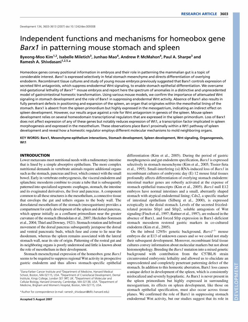
RESULTSUnique character of gastrointestinal homeosis inthe absence of Barx1We previously implicated the Barx1-null mutation in homeotictransformation of the rostral gut; however, unexplained embryoniclethality had restricted analysis to embryos at E12.5 or earlier.Crossing the null mutation [deletion of homeobox-encoding exons2, 3 and part of exon 4 (Kim et al., 2005)] to introduce the C57BL/6genetic background permitted Barx1–/– mice to survive to birth,although they succumb soon thereafter to respiratory distress that islikely to result from cleft palate; this defect probably reflectssignificant Barx1 expression in developing branchial arches (see Fig.S1A in the supplementary material) (Tissier-Seta et al., 1995).Barx1–/– pups appeared in the expected proportion in over 25 littersand our findings did not vary with C57BL/6 contributions between50% and 98%. Barx1–/– stomach was found to be greatly reduced insize (Fig. 1A) and escaped leftward rotation, thus presenting as amidline structure (data not shown). The villiform lining of neonatalBarx1–/– stomach (Fig. 1B) contrasts with the flat mucosa in controlmice (Fig. 1D) and carried two distinct surfaces: the distal 1/4 to 1/3is lined by villi of the intestinal variety, whereas the proximal
RESEARCH ARTICLE Development 134 (20)
DEVELO
PMENT
stomach has a highly atypical mucosa with gastric features. Periodicacid Schiff (PAS), which stains gastric but not intestinal epitheliumuniformly, highlighted this difference (Fig. 1C).
To define the nature of the homeotic transformation, we studiedgastric lineage markers. In wild-type mice, a stratified squamousepithelium extends from the esophagus to line the rostral dome(fundus) of the stomach (Fig. 1D,E), followed abruptly by theglandular mucosa of the corpus (Fig. 1D,F), which is flat and carriesthree major cell types in adults (Karam and Leblond, 1992). In mice,Muc5ac, H+/K+-ATPase and pepsinogen or intrinsic factor (IF; alsoknown as Gif – Mouse Genome Informatics) are specific molecularmarkers of these respective lineages: foveolar (pit), parietal (oxyntic)and chief (zymogenic) cells. The distal (antral-pyloric) stomach hasa similar lining, with modified cell ratios and folds that impart ascalloped appearance (Fig. 1D,G). Neutral mucins in gastric pit cellsstain with PAS, whereas intestinal goblet cells are fewer in numberand produce acidic mucins with affinity for both PAS and AlcianBlue; intestinal goblet cells also express trefoil factor 3 (Tff3)abundantly (Chinery et al., 1992). Two homeodomain transcriptionfactors permit further distinction between gut segments: Pdx1expression is scattered in distal stomach and uniform in duodenalepithelium (Offield et al., 1996), whereas Cdx2 is exquisitelyspecific to the intestine (Silberg et al., 2000). We used these featuresto characterize Barx1–/– stomach.
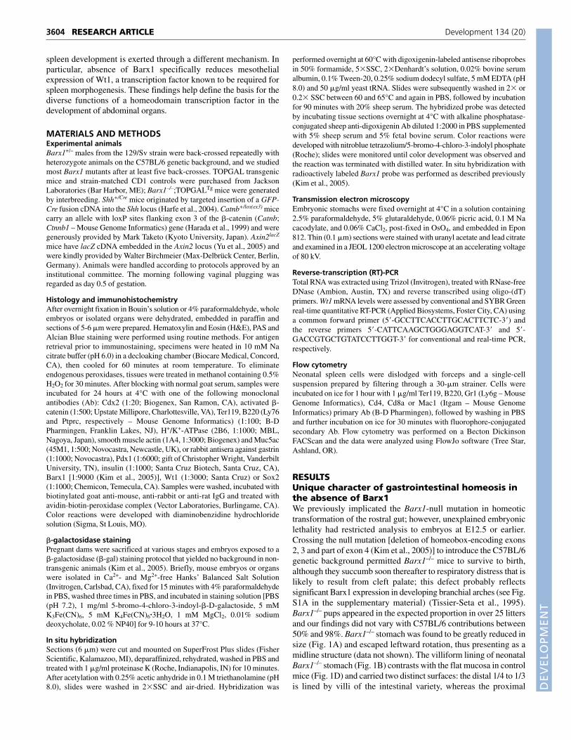
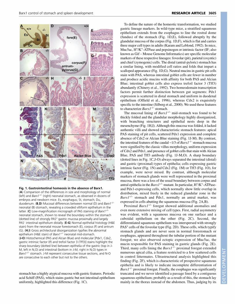
The mucosal lining of Barx1–/– mid-stomach was found to bethickly folded and the glandular morphology highly disorganized,with branching structures and epithelial nests deep in themesenchyme (Fig. 1H,I). Although this mucosa was folded, it lackedauthentic villi and showed characteristic stomach features: apicalPAS staining of pit cells, scattered Pdx1 expression and completeabsence of Cdx2 or Alcian Blue staining (Fig. 1J-M). By contrast,the intestinal features of the caudal ~1/3 of Barx1–/– stomach mucosawere signified by the classic villus morphology, uniform expressionof Cdx2 and Pdx1, and presence of goblet cells that stain with AlcianBlue, PAS and Tff3 antibody (Fig. 1J-M,O). A sharp boundary(dotted lines in Fig. 1C,J-O) always separated the intestinal (distal)and gastric (proximal) types of epithelia; cells expressing gastricintrinsic factor (Fig. 1N) and Cdx2 (Fig. 1M) or Tff3 (Fig. 1O), forexample, were never mixed. By contrast, although molecularmarkers of stomach glands were well represented in the proximalmucosa, there was a loss of the usual boundary between corpus andantral epithelia in the Barx1–/– mutant. In particular, H+/K+-ATPase-and Pdx1-expressing cells, which normally show little overlap indistribution, mixed freely in the reduced glandular zone in theBarx1–/– stomach and Pdx1, normally an antral marker, wasexpressed in cells abutting the squamous mucosa (Fig. 2A,B).
Proximal Barx1–/– foregut showed additional anomalies andeven more extensive mixing of cell types. First, radial asymmetrywas evident, with a squamous mucosa on one surface and acuboidal epithelium on the other (Fig. 2C). Second, thedifferentiated squamous epithelium was interspersed with stronglyPAS+ cells of the foveolar type (Fig. 2D). These cells, which typifystomach glands and are never seen in normal forestomach oresophagus, appeared throughout the tubular portion of the mutantforegut; we also observed ectopic expression of Muc5ac, themucin responsible for PAS staining in gastric glands (Fig. 2E).Third, many cells lining the Barx1–/– proximal foregut extendednumerous apical cilia, a feature restricted to a few scattered cellsin control littermates. Ultrastructural analysis highlighted thisfinding (Fig. 2F), which is characteristic of prospective squamousepithelia and is likely to indicate incomplete differentiation ofBarx1–/– proximal foregut. Finally, the esophagus was significantlytruncated and we never identified a passage lined by a contiguoussquamous epithelium; probably as a result of this, the stomach laymainly in the thorax instead of the abdomen. Thus, judging by its
3605RESEARCH ARTICLEBarx1 control of stomach and spleen development
Fig. 1. Gastrointestinal homeosis in the absence of Barx1.(A) Comparison of the differences in size and morphology of normal(left) and Barx1–/– (right) neonatal stomach, as observed in dozens ofembryos and newborn mice. Es, esophagus; St, stomach; Du,duodenum. (B,D) Mucosal differences between normal (D) and Barx1–/–
neonatal (B) stomach, revealing a crowded villiform epithelium in thelatter. (C) Low-magnification micrograph of PAS staining of Barx1–/–
neonatal stomach, shown to reveal the boundary within the stomach(dotted line) of strongly PAS+ gastric mucosa proximally and largelyPAS– intestinal epithelium distally. (E-G) Normal epithelial histology (H&Estain) from the neonatal mouse forestomach (E), corpus (F) and antrum(G). (H,I) Gross architectural disorganization typifies the abnormalepithelium (H&E stain) of Barx1–/– neonatal mid-stomach.(J-O) Histochemical (PAS and Alcian Blue) and molecular [Pdx1, Cdx2,gastric intrinsic factor (If) and trefoil factor 3 (Tff3)] stains highlight thesharp boundary (dotted line) between epithelia of the gastric (top in J-M, left in N,O) and intestinal (bottom in J-M, right in N,O) types inBarx1–/– stomach. J-M represent consecutive tissue sections, and N-Oare consecutive to each other but not to the others.
DEVELO
PMENT
3606
epithelium, the structure that overtly resembles an esophagusappeared to be a highly dysmorphic fundus with a mixed squamo-glandular lining. Sox2, a molecular marker of foregut squamous
epithelium (Que et al., 2007), was expressed in proximal but notdistal Barx1–/– foregut (Fig. 2G,H). Cells with smooth-musclemorphology and expression of smooth muscle actin appeared inthe correct distribution in the peripheral sub-epithelium, althoughthe muscle layer was discontinuous and less well differentiatedthan in controls (Fig. 2I,J).
Absence of Barx1 thus results in marked foregut shorteningand blurring of gastric squamo-glandular and corpus-antralboundaries (Fig. 2K). We never identified cells with dualstomach-intestine characteristics, but the sharply demarcatedgastro-duodenal junction was shifted anteriorly, into the stomachproper. An exterior boundary was always discernible between thestomach and duodenum, but the pyloric sphincter was absent (datanot shown). As Barx1 expression is exclusively sub-epithelial(Kim et al., 2005; Tissier-Seta et al., 1995), the mucosalanomalies in Barx1–/– mice must reflect mesenchymal influenceover the differentiation of the overlying endoderm. Structuraldefects such as reduced stomach size, fusiform shape, fundicdysmorphogenesis and pyloric sphincter agenesis, are likely torepresent functions intrinsic to the mesenchyme.
Genetic evidence that Barx1 inhibits stomachendodermal Wnt signalingWe previously proposed that Wnt antagonists are prominent targetsof Barx1 regulation in gastric mesenchyme (Kim et al., 2005). Theprospective stomach shows a wave of Wnt activity after E9, and weproposed that the usual decline in this activity results from Barx1-regulated production of secreted frizzled-related proteins (Sfrps).Recombinant fetal cell culture results supported this idea, but deathof Barx1–/– embryos precluded direct genetic confirmation. Havingovercome fetal lethality, we crossed 129/Sv-C57BL/6 hybridBarx1+/– and TOPGAL transgenic (Tg) mice, which carry lacZcDNA linked to multimerized Wnt-response elements and reportfaithfully on Wnt signaling (DasGupta and Fuchs, 1999). Ifthe model is correct, proximal stomach endoderm inBarx1–/–;TOPGALTg embryos should, unlike control TOPGALTg
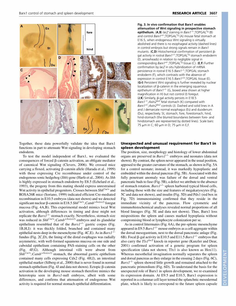
embryos, continue to express �-gal late in gestation. Indeed,between E16.5 (Fig. 3B) and birth, Barx1–/– embryos carrying onecopy of the Wnt-reporter transgene showed prominent �-gal activitythroughout the proximal foregut, a region we characterized as anatypical gastric fundus with mixed squamous-glandular epithelium(Fig. 2). Residual �-gal activity in control transgenic stomachs wasminimal by E16.5 (Fig. 3A,C) and undetectable in E18.5 stomach(data not shown) and at any stage in the developing esophagus. Bycontrast, the signal in Barx1–/–;TOPGALTg fundic stomach appearedsooner and stronger than in any other site of embryonic Wnt activity;this signal localized to the endoderm (Fig. 3D). We confirmed lacZexpression by RNA in situ hybridization in E16.5 foregut, wheresignal was readily detected in mutant (Fig. 3F) but not controlTOPGALTg (Fig. 3E) samples.
To monitor Wnt signaling independent of the TOPGAL reporter,we examined �-catenin localization. In E18.5 Barx1–/– foregut,innumerable cells showed unambiguous localization in the nucleus(Fig. 3G,H), whereas the signal in littermate control foregut alwaysappeared at cell-cell junctions (Fig. 3I). We also mated Barx1+/–
mice with another Wnt-reporter strain, Axin2lacZ. Insertion of lacZcDNA into the mouse Axin2 locus, a ubiquitous target of canonicalWnt signaling (Jho et al., 2002), accurately marks sites of Wntactivity (Yu et al., 2005). Again, we readily detected prominent �-gal activity in the atypical fundus in E18.5 Barx1–/–;Axin2lacZ
embryos (Fig. 3K), but only weak residual signal in the stomach andnone in the esophagus of Barx1+/–;Axin2lacZ littermates (Fig. 3J).
RESEARCH ARTICLE Development 134 (20)
Fig. 2. Severe mucosal abnormalities in the proximal foregut ofBarx1–/– mice. (A,B) Abnormally proximal expression of Pdx1 (near thesquamous lining) and mixing of H+/K+-ATPase+ and Pdx1+ cells in theglandular mucosa of Barx1–/– neonatal stomach. The two panels showstaining on adjacent sections. (C) Radial asymmetry of the most-proximal mucosa, with one surface lined by a flat squamoid epithelium(seen at the top of the image) and the other (bottom) by a cuboidalepithelium containing many ciliated cells (arrows). (D,E) PAS (D) andMuc5ac staining (E) reveal the presence in both surfaces of cuboidalcells with features of glandular pit cells. (F) Ultastructural confirmationof numerous apical cilia in cells lining neonatal Barx1–/– proximalforegut. (G,H) Immunohistochemical evidence for expression of Sox2, asquamous foregut marker, shown at low (G) and high (H)magnification. (I,J) Smooth muscle actin staining indicates smoothmuscle differentiation in Barx1–/– stomach (J), although both signalstrength and tissue continuity are reduced in comparison to littermatecontrols (I). (K) Graphic representation of gastrointestinal homeotic andregional anomalies that occur in the absence of Barx1.
DEVELO
PMENT
Together, these data powerfully validate the idea that Barx1functions in part to attenuate Wnt signaling in developing stomachendoderm.
To test the model independent of Barx1, we evaluated theconsequences of forced �-catenin activation, an obligate mediatorof canonical Wnt signaling (Clevers, 2006). We crossed micecarrying a floxed, activating �-catenin allele (Harada et al., 1999)with those expressing Cre recombinase under control of theendogenous sonic hedgehog (Shh) gene (Harfe et al., 2004). As Shhis highly expressed in stomach endoderm by E8.5 (Echelard et al.,1993), the progeny from this mating should express unrestrainedWnt activity in epithelial progenitors. Crosses between ShhCre/+ andROSA26R mice (Soriano, 1999) indicated efficient Cre-mediatedrecombination in E10.5 embryos (data not shown) and we detectedsignificant nuclear �-catenin in E18.5 ShhCre/+;Catnb+/lox(ex3) foregutmucosa (Fig. 4A,B). This experimental model mimics local Wntactivation, although differences in timing and dose might notreplicate the Barx1–/– stomach exactly. Nevertheless, stomach sizewas reduced in ShhCre/+;Catnb+/lox(ex3) embryos and its glandularepithelium resembled that of the Barx1–/– gastric corpus (Fig.1B,H,I): it was thickly folded, branched and contained manyepithelial nests deep in the mesenchyme (Fig. 4C,E). As in Barx1–/–
fundus (Fig. 2C,D), the lining of the distal esophagus was radiallyasymmetric, with well-formed squamous mucosa on one side andcuboidal epithelium containing PAS-staining cells on the other(Fig. 4F,G). Although intestinal villi were absent fromShhCre/+;Catnb+/lox(ex3) stomach, the abnormal gastric epitheliumcontained many cells expressing Cdx2 (Fig. 4H,I), an intestinalepithelial marker (Silberg et al., 2000) that is never found in normalstomach epithelium (Fig. 4J and data not shown). Ectopic �-cateninactivation in the developing mouse stomach therefore mimics theheterotopia seen in Barx1-null embryos, albeit with somedifferences, and confirms that attenuation of endogenous Wntactivity is required for normal stomach epithelial differentiation.
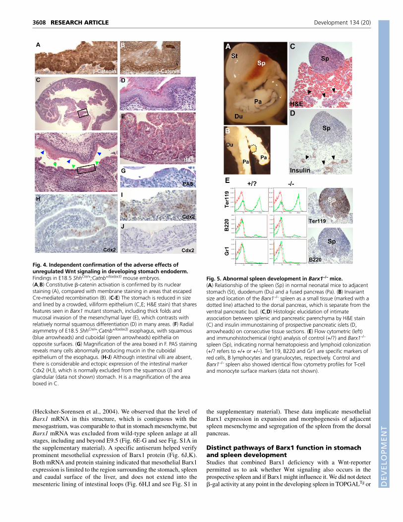
Unexpected and unusual requirement for Barx1 inspleen developmentThe position, size, morphology and histology of lower abdominalorgans are preserved in Barx1–/– embryos and neonates (data notshown). By contrast, the spleen never appeared in the usual position,apposed to the greater curvature of the stomach, as shown in Fig. 5Afor a control neonate; instead, it was markedly hypoplastic andembedded within the dorsal pancreas (Fig. 5B). Associated with thisfully penetrant anomaly was failure of the dorsal and ventralpancreatic buds to fuse (Fig. 5B), a defect we attribute to the absenceof stomach rotation. Barx1–/– spleen harbored typical blood cells,including those with the size and features of megakaryocytes (Fig.5C and data not shown), and insulin (Fig. 5D) and Pdx1 (red box inFig. 7D) immunostaining confirmed that they reside in theimmediate vicinity of the pancreas. Flow cytometric andimmunohistochemical analyses revealed normal proportions of allblood lineages (Fig. 5E and data not shown). Thus, Barx1 lossmispositions the spleen and causes marked hypoplasia withoutcompromising blood or lymphocyte colonization per se.
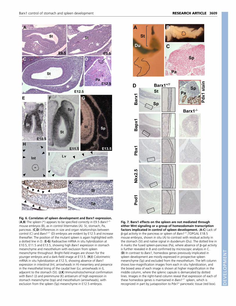
As in control littermates (Fig. 6A), the pre-splenic mesenchymeappeared in E9.5 Barx1–/– mouse embryos as a cell aggregate withinthe dorsal mesogastrium, next to the dorsal pancreatic anlage (Fig.6B). Focal �-gal activity in E10.5 and E11.5 Barx1–/– embryos thatalso carry the Tlx1lacZ knock-in reporter gene (Kanzler and Dear,2001) confirmed activation of a genetic program for spleenspecification (data not shown; Tlx1 is also known as Hox11).Whereas mesothelial invagination normally separates the spleenand dorsal pancreas as they enlarge in the ensuing 2 days (Fig. 6C),Barx1–/– spleen showed little growth and remained attached to thepancreatic primordium (Fig. 6D). To understand the basis for theunexpected role of Barx1 in spleen development, we re-examinedits expression domain. At E9.5 and E10.5, Barx1 expression isreported in a columnar cell layer termed the splanchnic mesodermalplate, which is likely to correspond to the future spleen capsule
3607RESEARCH ARTICLEBarx1 control of stomach and spleen development
Fig. 3. In vivo confirmation that Barx1 enablesattenuation of Wnt signaling in prospective stomachepithelium. (A,B) lacZ staining in Barx1–/–;TOPGALTg (B)and control Barx1+/–;TOPGALTg (A) mouse fetal stomach atE16.5, when endogenous Wnt signaling is virtuallyabolished and there is no esophageal activity (dashed lines)in control embryos but strong signals remain in Barx1mutants. (C,D) Histochemical confirmation of persistent �-gal activity in rostral Barx1–/–;TOPGALTg stomach endoderm(D, arrowheads) in relation to negligible signal incorresponding Barx1+/–;TOPGALTg tissue (C). (E,F) Furtherconfirmation by lacZ in situ hybridization of mRNApersistence in rostral E16.5 Barx1–/–;TOPGAL stomachendoderm (F), which contrasts with the absence ofexpression in control E16.5 Barx1+/+;TOPGAL tissue (E).(G-I) Persistent Wnt signaling is further revealed by nuclearlocalization of �-catenin in the emerging squamousepithelium of Barx1–/– (G, boxed area shown at highermagnification in H) but not control (I) foregut.(J,K) Similarly, �-gal activity persists in E18.5Barx1–/–;Axin2lacZ fetal stomach (K) compared withBarx1+/–;Axin2lacZ controls (J). Dashed and solid lines in Aand J demarcate normal esophagus (Es) and duodenum(Du), respectively. St, stomach; fore, forestomach; hind,hind-stomach (the blurred boundaries between fore- andhindstomach are represented by dotted lines). Scale bars:75 �m in C; 60 �m in D; 75 �m in E,F.
DEVELO
PMENT
3608
(Hecksher-Sorensen et al., 2004). We observed that the level ofBarx1 mRNA in this structure, which is contiguous with themesogastrium, was comparable to that in stomach mesenchyme, butBarx1 mRNA was excluded from wild-type spleen anlage at allstages, including and beyond E9.5 (Fig. 6E-G and see Fig. S1A inthe supplementary material). A specific antiserum helped verifyprominent mesothelial expression of Barx1 protein (Fig. 6J,K).Both mRNA and protein staining indicated that mesothelial Barx1expression is limited to the region surrounding the stomach, spleenand caudal surface of the liver, and does not extend into themesenteric lining of intestinal loops (Fig. 6H,I and see Fig. S1 in
the supplementary material). These data implicate mesothelialBarx1 expression in expansion and morphogenesis of adjacentspleen mesenchyme and segregation of the spleen from the dorsalpancreas.
Distinct pathways of Barx1 function in stomachand spleen developmentStudies that combined Barx1 deficiency with a Wnt-reporterpermitted us to ask whether Wnt signaling also occurs in theprospective spleen and if Barx1 might influence it. We did not detect�-gal activity at any point in the developing spleen in TOPGALTg or
RESEARCH ARTICLE Development 134 (20)
Fig. 4. Independent confirmation of the adverse effects ofunregulated Wnt signaling in developing stomach endoderm.Findings in E18.5 ShhCre/+;Catnb+/lox(ex3) mouse embryos.(A,B) Constitutive �-catenin activation is confirmed by its nuclearstaining (A), compared with membrane staining in areas that escapedCre-mediated recombination (B). (C-E) The stomach is reduced in sizeand lined by a crowded, villiform epithelium (C,E; H&E stain) that sharesfeatures seen in Barx1 mutant stomach, including thick folds andmucosal invasion of the mesenchymal layer (E), which contrasts withrelatively normal squamous differentiation (D) in many areas. (F) Radialasymmetry of E18.5 ShhCre/+;Catnb+/lox(ex3) esophagus, with squamous(blue arrowheads) and cuboidal (green arrowheads) epithelia onopposite surfaces. (G) Magnification of the area boxed in F. PAS stainingreveals many cells abnormally producing mucin in the cuboidalepithelium of the esophagus. (H-J) Although intestinal villi are absent,there is considerable and ectopic expression of the intestinal markerCdx2 (H,I), which is normally excluded from the squamous (J) andglandular (data not shown) stomach. H is a magnification of the areaboxed in C.
Fig. 5. Abnormal spleen development in Barx1–/– mice.(A) Relationship of the spleen (Sp) in normal neonatal mice to adjacentstomach (St), duodenum (Du) and a fused pancreas (Pa). (B) Invariantsize and location of the Barx1–/– spleen as a small tissue (marked with adotted line) attached to the dorsal pancreas, which is separate from theventral pancreatic bud. (C,D) Histologic elucidation of intimateassociation between splenic and pancreatic parenchyma by H&E stain(C) and insulin immunostaining of prospective pancreatic islets (D,arrowheads) on consecutive tissue sections. (E) Flow cytometric (left)and immunohistochemical (right) analysis of control (+/?) and Barx1–/–
spleen (Sp), indicating normal hematopoiesis and lymphoid colonization(+/? refers to +/+ or +/–). Ter119, B220 and Gr1 are specific markers ofred cells, B lymphocytes and granulocytes, respectively. Control andBarx1–/– spleen also showed identical flow cytometry profiles for T-celland monocyte surface markers (data not shown).
DEVELO
PMENT
3609RESEARCH ARTICLEBarx1 control of stomach and spleen development
Fig. 6. Correlates of spleen development and Barx1 expression.(A,B) The spleen (*) appears to be specified correctly in E9.5 Barx1–/–
mouse embryos (B), as in control littermates (A). St, stomach; Pa,pancreas. (C,D) Differences in size and organ relationships betweencontrol (C) and Barx1–/– (D) embryos are evident by E12.5 and increasethereafter. The position of the mutant spleen is again highlighted witha dotted line in D. (E-G) Radioactive mRNA in situ hybridization atE10.5, E11.5 and E13.5, showing high Barx1 expression in stomachmesenchyme and mesothelium with exclusion from spleenmesenchyme throughout. Bright-field images are shown for theyounger embryos and a dark-field image at E13.5. (H,I) ColorimetricmRNA in situ hybridization at E12.5, showing absence of Barx1expression in intestinal (Int; arrowheads in H) mesentery and presencein the mesothelial lining of the caudal liver (Lv; arrowheads in I),adjacent to the stomach (St). (J,K) Immunohistochemical confirmationwith Barx1 (J) and preimmune (K) antiserum of high expression instomach mesenchyme (top) and mesothelium (arrowheads), withexclusion from the spleen (Sp) mesenchyme in E12.5 embryos.
Fig. 7. Barx1 effects on the spleen are not mediated througheither Wnt signaling or a group of homeodomain transcriptionfactors implicated in control of spleen development. (A-C) Lack of�-gal activity in the pancreas or spleen of Barx1–/–;TOPGAL E18.5mouse embryos, shown in situ (A) to contrast with residual activity inthe stomach (St) and native signal in duodenum (Du). The dotted line inA marks the fused spleen-pancreas (Pa), where absence of �-gal activityis further revealed in B and confirmed by microscopic analysis in C.(D) In contrast to Barx1, homeobox genes previously implicated inspleen development are mostly expressed in prospective spleenmesenchyme (Sp) and excluded from the mesothelium. The left columnshows low-magnification images from each in situ hybridization, andthe boxed area of each image is shown at higher magnification in themiddle column, where the splenic capsule is demarcated by dottedlines. Images in the right-hand column reveal that expression of each ofthese homeobox genes is maintained in Barx1–/– spleen, which isrecognized in part by juxtaposition to Pdx1+ pancreatic tissue (red box).
DEVELO
PMENT
3610
Barx1–/–;TOPGALTg embryos (Fig. 7A-C and data not shown),which suggests that Barx1 controls spleen development through adifferent mechanism.
Mice carrying null mutations of the Tlx1 (Dear et al., 1995;Roberts et al., 1994), Bapx1 (Nkx3-2 – Mouse Genome Informatics)(Lettice et al., 1999; Tribioli and Lufkin, 1999), Pbx1 (Brendolan etal., 2005), Wt1 (Herzer et al., 1999) or Pod1 (Tcf21 – MouseGenome Informatics) (Lu et al., 2000) genes show splenic atrophyor asplenia, usually in conjunction with other defects. Mutantembryos typically initiate but fail to sustain spleen development ascells die, fail to proliferate, or change potential. Bapx1, Pbx1 andTlx1 are early splenic markers and another homeobox gene, Nkx2-5, is also suspected to regulate spleen development (Brendolan et al.,
2007). These factors therefore represent good candidates fordependence on Barx1 and their deficiencies might in part mediateaberrant spleen development in its absence. To address thispossibility, we assessed expression in Barx1–/– mice of transcriptionfactor genes implicated in spleen development. Expression of Tlx1,Nkx2-5, Pbx1, Bapx1 and Pod1 mRNAs appeared identical inBarx1–/– and control embryos (Fig. 7D) and �-gal staining in thesplenic anlage of Barx1–/–;Tlx1lacZ embryos was indistinguishablefrom that in Barx1+/–;Tlx1lacZ littermates (data not shown).
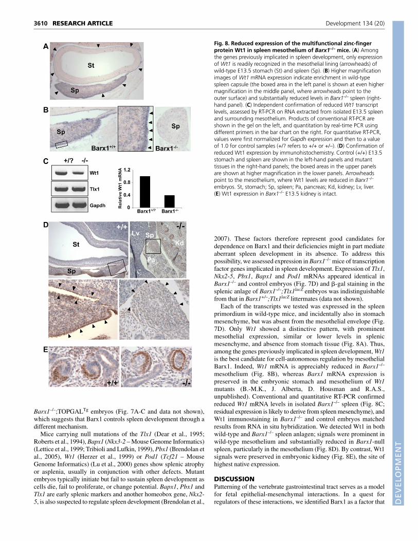
Each of the transcripts we tested was expressed in the spleenprimordium in wild-type mice, and incidentally also in stomachmesenchyme, but was absent from the mesothelial envelope (Fig.7D). Only Wt1 showed a distinctive pattern, with prominentmesothelial expression, similar or lower levels in splenicmesenchyme, and absence from stomach tissue (Fig. 8A). Thus,among the genes previously implicated in spleen development, Wt1is the best candidate for cell-autonomous regulation by mesothelialBarx1. Indeed, Wt1 mRNA is appreciably reduced in Barx1–/–
mesothelium (Fig. 8B), whereas Barx1 mRNA expression ispreserved in the embryonic stomach and mesothelium of Wt1mutants (B.-M.K., J. Alberta, D. Housman and R.A.S.,unpublished). Conventional and quantitative RT-PCR confirmedreduced Wt1 mRNA levels in isolated Barx1–/– spleen (Fig. 8C;residual expression is likely to derive from spleen mesenchyme), andWt1 immunostaining in Barx1–/– and control embryos matchedresults from RNA in situ hybridization. We detected Wt1 in bothwild-type and Barx1–/– spleen anlagen; signals were prominent inwild-type mesothelium and substantially reduced in Barx1-nullspleen, particularly in the mesothelium (Fig. 8D). By contrast, Wt1signals were preserved in embryonic kidney (Fig. 8E), the site ofhighest native expression.
DISCUSSIONPatterning of the vertebrate gastrointestinal tract serves as a modelfor fetal epithelial-mesenchymal interactions. In a quest forregulators of these interactions, we identified Barx1 as a factor that
RESEARCH ARTICLE Development 134 (20)
Fig. 8. Reduced expression of the multifunctional zinc-fingerprotein Wt1 in spleen mesothelium of Barx1–/– mice. (A) Amongthe genes previously implicated in spleen development, only expressionof Wt1 is readily recognized in the mesothelial lining (arrowheads) ofwild-type E13.5 stomach (St) and spleen (Sp). (B) Higher magnificationimages of Wt1 mRNA expression indicate enrichment in wild-typespleen capsule (the boxed area in the left panel is shown at even highermagnification in the middle panel, where arrowheads point to theouter surface) and substantially reduced levels in Barx1–/– spleen (right-hand panel). (C) Independent confirmation of reduced Wt1 transcriptlevels, assessed by RT-PCR on RNA extracted from isolated E13.5 spleenand surrounding mesothelium. Products of conventional RT-PCR areshown in the gel on the left, and quantitation by real-time PCR usingdifferent primers in the bar chart on the right. For quantitative RT-PCR,values were first normalized for Gapdh expression and then to a valueof 1.0 for control samples (+/? refers to +/+ or +/–). (D) Confirmation ofreduced Wt1 expression by immunohistochemistry. Control (+/+) E13.5stomach and spleen are shown in the left-hand panels and mutanttissues in the right-hand panels; the boxed areas in the upper panelsare shown at higher magnification in the lower panels. Arrowheadspoint to the mesothelium, where Wt1 levels are reduced in Barx1–/–
embryos. St, stomach; Sp, spleen; Pa, pancreas; Kd, kidney; Lv, liver.(E) Wt1 expression in Barx1–/– E13.5 kidney is intact.
DEVELO
PMENT
is expressed abundantly, transiently and selectively in themesenchyme and mesothelium of the developing stomach. We havenow characterized its functions in development of the stomach andspleen and its powerful role as a homeotic regulator of abdominalorganogenesis. Our results indicate that Barx1 influencesdevelopment of two adjacent organs by different mechanisms: non-cell-autonomous inhibition of canonical Wnt signaling in stomachendoderm and cell-autonomous disruption of Wt1 gene expressionin splenic mesothelium.
Mice lacking Barx1 present a severe, invariant and completelypenetrant form of visceral homeosis, with posteriorization of theproximal foregut. The esophagus is considerably shortened or, in theabsence of markers that can distinguish mouse esophagus fromsquamous forestomach, might be missing entirely. Instead of theusual domed morphology, the fundic stomach is tubular, andcuboidal cells expressing neutral mucins and Muc5ac, which areusually confined to the glandular stomach, interrupt its squamouslining. Zones similarly blur in the body of the stomach, where cellsnormally restricted to the antrum/pylorus mix freely with corpusgland cells. By contrast, the next epithelial boundary is strictlypreserved; intestinal villi occupy the entire distal stomach, butstomach and intestinal cells do not overlap in morphology orexpression of regional markers. Homeosis in the Barx1–/– gut thusharbors unique features, with blurring of rostral organ and epithelialboundaries and anterior shifting of intestinal mucosa.
These gastrointestinal abnormalities extend away from the Barx1expression domain both rostrally (esophagus) and caudally (pyloricsphincter), a phenomenon that is reminiscent of homeotictransformations in the limbs and axial skeleton (Capecchi, 1996;Izpisua-Belmonte and Duboule, 1992). However, the majoranomalies occur precisely in the domain of fetal Barx1 expression,in the gastric fundus and body, and suggest a dual role for Barx1 instomach mesenchyme. One group of functions, likely to be intrinsicto the mesenchyme, drives sub-epithelial differentiation andgenerates the correct organ size and shape. Mesenchymal mass isreduced in Barx1–/– mice but its viability seems intact and smoothmuscle appears in the right location; we have not addressed themechanisms behind the role of Barx1 in stomach morphogenesis. Asecond group of non-cell-autonomous functions helps specify theoverlying endoderm, as we previously inferred in part from findingsin recombinant embryonic cell cultures (Kim et al., 2005). Ourcharacterization of Barx1–/– stomach reinforces this function,extends our understanding and establishes the role of Barx1 insuppressing endodermal Wnt activity. We demonstrate that itsabsence permits persistent Wnt signaling in stomach endoderm,which is likely to disrupt mucosal specification and differentiationas a direct consequence. However, the scope of stomach and spleendefects in Barx1–/– embryos, coupled with the lack of canonical Wntsignaling in normal spleen primordium, implies that Wnt inhibitionrepresents only a facet of Barx1 mechanisms, albeit one that is vitalin stomach differentiation. Furthermore, we cannot rule out thepossibility that Barx1 regulation of spleen morphogenesis alsoinvolves Wnt signaling through non-canonical pathways.
Unexpectedly, Barx1–/– mice have a misplaced and severelyhypoplastic spleen of a form not observed in other animal models.Some reptiles (Falkmer, 1985) and mice lacking the pancreas-determining factor Ptf1a (Krapp et al., 1998) show isolatedendocrine pancreatic progenitors scattered within the spleen. Bycontrast, Barx1–/– mice reveal a novel phenotype in which a discretespleen is embedded within intact pancreatic parenchyma. Molecularunderstanding of spleen development is incomplete, but the organ isknown to originate as a mesenchymal condensation within dorsal
mesogastrium, in close apposition to the dorsal pancreas (Brendolanet al., 2007; Hecksher-Sorensen et al., 2004; Thiel and Downey,1921). Mice with defects in late pancreas development, in which themesenchyme is unaffected, usually have an intact spleen, whereasloss of pancreas mesenchyme, as observed, for example, intransgenic mice with ectopic Shh expression, is strongly correlatedwith asplenia (Ahlgren et al., 1996; Apelqvist et al., 1997; Harrisonet al., 1999); these observations signify a role for dorsal pancreaticmesenchyme in some aspects of spleen development. However,Barx1 mRNA and protein are conspicuously absent from spleen andpancreas anlagen, but appear at high levels in the epitheliod liningof these organ primordia. Barx1 is thus unique among regulators ofspleen development in exerting a pivotal influence exclusively fromthe mesothelium and its expression pattern suggests that it moderatesspleen development indirectly, much as mesenchymal Barx1 helpsspecify adjacent stomach endoderm. The splanchnic mesodermalplate is a known source of developmental signals, includingfibroblast growth factors 9 and 10 (Hecksher-Sorensen et al., 2004).A key role for the prospective capsule in spleen development isindependently revealed in dominant hemimelia (Dh) mutant mice,which lack this layer and are asplenic (Green, 1967; Hecksher-Sorensen et al., 2004); our findings suggest that some Dh effectsmight be mediated through Barx1.
All transcription factor genes expressed only in spleenprimordium and previously implicated in its maturation areexpressed normally in Barx1–/– spleen. These findings areconsistent with the preservation of hematopoietic potential andindicate that Barx1 is dispensable for their expression. Featuresof the mutant phenotype point instead to functions not previouslyexplored in spleen development. First, normal mesothelium seemsto exert a Barx1-dependent trophic effect that enlarges the organand imparts its characteristic shape. Alternatively, the mutantmesothelium might limit expansive and morphogenetic capacitiesinherent to the spleen anlage, and we cannot exclude thepossibility that the spleen defects in Barx1–/– mice follow mainlyfrom stomach malrotation and attendant disturbance inconfiguration of the omental bursa. A second function, separationof the spleen from the dorsal pancreas, is arguably betterattributed to cell-autonomous properties of the mesothelium, andit is here that Wt1 loss might be especially pertinent. Unlike othergenes implicated in the specification, survival or expansion of thespleen primordium, Wt1 alone is expressed in the mesothelium (inaddition to spleen mesenchyme); this overlap with the Barx1expression domain adds plausibility and significance to the resultthat mesothelial Wt1 expression depends on Barx1. In bothBarx1–/– and Wt1–/– embryos, the spleen is initially specified inthe correct location and ultimately much reduced in size but notabsent, and Tlx1 expression is not perturbed. Wt1–/– spleenprimordium is also reported to have a shorter connection to theprospective pancreas (Herzer et al., 1999), although perhaps notas short as we observe in Barx1–/– mice. Taken together, theseobservations raise the possibility that Barx1 control over spleendevelopment might be exercised in part through Wt1 generegulation in the dorsal mesothelium. It is interesting that Wt1mRNA is reduced in Tlx1–/– splenic mesenchyme, but not inTlx1–/– or Pbx1–/– mesothelium (Brendolan et al., 2005; Koehleret al., 2000).
Mice with targeted disruption of another homeobox gene, Bapx1,reveal markedly different consequences of failure of the spleen anddorsal pancreas to separate (Asayesh et al., 2006). Bapx1–/–
pancreatic endoderm undergoes metaplastic conversion to intestinalcyst-like structures, a defect attributed to persistent contact with
3611RESEARCH ARTICLEBarx1 control of stomach and spleen development
DEVELO
PMENT
3612
spleen mesenchyme past E13.5, the stage by which the two organshave normally separated. The authors argued that other mousemodels of asplenia avoid the same outcome because they do notexpose the pancreatic epithelial primordium directly to spleenmesenchyme (Asayesh et al., 2006). As such contact is evident inBarx1–/– embryos, we suggest that either the metaplastic defect inBapx1–/– pancreas is unique to that genotype, or the Barx1–/– spleenlacks the putative required factors.
Abdominal Barx1 expression is restricted to the stomach walland mesothelium and we identify significant and distinctdevelopmental functions in each of these locations. Our results alsomake a persuasive argument for Barx1-mediated inhibition of Wntsignaling in stomach endoderm and against a role for canonical Wntsignaling in spleen development. They hence demonstrate thatpositional and morphogenetic functions conferred by thishomeobox gene occur through distinct mechanisms, even over theshort distance that separates the stomach wall from its mesothelium.The pathways we have elucidated thus far – inhibition of canonicalWnt signaling in endoderm and regulation of Wt1 gene expressionin mesothelial cells – represent early steps in appreciating the basisfor homeobox gene functions in the gastrointestinal tract. Barx1 islikely to regulate additional events that contribute not only toforegut patterning and spleen expansion, but also to control ofstomach size and shape and pyloric sphincter formation.Characterization of other such pathways will add to the growingunderstanding of abdominal organogenesis.
This work was supported in part by grant number R01DK61139 (R.A.S.) andR01NS033642 (A.P.M.) from the National Institutes of Health. J.M. is a fellowof the Charles H. Hood Foundation. We are grateful to Susumu Ito forassistance with electron microscopy; Zhao Chen and SunTaek Kim for helpwith flow cytometry; Walter Birchmeier for providing Axin2lacZ reporter mice;Cliff Tabin and Mark Taketo for sharing ShhCre and Catnblox(ex3) mice,respectively; Terry Rabbitts and Licia Selleri for providing Tlx1lacZ mice; JuliaAlberta and David Housman for sharing Wt1-mutant mouse embryos;Christopher Wright and David Alper for generous gifts of Pdx1 and intrinsicfactor antisera, respectively; Richard Harvey, Thomas Lufkin, Robert Schwartzand Licia Selleri for plasmids; and Andrea Brendolan, Licia Selleri and MikeVerzi for critical review of the manuscript.
Supplementary materialSupplementary material for this article is available athttp://dev.biologists.org/cgi/content/full/134/20/3603/DC1
ReferencesAhlgren, U., Jonsson, J. and Edlund, H. (1996). The morphogenesis of the
pancreatic mesenchyme is uncoupled from that of the pancreatic epithelium inIPF1/PDX1-deficient mice. Development 122, 1409-1416.
Apelqvist, A., Ahlgren, U. and Edlund, H. (1997). Sonic hedgehog directsspecialized mesoderm differentiation in the intestine and pancreas. Curr. Biol. 7,801-804.
Asayesh, A., Sharpe, J., Watson, R. P., Hecksher-Sorensen, J., Hastie, N. D.,Hill, R. E. and Ahlgren, U. (2006). Spleen versus pancreas: strict control oforgan interrelationship revealed by analyses of Bapx1–/– mice. Genes Dev. 20,2208-2213.
Brendolan, A., Ferretti, E., Salsi, V., Moses, K., Quaggin, S., Blasi, F., Cleary,M. L. and Selleri, L. (2005). A Pbx1-dependent genetic and transcriptionalnetwork regulates spleen ontogeny. Development 132, 3113-3126.
Brendolan, A., Rosado, M. M., Carsetti, R., Selleri, L. and Dear, T. N. (2007).Development and function of the mammalian spleen. BioEssays 29, 166-177.
Capecchi, M. R. (1996). Function of homeobox genes in skeletal development.Ann. N. Y. Acad. Sci. 785, 34-37.
Chinery, R., Poulsom, R., Rogers, L. A., Jeffery, R. E., Longcroft, J. M.,Hanby, A. M. and Wright, N. A. (1992). Localization of intestinal trefoil-factormRNA in rat stomach and intestine by hybridization in situ. Biochem. J. 285, 5-8.
Clevers, H. (2006). Wnt/beta-catenin signaling in development and disease. Cell127, 469-480.
DasGupta, R. and Fuchs, E. (1999). Multiple roles for activated LEF/TCFtranscription complexes during hair follicle development and differentiation.Development 126, 4557-4568.
Dear, T. N., Colledge, W. H., Carlton, M. B., Lavenir, I., Larson, T., Smith, A. J.,Warren, A. J., Evans, M. J., Sofroniew, M. V. and Rabbitts, T. H. (1995). TheHox11 gene is essential for cell survival during spleen development.Development 121, 2909-2915.
Echelard, Y., Epstein, D. J., St-Jacques, B., Shen, L., Mohler, J., McMahon, J.A. and McMahon, A. P. (1993). Sonic hedgehog, a member of a family ofputative signaling molecules, is implicated in the regulation of CNS polarity. Cell75, 1417-1430.
Falkmer, S. (1985). Comparative morphology of pancreatic islets in animals. In TheDiabetic Pancreas (ed. B. W. Volk and K. F. Wellmann), pp. 17-52. New York:Plenum Press.
Finch, P. W., He, X., Kelley, M. J., Uren, A., Schaudies, R. P., Popescu, N. C.,Rudikoff, S., Aaronson, S. A., Varmus, H. E. and Rubin, J. S. (1997).Purification and molecular cloning of a secreted, Frizzled-related antagonist ofWnt action. Proc. Natl. Acad. Sci. USA 94, 6770-6775.
Green, M. C. (1967). A defect of the splanchnic mesoderm caused by the mutantgene dominant hemimelia in the mouse. Dev. Biol. 15, 62-89.
Harada, N., Tamai, Y., Ishikawa, T., Sauer, B., Takaku, K., Oshima, M. andTaketo, M. M. (1999). Intestinal polyposis in mice with a dominant stablemutation of the beta-catenin gene. EMBO J. 18, 5931-5942.
Harfe, B. D., Scherz, P. J., Nissim, S., Tian, H., McMahon, A. P. and Tabin, C. J.(2004). Evidence for an expansion-based temporal Shh gradient in specifyingvertebrate digit identities. Cell 118, 517-528.
Harrison, K. A., Thaler, J., Pfaff, S. L., Gu, H. and Kehrl, J. H. (1999). Pancreasdorsal lobe agenesis and abnormal islets of Langerhans in Hlxb9-deficient mice.Nat. Genet. 23, 71-75.
Hecksher-Sorensen, J., Watson, R. P., Lettice, L. A., Serup, P., Eley, L., DeAngelis, C., Ahlgren, U. and Hill, R. E. (2004). The splanchnic mesodermalplate directs spleen and pancreatic laterality, and is regulated by Bapx1/Nkx3.2.Development 131, 4665-4675.
Herzer, U., Crocoll, A., Barton, D., Howells, N. and Englert, C. (1999). TheWilms tumor suppressor gene wt1 is required for development of the spleen.Curr. Biol. 9, 837-840.
Izpisua-Belmonte, J. C. and Duboule, D. (1992). Homeobox genes and patternformation in the vertebrate limb. Dev. Biol. 152, 26-36.
Jho, E. H., Zhang, T., Domon, C., Joo, C. K., Freund, J. N. and Costantini, F.(2002). Wnt/beta-catenin/Tcf signaling induces the transcription of Axin2, anegative regulator of the signaling pathway. Mol. Cell. Biol. 22, 1172-1183.
Kanzler, B. and Dear, T. N. (2001). Hox11 acts cell autonomously in spleendevelopment and its absence results in altered cell fate of mesenchymal spleenprecursors. Dev. Biol. 234, 231-243.
Karam, S. M. and Leblond, C. P. (1992). Identifying and counting epithelial celltypes in the “corpus” of the mouse stomach. Anat. Rec. 232, 231-246.
Kim, B. M., Buchner, G., Miletich, I., Sharpe, P. T. and Shivdasani, R. A.(2005). The stomach mesenchymal transcription factor Barx1 specifies gastricepithelial identity through inhibition of transient Wnt signaling. Dev. Cell 8, 611-622.
Koehler, K., Franz, T. and Dear, T. N. (2000). Hox11 is required to maintainnormal Wt1 mRNA levels in the developing spleen. Dev. Dyn. 218, 201-216.
Krapp, A., Knofler, M., Ledermann, B., Burki, K., Berney, C., Zoerkler, N.,Hagenbuchle, O. and Wellauer, P. K. (1998). The bHLH protein PTF1-p48 isessential for the formation of the exocrine and the correct spatial organizationof the endocrine pancreas. Genes Dev. 12, 3752-3763.
Lettice, L. A., Purdie, L. A., Carlson, G. J., Kilanowski, F., Dorin, J. and Hill, R.E. (1999). The mouse bagpipe gene controls development of axial skeleton,skull, and spleen. Proc. Natl. Acad. Sci. USA 96, 9695-9700.
Lu, J., Chang, P., Richardson, J. A., Gan, L., Weiler, H. and Olson, E. N. (2000).The basic helix-loop-helix transcription factor capsulin controls spleenorganogenesis. Proc. Natl. Acad. Sci. USA 97, 9525-9530.
Offield, M. F., Jetton, T. L., Labosky, P. A., Ray, M., Stein, R. W., Magnuson,M. A., Hogan, B. L. and Wright, C. V. (1996). PDX-1 is required for pancreaticoutgrowth and differentiation of the rostral duodenum. Development 122, 983-995.
Que, J., Okubo, T., Goldenring, J. R., Nam, K. T., Kurotani, R., Morrisey, E. E.,Taranova, O., Pevny, L. H. and Hogan, B. L. (2007). Multiple dose-dependentroles for Sox2 in the patterning and differentiation of anterior foregutendoderm. Development 134, 2521-2531.
Rattner, A., Hsieh, J. C., Smallwood, P. M., Gilbert, D. J., Copeland, N. G.,Jenkins, N. A. and Nathans, J. (1997). A family of secreted proteins containshomology to the cysteine-rich ligand-binding domain of frizzled receptors. Proc.Natl. Acad. Sci. USA 94, 2859-2863.
Roberts, C. W., Shutter, J. R. and Korsmeyer, S. J. (1994). Hox11 controls thegenesis of the spleen. Nature 368, 747-749.
Silberg, D. G., Swain, G. P., Suh, E. R. and Traber, P. G. (2000). Cdx1 and cdx2expression during intestinal development. Gastroenterology 119, 961-971.
Soriano, P. (1999). Generalized lacZ expression with the ROSA26 Cre reporterstrain. Nat. Genet. 21, 70-71.
Thiel, G. A. and Downey, H. (1921). The development of the mammalianspleen with special reference to its hematopoietic activity. Am. J. Anat. 28,279-339.
RESEARCH ARTICLE Development 134 (20)

DEVELO
PMENT
Tissier-Seta, J. P., Mucchielli, M. L., Mark, M., Mattei, M. G., Goridis, C. andBrunet, J. F. (1995). Barx1, a new mouse homeodomain transcription factorexpressed in cranio-facial ectomesenchyme and the stomach. Mech. Dev. 51, 3-15.
Tribioli, C. and Lufkin, T. (1999). The murine Bapx1 homeobox gene plays a
critical role in embryonic development of the axial skeleton and spleen.Development 126, 5699-5711.
Yu, H. M., Jerchow, B., Sheu, T. J., Liu, B., Costantini, F., Puzas, J. E.,Birchmeier, W. and Hsu, W. (2005). The role of Axin2 in calvarialmorphogenesis and craniosynostosis. Development 132, 1995-2005.
3613RESEARCH ARTICLEBarx1 control of stomach and spleen development