Embed Size (px)
Citation preview

Distribution and Origin of Oxygen-Dependent and Oxygen-Independent Forms of Mg-Protoporphyrin Monomethylester Cyclaseamong Phototrophic Proteobacteria
Ekaterina N. Boldareva-Nuianzina,a Zuzana Bláhová,a,b Roman Sobotka,a,b Michal Koblížeka,b
Institute of Microbiology CAS, Department of Phototrophic Microorganisms, Algatech, Opatovický mlýn, Trebon, Czech Republica; Faculty of Science, University of SouthBohemia, Ceské Budejovice, Czech Republicb
Magnesium-protoporphyrin IX monomethylester cyclase is one of the key enzymes of the bacteriochlorophyll biosynthesis path-way. There exist two fundamentally different forms of this enzyme. The oxygen-dependent form, encoded by the gene acsF, cata-lyzes the formation of the bacteriochlorophyll fifth ring using oxygen, whereas the oxygen-independent form encoded by thegene bchE utilizes an oxygen atom extracted from water. The presence of acsF and bchE genes was surveyed in various pho-totrophic Proteobacteria using the available genomic data and newly designed degenerated primers. It was found that while themajority of purple nonsulfur bacteria contained both forms of the cyclase, the purple sulfur bacteria contained only the oxygen-independent form. All tested species of aerobic anoxygenic phototrophs contained acsF genes, but some of them also retainedthe bchE gene. In contrast to bchE phylogeny, the acsF phylogeny was in good agreement with 16S inferred phylogeny. Moreover,the survey of the genome data documented that the acsF gene occupies a conserved position inside the photosynthesis gene clus-ter, whereas the bchE location in the genome varied largely between the species. This suggests that the oxygen-dependent cyclasewas recruited by purple phototrophic bacteria very early during their evolution. The primary sequence and immunochemicalsimilarity with its cyanobacterial counterparts suggests that acsF may have been acquired by Proteobacteria via horizontal genetransfer from cyanobacteria. The acquisition of the gene allowed purple nonsulfur phototrophic bacteria to proliferate in themildly oxygenated conditions of the Proterozoic era.
The anoxygenic phototrophic bacteria are among the oldest or-ganisms on Earth. They evolved more than 3 billion years (3
Gyr) ago under the anoxic conditions of the Archean Ocean (1, 2).Hence, many species of anoxygenic phototrophs remained strictlyanaerobic organisms inhabiting anoxic environments. More evo-lutionary advanced oxygenic cyanobacteria evolved somewhatlater (approximately 2.7 Gyr ago), and their rapid spreading re-sulted in a gradual oxygenation of the Proterozoic Ocean (3, 4).The changing conditions forced many groups of anoxygenic pho-totrophs (such as purple sulfur bacteria [PSB]) to retreat to mar-ginal anaerobic niches, whereas some adapted to new oxic condi-tions and became facultative aerobes (i.e., purple nonsulfurbacteria [PNB]). Later, some phototrophic groups embarked on acompletely aerobic lifestyle; they lost the ability to fix inorganiccarbon and use light energy only as a supplement of their predom-inantly heterotrophic metabolism (5, 6). These organisms, calledaerobic anoxygenic phototrophs (AAPs), were found in marineenvironments (7–10), but recently their presence was also dem-onstrated in river estuaries (11) as well as freshwater and salinelakes (12, 13).
The main light-harvesting pigments in the anoxygenic pho-totrophs are porphyrin molecules called bacteriochlorophylls,whereas oxygenic organisms, cyanobacteria, algae, and plants uti-lize structurally very similar pigments called chlorophylls (14, 15).A universal structural feature of all these molecules is an isocyclicring, the formation of which is an essential step of (bacterio)chlo-rophyll biosynthesis (16). This complex reaction is catalyzed byMg-protoporphyrin IX monomethylester (oxidative) cyclase andresults in the conversion of Mg-protoporphyrin IX monomethylester into divinyl protochlorophyllide (see Fig. 1). The anaerobicphototrophs possess an oxygen-independent form of this enzyme
containing 4Fe-4S and cobalamin prosthetic groups. The enzymecatalyzes a complex multistep reaction in which the oxygen atomnecessary for the reaction is extracted from water (14, 17). Anessential component or perhaps the whole oxygen-independentcyclase enzyme is encoded by the bchE gene. A completely differ-ent form of this enzyme exists in cyanobacteria, algae, and plantsas well as in many PNB and AAP species. Unlike the oxygen-independent form, this enzyme catalyzes the formation of the iso-cyclic ring using oxygen as a substrate (18). Oxygen-dependentcyclase is expected to be active as a multisubunit complex (19);however, the only known subunit was identified by mutationalanalysis of the photosynthetic bacterium Rubrivivax gelatinosus(20) and denoted as AcsF (abbreviated from aerobic cyclase). Asubsequent genetic study revealed that Rubrivivax contained bothforms of the cyclase, which allows the cells to synthesize bacterio-chlorophyll under different oxygen concentrations (21). AcsF ho-mologs have since been identified in many cyanobacteria andphotosynthetic eukaryotes, including Synechocystis sp. strain PCC6803 (22), Chlamydomonas reinhardtii (23), and Arabidopsis thali-ana (24). More recently, the acsF gene was also identified in thegreen nonsulfur bacterium Chloroflexus aurantiacus (25).
Received 14 January 2013 Accepted 3 February 2013
Published ahead of print 8 February 2013
Address correspondence to Michal Koblížek, [email protected].
Supplemental material for this article may be found at http://dx.doi.org/10.1128/AEM.00104-13.
Copyright © 2013, American Society for Microbiology. All Rights Reserved.
doi:10.1128/AEM.00104-13
2596 aem.asm.org Applied and Environmental Microbiology p. 2596–2604 April 2013 Volume 79 Number 8
on April 11, 2019 by guest
http://aem.asm
.org/D
ownloaded from
The acquisition of the oxygen-dependent form of the cyclasewas proposed to be one of the key adaptations done by anoxygenicphototrophs during the evolutionary transition from anaerobic toaerobic conditions (26). In their recent review, Yurkov and Csot-onyi (5) speculated that the presence of the oxygen-dependentcyclase and the absence of the oxygen-independent cyclase couldserve as a convenient marker of predominantly aerobic pho-totrophic species. To test these hypotheses, we surveyed the pres-ence and organization of acsF and bchE genes among various an-oxygenic phototrophic Proteobacteria. The obtained molecularinformation was used to infer the evolutionary origin of the twoforms of the cyclase and their current role in anoxygenic pho-totrophic Proteobacteria.
MATERIALS AND METHODSStrains. Anoxygenic phototrophs belonging to alpha, beta, and gammaclasses of Proteobacteria were used for the study. Rhodobaca bogoriensis(strain LBB1), Rhodobaca barguzinensis (strain alga-05), Roseinatronobac-ter strains Dor-vul, Dor-3.5, Da, and Khil-ros, and Rhodobacterales strainsZun-kh and Chep-kr have been isolated from soda lakes as describedearlier (27, 28). Marine isolates SO3, SYOP2, BS110, COL2P, B09, andB11, belonging to the Roseobacter clade, Roseovarius sp. strain SL25, Eryth-robacter sp. strain NAP1, and Citromicrobium sp. strain CV44, were de-scribed previously (29–32). Freshwater betaproteobacterium strain VA01was isolated from Velka Amerika lake, Czech Republic, by the dilution-to-extinction technique (M. Mašín, unpublished data). Rhodobacter spha-eroides and Rhodobacter capsulatus were cultured photoheterotrophicallyin organic medium as described previously (32). Cultures of Roseibacaekhonensis (strain EL50), Roseisalinus antarcticus (strain EL88), and Sta-leya guttiformis (strain EL 38) were kindly provided by Matthias Labrenz.
Sandarakinorhabdus limnophila (strain so42) was provided by FredericGich. Congregibacter litoralis strain KT71 (DSM 17192), Roseateles depoly-merans (DSM 11813), Roseobacter litoralis (DSM 6996), and Rhodovulumsulfidophilum (DSM 1374) were obtained from the Deutsche Sammlungvon Mikroorganismen und Zellkulturen (DSMZ), Braunschweig, Ger-many. Synechocystis sp. strain PCC6803 was grown photoautotrophicallyin the BG11 mineral medium. The microbial cultures were grown at roomtemperature under a light/dark cycle with an irradiance of 150 �mol ofphotons m�2 s�1 in rotating Erlenmeyer flasks. If not stated otherwise, weused media recommended by the Deutsche Sammlung von Mikroorgan-ismen und Zellkulturen (DMSZ).
PCR amplification. Bacterial DNA was extracted using the RTP bac-teria DNA minikit (Invitek GmbH, Germany). Partial acsF gene se-quences were PCR amplified using forward primer acsF-F and reverseprimer acsF-R1, providing a 350-bp gene fragment (see Table 1). Theprogram consisted of 35 cycles with 1 min of denaturation at 94°C, 45 s ofannealing at 45°C, and 1 min of extension at 72°C. The final extension wasconducted for 10 min at 72°C. The bchE gene was amplified with theforward primer bchE-f (see Results) and reverse primer bchE-r2. The PCRprogram was 30 cycles with 1 min of denaturation at 94°C, 45 s of anneal-ing at 52°C, and 1 min of extension at 72°C. The size of the obtainedproduct was 640 bp. In some cases, bchE gene presence was amplifiedusing a bchE-f and bchE-r4 primer pair (Roseococcus sp. strain Da). Theamplification conditions remained the same, while annealing was con-ducted at 48°C. The size of the amplified fragments was 405 bp. Theobtained PCR products were purified using a GenElute PCR clean-up kit(Sigma-Aldrich Co. LLC) and directly sequenced using the appropriateforward primers. The sequencing results were analyzed and manually cor-rected using BioEdit (Sequence Alignment Editor) version 5.0.9, Chromasversion 1.5.
GenBank accession numbers. GenBank accession numbers of partialacsF sequences are as follows: Roseobacter sp. strain Zun-kholvo,JF917109; betaproteobacterium strain VA01, JF917110; Roseobacter sp.strain SY0P2, JF917111; Roseobacter sp. strain SO3, JF917112; Roseovariussp. strain SL25, JF917113; Sandarakinorhabdus limnophila so42,JF917114; Rhizobiales sp. strain RM11-8-1, JF917115; Roseinatronobactersp. strain Khil, JF917116; Staleya guttiformis EL38, JF917117; Rosein-atronobacter sp. strain Dor-vul, JF917118; Roseinatronobacter sp. strainDor 3.5, JF917119; Citromicrobium sp. strain CV44, JF917120; Roseobac-ter sp. strain COL2P, JF917121; Rhodobacterales strain Chep-kr,JF917122; Roseobacter sp. strain B11, JF917123; Roseobacter sp. strain B09,JF917124; Roseococcus sp. strain Da, JF917126; and Rhodobaca barguzi-nensis alga05, JF917125.
Partial bchE sequences are as follows: Roseobacter sp. strain B09,JF917127; Roseobacter sp. strain B11, JF917128; Citromicrobium sp. strainCV44, JF917129; Roseococcus sp. strain Da, JF917126; Rhodobacteralesstrain Chep-kr, JF917131; Roseinatronobacter sp. strain Dor 3.5, JF917132;Rhizobiales strain RM11-8-1, JN018414; Roseinatronobacter sp. strainKhil, JN018416; Roseinatronobacter sp. strain Dor-vul, JN018415; Roseo-bacter sp. strain Zun_kholvo, JN018417; Roseinatronobacter monicusROS35, JN018418; and Rhodobaca bogoriensis LBB1, JN018419.
Phylogenetic analyses. The obtained nucleotide sequences weretranslated into amino acids and aligned using the ClustalX2 programversion 2.0.10. The alignments were manually checked; ambiguously
FIG 1 A simplified scheme of the conversion of Mg-protoporphyrin IXmonomethylester into divinyl protochlorophyllide. The reaction is catalyzedby two fundamentally different forms of Mg-protoporphyrin monomethyl-ester cyclase. The oxygen-dependent form of the enzyme encoded by the acsFgene operates under aerobic (oxic) conditions, whereas the oxygen-indepen-dent form encoded by the bchE gene operates under anaerobic (anoxic) con-ditions.
TABLE 1 Primers designed for PCR amplification of bchE and acsF genes
Primer DirectionTarget positions in thecorresponding gene Primer sequence
acsF-F Forward 382–404 5=-ARTTYTCNGGCTGYGTNCTBTA-3=acsF-R1 Reverse 731–754 5=-TGSSDRAAYTCRTCRTTGCACCA-3=bchE-f Forward 640–662 5=-AATGGAAATTCTGGCGCGACTA-3=bchE-r2 Reverse 1320–1340 5=-GGRTARTGRAANAGCGCCTT-3=bchE-r4 Reverse 1045–1063 5=-ACGATGAACTGNGCYTCG-3=
Two Forms of Mg-Protoporphyrin Monomethylester Cyclase
April 2013 Volume 79 Number 8 aem.asm.org 2597
on April 11, 2019 by guest
http://aem.asm
.org/D
ownloaded from
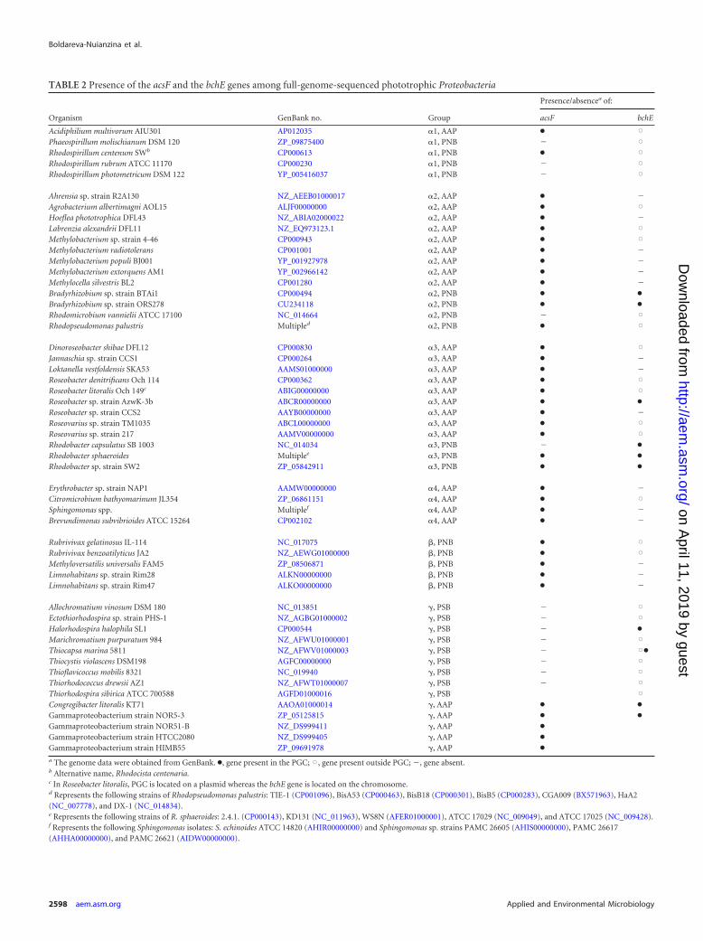
TABLE 2 Presence of the acsF and the bchE genes among full-genome-sequenced phototrophic Proteobacteria
Organism GenBank no. Group
Presence/absencea of:
acsF bchE
Acidiphilium multivorum AIU301 AP012035 �1, AAP ● XPhaeospirillum molischianum DSM 120 ZP_09875400 �1, PNB � XRhodospirillum centenum SWb CP000613 �1, PNB ● XRhodospirillum rubrum ATCC 11170 CP000230 �1, PNB � XRhodospirillum photometricum DSM 122 YP_005416037 �1, PNB � X
Ahrensia sp. strain R2A130 NZ_AEEB01000017 �2, AAP ● �Agrobacterium albertimagni AOL15 ALJF00000000 �2, AAP ● XHoeflea phototrophica DFL43 NZ_ABIA02000022 �2, AAP ● �Labrenzia alexandrii DFL11 NZ_EQ973123.1 �2, AAP ● XMethylobacterium sp. strain 4-46 CP000943 �2, AAP ● XMethylobacterium radiotolerans CP001001 �2, AAP ● �Methylobacterium populi BJ001 YP_001927978 �2, AAP ● �Methylobacterium extorquens AM1 YP_002966142 �2, AAP ● �Methylocella silvestris BL2 CP001280 �2, AAP ● �Bradyrhizobium sp. strain BTAi1 CP000494 �2, PNB ● ●
Bradyrhizobium sp. strain ORS278 CU234118 �2, PNB ● ●
Rhodomicrobium vannielii ATCC 17100 NC_014664 �2, PNB � XRhodopseudomonas palustris Multipled �2, PNB ● X
Dinoroseobacter shibae DFL12 CP000830 �3, AAP ● XJannaschia sp. strain CCS1 CP000264 �3, AAP ● �Loktanella vestfoldensis SKA53 AAMS01000000 �3, AAP ● �Roseobacter denitrificans Och 114 CP000362 �3, AAP ● XRoseobacter litoralis Och 149c ABIG00000000 �3, AAP ● XRoseobacter sp. strain AzwK-3b ABCR00000000 �3, AAP ● ●
Roseobacter sp. strain CCS2 AAYB00000000 �3, AAP ● �Roseovarius sp. strain TM1035 ABCL00000000 �3, AAP ● XRoseovarius sp. strain 217 AAMV00000000 �3, AAP ● XRhodobacter capsulatus SB 1003 NC_014034 �3, PNB � ●
Rhodobacter sphaeroides Multiplee �3, PNB ● ●
Rhodobacter sp. strain SW2 ZP_05842911 �3, PNB ● ●
Erythrobacter sp. strain NAP1 AAMW00000000 �4, AAP ● �Citromicrobium bathyomarinum JL354 ZP_06861151 �4, AAP ● XSphingomonas spp. Multiplef �4, AAP ● �Brevundimonas subvibrioides ATCC 15264 CP002102 �4, AAP ● �
Rubrivivax gelatinosus IL-114 NC_017075 �, PNB ● XRubrivivax benzoatilyticus JA2 NZ_AEWG01000000 �, PNB ● XMethyloversatilis universalis FAM5 ZP_08506871 �, PNB ● �Limnohabitans sp. strain Rim28 ALKN00000000 �, PNB ● �Limnohabitans sp. strain Rim47 ALKO00000000 �, PNB ● �
Allochromatium vinosum DSM 180 NC_013851 �, PSB � XEctothiorhodospira sp. strain PHS-1 NZ_AGBG01000002 �, PSB � XHalorhodospira halophila SL1 CP000544 �, PSB � ●
Marichromatium purpuratum 984 NZ_AFWU01000001 �, PSB � XThiocapsa marina 5811 NZ_AFWV01000003 �, PSB � X●
Thiocystis violascens DSM198 AGFC00000000 �, PSB � XThioflavicoccus mobilis 8321 NC_019940 �, PSB � XThiorhodococcus drewsii AZ1 NZ_AFWT01000007 �, PSB � XThiorhodospira sibirica ATCC 700588 AGFD01000016 �, PSB XCongregibacter litoralis KT71 AAOA01000014 �, AAP ● ●
Gammaproteobacterium strain NOR5-3 ZP_05125815 �, AAP ● ●
Gammaproteobacterium strain NOR51-B NZ_DS999411 �, AAP ●
Gammaproteobacterium strain HTCC2080 NZ_DS999405 �, AAP ●
Gammaproteobacterium strain HIMB55 ZP_09691978 �, AAP ●
a The genome data were obtained from GenBank. ●, gene present in the PGC; X, gene present outside PGC; �, gene absent.b Alternative name, Rhodocista centenaria.c In Roseobacter litoralis, PGC is located on a plasmid whereas the bchE gene is located on the chromosome.d Represents the following strains of Rhodopseudomonas palustris: TIE-1 (CP001096), BisA53 (CP000463), BisB18 (CP000301), BisB5 (CP000283), CGA009 (BX571963), HaA2(NC_007778), and DX-1 (NC_014834).e Represents the following strains of R. sphaeroides: 2.4.1. (CP000143), KD131 (NC_011963), WS8N (AFER01000001), ATCC 17029 (NC_009049), and ATCC 17025 (NC_009428).f Represents the following Sphingomonas isolates: S. echinoides ATCC 14820 (AHIR00000000) and Sphingomonas sp. strains PAMC 26605 (AHIS00000000), PAMC 26617(AHHA00000000), and PAMC 26621 (AIDW00000000).
Boldareva-Nuianzina et al.
2598 aem.asm.org Applied and Environmental Microbiology
on April 11, 2019 by guest
http://aem.asm
.org/D
ownloaded from
aligned regions and gaps were excluded from further analyses. The redun-dant sequences representing different strains of the same species (i.e.,Rhodopseudomonas palustris, Rhodobacter sphaeroides) were removed.The sequence identity matrix was calculated from the corrected alignmentusing the ClustalX2 program. The phylogenetic trees were computedfrom partial amino acid sequences using a neighbor-joining algorithmwith 1,000 bootstraps. In addition, the inferred phylogeny was confirmedby the maximum likelihood algorithm (PhyML software) using the LGmodel chosen by the ProtTest 2.4 algorithm.
Isolation of cell membranes and immunodetection of AcsF ho-mologs. The bacterial cells were pelleted by centrifugation and resus-pended in a breakage buffer containing 25 mM MES buffer (pH 6.5), 10mM MgCl2, 10 mM CaCl2, and 25% glycerol. The cells were disrupted ina beadbeater (BioSpec Products Inc.) using zirconia/silica beads 0.1 mmin diameter. The disruption was performed in four 1-min cycles inter-rupted by 5 min of cooling on ice to prevent potential protein degrada-tion. Cell membranes were pelleted by centrifugation at 40,000 � g at 4°Cfor 20 min, washed in the breakage buffer, and finally resuspended in 100�l of the same buffer.
The obtained membranes were solubilized by 2% dodecyl-�-malto-side, spun down to remove cell debris, and separated by 10% SDS poly-acrylamide gel electrophoresis. The proteins were then transferred ontothe Hybond-P polyvinylidene difluoride membrane (Amersham Biosci-ences, Sweden), which was incubated with polyclonal antibody raisedagainst the AcsF protein (CHL27) from Arabidopsis thaliana (Agrisera AB,Sweden) to detect the presence of AcsF homologs in the photosyntheticbacteria studied.
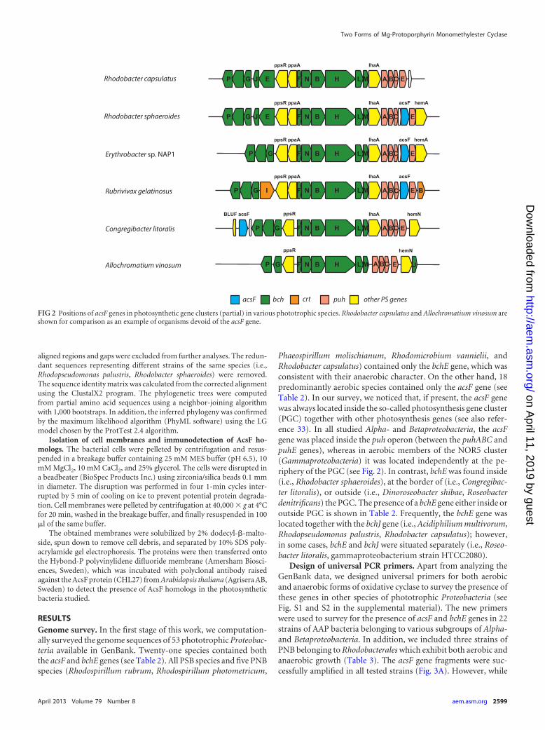
RESULTSGenome survey. In the first stage of this work, we computation-ally surveyed the genome sequences of 53 phototrophic Proteobac-teria available in GenBank. Twenty-one species contained boththe acsF and bchE genes (see Table 2). All PSB species and five PNBspecies (Rhodospirillum rubrum, Rhodospirillum photometricum,
Phaeospirillum molischianum, Rhodomicrobium vannielii, andRhodobacter capsulatus) contained only the bchE gene, which wasconsistent with their anaerobic character. On the other hand, 18predominantly aerobic species contained only the acsF gene (seeTable 2). In our survey, we noticed that, if present, the acsF genewas always located inside the so-called photosynthesis gene cluster(PGC) together with other photosynthesis genes (see also refer-ence 33). In all studied Alpha- and Betaproteobacteria, the acsFgene was placed inside the puh operon (between the puhABC andpuhE genes), whereas in aerobic members of the NOR5 cluster(Gammaproteobacteria) it was located independently at the pe-riphery of the PGC (see Fig. 2). In contrast, bchE was found inside(i.e., Rhodobacter sphaeroides), at the border of (i.e., Congregibac-ter litoralis), or outside (i.e., Dinoroseobacter shibae, Roseobacterdenitrificans) the PGC. The presence of a bchE gene either inside oroutside PGC is shown in Table 2. Frequently, the bchE gene waslocated together with the bchJ gene (i.e., Acidiphilium multivorum,Rhodopseudomonas palustris, Rhodobacter capsulatus); however,in some cases, bchE and bchJ were situated separately (i.e., Roseo-bacter litoralis, gammaproteobacterium strain HTCC2080).
Design of universal PCR primers. Apart from analyzing theGenBank data, we designed universal primers for both aerobicand anaerobic forms of oxidative cyclase to survey the presence ofthese genes in other species of phototrophic Proteobacteria (seeFig. S1 and S2 in the supplemental material). The new primerswere used to survey for the presence of acsF and bchE genes in 22strains of AAP bacteria belonging to various subgroups of Alpha-and Betaproteobacteria. In addition, we included three strains ofPNB belonging to Rhodobacterales which exhibit both aerobic andanaerobic growth (Table 3). The acsF gene fragments were suc-cessfully amplified in all tested strains (Fig. 3A). However, while
FIG 2 Positions of acsF genes in photosynthetic gene clusters (partial) in various phototrophic species. Rhodobacter capsulatus and Allochromatium vinosum areshown for comparison as an example of organisms devoid of the acsF gene.
Two Forms of Mg-Protoporphyrin Monomethylester Cyclase
April 2013 Volume 79 Number 8 aem.asm.org 2599
on April 11, 2019 by guest
http://aem.asm
.org/D
ownloaded from
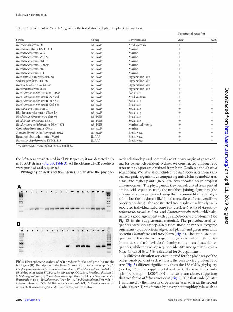
the bchE gene was detected in all PNB species, it was detected onlyin 10 AAP strains (Fig. 3B, Table 3). All the obtained PCR productswere purified and sequenced.
Phylogeny of acsF and bchE genes. To analyze the phyloge-
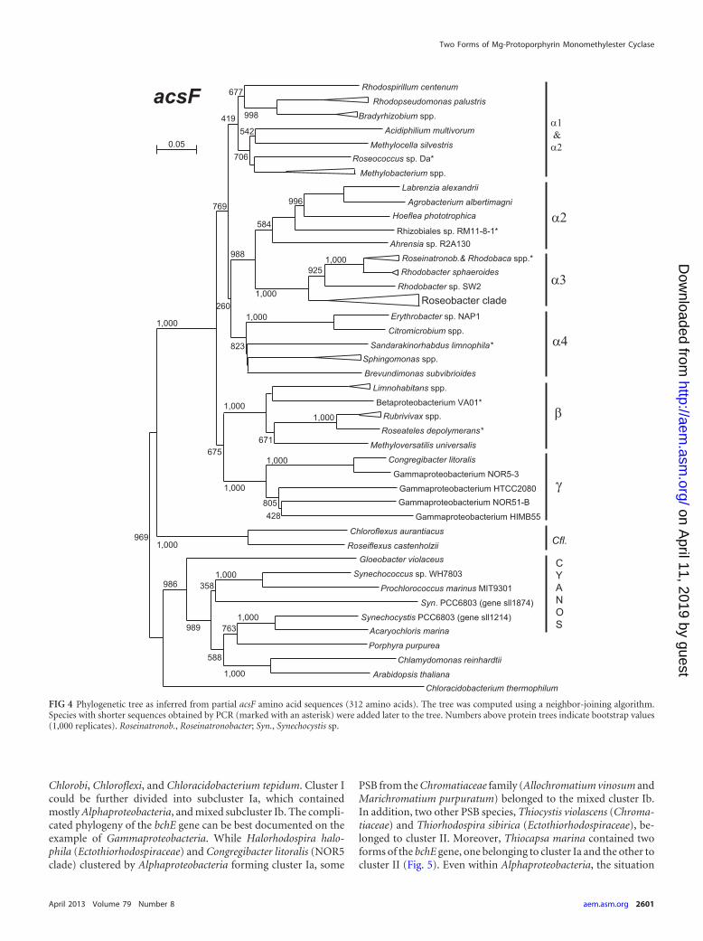
netic relationship and potential evolutionary origin of genes cod-ing for oxygen-dependent cyclase, we constructed phylogenetictrees using sequences obtained from both GenBank and de novosequencing. We have also included the acsF sequences from vari-ous oxygenic organisms encompassing unicellular cyanobacteria,algae, and higher plants (here, acsF was encoded on chloroplastchromosomes). The phylogenetic tree was calculated from partialamino acid sequences using the neighbor-joining algorithm (theanalysis was also performed using the maximum likelihood algo-rithm, but the maximum likelihood tree suffered from overall lowbootstrap values). The constructed tree displayed relatively well-separated individual subgroups (�-1, �-2, �-3, �-4) of Alphapro-teobacteria, as well as Beta- and Gammaproteobacteria, which sig-nalized a good agreement with 16S rRNA-derived phylogeny (seeFig. S3 in the supplemental material). The proteobacterial se-quences were clearly separated from those of various oxygenicorganisms (cyanobacteria, algae, and plants) and green nonsulfurbacteria Chloroflexus and Roseiflexus (Fig. 4). The amino acid se-quences of the selected oxygenic organisms had a 42% � 3%(mean � standard deviation) identity to the proteobacterial se-quences, while the average sequence identity among tested Proteo-bacteria was 61% � 7% (calculated for 34 organisms).
A different situation was encountered for the phylogeny of theoxygen-independent cyclase. Here, the constructed phylogenetictree (Fig. 5) differed significantly from the 16S rRNA phylogeny(see Fig. S3 in the supplemental material). The bchE tree clearlysplit (bootstrap � 1,000/1,000) into two main clades, suggestingthat two forms of bchE genes exist (Fig. 5). The first clade (clusterI) is formed by the majority of Proteobacteria, whereas the secondclade (cluster II) was formed by other phototrophic phyla, such as
TABLE 3 Presence of acsF and bchE genes in the tested strains of phototrophic Proteobacteria
Strain Group Environment
Presence/absencea of:
acsF bchE
Roseococcus strain Da �1, AAP Mud volcano Rhizobiales strain RM11-8-1 �2, AAP Marine Roseobacter strain SO3 �3, AAP Marine �Roseobacter strain SYOP2 �3, AAP Marine �Roseobacter strain BS110 �3, AAP Marine �Roseobacter strain COL2P �3, AAP Marine �Roseobacter strain B09 �3, AAP Marine Roseobacter strain B11 �3, AAP Marine Roseisalinus antarcticus EL-88 �3, AAP Hypersaline lake �Staleya guttiformis EL-38 �3, AAP Hypersaline lake �Roseibaca ekhonensis EL-50 �3, AAP Hypersaline lake �Roseovarius strain SL25 �3, AAP Hypersaline lake �Roseinatronobacter monicus ROS35 �3, AAP Soda lake Roseinatronobacter strain Dor-vul �3, AAP Mud volcano Roseinatronobacter strain Dor-3.5 �3, AAP Soda lake Roseinatronobacter strain Khil-ros �3, AAP Soda lake Roseobacter strain Zun-kh �3, AAP Soda lake Rhodobacterales strain Chep-kr �3, AAP Soda lake Rhodobaca barguzinensis alga-05 �3, PNB Soda lake Rhodobaca bogoriensis LBB1 �3, PNB Soda lake Rhodovulum sulfidophilum DSM 1374 �3, PNB Marine sediments Citromicrobium strain CV44 �4, AAP Marine Sandarakinorhabdus limnophila so42 �4, AAP Fresh water �Betaproteobacterium strain VA01 �, AAP Fresh water �Roseateles depolymerans DSM11813 �, AAP Fresh water �a , gene present; �, gene absent or not amplified.
FIG 3 Electrophoretic analysis of PCR products for the acsF gene (A) and thebchE gene (B). Description of the lines: M, marker; 1, Roseococcus sp. Da; 2,Hoeflea phototrophica; 3, Labrenzia alexandrii; 4, Rhodobacterales strain SO3; 5,Rhodobacterales strain SYOP2; 6, Roseobacter sp. COL2P; 7, Roseibaca ekhonensis;8, Staleya guttiformis; 9, Roseinatronobacter sp. Khil-ros; 10, Sandarakinorhabduslimnophila so42; 11, Roseobacter sp. Chep-kr; 12, Rhodobacterales sp. Dor-vul; 13,Citromicrobium sp. CV44; 14, Betaproteobacterium VA01; 15, Rhodobaca barguzi-nensis; 16, Rhodobacter sphaeroides (used as the positive control).
Boldareva-Nuianzina et al.
2600 aem.asm.org Applied and Environmental Microbiology
on April 11, 2019 by guest
http://aem.asm
.org/D
ownloaded from
Chlorobi, Chloroflexi, and Chloracidobacterium tepidum. Cluster Icould be further divided into subcluster Ia, which containedmostly Alphaproteobacteria, and mixed subcluster Ib. The compli-cated phylogeny of the bchE gene can be best documented on theexample of Gammaproteobacteria. While Halorhodospira halo-phila (Ectothiorhodospiraceae) and Congregibacter litoralis (NOR5clade) clustered by Alphaproteobacteria forming cluster Ia, some
PSB from the Chromatiaceae family (Allochromatium vinosum andMarichromatium purpuratum) belonged to the mixed cluster Ib.In addition, two other PSB species, Thiocystis violascens (Chroma-tiaceae) and Thiorhodospira sibirica (Ectothiorhodospiraceae), be-longed to cluster II. Moreover, Thiocapsa marina contained twoforms of the bchE gene, one belonging to cluster Ia and the other tocluster II (Fig. 5). Even within Alphaproteobacteria, the situation
FIG 4 Phylogenetic tree as inferred from partial acsF amino acid sequences (312 amino acids). The tree was computed using a neighbor-joining algorithm.Species with shorter sequences obtained by PCR (marked with an asterisk) were added later to the tree. Numbers above protein trees indicate bootstrap values(1,000 replicates). Roseinatronob., Roseinatronobacter; Syn., Synechocystis sp.
Two Forms of Mg-Protoporphyrin Monomethylester Cyclase
April 2013 Volume 79 Number 8 aem.asm.org 2601
on April 11, 2019 by guest
http://aem.asm
.org/D
ownloaded from
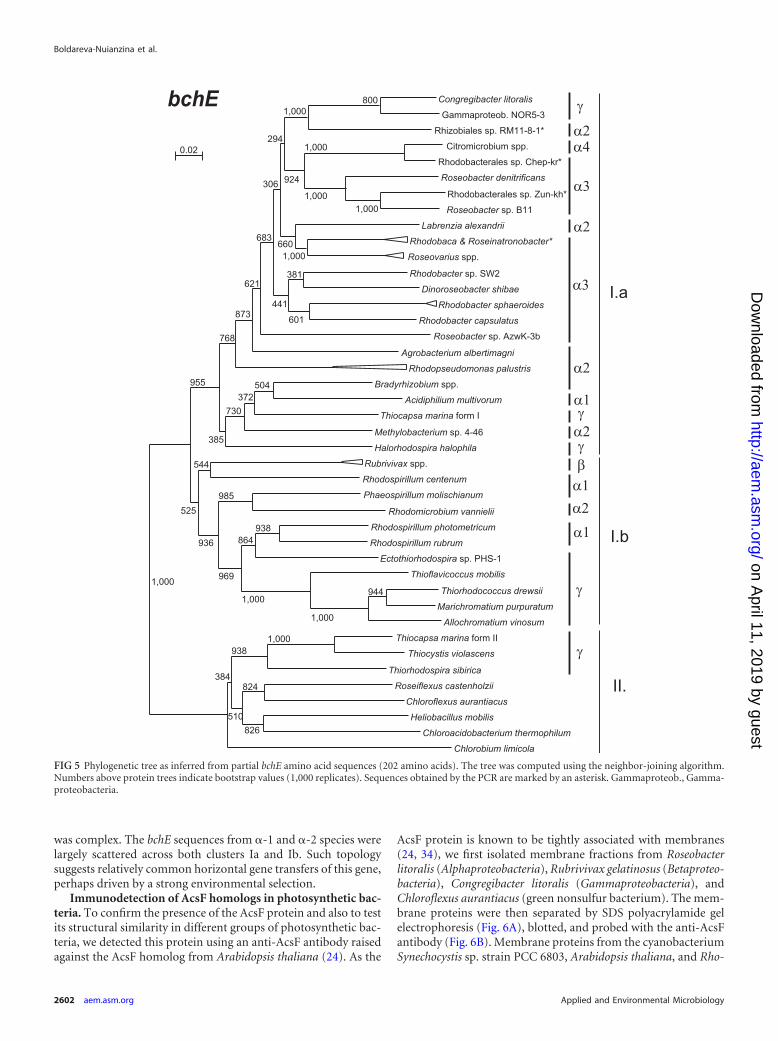
was complex. The bchE sequences from �-1 and �-2 species werelargely scattered across both clusters Ia and Ib. Such topologysuggests relatively common horizontal gene transfers of this gene,perhaps driven by a strong environmental selection.
Immunodetection of AcsF homologs in photosynthetic bac-teria. To confirm the presence of the AcsF protein and also to testits structural similarity in different groups of photosynthetic bac-teria, we detected this protein using an anti-AcsF antibody raisedagainst the AcsF homolog from Arabidopsis thaliana (24). As the
AcsF protein is known to be tightly associated with membranes(24, 34), we first isolated membrane fractions from Roseobacterlitoralis (Alphaproteobacteria), Rubrivivax gelatinosus (Betaproteo-bacteria), Congregibacter litoralis (Gammaproteobacteria), andChloroflexus aurantiacus (green nonsulfur bacterium). The mem-brane proteins were then separated by SDS polyacrylamide gelelectrophoresis (Fig. 6A), blotted, and probed with the anti-AcsFantibody (Fig. 6B). Membrane proteins from the cyanobacteriumSynechocystis sp. strain PCC 6803, Arabidopsis thaliana, and Rho-
FIG 5 Phylogenetic tree as inferred from partial bchE amino acid sequences (202 amino acids). The tree was computed using the neighbor-joining algorithm.Numbers above protein trees indicate bootstrap values (1,000 replicates). Sequences obtained by the PCR are marked by an asterisk. Gammaproteob., Gamma-proteobacteria.
Boldareva-Nuianzina et al.
2602 aem.asm.org Applied and Environmental Microbiology
on April 11, 2019 by guest
http://aem.asm
.org/D
ownloaded from
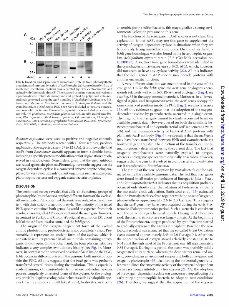
dobacter capsulatus were used as positive and negative controls,respectively. The antibody reacted with all four samples, produc-ing bands of the expected sizes (39 to 42 kDa). It is noteworthy thatAcsFs from Roseobacter litoralis appears to form a double band,indicating a specific protein modification or fast degradation not ob-served in cyanobacteria. Nonetheless, given that the used antibodywas raised against the plant AcsF homolog, our results suggest that theoxygen-dependent cyclase is a conserved enzyme despite being em-ployed by very evolutionarily distant organisms such as anoxygenicphototrophic bacteria and oxygenic cyanobacteria or plants.
DISCUSSION
The performed survey revealed that different functional groups ofphototrophic Proteobacteria employ different forms of the cyclase.All investigated PSB contained the bchE gene only, which is consis-tent with their strictly anaerobic lifestyle. The majority of the testedPNB species contained both genes. As could be expected from theiraerobic character, all AAP species contained the acsF gene; however,in contrast to Yurkov and Csotonyi’s original assumption (5), abouthalf of the AAP strains also contained the bchE gene.
The origin of the oxygen-independent form of the cyclaseamong phototrophic proteobacteria is not completely clear. Pre-sumably, it represents an ancient form of the cyclase, which isconsistent with its presence in all main phyla containing anoxy-genic phototrophs. On the other hand, the bchE phylogenetic treeindicates a very complex evolutionary history (see Fig. 4). More-over, in contrast to the conserved position of acsF inside the PGC,bchE occurs in different places in the genome, both inside or out-side the PGC. All this suggests that the bchE gene was probablytransferred several times during the evolution. This is especiallyevident among Gammaproteobacteria, where individual speciespossess completely unrelated forms of the cyclase. As the phylog-eny partially displays ecological subclusters such as halophilic spe-cies (marine and soda and salt lake strains), freshwater, or strictly
anaerobic purple sulfur bacteria, this may signalize a strong envi-ronmental selection pressure on this gene.
The function of the bchE gene in AAP species is not clear. Oneexplanation is that AAPs may use this gene to supplement theactivity of oxygen-dependent cyclase in situations when they aretemporarily facing anaerobic conditions. On the other hand, abchE gene homologue was also found in the heterotrophic organ-ism Acidiphilium cryptum strain JF-5 (GenBank accession no.CP000697). Also, three bchE gene homologues were identified inthe cyanobacterium Synechocystis sp. PCC 6803, which, however,did not seem to have any cyclase activity (22). All this indicatesthat the bchE genes in AAP species may encode proteins withanother enzymatic function.
A very different situation was encountered in the case of theacsF gene. Unlike the bchE gene, the acsF gene phylogeny corre-sponds relatively well with 16S rRNA-based phylogeny (Fig. 4; seealso Fig. S3 in the supplemental material). Moreover, in all inves-tigated Alpha- and Betaproteobacteria, the acsF genes occupy thesame conserved position inside the PGC (Fig. 2; see also reference33). All this evidence suggests that the adoption of the oxygen-dependent cyclase by proteobacteria occurred in a single event.The origin of the acsF gene cannot be clearly reconciled based onavailable genetic data. However, based on both the similarity be-tween proteobacterial and cyanobacterial acsF sequences (42% �3%) and the immunoreactivity of bacterial AcsF proteins withplant anti-AcsF antibody (Fig. 6), we speculate that the acsF genemay have been transferred between PNB and cyanobacteria viahorizontal gene transfer. The direction of the transfer cannot beunambiguously determined using the current data. The fact thatoxygenic cyanobacteria were naturally exposed to oxygen,whereas anoxygenic species were originally anaerobes, however,suggests that the gene first evolved in cyanobacteria and only laterwas it transferred to Proteobacteria.
The timing of the acsF adoption by Proteobacteria can be esti-mated using the available genomic data. The fact that acsF geneswere found in all major proteobacterial lineages (Alpha-, Beta-,and Gammaproteobacteria) indicates that its acquisition probablyoccurred only shortly after the radiation of Proteobacteria. Usingthe molecular clock calculation, Battistuzzi et al. (35) estimatedthat the Proteobacteria evolved together with the onset of oxygenicphotosynthesis approximately 2.6 to 2.3 Gyr ago. This suggeststhat the acsF gene may have been acquired during the early Pro-terozoic (Paleoproterozoic) period. Such a scenario is consistentwith the current biogeochemical models. During the Archean pe-riod, the Earth’s atmosphere was largely anoxic. At the beginningof the Proterozioc era, oxygen produced by cyanobacteria startedto gradually oxygenate the Earth’s atmosphere. Based on the geo-logical record, it was estimated that the so-called Great Oxidationevent occurred approximately 2.45 to 2.0 Gyr ago (4). After that,the concentration of oxygen stayed relatively constant (0.02 to0.04 atm) through most of the Proterozoic era (till approximately0.85 Gyr ago). During this period, the ocean was probably mildlyoxygenated at its surface, whereas the deep waters remained an-oxic, providing an environment supporting both anoxygenic andoxygenic phototrophs (36), facilitating the horizontal gene trans-fer event. Since the enzymatic activity of the oxygen-independentcyclase is strongly inhibited by free oxygen (21, 37), the adoptionof the oxygen-dependent cyclase was a necessary step, allowing theearly purple phototrophic bacteria to adapt to new conditions(26). Therefore, we suggest that the acquisition of the oxygen-
FIG 6 Isolation and separation of membrane proteins from photosyntheticorganisms and immunodetection of AcsF protein. (A) Approximately 10 �g ofsolubilized membrane proteins was separated by SDS electrophoresis andstained with Coomassie blue. (B) The separated proteins were transferred ontoa polyvinylidene difluoride membrane and probed by polyclonal anti-AcsFantibody generated using the AcsF homolog of Arabidopsis thaliana (see Ma-terials and Methods). Membrane fractions of Arabidopsis thaliana and thecyanobacterium Synechocystis PCC 6803 were included as positive controls,and anaerobic bacterium Rhodobacter capsulatus was included as a negativecontrol. Rvi. gelatinosus, Rubrivivax gelatinosus; Rsb. litoralis, Roseobacter lito-ralis; Rba. capsulatus, Rhodobacter capsulatus; Cfl. aurantiacus, Chloroflexusaurantiacus. Con. Litoralis, Congregibacter litoralis; Syn. PCC 6803, Synechocys-tis sp. PCC 6803; A. thaliana, Arabidopsis thaliana.
Two Forms of Mg-Protoporphyrin Monomethylester Cyclase
April 2013 Volume 79 Number 8 aem.asm.org 2603
on April 11, 2019 by guest
http://aem.asm
.org/D
ownloaded from
dependent cyclase by Proteobacteria was an important innovationwhich allowed the evolutionary transition from anaerobic to aerobicconditions and facilitated the radiation of purple nonsulfur speciesduring the Proterozoic era. We speculate that the fully aerobic AAPspecies probably evolved only later, at the end of the Proterozoic era,when oxygen concentrations reached approximate present levels.
ACKNOWLEDGMENTS
This research was supported by GACR projects P501/10/0221 and P501/12/G055 and project Algatech (CZ.1.05/2.1.00/03.0110).
We are indebted to Matthias Labrenz, Frederic Gich, and VladimirGorlenko for their kind gift of AAP strains, Dzmitry Hauruseu for his helpwith bacterial cultures, and Michal Mašín for providing DNA from hisVA01 isolate.
REFERENCES1. Blankenship RE. 2010. Early evolution of photosynthesis. Plant Physiol.
154:434 – 438.2. Des Marais DJ. 2000. Evolution: when did photosynthesis emerge on
Earth? Science 289:1703–1705.3. Buick R. 2008. When did oxygenic photosynthesis evolve? Philos. Trans.
R. Soc. B 363:2731–2743.4. Holland HD. 2006. The oxygenation of the atmosphere and oceans. Phi-
los. Trans. R. Soc. B 361:903–915.5. Yurkov VV, Csotonyi JT. 2009. New light on aerobic anoxygenic pho-
totrophs, p 31–55. In Hunter CN, Daldal F, Thurnauer MC, Beatty JT (ed),The purple phototrophic bacteria. Advances in photosynthesis and respi-ration, vol 28. Springer-Verlag, New York, NY.
6. Hauruseu D, Koblížek M. 2012. The influence of light on carbon utiliza-tion in aerobic anoxygenic phototrophs. Appl. Environ. Microbiol. 78:7414 –7419.
7. Kolber ZS, Plumley FG, Lang AS, Beatty JT, Blankenship RE, VanDoverCL, Vetriani C, Koblizek M, Rathgeber C, Falkowski PG. 2001. Contri-bution of aerobic photoheterotrophic bacteria to the carbon cycle in theocean. Science 292:2492–2495.
8. Jiao N, Zhang Y, Zeng Y, Hong N, Liu R, Chen F, Wang P. 2007.Distinct distribution pattern of abundance and diversity of aerobic anoxy-genic phototrophic bacteria in the global ocean. Environ. Microbiol.9:3091–3099.
9. Yutin N, Suzuki MT, Teeling H, Weber M, Venter JC, Rusch DB, BéjàO. 2007. Assessing diversity and biogeography of aerobic anoxygenic pho-totrophic bacteria in surface waters of the Atlantic and Pacific Oceansusing the Global Ocean Sampling expedition metagenomes. Environ. Mi-crobiol. 9:1464 –1475.
10. Koblížek M. 2011. Role of photoheterotrophic bacteria in the marinecarbon cycle, p 49 –51. In Jiao N, Azam F, Sanders S (ed), Microbial carbonpump in the ocean. Science/AAAS, Washington, DC.
11. Waidner LA, Kirchman DL. 2007. Aerobic anoxygenic phototrophicbacteria attached to particles in turbid waters of the Delaware and Chesa-peake estuaries. Appl. Environ. Microbiol. 73:3936 –3944.
12. Mašín M, Nedoma J, Pechar L, Koblížek M. 2008. Distribution ofaerobic anoxygenic phototrophs in temperate freshwater systems. Envi-ron. Microbiol. 10:1988 –1996.
13. Medová H, Boldareva E, Hrouzek P, Borzenko S, Namsaraev Z, Gor-lenko V, Namsaraev B, Koblížek M. 2011. High abundances of aerobicanoxygenic phototrophs in saline steppe lakes. FEMS Microbiol. Ecol.76:393– 400.
14. Willows R, Kriegel M. 2009. Biosynthesis of bacteriochlorophylls in pur-ple bacteria, p 57–79. In Hunter CN, Daldal F, Thurnauer MC, Beatty JT(ed), The purple phototrophic bacteria. Advances in photosynthesis andrespiration, vol 28. Springer-Verlag, New York, NY.
15. Chew AGM, Bryant DA. 2007. Chlorophyll biosynthesis in bacteria: theorigins of structural and functional diversity. Annu. Rev. Microbiol. 61:113–129.
16. Chereskin BM, Wong YS, Castelfranco PA. 1982. In vitro synthesis of thechlorophyll isocyclic ring: transformation of Mg-protoporphyrin IX andMg-protoporphyrin IX monomethyl ester into Mg-2,4-divinylpheopor-phyrin a5. Plant Physiol. 70:987–993.
17. Gough SP, Petersen BO, Duus JØ. 2000. Anaerobic chlorophyll isocyclicring formation in Rhodobacter capsulatus requires a cobalamin cofactor.Proc. Natl. Acad. Sci. U. S. A. 97:6908 – 6913.
18. Walker CJ, Mansfield KE, Smith KM, Castelfranco PA. 1989. Incorpo-ration of atmospheric oxygen into the carbonyl functionality of the pro-tochlorophyllide isocyclic ring. Biochem. J. 257:599 – 602.
19. Bollivar DW, Beale SI. 1996. The chlorophyll biosynthetic enzyme Mg-protoporphyrin IX monomethyl ester (oxidative) cyclase: characteriza-tion and partial purification from Chlamydomonas reinhardtii and Syn-echocystis sp. PCC 6803. Plant Physiol. 112:105–114.
20. Pinta V, Picaud M, Reiss-Husson F, Astier C. 2002. Rubrivivax gelati-nosus acsF (previously orf358) codes for a conserved, putative binuclear-iron-cluster-containing protein involved in aerobic oxidative cyclizationof Mg-protoporphyrin IX monomethylester. J. Bacteriol. 184:746 –753.
21. Ouchane S, Steunou A-S, Picaud M, Astier C. 2004. Aerobic and anaer-obic Mg-protoporphyrin monomethyl ester cyclases in purple bacteria. Astrategy adopted to bypass the repressive oxygen control system. J. Biol.Chem. 279:6385– 6394.
22. Minamizaki KK, Mizoguchi TT, Goto TT, Tamiaki HH, Fujita YY. 2008.Identification of two homologous genes, chlAI and chlAII, that are differen-tially involved in isocyclic ring formation of chlorophyll a in the cyanobacte-rium Synechocystis sp. PCC 6803. J. Biol. Chem. 283:2684–2692.
23. Moseley JL, Page MD, Alder NP, Eriksson M, Quinn J, Soto F, ThegSM, Hippler M, Merchant S. 2002. Reciprocal expression of two candi-date di-iron enzymes affecting photosystem I and light-harvesting com-plex accumulation. Plant Cell 14:673– 688.
24. Tottey S, Block MA, Allen M, Westergren T, Albrieux C, Scheller HV,Merchant S, Jensen PE. 2003. Arabidopsis CHL27, located in both enve-lope and thylakoid membranes, is required for the synthesis of protochlo-rophyllide. Proc. Natl. Acad. Sci. U. S. A. 100:16119 –16124.
25. Tang K-H, Wen J, Li X, Blankenship RE. 2009. Role of the AcsF proteinin Chloroflexus aurantiacus. J. Bacteriol. 191:3580 –3587.
26. Raimond J, Blankenship RE. 2004. Biosynthetic pathways, gene replace-ment and the antiquity of life. Geobiology 2:199 –220.
27. Boldareva EN, Bryantseva IA, Tsapin A, Nelson K, Sorokin DY, Tou-rova TP, Boichenko VA, Stadnichuk IN, Gorlenko VM. 2007. The newbacteriochlorophyll a-containing bacterium Roseinatronobacter monicussp. nov. from the hypersaline soda lake (Mono Lake, California, UnitedStates). Microbiology 76:82–92.
28. Boldareva EN, Akimov VN, Boichenko VA, Stadnichuk IN,Moskalenko AA, Makhneva ZK, Gorlenko VM. 2008. Rhodobaca bar-guzinensis sp. nov., a new alkalophylic purple nonsulfur bacteria isolatedfrom soda lake on the Barguzin valley (Buryat Republic, Russia). Micro-biology 77:206 –218.
29. Koblížek M, Béjà O, Bidigare RR, Christensen S, Benetiz-Nelson B,Vetriani C, Kolber MK, Falkowski PG, Kolber ZS. 2003. Isolation andcharacterization of Erythrobacter sp. strains from the upper ocean. Arch.Microbiol. 180:327–338.
30. Oz A, Sabehi G, Koblížek M, Massana R, Béjà O. 2005. Roseobacter-likebacteria in Red and Mediterranean Sea aerobic anoxygenic photosyntheticpopulations. Appl. Environ. Microbiol. 71:344 –353.
31. Rontani J-F, Christodoulous S, Koblížek M. 2005. GC-MS structuralcharacterization of fatty acids from marine aerobic anoxygenic pho-totrophic bacteria. Lipids 40:97–108.
32. Koblížek M, Mlcoušková J, Kolber Z, Kopecký J. 2010. On the photo-synthetic properties of marine bacterium COL2P belonging to Roseobacterclade. Arch. Microbiol. 192:41– 49.
33. Zheng Q, Zhang R, Koblížek M, Boldareva EN, Yurkov V, Yan S, JiaoN. 2011. Diverse arrangement of photosynthetic gene clusters in aerobicanoxygenic phototrophic bacteria. PLoS One 6:e25050. doi:10.1371/journal.pone.0025050.
34. Peter E, Salinas A, Wallner T, Jeske D, Dienst D, Wilde A, Grimm B.2009. Differential requirement of two homologous proteins encoded bysll1214 and sll1874 for the reaction of Mg protoporphyrin monomethyl-ester oxidative cyclase under aerobic and micro-oxic growth conditions.Biochim. Biophys. Acta 1787:1458 –1467.
35. Battistuzzi FU, Feijao A, Hedges SB. 2004. A genomic timescale ofprokaryote evolution: insights into the origin of methanogenesis, photot-rophy, and the colonization of land. BMC Evol. Biol. 4:44.
36. Johnston DT, Wolfe-Simon F, Pearson A, Knoll AH. 2009. Anoxygenicphotosynthesis modulated Proterozoic oxygen and sustained Earth’s mid-dle age. Proc. Natl. Acad. Sci. U. S. A. 106:16925–16929.
37. Bollivar DW. 2003. Intermediate steps in chlorophyll biosynthesis, p 49 –70. In Kadish K, Smith KM, Guillard R (ed), The porphyrin handbook.Academic Press, San Diego, CA.
Boldareva-Nuianzina et al.
2604 aem.asm.org Applied and Environmental Microbiology
on April 11, 2019 by guest
http://aem.asm
.org/D
ownloaded from