Embed Size (px)
Citation preview

Thomas Jefferson University Thomas Jefferson University
Jefferson Digital Commons Jefferson Digital Commons
Department of Pediatrics Faculty Papers Department of Pediatrics
1-1-2009
Increased susceptibility of spinal muscular atrophy fibroblasts to Increased susceptibility of spinal muscular atrophy fibroblasts to
camptothecin is p53-independent. camptothecin is p53-independent.
Chia-Yen Wu University of Delaware
Ilsa Gómez-Curet Alfred I. duPont Hospital for Children
Vicky L Funanage Alfred I. duPont Hospital for Children
Mena Scavina Alfred I. duPont Hospital for Children
Wenlan Wang Thomas Jefferson University Follow this and additional works at: https://jdc.jefferson.edu/pedsfp
Part of the Bioethics and Medical Ethics Commons, and the Pediatrics Commons
Let us know how access to this document benefits you
Recommended Citation Recommended Citation
Wu, Chia-Yen; Gómez-Curet, Ilsa; Funanage, Vicky L; Scavina, Mena; and Wang, Wenlan,
"Increased susceptibility of spinal muscular atrophy fibroblasts to camptothecin is
p53-independent." (2009). Department of Pediatrics Faculty Papers. Paper 23.
https://jdc.jefferson.edu/pedsfp/23
This Article is brought to you for free and open access by the Jefferson Digital Commons. The Jefferson Digital Commons is a service of Thomas Jefferson University's Center for Teaching and Learning (CTL). The Commons is a showcase for Jefferson books and journals, peer-reviewed scholarly publications, unique historical collections from the University archives, and teaching tools. The Jefferson Digital Commons allows researchers and interested readers anywhere in the world to learn about and keep up to date with Jefferson scholarship. This article has been accepted for inclusion in Department of Pediatrics Faculty Papers by an authorized administrator of the Jefferson Digital Commons. For more information, please contact: [email protected].
BioMed Central
Page 1 of 14(page number not for citation purposes)
BMC Cell Biology
Open AccessResearch articleIncreased susceptibility of spinal muscular atrophy fibroblasts to camptothecin is p53-independentChia-Yen Wu1,2, Ilsa Gómez-Curet2,3, Vicky L Funanage2,3, Mena Scavina2 and Wenlan Wang*2,3
Address: 1Department of Biological Science, University of Delaware, Newark, DE, USA, 2Nemours Biomedical Research, Alfred I. duPont Hospital for Children, Wilmington, DE, USA and 3Department of Pediatrics, Thomas Jefferson University, Philadelphia, PA, USA
Email: Chia-Yen Wu - [email protected]; Ilsa Gómez-Curet - [email protected]; Vicky L Funanage - [email protected]; Mena Scavina - [email protected]; Wenlan Wang* - [email protected]
* Corresponding author
AbstractBackground: Deletion or mutation(s) of the survival motor neuron 1 (SMN1) gene causes spinalmuscular atrophy (SMA). The SMN protein is known to play a role in RNA metabolism, neuriteoutgrowth, and cell survival. Yet, it remains unclear how SMN deficiency causes selective motorneuron death and muscle atrophy seen in SMA. Previously, we have shown that skin fibroblastsfrom SMA patients are more sensitive to the DNA topoisomerase I inhibitor camptothecin,supporting a role for SMN in cell survival. Here, we examine the potential mechanism ofcamptothecin sensitivity in SMA fibroblasts.
Results: Camptothecin treatment reduced the DNA relaxation activity of DNA topoisomerase Iin human fibroblasts. In contrast, kinase activity of DNA topoisomerase I was not affected bycamptothecin, because levels of phosphorylated SR proteins were not decreased. Uponcamptothecin treatment, levels of p53 were markedly increased. To determine if p53 plays a rolein the increased sensitivity of SMA fibroblasts to camptothecin, we analyzed the sensitivity of SMAfibroblasts to another DNA topoisomerase I inhibitor, -lapachone. This compound is known toinduce death via a p53-independent pathway in several cancer cell lines. We found that -lapachonedid not induce p53 activation in human fibroblasts. In addition, SMA and control fibroblasts showedessentially identical sensitivity to this compound. By immunofluorescence staining, SMN and p53co-localized in gems within the nucleus, and this co-localization was overall reduced in SMAfibroblasts. However, depletion of p53 by siRNA did not lessen the camptothecin sensitivity in SMAfibroblasts.
Conclusion: Even though p53 and SMN are associated, the increased sensitivity of SMA fibroblaststo camptothecin does not occur through a p53-dependent mechanism.
BackgroundSpinal muscular atrophy (SMA) is a neuromuscular dis-ease characterized by the loss of spinal motor neurons and
muscle atrophy [1]. SMA has an incidence of 1 in 6,000live births, and is one of the most common genetic causesof infant death [2,3]. Clinically, based on the age of onset
Published: 16 May 2009
BMC Cell Biology 2009, 10:40 doi:10.1186/1471-2121-10-40
Received: 13 May 2008Accepted: 16 May 2009
This article is available from: http://www.biomedcentral.com/1471-2121/10/40
© 2009 Wu et al; licensee BioMed Central Ltd. This is an Open Access article distributed under the terms of the Creative Commons Attribution License (http://creativecommons.org/licenses/by/2.0), which permits unrestricted use, distribution, and reproduction in any medium, provided the original work is properly cited.
BMC Cell Biology 2009, 10:40 http://www.biomedcentral.com/1471-2121/10/40
Page 2 of 14(page number not for citation purposes)
and severity of the disease, childhood SMA can be catego-rized into types I, II, and III [4,5]. Type I patients are diag-nosed between the ages of zero to six months and cannotsit unsupported or lift their heads, type II patients arediagnosed between the ages of seven and 18 months andcan sit, and type III patients are older than 18 monthswhen diagnosed and can stand alone and walk but maylater lose these motor milestones. Although SMA shows abroad spectrum of severity, genetic studies indicate that allclinical phenotypes of SMA are caused by deletion ormutation(s) of the survival motor neuron 1 (SMN1) gene[6].
The SMN protein is ubiquitously expressed and localizesin the cytoplasm as well as in the nucleus, where it is usu-ally concentrated in subnuclear structures referred to as"gems" (for Gemini of Cajal bodies [7,8]). The SMN pro-tein plays an essential role in the biogenesis of smallnuclear ribonucleoprotein (snRNP) and small nucleolarribonucleoprotein (snoRNP) complexes [9-11]. SMNappears to perform this function by associating withGemins 2–8 [12-14]. Recent studies have demonstratedthat the associated SMN/Gemin complex directly interactswith specific domains of Sm proteins and uridine-richsnRNAs to ensure stringent control of snRNP assembly[15,16]. In addition to RNP assembly, SMN has beenshown to play a role in neurite outgrowth [17,18],through its association with hnRNP R [19,20].
Complete loss of SMN in species ranging from S. pombe tomice is lethal, indicating that SMN is critical for survivalof multiple cell types [21-23]. More direct evidence to sup-port SMN's role in cell survival comes from studies in cul-tured cells [24-29]. For example, depletion of the SMNprotein in Drosophila S2 cells stimulates caspase activityand leads to increased cell death [25]. Importantly, SMNhas been shown to play a role in neuronal cell survival.Depletion of SMN in differentiated P19 cells activates cas-pase activity and increases cell death [27], whereas overex-pression of human SMN (hSMN) protects differentiatedPC12 cells from cell death induced by neurotrophic factorwithdrawal [26].
Previously, we investigated the role of SMN in cell survivalusing skin fibroblasts derived from SMA patients and age-matched controls [29]. We demonstrated that SMAfibroblasts display an increased sensitivity to camp-tothecin-induced cell death. Treatment with menadione,an agent causing cell death by generating oxidative stress[30], did not cause differences in survival between SMAand control fibroblasts. In addition, camptothecin treat-ment resulted in significantly higher caspase-3 activity inSMA fibroblasts when compared with control fibroblasts,and this activity directly correlated with levels of SMN in
fibroblasts. Thus, these data support an active role forSMN in cell survival.
Camptothecin is a specific DNA topoisomerase I inhibitorthat binds DNA topoisomerase I when the enzyme is com-plexed with DNA [31]. Consequently, camptothecin sta-bilizes the enzyme-DNA complex and suppresses theenzymatic activity of this protein. Camptothecin has beenshown to induce cell death in human ovarian adenocarci-noma cells via p53-dependent and independent pathways[32]. Interestingly, SMN has been shown to interact withp53, and this interaction is reduced when SMN harborsmutations derived from SMA patients [33]. Because SMNcan interact with p53 and camptothecin can induce celldeath via p53-dependent and independent mechanisms,this study addresses whether the increased sensitivity ofSMA fibroblasts to camptothecin occurs through a p53-dependent mechanism. We found that although SMNdirectly interacts with p53, the increased sensitivity ofSMN-depleted fibroblasts to camptothecin occursthrough a p53-independent mechanism.
ResultsCamptothecin inhibits DNA unwinding but not kinase activity of DNA topoisomerase I in human fibroblastsWe previously showed that fibroblasts derived from SMApatients have increased sensitivity to the DNA topoi-somerase I inhibitor camptothecin [29]. DNA topoi-somerase I has been shown to phosphorylate SR proteins[34] that regulate RNA splicing. Considering SMN's rolein RNA splicing, we examined whether camptothecintreatment would block phosphorylation of SR proteins inSMA fibroblasts. Control and SMA fibroblasts weretreated with 25 M camptothecin, and levels of phospho-rylated SR proteins in nuclear extracts were analyzed byWestern blotting using the mAb 104 antibody that specif-ically recognizes a phosphorylated epitope at thearginine/serine rich (RS) domain [35]. As shown in Figure1A, levels of phosphorylated SR proteins were not reducedin camptothecin-treated human fibroblasts. In fact, phos-phorylation of some SR proteins was slightly increased,which could be caused by activation of kinases other thanDNA topoisomerase I. These data suggest that camp-tothecin does not inhibit in vivo kinase activity of DNAtopoisomerase I. Thus, the camptothecin-induced celldeath in human fibroblasts must be mediated by suppres-sion of other enzymatic activities of DNA topoisomeraseI.
Next, we analyzed the DNA relaxation activity of DNAtopoisomerase I from human fibroblasts after camp-tothecin treatment. Control and SMA fibroblasts weretreated with 25 M camptothecin, and DNA topoisomer-ase I was immunoprecipitated. DNA relaxation activity ofthis enzyme was assayed on a supercoiled plasmid DNA

BMC Cell Biology 2009, 10:40 http://www.biomedcentral.com/1471-2121/10/40
Page 3 of 14(page number not for citation purposes)
Camptothecin inhibits DNA relaxation but not kinase activity of DNA topoisomerase IFigure 1Camptothecin inhibits DNA relaxation but not kinase activity of DNA topoisomerase I. (A) Control and SMA fibroblasts were treated with 25 M camptothecin, and levels of phosphorylated SR proteins in the nuclear extracts were ana-lyzed by Western blotting. The same blot was then stripped and reprobed with anti-histone 3 (H3) antibodies as a loading con-trol. (B) DNA topoisomerase I was immunoprecipitated from camptothecin treated fibroblasts and subjected to the DNA unwinding assay. Plasmid DNA in the supercoiled form (SC) and in the relaxed form (R) is indicated. (C) DNA topoisomerase I immunoprecipitated from untreated control fibroblasts was subjected to the DNA unwinding assay in the presence of camp-tothecin at the indicated concentrations. All the data shown here are representative of at least two independent experiments. CPT = camptothecin, H3 = histone 3, and Topo I = DNA topoisomerase I.
A
B C

BMC Cell Biology 2009, 10:40 http://www.biomedcentral.com/1471-2121/10/40
Page 4 of 14(page number not for citation purposes)
[36]. Figure 1B shows that in the absence of DNA topoi-somerase I, approximately half of the plasmid DNA wasfound in the supercoiled form. Upon addition of thisenzyme, the majority of plasmid DNA was in the relaxedform, and this DNA relaxation activity was inhibited bycamptothecin treatment. When immunoprecipitatedDNA topoisomerase I was mixed with camptothecin invitro, DNA relaxation activity of this enzyme was alsoreduced (Fig. 1C), which is consistent with data obtainedfrom studies with purified DNA topoisomerase I [36,37].Western blotting analyses indicated that upon camp-tothecin treatment, levels of DNA topoisomerases I (~100kD) in the immunoprecipitates and protein lysates werereduced by 80% or more, and SMA fibroblasts had morereduced levels of this enzyme than control fibroblasts(90% [SMA] vs. 80% [control] at 4 h and 100% [SMA] vs.88% at 8 h) (Figs. 2A and 2B). The reduction in DNAtopoisomerase I protein seems specific to camptothecinsince another DNA topoisomerase I inhibitor, -lapa-chone, did not drastically affect levels of this enzyme (Fig.2B). -lapachone directly binds to DNA topoisomerase Iand inhibits its enzymatic activity [38]. Thus, in vivo inhi-bition of DNA topoisomerase I by camptothecin likelyresults from a combination of decreased DNA relaxationactivity and reduced levels of this protein.
Activation of p53 and the sensitivity of SMA fibroblasts to the DNA topoisomerase I inhibitor -lapachoneCamptothecin has been shown to inhibit DNA topoi-somerase I activity by stabilizing the enzyme-DNA cleav-age complex [31]. As a result, camptothecin treatment
results in single- and double-stranded DNA breaks, causesG1 and G2 arrests, and leads to cell death via p53-depend-ent and independent pathways [32,39]. To elucidatewhether the increased sensitivity of SMA fibroblasts tocamptothecin is p53-dependent, we assessed p53 induc-tion after camptothecin treatment. As shown in Figure 3A,levels of p53 were markedly increased upon camptothecintreatment, and this induction was seen as early as 4 h andwas sustained for 24 h. Treatment with menadione, whichalso induces death in fibroblasts but does not show differ-ential sensitivity between control and SMA fibroblasts[29], did not elevate p53 levels (Fig. 3B). Unlike camp-tothecin, menadione causes cell death by generating oxi-dative stress [30]. This suggested that p53 could play a rolein the increased sensitivity of SMA fibroblasts to camp-tothecin. To further address the role of p53 in camp-tothecin sensitivity, we examined the sensitivity of SMAfibroblasts to another DNA topoisomerase I inhibitor -lapachone. This compound has been shown to induce celldeath via a p53-independent pathway in several cancercell lines [40]. Figure 3C shows that levels of p53 proteinwere not elevated after -lapachone treatment; and p53levels actually decreased by more than 70% under thiscondition. Note that under the same treatment condi-tions, levels of p53 were elevated two- to four-fold aftercamptothecin treatment. The SMA fibroblasts showed noincreased sensitivity to -lapachone when compared withcontrol fibroblasts using the percentage of cell death asthe readout (Fig. 3D). These data imply that p53 plays arole in the increased sensitivity of SMA fibroblasts tocamptothecin.
Camptothecin induces degradation of DNA topoisomerase I in fibroblastsFigure 2Camptothecin induces degradation of DNA topoisomerase I in fibroblasts. (A) Presence of DNA toposiomerase I in the immunocomplexes described in 1B was confirmed by Western blotting analyses. (B) Fibroblasts described in 1B were treated with 25 M camptothecin or 25 M -lapachone, and DNA topoisomerase I present in the lysates was detected by Western blotting. The same blots were stripped and reprobed with anti--tubulin antibodies as a loading control. Relative ratios of DNA topoisomerase I to tubulin levels are indicated. All the data shown here are representative of at least two inde-pendent experiments. CPT = camptothecin, Topo I = DNA topoisomerase I, IP = immunoprecipitation, IB = immunoblotting, and -LP = -lapachone.
A B

BMC Cell Biology 2009, 10:40 http://www.biomedcentral.com/1471-2121/10/40
Page 5 of 14(page number not for citation purposes)
Figure 3 (see legend on next page)
A
B
C
D

BMC Cell Biology 2009, 10:40 http://www.biomedcentral.com/1471-2121/10/40
Page 6 of 14(page number not for citation purposes)
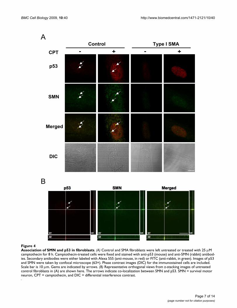
Association of p53 and SMN in SMA fibroblastsSince an interaction of p53 with SMN has been previouslyreported [33], we investigated whether p53 is associatedwith SMN in fibroblasts. Immunoprecipitation analysesof the endogenous p53 and SMN proteins did not showan association (data not shown), indicating that SMN andp53 interaction in fibroblasts is likely transient and notstable. We next determined if a fraction of endogenousp53 and SMN was associated by using confocal micros-copy to test for co-localization. Co-localization of thesetwo proteins was confirmed by visualizing orthogonalprojections of stacked images. Control and SMA fibrob-lasts were left untreated or treated with camptothecin, andp53 and SMN proteins were detected by double-labeledimmunofluorescence staining. As shown in Figure 4A,both p53 and SMN were localized in the cytoplasm andthe nucleus. Upon camptothecin treatment, p53 stainingin the nucleus was dramatically enhanced, which is con-sistent with the p53 induction measured by Western blot-ting analyses (see Figs. 3A and 3C). Nuclearimmunofluorescence staining showed that SMN wasmore concentrated in gems, and that SMA fibroblasts hadreduced numbers of gems in the nucleus both in theabsence and presence of camptothecin treatment (Fig. 4Aand Table 1). Gem size in SMA fibroblasts was alsosmaller than that in control fibroblasts (Table 1), likely asa result of reduced levels of SMN expression in these cells.In addition, SMN and p53 were seen to co-localize ingems in the absence and presence of camptothecin (Figs.4A and 4B). Overall, co-localization of SMN with p53 wasreduced in SMA fibroblasts (Table 1). For example, in theabsence of camptothecin treatment, control fibroblastshad 99% gems with SMN/p53 co-localization, whereasSMA fibroblasts had 75%–83%. Upon camptothecintreatment, both number of gems and percentage of gemswith co-localized SMN/p53 were reduced in control, typeII, and type III SMA fibroblasts. Interestingly, type I SMAfibroblasts had the fewest number of gems, but SMN andp53 co-localized in almost all gems (only one of all
counted gems did not contain co-localized SMN andp53).
Increased sensitivity of SMA fibroblasts to camptothecin is p53-independentHaving confirmed the association between SMN and p53in fibroblasts, we determined if the susceptibility of SMAfibroblasts to camptothecin is mediated by p53. Endog-enous p53 protein in fibroblasts was depleted by siRNA,and the sensitivity of SMA fibroblasts to camptothecinwas analyzed. Figures 5A and 5B showed a time course forp53 depletion by siRNA in fibroblasts. A reduction ofapproximately 85–90% in p53 mRNA levels was observedby addition of p53 siRNA nucleotides at each time pointanalyzed (Fig. 5A). Similarly, levels of the p53 proteinwere reduced by more than 90% in p53 siRNA transfectedcells (Fig. 5B). Levels of p53 in non-targeting control andmock transfected cells were indistinguishable, indicatingthat p53 depletion by siRNA is specific. Moreover, uponcamptothecin treatment, levels of p53 were markedly ele-vated in fibroblasts, and the increase in p53 expressionupon camptothecin treatment was completely eliminatedby p53 siRNA (Fig. 5C). Cell survival analyses indicatedthat SMA fibroblasts were more sensitive to camptothecinthan control fibroblasts (~70% survival in control vs.~35% in SMA fibroblasts after 25 M camptothecin treat-ment) (Fig. 6A). Surprisingly, depletion of p53 by siRNAdid not rescue either control or SMA fibroblasts fromcamptothecin-induced cell death. Figure 6A showed thatcell death induced by camptothecin was not significantlyreduced by p53 depletion. Our previous study showedthat SMA fibroblasts have significantly higher caspase-3activity upon camptothecin treatment than controlfibroblasts [29], thus we analyzed induction of camp-tothecin activated caspase-3 activity in p53 depletedfibroblasts after camptothecin treatment. Figure 6Bshowed that p53 depletion indeed decreased camp-tothecin-induced PARP cleavage, an in vivo caspase-3 sub-strate, in both control and SMA fibroblasts. This is
Activation of p53 and cell death induced by the DNA topoisomerase I inhibitors camptothecin and -lapachoneFigure 3 (see previous page)Activation of p53 and cell death induced by the DNA topoisomerase I inhibitors camptothecin and -lapa-chone. (A) A type I SMA fibroblasts as described [29] were treated with 25 M camptothecin and levels of p53 were analyzed by Western blotting. The same blots were stripped and reprobed with anti--tubulin antibodies as a loading control. (B) A type II/III SMA fibroblasts as described [29] were treated with 10 M menadione and levels of p53 were analyzed as described for (A). (C) Control and SMA fibroblasts were treated with 25 M camptothecin or 25 M -lapachone and levels of p53 were analyzed as described for (A). Relative ratios of p53 to tubulin levels are indicated. (D) Three control and three SMA fibroblasts (one type I, one type II, and one type III) were treated with camptothecin for 72 h or -lapachone for 24 h at the indicated con-centrations, respectively. Cell survival of treated cells was measured by the CellTiter-Blue assay, and the relative cell viability was calculated and presented as percentage of the untreated cells. Each condition was set up as replicates of four and repeated three times. The data presented here are combined mean values ± sem for three control and three SMA fibroblasts. Statistical analyses (unpaired t test) indicate that SMA fibroblasts are significantly sensitive to camptothecin at each tested concentration than control fibroblasts (*** p < 0.0001 and ** p < 0.001). CPT = camptothecin, -LP = -lapachone, MENA = menadione, and ND = non-detected.
BMC Cell Biology 2009, 10:40 http://www.biomedcentral.com/1471-2121/10/40
Page 7 of 14(page number not for citation purposes)
Association of SMN and p53 in fibroblastsFigure 4Association of SMN and p53 in fibroblasts. (A) Control and SMA fibroblasts were left untreated or treated with 25 M camptothecin for 8 h. Camptothecin-treated cells were fixed and stained with anti-p53 (mouse) and anti-SMN (rabbit) antibod-ies. Secondary antibodies were either labeled with Alexa 555 (anti-mouse, in red) or FITC (anti-rabbit, in green). Images of p53 and SMN were taken by confocal microscope (63×). Phase contrast images (DIC) for the immunostained cells are included. Scale bar is 10 m. Gems are indicated by arrows. (B) Representative orthogonal views from z-stacking images of untreated control fibroblasts in (A) are shown here. The arrows indicate co-localization between SMN and p53. SMN = survival motor neuron, CPT = camptothecin, and DIC = differential interference contrast.
A
B
SMN
Control
CPT
p53
Type I SMA
Merged
DIC
- + - +

BMC Cell Biology 2009, 10:40 http://www.biomedcentral.com/1471-2121/10/40
Page 8 of 14(page number not for citation purposes)
consistent with caspase-3 being downstream of p53 [41].Given that p53 depletion reduced caspase-3 activity butthis was not enough to rescue fibroblasts from camp-tothecin-induced cell death, non-caspase-3 pathwayscould also be involved in camptothecin-induced death inSMA fibroblasts. When these findings are taken together,we conclude that camptothecin-induced cell death inhuman fibroblasts is not p53-dependent, and p53 doesnot play a direct role in the increased sensitivity of SMAfibroblasts to camptothecin.
DiscussionThe critical feature in the pathogenesis of SMA is thatreduced levels of the SMN protein result in selective lossof motor neurons accompanied by muscle wasting. SMNhas been shown to play a role in the assembly of RNPcomplexes [9-11], transcription regulation [42], neuriteoutgrowth [17-19], and cell survival [24-29]. However,how SMN deficiency causes death of motor neurons andmuscle paralysis with little effect on other cells and tissuesremains unclear. Previously, we demonstrated that SMAfibroblasts are more sensitive to camptothecin-inducedcell death when compared with control fibroblasts [29],supporting a role for SMN in cell survival. In this study, wefurther explored the potential mechanism(s) underlyingthe susceptibility of SMA fibroblasts to camptothecin.
Camptothecin is a specific inhibitor of DNA topoisomer-ase I. The best-characterized enzymatic activity of DNAtopoisomerase I is the relaxation of supercoiled DNA dur-ing DNA replication and RNA synthesis [31]. Camp-tothecin suppresses the DNA relaxation activity of DNAtopoisomerase I by stabilizing the topoisomerase I-DNAcleavage complex [31,43]. As a result, camptothecin treat-ment introduces single- and double-stranded DNAbreaks, leading to chromosome fragmentation and, even-tually, cell death [39,43]. The action of camptothecin onthe DNA relaxation activity of DNA topoisomerase I hasbeen extensively characterized in vitro. Here, we usedimmunoprecipitated DNA topoisomerase I to analyze invivo DNA relaxation activity in camptothecin-treatedhuman fibroblasts. We found that DNA topoisomerase I
from camptothecin-treated fibroblasts had decreasedDNA relaxation activity (Fig. 1B), which may be due toreduced levels of this protein and/or reduced enzymaticactivity. A reduction in the levels of DNA topoisomerase Iin human fibroblasts after camptothecin treatment couldbe mediated by proteasome-dependent degradation aspreviously reported for some cancer cell lines [44]. Inter-estingly, the reduced levels of DNA topoisomerase I aftercamptothecin treatment were more pronounced in SMAfibroblasts (Figs. 2A and 2B). In addition, in the absenceof camptothecin, overall levels of DNA topoisomerase Iwere lower in SMA fibroblasts than control fibroblasts(Figs. 2A and 2B). Thus, the lower levels of DNA topoi-somerase I in SMA fibroblasts could account for theirincreased susceptibility to camptothecin. Further experi-ments are needed to determine if the increased sensitivityof SMA fibroblasts to camptothecin is due to lower levelsof DNA topoisomerase I in these cells. In addition to itsDNA relaxation activity, DNA topoisomerase I exhibits akinase activity that phosphorylates SR proteins [34]. Sincecamptothecin treatment of SMA fibroblasts did not resultin reduced levels of phosphorylated SR proteins, camp-tothecin probably did not inhibit kinase activity of DNAtopoisomerase I in vivo. Thus, inhibition of the DNA relax-ation activity of DNA topoisomerase I by camptothecinlikely accounts for its cytotoxicity in human fibroblasts.
In human ovarian adenocarcinoma cells, camptothecinhas been shown to induce cell death via p53-dependentand independent pathways [32]. The present studyshowed that levels of p53 were markedly increased inSMA fibroblasts upon camptothecin treatment (Figs. 3A,C, and 4A). Our data also confirmed previous findings[33] in which a fraction of p53 co-localized with SMN ingems, and this co-localization was overall decreased inSMA fibroblasts (Table 1). However, depletion of p53 bysiRNA did not lessen the susceptibility of SMA fibroblaststo camptothecin (Fig. 6). Thus, although p53 is activatedby camptothecin and p53 and SMN can associate in vivo,p53 does not play a direct role in the increased sensitivityof SMN-depleted fibroblasts to camptothecin. This agreeswith a previous study performed in SMA transgenic mice
Table 1: Co-localization of SMN with p53 in fibroblasts
Type I Type II Type III Control
UntreatedGem number/100 cells 6 16 24 114
Average gem size ± SD (nm) 538 ± 37 603 ± 73 649 ± 121 715 ± 153% of co-localization with p53 83.3 75.0 75.0 99.1
CPT treatedGem number/100 cells 6 11 17 80
Average gem size ± SD (nm) 544 ± 22 542 ± 53 689 ± 154 793 ± 173% of co-localization with p53 100.0 63.0 64.7 85.0

BMC Cell Biology 2009, 10:40 http://www.biomedcentral.com/1471-2121/10/40
Page 9 of 14(page number not for citation purposes)
Depletion of the p53 protein in fibroblasts by RNA interferenceFigure 5Depletion of the p53 protein in fibroblasts by RNA interference. (A) Control fibroblasts transfected with p53, non-tar-geting control, or no siRNA oligonucleotides (mock) were harvested at the indicated times. Levels of p53 mRNA was meas-ured by real-time TaqMan PCR using p53 as the target and gusB as the endogenous control. Statistical analyses (one-way ANOVA) indicate a significant reduction of p53 mRNA by the addition of p53 siRNA at each time point analyzed as compared to mock control (*** p < 0.0001). The data shown here are representative of two independent experiments. (B) A similar experiment was conducted as described for (A), and levels of the p53 protein were detected as described for 2A. (C) Control and SMA fibroblasts were transfected as described for (A). Twenty-four hours after transfection, cells were treated with 25 M camptothecin for 24 h. Levels of p53 were analyzed as described for (B). NT = non-targeting control, CPT = camptothecin, and ND = non-detected.
A
B
Moc
k
NT
p53
Moc
k
NT
p53
Moc
k
NT
p53
24h 48h 72h
p53
ß-tubulin
p53/tubulin 0.8 0.8 ND 1.3 2.0 ND 1.3 1.2 0.1
C

BMC Cell Biology 2009, 10:40 http://www.biomedcentral.com/1471-2121/10/40
Page 10 of 14(page number not for citation purposes)
Depletion of the p53 protein does not lessen the camptothecin susceptibility in SMA fibroblastsFigure 6Depletion of the p53 protein does not lessen the camptothecin susceptibility in SMA fibroblasts. (A) A similar experiment was conducted as described for 5C. Cell survival of treated cells was measured and presented as percentage of the untreated ones. At least two independent experiments were performed and each sample was set up in replicates of four. The mean value ± sem of one representative experiment is presented here. Statistical analyses (unpaired t test) indicate that SMA fibroblasts are significantly more sensitive to camptothecin than control fibroblasts (*** p < 0.0001). There is no difference in survival among cells transfected with non-targeting control, p53, or no siRNA oligonucleotides for either control or SMA fibroblasts. (B) A similar experiment was conducted as described for 5C except cells were treated with 25 M camptothecin for 16 h. Cell lysates were analyzed by Western blotting using antibodies against cleaved PARP, p53, and tubulin, respectively. NT = non-targeting control, CPT = camptothecin, and cPARP = cleaved poly ADP-ribose polymerase.
A
B

BMC Cell Biology 2009, 10:40 http://www.biomedcentral.com/1471-2121/10/40
Page 11 of 14(page number not for citation purposes)
[45]. This study indicated that elimination of p53 doesnot alleviate the disease severity or extend overall lifespanin type I and type III SMA mice. Thus, p53-independentapoptotic pathways may play a role in motor neuron losswhen SMN is depleted.
Analyses of the sensitivity of SMA fibroblasts to anotherDNA topoisomerase I inhibitor, -lapachone, indicatethat SMA and control fibroblasts showed similar sensitiv-ity to this compound (Fig. 3D). -lapachone is known toinduce cell death in several cancer cell lines through ap53-independent pathway [40]. Our data showed thatthis compound did not induce p53 in fibroblasts (Fig.3C), suggesting that p53 is not involved in -lapachone-induced cell death in fibroblasts. Unlike camptothecin, -lapachone directly binds to DNA topoisomerase I andinhibits its enzymatic activity [38]. Thus, -lapachonetreatment usually does not cause DNA damage [38]. Wenoticed that -lapachone did not induce a drastic reduc-tion in the levels of DNA topoisomerase I (Fig. 2B). Sincea reduction in DNA topoisomerase I protein by camp-tothecin seems to be triggered by DNA damage [44], it ispossible that -lapachone did not cause DNA damage inhuman fibroblasts, so levels of DNA topoisomerase I pro-tein remained unaltered after -lapachone treatment.Thus, the increased sensitivity of SMA fibroblasts to camp-tothecin but not to -lapachone suggests that cell deathpathways activated by DNA damage may be responsiblefor the susceptibility of SMA fibroblasts to camptothecin.SMN may protect fibroblasts from camptothecin-inducedcell death through this pathway. This hypothesis is furthersupported by our observation that in addition to camp-tothecin, SMA fibroblasts show an increased sensitivity toother DNA damaging reagents (C. Wu, unpublisheddata). It will be interesting to find out whether otherapoptotic molecules in this pathway such as Bax play arole in the vulnerability of SMA fibroblasts to camp-tothecin, since abolishing this apoptotic protein clearlyprotects SMA mice from motor neuron loss [46].
ConclusionOur results confirm that p53 is activated by camptothecinin human fibroblasts. In addition, p53 co-localizes withSMN in gems, and this co-localization is overall reducedin SMA fibroblasts. However, p53 does not directly affectcamptothecin sensitivity when SMN is depleted.
MethodsCell culture and transfectionSkin biopsies from SMA patients and controls wereobtained as part of a study approved by the InstitutionalReview Board of the Alfred I. duPont Hospital for Chil-dren. Human fibroblast cell lines were established fromthese biopsies and maintained according to standard pro-tocols [29]. In brief, fibroblasts were maintained in
DMEM supplemented with 20% fetal bovine serum andantibiotics. Passage numbers for control and SMA fibrob-lasts were matched as closely as possible for all experi-mental procedures and always kept #25. Thesefibroblasts were used in our previous studies [29,47].Unless specifically denoted, SMA fibroblasts used in thisstudy are not the cell lines used in our initial publishedstudy [29]. The number of SMN1 and SMN2 gene copiesfor control and SMA fibroblasts were determined by quan-titative real-time PCR as described [47]. Control fibrob-lasts carry two copies of SMN1 and two copies of SMN2.All SMA fibroblasts have zero copies of SMN1. For theSMN2 gene, most type I fibroblasts contain two copies,type II mainly carry three copies, and type III carry three ormore copies. For RNA interference (RNAi) analyses, 1 ×10⁶ fibroblasts were electroporated with 100 nM of smallinterference RNA (siRNA) oligonucleotides in nucleofec-tor solution optimized for primary fibroblasts followingmanufacturer's instruction (Amaxa, Gaithersburg, MD).
siRNA oligonucleotides (SMARTpool kits) for human p53(p53 SMART pool, L-003329) and non-targeting control(siGenome non-targeting siRNA, D-001206-14) were pur-chased from Dharmacon (Chicago, IL).
Analysis of p53 transcript levels by real-time PCRFor p53 RNAi validation, control fibroblasts were trans-fected with no siRNA (mock), non-targeting control, orp53 siRNA oligonucleotides. Cells were harvested at 24,48, and 72 h after transfection. Total RNA was isolatedusing the RNeasy kit with on-column DNase treatment(Qiagen, Los Angeles, CA). First-strand cDNA synthesiswas carried out with the Omniscript kit (Qiagen). Thereal-time PCR was performed in a total volume of 15 l,containing 10 ng of cDNA, 1× TaqMan Universal PCRmaster mix (Applied Biosystems, Atlanta, GA), and 1×p53 gene expression assay (Hs01034253) from AppliedBiosystems. The real-time PCR was performed on a 7900HT Sequence Detection System (Applied Biosystems)using a 384-well format, and amplification was achievedusing the standard amplification protocol. To enable nor-malization of the input target cDNA added to each well,the endogenous control GusB (GusB gene expressionassay, 4333767F, Applied Biosystems) was amplifiedsimultaneously in a separate reaction well but under iden-tical thermal cycling conditions. Each reaction was run intriplicate and each sample was run at least twice. Amplifi-cation data were analyzed using the Sequence DetectionSoftware SDS 2.2 (Applied Biosystems) and running rela-tive quantification (RQ) studies where p53 was identifiedas the target and GusB as the endogenous control.
Western blotting analyses and immunoprecipitationFor p53 RNAi validation at the protein levels, controlfibroblasts were transfected with no siRNA (mock), non-
BMC Cell Biology 2009, 10:40 http://www.biomedcentral.com/1471-2121/10/40
Page 12 of 14(page number not for citation purposes)
targeting control, or p53 siRNA oligonucleotides. Cellswere harvested at 24, 48, and 72 h after transfection.Lysates from fibroblasts were prepared, protein concentra-tion was measured by the BCA assay, and Western blottinganalyses were performed as previously described [29]. Inbrief, 50 g of protein lysates was resolved on 7.5% SDS-PAGE for DNA topoisomerase I detection, 10% SDS-PAGE for phosphorylated SR proteins, histone 3 (H3),and cleaved PARP detection, or 12% SDS-PAGE for p53,SMN, and -tubulin detection. Blots were probed withantibodies against DNA topoisomerase I (1:50, hybrid-oma 8G6 supernatant, a kind gift from Dr. Daniel Sim-mons at the University of Delaware, USA) [37]),phosphorylated SR proteins (mAB 104, 1:1000, a kind giftfrom Dr. Paula Grabowski at the University of Pittsburgh,USA) [35]), histone 3 (1:1000, Cell Signaling, Danvers,MA), cleaved PARP (1:200, Millipore, Billerica, MA), p53(1:500, Santa Cruz Biotechnology, Santa Cruz, CA), SMN(1:1000, BD Sciences, San Jose, CA), and -tubulin(1:500, Santa Cruz). The blots were then incubated withthe appropriate secondary HRP-conjugated antibodies,and proteins were detected using enhanced chemilumi-nescence (AmershamPharmacia). Signals were quantifiedusing Image J (National Institute of Health, Bethesda,MA). The ratios of p53 or DNA topoisomerase I to tubulinlevels were calculated.
For immunoprecipitation of human DNA topoisomeraseI, fibroblasts were left untreated or treated with 25 Mcamptothecin for 4 or 8 h. Cell lysates were prepared, and750 g of cell lysates in 1 ml of lysis buffer as describedabove was incubated with 2.5 g of purified monoclonalanti-DNA topoisomerase I antibody 8G6 plus protein A/G beads (Santa Cruz) at 4°C overnight. The immunocom-plex was extensively washed with lysis buffer and thenwith DNA relaxation assay buffer and subjected to DNAunwinding assay (see below), or eluted with SDS samplebuffer, which preceded Western blotting analyses. Similarresults were obtained for both time points, and onlyresults obtained at 4 h are shown in Figure 2A.
DNA unwinding assaysFibroblasts were left untreated or treated with 25 Mcamptothecin for 4 or 16 h. DNA topoisomerase I wasimmunoprecipitated and assayed for DNA unwindingactivity as described [36]. In brief, immunoprecipitatedDNA topoisomerase I was incubated with 1 g of pBlue-script plasmid DNA (Stratagene, La Jolla, CA) in 20 l ofrelaxation buffer (10 mM Tris-HCl, pH 7.5, 50 mM KCl, 5mM MgCl2, 0.1 mM EDTA, 0.5 g/ml BSA, and 0.2 mMDTT) for 30 min at 37°C. The reaction was stopped byadding 6 l of loading buffer containing 50 mM EDTA,0.5% SDS, 0.1% bromophenol blue, and 50% (w/v)sucrose. The samples were separated by electrophoresis in1% agarose gels in TBE buffer (30 mM Tris base, 90 mM
boric acid, and 2 mM EDTA, pH 8.0). DNA bands werevisualized by ethidium bromide staining. Similar resultswere obtained for both time points, and only resultsobtained at 4 h are shown in Figure 1B. To assess in vitrothe inhibitory effect of camptothecin on enzymatic activ-ity, the immunoprecipitated DNA topoisomerase I fromuntreated fibroblasts was incubated with plasmid DNA inthe presence of camptothecin, and the DNA unwindingactivity was assayed as described above.
Nuclear extract preparationsControl and SMA fibroblasts seeded on 100-mm dishes ata density of 1 × 10⁶ per dish were left untreated or treatedwith 25 M camptothecin. Treated cells were harvested at0, 4, and 16 h after treatment, and resuspended in hypot-onic lysis buffer (10 mM HEPES, pH 7.9 containing 1.5mM MgCl2, 10 mM KCl, 0.2 mM PMSF, and 0.5 mM dithi-othreitol). Cells were allowed to swell for 10 min and afterthat homogenized. The nuclei were collected by centrifu-gation, and resuspended in lysis buffer as describedabove. After 20-min incubation on ice, lysates were centri-fuged at 13,200 rpm for 15 min at 4°C, and the proteinconcentration of nuclear extracts was measured by theBCA assay. Fifty micrograms of nuclear extracts was sub-jected to Western blotting analyses using antibodiesagainst the phosphorylated SR proteins and histone 3.
Chemical treatments and cell survival assaysFor p53 activation analysis, approximately 1 × 10⁶ fibrob-lasts were seeded on 100-mm dishes. Eighteen hours afterseeding, cells were left untreated or treated with camp-tothecin (Sigma, St. Louis, MO), -lapachone (Biomol,Plymouth Meeting, PA), or menadione (Sigma) at theindicated time points (Fig. 3). Activation of p53 was ana-lyzed by Western blotting (see above). For analysis of cellsurvival, fibroblasts were seeded on 96-well plates at adensity of 3 × 10⁴ cells/well. Eighteen hours after seeding,cells were washed three times with 0.5% BSA in DMEMand exposed to either camptothecin for 72 h or -lapa-chone for 24 h at the indicated concentrations (Fig. 3D).Cell viability of treated cells was measured by the Cell-Titer-Blue assay following manufacturer's recommenda-tions (Promega, Madison, WI). All treatment conditionswere set up on three control and three SMA fibroblastsand each condition was assayed in quadruplicate. The rel-ative cell viability was calculated for each condition. Theresults for three control and SMA fibroblasts were com-bined and presented as the percentage of the untreatedcells. For cell survival analyses in p53-depleted fibroblasts,approximately 6 × 10⁴ transfected fibroblasts were seededon 96-well plates. Twenty-four hours later, cells weretreated with 5 or 25 M camptothecin for 24 or 48 h. Cellsurvival was measured by the CellTiter-Blue assay(Promega). Similar results were obtained for both timepoints, and only results obtained at 24 h are shown in Fig-
BMC Cell Biology 2009, 10:40 http://www.biomedcentral.com/1471-2121/10/40
Page 13 of 14(page number not for citation purposes)
ure 6A. For the activation of caspase-3 in p53-depletedfibroblasts, approximately 1 × 10⁶ transfected cells wereseeded on 100-mm dishes. Twenty-four hours later, cellswere treated with 25 M camptothecin for 16 h, and theactivation of caspase-3 in vivo was analyzed by detectionof cleaved PARP by Western blotting as described above.
ImmunofluorescenceFibroblasts were washed in phosphate-buffered saline(PBS) and fixed with 4% paraformaldehyde in PBS for 10min at room temperature. After two washes with PBS, cellswere permeabilized with 0.2% Triton X100 for 15 min,washed twice with PBS, and blocked with 5% bovineserum albumin in Tris-buffered saline containing 0.05%Tween-20 (TBST) for 1 h. Cells were then incubated over-night at 4°C with polyclonal anti-SMN antibodies (1:100,Santa Cruz), or monoclonal anti-p53 antibody (1:100,Santa Cruz) diluted in blocking buffer. After three TBSTwashes, the cells were then incubated with Alexa fluor-555 conjugated anti-mouse antibodies (1:600, Invitrogen,Chicago, IL) or FITC-conjugated anti-rabbit antibodies(1:200; Jackson ImmunoResearch Laboratories, WestGrove, PA) for 1 h and mounted on glass slides usingVectashield (Vector Laboratories, Southfield, MI). Serialimages were taken on a confocal TCS-SP2 laser-scanningmicroscope with overlapped excitation and emissionwavelength removed (Leica Microsystems, Inc., Bannock-burn, IL). Co-localization of SMN and p53 was visualizedin x-, y-, and z-planes using orthogonal views of stackedimages. Number of gems with or without co-localizedSMN/p53 per 100 cells was counted in control and eachtype of SMA fibroblasts. Only gems sizes 0.5 m indiameter were included because they can be easilydetected on a single orthogonal plane.
Authors' contributionsCW carried out cell survival, immunofluorescence stain-ing, Western blotting studies, and statistical analyses. Shealso participated in writing of the manuscript. IG gave val-uable suggestions in experimental designing, carried outreal-time PCR, and helped with writing of the manuscript.VF and MS recruited control and SMA fibroblasts and par-ticipated in the writing of the manuscript. WW carried outnuclear extract preparation and helped with Western blot-ting analyses. WW also participated in the overall designof the study and the writing of the manuscript. All authorshave read and approved the final manuscript.
AcknowledgementsWe thank Dr. Paula Grabowski at the University of Pittsburgh for the anti-phosphorylated SR protein antibody and Dr. Daniel Simmons at the Univer-sity of Delaware for the anti-DNA topoisomerase I antibody. We are also thankful to Drs. Robert Mason, Andre Salama, and Jeffery Twiss for critical comments on this work. This work was supported by Nemours and a COBRE grant award from the National Institute of Health (1 P20 RR020173-04) to support the Center for Pediatric Research at the Alfred
I. duPont Hospital for Children, USA. This work was also supported in part by the Biomolecular Core Laboratory at Nemours.
References1. Crawford TO, Pardo CA: The neurobiology of childhood spinal
muscular atrophy. Neurobiol Dis 1996, 3(2):97-110.2. McAndrew PE, Parsons DW, Simard LR, Rochette C, Ray PN, Mendell
JR, Prior TW, Burghes AH: Identification of proximal spinalmuscular atrophy carriers and patients by analysis of SMNTand SMNC gene copy number. Am J Hum Genet 1997,60(6):1411-1422.
3. Pearn J: Classification of spinal muscular atrophies. Lancet1980, 1(8174):919-922.
4. Munsat TL, Davies KE: International SMA consortium meeting.(26–28 June 1992, Bonn, Germany). Neuromuscul Disord 1992,2(5–6):423-428.
5. Lunn MR, Wang CH: Spinal muscular atrophy. Lancet 2008,371(9630):2120-2133.
6. Lefebvre S, Burglen L, Reboullet S, Clermont O, Burlet P, Viollet L,Benichou B, Cruaud C, Millasseau P, Zeviani M, et al.: Identificationand characterization of a spinal muscular atrophy-determin-ing gene. Cell 1995, 80:155-165.
7. Liu Q, Dreyfuss G: A novel nuclear structure containing thesurvival of motor neurons protein. Embo J 1996,15(14):3555-3565.
8. Young PJ, Le TT, thi Man N, Burghes AH, Morris GE: The relation-ship between SMN, the spinal muscular atrophy protein, andnuclear coiled bodies in differentiated tissues and culturedcells. Exp Cell Res 2000, 256(2):365-374.
9. Terns MP, Terns RM: Macromolecular complexes: SMN – themaster assembler. Curr Biol 2001, 11(21):R862-864.
10. Yong J, Wan L, Dreyfuss G: Why do cells need an assemblymachine for RNA-protein complexes? Trends Cell Biol 2004,14(5):226-232.
11. Eggert C, Chari A, Laggerbauer B, Fischer U: Spinal muscular atro-phy: the RNP connection. Trends Mol Med 2006, 12(3):113-121.
12. Paushkin S, Gubitz AK, Massenet S, Dreyfuss G: The SMN com-plex, an assemblyosome of ribonucleoproteins. Curr Opin CellBiol 2002, 14(3):305-312.
13. Gubitz AK, Feng W, Dreyfuss G: The SMN complex. Exp Cell Res2004, 296(1):51-56.
14. Carissimi C, Saieva L, Baccon J, Chiarella P, Maiolica A, Sawyer A,Rappsilber J, Pellizzoni L: Gemin8 is a novel component of thesurvival motor neuron complex and functions in smallnuclear ribonucleoprotein assembly. J Biol Chem 2006,281(12):8126-8134.
15. Pellizzoni L, Yong J, Dreyfuss G: Essential role for the SMN com-plex in the specificity of snRNP assembly. Science 2002,298(5599):1775-1779.
16. Wan L, Battle DJ, Yong J, Gubitz AK, Kolb SJ, Wang J, Dreyfuss G:The survival of motor neurons protein determines thecapacity for snRNP assembly: biochemical deficiency in spi-nal muscular atrophy. Mol Cell Biol 2005, 25(13):5543-5551.
17. McWhorter ML, Monani UR, Burghes AH, Beattie CE: Knockdownof the survival motor neuron (Smn) protein in zebrafishcauses defects in motor axon outgrowth and pathfinding. JCell Biol 2003, 162(5):919-931.
18. Winkler C, Eggert C, Gradl D, Meister G, Giegerich M, Wedlich D,Laggerbauer B, Fischer U: Reduced U snRNP assembly causesmotor axon degeneration in an animal model for spinal mus-cular atrophy. Genes Dev 2005, 19(19):2320-2330.
19. Rossoll W, Jablonka S, Andreassi C, Kroning AK, Karle K, Monani UR,Sendtner M: Smn, the spinal muscular atrophy-determininggene product, modulates axon growth and localization ofbeta-actin mRNA in growth cones of motoneurons. J Cell Biol2003, 163(4):801-812.
20. Rossoll W, Kroning AK, Ohndorf UM, Steegborn C, Jablonka S, Send-tner M: Specific interaction of Smn, the spinal muscular atro-phy determining gene product, with hnRNP-R and gry-rbp/hnRNP-Q: a role for Smn in RNA processing in motor axons?Hum Mol Genet 2002, 11(1):93-105.
21. Paushkin S, Charroux B, Abel L, Perkinson RA, Pellizzoni L, DreyfussG: The survival motor neuron protein of Schizosacharomy-ces pombe. Conservation of survival motor neuron interac-

Publish with BioMed Central and every scientist can read your work free of charge
"BioMed Central will be the most significant development for disseminating the results of biomedical research in our lifetime."
Sir Paul Nurse, Cancer Research UK
Your research papers will be:
available free of charge to the entire biomedical community
peer reviewed and published immediately upon acceptance
cited in PubMed and archived on PubMed Central
yours — you keep the copyright
Submit your manuscript here:http://www.biomedcentral.com/info/publishing_adv.asp
BioMedcentral
BMC Cell Biology 2009, 10:40 http://www.biomedcentral.com/1471-2121/10/40
Page 14 of 14(page number not for citation purposes)
tion domains in divergent organisms. J Biol Chem 2000,275(31):23841-23846.
22. Miguel-Aliaga I, Culetto E, Walker DS, Baylis HA, Sattelle DB, DaviesKE: The Caenorhabditis elegans orthologue of the humangene responsible for spinal muscular atrophy is a maternalproduct critical for germline maturation and embryonic via-bility. Hum Mol Genet 1999, 8(12):2133-2143.
23. Schrank B, Gotz R, Gunnersen JM, Ure JM, Toyka KV, Smith AG,Sendtner M: Inactivation of the survival motor neuron gene, acandidate gene for human spinal muscular atrophy, leads tomassive cell death in early mouse embryos. Proc Natl Acad SciUSA 1997, 94(18):9920-9925.
24. Wang J, Dreyfuss G: A cell system with targeted disruption ofthe SMN gene: functional conservation of the SMN proteinand dependence of Gemin2 on SMN. J Biol Chem 2001,276(13):9599-9605.
25. Ilangovan R, Marshall WL, Hua Y, Zhou J: Inhibition of apoptosisby Z-VAD-fmk in SMN-depleted S2 cells. J Biol Chem 2003,278(33):30993-30999.
26. Vyas S, Bechade C, Riveau B, Downward J, Triller A: Involvementof survival motor neuron (SMN) protein in cell death. HumMol Genet 2002, 11(22):2751-2764.
27. Trulzsch B, Garnett C, Davies K, Wood M: Knockdown of SMN byRNA interference induces apoptosis in differentiated P19neural stem cells. Brain Res 2007, 1183:1-9.
28. Iwahashi H, Eguchi Y, Yasuhara N, Hanafusa T, Matsuzawa Y, Tsujim-oto Y: Synergistic anti-apoptotic activity between Bcl-2 andSMN implicated in spinal muscular atrophy. Nature 1997,390(6658):413-417.
29. Wang W, Dimatteo D, Funanage VL, Scavina M: Increased suscep-tibility of spinal muscular atrophy fibroblasts to camp-tothecin-induced cell death. Mol Genet Metab 2005, 85(1):38-45.
30. Gerasimenko JV, Gerasimenko OV, Palejwala A, Tepikin AV, PetersenOH, Watson AJ: Menadione-induced apoptosis: roles ofcytosolic Ca(2+) elevations and the mitochondrial permea-bility transition pore. J Cell Sci 2002, 115(Pt 3):485-497.
31. Hertzberg RP, Caranfa MJ, Hecht SM: On the mechanism oftopoisomerase I inhibition by camptothecin: evidence forbinding to an enzyme-DNA complex. Biochemistry 1989,28(11):4629-4638.
32. McDonald AC, Brown R: Induction of p53-dependent and p53-independent cellular responses by topoisomerase 1 inhibi-tors. Br J Cancer 1998, 78(6):745-751.
33. Young PJ, Day PM, Zhou J, Androphy EJ, Morris GE, Lorson CL: Adirect interaction between the survival motor neuron pro-tein and p53 and its relationship to spinal muscular atrophy.J Biol Chem 2002, 277(4):2852-2859.
34. Rossi F, Labourier E, Forne T, Divita G, Derancourt J, Riou JF, AntoineE, Cathala G, Brunel C, Tazi J: Specific phosphorylation of SRproteins by mammalian DNA topoisomerase I. Nature 1996,381(6577):80-82.
35. Roth MB, Murphy C, Gall JG: A monoclonal antibody that recog-nizes a phosphorylated epitope stains lampbrush chromo-some loops and small granules in the amphibian germinalvesicle. J Cell Biol 1990, 111(6 Pt 1):2217-2223.
36. Liu LF, Miller KG: Eukaryotic DNA topoisomerases: two formsof type I DNA topoisomerases from HeLa cell nuclei. ProcNatl Acad Sci USA 1981, 78(6):3487-3491.
37. Trowbridge PW, Roy R, Simmons DT: Human topoisomerase Ipromotes initiation of simian virus 40 DNA replication invitro. Mol Cell Biol 1999, 19(3):1686-1694.
38. Li CJ, Averboukh L, Pardee AB: beta-Lapachone, a novel DNAtopoisomerase I inhibitor with a mode of action differentfrom camptothecin. J Biol Chem 1993, 268(30):22463-22468.
39. Tazi J, Rossi F, Labourier E, Gallouzi I, Brunel C, Antoine E: DNAtopoisomerase I: customs officer at the border betweenDNA and RNA worlds? J Mol Med 1997, 75(11–12):786-800.
40. Planchon SM, Wuerzberger S, Frydman B, Witiak DT, Hutson P,Church DR, Wilding G, Boothman DA: Beta-lapachone-mediatedapoptosis in human promyelocytic leukemia (HL-60) andhuman prostate cancer cells: a p53-independent response.Cancer Res 1995, 55(17):3706-3711.
41. Shen Y, White E: p53-dependent apoptosis pathways. Adv Can-cer Res 2001, 82:55-84.
42. Pellizzoni L, Charroux B, Rappsilber J, Mann M, Dreyfuss G: A func-tional interaction between the survival motor neuron com-plex and RNA polymerase II. J Cell Biol 2001, 152(1):75-85.
43. Wang JC: DNA topoisomerases. Annu Rev Biochem 1996,65:635-692.
44. Desai SD, Li TK, Rodriguez-Bauman A, Rubin EH, Liu LF: Ubiquitin/26S proteasome-mediated degradation of topoisomerase Ias a resistance mechanism to camptothecin in tumor cells.Cancer Res 2001, 61(15):5926-5932.
45. Tsai MS, Chiu YT, Wang SH, Hsieh-Li HM, Li H: Abolishing Trp53-dependent apoptosis does not benefit spinal muscular atro-phy model mice. Eur J Hum Genet 2006, 14(3):372-375.
46. Tsai MS, Chiu YT, Wang SH, Hsieh-Li HM, Lian WC, Li H: Abolish-ing Bax-dependent apoptosis shows beneficial effects on spi-nal muscular atrophy model mice. Mol Ther 2006,13(6):1149-1155.
47. Gomez-Curet I, Robinson KG, Funanage VL, Crawford TO, ScavinaM, Wang W: Robust quantification of the SMN gene copynumber by real-time TaqMan PCR. Neurogenetics 2007,8(4):271-278.